Review

Dysregulation of the Bone Marrow Microenvironment in Pediatric Tumors: The Role of Extracellular Vesicles in Acute Leukemias and Neuroblastoma

1
Tettamanti Center, Fondazione IRCCS San Gerardo dei Tintori, 20900 Monza, Italy
2
Laboratorio di Terapie Cellulari, IRCCS Istituto Giannina Gaslini, 16147 Genova, Italy
*
Author to whom correspondence should be addressed.
These authors contributed equally to this work.
Int. J. Mol. Sci. 2025, 26(11), 5380; https://doi.org/10.3390/ijms26115380
Submission received: 28 March 2025 / Revised: 26 May 2025 / Accepted: 29 May 2025 / Published: 4 June 2025

Abstract

The role of extracellular vesicles has been extensively studied in physiological and pathological conditions, and growing evidence has pinpointed them as key players in tumor progression, regulation of the metastatic niche, and modulation of anti-tumor immune responses. Indeed, a dynamic transfer of extracellular vesicles between cancer cells and immunological or non-immunological cells homing in the tumor microenvironment exists, and the balance between their release by cancer cells and by normal cells determines cancer progression. Here, we focused on the role of extracellular vesicles in the dysregulation of the bone marrow environment in pediatric tumors such as acute leukemias and neuroblastomata, whose poor prognosis is strictly related to the involvement of such anatomical site. Acute leukemias arise from bone marrow progenitors, whereas approximately 50% of neuroblastoma patients have bone marrow metastases at diagnosis. Thus, here, we discuss the mechanisms underlying the bone marrow dysregulation in pediatric acute leukemias and neuroblastomata with particular emphasis on the involvement of extracellular vesicles.

1. Introduction

Extracellular vesicles (EVs) are small particles delimited by a phospholipid bilayer, oriented similarly to that of the plasma membrane, but enriched in cholesterol, ceramide, sphingomyelin, and phosphatidylserine, that are released in the extracellular space by all cell types, irrespective of their origin. EVs express on the surface tetraspanins, including CD9, CD63, CD81, and integrins, but also acquire surface markers of the cell of origin. They contain metabolites, ions, proteins, lipids, and nucleic acids and may be distinguished into several subtypes that differ in size, morphology, and composition. Although the biological role of EVs has been largely underestimated for a long time, it is now recognized that they are crucially important in the cell-to-cell relationship and cell environment communications even at distant sites via biofluids [1,2,3].
EVs were initially divided into four major subclasses, according to their different dimensions and biogenesis: exosomes, microvesicles, apoptotic bodies, and large oncosomes. Afterwards, the nomenclature of EV was updated following the Minimal Information for Studies of Extracellular Vesicles (MISEV) guidelines in 2018 on the basis of the size of EVs. Thus, microvesicles have also been defined as large EVs and exosomes as small EVs [4]. However, in MISEV 2023, it was established that there is no strict consensus on the upper and lower size cut-off (200 nm). Therefore, this type of nomenclature is “recommended with caution” because the size is related to a specific characterization method [5].
Exosomes are spherical vesicles of 50–150 nm released by exocytosis and presumably originated by endocytosis. Microvesicles have an irregular shape with dimensions of 100–1000 nm and are produced by bubble formation on the cellular membrane. Apoptotic bodies, with a diameter of 100–5000 nm, are released by a rigorous apoptotic program, whereas large oncosomes, with dimensions of 1000–10,000 nm, are shed by cancer cells and contain abnormal/transforming macromolecules and other cargo [6,7]. Recent advances in isolation and analytical methods have led to the identification of an ever-increasing number of EV types [8]. Despite such differences, they share the common properties of reflecting the content of the originating cells [9,10].
The functional activities of EVs on recipient cells are mediated by their interactions with surface molecules, including specific ligands, glycans, or lipids, followed by their uptake through both selective and non-selective mechanisms depending on the expression of surface molecules and/or on their overall negative charge. Furthermore, it has been reported that EVs may function by remaining bound to the cell surface without any internalization [9,10]. Thus, the EV signal occurs by cell surface interactions as well as intracellular molecules and implies wide effects on recipient cells due to their pleiotropic functions.
The role of EVs has been extensively studied in physiological and pathological conditions with particular emphasis on their implication in immune regulation and tumor progression with the aim of exploring their potential use as new disease biomarkers, therapeutic tools, or therapy monitoring, as reviewed in [11]. Indeed, growing evidence highlights tumor-derived EVs as main actors in tumor progression, formation of the metastatic niche, and inhibition of anti-tumor immune responses [12,13,14]. Of note, a dynamic transfer of EVs between cancer cells and immunological or non-immunological cells homing in the tumor microenvironment occurs, and the balance between EVs released by cancer cells and those secreted by normal cells, as well as their cargo, determines cancer progression. Cancer cells generally increase the EV release compared to non-malignant cells.
We focused our review on the crosstalk between cancer cells and the bone marrow (BM) highlighting the crucial role exerted by EVs in the dysregulation of such microenvironment in pediatric acute leukemias, both of myeloid and lymphoid origin, and in neuroblastomata. Such malignancies are characterized by a clear involvement of the BM, since acute leukemias arise from BM progenitors, whereas approximately 50% of neuroblastoma patients have BM metastases at diagnosis and present a very poor prognosis.

2. Role of EVs in Immune Modulation and Anti-Tumor Responses

EVs may be released by all cells of the innate and adaptive immunity, exerting a prominent role on immune cell functions by signaling in an autocrine, juxtacrine, paracrine, and endocrine manner [13]. They have pro-inflammatory capacities, mediating the differentiation and polarization of type 1 macrophages (M1) and T lymphocytes by carrying mediators such as the high-mobility group box 1 (HMGB1), heat shock proteins (HSPs), damage-associated molecular patterns (DAMPs), and cytokines that may be carried on the EV membrane or as internal cargo (e.g., tumor necrosis factor and transforming growth factor-β) [14,15,16]. Nonetheless, an anti-inflammatory function has been also reported and associated, for example, with M2-derived EVs that transport high levels of the immunosuppressive cytokine IL-10 [17].
Regulation of adaptive immunity is provided for both T and B lymphocytes through different complex mechanisms that include i) differentiation of T lymphocytes into the thymus and of B cells in the BM by molecules such as sphingosine-1-phosphatase lyase 1 and CD24, respectively [18,19], ii) antigen presentation (e.g., carrying functional peptide MHC class I complexes and co-stimulatory molecules) [20,21], iii) cross-presentation by cross-dressing or EV uptake and processing for indirect presentation [22], iv) immune synapses between both T-dendritic cells (DCs) and T–B lymphocytes [23,24], v) immune regulation mediated by immune-checkpoint molecules, including programmed cell death ligand (PDL)-1 and cytotoxic T lymphocyte antigen (CTLA)-4 [25], ectoenzymes, and cytokines, and vi) modulation of anti-microbial responses [26]. Of note, all these activities may also be regulated by nucleic acids and micro-RNA (miRNA) shuttled by EVs.
Finally, the relationship between EVs derived from tumors and the immune response has been largely studied, and their role in suppressing anti-tumor immune responses by acting on macrophages [27], myeloid-derived suppressor cells (MDSCs) [28], DCs [29], NK, T lymphocytes, and regulatory B cells (Breg) [30,31] is now clearly established. EVs function, for example, as carriers of i) immunosuppressive cytokines such as IL-10, transforming growth factor (TGF-β)1, activating MDSCs, Tregs, and down-regulating NKG2D on NK cells [32,33,34], ii) PDL-1, tumor necrosis factor-related apoptosis-inducing ligand (TRAIL), and FAS-L inducing death in T and NK cells [35], iii) HLA-G and adenosine necessary to generate ectoenzymes CD38, CD39, and CD73 [36,37,38,39], and iv) non-coding RNA and miRNA [14]. These mechanisms are involved in tumor progression, but also in the dysregulation of the BM environment of acute leukemias and neuroblastomata, as reported in the following paragraphs.

3. Acute Leukemia (ALL and AML)

Acute lymphoblastic leukemia (ALL) is the most common pediatric cancer that occurs consequently to the arrest of typical lymphoid cell maturation in a specific stage of development, thus manifesting as accumulation of malignant and poorly differentiated lymphoid cells within the BM, peripheral blood, and some extramedullary sites. A slight male predominance has been observed, with a peak of incidence at 1–4 years of age [40,41]. As documented by Namayandeh et al. [42], leukemia accounts for 27% of childhood cancers in the United States, 30% in Ireland and France, and 33% in Germany. ALL is by far the most common type of leukemia, accounting for 15–20% of all leukemia cases, followed by acute myeloid leukemia (AML), which is responsible for only a minority of childhood leukemia cases. The mortality rate of leukemia, especially ALL, in children has declined in Europe, the United States, and Japan thanks to the advancement of therapies. Recent studies have reported 5-year overall survival (OS) rates exceeding 90% for ALL [43] and around 65–70% for AML pediatric patients in high-income countries [44]. Despite therapy advancements, acute leukemia still represents one of the leading causes of mortality in children.
Approximately 80% of ALL is represented by B-ALL, a heterogeneous disease consisting of several distinct genetic subtypes characterized by molecular changes such as aneuploidy, chromosome rearrangements, DNA copy number changes, and sequence mutation [45]. Of the subtypes characterized by translocations, the most common in childhood B-ALL is t(12;21)(p13;q22) encoding ETV6-RUNX1, which is typically cryptic on cytogenetic analysis and is associated with a favorable prognosis. By contrast, the t(9;22)(q34;q11.2) translocation results in the formation of the Philadelphia chromosome that encodes BCR-ABL1 and is found in a subset of childhood ALL that is also associated with unfavorable outcomes, although the prognosis has now been improved with combined chemotherapy and tyrosine kinase inhibition [46]. Genomic analyses, particularly transcriptome sequencing, have identified multiple new subtypes not evident on cytogenetic analysis because of cryptic and/or diverse rearrangements or sequence mutations acting as driver lesions. Of these, it is worth mentioning the translocation of DUX4, encoding a double-homeobox transcription factor, to the immunoglobulin heavy-chain locus (IGH) that is found in 5–10% of B-ALL or the ETV6-RUNX1-like ALL occurring almost exclusively in children (representing ~3% of pediatric ALL) and associated with a relatively favorable prognosis [47].
The relevance of these alterations is highlighted by the finding that they are directly involved in the abnormal proliferation of immature lymphoid cells, leading to embryonic and after-birth tumor initiation [43]. Genetic abnormalities are used as diagnostic, prognostic, and predictive biomarkers that play an important role in earlier disease detection, risk stratification, and treatment [45,48]. In particular, in the last few years, the evaluation of minimal residual disease (MRD) has emerged as having an important prognostic and therapeutic impact, equally to genetic subclassification, for risk stratification [49].
Among the various subtypes, the most common is caused by an abnormal clonal proliferation of B cell progenitors, while in the remaining cases, there is the uncontrolled proliferation of mature B cells [50]. Approximately 20% of ALL originate from T cell progenitors. B-ALL derived from B cell progenitors is defined as a neoplasm of precursor lymphoid cells committed to the B cell lineage, typically composed of small-to-medium-sized blast cells with scant cytoplasm, moderately condensed to dispersed chromatin and inconspicuous nucleoli [51]. Current risk assignment incorporates clinical characteristics (age, sex), laboratory studies (white blood cell count and the presence or absence of leukemia in cerebrospinal fluid), as well as characteristics of leukemic blasts (immunophenotype, cytogenetics, molecular diagnostics for the presence of translocation-encoded fusion transcripts, and response to therapy) [52]. The interaction of leukemic blasts with the BM microenvironment is crucial for the development of pre-leukemic cells and in the transition from pre-leukemia to overt leukemia [53]. Furthermore, BM infiltration and early BM response, in terms of tumor burden reduction, represent predictive factors for a successful treatment [54].
Acute myeloid leukemia (AML) is the second most common hematologic malignancy in children. AML is a clonal disorder arising from the transformation of hematopoietic stem or progenitor cells, leading to the accumulation of malignant cells in the BM and other organs [55]. The prognosis for childhood AML has significantly improved over the past 35 years. In the 1980s, nearly all children diagnosed with AML succumbed to the disease. Today, the five-year survival rate for pediatric AML patients has risen to 65–70%. Despite this progress, the prognosis remains less favorable compared to other childhood cancers [44,56]. Approximately 30% of pediatric AML patients experience relapse, and 5–10% ultimately die from disease-related complications or treatment side effects [57]. Although the pathophysiological mechanisms underlying AML development in children are similar to those in adults, the frequency of certain genetic alterations differs between the two populations. Several distinct genetic subgroups have been identified, including core-binding factor (CBF) AML, KMT2A/11q23 rearrangement AML, normal karyotype AML with somatic mutations, AML with unbalanced cytogenetic abnormalities, nucleoporin (NUP)98 11p15/NUP09 rearrangement AML, and acute promyelocytic leukemia (APL) with the PML–RARA rearrangement [56]. Risk group stratification is a key component of therapy planning and is based on classifying patients into different risk groups according to prognostically significant genetic abnormalities and treatment response, assessed through MRD monitoring [58,59]. The role of the BM microenvironment in AML onset and progression has been clearly described [60].

4. Neuroblastoma

Neuroblastoma (NB) is the most widespread extracranial solid tumor in childhood, and 90% of cases are diagnosed before 10 years of age, with an average age of 2 years at diagnosis. NB arises from the sympathetic nervous system, mainly from the adrenal medulla, and appears with a heterogeneous clinical presentation, ranging from asymptomatic tumors to diffuse metastases with systemic manifestations [61]. Such differences impact the outcomes of patients since NB may evolve into spontaneous regression as well as into aggressive metastases and death of the patients. Metastases at diagnosis are present in about 50% of patients and mainly involve the BM, but also the bone and lymph nodes.
Diagnosis is based on radiographic imaging and pathology, laboratory tests associated with histologic and cytogenetic analysis, in particular, the amplification of the MYCN oncogene [62]. It has been recently reported [63] that the majority of tumors (69%) combine chromosomal gains or losses with candidate driver mutations at smaller genomic scales, including focal gene amplifications (e.g., MYCN, CDK4, ALK), structural rearrangements (for example, in TERT and ATRX), large deletions (e.g., CDKN2A and ATRX), small insertions/deletions (as in ATRX and NF1), and somatic single-nucleotide variants such as in ALK and those coding for components of the mitogen-activated protein kinase (MAPK) pathways such as HRAS, KRAS, NRAS, and BRAF. In addition, these additional oncogenic drivers support telomere maintenance and MYCN amplification or rearrangement in the TERT locus, representing molecular predictors of a poor outcome.
Regarding the statistical analysis of incidence and mortality, Nong et al. [64] revealed that, globally, the incidence of neuroblastoma in children in 2021 was 5560 cases with 1977 deaths, and from 1990 to 2021, the incidence increased by 30.26%, mortality—by 20.35%. Due to its poor prognosis, many efforts have been made worldwide to stratify patients into different risk groups in order to minimize the therapy and predict potential chemo-resistance. In accordance with the International Neuroblastoma Risk Group Staging System, NB patients are currently stratified as low-risk, intermediate-risk, and high-risk (HR). Low- and intermediate-risk patients have a good prognosis and reach an overall survival rate higher than 90%. By contrast, HR-NB has a poor prognosis, and long-term survival rates remain below 40% despite aggressive therapies. This group represents half of newly diagnosed NB patients, and when metastatic BM disease is present at diagnosis, approximately 10% survive at relapse [65,66]. Thus, BM is commonly used for molecular quantification of MRD and outcome prediction and is the focus of ongoing clinical and research studies.
Since BM infiltration is a common feature in patients with metastatic disease and is correlated with a worse clinical outcome, a better knowledge of the physiological and pathological BM environment may be important to develop novel tools for diagnosis, prognosis, and treatment.

5. The BM Niche in Physiological Condition

The BM niche is a complex and highly specialized microenvironment consisting of different cell types, extracellular components, chemical and physical factors. The BM hosts several hematopoietic and non-hematopoietic subpopulations, such as vascular and endothelial cells, megakaryocytes, macrophages, and mesenchymal stromal cells (MSCs) [67]. Two distinct types of niches have been identified, known as the osteoblastic niche and the vascular niche, which work in concert and are regulated by several cell subsets, of which MSCs represent key elements [68,69,70].
The osteoblastic niche, located in the endosteum, includes various cell types such as osteoblasts, osteoclasts, MSCs, and non-myelinating Schwann cells [71]. Osteoblasts work in concert with osteoclasts to maintain bone homeostasis, support and expand hematopoietic stem cells (HSCs) mainly by the Notch signaling [72], and to induce quiescence in HSCs through the production of molecules such as chemokine (C–X–C motif) ligand (CXCL)12 or angiopoietin-1 [69]. Osteoclasts are crucial for the localization of HSCs within the osteoblastic niche and are involved in the formation and maintenance of the cavities that constitute the osteoblastic niche through the receptor activator of nuclear factor kappa B ligand (RANKL) [73].
MSCs are a rare population of multipotent stem cells first identified as adherent cells present in the BM, which exhibit a fibroblast-like morphology and the ability to differentiate in vitro into mesodermal lineages. They are characterized by the expression of specific surface antigens, such as CD105, CD73, CD90, while they do not express hematopoietic, endothelial, and HLA class II markers [74]. MSCs have broad and potent immunoregulatory and regenerative properties both in vitro and in vivo that support their application in clinical settings [75]. More importantly, several scientific studies have demonstrated that, in cases of leukemia onset as well as of metastatic NB, MSCs can support malignant proliferation, survival, and chemoresistance by direct and indirect mechanisms, including the release of EVs [37,76,77,78].

6. BM Environment in Acute Leukemia

In recent years, it has been documented that the leukemic BM niche represents an altered microenvironment that supports malignant cells [53,79]. Cancer cells remodel their niche by generating signals and modifying the function of the surrounding cells within the BM. Such remodeling is a significant event in maintaining and controlling the activity of leukemic stem cells during the early stages of the disease and self-reinforcing the malignant environment [80]. These features impact disease progression at the expense of normal hematopoiesis since the progressive changes in both the cellular composition and function of the microenvironment are incompatible with healthy hematopoiesis [70,81,82]. Chemokines, cytokines, enzymes, and other non-protein mediators have been described as part of the signals released by leukemic cells that are able to alter the surrounding microenvironment. AML blasts, through the release of IFN-γ [83], have been shown to upregulate the expression of IFN-induced genes in MSCs. Moreover, thanks to the release of IL-10, IL-35, TGF-β, and indoleamine 2 3-dioxygenase-1 (IDO-1), AML cells have been reported to induce regulatory T cell (Treg) differentiation [79]. Similarly, the release of the enzyme arginase-2 by AML blasts has been correlated with the inhibition of T cell proliferation and the promotion of an M2-like phenotype in surrounding monocytes, thus impacting the BM immune infiltrate. In addition, arginase-2 was demonstrated to inhibit proliferation and differentiation of murine granulocyte–monocyte progenitors and human CD34+ cells, impairing healthy hematopoiesis [84]. More recently, the aberrant expression of S100A8 in AML cells has been associated with an increased production of reactive oxygen species (ROS), which are able to induce a senescence program in BM MSCs [85]. Concerning the ability of leukemia to shape the vascular niche and the surrounding extracellular matrix, AML cells have been shown to secrete metalloprotease-2 (MMP-2) and MMP-9, responsible, through the destruction of tight junction proteins, for the increase in blood–brain barrier permeability, thus facilitating blast dissemination [86]. In addition, in the context of B-ALL, several reports described the expression in membrane-bound form and the release of MMPs, including MMP-9 and MMP-14, associated with relapse and peripheral infiltration [87]. B-ALL cells have also been shown to secrete inflammatory cytokines such as IL-1α, IL-1β, and tumor necrosis factor (TNF)-α and hematopoietic growth factors [88]. Accordingly, the inflammatory mediators IL-6, IL-1β, and TNF-α were described as upregulated in the BM plasma of B-ALL patients compared to HDs and were shown to induce the production of the pro-leukemic factor activin A by BM MSCs [89].
Concerning other microenvironment modifications, studies have highlighted that acute leukemia cells in BM i) prevent the osteogenic differentiation of MSC, ii) shape the extracellular matrix composition of the BM niche, iii) reduce the number of osteoblasts, and iv) modify the vascular composition by increasing the BM microvessel density and blood vessel content [90,91,92,93]. The latter activity is mediated by the ALL secretion of the vascular endothelial growth factor (VEGF), granulocyte–macrophage colony stimulating factor (GM-CSF), M-CSF, granulocyte-CSF, IL-6, and stem cell factor (SCF). In addition, in AML patient-derived xenograft (PDX) models, vascular leakiness and increased hypoxia have been identified as the key features of BM vessel architecture, with nitric oxide (NO) recognized as the primary mediator of this phenotype [94]. Furthermore, leukemic cells secrete factors that disrupt healthy BM, inducing an imbalance in chemokines (e.g., CXCL9, CXCL10) and cytokine production, such as IL-6, IL-10, and TNF-α, that provide an immunosuppressive and permissive microenvironment for leukemic progression [95].
An additional cell component with an important role in leukemic progression is represented by MSCs that can sustain leukemic blasts not only by cell-to-cell contact, but also through the release of cytokines and chemokines. Several studies demonstrated that ALL modulates the BMMSC secretome by increasing the release of CXCL10, CXCL8, CXCL1, CCL22, CCL2, CXCL11, CCL7, and CXCL2 [96]. In this regard, in vivo studies demonstrated that CCL2 increase is related to leukemia/MSC interaction mediated by an osteopontin-dependent mechanism. Accordingly, independent studies found significantly higher levels of CXCL8 and CCL2 in the BM of B-ALL patients at diagnosis compared to healthy donors. In addition to chemokines, blasts also alter the release of cytokines by MSCs such as TGF-β family members endowed with a pro-tumorigenic role, promoting epithelial-to-mesenchymal transition (EMT), invasion, and metastasis [97]. Another molecule involved in the dysregulation of BM is the bone-morphogenetic protein (BMP)-4 that is highly secreted by ALL MSCs and can induce immunosuppressive DCs and polarize macrophages to an M2 pro-tumoral phenotype [98].

7. Role of EVs in BM Dysregulation in Acute Leukemia

Several studies have highlighted the significant role of leukemia-derived EVs in modulating the BM niche, establishing a microenvironment that supports leukemia progression [99,100]. Georgievski et al. [101] demonstrated in T and B-ALL PDX that leukemia-derived EVs can induce BM niche transformation. In particular, fluorescently labeled ALL EVs were demonstrated to target murine hematopoietic stem progenitor cells (HSPCs) in the BM, disturbing their quiescence and maintenance. Indeed, metabolomic analysis revealed that ALL-derived EVs were enriched with cholesterol and other metabolites able to promote mitochondrial function in HSPCs and induce their exhaustion. EVs may also vehiculate molecules involved in angiogenesis (e.g., VEGF and miR-210) and immune regulation. Liu et al. demonstrated in vitro and in vivo in a T-ALL model that leukemia-associated EVs activate protein kinase RNA-like endoplasmic reticulum kinase (PERK)-ATF4-JAG1 in endothelial cells, impacting angiocrine factor expression and inducing vascular niche remodeling [102]. Regarding immune modulation, ALL-derived exosomes isolated from the serum of pediatric B-ALL patients were shown to modulate T cell functions by inducing apoptosis and expression of FOXP3 and Treg-related cytokines (e.g., TGF-β and IL-10) [103]. On the same line, 4-1BB-L-enriched EVs released from AML cells were shown to promote the suppressive activity of Tregs [104].
Furthermore, ALL-derived EVs may be internalized by other leukemic cells, contributing to tumor survival and the induction of an aggressive phenotype. In particular, Haque et al. demonstrated that exosomes derived from B-ALL cells are internalized by other leukemic cells, where they promote proliferation by modulating the balance between pro-apoptotic and pro-survival genes [105]. This effect could be ascribed to miR-181-a upregulation in leukemia-associated exosomes. Notably, exosomes derived from leukemic cells at diagnosis and relapse exhibited a stronger ability to promote leukemic proliferation compared to exosomes from the first and second remission [106]. These observations are in accordance with a recent paper from our group, showing that EVs isolated from a B-ALL cell line are efficiently internalized by other leukemic cells, sustaining their survival under nutritional stress culture conditions. Interestingly, activin A, a cytokine released by MSCs upon interaction with leukemic cells, was able to boost the intra-leukemic EV-mediated crosstalk, further improving B-ALL survival [107]. Specifically, we showed that activin A increases the release of EVs by B-ALL cells and modifies EV-associated miRNA cargo. Among the deregulated miRNAs, miR-491-5p was increased, while miR-1236-3p was downregulated. Of note, both miRNAs have been correlated with cancer progression in several tumor types. Similarly, in the context of AML, miR-1246-containing EVs were shown to promote the survival of leukemia stem cells [108].
Leukemia-derived EVs can alter the biological function of MSCs by delivering several types of miRNAs. In this context, miR-146a-5p-, miR-181b-5p-, and miR-199b-3p-enriched exosomes produced by B-ALL cells were shown to activate pro-inflammatory cytokine production in BM MSCs by toll-like receptor (TLR)8 ligation [109]. This phenomenon contributes to the definition of a pro-leukemic microenvironment, detrimental for normal hematopoiesis. Leukemic cells could reshape the BM niche, also affecting the ability of MSCs to differentiate into the osteogenic lineage. Co-culturing the EVs derived from T-ALL cells with BM MSCs led to a reduction in osteogenic differentiation markers and bone mineralization through a miR-34a-5p/Wingless/Integrated (WNT)1-dependent mechanism [110].
In the context of AML, Huan et al. reported that AML-derived EVs participate in the modulation of residual hematopoiesis, indirectly via dysregulation of the BM stromal cell secretome and directly by modulating the biological properties of HSPCs. Indeed, in vitro experiments demonstrated that AML EVs i) promote the loss of clonogenicity and decrease the CXCR4 expression in HSPCs, ii) contribute to the downregulation of SCF and CXCL12 in stromal cells, and iii) induce IL-8 secretion in stromal cells, thus contributing to the etoposide resistance by a Snail-mediated mechanism [111,112]. In particular, the deregulation of the CXCR4/CXCL12 axis was shown to cause the redistribution of HSPCs to the peripheral circulation and a reduction of the BM hematopoietic function [112].
On the other hand, leukemia cells may also represent the specific targets of EVs released by MSCs. In vitro studies by Lyu et al. showed that MSC EVs are able to induce chemoresistance, proliferation, and invasiveness in AML cells by upregulating the calcium-binding protein S100A4 [77]. In the context of B-ALL, we demonstrated that EVs released by MSCs can be actively internalized by ALL cells (manuscript in preparation). Similarly, a recent paper from Karantanou et al. showed an EV-mediated crosstalk between MSCs and BCR-ABL+ B-ALL cells [113]. In particular, the authors identified a mechanism whereby TNF-α secreted by BCR-ABL1+ B-ALL cells created a favorable niche for B-ALL progression. This occurs through the downregulation of PLEKHM1 in MSCs, leading to the increased release of EVs enriched in syntenin and syndecan-1. Upon internalization by B-ALL cells, these EVs enhance the phosphorylation of protein kinase B (AKT) and focal adhesion kinase (FAK), thereby promoting BCR-ABL1+ B-ALL cell proliferation, migration, and progression. Moreover, Fei et al. demonstrated that stromal-derived exosomes induce galectin-3 upregulation in ALL target cells, promoting leukemia survival under chemotherapy treatment [114]. The mechanisms involved in the dysregulation of the BM microenvironment driven by EVs are summarized in Figure 1.

8. BM Environment in Metastatic NB

Several studies showed that metastatic NB cells display distinctive features from primary tumor cells, with upregulation of immunosuppressive molecules (HLA-G and calprotectin) and downmodulation of tumor suppressor genes (i.e., SIRT6, BBC3/PUMA, and CADM4). Nonetheless, more recently, Fetahu et al. [115] reported that BM metastases have tumor cell phenotypes comparable to the primary site, but marked transcriptional differences as tested by single-cell transcriptomic and epigenomic analysis. The authors unraveled the role of monocytes in NB BM metastasis and identified MIF and midkine as specific NB factors rewiring monocytes, which exhibit M1 and M2 features, marked by activation of pro- and anti-inflammatory programs, and express tumor-promoting factors, reminiscent of tumor-associated macrophages. Moreover, the MIF pathway serves as the main communication with stem cells, myeloid cells, B cells, and plasmacytoid dendritic cells, and to a lesser extent with NK and T cells. Next, they assessed changes in cell type abundances and reported an enrichment in T and NK cells, and a depletion of B and myeloid cells in NB metastases compared to controls. Other groups focused on the role of MSC and NB metastasis starting from the observation that MSCs were found increased in number in these patients [116]. MSCs enhanced NB cell growth and showed a reduced ability to differentiate towards osteoblasts and a higher osteogenic potential compared to healthy BM MSCs. In addition, the osteolytic activity was supported by NB cells expressing high levels of the receptor activator of nuclear factor-kB ligand (RANKL) that directly activates osteoclasts, creating the physical space for NB cell growth [117]. The group of Hochheuser [118] demonstrated that in primary NB patient samples, the number of MSCs was significantly increased in metastatic BM compared with NB-free BM, pointing toward a direct or indirect effect of NB cells on MSCs. The same group recently reviewed the role of MSCs in NB, exploring their crosstalk and the potential therapeutic implications in [116].
Similarly to that reported for acute leukemias, another important mechanism underlying the modulation of the BM in NB, thus impacting tumor progression, is represented by EVs. This issue is described in the following section.

9. Role of EVs in the Dysregulation of NB BM

Regarding the cross-talk between NB cells and other cell subsets in the BM through the release of EVs, we and others have greatly improved the knowledge in the field. Marimpietri et al. [36,119] first reported the presence of EVs in NB and highlighted the impact of EV cargo in tumor progression. The majority of proteins in the EV-derived NB originated from the cytoplasm and only marginally from the membrane or the nucleus. Recent data from proteomic analyses [120] performed on EVs collected from the serum of NB patients revealed that CD147, HSP90AB1, SLC44A1, CHGA, ATP6V0A1, LFA-1, and CD62L are closely associated with NB and may distinguish NB patients from other individuals. BSG, HSP90AB1, SLC44A1, and CHGA regulate NB progression, whereas ATP6V0A1, LFA-1, and CD62L are involved in immunological synapse formation and immune regulation. Additional proteomic studies on EVs derived from metastatic NB cell lines identified the EV cargo of molecules involved in cell differentiation and proliferation, cell death, metabolic processes, and defense response. These include cell division cycle-associated protein (CDCA)-3, calcium and integrin-binding protein (CIB)-1, and the nuclear pore complex protein Nup107. Of note, hormones, cytokines, and growth factors were not enriched in NB EVs, whereas miRNA or immunosuppressive molecules were present. We [62,121] reported that EVs isolated from BM plasma samples express adenosinergic ectoenzymes that can modulate the BM infiltration by NB cells. NB-derived EVs display higher levels than healthy controls of surface CD38, CD39, CD73, and CD203a/PC-1 and are mostly characterized by higher enzymatic functions. Indeed, the activity of CD39 (witnessed by the conversion of ATP to ADP and AMP) and of CD73 (demonstrated by the ability to convert AMP to ADO) was higher in EVs from NB patients than in those from controls, whereas the enzymatic functions of CD203a/PC-1 and CD38 were similar. Of note, the analyzed adenosinergic ectoenzymes were highly expressed not only by metastatic NB, but also by BM resident cells, including monocytes, granulocytes, and lymphocytes, thus supporting the concept that BM EVs may derive either from mononuclear cells or NB. However, these findings were paralleled by the observation that NB-derived EVs impair T cell proliferation in vitro, suggesting their potential role in the modulation of anti-tumor immune responses.
More recently, we [37] expanded our studies and demonstrated that the large majority of BM EVs present in NB patients derive from MSCs with a smaller contribution from HSCs, mature leukocytes, and NB cells. Such EVs express high levels of CD56, a marker of immune cell activation, and of the immunomodulatory mediators HLA-G, PD-1, and PD-L1. Functional assays revealed these molecules are involved in i) the enhancement of GM-CSF secretion by activated mononuclear cells, ii) inhibition of IL-6 secretion, iii) modulation of IL-2 and IFN-γ production, and iv) inhibition of T cell proliferation. The role of EVs in the dysregulation of NB BM is shown in Figure 2.

10. Cancer Vaccines Based on EVs

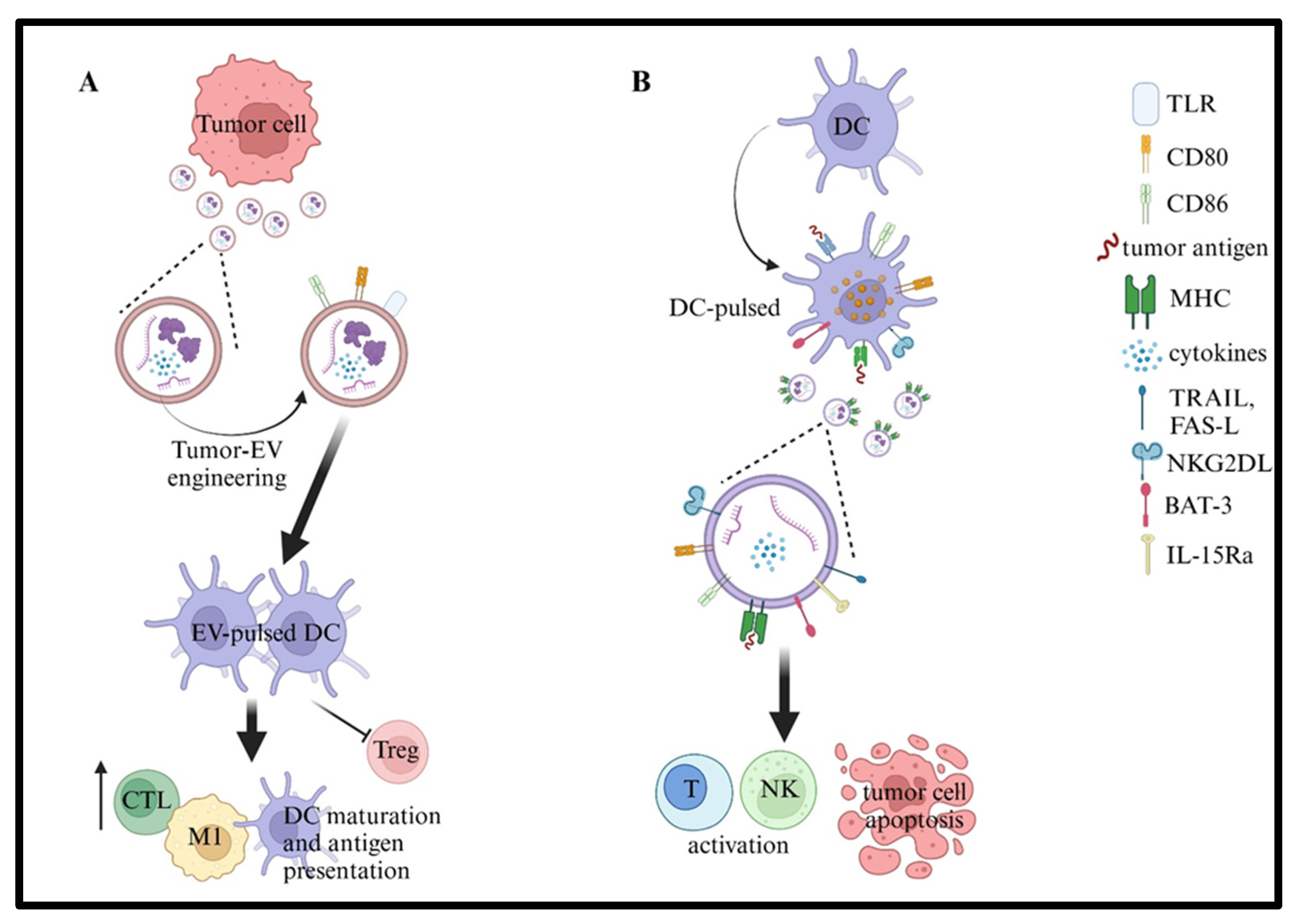
Beyond the role of EVs in tumor chemoresistance (mainly due to the horizontal transfer of drug resistance proteins and genetic material from chemotherapy-resistant tumor cells) and their potential use as biomarkers of tumor progression as well as therapy monitoring (e.g., thanks to their release in biological fluids), EVs have gained particular attention as a novel immunotherapeutic tool [2,11,122,123]. Indeed, due to their unique ability to vehiculate and deliver bioactive molecules, associated with their resistance in blood and biological fluids, EVs me be used as cancer vaccines and for drug delivery [124,125]. Two main approaches have been investigated in the context of cancer vaccines: the engineering of EVs derived from tumors (Figure 3A) or, alternatively, from immune cells, mainly represented by DCs (Figure 3B) [29]. Gu et al. [126] initially reported that DCs pulsed with tumor-derived EVs were effective in inducing anti-tumor responses despite their cargo of immunosuppressive molecules. Keeping in mind that tumor-derived EVs represent a privileged system to deliver antigenic material to DCs, they have been engineered to increase their immunogenicity and reduce the immunosuppressive features. For example, loading of TLR-activating microRNAs (i.e., miR-142) [127] or IL-12 on tumor-derived EVs and silencing of TGF-β1 induced greater anti-tumor responses compared to unmanipulated EVs [128,129]. Other studies reported satisfactory anti-leukemia responses using leukemia cell-derived EVs engineered to express high levels of the co-stimulatory molecules CD80 and CD86, or low levels of PDL-1. Such approaches improved the DC maturation and antigen presentation, induced cytotoxic T cell responses, increased the M1 level within the tumor, and reduced regulatory T cells, both in vitro and in animal models [130,131]. Nonetheless, it has been suggested that manipulation of DC-derived EVs could be more promising as a cancer vaccine compared to tumor-derived EVs. This is based on the knowledge that DC-derived EVs are free of immunosuppressive cargo, but vehiculate, among others, i) antigens on MHC class I and II molecules, ii) CD80 and CD86 with co-stimulatory functions, iii) NK cell-activating ligands BAT3 and NKG2DL, iv) TNFα, FAS-L, and TRAIL inducing apoptosis on tumors and activating NK cells, v) IL-15Rα inducing activation and proliferation of NK cells, and vi) HSP-70 and -73 with anti-tumor effects [29]. Of note, DC-derived EVs are smaller than intact DCs and can penetrate different biological barriers, including the blood–brain barrier and the blood–tumor barrier, as well as efficiently reach secondary lymphoid organs. DC-derived EVs are commonly obtained by mature DCs generated from peripheral blood monocytes and may be induced to carry tumor-specific antigens, proteins, or peptides derived from different malignant diseases [132]. In this context, it is worth mentioning the ability of DCs pulsed with MAGE or alpha-fetoprotein to release EVs expressing antigens and used against melanoma and hepatocellular carcinoma, respectively. Such DC-derived EVs were able to induce a strong NK and CD8+ T cell activation, IFN-γ production, and inhibition of immunosuppressive cytokines (IL-10 and TGF-β1) and regulatory T cell activities [133].

11. Concluding Remarks

The role of tumor-derived EVs in the progression of different tumors has been clearly defined in recent years. EVs have a key function in modifying the BM microenvironment to form the pre-metastatic niche through the increase in vascular permeability, the re-modeling of the extracellular matrix, BM cell recruitment, the increase in angiogenesis, and immunosuppression. These features are related to proteins, miRNA, mRNA, and surface receptors derived from parental tumor cells [134,135]. Here, we summarized data present in the literature regarding the role of EVs in the dysregulation of the BM microenvironment in NB, ALL, and AML. Recent studies have characterized the phenotype of EVs from BM samples of patients with NB and hematopoietic malignancies, and other studies are ongoing (Marimpietri et al., in preparation). These studies will help to understand the mechanism(s) underlying their immunosuppressive activities, their role in tumor progression, and their possible contribution to the failure of immunotherapeutic strategies. Indeed, EVs may sequester antibodies directed against tumor antigens (which are also expressed on the surface of tumor-derived EVs) or prime DCs to generate tolerance against tumors.
In addition, a comprehensive analysis of surface markers and a genomic/proteomic analysis of EVs may lead in the future to the use of BM-derived EVs as novel biomarkers for diagnosis and response to treatment in NB and ALL/AML patients, as already investigated for other human solid and hematological tumors [136].

Funding

(1) “Research to care” grant from Sanofi to F.M. (2) Italian Ministry of Health (Ricerca Corrente 2024 n°2042 and 5x1000 project 2017 UAUT 5/19). (3) Associazione Italiana per la Ricerca contro il Cancro (AIRC), IG 2019 n. 23354 to G.D.

Acknowledgments

We thank BioRender for providing excellent tools for the creation of Figures (www.biorender.com). Publication licenses for the figures were obtained on 28 March 2025 (agreement numbers RA282UK5GC, QE282UJQ2Z, and YW282UJYVJ).

Conflicts of Interest

The authors declare no conflict of interest.

References

  1. Jeppesen, D.K.; Fenix, A.M.; Franklin, J.L.; Higginbotham, J.N.; Zhang, Q.; Zimmerman, L.J.; Liebler, D.C.; Ping, J.; Liu, Q.; Evans, R.; et al. Reassessment of Exosome Composition. Cell 2019, 177, 428–445.e18. [Google Scholar] [CrossRef] [PubMed]
  2. Mathieu, M.; Martin-Jaular, L.; Lavieu, G.; Théry, C. Specificities of secretion and uptake of exosomes and other extracellular vesicles for cell-to-cell communication. Nat. Cell Biol. 2019, 21, 9–17. [Google Scholar] [CrossRef] [PubMed]
  3. van Niel, G.; D’Angelo, G.; Raposo, G. Shedding light on the cell biology of extracellular vesicles. Nat. Rev. Mol. Cell Biol. 2018, 19, 213–228. [Google Scholar] [CrossRef] [PubMed]
  4. Théry, C.; Witwer, K.W.; Aikawa, E.; Alcaraz, M.J.; Anderson, J.D.; Andriantsitohaina, R.; Antoniou, A.; Arab, T.; Archer, F.; Atkin-Smith, G.K.; et al. Minimal information for studies of extracellular vesicles 2018 (MISEV2018): A position statement of the International Society for Extracellular Vesicles and update of the MISEV2014 guidelines. J. Extracell. Vesicles 2018, 7, 1535750. [Google Scholar] [CrossRef]
  5. Welsh, J.A.; Goberdhan, D.C.I.; O’Driscoll, L.; Buzas, E.I.; Blenkiron, C.; Bussolati, B.; Cai, H.; Di Vizio, D.; Driedonks, T.A.P.; Erdbrügger, U.; et al. Minimal information for studies of extracellular vesicles (MISEV2023): From basic to advanced approaches. J. Extracell. Vesicles 2024, 13, e12404. [Google Scholar] [CrossRef]
  6. Jeppesen, D.K.; Zhang, Q.; Franklin, J.L.; Coffey, R.J. Extracellular vesicles and nanoparticles: Emerging complexities. Trends Cell Biol. 2023, 33, 667–681. [Google Scholar] [CrossRef]
  7. Kalluri, R.; McAndrews, K.M. The role of extracellular vesicles in cancer. Cell 2023, 186, 1610–1626. [Google Scholar] [CrossRef]
  8. Sheta, M.; Taha, E.A.; Lu, Y.; Eguchi, T. Extracellular Vesicles: New Classification and Tumor Immunosuppression. Biology 2023, 12, 110. [Google Scholar] [CrossRef]
  9. Kalluri, R.; LeBleu, V.S. The biology, function, and biomedical applications of exosomes. Science 2020, 367, eaau6977. [Google Scholar] [CrossRef]
  10. O’Brien, K.; Breyne, K.; Ughetto, S.; Laurent, L.C.; Breakefield, X.O. RNA delivery by extracellular vesicles in mammalian cells and its applications. Nat. Rev. Mol. Cell Biol. 2020, 21, 585–606. [Google Scholar] [CrossRef]
  11. Dhamdhere, M.R.; Spiegelman, V.S. Extracellular vesicles in neuroblastoma: Role in progression, resistance to therapy and diagnostics. Front. Immunol. 2024, 15, 1385875. [Google Scholar] [CrossRef] [PubMed]
  12. Fusco, C.; De Rosa, G.; Spatocco, I.; Vitiello, E.; Procaccini, C.; Frigè, C.; Pellegrini, V.; La Grotta, R.; Furlan, R.; Matarese, G.; et al. Extracellular vesicles as human therapeutics: A scoping review of the literature. J. Extracell. Vesicles 2024, 13, e12433. [Google Scholar] [CrossRef] [PubMed]
  13. Kalluri, R. The biology and function of extracellular vesicles in immune response and immunity. Immunity 2024, 57, 1752–1768. [Google Scholar] [CrossRef] [PubMed]
  14. Marar, C.; Starich, B.; Wirtz, D. Extracellular vesicles in immunomodulation and tumor progression. Nat. Immunol. 2021, 22, 560–570. [Google Scholar] [CrossRef]
  15. Buzas, E.I. The roles of extracellular vesicles in the immune system. Nat. Rev. Immunol. 2023, 23, 236–250. [Google Scholar] [CrossRef]
  16. Kuang, L.; Wu, L.; Li, Y. Extracellular vesicles in tumor immunity: Mechanisms and novel insights. Mol. Cancer 2025, 24, 45. [Google Scholar] [CrossRef]
  17. Chen, J.G.; Du, Y.T.; Guan, C.H.; Fan, H.Y.; Liu, Y.A.; Wang, T.; Li, X.; Chen, G. Extracellular Vesicles Derived from Plasmodium-infected Hosts as Stimuli of “Trained” Innate Immunity. Curr. Med. Chem. 2023, 30, 4450–4465. [Google Scholar] [CrossRef]
  18. Ayre, D.C.; Elstner, M.; Smith, N.C.; Moores, E.S.; Hogan, A.M.; Christian, S.L. Dynamic regulation of CD24 expression and release of CD24-containing microvesicles in immature B cells in response to CD24 engagement. Immunology 2015, 146, 217–233. [Google Scholar] [CrossRef]
  19. Lundberg, V.; Berglund, M.; Skogberg, G.; Lindgren, S.; Lundqvist, C.; Gudmundsdottir, J.; Thörn, K.; Telemo, E.; Ekwall, O. Thymic exosomes promote the final maturation of thymocytes. Sci. Rep. 2016, 6, 36479. [Google Scholar] [CrossRef]
  20. Raposo, G.; Nijman, H.W.; Stoorvogel, W.; Liejendekker, R.; Harding, C.V.; Melief, C.J.; Geuze, H.J. B lymphocytes secrete antigen-presenting vesicles. J. Exp. Med. 1996, 183, 1161–1172. [Google Scholar] [CrossRef]
  21. Théry, C.; Zitvogel, L.; Amigorena, S. Exosomes: Composition, biogenesis and function. Nat. Rev. Immunol. 2002, 2, 569–579. [Google Scholar] [CrossRef] [PubMed]
  22. Balan, S.; Bhardwaj, N. Cross-Presentation of Tumor Antigens Is Ruled by Synaptic Transfer of Vesicles among Dendritic Cell Subsets. Cancer Cell 2020, 37, 751–753. [Google Scholar] [CrossRef] [PubMed]
  23. Céspedes, P.F.; Jainarayanan, A.; Fernández-Messina, L.; Valvo, S.; Saliba, D.G.; Kurz, E.; Kvalvaag, A.; Chen, L.; Ganskow, C.; Colin-York, H.; et al. T-cell trans-synaptic vesicles are distinct and carry greater effector content than constitutive extracellular vesicles. Nat. Commun. 2022, 13, 3460. [Google Scholar] [CrossRef] [PubMed]
  24. Choudhuri, K.; Llodrá, J.; Roth, E.W.; Tsai, J.; Gordo, S.; Wucherpfennig, K.W.; Kam, L.C.; Stokes, D.L.; Dustin, M.L. Polarized release of T-cell-receptor-enriched microvesicles at the immunological synapse. Nature 2014, 507, 118–123. [Google Scholar] [CrossRef]
  25. Chen, G.; Huang, A.C.; Zhang, W.; Zhang, G.; Wu, M.; Xu, W.; Yu, Z.; Yang, J.; Wang, B.; Sun, H.; et al. Exosomal PD-L1 contributes to immunosuppression and is associated with anti-PD-1 response. Nature 2018, 560, 382–386. [Google Scholar] [CrossRef]
  26. Manning, A.J.; Kuehn, M.J. Contribution of bacterial outer membrane vesicles to innate bacterial defense. BMC Microbiol. 2011, 11, 258. [Google Scholar] [CrossRef]
  27. Adamczyk, A.M.; Leicaj, M.L.; Fabiano, M.P.; Cabrerizo, G.; Bannoud, N.; Croci, D.O.; Witwer, K.W.; Remes Lenicov, F.; Ostrowski, M.; Pérez, P.S. Extracellular vesicles from human plasma dampen inflammation and promote tissue repair functions in macrophages. J. Extracell. Vesicles 2023, 12, e12331. [Google Scholar] [CrossRef]
  28. Ramil, C.P.; Xiang, H.; Zhang, P.; Cronin, A.; Cabral, L.; Yin, Z.; Hai, J.; Wang, H.; Ruprecht, B.; Jia, Y.; et al. Extracellular vesicles released by cancer-associated fibroblast-induced myeloid-derived suppressor cells inhibit T-cell function. Oncoimmunology 2024, 13, 2300882. [Google Scholar] [CrossRef]
  29. Schioppa, T.; Gaudenzi, C.; Zucchi, G.; Piserà, A.; Vahidi, Y.; Tiberio, L.; Sozzani, S.; Del Prete, A.; Bosisio, D.; Salvi, V. Extracellular vesicles at the crossroad between cancer progression and immunotherapy: Focus on dendritic cells. J. Transl. Med. 2024, 22, 691. [Google Scholar] [CrossRef]
  30. Beltraminelli, T.; Perez, C.R.; De Palma, M. Disentangling the complexity of tumor-derived extracellular vesicles. Cell Rep. 2021, 35, 108960. [Google Scholar] [CrossRef]
  31. Del Vecchio, F.; Martinez-Rodriguez, V.; Schukking, M.; Cocks, A.; Broseghini, E.; Fabbri, M. Professional killers: The role of extracellular vesicles in the reciprocal interactions between natural killer, CD8+ cytotoxic T-cells and tumour cells. J. Extracell. Vesicles 2021, 10, e12075. [Google Scholar] [CrossRef] [PubMed]
  32. Clayton, A.; Mitchell, J.P.; Court, J.; Linnane, S.; Mason, M.D.; Tabi, Z. Human tumor-derived exosomes down-modulate NKG2D expression. J. Immunol. 2008, 180, 7249–7258. [Google Scholar] [CrossRef] [PubMed]
  33. Wieckowski, E.U.; Visus, C.; Szajnik, M.; Szczepanski, M.J.; Storkus, W.J.; Whiteside, T.L. Tumor-derived microvesicles promote regulatory T cell expansion and induce apoptosis in tumor-reactive activated CD8+ T lymphocytes. J. Immunol. 2009, 183, 3720–3730. [Google Scholar] [CrossRef] [PubMed]
  34. Xiang, X.; Poliakov, A.; Liu, C.; Liu, Y.; Deng, Z.B.; Wang, J.; Cheng, Z.; Shah, S.V.; Wang, G.J.; Zhang, L.; et al. Induction of myeloid-derived suppressor cells by tumor exosomes. Int. J. Cancer 2009, 124, 2621–2633. [Google Scholar] [CrossRef]
  35. Huber, V.; Fais, S.; Iero, M.; Lugini, L.; Canese, P.; Squarcina, P.; Zaccheddu, A.; Colone, M.; Arancia, G.; Gentile, M.; et al. Human colorectal cancer cells induce T-cell death through release of proapoptotic microvesicles: Role in immune escape. Gastroenterology 2005, 128, 1796–1804. [Google Scholar] [CrossRef]
  36. Marimpietri, D.; Airoldi, I.; Faini, A.C.; Malavasi, F.; Morandi, F. The Role of Extracellular Vesicles in the Progression of Human Neuroblastoma. Int. J. Mol. Sci. 2021, 22, 3964. [Google Scholar] [CrossRef]
  37. Marimpietri, D.; Corrias, M.V.; Tripodi, G.; Gramignoli, R.; Airoldi, I.; Morandi, F. Immunomodulatory properties of extracellular vesicles isolated from bone marrow of patients with neuroblastoma: Role of PD-L1 and HLA-G. Front. Immunol. 2024, 15, 1469771. [Google Scholar] [CrossRef]
  38. Morandi, F.; Marimpietri, D.; Görgens, A.; Gallo, A.; Srinivasan, R.C.; El-Andaloussi, S.; Gramignoli, R. Human Amnion Epithelial Cells Impair T Cell Proliferation: The Role of HLA-G and HLA-E Molecules. Cells 2020, 9, 2123. [Google Scholar] [CrossRef]
  39. Morandi, F.; Marimpietri, D.; Horenstein, A.L.; Bolzoni, M.; Toscani, D.; Costa, F.; Castella, B.; Faini, A.C.; Massaia, M.; Pistoia, V.; et al. Microvesicles released from multiple myeloma cells are equipped with ectoenzymes belonging to canonical and non-canonical adenosinergic pathways and produce adenosine from ATP and NAD+. Oncoimmunology 2018, 7, e1458809. [Google Scholar] [CrossRef]
  40. Hunger, S.P.; Mullighan, C.G. Acute Lymphoblastic Leukemia in Children. N. Engl. J. Med. 2015, 373, 1541–1552. [Google Scholar] [CrossRef]
  41. Kato, M.; Manabe, A. Treatment and biology of pediatric acute lymphoblastic leukemia. Pediatr. Int. 2018, 60, 4–12. [Google Scholar] [CrossRef] [PubMed]
  42. Namayandeh, S.M.; Khazaei, Z.; Lari Najafi, M.; Goodarzi, E.; Moslem, A. GLOBAL Leukemia in Children 0-14 Statistics 2018, Incidence and Mortality and Human Development Index (HDI): GLOBOCAN Sources and Methods. Asian Pac. J. Cancer Prev. APJCP 2020, 21, 1487–1494. [Google Scholar] [CrossRef] [PubMed]
  43. Inaba, H.; Mullighan, C.G. Pediatric acute lymphoblastic leukemia. Haematologica 2020, 105, 2524–2539. [Google Scholar] [CrossRef] [PubMed]
  44. Rubnitz, J.E.; Kaspers, G.J.L. How I treat pediatric acute myeloid leukemia. Blood 2021, 138, 1009–1018. [Google Scholar] [CrossRef]
  45. Lejman, M.; Chałupnik, A.; Chilimoniuk, Z.; Dobosz, M. Genetic Biomarkers and Their Clinical Implications in B-Cell Acute Lymphoblastic Leukemia in Children. Int. J. Mol. Sci. 2022, 23, 2755. [Google Scholar] [CrossRef]
  46. Slayton, W.B.; Schultz, K.R.; Kairalla, J.A.; Devidas, M.; Mi, X.; Pulsipher, M.A.; Chang, B.H.; Mullighan, C.; Iacobucci, I.; Silverman, L.B.; et al. Dasatinib Plus Intensive Chemotherapy in Children, Adolescents, and Young Adults With Philadelphia Chromosome-Positive Acute Lymphoblastic Leukemia: Results of Children’s Oncology Group Trial AALL0622. J. Clin. Oncol. 2018, 36, 2306–2314. [Google Scholar] [CrossRef]
  47. Gu, Z.; Churchman, M.L.; Roberts, K.G.; Moore, I.; Zhou, X.; Nakitandwe, J.; Hagiwara, K.; Pelletier, S.; Gingras, S.; Berns, H.; et al. PAX5-driven subtypes of B-progenitor acute lymphoblastic leukemia. Nat. Genet. 2019, 51, 296–307. [Google Scholar] [CrossRef]
  48. Inaba, H.; Pui, C.H. Advances in the Diagnosis and Treatment of Pediatric Acute Lymphoblastic Leukemia. J. Clin. Med. 2021, 10, 1926. [Google Scholar] [CrossRef]
  49. Bassan, R.; Spinelli, O.; Oldani, E.; Intermesoli, T.; Tosi, M.; Peruta, B.; Rossi, G.; Borlenghi, E.; Pogliani, E.M.; Terruzzi, E.; et al. Improved risk classification for risk-specific therapy based on the molecular study of minimal residual disease (MRD) in adult acute lymphoblastic leukemia (ALL). Blood 2009, 113, 4153–4162. [Google Scholar] [CrossRef]
  50. Mullighan, C.G. Molecular genetics of B-precursor acute lymphoblastic leukemia. J. Clin. Investig. 2012, 122, 3407–3415. [Google Scholar] [CrossRef]
  51. Ross, M.E.; Zhou, X.; Song, G.; Shurtleff, S.A.; Girtman, K.; Williams, W.K.; Liu, H.C.; Mahfouz, R.; Raimondi, S.C.; Lenny, N.; et al. Classification of pediatric acute lymphoblastic leukemia by gene expression profiling. Blood 2003, 102, 2951–2959. [Google Scholar] [CrossRef] [PubMed]
  52. Temple, W.C.; Mueller, S.; Hermiston, M.L.; Burkhardt, B. Diagnosis and management of lymphoblastic lymphoma in children, adolescents and young adults. Best Pract. Res. Clin. Haematol. 2023, 36, 101449. [Google Scholar] [CrossRef] [PubMed]
  53. Dander, E.; Palmi, C.; D’Amico, G.; Cazzaniga, G. The Bone Marrow Niche in B-Cell Acute Lymphoblastic Leukemia: The Role of Microenvironment from Pre-Leukemia to Overt Leukemia. Int. J. Mol. Sci. 2021, 22, 4426. [Google Scholar] [CrossRef] [PubMed]
  54. Lauten, M.; Möricke, A.; Beier, R.; Zimmermann, M.; Stanulla, M.; Meissner, B.; Odenwald, E.; Attarbaschi, A.; Niemeyer, C.; Niggli, F.; et al. Prediction of outcome by early bone marrow response in childhood acute lymphoblastic leukemia treated in the ALL-BFM 95 trial: Differential effects in precursor B-cell and T-cell leukemia. Haematologica 2012, 97, 1048–1056. [Google Scholar] [CrossRef]
  55. Reinhardt, D.; Antoniou, E.; Waack, K. Pediatric Acute Myeloid Leukemia-Past, Present, and Future. J. Clin. Med. 2022, 11, 504. [Google Scholar] [CrossRef]
  56. Tseng, S.; Lee, M.E.; Lin, P.C. A Review of Childhood Acute Myeloid Leukemia: Diagnosis and Novel Treatment. Pharmaceuticals 2023, 16, 1614. [Google Scholar] [CrossRef]
  57. de Rooij, J.D.; Zwaan, C.M.; van den Heuvel-Eibrink, M. Pediatric AML: From Biology to Clinical Management. J. Clin. Med. 2015, 4, 127–149. [Google Scholar] [CrossRef]
  58. Schuurhuis, G.J.; Heuser, M.; Freeman, S.; Béné, M.C.; Buccisano, F.; Cloos, J.; Grimwade, D.; Haferlach, T.; Hills, R.K.; Hourigan, C.S.; et al. Minimal/measurable residual disease in AML: A consensus document from the European LeukemiaNet MRD Working Party. Blood 2018, 131, 1275–1291. [Google Scholar] [CrossRef]
  59. Tran, T.H.; Hunger, S.P. The genomic landscape of pediatric acute lymphoblastic leukemia and precision medicine opportunities. Semin. Cancer Biol. 2022, 84, 144–152. [Google Scholar] [CrossRef]
  60. Bakhtiyari, M.; Liaghat, M.; Aziziyan, F.; Shapourian, H.; Yahyazadeh, S.; Alipour, M.; Shahveh, S.; Maleki-Sheikhabadi, F.; Halimi, H.; Forghaniesfidvajani, R.; et al. The role of bone marrow microenvironment (BMM) cells in acute myeloid leukemia (AML) progression: Immune checkpoints, metabolic checkpoints, and signaling pathways. Cell Commun. Signal. CCS 2023, 21, 252. [Google Scholar] [CrossRef]
  61. Matthay, K.K.; Maris, J.M.; Schleiermacher, G.; Nakagawara, A.; Mackall, C.L.; Diller, L.; Weiss, W.A. Neuroblastoma. Nat. Rev. Dis. Primers 2016, 2, 16078. [Google Scholar] [CrossRef] [PubMed]
  62. Morandi, F.; Sabatini, F.; Podestà, M.; Airoldi, I. Immunotherapeutic Strategies for Neuroblastoma: Present, Past and Future. Vaccines 2021, 9, 43. [Google Scholar] [CrossRef] [PubMed]
  63. Körber, V.; Stainczyk, S.A.; Kurilov, R.; Henrich, K.O.; Hero, B.; Brors, B.; Westermann, F.; Höfer, T. Neuroblastoma arises in early fetal development and its evolutionary duration predicts outcome. Nat. Genet. 2023, 55, 619–630. [Google Scholar] [CrossRef] [PubMed]
  64. Nong, J.; Su, C.; Li, C.; Wang, C.; Li, W.; Li, Y.; Chen, P.; Li, Y.; Li, Z.; She, X.; et al. Global, regional, and national epidemiology of childhood neuroblastoma (1990-2021): A statistical analysis of incidence, mortality, and DALYs. EClinicalMedicine 2025, 79, 102964. [Google Scholar] [CrossRef]
  65. Burchill, S.A.; Beiske, K.; Shimada, H.; Ambros, P.F.; Seeger, R.; Tytgat, G.A.; Brock, P.R.; Haber, M.; Park, J.R.; Berthold, F. Recommendations for the standardization of bone marrow disease assessment and reporting in children with neuroblastoma on behalf of the International Neuroblastoma Response Criteria Bone Marrow Working Group. Cancer 2017, 123, 1095–1105. [Google Scholar] [CrossRef]
  66. Park, J.R.; Bagatell, R.; Cohn, S.L.; Pearson, A.D.; Villablanca, J.G.; Berthold, F.; Burchill, S.; Boubaker, A.; McHugh, K.; Nuchtern, J.G.; et al. Revisions to the International Neuroblastoma Response Criteria: A Consensus Statement From the National Cancer Institute Clinical Trials Planning Meeting. J. Clin. Oncol. 2017, 35, 2580–2587. [Google Scholar] [CrossRef]
  67. Boulais, P.E.; Frenette, P.S. Making sense of hematopoietic stem cell niches. Blood 2015, 125, 2621–2629. [Google Scholar] [CrossRef]
  68. Asada, N.; Kunisaki, Y.; Pierce, H.; Wang, Z.; Fernandez, N.F.; Birbrair, A.; Ma’ayan, A.; Frenette, P.S. Differential cytokine contributions of perivascular haematopoietic stem cell niches. Nat. Cell Biol. 2017, 19, 214–223. [Google Scholar] [CrossRef]
  69. Ehninger, A.; Trumpp, A. The bone marrow stem cell niche grows up: Mesenchymal stem cells and macrophages move in. J. Exp. Med. 2011, 208, 421–428. [Google Scholar] [CrossRef]
  70. Man, Y.; Yao, X.; Yang, T.; Wang, Y. Hematopoietic Stem Cell Niche During Homeostasis, Malignancy, and Bone Marrow Transplantation. Front. Cell Dev. Biol. 2021, 9, 621214. [Google Scholar] [CrossRef]
  71. Le, P.M.; Andreeff, M.; Battula, V.L. Osteogenic niche in the regulation of normal hematopoiesis and leukemogenesis. Haematologica 2018, 103, 1945–1955. [Google Scholar] [CrossRef] [PubMed]
  72. Wang, W.; Stiehl, T.; Raffel, S.; Hoang, V.T.; Hoffmann, I.; Poisa-Beiro, L.; Saeed, B.R.; Blume, R.; Manta, L.; Eckstein, V.; et al. Reduced hematopoietic stem cell frequency predicts outcome in acute myeloid leukemia. Haematologica 2017, 102, 1567–1577. [Google Scholar] [CrossRef] [PubMed]
  73. Boyle, W.J.; Simonet, W.S.; Lacey, D.L. Osteoclast differentiation and activation. Nature 2003, 423, 337–342. [Google Scholar] [CrossRef] [PubMed]
  74. Kobolak, J.; Dinnyes, A.; Memic, A.; Khademhosseini, A.; Mobasheri, A. Mesenchymal stem cells: Identification, phenotypic characterization, biological properties and potential for regenerative medicine through biomaterial micro-engineering of their niche. Methods 2016, 99, 62–68. [Google Scholar] [CrossRef]
  75. Uccelli, A.; Moretta, L.; Pistoia, V. Mesenchymal stem cells in health and disease. Nat. Rev. Immunol. 2008, 8, 726–736. [Google Scholar] [CrossRef]
  76. Sun, G.; Gu, Q.; Zheng, J.; Cheng, H.; Cheng, T. Emerging roles of extracellular vesicles in normal and malignant hematopoiesis. J. Clin. Investig. 2022, 132, e160840. [Google Scholar] [CrossRef]
  77. Lyu, T.; Wang, Y.; Li, D.; Yang, H.; Qin, B.; Zhang, W.; Li, Z.; Cheng, C.; Zhang, B.; Guo, R.; et al. Exosomes from BM-MSCs promote acute myeloid leukemia cell proliferation, invasion and chemoresistance via upregulation of S100A4. Exp. Hematol. Oncol. 2021, 10, 24. [Google Scholar] [CrossRef]
  78. Crompot, E.; Van Damme, M.; Pieters, K.; Vermeersch, M.; Perez-Morga, D.; Mineur, P.; Maerevoet, M.; Meuleman, N.; Bron, D.; Lagneaux, L.; et al. Extracellular vesicles of bone marrow stromal cells rescue chronic lymphocytic leukemia B cells from apoptosis, enhance their migration and induce gene expression modifications. Haematologica 2017, 102, 1594–1604. [Google Scholar] [CrossRef]
  79. Tettamanti, S.; Pievani, A.; Biondi, A.; Dotti, G.; Serafini, M. Catch me if you can: How AML and its niche escape immunotherapy. Leukemia 2022, 36, 13–22. [Google Scholar] [CrossRef]
  80. Kasherwal, V.; Kale, V.; Vaidya, A. Extracellular vesicles secreted by leukemic cells as mediators of dysregulated hematopoiesis: Acute myeloid leukemia as a case in point. Expert Rev. Hematol. 2025, 18, 225–237. [Google Scholar] [CrossRef]
  81. Balandrán, J.C.; Purizaca, J.; Enciso, J.; Dozal, D.; Sandoval, A.; Jiménez-Hernández, E.; Alemán-Lazarini, L.; Perez-Koldenkova, V.; Quintela-Núñez Del Prado, H.; Rios de Los Ríos, J.; et al. Pro-inflammatory-Related Loss of CXCL12 Niche Promotes Acute Lymphoblastic Leukemic Progression at the Expense of Normal Lymphopoiesis. Front. Immunol. 2016, 7, 666. [Google Scholar] [CrossRef] [PubMed]
  82. Crippa, S.; Bernardo, M.E. Mesenchymal Stromal Cells: Role in the BM Niche and in the Support of Hematopoietic Stem Cell Transplantation. HemaSphere 2018, 2, e151. [Google Scholar] [CrossRef] [PubMed]
  83. Corradi, G.; Bassani, B.; Simonetti, G.; Sangaletti, S.; Vadakekolathu, J.; Fontana, M.C.; Pazzaglia, M.; Gulino, A.; Tripodo, C.; Cristiano, G.; et al. Release of IFNγ by Acute Myeloid Leukemia Cells Remodels Bone Marrow Immune Microenvironment by Inducing Regulatory T Cells. Clin. Cancer Res. 2022, 28, 3141–3155. [Google Scholar] [CrossRef] [PubMed]
  84. Mussai, F.; De Santo, C.; Abu-Dayyeh, I.; Booth, S.; Quek, L.; McEwen-Smith, R.M.; Qureshi, A.; Dazzi, F.; Vyas, P.; Cerundolo, V. Acute myeloid leukemia creates an arginase-dependent immunosuppressive microenvironment. Blood 2013, 122, 749–758. [Google Scholar] [CrossRef]
  85. Gu, Y.; Xia, J.; Guo, Y.; Tao, L.; Zhang, G.; Xu, J. Leukemia cells remodel bone marrow stromal cells to generate a protumoral microenvironment via the S100A8-NOX2-ROS signaling pathway. Sci. Rep. 2025, 15, 17179. [Google Scholar] [CrossRef]
  86. Feng, S.; Cen, J.; Huang, Y.; Shen, H.; Yao, L.; Wang, Y.; Chen, Z. Matrix metalloproteinase-2 and -9 secreted by leukemic cells increase the permeability of blood-brain barrier by disrupting tight junction proteins. PLoS ONE 2011, 6, e20599. [Google Scholar] [CrossRef]
  87. Schneider, P.; Costa, O.; Legrand, E.; Bigot, D.; Lecleire, S.; Grassi, V.; Vannier, J.P.; Vasse, M. In vitro secretion of matrix metalloprotease 9 is a prognostic marker in childhood acute lymphoblastic leukemia. Leuk. Res. 2010, 34, 24–31. [Google Scholar] [CrossRef]
  88. Vilchis-Ordoñez, A.; Contreras-Quiroz, A.; Vadillo, E.; Dorantes-Acosta, E.; Reyes-López, A.; Quintela-Nuñez del Prado, H.M.; Venegas-Vázquez, J.; Mayani, H.; Ortiz-Navarrete, V.; López-Martínez, B.; et al. Bone Marrow Cells in Acute Lymphoblastic Leukemia Create a Proinflammatory Microenvironment Influencing Normal Hematopoietic Differentiation Fates. BioMed Res. Int. 2015, 2015, 386165. [Google Scholar] [CrossRef]
  89. Portale, F.; Cricrì, G.; Bresolin, S.; Lupi, M.; Gaspari, S.; Silvestri, D.; Russo, B.; Marino, N.; Ubezio, P.; Pagni, F.; et al. ActivinA: A new leukemia-promoting factor conferring migratory advantage to B-cell precursor-acute lymphoblastic leukemic cells. Haematologica 2019, 104, 533–545. [Google Scholar] [CrossRef]
  90. Chen, Y.; Hoffmeister, L.M.; Zaun, Y.; Arnold, L.; Schmid, K.W.; Giebel, B.; Klein-Hitpass, L.; Hanenberg, H.; Squire, A.; Reinhardt, H.C.; et al. Acute myeloid leukemia-induced remodeling of the human bone marrow niche predicts clinical outcome. Blood Adv. 2020, 4, 5257–5268. [Google Scholar] [CrossRef]
  91. Polak, R.; de Rooij, B.; Pieters, R.; den Boer, M.L. B-cell precursor acute lymphoblastic leukemia cells use tunneling nanotubes to orchestrate their microenvironment. Blood 2015, 126, 2404–2414. [Google Scholar] [CrossRef] [PubMed]
  92. Veiga, J.P.; Costa, L.F.; Sallan, S.E.; Nadler, L.M.; Cardoso, A.A. Leukemia-stimulated bone marrow endothelium promotes leukemia cell survival. Exp. Hematol. 2006, 34, 610–621. [Google Scholar] [CrossRef] [PubMed]
  93. Verma, D.; Zanetti, C.; Godavarthy, P.S.; Kumar, R.; Minciacchi, V.R.; Pfeiffer, J.; Metzler, M.; Lefort, S.; Maguer-Satta, V.; Nicolini, F.E.; et al. Bone marrow niche-derived extracellular matrix-degrading enzymes influence the progression of B-cell acute lymphoblastic leukemia. Leukemia 2020, 34, 1540–1552. [Google Scholar] [CrossRef] [PubMed]
  94. Passaro, D.; Di Tullio, A.; Abarrategi, A.; Rouault-Pierre, K.; Foster, K.; Ariza-McNaughton, L.; Montaner, B.; Chakravarty, P.; Bhaw, L.; Diana, G.; et al. Increased Vascular Permeability in the Bone Marrow Microenvironment Contributes to Disease Progression and Drug Response in Acute Myeloid Leukemia. Cancer Cell 2017, 32, 324–341.e6. [Google Scholar] [CrossRef]
  95. Méndez-Ferrer, S.; Bonnet, D.; Steensma, D.P.; Hasserjian, R.P.; Ghobrial, I.M.; Gribben, J.G.; Andreeff, M.; Krause, D.S. Bone marrow niches in haematological malignancies. Nat. Rev. Cancer 2020, 20, 285–298. [Google Scholar] [CrossRef]
  96. La Spina, E.; Giallongo, S.; Giallongo, C.; Vicario, N.; Duminuco, A.; Parenti, R.; Giuffrida, R.; Longhitano, L.; Li Volti, G.; Cambria, D.; et al. Mesenchymal stromal cells in tumor microenvironment remodeling of BCR-ABL negative myeloproliferative diseases. Front. Oncol. 2023, 13, 1141610. [Google Scholar] [CrossRef]
  97. Han, G.; Lu, S.L.; Li, A.G.; He, W.; Corless, C.L.; Kulesz-Martin, M.; Wang, X.J. Distinct mechanisms of TGF-beta1-mediated epithelial-to-mesenchymal transition and metastasis during skin carcinogenesis. J. Clin. Investig. 2005, 115, 1714–1723. [Google Scholar] [CrossRef]
  98. Vicente López, Á.; Vázquez García, M.N.; Melen, G.J.; Entrena Martínez, A.; Cubillo Moreno, I.; García-Castro, J.; Orellana, M.R.; González, A.G. Mesenchymal stromal cells derived from the bone marrow of acute lymphoblastic leukemia patients show altered BMP4 production: Correlations with the course of disease. PLoS ONE 2014, 9, e84496. [Google Scholar] [CrossRef]
  99. Magalhães-Gama, F.; Malheiros Araújo Silvestrini, M.; Neves, J.C.F.; Araújo, N.D.; Alves-Hanna, F.S.; Kerr, M.W.A.; Carvalho, M.; Tarragô, A.M.; Soares Pontes, G.; Martins-Filho, O.A.; et al. Exploring cell-derived extracellular vesicles in peripheral blood and bone marrow of B-cell acute lymphoblastic leukemia pediatric patients: Proof-of-concept study. Front. Immunol. 2024, 15, 1421036. [Google Scholar] [CrossRef]
  100. Pando, A.; Reagan, J.L.; Quesenberry, P.; Fast, L.D. Extracellular vesicles in leukemia. Leuk. Res. 2018, 64, 52–60. [Google Scholar] [CrossRef]
  101. Georgievski, A.; Michel, A.; Thomas, C.; Mlamla, Z.; Pais de Barros, J.P.; Lemaire-Ewing, S.; Garrido, C.; Quéré, R. Acute lymphoblastic leukemia-derived extracellular vesicles affect quiescence of hematopoietic stem and progenitor cells. Cell Death Dis. 2022, 13, 337. [Google Scholar] [CrossRef] [PubMed]
  102. Liu, C.; Chen, Q.; Shang, Y.; Chen, L.; Myers, J.; Awadallah, A.; Sun, J.; Yu, S.; Umphred-Wilson, K.; Che, D.; et al. Endothelial PERK-ATF4-JAG1 axis activated by T-ALL remodels bone marrow vascular niche. Theranostics 2022, 12, 2894–2907. [Google Scholar] [CrossRef] [PubMed]
  103. Gholipour, E.; Kahroba, H.; Soltani, N.; Samadi, P.; Sarvarian, P.; Vakili-Samiani, S.; Hosein Pour Feizi, A.A.; Soltani-Zangbar, M.S.; Baghersalimi, A.; Darbandi, B.; et al. Paediatric pre-B acute lymphoblastic leukaemia-derived exosomes regulate immune function in human T cells. J. Cell. Mol. Med. 2022, 26, 4566–4576. [Google Scholar] [CrossRef] [PubMed]
  104. Swatler, J.; Turos-Korgul, L.; Brewinska-Olchowik, M.; De Biasi, S.; Dudka, W.; Le, B.V.; Kominek, A.; Cyranowski, S.; Pilanc, P.; Mohammadi, E.; et al. 4-1BBL-containing leukemic extracellular vesicles promote immunosuppressive effector regulatory T cells. Blood Adv. 2022, 6, 1879–1894. [Google Scholar] [CrossRef] [PubMed]
  105. Chen, L.; Guo, Z.; Zhou, Y.; Ni, J.; Zhu, J.; Fan, X.; Chen, X.; Liu, Y.; Li, Z.; Zhou, H. microRNA-1246-containing extracellular vesicles from acute myeloid leukemia cells promote the survival of leukemia stem cells via the LRIG1-meditated STAT3 pathway. Aging 2021, 13, 13644–13662. [Google Scholar] [CrossRef] [PubMed]
  106. Haque, S.; Vaiselbuh, S.R. Silencing of Exosomal miR-181a Reverses Pediatric Acute Lymphocytic Leukemia Cell Proliferation. Pharmaceuticals 2020, 13, 241. [Google Scholar] [CrossRef]
  107. Licari, E.; Cricrì, G.; Mauri, M.; Raimondo, F.; Dioni, L.; Favero, C.; Giussani, A.; Starace, R.; Nucera, S.; Biondi, A.; et al. ActivinA modulates B-acute lymphoblastic leukaemia cell communication and survival by inducing extracellular vesicles production. Sci. Rep. 2024, 14, 16083. [Google Scholar] [CrossRef]
  108. Zhai, X.; You, F.; Xiang, S.; Jiang, L.; Chen, D.; Li, Y.; Fan, S.; Han, Z.; Zhang, T.; An, G.; et al. MUC1-Tn-targeting chimeric antigen receptor-modified Vγ9Vδ2 T cells with enhanced antigen-specific anti-tumor activity. Am. J. Cancer Res. 2021, 11, 79–91. [Google Scholar]
  109. Rios de Los Rios, J.; Enciso, J.; Vilchis-Ordoñez, A.; Vázquez-Ramírez, R.; Ramirez-Ramirez, D.; Balandrán, J.C.; Rodríguez-Martínez, A.; Ruiz-Tachiquín, M.; Pompa-Mera, E.; Mendoza, L.; et al. Acute lymphoblastic leukemia-secreted miRNAs induce a proinflammatory microenvironment and promote the activation of hematopoietic progenitors. J. Leukoc. Biol. 2022, 112, 31–45. [Google Scholar] [CrossRef]
  110. Yuan, T.; Shi, C.; Xu, W.; Yang, H.L.; Xia, B.; Tian, C. Extracellular vesicles derived from T-cell acute lymphoblastic leukemia inhibit osteogenic differentiation of bone marrow mesenchymal stem cells via miR-34a-5p. Endocr. J. 2021, 68, 1197–1208. [Google Scholar] [CrossRef]
  111. Chen, T.; Zhang, G.; Kong, L.; Xu, S.; Wang, Y.; Dong, M. Leukemia-derived exosomes induced IL-8 production in bone marrow stromal cells to protect the leukemia cells against chemotherapy. Life Sci. 2019, 221, 187–195. [Google Scholar] [CrossRef] [PubMed]
  112. Huan, J.; Hornick, N.I.; Goloviznina, N.A.; Kamimae-Lanning, A.N.; David, L.L.; Wilmarth, P.A.; Mori, T.; Chevillet, J.R.; Narla, A.; Roberts, C.T., Jr.; et al. Coordinate regulation of residual bone marrow function by paracrine trafficking of AML exosomes. Leukemia 2015, 29, 2285–2295. [Google Scholar] [CrossRef] [PubMed]
  113. Karantanou, C.; Minciacchi, V.R.; Kumar, R.; Zanetti, C.; Bravo, J.; Pereira, R.S.; Tascher, G.; Tertel, T.; Covarrubias-Pinto, A.; Bankov, K.; et al. Impact of mesenchymal stromal cell-derived vesicular cargo on B-cell acute lymphoblastic leukemia progression. Blood Adv. 2023, 7, 1190–1203. [Google Scholar] [CrossRef] [PubMed]
  114. Fei, F.; Joo, E.J.; Tarighat, S.S.; Schiffer, I.; Paz, H.; Fabbri, M.; Abdel-Azim, H.; Groffen, J.; Heisterkamp, N. B-cell precursor acute lymphoblastic leukemia and stromal cells communicate through Galectin-3. Oncotarget 2015, 6, 11378–11394. [Google Scholar] [CrossRef]
  115. Fetahu, I.S.; Esser-Skala, W.; Dnyansagar, R.; Sindelar, S.; Rifatbegovic, F.; Bileck, A.; Skos, L.; Bozsaky, E.; Lazic, D.; Shaw, L.; et al. Single-cell transcriptomics and epigenomics unravel the role of monocytes in neuroblastoma bone marrow metastasis. Nat. Commun. 2023, 14, 3620. [Google Scholar] [CrossRef]
  116. Hochheuser, C.; Windt, L.J.; Kunze, N.Y.; de Vos, D.L.; Tytgat, G.A.M.; Voermans, C.; Timmerman, I. Mesenchymal Stromal Cells in Neuroblastoma: Exploring Crosstalk and Therapeutic Implications. Stem Cells Dev. 2021, 30, 59–78. [Google Scholar] [CrossRef]
  117. Colletti, M.; Tomao, L.; Galardi, A.; Paolini, A.; Di Paolo, V.; De Stefanis, C.; Mascio, P.; Nazio, F.; Petrini, S.; Castellano, A.; et al. Neuroblastoma-secreted exosomes carrying miR-375 promote osteogenic differentiation of bone-marrow mesenchymal stromal cells. J. Extracell. Vesicles 2020, 9, 1774144. [Google Scholar] [CrossRef]
  118. Hochheuser, C.; van Zogchel, L.M.J.; Kleijer, M.; Kuijk, C.; Tol, S.; van der Schoot, C.E.; Voermans, C.; Tytgat, G.A.M.; Timmerman, I. The Metastatic Bone Marrow Niche in Neuroblastoma: Altered Phenotype and Function of Mesenchymal Stromal Cells. Cancers 2020, 12, 3231. [Google Scholar] [CrossRef]
  119. Marimpietri, D.; Petretto, A.; Raffaghello, L.; Pezzolo, A.; Gagliani, C.; Tacchetti, C.; Mauri, P.; Melioli, G.; Pistoia, V. Proteome profiling of neuroblastoma-derived exosomes reveal the expression of proteins potentially involved in tumor progression. PLoS ONE 2013, 8, e75054. [Google Scholar] [CrossRef]
  120. Cheng, J.; Ji, D.; Ma, J.; Zhang, Q.; Zhang, W.; Yang, L. Proteomic analysis of serum small extracellular vesicles identifies diagnostic biomarkers for neuroblastoma. Front. Oncol. 2024, 14, 1367159. [Google Scholar] [CrossRef]
  121. Morandi, F.; Airoldi, I.; Marimpietri, D.; Bracci, C.; Faini, A.C.; Gramignoli, R. CD38, a Receptor with Multifunctional Activities: From Modulatory Functions on Regulatory Cell Subsets and Extracellular Vesicles, to a Target for Therapeutic Strategies. Cells 2019, 8, 1527. [Google Scholar] [CrossRef] [PubMed]
  122. Álvarez-Zúñiga, C.D.; Garza-Veloz, I.; Martínez-Rendón, J.; Ureño-Segura, M.; Delgado-Enciso, I.; Martinez-Fierro, M.L. Circulating Biomarkers Associated with the Diagnosis and Prognosis of B-Cell Progenitor Acute Lymphoblastic Leukemia. Cancers 2023, 15, 4186. [Google Scholar] [CrossRef] [PubMed]
  123. Hornick, N.I.; Huan, J.; Doron, B.; Goloviznina, N.A.; Lapidus, J.; Chang, B.H.; Kurre, P. Serum Exosome MicroRNA as a Minimally-Invasive Early Biomarker of AML. Sci. Rep. 2015, 5, 11295. [Google Scholar] [CrossRef] [PubMed]
  124. Kumar, M.A.; Baba, S.K.; Sadida, H.Q.; Marzooqi, S.A.; Jerobin, J.; Altemani, F.H.; Algehainy, N.; Alanazi, M.A.; Abou-Samra, A.B.; Kumar, R.; et al. Extracellular vesicles as tools and targets in therapy for diseases. Signal Transduct. Target. Ther. 2024, 9, 27. [Google Scholar] [CrossRef]
  125. Payandeh, Z.; Tangruksa, B.; Synnergren, J.; Heydarkhan-Hagvall, S.; Nordin, J.Z.; Andaloussi, S.E.; Borén, J.; Wiseman, J.; Bohlooly, Y.M.; Lindfors, L.; et al. Extracellular vesicles transport RNA between cells: Unraveling their dual role in diagnostics and therapeutics. Mol. Asp. Med. 2024, 99, 101302. [Google Scholar] [CrossRef]
  126. Gu, X.; Erb, U.; Büchler, M.W.; Zöller, M. Improved vaccine efficacy of tumor exosome compared to tumor lysate loaded dendritic cells in mice. Int. J. Cancer 2015, 136, E74–E84. [Google Scholar] [CrossRef]
  127. Khani, A.T.; Sharifzad, F.; Mardpour, S.; Hassan, Z.M.; Ebrahimi, M. Tumor extracellular vesicles loaded with exogenous Let-7i and miR-142 can modulate both immune response and tumor microenvironment to initiate a powerful anti-tumor response. Cancer Lett. 2021, 501, 200–209. [Google Scholar] [CrossRef]
  128. Huang, F.; Wan, J.; Hu, W.; Hao, S. Enhancement of Anti-Leukemia Immunity by Leukemia-Derived Exosomes Via Downregulation of TGF-β1 Expression. Cell. Physiol. Biochem. Int. J. Exp. Cell. Physiol. Biochem. Pharmacol. 2017, 44, 240–254. [Google Scholar] [CrossRef]
  129. Rossowska, J.; Anger, N.; Wegierek, K.; Szczygieł, A.; Mierzejewska, J.; Milczarek, M.; Szermer-Olearnik, B.; Pajtasz-Piasecka, E. Antitumor Potential of Extracellular Vesicles Released by Genetically Modified Murine Colon Carcinoma Cells With Overexpression of Interleukin-12 and shRNA for TGF-β1. Front. Immunol. 2019, 10, 211. [Google Scholar] [CrossRef]
  130. Huang, F.; Li, Z.; Zhang, W.; Li, J.; Hao, S. Enhancing the anti-leukemia immunity of acute lymphocytic leukemia-derived exosome-based vaccine by downregulation of PD-L1 expression. Cancer Immunol. Immunother. CII 2022, 71, 2197–2212. [Google Scholar] [CrossRef]
  131. Zhang, D.; Jiang, Y.; Wang, M.; Zhao, J.; Wan, J.; Li, Z.; Huang, D.; Yu, J.; Li, J.; Liu, J.; et al. A novel costimulatory molecule gene-modified leukemia cell-derived exosome enhances the anti-leukemia efficacy of DC vaccine in mouse models. Vaccine 2024, 42, 126097. [Google Scholar] [CrossRef] [PubMed]
  132. Luo, S.; Chen, J.; Xu, F.; Chen, H.; Li, Y.; Li, W. Dendritic Cell-Derived Exosomes in Cancer Immunotherapy. Pharmaceutics 2023, 15, 2070. [Google Scholar] [CrossRef] [PubMed]
  133. Liu, C.; Liu, X.; Xiang, X.; Pang, X.; Chen, S.; Zhang, Y.; Ren, E.; Zhang, L.; Liu, X.; Lv, P.; et al. A nanovaccine for antigen self-presentation and immunosuppression reversal as a personalized cancer immunotherapy strategy. Nat. Nanotechnol. 2022, 17, 531–540. [Google Scholar] [CrossRef] [PubMed]
  134. Xu, R.; Rai, A.; Chen, M.; Suwakulsiri, W.; Greening, D.W.; Simpson, R.J. Extracellular vesicles in cancer—Implications for future improvements in cancer care. Nat. Rev. Clin. Oncol. 2018, 15, 617–638. [Google Scholar] [CrossRef]
  135. Dong, Q.; Liu, X.; Cheng, K.; Sheng, J.; Kong, J.; Liu, T. Pre-metastatic Niche Formation in Different Organs Induced by Tumor Extracellular Vesicles. Front. Cell Dev. Biol. 2021, 9, 733627. [Google Scholar] [CrossRef]
  136. Hoshino, A.; Kim, H.S.; Bojmar, L.; Gyan, K.E.; Cioffi, M.; Hernandez, J.; Zambirinis, C.P.; Rodrigues, G.; Molina, H.; Heissel, S.; et al. Extracellular Vesicle and Particle Biomarkers Define Multiple Human Cancers. Cell 2020, 182, 1044–1061.e18. [Google Scholar] [CrossRef]
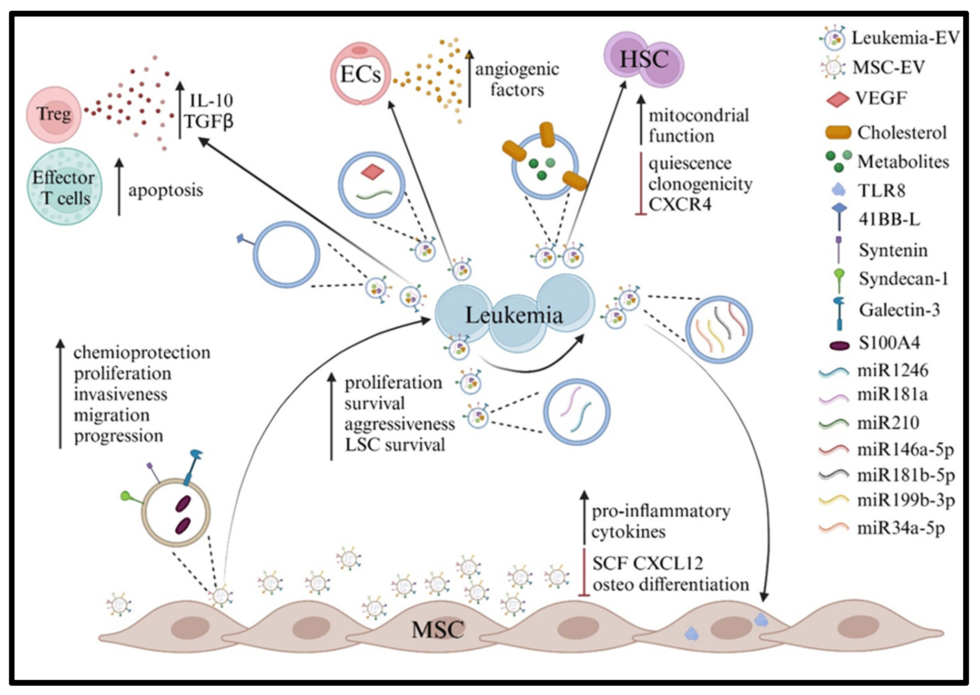
Figure 1. Created in BioRender. D’Amico, G. (2025); https://BioRender.com/na8vkbw.
Ijms 26 05380 g001
Figure 2. Created in BioRender. Airoldi, I. (2025); https://BioRender.com/45fho1i.
Ijms 26 05380 g002
Figure 3. Created in BioRender. Airoldi, I. (2025); https://BioRender.com/mplge2w.
Ijms 26 05380 g003
Disclaimer/Publisher’s Note: The statements, opinions and data contained in all publications are solely those of the individual author(s) and contributor(s) and not of MDPI and/or the editor(s). MDPI and/or the editor(s) disclaim responsibility for any injury to people or property resulting from any ideas, methods, instructions or products referred to in the content.