Review

Review of Four Refined Clinical Entities in Hereditary Retinal Disorders from Japan

Nagoya University, 3-707 Suishouenn, Moriyamaku, Nagoya 463-0010, Japan
Int. J. Mol. Sci. 2025, 26(11), 5166; https://doi.org/10.3390/ijms26115166
Submission received: 19 March 2025 / Revised: 22 May 2025 / Accepted: 22 May 2025 / Published: 28 May 2025

Abstract

:
In the past, only Oguchi disease was reported as a hereditary retinal disease from Japan. Dr. Chuuta Oguch was a Professor of Nagoya University in Japan. During the past 40 years, four new clinical entities in hereditary retinal disorders have been detected by the Miyake group from Nagoya, Japan. All disorders show essentially normal fundi, and the diagnosis was made mainly by the analysis of an electroretinogram (ERG). Gene mutations are detected in three of them. Bipolar cell (BP) dysfunction syndrome: Congenital stationary night blindness (CSNB) with negative ERG (a-wave is larger than b-wave) was named as the Schubert–Bornschein type in 1952 and considered to be an independent clinical entity. In 1986, Miyake group classified ninety patients with the Schubert–Bornschein type into two types (complete and incomplete type). The complete type of CSNB (CSNB1) showed no rod function, but the incomplete type CSNB (CSNB2) showed remaining rod function in both subjective dark adaptation and rod ERG. In order to investigate the pathogenesis, these two types of CSNB were analyzed by comparing the monkey ERGs using different glutamate analogs to the retina. The ERG analysis demonstrated that CSNB1 has a complete functional defect in the ON type BP, while CSNB2 has incomplete functional defects in the ON and OFF type BP in both rod and cone visual pathways. Evidence of several different genetic heterogeneities was reported in both diseases, indicating CSNB1 and CSNB2 are independent clinical entities. Another entity, showing total complete defect of both ON and OFF BP, was detected in 1974 and was reported by Miyake group in a brother and younger sister, showing severe photophobia, nystagmus, extremely low visual acuity, and disappearance of color vision (total color blindness). This disorder is a congenital stational condition, and subjective visual functions were severely deteriorated from birth but remained unchanged through life. This disease was termed “Total complete bipolar cell dysfunction syndrome (CSNB3)”. The relationship between BP and subjective visual function was unknown. These three kinds of BP diseases can provide information on how BP relates to subjective visual functions. Occult macular dystrophy (OMD): Occult macular dystrophy (OMD) was discovered by Miyake group in 1989. This disease shows an unusual, inherited macular dystrophy characterized by progressive decrease visual acuity due to macular dysfunction, but the fundus and fluorescein angiography are essentially normal. The full-field rod and cone ERG do not show any abnormality, but the focal macular ERG (FERG) or multifocal ERG is abnormal and the only method for diagnosis. Many pedigrees of this disorder suggest autosomal dominant heredity, showing a genetic mutation of RP1L1. This disease was termed “occult macular dystrophy”. “Occult” means “hidden from sight”. Recently, it has been called “Miyake disease”.

1. Background

Reviewing the history of the discovery of hereditary retinal disorders in Japan, Oguchi disease is the only one that was found by Oguchi in 1907 [1]. Oguchi disease is a type of CSNB and an autosomal recessive disease with a genetic mutation of the arrestin gene [2,3] or the rhodopsin kinase gene [4]. The fundus shows a peculiar golden reflex, and the diagnosis is not difficult [5]. Dr. Oguchi was a Professor of Ophthalmology at Nagoya University, Japan.
In this paper, I summarize four additional newly identified hereditary retinal disorders with normal fundus (Figure 1). We have termed these four diseases as “complete type congenital stationary night blindness (CSNB1)” (A) [6,7,8,9], “incomplete type congenital stationary night blindness (CSNB2)” (B) [6,7,8,9], “total complete bipolar cell dysfunction syndrome (CSNB3)” [10] (C), and “occult macular dystrophy (OMD)” [11,12]. All were reported from Nagoya University.

2. Introduction

2.1. Congenital Stationary Night Blindness (CSNB) with Normal Fundus: A New Classification

In the past, a type of CSNB with normal fundus was called Schubert–Bornschein type [13], where a unique electroretinogram (ERG) was shown as a negative ERG. In a single bright flash ERG in the dark, the a-wave is normal, but the b-wave is smaller than the a-wave (negative ERG). Since the origin of a-wave is nearly the photoreceptor, while that of the b-wave is the middle retinal layer, negative ERG means middle retinal layer dysfunction, possibly bipolar cell (BP). In 1986, Miyake et al. reported a new classification of CSNB with negative ERG [6]. CSNB with negative ERG (Schubert–Bornschein type) was classified into two types: complete type (CSNB1) and incomplete type (CSNB2), according to the rod function in subjective dark adaptation and rod ERG. CSNB1 lacks rod function completely, but CSNB2 shows some residual rod function [6,7,8,9]. In addition to rod ERG, cone ERG also showed significant differences. Cone ERG looks good in CSNB1, while it looks poor in CSNB2 [6,7,8,9]. Although these two types have common findings, such as a normal fundus, negative ERGs, and normal electrooculograms (EOGs), there are several significant differences, as shown below.

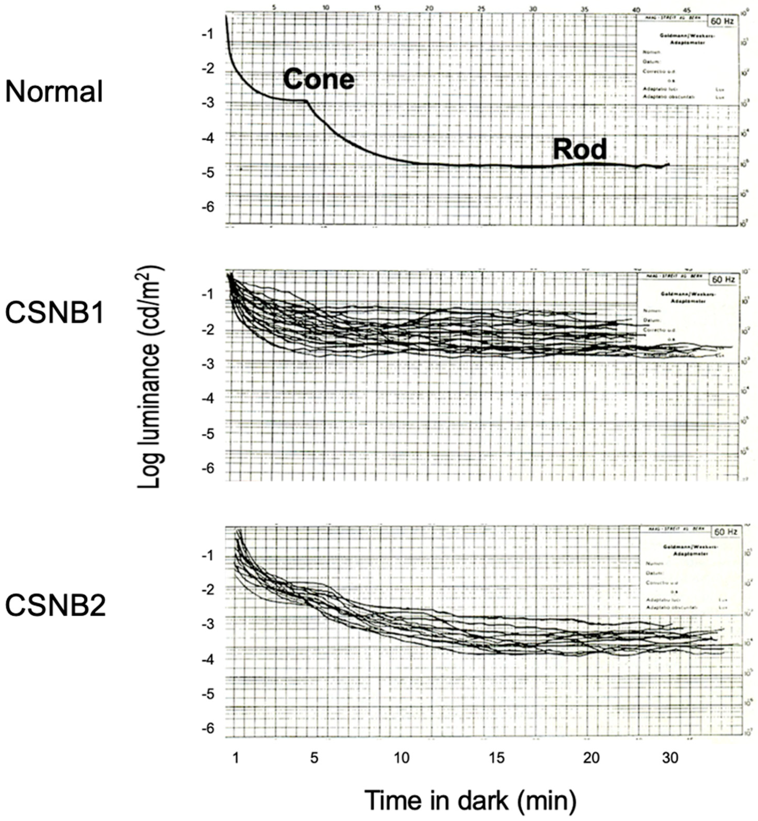
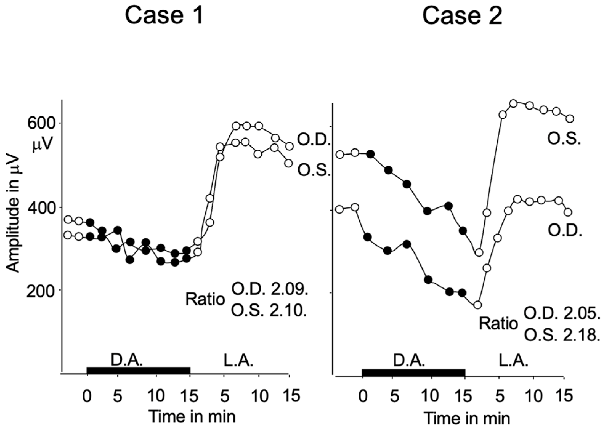
2.2. Subjective Dark Adaptation

Subjective dark adaptation (Figure 2) was tested by a Goldmann–Weekers adaptometer [6,8,9]. The measurements were taken 15° in the superior retina. The superimposed dark adaptation curves in 33 patients with CSNB1 (27 male, 6 female) and 19 patients with CSNB2 (all female) are compared with a normal control.
In CSNB1, the cone threshold is elevated as compared with a normal control, and the rod threshold is absent. In CSNB2, the cone threshold is slightly elevated, and the final threshold of the rod is elevated by approximately 1.0–1.5 log units above normal.

2.3. Visual Acuity and Refractive Error

The distribution of the corrected visual acuity of patients with both types is shown in Figure 3. The corrected visual acuity of 88 eyes in CSNB1 (79 male, 9 female) and 80 eyes of CSNB2 (all male) were compared. Both types showed moderately low visual acuity in many patients (CSNB1: mean 0.45, CSNB2: mean 0.49) [8]. These do not differ significantly. However, it should be noted that eight eyes of CSNB1 patients showed normal visual acuity (1.0 or better) [6,8].
The distribution of the refractive errors in the two groups is shown in Figure 3 [6,8]. Most CSNB1 patients have moderate to high myopic refractive error, whereas CSNB2 patients show a wide range of refractive error, from myopia to hyperopia. The mean refractive error was −8.7 diopter in CSNB1 and −2.5 diopter in CSNB2, which is significantly different (p < 0.001) [8].

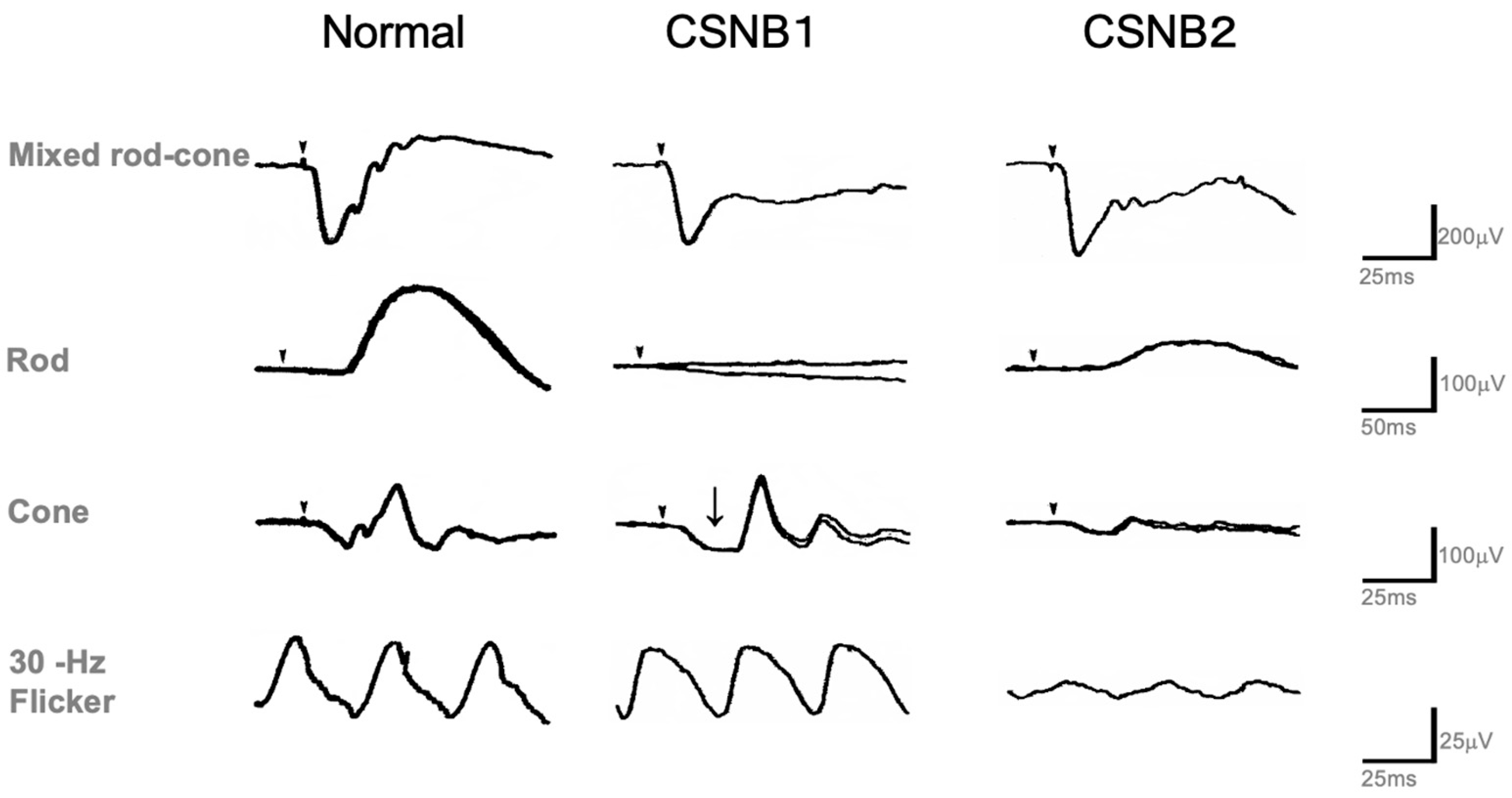
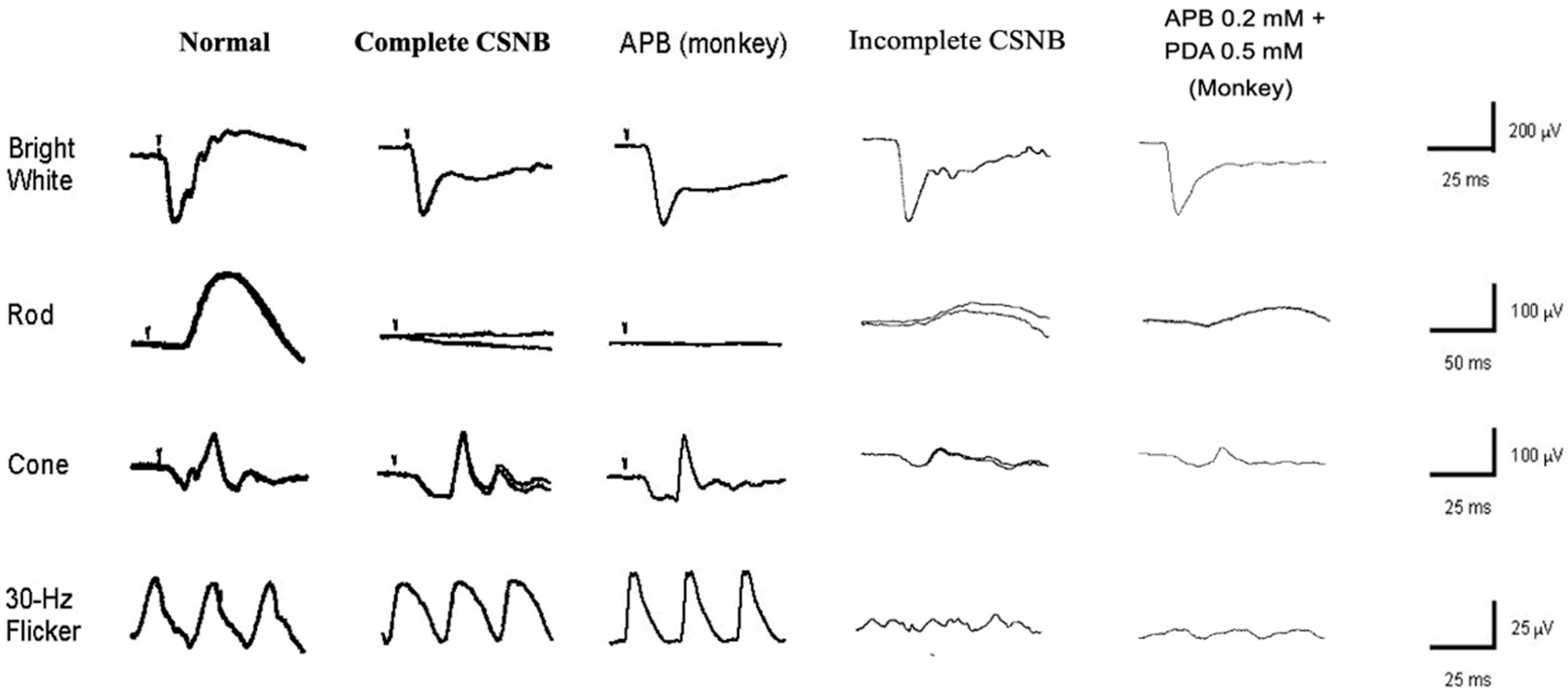
2.4. Standard Full-Field ERG (ISCEV Protocol)

Figure 4 [9] shows representative examples of the standard full-field ERGs recorded from a normal subject and CSNB1 and CSNB2 patients recorded by the ISCEV protocol [14]. For the diagnosis of both types, standard full-field ERG is the most helpful. Bright flash (mixed rod and cone) ERG recorded after 30 min of dark adaptation shows negative ERG (normal a-wave with extremely reduced b-wave) in both types. Oscillatory potentials are often recordable only in CSNB2. Rod ERG is absent in CSNB1 but recordable (although smaller than normal) in CSNB2. This result is consistent with subjective dark adaptation (Figure 2). On the other hand, cone and 30 Hz flicker ERGs are the function of the cone visual pathway. Both look normal in CSNB1 in terms of amplitude and implicit time. However, the a-wave bottom of the cone ERG is not an acute angle, but it shows a plateau (arrow). This wave shape is caused by the OFF wave, which appears when the ON wave disappears. In CSNB2, both cone and 30 Hz flicker ERG are very deteriorated. This is a key finding of CSNB2 in the differentiation of CSNB1. It also provides a very interesting finding that, in spite of no significant difference in visual acuity between the two types (Figure 3), the ERG of the cone system shows such a big difference. In addition, although the cone threshold in subjective dark adaptation (Figure 2) was elevated more in CSNB1 than CSNB2, cone ERGs look better in CSNB1 than CSNB2.

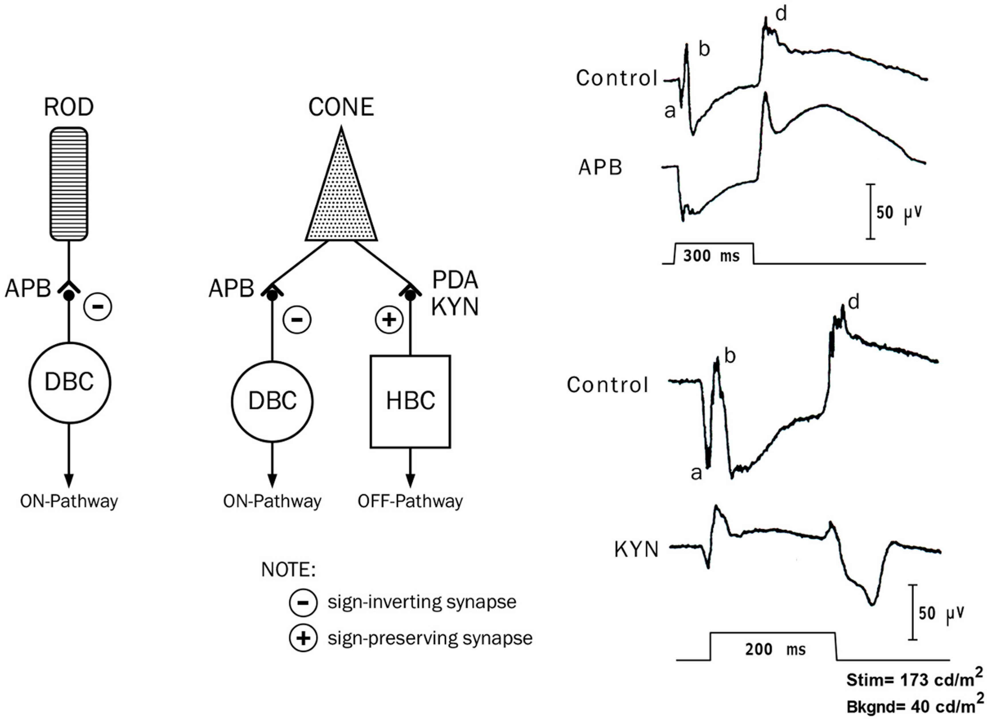
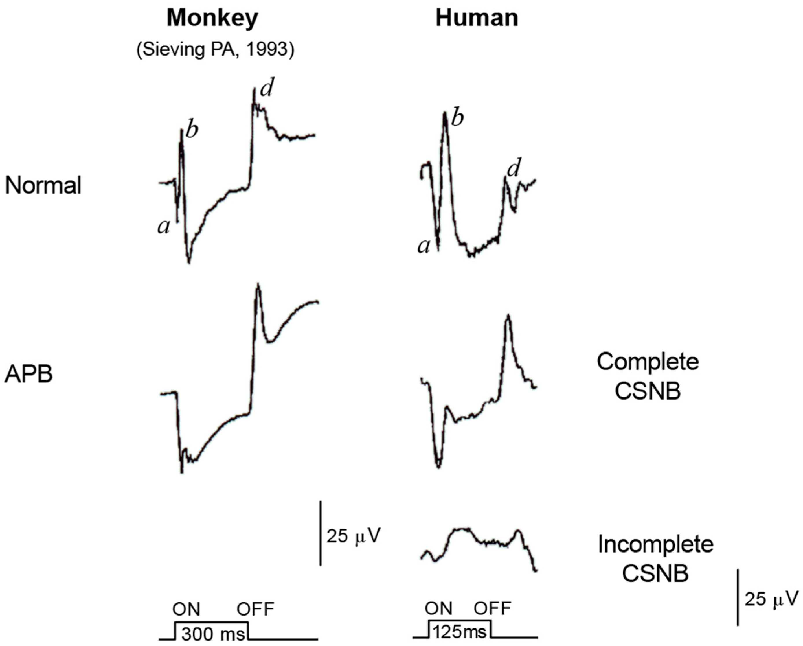
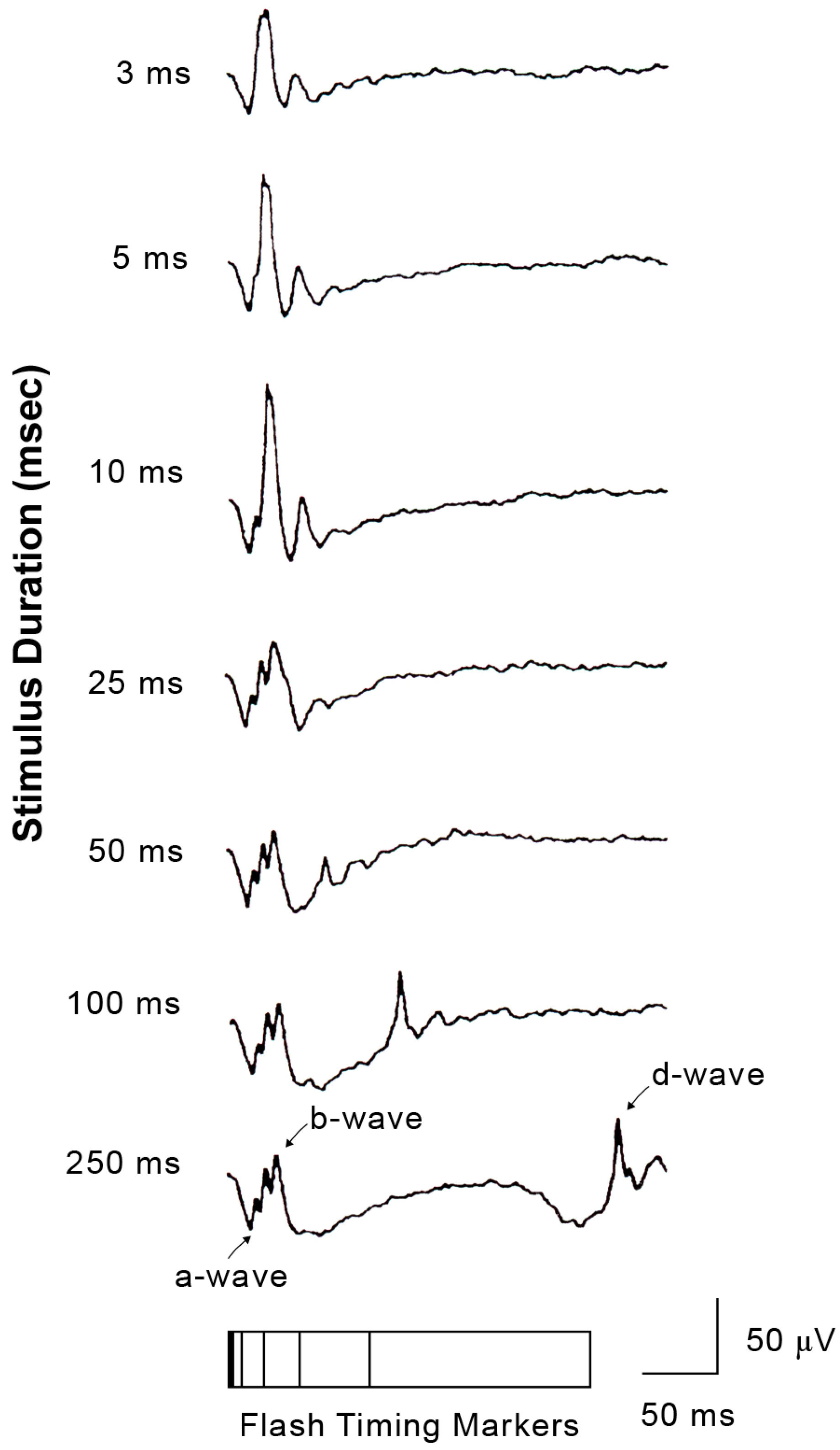
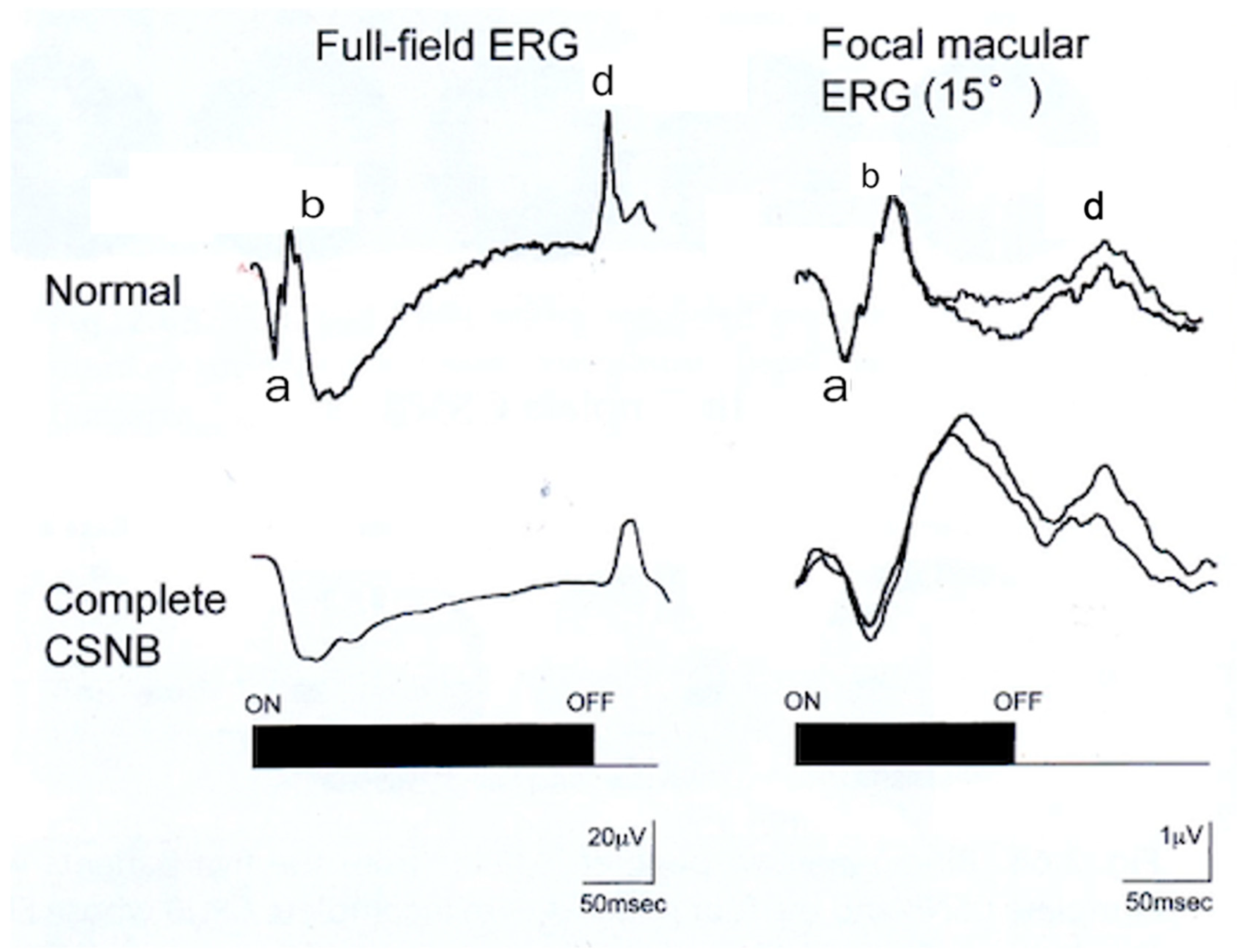
2.5. ON and OFF Responses in Photopic ERG

There are significant potential changes when a light stimulus is turned off. These potentials are called the OFF response, or d-wave, of the ERG. For conventional recordings, a stroboscopic flash is used to elicit the ERGs, and the OFF response is embedded in the ON response. Because there are retinal diseases in which the ON and OFF responses are affected differentially, it became extremely important to record ON and OFF responses separately using a long-duration stimulus [7,15].
Figure 5 shows a simplified schema of the rod and cone visual pathways in the mammalian retina (left) and the long-flash photopic ERGs (right), which were shown by Sieving [15]. The photoreceptors transmit visual information to the BP, with the rods containing only depolarizing bipolar cells (DBCs) through sign-inverting (-)synapses (ON synapse). The cones contact both DBCs and hyperpolarizing bipolar cells (HBCs) through sign-inverting (+) synapses (OFF synapse), respectively. The two types of synapses from the photoreceptors to the BPs are selectively sensitive to different glutamate analogs. The sign-inverting synapse (ON synapse) can be blocked by 2-amino-4-phosphobutyric acid (APB) [16]. The sign-preserving synapse (OFF synapse) can be blocked by either cis-2,3-piperidine dicarboxylic acid (PDA) or kynurenic acid (KYN) [17]. Sieving demonstrated the contribution of these glutamate analogs to the DBCs and HBCs to the monkey long-flash photopic ERG [15].
As shown in Figure 5 (right), the control ERG exhibits ON responses (a-waves and b-waves) and OFF responses (d-waves), with a negative plateau between the b-waves and d-waves. By blocking DBC activity, the photopic b-wave was suppressed, and the a- and d-waves were enhanced. After blocking the HBCs with KYN, the a- and d-waves became smaller, and the plateau was elevated above the baseline (Figure 5, arrowheads). Based on the results, it has been proposed that the “push-pull” activity of the HBCs and DBCs is summated in the photopic ERG recorded at the cornea [15]. The a-wave of the photopic ERG evoked by long- and short-duration flashes arises not only from the neural activity of the photoreceptors but also from HBCs. In addition, the b- and d-waves of the photopic ERG elicited by long-duration flashes are produced by an interaction of the HBCs and DBCs, and the cornea-positive peak of the short-flash ERG results from a summation of the b-wave at light onset and the d-wave at light offset [15].

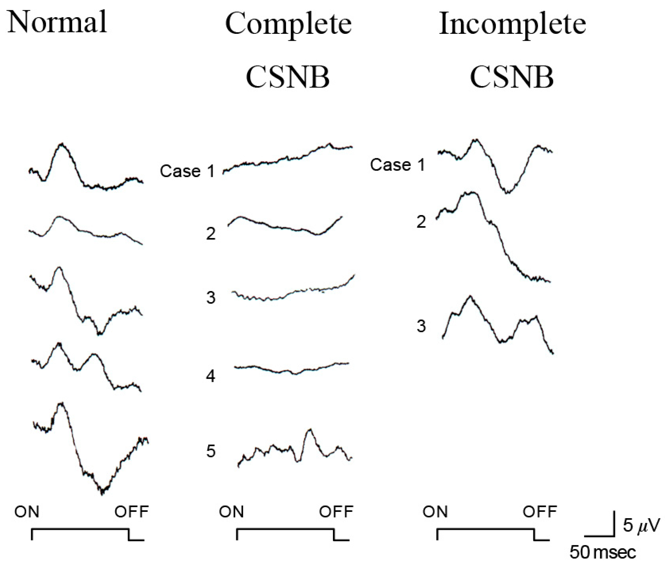
2.6. ON and OFF Responses in CSNB

Using photopic ERGs elicited by long-duration square-wave stimuli, we found that the cone “ON” response generated by depolarizing ON bipolar cells is selectively and severely depressed in patients with CSNB1 [7] (Figure 6). The waveform is similar to that of monkeys after APB is injected into the vitreous to block the ON synapse between photoreceptors and bipolar cells [8].
The OFF response, on the other hand, which is generated by hyperpolarizing bipolar cells, is intact in patients with CSNB1, leading us to hypothesize that the ON function of both the rod and cone visual pathways is completely blocked in eyes with CSNB1. The complete defect of the visual pathway results in complete night blindness, because rods connect only to the ON bipolar cells.
It is interesting to consider why the standard brief flash cone ERG consists of a normal-appearing response (Figure 4) despite the defects of the ON component evaluated by the long-flash photopic ERG in CSNB1. The mechanism involved in generating this phenomenon is as follows.
With long-duration stimuli, the a-, b-, and d-waves are clearly separated. As the stimulus duration is short (brief-flash stimuli), the positive component of the photopic ERG consists mainly of d-waves (Figure 7). Therefore, even when the b-wave, a component of the ON response, is absent (as in CSNB1), the d-wave replaces the b-wave, and a positive wave is recorded with brief-flash stimuli.
In CSNB2, on the other hand, the story is more complex, with both subnormal ON and OFF responses (Figure 6). We hypothesized that the ON and OFF systems are incompletely disturbed at the level of the bipolar cells in patients with CSNB2. This hypothesis was confirmed by the standard full-field ERGs recorded from the monkey’s eye after being treated with neurotransmitter blocking agents. The technique of full-field ERG recording from monkeys under the same conditions as human patients is shown in Figure 8. The monkey ERGs recorded after the ON synapses were completely blocked by APB were identical to those recorded from CSNB1 patients (Figure 8). After the monkey eye was treated with low levels of APB and PDA to block the ON and OFF synapses incompletely, the shape of the full-field ERG is similar to that for CSNB2 (Figure 8). These results indicate that a regular full-field ERG recording (ISCEV protocol [14]) can provide the ON and OFF BP functions.

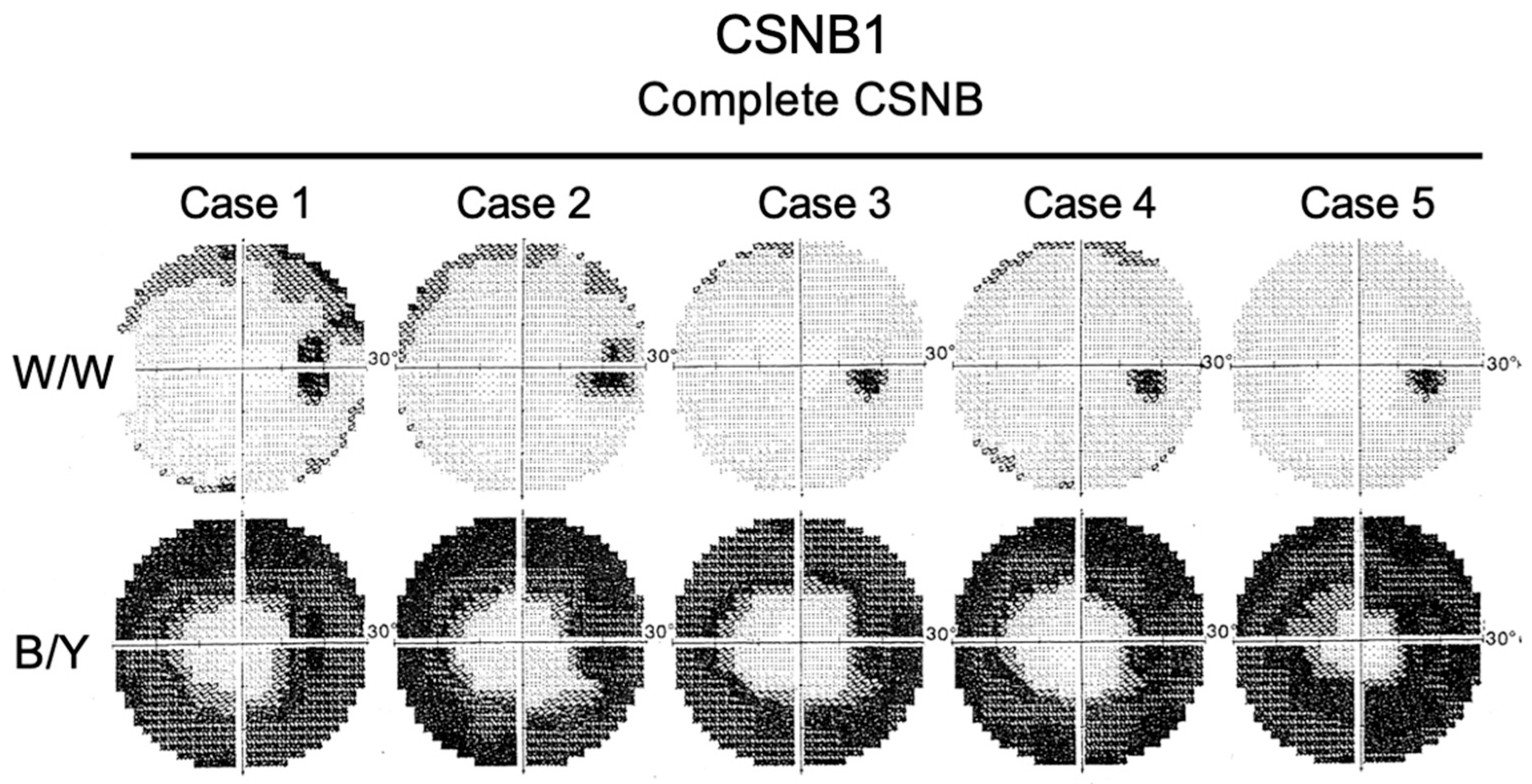
2.7. Short-Wavelength Cone (S-Cone) ERG and Subjective Blue Sensitivity

The S-cone ERGs are markedly different in the two types of CSNB. Because the S-cones connect only to the ON BC, whereas middle- and long-wavelength cones (LM-cones) connect to both ON and OFF BCs [18]. Thus, the S-cone function should be absent in CSNB1. Indeed, the full-field S-cone ERG was nonrecordable in CSNB1 patients (Figure 9).
However, surprisingly enough, the subjective blue sensitivity is normal in CSNB1 [19,20]. Two-color perimetry was performed to evaluate the subjective S-cone function of the entire retina in five patients with CSNB1 (Figure 10) [21].
White-on-white (W/W) perimetry was essentially normal, but blue-on-yellow (B/Y) perimetry showed diffuse dysfunction of S-cones; however, only the central field was preserved. The blue sensitivity is nearly normal only in the central 10° to 15° in CSNB1 [21]. Psychophysically determined color vision is influenced mainly by the central visual field. These findings lead us to believe that the macula of CSNB1 patients must have a unique pathology that is different from that in other parts of the retina in CSNB1.
To solve this interesting finding, the focal macular ERG (FERG) was recorded with a 15° spot from the macula of CSNB1. Full-field photopic ERG and FERG recorded by long-flash stimuli in a CSNB1 patient (Figure 11) [9]. ON and OFF responses were separately recorded.
In a normal control, a-wave, b-wave, oscillatory potentials (OPs), and OFF wave (d-wave) are shown in Figure 11. In CSNB1, the full-field ERG shows that the a-wave and d-wave are clearly recordable, but the b-wave is extremely small, and OPs are absent. In focal macular ERG, in addition to an a-wave, a big b-wave is also recorded in CSNB1. The wave shape of focal macular ERG is quite different from that of full-field ERG. The amplitude of the b-wave is normal; however, the implicit time of the b-wave is far delayed.
The implicit time of the b-wave is similar to that of the S-cone ERG in normal subjects. This result may indicate that only the macular function of the S-cone may still be alive in CSNB1.

2.8. Molecular Genetics

Since we reported a new classification of CSNB in 1986 [6], genetic research has been performed by several institutes to prove that CSNB1 and CSNB2 are different clinical entities [22]. Evidence of genetic heterogeneity in X-linked (XR) and autosomal recessive (AR) CSNB has been reported by several authors. In XR CSNB2, CACNA1F was reported [23,24,25,26]. In XR CSNB1, NYX was reported [27,28]. In AR CSNB1, GRM6 [29], TRPM1 [30,31,32], GPR179 [33], GRM6 [29], and LRIT3 [26,34] were reported. In AR CSNB2, CABP4 [35,36,37,38,39,40,41,42] and CACNA2D4 [43] were reported. Regardless of the mutated gene, CSNB1 always shows BPs are affected by involvement in signal transmission from rods or the unsuitable localization of ion channels in BPs. On the other hand, CSNB2 always shows calcium channel problems. This gene searching information was obtained from phenotype analysis, mainly by ERG.

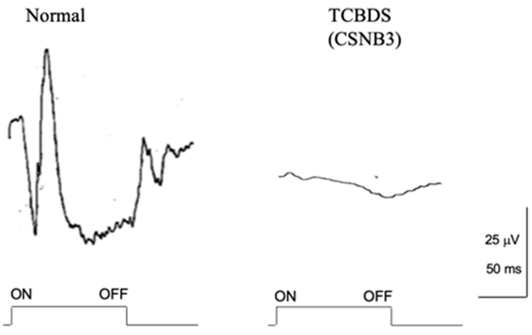
2.9. Total Complete Bipolar Cell Dysfunction Syndrome (TCBDS)

We detected two clinical diseases of BPs, CSNB1 and CSNB2, which showed different BC dysfunctions. By analyzing the pathophysiology of these diseases, we could determine the correlation between psychophysical and electrophysiological abnormalities in relation to BPs.
In 1980, Miyake et al. reported a brother and a sister who showed complete dysfunction of both ON and OFF BCs severely from birth [10]. We followed these patients for longer than 34 years and found that the condition is stationary. We termed this disease as total complete bipolar cell dysfunction syndrome (TCBDS) (CSNB3).
A pair of siblings (5-year-old girl and 7-year-old boy) visited our clinic in 1971. Both suffered severe photophobia, extremely low visual acuity (0.04~0.06), nystagmus, and total color blindness (Figure 12). The fundus and fluoresceine angiography were essentially normal (Figure 1). Neither complained of night blindness. The first impression was rod monochromat because of the color vision tests (Figure 12). The results of the Panel D-15 Dichotomous Color Vision Test demonstrated a characteristic scotoma pattern. The 100 Hue Test developed by the Japan Color Research Institute (JCR) did not reveal any characteristic axis, but it indicated a severely impaired color discrimination ability. The Nagel Anomaloscope results indicated a characteristic matching range consisted with rod monochromacy. However, the full-field ERG showed a negative shape with nonrecordable rod ERG and cone ERG. Taking the bipolar cell functions of CSNB1 and CSNB2 into consideration, the results from these two patients strongly suggest complete dysfunction of ON and OFF BPs. As far as I know, the mutated gene has not been reported in the past. Since many CSNB2 patients who show calcium channel abnormalities reveal ON and OFF BP problems, this disease can be one of them [42]. However, no patient has been reported to have shown absent rod and cone ERG in CSNB2.
These two patients were followed for 34 years, and several examinations were performed during this time. In 1983, the ISCEV protocol was used for ERG testing (Figure 13). The absent rod ERG with negative ERG is similar to CSNB1; however, cone and flicker ERGs were completely absent. The photopic long-flash ERG was nearly nonrecordable (Figure 14), indicating severe dysfunction of ON and OFF BCs.
These results strongly suggest that ON and OFF BCs are both completely impaired in both the rod and cone visual pathways. In the case where the BC function is gone, it is reasonable that almost the entire visual function is wiped out. Indeed, the visual function, including very poor visual acuity, total color blindness, severe photophobia, and nystagmus, looks like rod monochromat (Figure 12). However, one mystery was that neither patient complained of night blindness despite a nonrecordable rod ERG (Figure 13). Night blindness is congenital, and the photophobia is such that the patient may not become severely aware of night blindness. Another surprise was that the Goldmann–Weeker visual field did not show severe abnormality (Figure 12).
The EOG is completely normal (Figure 15). It is interesting that the EOG in all three DC diseases mentioned above is normal. This result suggests that the BC function does not significantly influence the retinal pigment epithelium function.
The above three diseases are all hereditary, stationary disorders with BC dysfunction. One can assume what kind of BC is disturbed and how the visual function may change accordingly.

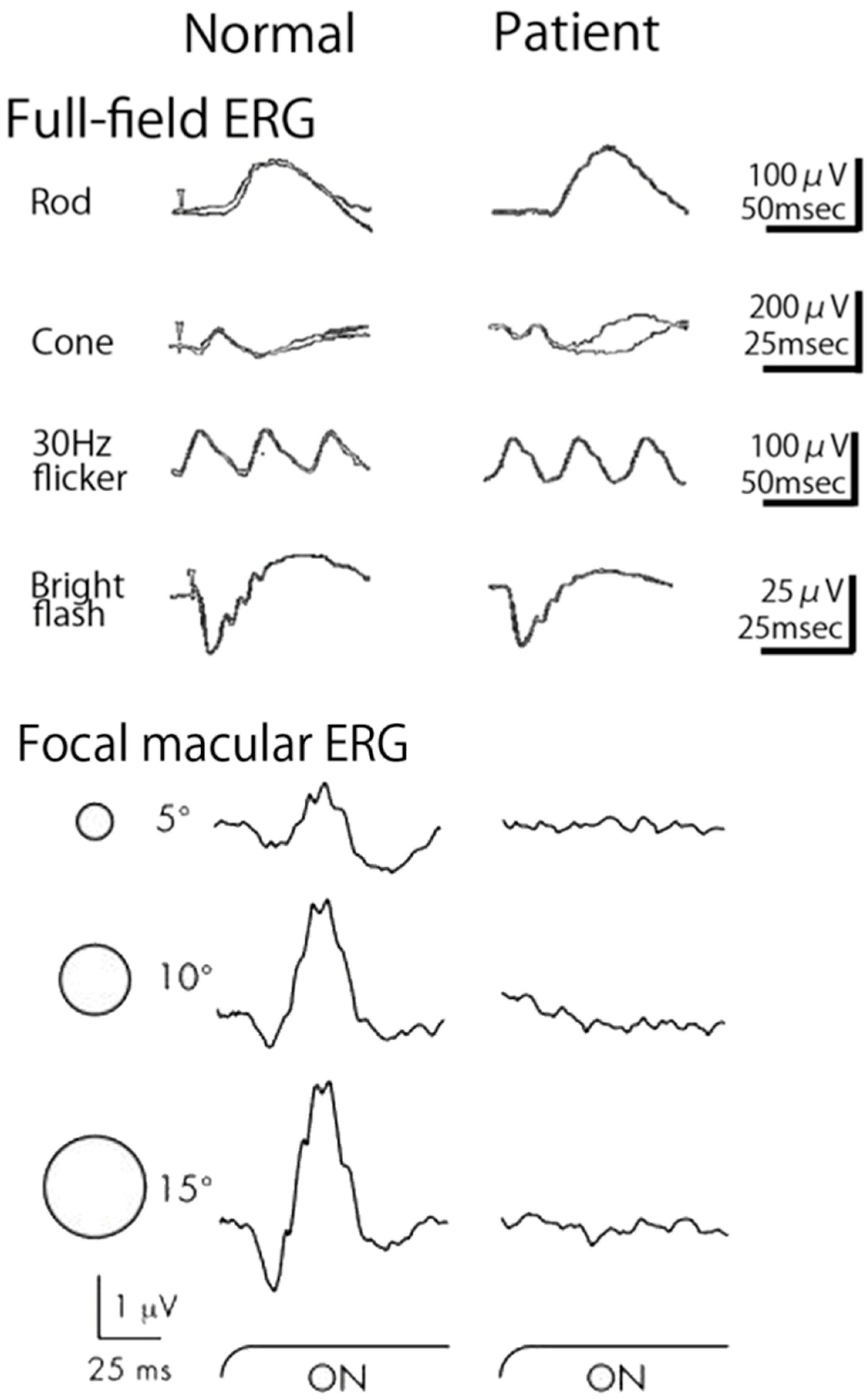
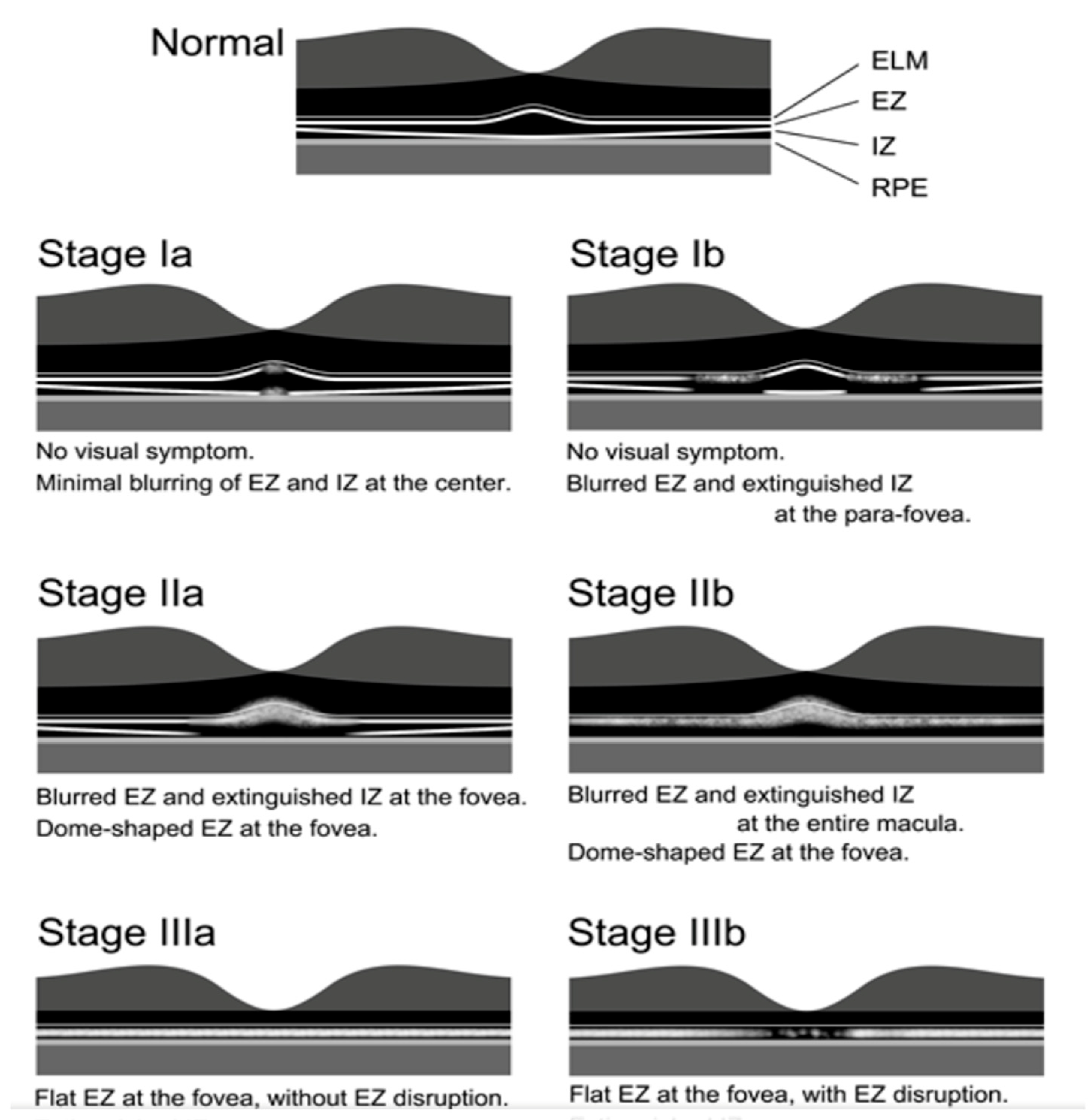
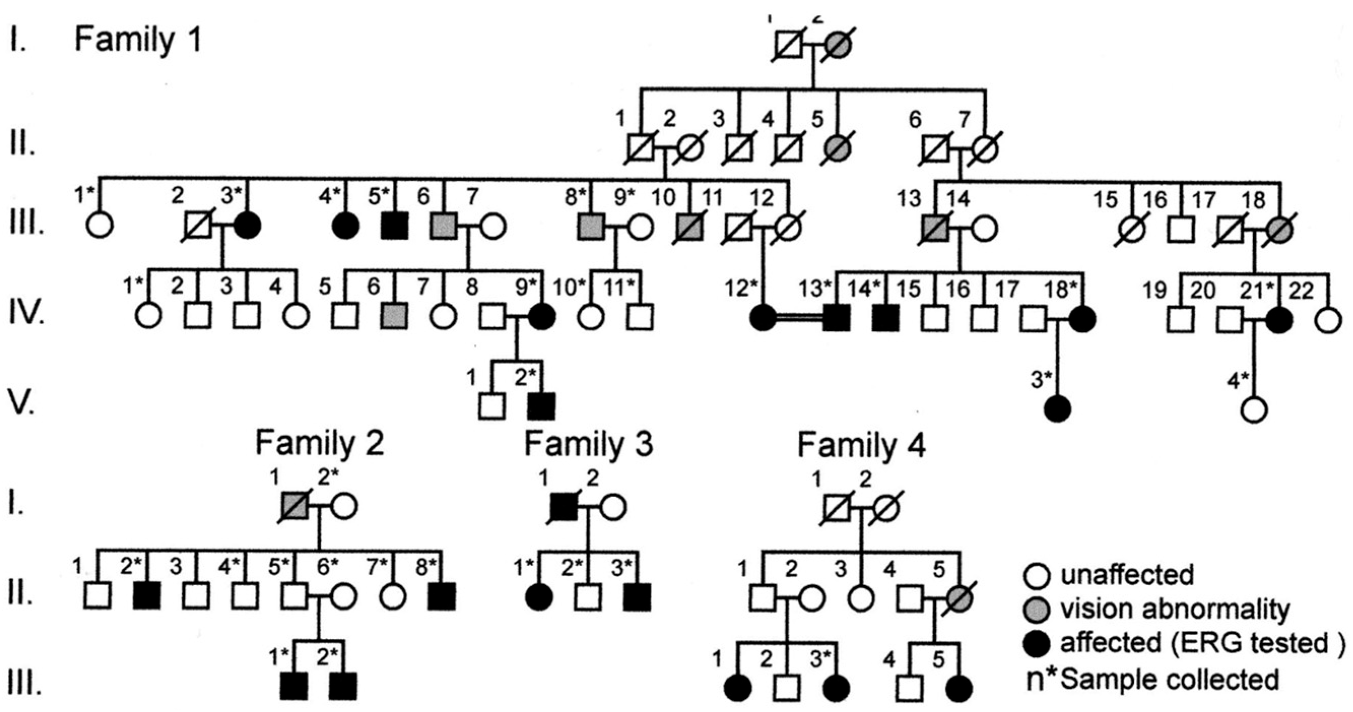
2.10. Occult Macular Dystrophy

In addition to the three kinds of hereditary retinal diseases of BC dysfunction mentioned above, where the fundus is normal, one more hereditary macular disease with normal fundus appearance was included. From 1976, Miyake started to build a focal macular ERG (FERG) with Dr. Tatsuo Hirose in Boston [44]. After long-term efforts to build the FERG, the Miyake group succeeded in making the most informative system under the fundus monitor through an infrared fundus camera (Cannon) in 1988 [45,46]. By using this system, most ERG components could be precisely recorded. They included a-wave, b-wave, oscillatory potentials, OFF d-wave, and PhNR [45,46].
The Miyake group tried to record FERG from many patients and found a new disease, “occult macular dystrophy (OMD)” [11,12]. Our first patient (28-year-old-woman) complained of a gradual decrease in visual acuity (0.3~0.4) during the past 10 years in both eyes. The fundus and fluorescein angiography were normal. The full-field ERGs were normal, but the FERG was enormously abnormal, indicating that only the macula is abnormal. Other family members were examined, and another two members of this family (24-year-old-brother, 54-year-old-father) showed similar abnormalities. The ERG findings in a representative patient with OMD are shown in Figure 16.
The full-field ERGs (left) and the FERGs (right) of three different spot sizes (5°,10°, 15° in diameter) are shown in a representative OMD patient in Figure 16. The full-field ERG is normal, but the focal macular ERGs are very abnormal. The multifocal ERG [47] also shows clearly the pathology of the affected retina (Figure 17).
In 1996, Miyake et al. reported 16 patients with this disease and analyzed them from several points of view [12]. The disease name was termed as “Occult macular dystrophy (OMD)” [12]. “Occult” means “hidden from sight”. The distribution of visual acuity and the age of OMD patients are shown in Figure 18. Visual acuity shows a large variation and little correlation with age [48].
OCT provides some information as to the progress of OMD [49,50,51] (Figure 19). The photoreceptor structures at the fovea and parafovea region determined by OCT gradually changed as the duration of OMD increased. Nakamura et al. graded the OCT changes in the photoreceptor layers in 61 cases. All of the patients at stage 1 were asymptomatic with good visual acuity. Stage 11 was the largest group among OMD patients; disease duration ranged from 1 to 53 years. Stage III was the late stage of OMD; the disease duration ranged from 6 to 65 years, and the corrected visual acuity of stage III b ranged from 0.08 to 0.31.
Thereafter, a lot of attention was paid to detecting gene mutations. The hereditary mode of this family is autosomal dominant, and in 2010, a mutation of the RP1L1 gene after gene analysis of a large family [52] was found (Figure 20, Family 1) by the Miyake group.
The RP1L1 gene was originally identified by human and mouse genome sequencing, and it contained four exons that span 50 kb on chromosome 8p. Immunohistochemistry showed that it is expressed in the rod and cone photoreceptors of cynomolgus monkeys [53]. The RP1L1 protein was suggested to be involved in the morphologic and functional maintenance of photoreceptors [54,55]. The cellular mechanisms that explain why only the macular region is impaired in human OMD patients have not been determined.
In the Japanese population, RP1L1 gene mutations are rarely found in sporadic cases [56,57]. There are OMD families without the RP1L1 mutation where autosomal recessive inheritance is assumed. The genetic background leading to the OMD may be a variant, and other genetic causes will probably be determined in the future.
The Miyake group developed the focal macular ERG system in 1986 after long-term efforts. This focal macular ERG system is very informative [45,46], and this system was in the forefront of the multifocal ERG developed by Sutter in 1994 [47]. By using this method, OMD was discovered in 1989 [11]. In 2010, the gene mutation of OMD, RP1L1, was detected by Akahori et al. [52].
Accordingly, the development of diagnostic instruments, the discovery of new diseases, and the discovery of gene mutations are all achievements completed by Miyake’s group, which may be only one brilliant feat in medical history. Because of the substantial contribution of our group, OMD diseases with the RP1L1 gene mutations are now referred to as “Miyake disease” [57,58,59]. In 2015, the Japanese Ministry of Health, Labor and Welfare for medical care declared Miyake disease as one of the limited diseases for priority functional support.

Funding

This research received no external funding.

Conflicts of Interest

The authors declare no conflict of interest.

References

  1. Oguchi, C. Uber eine Abart von Hemeralopie. Acte Soc. Ophthalmol. Jpn. 1907, 11, 123–134. [Google Scholar]
  2. Fuchs, S.; Nakazawa, M.; Maw, M.; Tamaio, M.; Ogucjhi, Y.; Gal, A. A homozygous 1-base pair deletion in the arrestin gene is a frequent cause of Oguchi disease in Japanese. Nat. Genet. 1995, 10, 360–362. [Google Scholar] [CrossRef]
  3. Nakamura, M.; Yamamoto, S.; Okada, M.; Ito, S.; Tano, Y.; Miyake, Y. Novel mutations in arrestin gene and associated clinical features in Japanese patients with Oguchi disease. Ophthalmology 2004, 111, 1410–1414. [Google Scholar] [CrossRef]
  4. Yamamoto, S.; Sippel, K.C.; Berson, E.L.; Dryja, T.P. Defects in the rhodopsin kinase gene in patients with the Oguchi form of stationary night blindness. Nat. Genet. 1997, 15, 175–178. [Google Scholar] [CrossRef]
  5. Miyake, Y.; Horiguchi, M.; Suzuki, S.; Kondo, M.; Tanikawa, A. Electrophysiological findings in patients with Oguchi’s disease. Jpn. J. Ophthalmol. 1996, 40, 511–519. [Google Scholar]
  6. Miyake, Y.; Yagasaki, K.; Horiguchi, M.; Kawase, Y.; Kanda, T. Congenital stationary night blindness with negative electroretinogram. A new classification. Arch. Ophthalmol. 1986, 104, 1013–1020. [Google Scholar] [CrossRef]
  7. Miyake, Y.; Yagasaki, K.; Horiguchi, M.; Kawase, Y. On- and off-responses in photopic electroretinogram in complete and incomplete types of congenital stationary night blindness. Jpn. J. Ophthalmol. 1987, 31, 81–87. [Google Scholar]
  8. Miyake, Y. Establishment of the concept of new clinical entities. Complete and incomplete form of congenital stationary night blindness. Jpn. Ophthalmol. Soc. 2002, 106, 737–756. [Google Scholar]
  9. Miyake, Y. Hereditary retinal and allied diseases. Complete and incomplete types of CSNB. In Electrodiagnosis of Retinal Diseases; Springer: Tokyo, Japan; Berlin, Germany; New York, NY, USA, 2008; pp. 90–118. [Google Scholar]
  10. Miyake, Y.; Yasuma, T.; Awaya, S. Familial cone-rod dysfunction syndrome. Jpn. Clin. Ophthalmol. 1980, 34, 377–385. [Google Scholar]
  11. Miyake, Y.; Ichikawa, K.; Shiose, Y.; Kawase, Y. Hereditary macular dystrophy without visible fundus abnormality. Am. J. Ophthalmol. 1989, 108, 292–299. [Google Scholar] [CrossRef]
  12. Miyake, Y.; Horiguchi, M.; Tomita, N.; Kondo, M.; Tanikawa, A.; Takahashi, H.; Suzuki, S.; Terasaki, H. Occult macular dystrophy. Am. J. Ophthalmol. 1996, 122, 644–653. [Google Scholar] [CrossRef]
  13. Schubert, G.; Bornschein, H. Beiteitrag zur Analyse des menshlichen Electroretinogram. Ophthalmologica 1952, 123, 396–412. [Google Scholar] [CrossRef]
  14. Marmor, M.F.; Fulton, A.B.; Holder, G.E.; Miyake, Y.; Brigell Bach, M. International Society for Clinical Electrophysiology of Vision ISCEV Standard for full-field clinical electroretinography (2008 update). Doc. Ophthalmol. 2009, 118, 69–77. [Google Scholar] [CrossRef]
  15. Sieving, P.A. Photopic on- and off-pathway abnormalities in retinal dystrophies. Trans. Am. Ophthalmol. Soc. 1993, 91, 701–773. [Google Scholar]
  16. Slaughter, M.M.; Miller, R.F. 2 amino-4-phosphenobutric acid: A new pharmacological tool for retina research. Science 1981, 211, 182–185. [Google Scholar] [CrossRef]
  17. Slaughter, M.M.; Miller, R.F. An excitatory amino acid antagonist blocks cone input to signal conserving second-order retinal neuros. Science 1983, 219, 1230–1232. [Google Scholar] [CrossRef]
  18. Kolb, H.; Lipets, L.E. The anatomical basis for color vision in the vertebrate retina. In The Perception of Colour; Gouras, P., Ed.; Macimillan: London, UK, 1991; pp. 128–145. [Google Scholar]
  19. McKay, C.J.; Saeki, M.; Gouras, P.; Roy, M. Congenital and acquired nyctalopia eliminate the S-cone without disturbing color vision. Investig. Ophthalmol. Vis. Sci. (Supple) 1995, 36, 3925. [Google Scholar]
  20. Kamiyama, M.; Yamamoto, S.; Nitta, K.; Hayasaka, S. Undetectable S-cone electroretinogram b-wave in complete congenital stationary night blindness. Br. J. Ophthalmol. 1996, 80, 637–639. [Google Scholar] [CrossRef]
  21. Terasaki, H.; Miyake, Y.; Nomura, R.; Horiguchi, M.; Suzuki, K.; Kondo, M. Blue-on-yellow perimetry in the complete type of congenital stationary night blindness. Investig. Ophthalmol. Vis. Sci. 1999, 40, 2761–2764. [Google Scholar]
  22. Boycott, K.M.; Pearce, W.G.; Musarella, M.A.; Weleber, R.G.; Maybaum, T.A.; Birch, D.G.; Miyake, Y.; Young, R.S.; Bech-Hansen, N.T. Evidence for genetic heterogeneity in X-linked congenital stationary night blindness. Am. J. Hum. Genet. 1998, 62, 865–875. [Google Scholar] [CrossRef]
  23. Strom, T.M.; Nyakatura, G.; Apfelstedt-Sylla, E.; Hellebrand, H.; Lorenz, B.; Weber, B.H.; Wutz, K.; Gutwillinger, N.; Rüther, K.; Drescher, B.; et al. An L-type calcium-channel gene mutated in incomplete X-linked congenital stationary night blindness. Nat. Genet. 1998, 19, 260–263. [Google Scholar] [CrossRef] [PubMed]
  24. Bech-Hansen, N.T.; Naylor, M.J.; Maybaum, T.A.; Pearce, W.G.; Koop, B.; Fishman, G.A.; Mets, M.; Musarella, M.A.; Boycott, K.M. Loss-of-function mutations in a calcium-channel altha1-subunit gene in Xp11.23 cause incomplete X-linked congenital stationary night blindness. Nat. Genet. 1998, 13, 264–267. [Google Scholar] [CrossRef] [PubMed]
  25. Nakamura, M.; Ito, D.; Terasaki, H.; Miyake, Y. Novel CACNA1F mutation in Japanese patients with incomplete congenital stationary night blindness. Investig. Ophthalmol. Vis. Sci. 2001, 42, 1610–1616. [Google Scholar]
  26. Katta, M.; de Guimaraes, T.A.C.; Fujinami-Yokokawa, Y.; Fujinami, K.; Georgiou, M.; Mahroo, O.A.; Webster, A.R.; Michaelides, M. Congenital stationary night blindness: Structure, function and genotype-phenotype correlations in cohort of 122 patients. Ophthalmol. Retina 2024, 8, 932–941. [Google Scholar] [CrossRef]
  27. Bech-Hansen, N.T.; Naylor, M.J.; Maybaum, T.A.; Sparkes, R.L.; Koop, B.; Birch, D.G.; Bergen, A.A.; Prinsen, C.F.; Polomeno, R.C.; Gal, A.; et al. Mutations in NYX, encoding the leucine-rich proteoglycan nyctalopin, cause X-linked complete congenital stationary night blindness. Nat. Genet. 2000, 26, 319–323. [Google Scholar] [CrossRef] [PubMed]
  28. Pusch, C.M.; Zeitz, C.; Brandau, O.; Pesch, K.; Achatz, H.; Feil, S.; Scharfe, C.; Maurer, J.; Jacobi, F.K.; Pinckers, A.; et al. The complete form of X-linked congenital stationary night blindness is caused by mutations in a gene encoding a leucine-rich repeat protein. Nat. Genet. 2000, 26, 324–327. [Google Scholar] [CrossRef]
  29. Dryja, T.P.; McGee, T.L.; Berson, E.L.; Fishman, G.A.; Sandberg, M.A.; Alexander, K.R.; Derlacki, D.J.; Rajagopalan, A.S. Night blindness and abnormal cone electroretinogram ON responses in patients with mutations in the GRM6 gene encoding mGluR6. Proc. Natl. Acad. Sci. USA 2005, 102, 4884–4889. [Google Scholar] [CrossRef]
  30. Audo, I.; Kohl, S.; Leroy, B.P.; Munier, F.L.; Guillonneau, X.; Mohand-Said, S. TRPM1 is mutated in patients with autosomal-recessive complete congenital stationary night blindness. Am. J. Hum. Genet. 2009, 85, 720–7229. [Google Scholar] [CrossRef] [PubMed]
  31. van Genderen, M.M.; Bijveld, M.M.; Claassen, Y.B.; Florijn, R.J.; Pearring, J.N.; Meire, F.M.; McCall, M.A.; Riemslag, F.C.; Gregg, R.G.; Bergen, A.A.; et al. Mutations in TRPM1 are a common cause of complete congenital stationary night blindness. Am. J. Hum. Genet. 2009, 85, 730–736. [Google Scholar] [CrossRef]
  32. Nakamura, M.; Sanuki, R.; Yasuma, T.R.; Onishi, A.; Nishiguchi, K.M.; Koike, C. TRPM1 mutations are associated with the complete form congenital stationary night blindness. Mol. Vis. 2010, 16, 425–437. [Google Scholar]
  33. Audo, I.; Bujakowska, K.; Orhan, E.; Poloschek, C.M.; Defoort-Dhellemmes, S.; Drumare, I.; Kohl, S.; Luu, T.D.; Lecompte, O.; Zrenner, E.; et al. Whole-exome sequencing identifies mutations in GPR179 leading to autosomal-recessive complete congenital stationary night blindness. Am. J. Hum. Genet. 2012, 90, 321–330. [Google Scholar] [CrossRef] [PubMed]
  34. Zeitz, C.; Jacobson, S.G.; Hamel, C.P.; Bujakowska, K.; Neuillé, M.; Orhan, E.; Zanlonghi, X.; Lancelot, M.E.; Michiels, C.; Schwartz, S.B.; et al. Whole-exome sequencing identifies LRIT3 mutatons as a cause of autosomal-recessive complete congenital night blindness. Am. J. Hum. Genet. 2013, 92, 67–75. [Google Scholar] [CrossRef] [PubMed]
  35. Zeitz, C.; Kloeckener-Gruissem, B.; Forster, U.; Kohl, S.; Magyar, I.; Wissinger, B.; Mátyás, G.; Borruat, F.-X.; Schorderet, D.F.; Zrenner, E.; et al. Mutations in CABP4, the gene encoding the Ca2+-binding protein 4, cause autosomal recessive night blindness. Am. J. Hum. Genet. 2006, 79, 657–667. [Google Scholar] [CrossRef]
  36. Littink, K.W.; van Genderen, M.M.; Collin, R.W.J.; Roosing, S.; de Brouwer, A.P.M.; Riemslag, F.C.C.; Venselaar, H.; Thiadens, A.A.H.J.; Hoyng, C.B.; Rohrschneider, K.; et al. A novel homozygous nonsense mutation in CABP4 causes congenital cone-rod synaptic disorder. Investig. Ophthalmol. Vis. Sci. 2009, 50, 2344–2359. [Google Scholar] [CrossRef] [PubMed]
  37. Khan, A.O.; Alrashed, M.; Alkuraya, F.S. Clinical characterization of the CABP4 related retinal phenotype. Br. J. Ophthalmol. 2013, 97, 262–265. [Google Scholar] [CrossRef]
  38. Khan, A.O. CABP4 mutations do not cause congenital stationary night blindness. Ophthalmology 2013, 121, e15. [Google Scholar] [CrossRef]
  39. Bijveld, M.M.; Florijn, R.J.; Bergen, A.A.; van den Born, L.I.; Kamermans, M.; Prick, L.; Riemslag, F.C.; van Schooneveld, M.J.; Kappers, A.M.; van Genderen, M.M. Genotype and phenotype of 101 dutch patients with congenital stationary night blindness. Ophthalmology 2013, 120, 2072–2081. [Google Scholar] [CrossRef]
  40. Schatz, P.; Abdalla Elsayed, M.E.A.; Khan, A.O. Mutation modal imaging in CABP-4 related retinopathy. Ophthalmic Genet. 2017, 38, 459–464. [Google Scholar] [CrossRef]
  41. Smirnov, V.M.; Zeitz, C.; Soumittra, N.; Audo, I.; Defoort-Dhellemmes, S. Retinal findings in a patient of French ancestry with CABP-4 related retinal disease. Doc. Ophthalmol. 2018, 136, 135–143. [Google Scholar] [CrossRef]
  42. Tan, J.K.; Arno, G.; Josifova, D.; Mohamed, M.D.; Mahroo, O.A. Unusual findings in a patient with CABP4-associated cone-rod synaptic disorder. Doc. Ophthalmol. 2024, 148, 1115–1120. [Google Scholar] [CrossRef]
  43. Wycisk, K.A.; Zeitz, C.; Feil, S.; Wittmer, M.; Forster, U.; Neidhardt, J.; Wissinger, B.; Zrenner, E.; Wilke, R.; Kohl, S.; et al. Mutation in the auxiliary calcium-channel subunit CACNA2D4 causes autosomal recessive cone dystrophy. Am. J. Hum. Genet. 2006, 79, 973–977. [Google Scholar] [CrossRef] [PubMed]
  44. Hirose, T.; Miyake, Y.; Hara, A. Simultaneous recording pf electroretinogram and visual evoked response. Focal stimulation under direct observation. Arch. Ophthalmol. 1977, 95, 1205–1208. [Google Scholar] [CrossRef]
  45. Miyake, Y. Studies of local macular ERG. Jpn. Ophthamol. Soc. 1988, 92, 1419–1449. [Google Scholar]
  46. Miyake, Y.; Shiroyama, N.; Ota, I.; Horiguchi, M. Oscillatory potentials in electroretinograms of the human macular region. Investig. Ophthalmol. Vis. Sci. 1988, 29, 1631–1635. [Google Scholar]
  47. Sutter, E.E.; Tran, D. The field topography of ERG components in man. 1. The photopic luminance response. Vis. Res. 1992, 32, 433–446. [Google Scholar] [CrossRef] [PubMed]
  48. Miyake, Y.; Tsunoda, K. Occult macular dystrophy. Jpn. J. Ophthalmol. 2015, 59, 71–80. [Google Scholar] [CrossRef]
  49. Nakanishi, A.; Ueno, S.; Kawano, K.; Ito, Y.; Kominami, T.; Yasuda, S.; Kondo, M.; Tsunoda, K.; Iwata, T.; Terasaki, H. Pathologic changes of cone photoreceptors in eyes with occult macular dystrophy. Investig. Ophthalmol. Vis. Sci. 2015, 56, 7243–7249. [Google Scholar] [CrossRef]
  50. Fujinami, K.; Kameya, S.; Kikuchi, S.; Ueno, S.; Kondo, M.; Hayashi, T.; Shinoda, K.; Machida, S.; Kuniyoshi, K.; Kawamura, Y.; et al. Novel RP1L1 variants and genotype-photoreceptor microstructural phenotype association in cohort of Japanese patients with occult macular dystrophy. Investig. Ophthalmol. Vis. Sci. 2016, 57, 4837–4846. [Google Scholar] [CrossRef]
  51. Nakamura, N.; Tsunoda, K.; Mizuno, Y.; Usui, T.; Hatase, T.; Ueno, S.; Kuniyoshi, K.; Hayashi, T.; Katagiri, S.; Kondo, M.; et al. Clinical stages of occult macular dystrophy based on optical coherence tomographic findings. Investig. Ophthalmol. Vis. Sci. 2019, 60, 4691–5700. [Google Scholar] [CrossRef]
  52. Akahori, M.; Tsunoda, K.; Miyake, Y.; Fukuda, Y.; Ishiura, H.; Tsuji, S.; Usui, T.; Hatase, T.; Nakamura, M.; Ohde, H.; et al. Dominant mutations in RP1L1are responsible for occult macular dystrophy. Am. J. Hum. Genet. 2010, 10, 424–429. [Google Scholar] [CrossRef]
  53. Noel, N.C.L.; MacDonald, I.M. RP1L1 and inherited photoreceptor disease. A review. Surv. Ophthalmol. 2020, 65, 725–739. [Google Scholar] [CrossRef] [PubMed]
  54. Conte, I.; Lestingi, M.; den Hollander, A.; Alfano, G.; Ziviello, C.; Pugliese, M.; Circolo, D.; Caccioppoli, C.; Ciccodicola, A.; Banfi, S. Identification and characterization of the retinitis pigmentosa 1-like gene (RP1L1) a novel candidate for retinal degenerations. Eur. J. Human. Genet. EJHC 2003, 11, 155–162. [Google Scholar] [CrossRef] [PubMed]
  55. Bowne, S.J.; Daiger, S.P.; Malone, K.A.; Heckenlively, J.R.; Kennan, A.; Humphries, P.; Hughbanks-Wheaton, D.; Birch, D.G.; Liu, Q.; Pierce, E.A.; et al. Characterization of RPL1L, a highly polymorphic paralog of retinitis pigmentosa 1 (RP1) gene. Mol. Vis. 2003, 9, 129–137. [Google Scholar]
  56. Tsunoda, K.; Usui, T.; Hatase, T.; Yamai, S.; Fujinami, K.; Hanazono, G.; Shinoda, K.; Ohde, H.; Akahori, M.; Iwata, T.; et al. Clinical characteristics of occult macular dystrophy in family with mutation of RP1L1 gene. Retina 2012, 32, 1135–1147. [Google Scholar] [CrossRef]
  57. Fujinami, K.; Yang, L.; Joo, K.; Tsunoda, K.; Kameya, S.; Hanazono, G.; Fujinami-Yokokawa, Y.; Arno, G.; Kondo, M.; Nakamura, N.; et al. Clinical and genetic characteristics of east Asian patients with occult macular dystrophy (Miyake Disease): East Asia Occult Macular Dystrophy Studies Report Number 3. Ophthalmology 2019, 126, 1432–1444. [Google Scholar] [CrossRef] [PubMed]
  58. Kato, Y.; Hanazono, G.; Fujinami, K.; Hatase, T.; Kawamura, Y.; Iwata, T.; Miyake, Y.; Tsunoda, K. Parafoveal photoreceptor abnormalities in asymptomatic patients with RP1L1 mutations in families with occult macular dystrophy (Miyake Disease). Investig. Ophthalmol. Vis. Sci. 2017, 58, 6020–6029. [Google Scholar] [CrossRef]
  59. Fujinami-Yokokawa, Y.; Joo, K.; Liu, X.; Tsunoda, K.; Kondo, M.; Ahn, S.J.; Robson, A.G.; Naka, I.; Ohashi, J.; Li, H.; et al. Distinct clinical effects of two RP1L1 hotspots in east Asian with occult macular dystrophy (Miyake Disease): EAOMO Report 4. Investig. Ophthalmol. Vis. Sci. 2024, 65, 41–47. [Google Scholar] [CrossRef]
Figure 1. Fundus of complete type congenital stationary night blindness (CSNB1) (A) [6]. Incomplete type congenital stationary night blindness (CSNB2) (B) [6]. Total complete bipolar cell dysfunction syndrome (CSNB3) (C) [10]. Occult macular dystrophy (OMD) (D) [11]. All diseases show a normal fundus. (From Miyake [6,10,11]).
Ijms 26 05166 g001
Figure 2. Superimposed subjective dark adaptation curve in a normal patient and CSNB1 and CSNB2 patients. CSNB1 patients lack rod adaptation completely, but CSNB2 patients show preserved rod adaptation [6,8].
Ijms 26 05166 g002
Figure 3. Visual acuity (left) and refractive error (right) in CSNB1 (upper) and CSNB2 (lower). Moderately low visual acuity shows in both types of CSNB. Most CSNB1 patients show high to moderate myopia, while CSNB2 patients show both moderate myopia and hyperopia [8]. Visual acuity is shown as decimal visual acuity.
Ijms 26 05166 g003
Figure 4. Full-field standard ERGs recorded by the ISCEV protocol in a normal control, CSNB1, and CSNB2. Both types show negative ERG. Rod ERG is absent (CSNB1) but recordable (CSNB2). In spite of the fact that visual acuity shows little difference between the two CSNB, photopic ERGs (cone and 30 Hz flicker) show a significant difference. The cone ERG looks normal in CSNB1, but the bottom of the a-wave shows a plateau shape (arrow). Small arrows in each response indicate stimulus onset.
Ijms 26 05166 g004
Figure 5. Simplified schema of the rod and cone visual pathway in the mammalian retina [15]. a, b and d on the wave are names of component in ERG.
Ijms 26 05166 g005
Figure 6. Long-flash photopic ERGs in monkeys are compared with those of patients. Normal responses were compared with APB-treated monkey and CSNB1 and CSNB2 patients. The waveform of CSNB1 is similar to APB-treated monkey ERG [8]. a, b and d on the wave are names of component in ERG.
Ijms 26 05166 g006
Figure 7. Photopic ERGs recorded with various stimulus durations from a normal subject.
Ijms 26 05166 g007
Figure 8. From the left: standard full-field ERGs in a normal human control, CSNB1 patient, monkey model of CSNB1 with APB injected into the vitreous, CSNB2 patient, monkey model of CSNB2 with a small amount of APB and PDA injected into the vitreous. ERGs of CSNB1 and CSNB2 are very similar to each monkey model ERG. Small arrows in each response indicate stimulus onset.
Ijms 26 05166 g008
Figure 9. Full-field S-cone ERGs in normal subjects and CSNB1 and CSNB2 patients. S-cone ERG is absent in CSNB1 but subnormal in CSNB2 patients.
Ijms 26 05166 g009
Figure 10. Two-color perimetry in white-on-white (W/W) and blue-on-yellow (B/Y) in 5 patients with CSNB1. In B/Y perimetry, only the central zone is preserved [21].
Ijms 26 05166 g010
Figure 11. Long-flash photopic full-field ERG (left) and focal macular ERG (right) in a normal subject (upper) and a patient with CSNB1 (lower) [9]. a, b, and d on the wave are components of ERG.
Ijms 26 05166 g011
Figure 12. Visual fields and color vision in two patients with TCBDS (CSNB3). Panel D-15 test, 100 Hue Test and Nagel Anomaloscope show similar results of total color blindness (rod monochromat). P: protan, D: deutan, N: normal (from Miyake et al. [10]).
Ijms 26 05166 g012
Figure 13. Full-field ERG in 2 patients with TCBDS (CSNB3). Bright-flash ERG shows a negative shape. Both rod and cone ERGs are completely absent [10]. Arrow indicates stimulus onset.
Ijms 26 05166 g013
Figure 14. Photopic long-flash full-field ERG in a normal subject (left) and TCBDS (CSNB3) (right) [10].
Ijms 26 05166 g014
Figure 15. EOG in two patients with CSNB3. EOG is normal.
Ijms 26 05166 g015
Figure 16. Full-field ERGs (left) and focal macular ERG (FERG) recorded by 3 different sizes of stimulus spots (5°, 10°, 15°) in diameter (right) in a normal subject and a patient with occult macular dystrophy (OMD). Full-field ERG is normal, but FERG is very abnormal. Arrow indicates stimulus onset.
Ijms 26 05166 g016
Figure 17. Multifocal ERG in a normal subject (A) and occult macular dystrophy (OMD) (B). Only the central retina is depressed, and the other part is normal.
Ijms 26 05166 g017
Figure 18. Corrected visual acuity of OMD and age of the patients. There is little correlation between visual acuity and age. Visual acuity is shown as decimal visual acuity.
Ijms 26 05166 g018
Figure 19. OCT and progress of OMD [51]. ELM: external limiting membrane, EZ: ellipsoid zone, IZ: internal segment ellipsoid, RPE: retinal pigment epithelium.
Ijms 26 05166 g019
Figure 20. Examples of OMD families with RP1L1 gene mutation. From analysis of Family 1, RP1L1 gene mutation was found for the first time [47,48].
Ijms 26 05166 g020
Disclaimer/Publisher’s Note: The statements, opinions and data contained in all publications are solely those of the individual author(s) and contributor(s) and not of MDPI and/or the editor(s). MDPI and/or the editor(s) disclaim responsibility for any injury to people or property resulting from any ideas, methods, instructions or products referred to in the content.

