Article

Doping of Hollow Urchin-like MnO2 Nanoparticles in Beta-Tricalcium Phosphate Scaffold Promotes Stem Cell Osteogenic Differentiation

1
Department of Ocean & Mechanical Engineering, Florida Atlantic University, Boca Raton, FL 33431, USA
2
Department of Biomedical Engineering, Florida Atlantic University, Boca Raton, FL 33431, USA
3
Department of Biomedical Science, Florida Atlantic University, Boca Raton, FL 33431, USA
4
Faculty of Integrative Biology PhD Program, Department of Biological Science, Florida Atlantic University, Boca Raton, FL 33431, USA
5
Department of Chemistry and Biochemistry, Florida Atlantic University, Boca Raton, FL 33431, USA
*
Author to whom correspondence should be addressed.
Int. J. Mol. Sci. 2025, 26(11), 5092; https://doi.org/10.3390/ijms26115092
Submission received: 26 March 2025 / Revised: 19 May 2025 / Accepted: 21 May 2025 / Published: 26 May 2025

Abstract

Effective osteogenesis for bone regeneration is still considerably challenging for a porous β-tricalcium phosphate (β-TCP) scaffold to achieve. To overcome this challenge, hollow manganese dioxide (H-MnO2) nanoparticles with an urchin-like shell structure were prepared and added in the porous β-TCP scaffold. A template-casting method was used to prepare the porous H-MnO2/β-TCP scaffolds. As a control, solid manganese dioxide (S-MnO2) nanoparticles were also added into β-TCP scaffolds. Human bone mesenchymal stem cells (hBMSC) were seeded in the porous scaffolds and characterized through cell viability assay and alkaline phosphatase (ALP) assay. Results from in vitro protein loading and releasing experiments showed that H-MnO2 can load significantly higher proteins and release more proteins compared to S-MnO2 nanoparticles. When they were doped into β-TCP, MnO2 nanoparticles did not significantly change the surface wettability and mechanical properties of porous β-TCP scaffolds. In vitro cell viability results showed that MnO2 nanoparticles promoted cell proliferation in a low dose, but inhibited cell growth when the added concentration went beyond 0.5%. At a range of lower than 0.5%, H-MnO2 doped β-TCP scaffolds promoted the early osteogenesis of hBMSCs. These results suggested that H-MnO2 in the porous β-TCP scaffold has promising potential to stimulate osteogenesis. More studies would be performed to demonstrate the other functions of urchin-like H-MnO2 nanoparticles in the porous β-TCP.

1. Introduction

Rapid and efficient bone formation is still a considerable challenge for a tissue engineered β-tricalcium phosphate (β-TCP) scaffold. Tissue engineering strategies using β-TCP biomaterial scaffolds, cells, and growth factors have been developed to overcome the issues associated with autografts and allografts for bone tissue repairs [1,2,3,4,5]. However, translating tissue-engineered products into clinical applications is facing enormous difficulties and challenges. The progress achieved in laboratory studies has not yielded true benefits to clinical patients [6]. The reason may come from the following issues: classic stem-cell- or growth-factor-centric tissue regenerative strategies require seeding reproducible stem cells [7,8,9,10], maintaining the homogeneity of cultured cells in vitro [11,12,13], effectively directing stem cell differentiation, and ensuring a high survival rate and effective engraftment of seeded cells in vivo [14,15]. Obviously, it is extremely challenging for current biomaterial scaffolds to achieve the above-stated requirements.
For stimulating stem cell osteogenesis, many strategies have been studied; for example, using osteoinductive growth factor bone morphogenesis proteins (BMPs). However, speculations as to the safety and efficacy concerning the clinical application of BMPs remain, as a satisfactory level of data has not been generated yet, although the concern is based on the limited number of reports linking osteosarcomas with BMP activity [16,17]. Additionally, as BMPs are rapidly cleared from circulation by the liver, a well-characterized BMPs delivery carrier system is required to deliver BMPs in the implanted sites. Even so, 25 to 90% of the implanted dose after 3 h of implantation is lost, depending on the carriers [18,19]. Therefore, many studies explored alternative ways to add osteogenesis-stimulating materials into the scaffold to develop a new-generation bone substitute with excellent osteoinductivity and without the administration of any growth factor; for example, adding the elements magnesium [20], silicon [21], or strontium [22]. However, the effect of manganese ions (Mn2+) on osteogenesis seems to be overlooked, and has not been studied yet. Manganese is an essential trace element that is naturally present in many foods and available as a dietary supplement, and the human body contains about 10 to 20 mg manganese, of which 25% to 40% is in bone [23,24]. Manganese is a cofactor for several enzymes involved in bone formation [25]. In animals, manganese deficiency can impair bone formation and reduce bone mineral density [26], and manganese supplementation can increase both bone mineral density and bone formation [27].
Manganese dioxide (MnO2), an important and well-studied class of materials in catalysts, ion-exchangers, and batteries [28], has been proven to be biodegradable [29,30]. It displays excellent drug delivery capabilities, as it can react with the intracellular redox reagent glutathione (GSH), and produces vast amounts of water soluble Mn2+ ions [31,32,33]. Studies showed that MnO2-containing scaffolds promoted high survival rates, controlled differentiation of stem cells [34], and functional nerve recovery in a spinal cord injury animal model [35,36,37].
In our recent study [38], we successfully prepared hollow urchin-like MnO2 (H-MnO2) nanoparticles. The study results showed that hollow urchin-like MnO2 nanoparticles have higher surface area for drug loading, faster degradation rates for drug release, and improved colloidal stability, as compared to other potential drug delivery candidates, and they can be coated, loaded with a drug, and can target cells. However, whether the addition of such hollow urchin-like MnO2 nanoparticles into a β-TCP scaffold can enhance osteogenesis remains to be investigated. In this study, we added MnO2 nanoparticles in the porous β-TCP scaffold. We hypothesized that the addition of hollow urchin-like MnO2 nanoparticles in the porous β-TCP scaffold would significantly promote osteogenesis. To this end, solid and hollow MnO2 nanoparticles were doped into the β-TCP scaffolds. The porous β-TCP scaffolds with manganese dioxide nanoparticles were fabricated by our published method [39,40,41]. Human bone mesenchymal stem cells (hBMSC) were seeded on the scaffolds to characterize their behaviors through alkaline phosphatase (ALP) assay. The results showed that in a safety concentration range the MnO2/β-TCP scaffolds proved to have no adverse effects on the viability of hBMSCs and promoted their osteogenic differentiation and proliferation. These results suggested that the doping of MnO2 nanoparticles in the porous β-TCP scaffold potentially stimulated bone tissue regeneration.

2. Results

2.1. Morphology of H-MnO2 Nanoparticles

In this study, we prepared H-MnO2 nanoparticles first, and then added these nanoparticles into β-TCP for nanoparticle-doped scaffolds. The SEM image indicates that most of the nanoparticles have a similar size, which was determined by the diameter of the SiO2 template nanoparticles (Figure 1A). A TEM image clearly shows that the H-MnO2 nanoparticles have hollow structures (Figure 1B), but the S-MnO2 nanoparticles (US Research Nanomaterials, Inc., Houston, TX, USA) show a solid structure with a smooth surface (Figure 1C). Interestingly, the shell of H-MnO2 nanoparticles has many stings, which is mimetic to the shell of sea urchin (Figure 1D). The diameter of the nanoparticle is around 250 nm, with an around 25–40 nm shell (Figure 1D(i)).

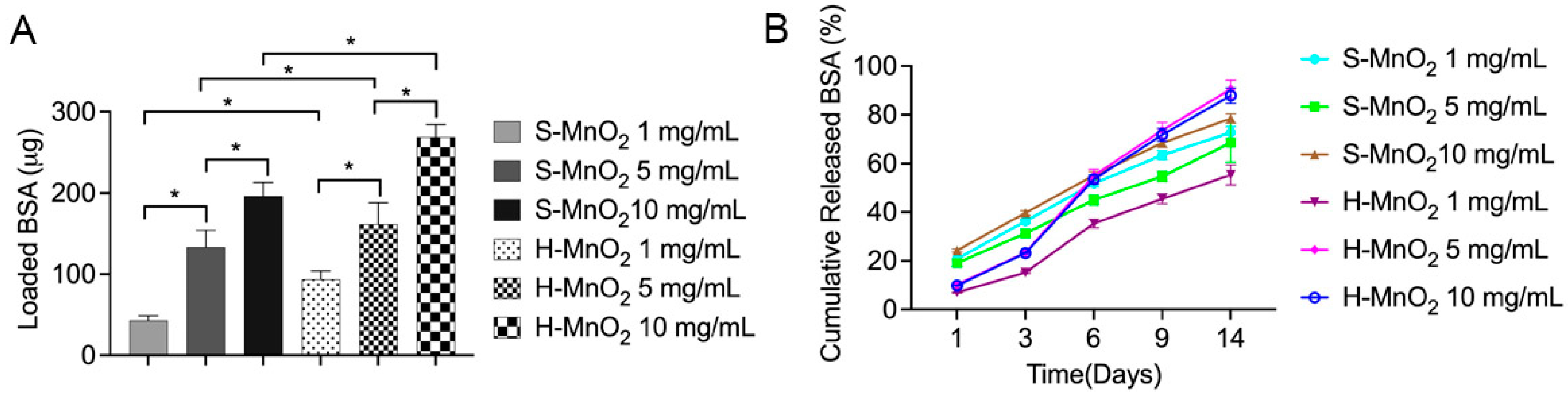
2.2. Loading and Release Profile of the Proteins on H-MnO2 Nanoparticles

To further confirm the function of such an urchin-like shell and hollow structure, protein loading and release experiments were performed. The results show that the hollow MnO2 nanoparticles can load significantly higher bovine serum albumin (BSA) proteins compared to S-MnO2 nanoparticles at the same addition of MnO2 nanoparticles mass (Figure 2A). Meanwhile, with the increase in the MnO2 nanoparticles (from 1 mg/mL to 10 mg/mL), regardless of hollow or solid nanoparticles, the BSA loading increased with the increased addition of MnO2 nanoparticles (Figure 2A). This result implies that the hollow structure can load more proteins than solid structure.
After we placed the BSA-loaded MnO2 nanoparticles into a phosphate-buffer saline (PBS) released medium, the released BSA was measured. The results show that both S-MnO2 and H-MnO2 nanoparticles can slowly release the adsorbed BSA with time (Figure 2B). Before 6 days, the H-MnO2 nanoparticles can slowly release less BSA from the nanoparticles compared to the S-MnO2 nanoparticles, and they can sustainably release BSA. This result implies that the hollow structure has better capacity that can not only load higher proteins, but also release more BSA continuously. This capacity may be related to the hollow urchin-like structure.

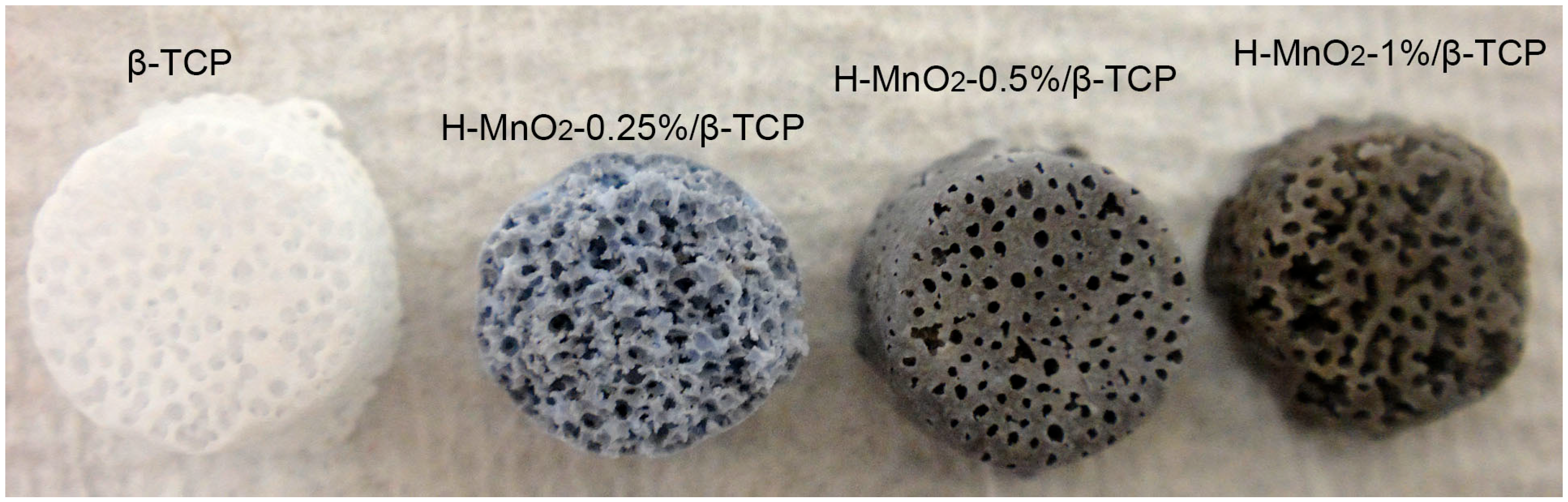
2.3. H-MnO2/β-TCP Scaffold Preparation

In this study, we added S-MnO2 and H-MnO2 nanoparticles into β-TCP to prepare MnO2-containing β-TCP scaffolds, and to evaluate their effect on the properties of the scaffolds. Various MnO2-doped β-TCP scaffold groups were created with MnO2 nanoparticles at weight concentrations of 0.25%, 0.5%, 1%, 3%, and 5% (w/w) for both H-MnO2 and S-MnO2.
We can see that the addition of MnO2 nanoparticles into β-TCP made the β-TCP scaffold change from white to black brown (Figure 3). With the increased addition of H-MnO2 nanoparticles, the black color of the H-MnO2/β-TCP becomes deeper. The diameter of the porous β-TCP scaffolds is around 7 mm, and the height is about 4 mm. This dimension was determined by the template. If the template was changed, the dimension of the resultant scaffold would be simultaneously changed.
SEM images at a low magnification showed that the pores are interconnected, and the average pore size is about 400–450 μm (Figure 4A). The pore morphologies and porosity of these MnO2-containing β-TCP scaffolds are consistent with what we observed in the porous β-TCP scaffolds in the previous studies [39,40,41], as the preparation method for the scaffolds are the same as the ones used in our previous studies. From all cross-sections, the pores of the scaffolds are open to interconnect with each other, which could provide efficient paths for nutrient transportation and cell migrations. Further higher magnified SEM images showed that scaffolds without doped nanoparticles have a rough protruding structure (Figure 4B). However, as the percentage of MnO2 nanoparticles increases, the surface structure of inner struts became increasingly smooth, and the grain boundaries became blurry (Figure 4C–L). When the concentration of H-MnO2 nanoparticles increased to 5%, the grain boundary almost disappeared (Figure 4K,L).

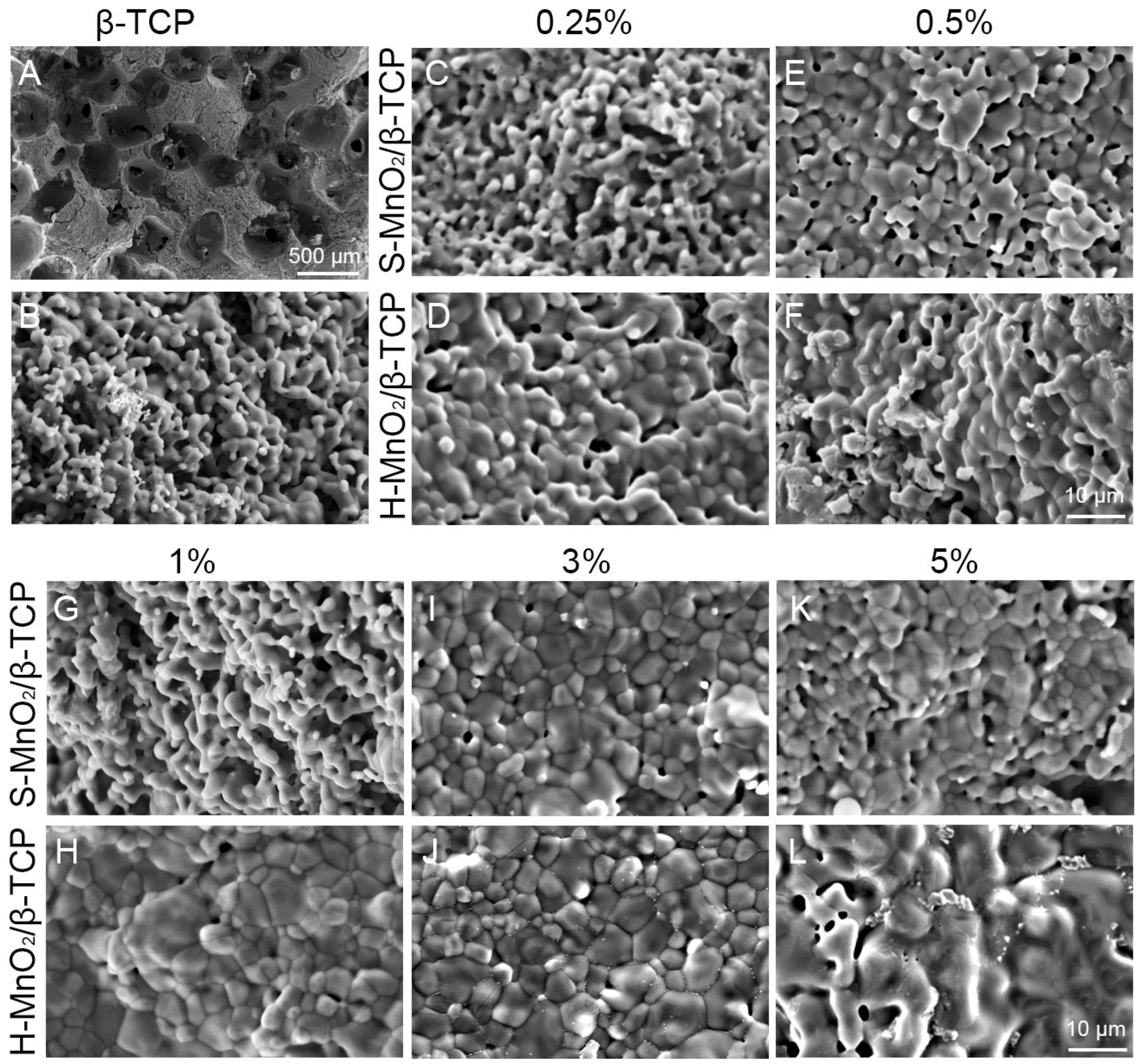
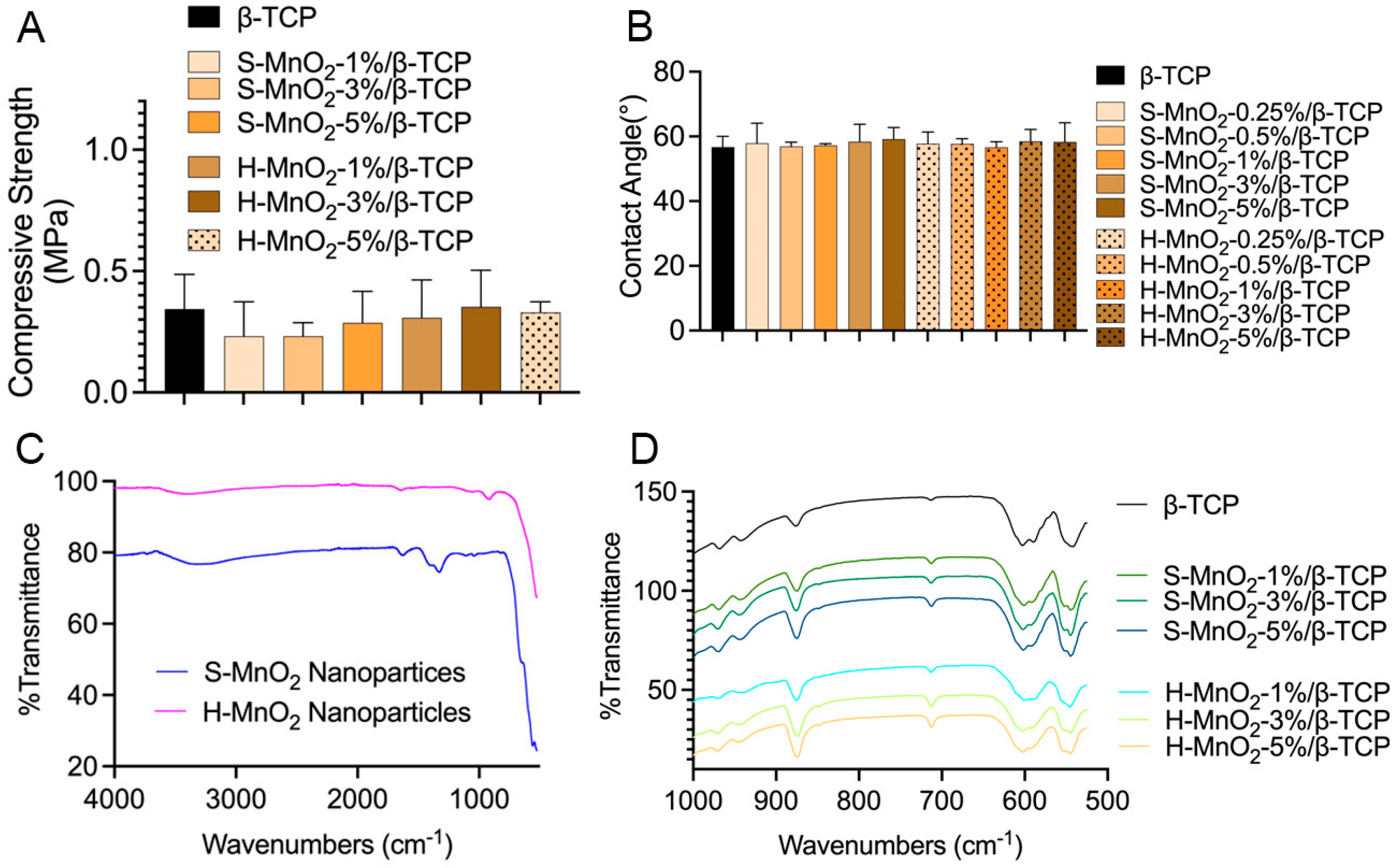
2.4. Characterizations of the Scaffolds

To investigate the effect of MnO2 nanoparticles on the mechanical properties of the porous scaffolds, both S-MnO2 and H-MnO2 nanoparticles with different doped concentrations were added into β-TCP scaffolds. The results show that the addition of S-MnO2 and H-MnO2 nanoparticles did not significantly influence the mechanical properties of the scaffolds (Figure 5A). There is no significant difference between those groups with different doped MnO2 nanoparticles.
Contact angle tests were performed to evaluate if the doping of H-MnO2 nanoparticles changes the hydrophilicity of the β-TCP scaffolds. The result shows there was no effect on the contact angle between the deionized water and the material. The contact angles of all types of scaffolds are about 57 degrees, which shows that the surface of all the scaffolds with the doped nanoparticles is hydrophilic, and no effect for the surface wettability was found (Figure 5B).
The FTIR analysis was performed to further characterize and confirm the possible effect of MnO2 on the chemical composition of the scaffolds (Figure 5C,D). The characteristic Mn-O vibration was expected at 550 cm−1 [42,43] (Figure 5C), which resulted in the appearance of a small duplet peak at 556 cm−1 in near position to the O-P-O bending peak at 550 cm−1 in the scaffold (Figure 5D). The absorption peak at around 1328.7 cm−1 is ascribed to O-Mn-O vibrations of MnO2 [44]. This peak in solid MnO2 nanoparticles is significant, but it disappeared in hollow MnO2 nanoparticles (Figure 5C). The disappearance could be derived of the hollow structure of H-MnO2 nanoparticles. The peaks at 550 and 605 cm−1 corresponds to the O-P-O bending vibration of β-TCP. The absorption band at around 720 cm−1 is due to the stretching vibrations of the oxide group of MnO2 in the form of Mn-O-Mn [45]. It was noted that, with the increase in MnO2 concentrations, the absorption band at around 720 cm−1 seems to became gradually more evident, and the hollow MnO2 groups have a more pronounced peak at 720 cm−1, compared to the solid MnO2 groups with the same concentration (Figure 5D).

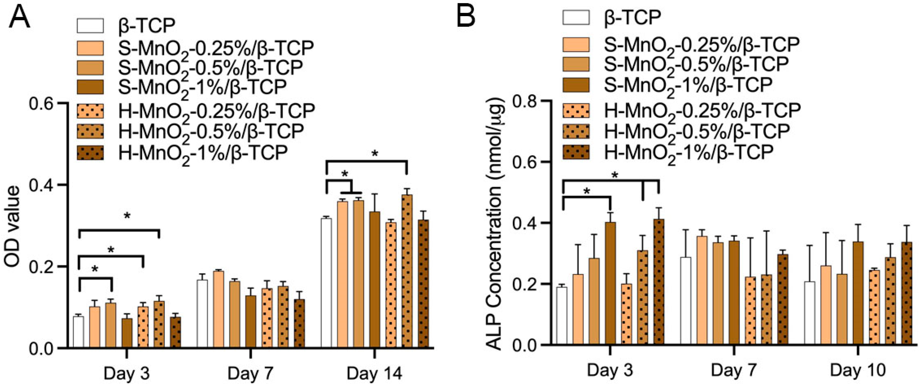
2.5. Cell Viability and Osteogenesis

Figure 6A showed the result that the addition of MnO2 nanoparticles affected the cell growth. When β-TCP scaffolds contain 0.5% S-MnO2 and H-MnO2 nanoparticles, the doped nanoparticles promoted cell growth at day 3 and day 14. However, with the increased addition of S-MnO2 or H-MnO2 nanoparticles to 1%, the doped nanoparticles exhibited either no effect or inhibition to hBMSCs growth. It is worth noting that the hollow nanoparticles exhibit higher inhibition than the solid MnO2 nanoparticles when the mass ratio of MnO2 nanoparticles was higher than 1%, but it seems that 0.5% H-MnO2 nanoparticles promoted higher cells growth compared to 0.5% S-MnO2 group. Beyond 1%, including 3% and 5%, regardless of hollow or solid MnO2 nanoparticle-doped groups, there was a significant inhibition on cell growth (Supplementary Figure S1). These results seem that there is no consistent trend in the cell growth rate in the different MnO2 nanoparticle-doped scaffolds. However, we can still see that 0.5% doped concentration is a peak.
ALP activity measurement showed that at day 3, both 0.5% and 1% H-MnO2 nanoparticle-doped scaffolds had significantly higher ALP levels compared to β-TCP scaffolds, but only 1% S-MnO2 nanoparticle-doped β-TCP scaffolds show a stimulated effect (Figure 6B). This result implies that the H-MnO2 nanoparticles may be more effective to promote osteogeneses of hBMSCs. At day 7 or 14, the significant difference between all groups disappeared.

3. Discussion

Enormous studies have been using β-TCP scaffolds for bone tissue engineering applications. However, the ideal β-TCP scaffold has not been achieved yet. In this study, porous β-TCP scaffolds were doped with different concentrations of either hollow or solid MnO2 nanoparticles (0.25%, 0.5%, 1%, 3% or 5%) to achieve the osteogenesis-stimulating function. Studies showed that MnO2 have free radical scavenging effect [46], which makes MnO2 an attractive doping material for bone regeneration after infection or cancer therapy [47]. However, the in vitro interaction between hBMSCs and MnO2-doped β-TCP scaffolds had not been well investigated.
In this study, we used SiO2 as a template to prepare hollow MnO2 nanoparticles, and the H-MnO2 nanoparticles showed a hollow structure resembling a sea urchin, which increases the surface area and accelerates the degradation of manganese dioxide [38]. Regarding the effect of the addition of such hollow MnO2 nanoparticles into β-TCP scaffolds on the morphological properties of the scaffolds, the surface structure of the scaffolds became increasingly smooth as the percentage of MnO2 nanoparticles increases (Figure 4). It is likely that, during the sintering process, the MnO2 promoted the sintering of β-TCP granules to be densified. It is worth noting that, at low concentrations of MnO2 doping (0.25, 0.5, and 1%), hollow MnO2 groups also had a smoother surface than solid MnO2-doped scaffold groups. This phenomenon could be due to the larger surface area of the hollow nanoparticles, which can promote the migration and densification of the β-TCP particles during the sinter process. The real reasons remain unknown, as there was no intended experiment set in this study to investigate the sintering mechanisms. However, it is worth noting that the hollow structure of H-MnO2 nanoparticles may still be maintained in the β-TCP scaffold, although it looks like “melting”. This is because the early osteogenesis result (Figure 6B) showed that the H-MnO2-doped scaffolds promoted early osteogenesis compared to S-MnO2-doped scaffolds, which indirectly implied that the hollow structure may still be maintained in the porous β-TCP scaffolds during the sintering process to take effects on osteogenesis.
The contact angle was used to verify whether the change in microstructure of a hollow structure affected the surface free energy of the β-TCP scaffold. As samples with porous structures are challenging to test through the contact angle settings, the MnO2-doped β-TCP composite materials were prepared into a dense disk-like shape to represent the surface of a porous scaffold. We found that the doping of MnO2 did not change the surface hydrophilicity of all the groups of MnO2-doped β-TCP disks compared to β-TCP (Figure 5B). Based on this result, we theoretically assume that the addition of MnO2 nanoparticles did not change the surface hydrophilicity of the porous MnO2-doped β-TCP scaffolds compared to the porous β-TCP scaffold. However, the addition of MnO2 made the surface of the porous MnO2-doped β-TCP scaffolds rougher compared to the β-TCP scaffold (Figure 4) [48,49].
Regarding the biological properties of the scaffolds, the cell viability experiment showed that scaffolds with less than 1% MnO2 content are biocompatible, and groups that contain high hollow nanoparticles exhibited a greater inhibition on cell growth. This greater inhibition may be due to the faster degradation of hollow particles, which cause the faster release of the Mn ions. More Mn ions may induce toxicity. This assumed reason may need to be verified as we did not perform a degradation test on the scaffold in this study, but the degraded rate of H-MnO2 nanoparticles was faster than S-MnO2 nanoparticles in our previously published study [38].
For further investigating the effect of MnO2 nanoparticles on cell proliferation and differentiation of hBMSC, we analyzed the cell growth on those scaffolds. We found that the addition of MnO2 nanoparticles significantly affected cell proliferation, especially in a high-doped MnO2 scaffold group. More surprisingly, this effect is more significant in hollow MnO2-doped scaffolds when the doping increased to beyond 1%. This result may imply that there may be a threshold of doped concentration of MnO2 nanoparticles. These results seemed to be consistent with F. Qian’s study [42], where they found that with the increase of MnO2 to 0.5%, the cell proliferation was inhibited. On the other hand, ALP results demonstrated that 0.5% H-MnO2 nanoparticle-doped scaffold promoted early osteogenic differentiation of hBMSCs (at day 3), while S-MnO2 nanoparticles did not at this doped concentration until it increased 1%, although the difference in ALP level at both day 7 and 14 was statistically insignificant between all test groups. This result implied that hollow structure promoted both proliferation and differentiation when it was doped at a low dose (higher than 0.25% but less than 0.5%). At present, there are few studies on the effect of doped hollow MnO2 nanoparticles on early osteogenesis. Many studies are exploring the use of MnO2 nanoparticles for scavenging superoxide’s radicals in inflammatory conditions, such as arthrosclerosis [50], cancers, and cytoprotection of pancreatic islets of Langerhans in vitro [51]. For stimulating the osteogenesis of bone marrow stem cells for bone regeneration, to the best of our knowledge, very few studies show that solid MnO2 nanoparticles in a hydrogel or Mn-containing ceramic promoted the repair of osteoporotic bone defects [52,53]. However, little research has been performed on urchin-like hollow structure of MnO2 nanoparticles for bone regeneration. In this study, we doped hollow MnO2 nanoparticles into β-TCP scaffolds. This doping brings a unique function, which has not only the osteoconductivity of β-TCP and the porous structure of the scaffold, but also the osteogenesis-promoting function of hollow-structured MnO2 nanoparticles.
Although there are many unique advantages of this new structured MnO2 nanoparticle-doped β-TCP scaffold, some limitations in this study exist; for example, there is no test on the effect of doped MnO2 nanoparticles on the scavenging of radicals like H2O2 that was performed in [42]. It is also worth noting that the integrity of the hollow structure of H-MnO2 nanoparticles after doping in β-TCP scaffolds was not verified due to the technical challenges by SEM. As the doped concentration of nanoparticles in the scaffolds was less than 1–5%, it is challenging for SEM to identify them. Using protein loading and release profiles on the nanoparticles before doping, and on the scaffolds after doping, may be ways to indirectly prove the integrity of the hollow structure. Investigating the radical-scavenging function of the nanoparticles before doping and the scaffolds after doping could also be another indirect approach to verify the structural integrity and related functions. Another limitation in this study is that there was no further characterization on how hollow MnO2 nanoparticles affects cell behaviors including cell morphologies, osteogenic protein expression, and bone-related gene expressions. The related mechanisms that hollow MnO2 nanoparticles stimulate osteogenesis and potentially scavenge bone defect-related reactive oxygen species radicals [54] were not explored in this study. These functional and mechanistic studies will be performed in the future.
Even though these limitations, this study successfully provided a preliminary data on the preparation of urchin-like hollow structured nanoparticles in the β-TCP scaffold and the maximum doped concentration of H-MnO2 nanoparticles, which built the foundation for the future studies. Our current study brought a new potential of using the urchin-like hollow structure for bone tissue regeneration applications.

4. Materials and Methods

4.1. Materials

β-TCP nanopowders were purchased from Nanocerox (Ann Arbor, MI, USA). Magnesium acetate was purchased from Sigma-Aldrich (St. Louis, MO, USA). Carboxymethyl cellulose powder, dispersant (Darvan C), and surfactant antifoam solution (Surfonals) were obtained from Fisher Scientific (Waltham, MA, USA). Solid MnO2 (S-MnO2) nanoparticles were obtained from US Research Nanomaterials, Inc. (Houston, TX, USA). Mesenchymal Stem Cell Growth Kit (MSCGM) was purchased from ATCC (Manassas, VA, USA).

4.2. Preparation and Characterization of H-MnO2 Nanoparticles

Hollow MnO2 nanoparticles were fabricated using an SiO2 template [38,55]. Briefly, 100 mL of ethanol, 20 mL of deionized water, and 2 mL of 28% ammonia were mixed in a beaker with continuous stirring. A total of 8 mL of tetrapropyl orthosilicate (Fisher Scientific, Waltham, MA, USA) were then added to the mixture solution, keeping stirring for overnight. The milky solution was centrifuged at 6000 rpm for 20 min. The supernatant was discarded, and the precipitated pellets were washed three times with deionized water. The final precipitate pellets were then resuspended in 60 mL of DI water and mixed with 1.96 g of potassium permanganate (Sigma-Aldrich). The suspension was sonicated for 30 min, and then transferred into a Teflon-lined autoclave inside an oven. The suspension was incubated in the oven for 48 h at 150 °C. After cooling down, the mixture was centrifuged and washed with water until the purple color was clear. A solution of 300 mL of 2 M sodium carbonate (Fisher Scientific) was then added to etch the template SiO2 nanoparticles at 60 °C. To completely etch the template SiO2 nanoparticles, up to 2 days’ incubation in the sodium carbonate solution was carried out. The final hollow MnO2 nanoparticles were obtained after several times of washing, and then freeze-dried.
The dried H-MnO2 nanoparticles were observed by scanning electron microscope (SEM, JCM-6000Plus, Tokyo, Japan). To prepare the samples for the SEM observation, H-MnO2 nanoparticles were tapped on a SEM holder stage, and then coated with gold in a Pelco SC-7 sputter coater (Leica, Teaneck, NJ, USA).
To further measure the size and observe the detailed morphology, H-MnO2 nanoparticles were prepared for transmission electron microscopy (TEM, JEM-1400Flash, Tokyo, Japan). Briefly, a few milligrams of nanoparticles were dispersed in an ethanol solution, and the suspension was then dropped on a Formvar/Carbon 200 mesh. After drying, the holder was loaded to the TEM with 120 kV of acceleration voltage for imaging.
To show the function of hollow structure of the MnO2 nanoparticle in loading proteins, a model protein bovine serum albumin (BSA) (Fisher Scientific) was used. Different mass of hollow MnO2 and S-MnO2 nanoparticles were weighed and added into a BSA solution to load BSA. The concentration of the BSA solution is 100 μg/mL. The nanoparticles were added into the BSA solutions for 24 h at room temperature on a rotator with 30 rpm. At the end of the time, the BSA solution was collected and measured by Micro BCA™ Protein Assay Kit (Thermofisher Scientific, Waltham, MA, USA) and the optical density (OD) value was read by a SpectraMax 190 microplate reader (Molecular Devices LLC, San Jose, CA, USA) at the wavelength of 562 nm. The loading amount was obtained from the difference between masses before and after adsorption of BSA proteins. After 24 h, the nanoparticles were centrifuged and collected for in vitro release. The released BSA was calculated and the cumulative released mass of BSA was profiled against the time.

4.3. Preparation of MnO2/β-TCP Scaffolds

To prepare H-MnO2/β-TCP and S-MnO2/β-TCP scaffolds, different doped weight of MnO2 were set with MnO2 nanoparticles at weight concentrations of 0.25%, 0.5%, 1%, 3%, and 5% (w/w) for both H-MnO2 and S-MnO2. A template-casting method, as previously described, was used to prepare H-MnO2/β-TCP and S-MnO2/β-TCP scaffolds [41]. Briefly, a 24-well plate was coated with paraffin solution, then paraffin beads with 1 mm diameter were packed into the wells. β-TCP nanopowder from Nanocerox (Ann Arbor, MI, USA), dispersant (Darvan C), antifoam solution, magnesium acetate and MnO2 nanoparticles were mixed in distilled water to form a MnO2/β-TCP slurry. The mixed MnO2/β-TCP slurry was then cast into the molds under vacuum, solidified in 70% ethanol for two days, and then dehydrated by gradient ethanol from 70% to 95%. After complete dehydration, the green bodies were sintered at 1250 °C for 3 h. Porous MnO2/β-TCP scaffolds with different concentrations of S-MnO2 and H-MnO2 were prepared and stored for physicochemical and biological characterizations.

4.4. Characterization of the Porous Scaffolds

4.4.1. SEM Observations

The surface and pore morphologies of porous scaffolds were observed by scanning electron microscope (SEM, JCM-6000Plus, Tokyo, Japan). Briefly, the scaffolds were washed by ethanol, and air-dried in a fume hood. The dried scaffolds were cut longitudinally or transversely and then taped on a SEM stage, and coated with gold in a Pelco SC-7 sputter coater. The scaffolds were observed under a voltage of 15 KV.

4.4.2. Contact Angle Measurement

In order to reduce the influence of the porous structure on the contact angle measurement, the MnO2/β-TCP composites were specially prepared to a thin disk shape. An Ossila contact angle measurement system (South Yorkshire, UK) was used to test the contact angle between the material and the DI water. The raw images were analyzed by the software and output the angle data automatically.

4.4.3. FTIR Measurement

The chemical structure of scaffolds was analyzed by Fourier transform infrared (FTIR). MnO2 nanoparticles, β-TCP, S-MnO2/β-TCP and H-MnO2/β-TCP scaffolds were tested through the Thermo Scientific Nicolet iS10 ATR-FTIR spectrometer (Waltham, MA, USA) to identify the chemical groups. The dried scaffolds were grounded to powders and made into a thin film. FTIR spectra was collected with 20 scans at 4.0 cm−1 resolution.

4.4.4. Compressive Strength of Scaffolds

To measure the compressive strength of the scaffolds, a Zwick-Roell universal tension-compression machine Z50 (Ulm, Germany) was used following the American Society for Testing and Materials (ASTM) standards. To ensure the two ends of the scaffolds were parallel to the crosshead of the testers, the top and bottom sides of the scaffolds were polished using a LANHU 600 Grit electro coated abrasive sandpaper obtained from Amazon (Seattle, WA, USA). The diameter of each scaffold was individually measured. A crosshead speed of 0.5 mm/min was applied to each scaffold until fracture. Five samples per group were measured.

4.5. hBMSC Cell Behaviors

4.5.1. hBMSC Culture

hBMSCs were purchased from ATCC (Manassas, VA, USA). The cells were cultured with Mesenchymal Stem Cell Growth Kit (MSCGM, ATCC, USA) under a standard condition (5% CO2, 95% humidity, and 37 °C). The cells at the passages 4–7 were used for all of the experiments.

4.5.2. Cell Proliferation on the Scaffolds

A total of 5 × 10⁴ cells in 100 µL cell suspension were pre-seeded on the porous scaffold for 90 min, and MSCGM medium was gently added to incubate the cells in a 24-well plate at 37 °C. After 3, 7, and 14 days, the porous scaffolds were washed by PBS and transferred to a new well plate. A PBS with 10% MTT was added and incubated for 4 h. The medium was then carefully removed, and dimethyl sulfoxide (DMSO) was added to dissolve the formazan. Repeatedly the scaffolds were rinsed by DMSO during the process to ensure that all formazan crystals were dissolved. All of the solutions were collected and centrifuged, and the supernatant DMSO was then transferred to a 96-well plate. The OD value was read by plate reader at the wavelength of 490 nm.

4.5.3. Alkaline Phosphatase (ALP)

ALP activity was evaluated to investigate the effect of MnO2 on potential osteogenic differentiation of hBMSCs. The cells were cultured with scaffolds in an osteogenic differentiation medium, which contained 10% FBS, 10 mM β-glycerophosphate, 10 nM dexamethasone, and 50 mg/mL ascorbic acid in the MCSBM. The hBMSCs were continuously cultured for 3, 7, and 14 days. After rinsing by PBS, the cells and scaffolds were collected and stored in −80 °C. To determine the ALP activity quantitatively, the total protein and total ALP were measured based on the published protocols [40]. Briefly, cell lysate of hBMSCs was prepared. The total protein in the hBMSCs cell lysate was measured by Micro BCA™ Protein Assay Kit (Thermofisher Scientific), and the OD value was read by a SpectraMax 190 microplate reader at the wavelength of 562 nm. The ALP was determined by a p-nitrophenyl phosphate (pNPP) method following our published protocol [56]. SIGMAFAST™ p-Nitrophenyl phosphate Tablets (Millipore Sigma, St. Louis, MO, USA) were used to prepare the working reagent and incubated at 37 °C for 3 h. The OD value was read by a plate reader at the wavelength of 405 nm. The final total ALP activity was normalized by the total protein contents of each sample.

4.6. Statistical Analysis

All of the collected data were analyzed using unpaired t-tests or one-way analyses of variance (ANOVA) with GraphPad Prism 7 (GraphPad, Boston, MA, USA), and p < 0.05 was considered statistically significant.

5. Conclusions

In this study, we successfully prepared urchin-like hollow MnO2 nanoparticles and then doped them into β-TCP scaffolds to make hollow MnO2 nanoparticle-doped β-TCP scaffolds. The scaffold shows interconnected pores, and H-MnO2 nanoparticles enhanced the sintering of β-TCP scaffolds. The addition of hollow MnO2 nanoparticles did not significantly change the mechanical properties and surface hydrophilicity of the scaffolds. The low dose H-MnO2-doped β-TCP scaffolds promoted the cell proliferation and early osteogenesis of human mesenchymal stem cells. In the whole, H-MnO2 nanoparticles demonstrated more advantages in promoting cell growth and osteogenesis compared to solid MnO2 nanoparticles when they were doped into the β-TCP scaffolds at a low concentration. More studies would be needed to further investigate the function and mechanisms of H-MnO2 in bone regeneration.

Supplementary Materials

The following supporting information can be downloaded at: https://www.mdpi.com/article/10.3390/ijms26115092/s1.

Author Contributions

Y.K.: Conceptualization, writing, project administration, funding acquisition; E.Q.: Investigation; A.E.: Editing. All authors have read and agreed to the published version of the manuscript.

Funding

This research was funded by National Institute of Dental & Craniofacial Research of the National Institutes of Health, grant number 1R15DE032189-01, 1R03DE028406-01 and also supported by National Science Foundation under award number 2223702. The content is solely the responsibility of the authors and does not necessarily represent the official views of the National Institutes of Health and National Science Foundation.

Institutional Review Board Statement

Not applicable.

Informed Consent Statement

Not applicable.

Data Availability Statement

Data will be made available on request.

Conflicts of Interest

The authors declare no conflicts of interest.

References

  1. Lee, K.Y.; Jeong, L.; Kang, Y.O.; Lee, S.J.; Park, W.H. Electrospinning of polysaccharides for regenerative medicine. Adv. Drug Deliv. Rev. 2009, 61, 1020–1032. [Google Scholar] [CrossRef] [PubMed]
  2. Cen, L.; Liu, W.; Cui, L.; Zhang, W.; Cao, Y. Collagen tissue engineering: Development of novel biomaterials and applications. Pediatr. Res. 2008, 63, 492–496. [Google Scholar] [CrossRef] [PubMed]
  3. Amini, A.R.; Laurencin, C.T.; Nukavarapu, S.P. Bone tissue engineering: Recent advances and challenges. Crit. Rev. Biomed. Eng. 2012, 40, 363–408. [Google Scholar] [CrossRef] [PubMed]
  4. Mano, J.F.; Silva, G.A.; Azevedo, H.S.; Malafaya, P.B.; Sousa, R.A.; Silva, S.S.; Boesel, L.F.; Oliveira, J.M.; Santos, T.C.; Marques, A.P.; et al. Natural origin biodegradable systems in tissue engineering and regenerative medicine: Present status and some moving trends. J. R. Soc. Interface/R. Soc. 2007, 4, 999–1030. [Google Scholar] [CrossRef]
  5. Zhu, J.; Marchant, R.E. Design properties of hydrogel tissue-engineering scaffolds. Expert Rev. Med. Devices 2011, 8, 607–626. [Google Scholar] [CrossRef]
  6. Fisher, M.B.; Mauck, R.L. Tissue engineering and regenerative medicine: Recent innovations and the transition to translation. Tissue Eng. Part B Rev. 2013, 19, 1–13. [Google Scholar] [CrossRef]
  7. Tanaka, A.; Woltjen, K.; Miyake, K.; Hotta, A.; Ikeya, M.; Yamamoto, T.; Nishino, T.; Shoji, E.; Sehara-Fujisawa, A.; Manabe, Y.; et al. Efficient and reproducible myogenic differentiation from human iPS cells: Prospects for modeling Miyoshi Myopathy in vitro. PLoS ONE 2013, 8, e61540. [Google Scholar] [CrossRef]
  8. Hussain, W.; Moens, N.; Veraitch, F.S.; Hernandez, D.; Mason, C.; Lye, G.J. Reproducible culture and differentiation of mouse embryonic stem cells using an automated microwell platform. Biochem. Eng. J. 2013, 77, 246–257. [Google Scholar] [CrossRef]
  9. Dighe, N.; Khoury, M.; Mattar, C.; Chong, M.; Choolani, M.; Chen, J.; Antoniou, M.N.; Chan, J.K. Long-term reproducible expression in human fetal liver hematopoietic stem cells with a UCOE-based lentiviral vector. PLoS ONE 2014, 9, e104805. [Google Scholar] [CrossRef]
  10. Peters, A.; Burridge, P.W.; Pryzhkova, M.V.; Levine, M.A.; Park, T.S.; Roxbury, C.; Yuan, X.; Peault, B.; Zambidis, E.T. Challenges and strategies for generating therapeutic patient-specific hemangioblasts and hematopoietic stem cells from human pluripotent stem cells. Int. J. Dev. Biol. 2010, 54, 965–990. [Google Scholar] [CrossRef]
  11. Das, B.; Bayat-Mokhtari, R.; Tsui, M.; Lotfi, S.; Tsuchida, R.; Felsher, D.W.; Yeger, H. HIF-2alpha suppresses p53 to enhance the stemness and regenerative potential of human embryonic stem cells. Stem Cells 2012, 30, 1685–1695. [Google Scholar] [CrossRef]
  12. Safwani, W.K.; Makpol, S.; Sathapan, S.; Chua, K. Impact of adipogenic differentiation on stemness and osteogenic gene expression in extensive culture of human adipose-derived stem cells. Arch. Med. Sci. AMS 2014, 10, 597–606. [Google Scholar] [CrossRef] [PubMed]
  13. Ito, K.; Suda, T. Metabolic requirements for the maintenance of self-renewing stem cells. Nat. Rev. Mol. Cell Biol. 2014, 15, 243–256. [Google Scholar] [CrossRef] [PubMed]
  14. Lovett, M.; Lee, K.; Edwards, A.; Kaplan, D.L. Vascularization strategies for tissue engineering. Tissue Eng. Part B Rev. 2009, 15, 353–370. [Google Scholar] [CrossRef] [PubMed]
  15. Cao, L.; Mooney, D.J. Spatiotemporal control over growth factor signaling for therapeutic neovascularization. Adv. Drug Deliv. Rev. 2007, 59, 1340–1350. [Google Scholar] [CrossRef]
  16. Yoshikawa, H.; Rettig, W.J.; Takaoka, K.; Alderman, E.; Rup, B.; Rosen, V.; Wozney, J.M.; Lane, J.M.; Huvos, A.G.; Garin-Chesa, P. Expression of bone morphogenetic proteins in human osteosarcoma. Immunohistochemical detection with monoclonal antibody. Cancer 1994, 73, 85–91. [Google Scholar] [CrossRef]
  17. Raval, P.; Hsu, H.H.; Schneider, D.J.; Sarras, M.P., Jr.; Masuhara, K.; Bonewald, L.F.; Anderson, H.C. Expression of bone morphogenetic proteins by osteoinductive and non-osteoinductive human osteosarcoma cells. J. Dent. Res. 1996, 75, 1518–1523. [Google Scholar] [CrossRef]
  18. Louis-Ugbo, J.; Kim, H.S.; Boden, S.D.; Mayr, M.T.; Li, R.C.; Seeherman, H.; D’Augusta, D.; Blake, C.; Jiao, A.; Peckham, S. Retention of 125I-labeled recombinant human bone morphogenetic protein-2 by biphasic calcium phosphate or a composite sponge in a rabbit posterolateral spine arthrodesis model. J. Orthop. Res. Off. Publ. Orthop. Res. Soc. 2002, 20, 1050–1059. [Google Scholar] [CrossRef]
  19. Hollinger, J.O.; Uludag, H.; Winn, S.R. Sustained release emphasizing recombinant human bone morphogenetic protein-2. Adv. Drug Deliv. Rev. 1998, 31, 303–318. [Google Scholar]
  20. Gu, Y.; Zhang, J.; Zhang, X.; Liang, G.; Xu, T.; Niu, W. Three-dimensional Printed Mg-Doped beta-TCP Bone Tissue Engineering Scaffolds: Effects of Magnesium Ion Concentration on Osteogenesis and Angiogenesis In Vitro. Tissue Eng. Regen. Med. 2019, 16, 415–429. [Google Scholar] [CrossRef]
  21. Bose, S.; Banerjee, D.; Robertson, S.; Vahabzadeh, S. Enhanced In Vivo Bone and Blood Vessel Formation by Iron Oxide and Silica Doped 3D Printed Tricalcium Phosphate Scaffolds. Ann. Biomed. Eng. 2018, 46, 1241–1253. [Google Scholar] [CrossRef] [PubMed]
  22. Tohidnezhad, M.; Kubo, Y.; Lichte, P.; Heigl, T.; Roch, D.; Barahmand Pour, N.; Bergmann, C.; Sonmez, T.T.; Hock, J.V.P.; Fragoulis, A.; et al. Effects of Strontium-Doped beta-Tricalcium Scaffold on Longitudinal Nuclear Factor-Kappa Beta and Vascular Endothelial Growth Factor Receptor-2 Promoter Activities during Healing in a Murine Critical-Size Bone Defect Model. Int. J. Mol. Sci. 2020, 21, 3208. [Google Scholar] [CrossRef] [PubMed]
  23. Buchman, A.R. Manganese. In Modern Nutrition in Health and Disease, 11th ed.; Ross, A.C., Caballero, B., Cousins, R.J., Tucker, K.L., Ziegler, T.R., Eds.; Lippincott Williams Wilkins: Baltim, MD, USA, 2014; p. 7. [Google Scholar]
  24. Nielsen, F.H. Manganese, Molybdenum, Boron, Chromium, and Other Trace Elements. In Present Knowledge in Nutrition, 10th ed.; Erdman, J.W., Jr., Macdonald, I.A., Zeisel, S.H., Eds.; Wiley-Blackwell: Hoboken, NJ, USA, 2012; p. 22. [Google Scholar]
  25. Palacios, C. The role of nutrients in bone health, from A to Z. Crit. Rev. Food Sci. Nutr. 2006, 46, 621–628. [Google Scholar] [CrossRef] [PubMed]
  26. Saltman, P.D.; Strause, L.G. The role of trace minerals in osteoporosis. J. Am. Coll. Nutr. 1993, 12, 384–389. [Google Scholar] [CrossRef]
  27. Bae, Y.J.; Kim, M.H. Manganese supplementation improves mineral density of the spine and femur and serum osteocalcin in rats. Biol. Trace Elem. Res. 2008, 124, 28–34. [Google Scholar] [CrossRef]
  28. Fischer, A.E.; Pettigrew, K.A.; Rolison, D.R.; Stroud, R.M.; Long, J.W. Incorporation of homogeneous, nanoscale MnO2 within ultraporous carbon structures via self-limiting electroless deposition: Implications for electrochemical capacitors. Nano Lett. 2007, 7, 281–286. [Google Scholar] [CrossRef]
  29. Zhao, Z.; Fan, H.; Zhou, G.; Bai, H.; Liang, H.; Wang, R.; Zhang, X.; Tan, W. Activatable fluorescence/MRI bimodal platform for tumor cell imaging via MnO2 nanosheet-aptamer nanoprobe. J. Am. Chem. Soc. 2014, 136, 11220–11223. [Google Scholar] [CrossRef]
  30. Chen, Y.; Ye, D.; Wu, M.; Chen, H.; Zhang, L.; Shi, J.; Wang, L. Break-up of two-dimensional MnO2 nanosheets promotes ultrasensitive pH-triggered theranostics of cancer. Adv. Mater. 2014, 26, 7019–7026. [Google Scholar] [CrossRef]
  31. Wang, Y.; Jiang, K.; Zhu, J.; Zhang, L.; Lin, H. A FRET-based carbon dot-MnO2 nanosheet architecture for glutathione sensing in human whole blood samples. Chem. Commun. 2015, 51, 12748–12751. [Google Scholar] [CrossRef]
  32. Li, P.; Wei, M.; Zhang, F.; Su, J.; Wei, W.; Zhang, Y.; Liu, S. Novel Fluorescence Switch for MicroRNA Imaging in Living Cells Based on DNAzyme Amplification Strategy. ACS Appl. Mater. Interfaces 2018, 10, 43405–43410. [Google Scholar] [CrossRef]
  33. Jiang, Y.; Zhao, J.; Zhang, D. Manganese Dioxide-Based Nanomaterials for Medical Applications. ACS Biomater. Sci. Eng. 2024, 10, 2680–2702. [Google Scholar] [CrossRef] [PubMed]
  34. Zhao, F.; Jia, Z.; Zhang, L.; Liu, G.; Li, J.; Zhao, J.; Xie, Y.; Chen, L.; Jiang, H.; He, W.; et al. A MnO2 nanosheets doping double crosslinked hydrogel for cartilage defect repair through alleviating inflammation and guiding chondrogenic differentiation. Biomaterials 2025, 314, 122875. [Google Scholar] [CrossRef] [PubMed]
  35. Rathnam, C.; Yang, L.; Castro-Pedrido, S.; Luo, J.; Cai, L.; Lee, K.B. Hybrid SMART spheroids to enhance stem cell therapy for CNS injuries. Sci. Adv. 2021, 7, eabj2281. [Google Scholar] [CrossRef] [PubMed]
  36. Yang, L.; Chueng, S.D.; Li, Y.; Patel, M.; Rathnam, C.; Dey, G.; Wang, L.; Cai, L.; Lee, K.B. A biodegradable hybrid inorganic nanoscaffold for advanced stem cell therapy. Nat. Commun. 2018, 9, 3147. [Google Scholar] [CrossRef]
  37. Li, L.; Xiao, B.; Mu, J.; Zhang, Y.; Zhang, C.; Cao, H.; Chen, R.; Patra, H.K.; Yang, B.; Feng, S.; et al. A MnO2 Nanoparticle-Dotted Hydrogel Promotes Spinal Cord Repair via Regulating Reactive Oxygen Species Microenvironment and Synergizing with Mesenchymal Stem Cells. ACS Nano 2019, 13, 14283–14293. [Google Scholar] [CrossRef]
  38. Greene, A.; Hashemi, J.; Kang, Y. Development of MnO2 hollow nanoparticles for potential drug delivery applications. Nanotechnology 2021, 32, 025713. [Google Scholar] [CrossRef]
  39. Kang, Y.; Kim, S.; Bishop, J.; Khademhosseini, A.; Yang, Y. The osteogenic differentiation of human bone marrow MSCs on HUVEC-derived ECM and beta-TCP scaffold. Biomaterials 2012, 33, 6998–7007. [Google Scholar] [CrossRef]
  40. Kang, Y.; Kim, S.; Khademhosseini, A.; Yang, Y. Creation of bony microenvironment with CaP and cell-derived ECM to enhance human bone-marrow MSC behavior and delivery of BMP-2. Biomaterials 2011, 32, 6119–6130. [Google Scholar] [CrossRef]
  41. Kang, Y.; Scully, A.; Young, D.A.; Kim, S.; Tsao, H.; Sen, M.; Yang, Y. Enhanced mechanical performance and biological evaluation of a PLGA coated beta-TCP composite scaffold for load-bearing applications. Eur. Polym. J. 2011, 47, 1569–1577. [Google Scholar] [CrossRef]
  42. Qian, F.; Huang, Z.; Liu, W.; Liu, Y.; He, X. Functional β-TCP/MnO/PCL artificial periosteum promoting osteogenic differentiation of BMSCs by reducing locally reactive oxygen species level. J. Biomed. Mater. Res. Part A 2023, 111, 1678–1691. [Google Scholar] [CrossRef]
  43. Wang, H.; Lu, Z.; Qian, D.; Li, Y.; Zhang, W. Single-crystal α-MnO2 nanorods: Synthesis and electrochemical properties. Nanotechnology 2007, 18, 115616. [Google Scholar] [CrossRef]
  44. Gangwar, D.; Rath, C. Structural, optical and magnetic properties of α- and β-MnO2 nanorods. Appl. Surf. Sci. 2021, 557, 149693. [Google Scholar] [CrossRef]
  45. Xie, Y.; Yu, Y.; Gong, X.; Guo, Y.; Guo, Y.; Wang, Y.; Lu, G. Effect of the crystal plane figure on the catalytic performance of MnO2 for the total oxidation of propane. CrystEngComm 2015, 17, 3005–3014. [Google Scholar] [CrossRef]
  46. Lin, S.; Xu, Z.; Liu, Y.; Yang, G.; Qi, X.; Huang, Y.; Zhou, M.; Jiang, X. Engineered Macrophage Membrane-Camouflaged Nanodecoys Reshape the Infectious Microenvironment for Efficient Periodontitis Treatment. ACS Nano 2025, 19, 15345–15362. [Google Scholar] [CrossRef] [PubMed]
  47. Xu, W.; Qing, X.; Liu, S.; Yang, D.; Dong, X.; Zhang, Y. Hollow Mesoporous Manganese Oxides: Application in Cancer Diagnosis and Therapy. Small 2022, 18, e2106511. [Google Scholar] [CrossRef]
  48. Tamai, Y.; Aratani, K. Experimental study of the relation between contact angle and surface roughness. J. Phys. Chem. 1972, 76, 3267–3271. [Google Scholar] [CrossRef]
  49. Wenzel, R.N. Surface Roughness and Contact Angle. J. Phys. Colloid Chem. 1949, 53, 1466–1467. [Google Scholar] [CrossRef]
  50. Bizeau, J.; Tapeinos, C.; Marella, C.; Larranaga, A.; Pandit, A. Synthesis and characterization of hyaluronic acid coated manganese dioxide microparticles that act as ROS scavengers. Colloids Surf. B Biointerfaces 2017, 159, 30–38. [Google Scholar] [CrossRef]
  51. Tootoonchi, M.H.; Hashempour, M.; Blackwelder, P.L.; Fraker, C.A. Manganese oxide particles as cytoprotective, oxygen generating agents. Acta Biomater. 2017, 59, 327–337. [Google Scholar] [CrossRef]
  52. Chen, Q.; Li, J.; Han, F.; Meng, Q.; Wang, H.; Wei, Q.; Li, Z.; Li, F.; Xie, E.; Qin, X.; et al. A Multifunctional Composite Hydrogel That Rescues the ROS Microenvironment and Guides the Immune Response for Repair of Osteoporotic Bone Defects. Adv. Funct. Mater. 2022, 32, 2201067. [Google Scholar] [CrossRef]
  53. Li, J.; Deng, C.; Liang, W.; Kang, F.; Bai, Y.; Ma, B.; Wu, C.; Dong, S. Mn-containing bioceramics inhibit osteoclastogenesis and promote osteoporotic bone regeneration via scavenging ROS. Bioact. Mater. 2021, 6, 3839–3850. [Google Scholar] [CrossRef] [PubMed]
  54. Yang, Y.; Yao, Z.; Sun, Y.; Nie, Y.; Zhang, Y.; Li, Z.; Luo, Z.; Zhang, W.; Wang, X.; Du, Y.; et al. 3D-printed manganese dioxide incorporated scaffold promotes osteogenic-angiogenic coupling for refractory bone defect by remodeling osteo-regenerative microenvironment. Bioact. Mater. 2025, 44, 354–370. [Google Scholar] [CrossRef] [PubMed]
  55. Guo, X.; Li, J.; Jin, X.; Han, Y.; Lin, Y.; Lei, Z.; Wang, S.; Qin, L.; Jiao, S.; Cao, R. A Hollow-Structured Manganese Oxide Cathode for Stable Zn-MnO2 Batteries. Nanomater 2018, 8, 301. [Google Scholar] [CrossRef] [PubMed]
  56. Liu, Y.; Kim, J.H.; Young, D.; Kim, S.; Nishimoto, S.K.; Yang, Y. Novel template-casting technique for fabricating beta-tricalcium phosphate scaffolds with high interconnectivity and mechanical strength and in vitro cell responses. J. Biomed. Mater. Res. A 2010, 92, 997–1006. [Google Scholar] [CrossRef]
Figure 1. SEM photograph shows the morphologies of hollow MnO2 nanoparticles (A). TEM shows the hollow MnO2 nanoparticles (B), solid MnO2 nanoparticles (C), and the high magnified image of H-MnO2 nanoparticles (D) with a close view of the shell stings (i).
Ijms 26 05092 g001
Figure 2. Loading amount of BSA on H-MnO2 nanoparticles and S-MnO2 nanoparticles (A), and their in vitro cumulative release (B) (The asterisk (*) represents a statistically significant difference) (p < 0.05).
Ijms 26 05092 g002
Figure 3. Digital images of porous β-TCP scaffolds: 0.25%, 0.5%, 1% H-MnO2 nanoparticle-doped β-TCP scaffolds.
Ijms 26 05092 g003
Figure 4. SEM images of porous β-TCP scaffolds (A,B), 0.25% S-MnO2 and H-MnO2 (C,D), 0.5% S-MnO2 and H-MnO2 (E,F), 1% S-MnO2 and H-MnO2 (G,H), 3% S-MnO2 and H-MnO2 (I,J), and 5% S-MnO2 and H-MnO2 doped β-TCP scaffolds (K,L), respectively. (Scale bar: A, 500 μm; BL, 10 μm).
Ijms 26 05092 g004
Figure 5. Compressive strength (A), contact angle (B), and FTIR (C,D) of porous β-TCP scaffolds, H-MnO2 nanoparticle-doped β-TCP scaffolds, and S-MnO2 nanoparticle-doped β-TCP scaffolds.
Ijms 26 05092 g005
Figure 6. Cell viability (A) and ALP concentration (B) produced by hBMSCs on porous β-TCP scaffolds, H-MnO2 nanoparticle-doped β-TCP scaffolds and S-MnO2 nanoparticle-doped β-TCP scaffolds. (The asterisk (*) represents a statistically significant difference) (p < 0.05).
Ijms 26 05092 g006
Disclaimer/Publisher’s Note: The statements, opinions and data contained in all publications are solely those of the individual author(s) and contributor(s) and not of MDPI and/or the editor(s). MDPI and/or the editor(s) disclaim responsibility for any injury to people or property resulting from any ideas, methods, instructions or products referred to in the content.

