Article

Immune Responses of Mango Callus Infected by Agrobacterium tumefaciens Inhibited Transformation

1
Tropical Crops Genetics Resources Institute, Chinese Academy of Tropical Agricultural Sciences, Haikou 572025, China
2
Sanya Research Institute, Chinese Academy of Tropical Agricultural Sciences, Sanya 572025, China
3
National Key Laboratory for Tropical Crop Breeding, Sanya 572024, China
4
Key Laboratory of Crop Gene Resources and Germplasm Enhancement in Southern China, Ministry of Agriculture and Rual Affairs, Haikou 571101, China
5
Key Laboratory of Tropical Crops Germplasm Resources Genetic Improvement and Innovation of Hainan Province, Haikou 571101, China
6
Faculty of Agriculture, Gopalganj Science and Technology University, Gopalganj 8100, Bangladesh
7
Department of Biotechnology and Genetic Engineering, Gopalganj Science and Technology University, Gopalganj 8100, Bangladesh
8
College of Tropical Crops, Yunan Agricultural University, Puer 665099, China
*
Authors to whom correspondence should be addressed.
These authors contributed equally to this work.
Int. J. Mol. Sci. 2025, 26(11), 5006; https://doi.org/10.3390/ijms26115006
Submission received: 2 April 2025 / Revised: 9 May 2025 / Accepted: 18 May 2025 / Published: 22 May 2025
(This article belongs to the Section Molecular Plant Sciences)

Abstract

Mango is a vital fruit crop in tropical and subtropical regions, yet pests and diseases cause 30–70% production losses. Developing disease-resistant cultivars through transgenic methods could mitigate these issues. Agrobacterium-mediated callus transformation is a common genetic engineering approach, but successful transgenic mango plants from callus remain unreported due to severe browning and necrosis post-infection. We hypothesized that Agrobacterium-induced immune responses trigger callus death, hindering transformation. To improve efficiency, we engineered an Agrobacterium strain carrying the type III secretion system (T3SS) and effector gene AvrPto. Compared to controls, infected calluses exhibited elevated reactive oxygen species (ROS), along with up-regulated ROS-related, gallic acid biosynthesis, and defense genes. Calluses infected with T3SS-AvrPto-harboring Agrobacterium showed delayed browning and necrosis versus those infected with the empty vector (NV). The transformation rate with Agrobacterium (T3SS-AvrPto-EGFP) reached 1.6%, while Agrobacterium (NV-EGFP) failed entirely. These findings demonstrate that T3SS and AvrPto enhance mango transformation efficiency, offering a promising strategy for breeding multi-resistant varieties.

1. Introduction

Mango (Mangifera indica L.) is the world’s second-largest tropical fruit in yield annually (2023 data from the Office of Tropical Crops in Southern Asia, Ministry of Agriculture, China). The global mango planting area exceeds 10 million hectares annually (data from the Food and Agriculture Organization of the United Nations). India is the largest mango producer, with a planting area of 2.625 million hectares. In 2023, China’s mango planting area was 386,200 hectares, and the mango output value in 2021 was CNY 21.14 billion. Other major mango-producing countries include Thailand (204,000 hectares), Indonesia (266,700 hectares), Pakistan (213,500 hectares), Mexico (215,600 hectares), Brazil (98,200 hectares), Malawi (69,900 hectares), Bangladesh (139,800 hectares), Egypt (148,300 hectares), and so on.
During mango planting, the losses caused by pests and diseases account for 30–70% of mango production generally [1,2]. Mango anthracnose disease (MAD) can cause up to 100% yield loss in unmanaged orchards [1]. In India, the disease leads to an estimated yield loss of about 39% [1]. In Indonesia, the yield loss due to MAD is reported to be 50.28% [1]. In China, MAD causes an annual yield loss of 30–60% in mango production [1]. The avoidable yield loss caused by mango pests is about 61.51% [2]. Among them, mango hoppers and thrips are the main pests [2]. Cultivating new varieties with various resistances is the basic way to resolve these problems. The conventional method for generating new mango varieties is cross-breeding. A complete cycle of cross-breeding for mangoes always takes about 10–20 years [3]. Transgene has high breeding efficiency and can solve the problems above in a short time [4,5]. After two-week-old mango seedlings were punctured at the apical meristem, the apical meristem was infected with Agrobacterium harboring GFP and GUS genes individually [6]. Some plants were found to have been transformed successfully [6]. Adjei et al. (2023) optimized the conditions of the protoplast isolation and transient expression system for mango [7]. GFP fluorescence was observed in the transformed protoplasts [7]. The target gene was successfully induced into the protoplast [7].
Stem apex meristematic tissue is a highly differentiated tissue. When stem apex meristematic tissues were transformed, some cells were transformed and some failed, which always resulted in a chimera [8,9]. When cells are divided, transformation-failed cells always stand ahead of the competition, and the transformed cells stop division, resulting in foreign genes being lost and the failure of the experiment [8,9]. The experiment that Adjei et al. (2023) did was a transient transformation and did not produce transformed seedlings [7]. Plant embryogenic callus is a loose and crisp tissue. When Agrobacterium infected the embryogenic callus, callus cells had a high chance of being infected and transformed. Infecting calluses using Agrobacterium-harboring target genes is a conventional method for constructing a transgene protocol [10,11,12]. However, the transgenic-mango plant from callus has not been reported until now. In this research, we found that most mango calluses necrotized and died after Agrobacterium infected them. No seedling was differentiated from the surviving callus. Immune response can injure and kill plant cells, resulting in callus browned and necrotized [13]. When infecting plant cells, Agrobacterium tumefaciens sends virulence proteins into host cells through the type IV secretion system (T4SS) [14]. Active plant defense relying on innate immune responses was triggered [15]. Some Gram-negative plant pathogenic bacteria, such as Pseudomonas syringae pv. tomato, deliver bacterial proteins into eukaryotic hosts through the type III secretion system (T3SS), which can escape being perceived by plant cells and do not induce the plant basal defense [15].
We propose that the browning and necrosis of callus tissue after Agrobacterium infection are the main reasons for the failure of mango genetic transformation. The browning and necrosis of mango calluses might be due to immune responses triggered by Agrobacterium infection. Constructing an Agrobacterium-harboring type III secretion gene cluster (T3SS) and the effector gene AvrPto might improve the transformation efficiency of mango callus. To identify these hypotheses, metabolome and transcriptome experiments were performed to find substances and genes that play crucial roles in the immune responses of mango callus against Agrobacterium infection in this research. An Agrobacterium strain harboring T3SS and its effector gene AvrPto, isolated from Pseudomonas syringae pv. tomato strain DC3000, was constructed. The mango callus infected by Agrobacterium (T3SS-AvrPto) showed slower browning and necrosis than those infected by Agrobacterium harboring the net vector (NV). The transformation rate of mango callus infected with Agrobacterium (T3SS-AvrPto-EGFP) was about 1.6%, a promising finding encouraging further exploration. These findings provide hope for the potential application of the transgenic method in mango breeding.

2. Results

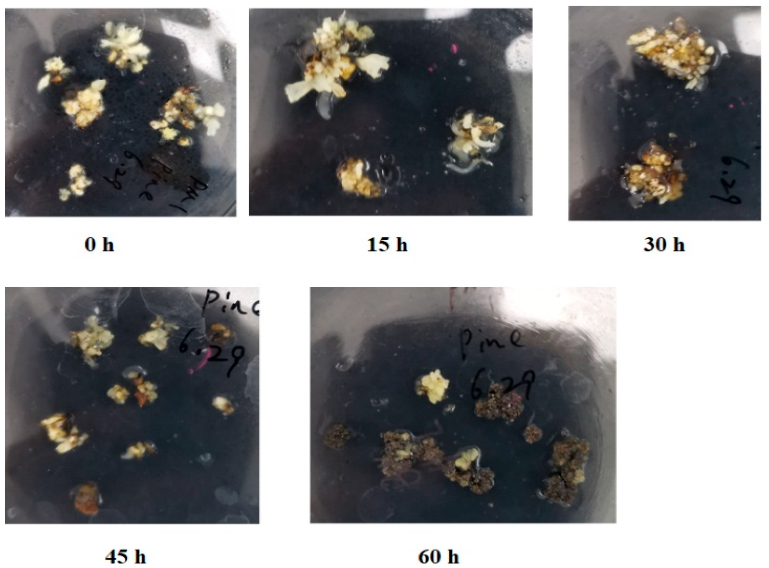
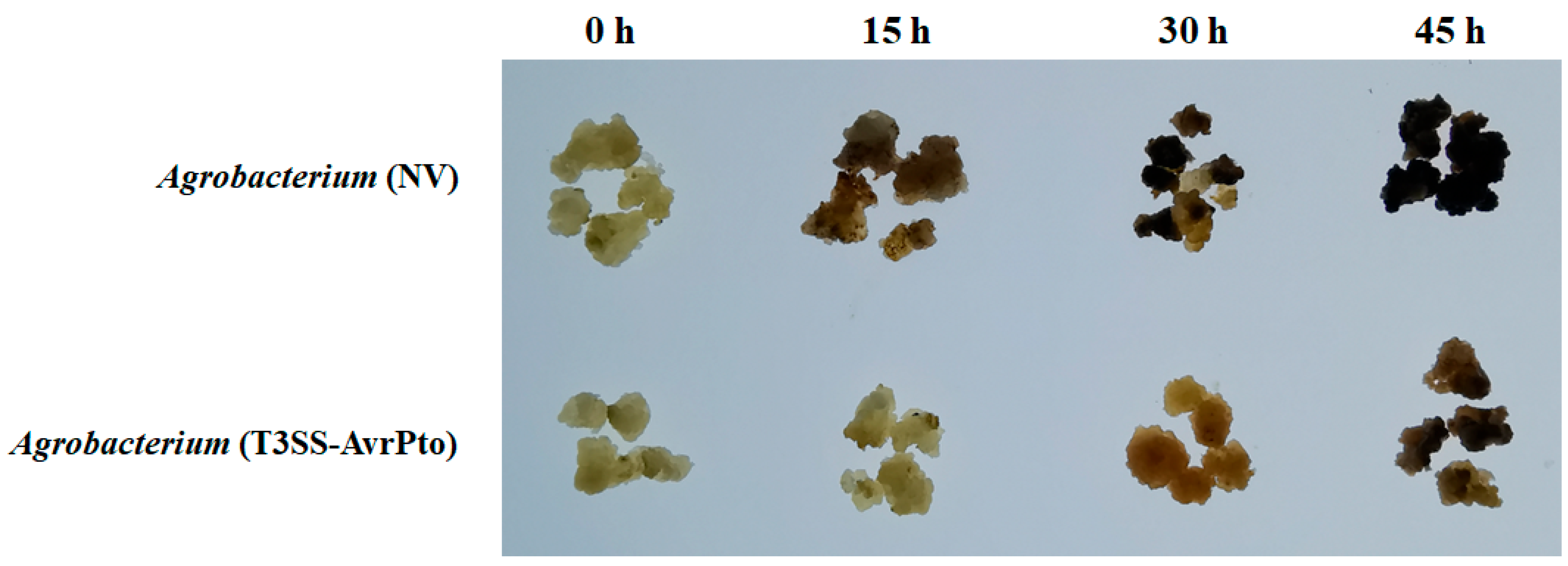
2.1. Browning and Necrosis of Mango Callus After Agrobacterium Infection

When infected with Agrobacterium, most mango calluses turned brown and underwent necrosis within 60 h (Figure 1). In contrast, most of the uninfected calluses remained white or yellow. At 15 h post-infection (hpi), no browning or necrosis was observed in mango calluses. By 30 hpi, partial browning appeared in some calluses, accounting for approximately 7% of the total fresh weight. Browning and necrosis progressed rapidly by 45 hpi, with affected tissue increasing to 39% of the total callus mass. At 60 hpi, the majority of calluses exhibited severe browning and necrosis, constituting about 92% of the total fresh weight (Figure 1). After the browned and necrotized calluses were transferred onto a fresh medium and cultured for an additional two days, the browning and necrosis were not alleviated. The browning and necrosis continued until the calluses were necrotized completely. These demonstrated that Agrobacterium infection can induce irreversible browning and necrosis in mango callus, underscoring the urgent need for a solution.

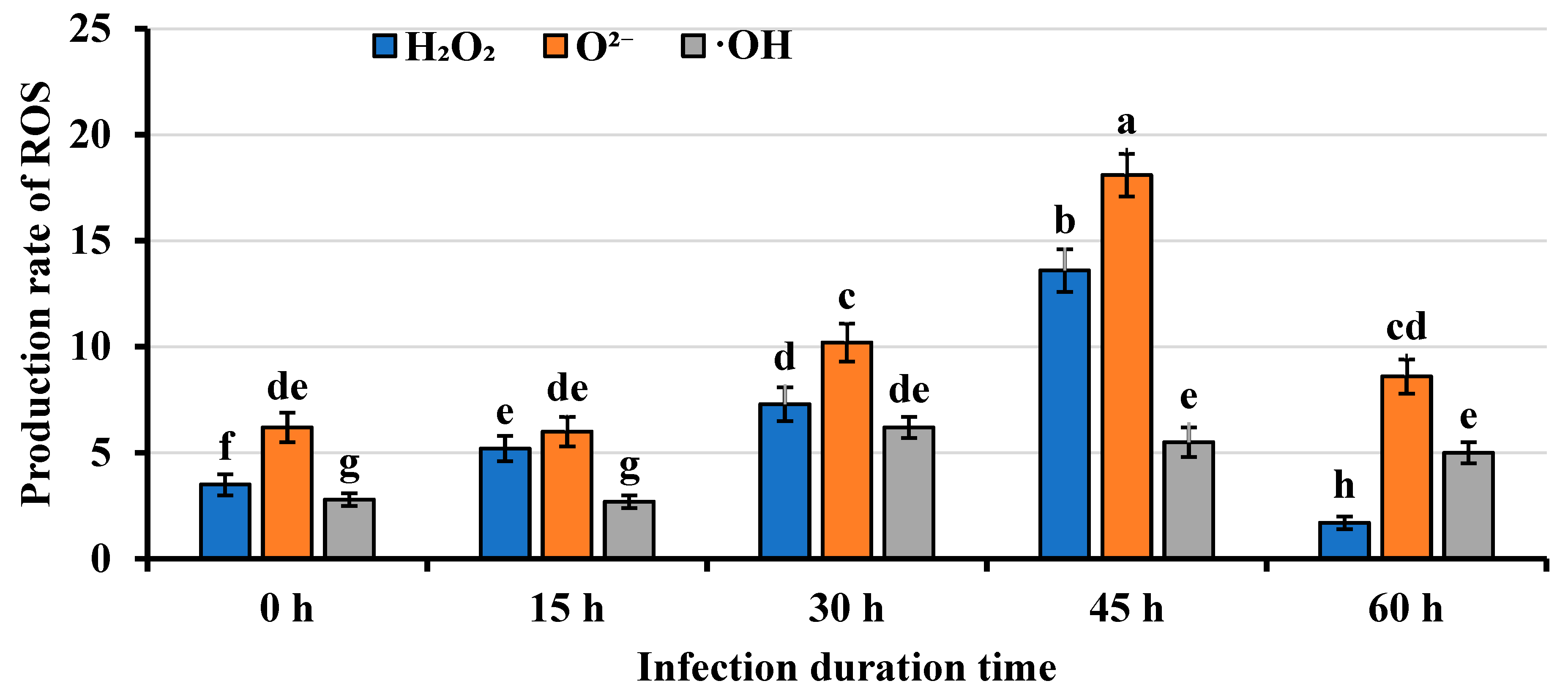
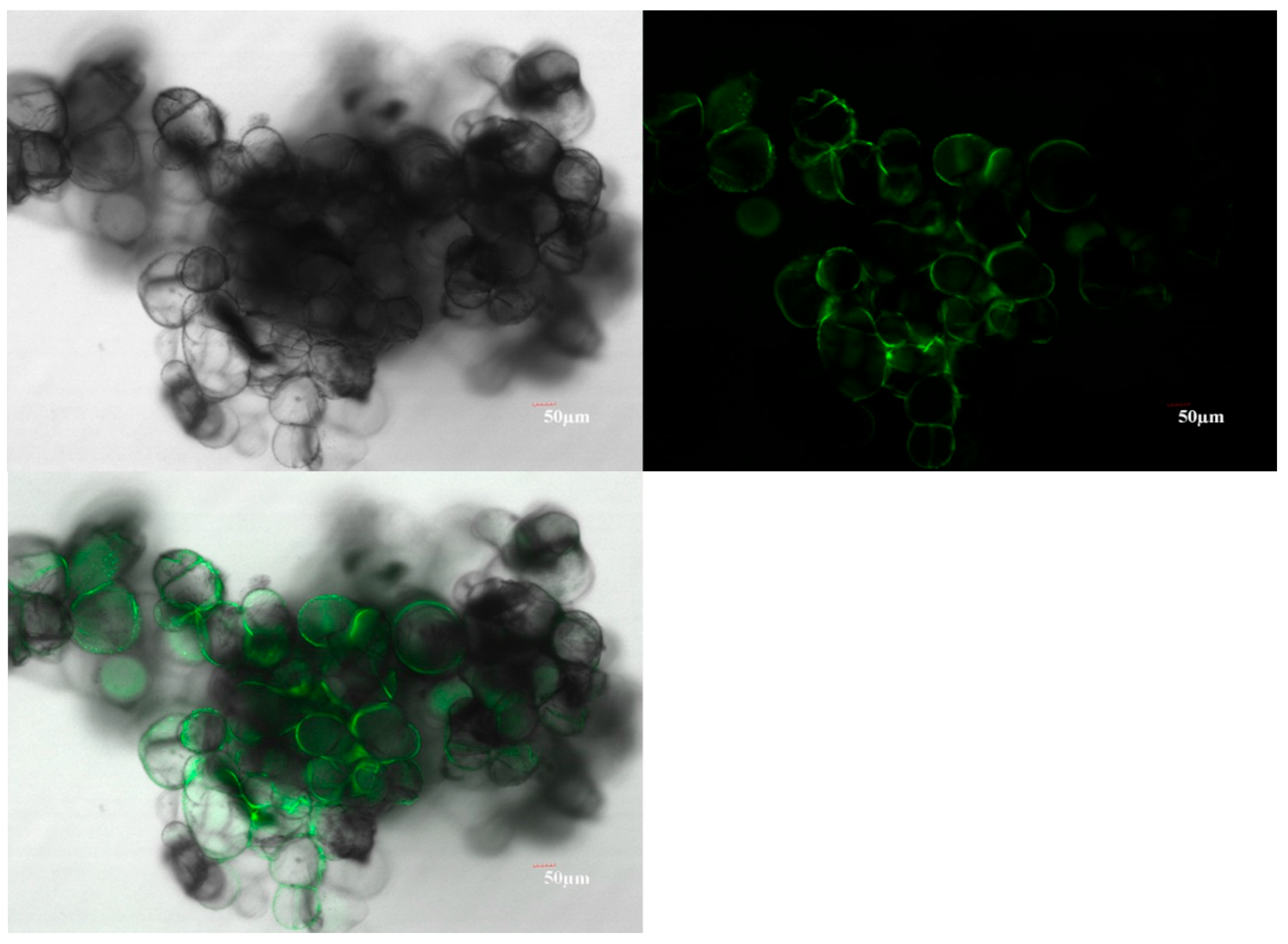
2.2. Mango Callus Infected by Agrobacterium Contained More Reactive Oxygen Species (ROS) than Control

To investigate whether the browning and necrosis in Agrobacterium-infected mango callus infected by Agrobacterium was associated with ROS production, the levels of hydrogen peroxide (H2O2), superoxide anion (O2−), and hydroxyl radical (OH) in the infected callus were measured. The results revealed a significant increase in ROS levels following Agrobacterium infection. At 15 h post-infection (hpi), H2O2 content in infected callus (5.2 ± 0.6 μmol/mg protein) was already significantly higher than in uninfected controls (3.5 ± 0.5 μmol/mg protein) (Figure 2). This increase continued over time, reaching 7.3 ± 0.8 μmol/mg protein at 30 hpi and peaking at 13.6 ± 1.0 μmol/mg protein at 45 hpi, followed by a gradual decline. Similarly, O2− levels showed a comparable accumulation pattern. While no significant difference was observed between infected (6.0 ± 0.7 nmol/(min mg protein)) and uninfected callus (6.2 ± 0.7 nmol/(min mg protein)) at 15 hpi, the O2− content increased markedly to 10.2 ± 0.9 nmol/(min mg protein) by 30 hpi and reached its maximum (18.1 ± 1.0 nmol/(min mg protein)) at 45 hpi before decreasing. The OH content exhibited a similar temporal pattern, with no significant difference at 15 hpi but reaching maximum levels at 30 hpi (Figure 2). Notably, all three ROS species maintained stable baseline levels in uninfected callus throughout the experimental period. These findings demonstrated that Agrobacterium infection triggered a rapid and substantial accumulation of ROS in mango callus, suggesting a potential role of oxidative stress in the browning and necrosis phenomena. The differential timing of peak accumulation among the various ROS species may reflect their distinct roles in the plant’s defense response.

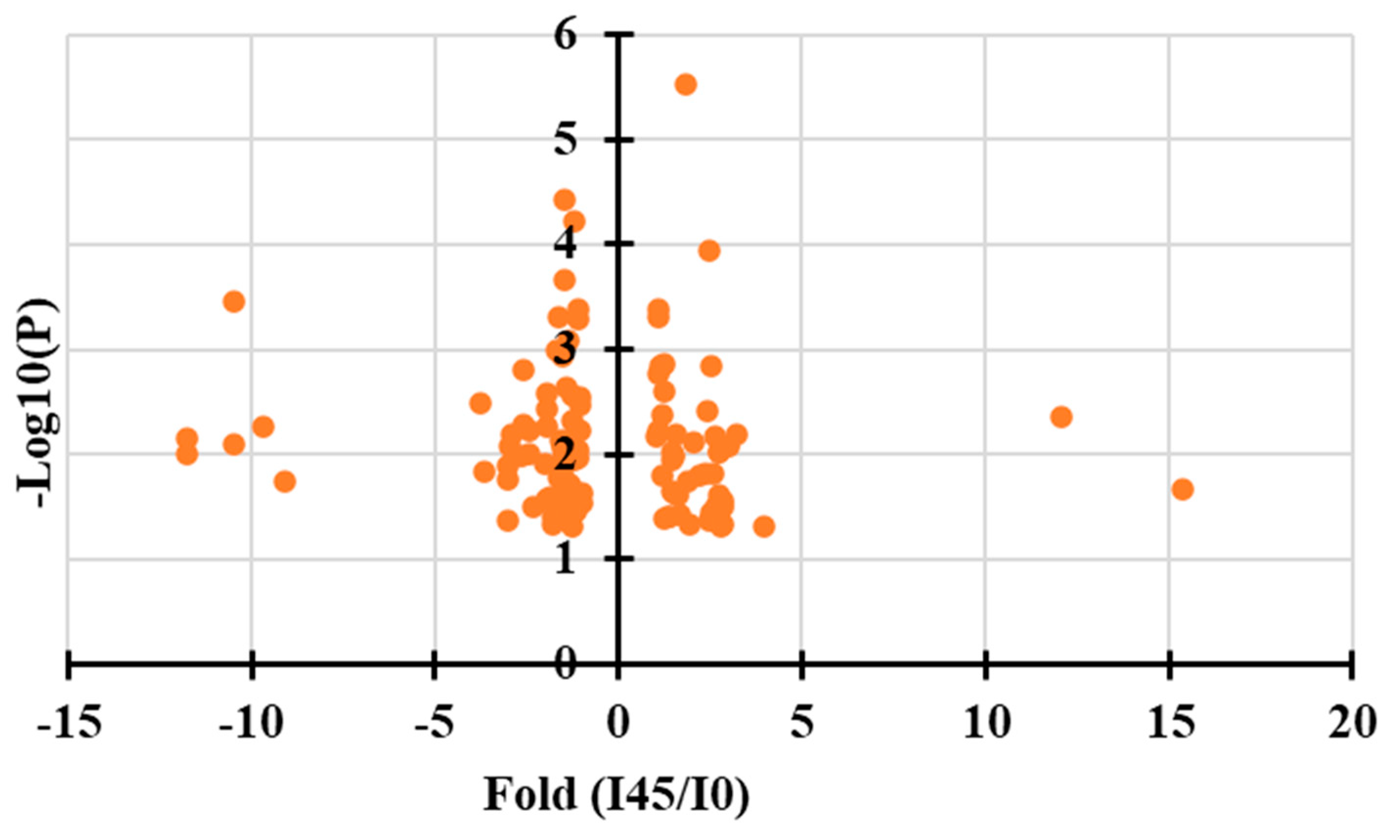
2.3. Identification of Key Metabolites Associated with Browning in Mango Calluses Infected by Agrobacterium

To elucidate the metabolic basis of browning and necrosis in Agrobacterium-infected mango callus, a comparative metabolomic analysis between infected (45 hpi) and uninfected callus tissues was conducted. A total of 516 metabolites were detected in the samples. Using a threshold of fold-change >2.0 or <0.5 (infected vs. uninfected), 106 differentially accumulated metabolites, including 38 up-regulated and 68 down-regulated compounds, were identified (Figure 3). The seven most significantly up-regulated compounds were (in descending order of fold-change) gallic acid (15.2-fold increase), ubiquinone (11.5-fold), D-erythrose-4-phosphate, phosphoenolpyruvic acid, dihydrokaempferol, cyanidin, and dihydromyricetin. Variable Importance in Projection (VIP) scores further highlighted the significance of these metabolites (Table 1). Gallic acid exhibited both the highest fold-change (15.2) and VIP score (1.96) among all up-regulated metabolites, strongly suggesting its pivotal role in the browning and necrosis processes. The identified metabolites, particularly those involved in phenolic and flavonoid pathways (gallic acid, dihydrokaempferol, cyanidin), as well as key intermediates in energy metabolism (phosphoenolpyruvic acid, D-erythrose-4-phosphate), may collectively contribute to the observed physiological responses through their involvement in plant defense mechanisms and oxidative stress responses. These findings provided valuable insights into the metabolic change associated with Agrobacterium infection in mango callus, with gallic acid emerging as a potential key mediator of the browning phenotype.

2.4. The Expression Levels of ROS-Related Genes and Gallic Acid Synthesis Genes Were Up-Regulated in Callus Infected by Agrobacterium

To investigate the molecular mechanisms underlying immune responses in Agrobacterium-infected mango callus, transcriptome profiling of infected (45 hpi) and uninfected tissues was performed. Results showed that 1256 genes were up-regulated and 1528 down-regulated using the |log2FC| > 1 criteria and FDR < 0.05. Pathways were analyzed using the Kyoto Encyclopedia of Genes and Genomes (KEGG) database. Results showed that the up-regulated genes were mainly grouped in phenylpropanoid biosynthesis, phenylalanine metabolism, and antioxidant biosynthesis. The down-regulated genes were significantly enriched in photosynthesis, nucleic acid metabolism, DNA replication, and base excision repair. These demonstrated that after Agrobacterium infected mango callus, expressions of genes participating in phenylpropanoid biosynthesis, phenylalanine metabolism, and antioxidant biosynthesis were induced. The expressions of genes participating in photosynthesis, nucleic acid metabolism, DNA replication, and base excision repair were inhibited. The top 20 up-regulated genes are listed in Table 2. These genes mainly participated in gallic acid synthesis (MiDAHPS1, MiDHQD1, MiDHQD2, MiSHD1, MiSHD2), ROS biosynthesis (MiCAT1, MiCAT2, MiGOX1, MiGOX2, MiFOX1, MiFOX2, MiNADO1, MiNADO2), and defense reactions (MiFLS2-1, MiMPK3-1, MiMPK6-1, MiWRKY22-1, MiNHL10-1) (Table 2). These findings demonstrated that Agrobacterium infection triggers a coordinated transcriptional reprogramming in mango callus, characterized by activation of phenolic compound biosynthesis (particularly gallic acid pathway), induction of ROS-generating and scavenging systems, up-regulation of defense signaling components, and concurrent suppression of growth-related processes. The simultaneous up-regulation of gallic acid biosynthesis genes and ROS-related enzymes suggests their potential synergistic role in the browning phenotype and defense responses against Agrobacterium infection.

2.5. Validation of Immune-Related Gene Expression Patterns in Agrobacterium-Infected Mango Callus

To confirm the transcriptome findings, quantitative real-time PCR (qRT-PCR) analysis on partially up-regulated genes was conducted. The qRT-PCR results strongly corroborated the transcriptome data, demonstrating high consistency in expression patterns between both methodologies (Table 3). For example, the transcriptome experiment showed that the expression of MiDAHPS1 in mango callus infected by Agrobacterium for 45 h was 5.2-fold of that in callus uninfected (Table 2). QRT-PCR results showed that transcripts of MiDAHPS1 in mango calluses infected by Agrobacterium for 45 h were 5-fold of those in calluses uninfected (Table 3). Expressions of other up-regulated genes between transcriptome and qRT-PCR also showed similar trends (Table 2 and Table 3). Furthermore, the changing trends of the expressions of the up-regulated genes were highly consistent with those of the ROS contents in mango calluses infected by Agrobacterium. For example, the contents of H2O2, O2−, and OH in mango callus increased after the callus was infected by Agrobacterium (Figure 2). ROS contents in the callus reached their most after the callus was infected for 45 h (Table 3), and then, they decreased (Figure 2). Transcripts of the up-regulated genes using qRT-PCR showed the same trends (Table 3). For example, the expression of MiDAHPS1 in callus infected for 45 h was the highest among the five treatments (Table 3). Expression of MiDAHPS1 in callus infected for 60 h was less than that in callus infected for 45 h (Table 3). The changing trends of the expressions of genes participating in ROS biosynthesis, defense responses, and gallic acid biosynthesis were consistent with those of the ROS contents in the callus and those of the callus browning and necrosis. These genes essentially participated in the immune responses in mango calluses infected by Agrobacterium. These results validate that Agrobacterium infection triggers a coordinated transcriptional reprogramming in mango callus, characterized by temporally coupled activation of ROS metabolic pathways, phenolic compound (particularly gallic acid) biosynthesis, and defense signaling cascades. The correlation between molecular responses (gene expression), biochemical changes (ROS levels), and phenotypic manifestations (browning/necrosis) strongly supports the conclusion that these pathways constitute integral components of the mango callus immune response to Agrobacterium infection.

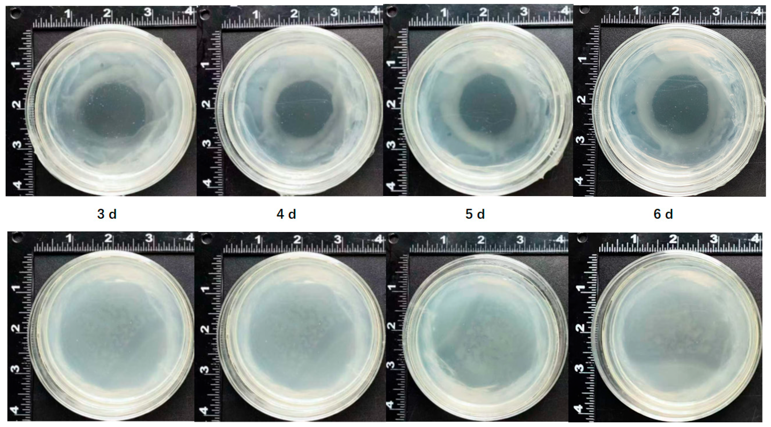
2.6. The Inhibitory Effect of Gallic Acid on Agrobacterium Growth

Both metabolome and transcriptome experiments showed that gallic acid participated in immune responses in mango calluses infected by Agrobacterium (Table 1 and Table 2). To study the relationship between gallic acid and the immune response of mango callus infected by Agrobacterium, 100 μL of Agrobacterium culture was spread on an MS plate. One hundred microliters of 10 mg/L gallic acid was pipetted onto the center of the plate. The MS plate was put in an incubator at a temperature of 28 °C for 3–6 days. Results showed that after Agrobacterium was cultured for 3 days, there was an empty circular area on the plate (Figure 4). Agrobacterium cannot propagate in this circular area. After Agrobacterium was cultured for 4 d, 5 d, or 6 d, more Agrobacterium cells accumulated outside the circular area (Figure 4). Especially for MS plates pipetted with gallic acid and cultured for 5 d and 6 d, a dense Agrobacterium band can be found outside and close to the circle (Figure 4). However, no Agrobacterium cell was found in the circular area (Figure 4). These demonstrated that Agrobacterium cells cannot grow on the MS plate area pipetted with gallic acid. After being cultured for a longer time, although more Agrobacterium cells accumulated in the MS plate area without gallic acid, they did not grow in the field containing gallic acid. These indicated that gallic acid can inhibit the growth of Agrobacterium, which might decrease the efficiency of Agrobacterium infection.

2.7. Suppression of Immune Responses in Mango Callus by Agrobacterium (T3SS-AvrPto)

To study whether T3SS and its effector protein can inhibit the immune response in mango callus induced by Agrobacterium, P. syringae pv. tomato strain DC3000 T3SS gene cluster (GenBank accession number: AF232004.3), effector gene AvrPto (NC_004578.1), and its promoter (116 bp upstream of ATG) were transformed into Agrobacterium EHA105 in this research. The transformed Agrobacterium was named Agrobacterium (T3SS-AvrPto). Agrobacterium transformed with a net vector was named Agrobacterium (NV). Mango callus was infected with Agrobacterium (T3SS-AvrPto) and Agrobacterium (NV), respectively, according to the method described in the Materials and Methods Section 4. The callus browning and the necrosis process were monitored. Results showed that after being infected by Agrobacterium (NV) for 15 h, about 30% of the calluses were browned and necrotized (Figure 5). After being infected with Agrobacterium (NV) for 30 h, about 90% of the calluses were browned and necrotized (Figure 5). If mango callus was infected with Agrobacterium (T3SS-AvrPto) for 15 h, a few calluses were browned or necrotized (Figure 5). When the callus was infected with Agrobacterium (T3SS-AvrPto) for 45 h, only about 10% of the calluses were browned (Figure 5). Furthermore, most of the browned calluses did not necrotize (Figure 5). Mango calluses infected by Agrobacterium (T3SS-AvrPto) browned and necrotized remarkably slower than those infected by Agrobacterium (NV) (Figure 5). These indicated that mango calluses infected by Agrobacterium (T3SS-AvrPto) had less immune response than those infected with Agrobacterium (NV).

2.8. Mango Callus Infected by Agrobacterium (T3SS-AvrPto) Produced Less ROS than Those Infected by Agrobacterium (NV)

To identify whether the mango callus infected by Agrobacterium (T3SS-AvrPto) browned and necrotized slower than Agrobacterium (NV) was due to that the callus infected by Agrobacterium (T3SS-AvrPto) had weaker immune responses, ROS in calluses infected by Agrobacterium (T3SS-AvrPto) and those infected by Agrobacterium (NV) were determined. Results showed that after the mango calluses were infected for 15 h, H2O2 content in the calluses infected by Agrobacterium (NV) was 5.0 ± 0.6 μmol/mg protein, while that in calluses infected by Agrobacterium (T3SS-AvrPto) was 3.9 ± 0.5 μmol/mg protein (Table 4). The H2O2 content in the calluses infected by Agrobacterium increased with more time, and the H2O2 content in the callus reached the maximum when the callus was infected for 45 h, and then decreased. At every time point, H2O2 content in mango callus infected by Agrobacterium (T3SS-AvrPto) was significantly less than that in callus infected by Agrobacterium (NV) (Table 4). Except for 15 h, O2− content and OH content in mango callus infected by Agrobacterium (T3SS-AvrPto) were less than those in callus infected by Agrobacterium (NV), correspondingly (Table 4). ROS contents in mango calluses infected by Agrobacterium (T3SS-AvrPto) were less than those in calluses infected by Agrobacterium (NV) (Table 4). These demonstrated that mango calluses infected by Agrobacterium (T3SS-AvrPto) have weaker immune responses than those infected by Agrobacterium (NV), resulting in calluses infected by Agrobacterium (T3SS-AvrPto) browned and necrotized slower than those infected by Agrobacterium (NV).

2.9. Suppression of Immune Responses in Mango Callus Infected by Agrobacterium (T3SS-AvrPto) Compared to Agrobacterium (NV)

To study the mechanism underlying the phenomenon that mango callus infected by Agrobacterium (T3SS-AvrPto) had less immune response than callus infected by Agrobacterium (NV). The expressions of the top up-regulated genes in calluses infected by Agrobacterium were measured in mango calluses infected by Agrobacterium (T3SS-AvrPto) and those infected by Agrobacterium (NV) using qRT-PCR. Results showed that the expressions of these genes in calluses infected by Agrobacterium (T3SS-AvrPto) for 45 h were significantly less than those in calluses infected by Agrobacterium (NV) for 45 h (Table 5). At 15 h, 30 h, and 60 h, the expressions of these genes in calluses infected by Agrobacterium (T3SS-AvrPto) were less than or similar to those in calluses infected by Agrobacterium (NV) (Table 5). For example, the relative expression of MiDAHPS1 in calluses infected by Agrobacterium (T3SS-AvrPto) for 45 h was 2.0 ± 0.2 (Table 5). The value in calluses infected by Agrobacterium (NV) for 45 h was 5.1 ± 0.3 (Table 5). Relative expression of MiDAHPS1 in calluses infected by Agrobacterium (T3SS-AvrPto) for 45 h was significantly less than that in calluses infected by Agrobacterium (NV) for 45 h (Table 5). Relative expression of MiDHQD1 in calluses infected by Agrobacterium (T3SS-AvrPto) for 15 h was 0.5 ± 0.1 (Table 5). The value in callus infected by Agrobacterium (NV) for 15 h was 0.6 ± 0.1 (Table 5). Relative expression of MiDHQD1 in calluses infected by Agrobacterium (T3SS-AvrPto) was similar to that in calluses infected by Agrobacterium (NV) for 15 h (Table 5). These indicated that when the mango callus was infected by Agrobacterium (T3SS-AvrPto), the expression of the genes up-regulated during immune responses was either inhibited or maintained. Immune reactions in calluses infected by Agrobacterium (T3SS-AvrPto) were suppressed.

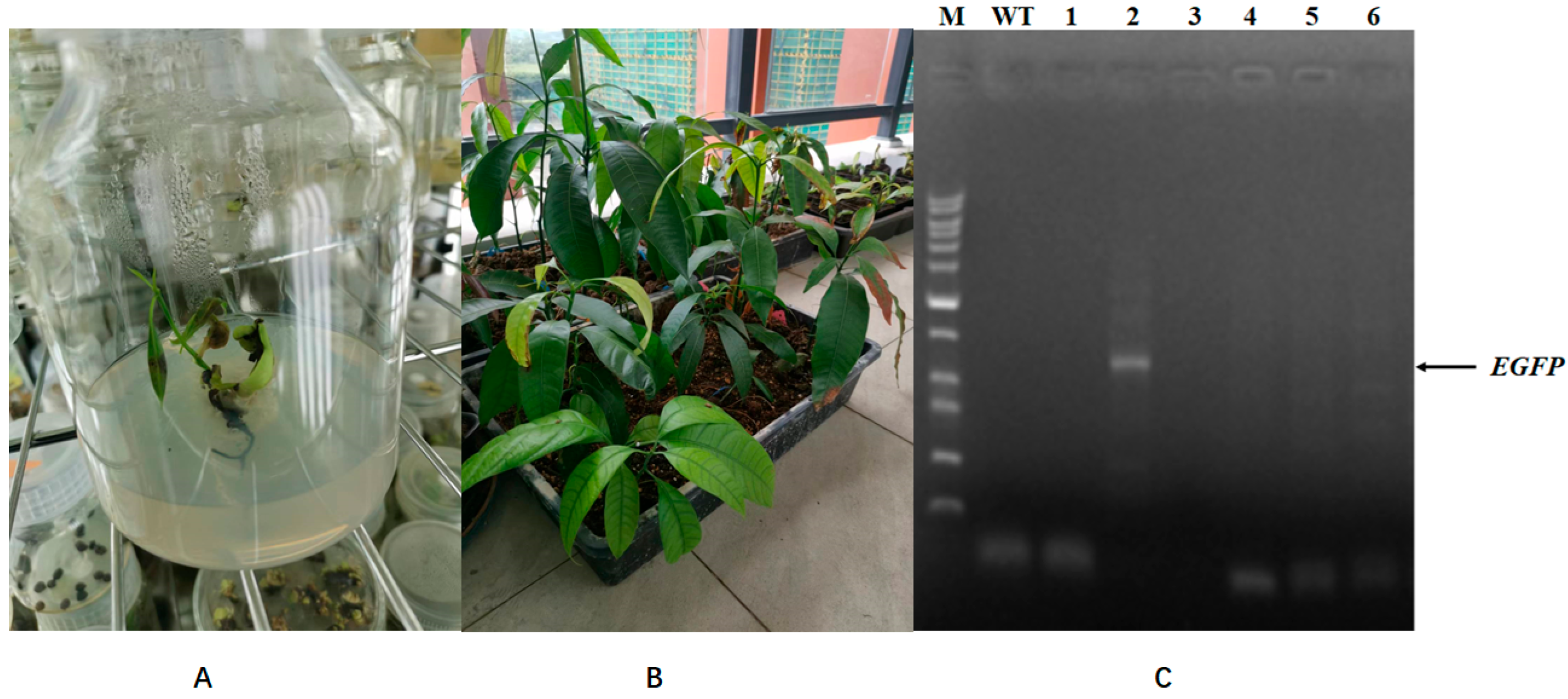
2.10. Agrobacterium Harboring Type III Secretion Gene Cluster (T3SS) and Effector Gene AvrPto Had Higher Infection Efficiency than Control Agrobacterium
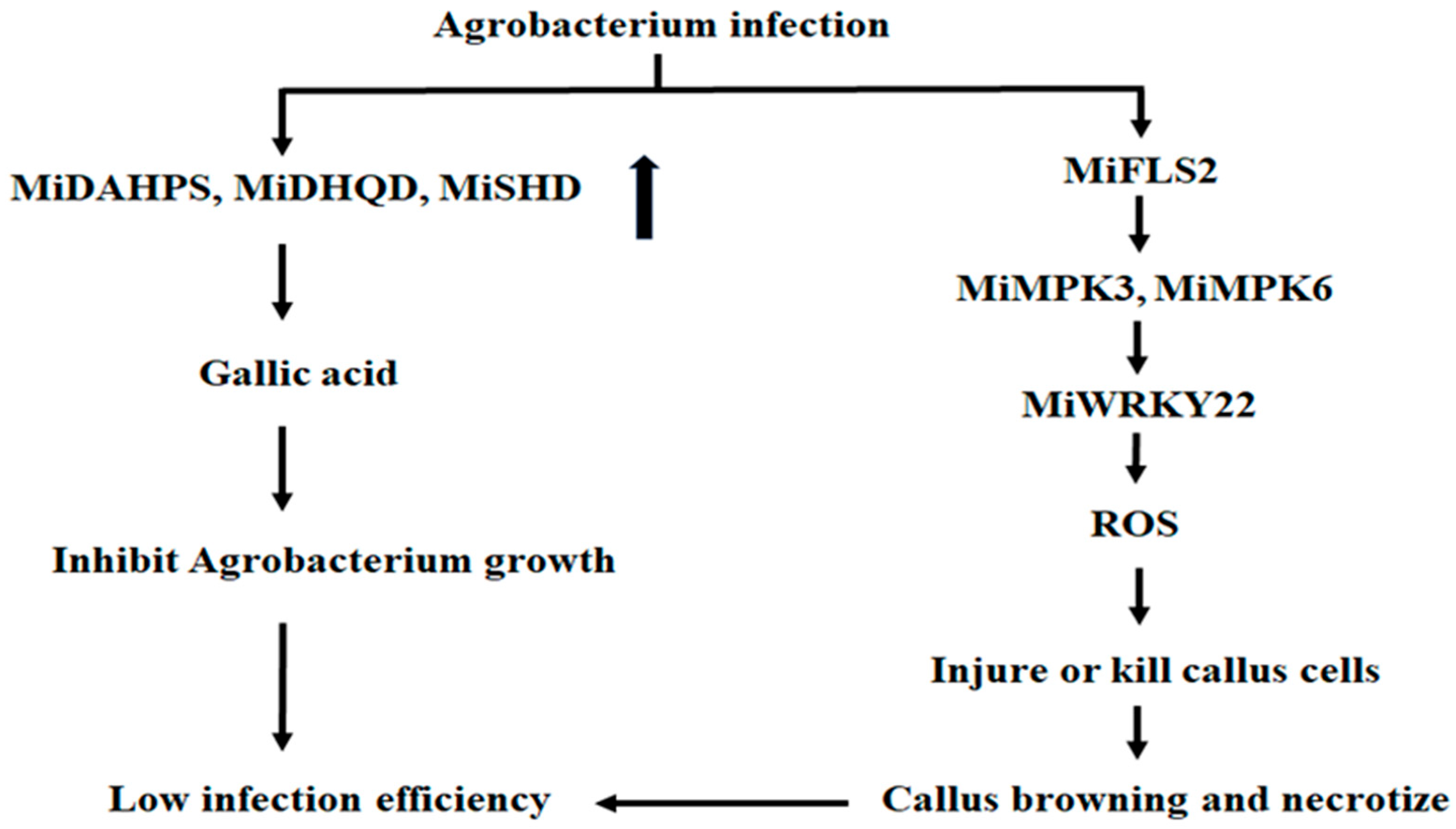
To study whether the transgenic efficiency of mango calluses can be improved using Agrobacterium harboring a type III secretion gene cluster (T3SS) and effector gene AvrPto, Agrobacterium (T3SS-AvrPto-EGFP) and Agrobacterium (NV-EGFP) were constructed as described in Materials and Methods. Mango calluses were infected with Agrobacterium (T3SS-AvrPto-EGFP), and EGFP fluorescence in calluses was observed. Results showed that about 1.6% of calluses infected by Agrobacterium (T3SS-AvrPto-EGFP) had successfully been transformed (Figure 6). No EGFP fluorescence was found in the callus infected with Agrobacterium (NV-EGFP). Mango calluses infected by Agrobacterium (T3SS-AvrPto-EGFP) had remarkably higher transgene efficiency than those infected by Agrobacterium (NV-EGFP). Agrobacterium-harboring T3SS-AvrPto had higher infection efficiency than Agrobacterium (NV). After the infected calluses were cultured on medium containing 20 μg/mL chloromycetin and 400 mg/L timentin for 1 month, the transformed calluses differentiated into seedlings. The possible transgenic seedlings were identified using PCR. Results showed that some seedlings had been transformed successfully (Figure 7). These findings underscore the potential of inhibiting the immune responses in mango calluses to enhance their transgene efficiency and the possibility of improving the transgene efficiency of mango calluses by constructing an Agrobacterium strain harboring T3SS and AvrPto.

3. Discussion

After multiple infections of the mango embryogenic callus using Agrobacterium tumefaciens, we observed that the majority of the callus rapidly browned and necrotized post-infection. The surviving callus eventually died during the differentiation culture process, and no regenerated plants were obtained. When pathogenic bacteria infect plants, the plants trigger immune responses, leading to programmed cell death to prevent the spread of the pathogen within the host [16,17]. The infection of mango embryogenic callus by Agrobacterium tumefaciens may have similarly induced such immune responses. Suppressing these immune reactions could significantly improve the transgenic efficiency of mango embryogenic calluses.
The secretion system of Agrobacterium tumefaciens is a type IV secretion system (T4SS), which facilitates the extracellular delivery of bacterial effector proteins [18]. However, many plants exhibit immune responses against T4SS and Agrobacterium effector proteins [19]. For example, Pseudomonas syringae employs a type III secretion system (T3SS) to translocate its effector proteins. Agrobacterium transformed with the T3SS gene cluster and the effector gene AvrPto from P. syringae can escape being perceived by plant cells [15]. Using the engineered Agrobacterium, the transformation rate of wheat, alfalfa, and switchgrass increased by 250–400% [15]. The transformation rate of mango calluses infected with Agrobacterium (T3SS-AvrPto-EGFP) was about 1.6%, which is a promising finding for the potential application of the transgenic method in mango breeding. This strategy can also be used for constructing stable transgenic plants of recalcitrant species.
ROS can injure or kill plant cells, inducing callus browning and necrotized. High content of ROS induced plant cells to necrotize and die [20]. Immune response can be triggered by ROS (Tang et al., 2017) [21]. After the grape embryogenic calluses were infected by Agrobacterium, key ROS-removal enzymes were down-regulated and ROS-production-related proteins were up-regulated [22]. H2O2 concentration and peroxidase activity increased in the callus [22]. The expression of ascorbate peroxidase (APX) decreased. APX is involved in the cell response to oxidative stress and plays a key role in regulating H2O2 levels in plants [22]. We found that after being infected by Agrobacterium for 15 h, H2O2 content in mango callus was 5.2 ± 0.6 μmol/mg protein. H2O2 content in uninfected mango calluses was 3.5 ± 0.5 μmol/mg protein. After the callus was infected for 45 h, H2O2 content in the mango callus was 13.6 ± 1.0 μmol/mg protein. O2− in calluses infected by Agrobacterium at different time points showed a similar trend. OH content in calluses infected for 15 h was similar to that in uninfected calluses. After being infected for 30 h, OH content in the callus reached the maximum. ROS content in the calluses increased rapidly after they were infected by Agrobacterium. Among the 20 top up-regulated genes in the mango callus, MiCAT1, MiCAT2, MiGOX1, MiGOX2, MiFOX1, MiFOX2, MiNADO1, and MiNADO2 participated in ROS biosynthesis. These indicated that ROS played important roles in inhibiting mango transformation upon being infected by Agrobacterium.
Darkening of explants frequently observed during Agrobacterium-mediated plant transformation has also generally been attributed to phenolic production, which may eventually lead to the death of plant cells [23,24,25]. When plant cells were injured, phenolic compounds in the cells were oxidized, and ubiquinone formed [26]. Ubiquinone was the brown compound in the callus [27]. Dihydrotricetin, dihydrokaempferol, and cyanidin were all phenolic compounds. When the mango callus was infected by Agrobacterium, the contents of dihydrotricetin, dihydrokaempferol, and cyanidin in the mango callus increased. More ubiquinone will be synthesized in the callus, and the callus will brown, as shown in the metabolome experiment in this research.
DAHPS, DHQD, and SDH participated in gallic acid biosynthesis [28]. In this research, it was found that the expressions of these genes were up-regulated after Agrobacterium infected the callus. This might improve the synthesis of gallic acid, as seen in the metabolome experiment in this research. Gallic acid is a common phenolic acid widely used as an antibacterial agent in the food industry [29]. It is recognized as a compound with broad-spectrum antibacterial and long-lasting sterilizing properties [29]. This research showed that Agrobacterium did not grow on an MS plate containing gallic acid. The growth of Agrobacterium cells can be inhibited by gallic acid. Failure of the mango transgene might be partially due to the gallic acid produced in the callus. If the reagents that can degrade gallic acid or inhibit the function of gallic acid were added to the liquid medium when mango calluses were infected by Agrobacterium, more Agrobacterium cells would survive, and more mango calluses would be infected. The infection efficiency of the Agrobacterium and the transformation rate of the mango calluses might also be enhanced. These are worth being investigated in the future.
After the grape embryogenic callus was infected by Agrobacterium, PR10 was one of the most strongly up-regulated proteins [22]. PR10 was a protein with a role in the plant defense reaction [22]. In this study, after mango calluses were infected with Agrobacterium, the expressions of MiFLS2-1, MiMPK3-1, MiMPK6-1, MiWRKY22-1, and MiNHL10-1 increased significantly. They all participated in plant defense reactions, demonstrating that immune responses and defense reactions played key roles in the browning and necrosis of mango callus after being infected by Agrobacterium.
Pattern-recognized receptors (PRRs) on the plant cell membranes can perceive pathogens and activate the immune system of plant cells [30]. Arabidopsis FLAGELIN-SENSITIVE2 (FLS2) was an identified PRR, which can perceive the 22 conserved amino acids in the N-terminal of bacterial flagellin (flg22) and interact with them [31]. After perceiving flg22, FLS2 combined with Brassinosteroid receptor-associated kinase 1 (BAK1) and formed a complex, which activated the downstream mitogen-activated protein kinase 3 (MPK3) and mitogen-activated protein kinase 6 (MPK6) [32]. MPK3 and MPK6 induced the expression of WRKY22, WRKY29, FRK1 (FLG22-induced receptor-like kinase 1), and other defense-related genes, producing more ROS [32]. The high content of ROS induced necrosis and death in plant cells [26]. Arabidopsis EF-Tu receptor (EFR) was also a PRR [33]. EFR can perceive Agrobacterium elongation factor Tu (EF-Tu) and activate plant immune response [34]. The polypeptide constituted by 18 amino acids located in the N-terminal of EF-Tu (elf18) can induce ROS production quickly, which causes the expression of defense genes FRK1, NHL10 (NDR1/HIN1-like 10), and so on [35]. It was found that the expressions of MiFLS2-1, MiMPK3-1, MiMPK6-1, MiWRKY22-1, and MiNHL10-1 in mango calluses infected by Agrobacterium were all higher than those in uninfected calluses, suggesting that when Agrobacterium infected calluses, these proteins were synthesized rapidly, which further induced the expression of the ROS-related genes. ROS was quickly produced. Some or all of the callus was damaged or killed by excessive ROS. Some infected calluses browned, and some necrotized, resulting in a low transformation rate (Figure 8). Gallic-acid synthesis genes and defense-related genes constituted the immune response system in the mango callus (Figure 8). This differed from Arabidopsis, which was caused only by defense-related genes. This is probably why the efficiency of conventional Agrobacterium infection on the mango callus was low, and the mango transgene experiment was challenging.

4. Materials and Methods

4.1. Agrobacterium Strain

The Agrobacterium strain used in this research was EHA105, which is a derivative of the soil bacterium Agrobacterium tumefaciens, widely used in plant genetic engineering due to its ability to transfer foreign DNA into plant cells. It is a disarmed strain, meaning its tumor-inducing (Ti) plasmid has been modified to remove pathogenic genes while retaining the DNA transfer mechanism essential for genetic transformation. EHA105 is derived from the wild-type strain A. tumefaciens C58, specifically from its helper strain EHA101. The Ti plasmid (pTiBo542) is disarmed by removing oncogenes (e.g., iaaM, ipt) but retaining virulence (vir) genes required for T-DNA transfer. A binary vector system: A separate plasmid (e.g., pCAMBIA series) carries the gene of interest and T-DNA borders, while the disarmed Ti plasmid provides vir genes in trans. Agrobacterium EHA105 is non-pathogenic, which cannot cause crown gall disease due to the absence of oncogenes. It has high transformation efficiency for dicotyledonous plants and some monocots.

4.2. Construction of Agrobacterium Strains

The DNA fragment containing the P. syringae pv. tomato strain DC3000 T3SS gene cluster (GenBank accession number: AF232004.3), effector gene AvrPto (NC_004578.1), and its promoter (116 bp upstream of ATG) was meticulously synthesized with attB sequences at the two terminals. The BP recombination reaction was carried out with the same level of meticulousness, inserting the synthesized DNA into pENTRTM5′-TOPO (Invitrogen, Carlsbad, CA, USA) using the pENTRTM5′-TOPO Cloning Kit (Invitrogen). E. coli TOP10 was used to propagate the plasmid. The plasmids pENTRTM5′-TOPO-T3SS-AvrPto were extracted using the GeneJET kit (Thermo Fisher Scientific, Waltham, MA, USA). The LR recombination reaction was performed with the same level of meticulousness, transferring T3SS-AvrPto in pENTRTM5′-TOPO-T3SS-AvrPto into pEarleyGate302 (BIOESN, Shanghai, China) using the LR Clonase™ Enzyme Mix (Invitrogen). E. coli TOP10 was used to propagate the plasmid. The plasmids pEarleyGate302-T3SS-AvrPto were extracted and transformed into Agrobacterium EHA105. Triparental mating [36] was used to mobilize pEarleyGate302-T3SS-AvrPto into A. tumefaciens EH105. E. coli HB101 harboring the helper plasmid pRK2013 was bought from Beijing Biobw Biotechnology Co., Ltd. (Beijing, China) (Bio-133891) [37,38]. A single clone of E. coli DH10B harboring pEarleyGate302-T3SS-AvrPto was picked up from the LB plate and cultured in LB liquid medium containing kanamycin until OD600 = 0.5–0.8. E. coli HB101 harboring the helper plasmid pRK2013 was also cultured in LB liquid medium containing 50 μg/mL kanamycin until OD600 = 0.5–0.8. Agrobacterium EHA105 was cultured in YEB liquid medium containing 100 μg/mL rifampicin until OD600 = 0.5–0.8. One hundred and fifty microliters of the 3 cultures were mixed and centrifuged at 800 rpm for 20 min. The supernatant was discarded, and the pellet was resuspended in 300 μL LB liquid medium. The mixture was spread on a YEB plate containing 50 μg/mL kanamycin and 100 μg/mL rifampicin. The plate was put at 28 °C for 24 h. Single clones were picked up and cultured in YEB liquid medium containing 50 μg/mL kanamycin and 100 μg/mL rifampicin, and PCR was performed. P2F: 5′-GGGGACAAGTTTGTACAAAAAAGCAGGCT-3′, P2R: 5′-CTACCCATACGATGTTCCAG-3′. Clones with the expected PCR result were stocked to infect mango calluses. To construct Agrobacterium (T3SS-AvrPto-EGFP), Agrobacterium (T3SS-AvrPto) was transformed with plasmid pBI221-GFP (HonorGene, Changsha, China), and the transformed Agrobacterium was screened on a plate containing 100 μg/mL ampicillin and 100 μg/mL rifampicin.

4.3. Mango Embryogenic Callus Induction

Mango callus was induced and modified according to the published method [39]. Fruitlet in the cotyledon stage (45 days after the flower) was collected. Contaminants on the surface were removed using a brush. The fruit was rinsed for 30 min using tap water. It was transferred into 1% detergent and marinated for 20 min. The fruitlet was washed using flush water for 20 min. 75% ethanol was sprayed onto the surface. The fruitlet was cut using a scalpel in the super clean bench, and the seed was fetched. The seed was longitudinally cut. The embryonic matter was collected from the inner surface of the seed and cultured on medium M1 (Table 6). The plates were put in the dark at 26 °C for one month. And then, the callus induced was transferred onto medium M2 (Table 6). The plates were put in a dark place at 26 °C and cultured for one month. A large amount of embryogenic callus will be obtained.

4.4. Mango Callus Transformation

One gram of mango embryogenic callus is placed on a medium containing chloromycetin concentrations. The plates are then kept in the dark at 25 °C for one month to identify the concentration at which the callus cannot grow. Agrobacterium cells transformed with pEarleyGate302-T3SS-AvrPto or pBI221-GFP are cultured overnight; 1.5 mL of the Agrobacterium culture is centrifuged at 500 rpm, and the supernatant is discarded. The pellet is resuspended with an AAM liquid medium containing 200 µmolL−1 acetosyringone and shaken at 100 rpm, 26 °C for 1 h. The mango callus is placed in the AAM medium and cultured for 30 min. The liquid is discarded, and the callus is put on sterilized filter paper on a super-clean bench for 60 min. Next, the callus is transferred onto medium M3 (Table 6). The plates are observed in the dark at 26 °C for 30 days.

4.5. Metabolome and Transcriptome Experiments

Metabolome and transcriptome experiments were performed as described before [40]. Agrobacterium was cultured at 26 °C in the dark for two days. The OD600 was adjusted to 0.4; 1.5 mL of Agrobacterium culture was collected and centrifuged at 500 rpm. The supernatant was discarded. The pellet was resuspended in 1 mL AAM medium containing 200 µmolL−1 acetosyringone. The mixture was transferred into 100 mL AAM medium containing 200 µmolL−1 acetosyringone and cultured for 1 h in the dark at 26 °C; 10 g of mango embryogenic callus was put in AAM medium and shaken at 500 rpm, 26 °C in the dark for 30 min. The liquid was discarded. The callus was washed with sterilized water ten times. The callus was stored at −80 °C until metabolome and transcriptome experiments were performed.
For analyzing the metabolome results, unsupervised PCA (principal component analysis) was performed by the statistics function prcomp within R (www.r-project.org, accessed on 6 May 2023). The data were unit variance scaled before unsupervised PCA. The HCA (hierarchical cluster analysis) results of samples and metabolites were presented as heatmaps with dendrograms, while Pearson correlation coefficients (PCC) between samples were calculated by the cor function in R and presented as only heatmaps. Both HCA and PCC were carried out by the R package pheatmap. For HCA, normalized signal intensities of metabolites (unit variance scaling) are visualized as a color spectrum. Significantly regulated metabolites between groups were determined by VIP ≥ 1 and absolute Log2FC (fold change) ≥ 1. VIP values were extracted from the OPLS-DA result, which also contains score plots and permutation plots, and was generated using the R package MetaboAnalystR 3.0. The data were log-transformed (log2) and mean-centered before OPLS-DA. In order to avoid overfitting, a permutation test (200 permutations) was performed. Identified metabolites were annotated using the KEGG Compound database (http://www.kegg.jp/kegg/compound/, accessed on 10 May 2023); annotated metabolites were then mapped to the KEGG Pathway database (http://www.kegg.jp/kegg/pathway.html, accessed on 8 July 2023). Pathways with significantly regulated metabolites mapped were then fed into MSEA (metabolite sets enrichment analysis); their significance was determined by the hypergeometric test’s p-values.

4.6. QRT-PCR

NucleoSpin(R) 96 RNA core kit (Thermo Fisher Scientific, Waltham, MA, USA) was used to extract mango callus RNA. RNA was transcribed into cDNA using Oligo(dT) 12–18 and SuperScript III Reverse Transcriptase (Invitrogen). QRT-PCR was performed using a CFX Real-time PCR system (Applied Biosystems, Foster City, CA, USA) and KiCqStart SYBR Green qPCR ReadyMix (Catalog No. KCQS01, MilliporeSigma, Burlington, MA, USA). QRT-PCR data were collected using the software Bio-Rad CFX Manager Version 2.1.1022.0523. The relative expression was calculated using 2−ΔΔCT. Housekeeping gene tubulin (XM_044625649.1) was used as a control. Genomic DNA in RNA samples was removed using TURBO DNase (Catalog No. AM1907, Invitrogen). The raw Ct values were transformed into relative quantities or copy numbers. The target gene Ct values were normalized using the reference gene (tubulin, XM_044625649.1): ΔCt = ΔCttarget gene − ΔCtreference gene. A curve was generated using standards of known concentrations to calculate absolute copy numbers. If the data follow an exponential distribution, a log transformation (log2) was applied to stabilize variance. ΔΔCt was calculated, and ΔCt values between experimental and control groups were compared: ΔΔCt = ΔCtexperimental group − ΔCtcontrol group. Fold change was calculated as follows. Fold Change = 2−ΔΔCt (assuming 100% amplification efficiency). The Shapiro–Wilk test was used to assess whether the data follow a normal distribution. If it were a normal distribution, ANOVA was applied to compare differences among multiple groups, followed by post-hoc tests (Tukey’s HSD). If it were non-normally distributed, the Kruskal–Wallis test was used for comparisons among multiple groups. Bonferroni correction was applied to control the false discovery rate.

4.7. Confocal Observation

Green fluorescence in transformed callus was observed using confocal, as described in a published paper [41]. The exciting light wavelength was 488 nm. The scattered light wavelength was 493–550 nm. The eyepiece magnification is 10×, and the objective lens magnification is 20×. The results were analyzed using the software Leica Application Suite X (LAS X) version 3.5.5.19976.

4.8. Determination of ROS Contents in Mitochondria Isolated from Mango Callus

According to the published paper, mitochondria were isolated and purified from mango calluses [42]. The mitochondrial pellets were resuspended in a buffer (20% glycerol, 0.1 M Tris, pH 7.0). The contents of H2O2 and O2− were measured using established methods [43], and the OH content was determined as previously described [44]. A 0.5 mL suspended mitochondrial buffer was added to a 0.5 mL basic reaction system (120 mmolL−1 KCl, 0.4 molL−1 NADH, 30 mmolL−1 DMSO, 40 mmolL−1 phosphate buffer solution, pH 7.0). The mixture was then incubated at 25 °C for 10 min. The production of formaldehyde was calculated from the Nash reaction to determine the content of OH [45].

4.9. Statistical Analysis

Each treatment consisted of three replicates. The significant differences between the treatments were tested by analysis of variance (ANOVA) and Duncan’s multiple range test (DMRT). SPSS software was used to determine the significant difference at p ≤ 0.05. Data are presented as mean ± standard error (SE).

5. Conclusions

The low efficiency of the mango transgene was due to immune responses activated in the callus. The infection and transformation efficiency can be significantly improved if the mango calluses are infected by Agrobacterium harboring a T3SS gene cluster and its effector gene. The transformation rate of mango callus can be enhanced to 1.6% when they are infected with Agrobacterium (T3SS-AvrPto), which is a promising finding. These may play important roles in developing new mango varieties with multiple resistance traits in the future.

Author Contributions

Conceptualization, H.S. and S.C.; formal analysis, H.S., Z.A.J. and O.F.; investigation, H.S., L.H., Q.W. and R.Z.; resources, S.C. and R.Z.; writing—original draft preparation, H.S. and S.C.; writing—review and editing, O.F. and Z.A.J.; supervision, S.C.; funding acquisition, R.Z. All authors have read and agreed to the published version of the manuscript.

Funding

This research was supported by the State Key Project for Research and Development of China “Synergistic technology model and application for eco-friendly scheduling and quality improvement in Hainan mango production season” (No. 2023YFD2300803-2).

Institutional Review Board Statement

No animals were used in this research.

Informed Consent Statement

Not applicable.

Data Availability Statement

Data are contained within the article.

Conflicts of Interest

The authors have no competing interests to declare that are relevant to the content of this article.

References

  1. Dofuor, A.K.; Quartey, N.K.; Osabutey, A.F.; Antwi-Agyakwa, A.K.; Asante, K.; Boateng, B.O.; Ablormeti, F.K.; Lutuf, H.; Osei-Owusu, J.; Osei, J.H.N.; et al. Mango anthracnose disease: The current situation and direction for future research. Front. Microbiol. 2023, 14, 1168203. [Google Scholar] [CrossRef] [PubMed]
  2. Patel, K.B.; Saxena, S.P.; Patel, K.M.; Patel, A.T. Estimation of crop loss caused by important insect-pests of mango. Int. J. Biol. Sci. 2022, 4, 194–197. [Google Scholar] [CrossRef]
  3. Bally, I.S.E.; Dillon, N.L. Mango (Mangifera indica L.) Breeding. In Advances in Plant Breeding Strategies: Fruits; Springer Nature: Berlin, Germany, 2018; pp. 811–896. ISBN 303023415X. [Google Scholar]
  4. Litz, R.E.; Hormaza, J.I. Mangifera indica mango. In Biotechnology of Fruit and Nut Crops, 2nd ed.; Litz, R.E., Pliego-Alfaro, F., Hormaza, J.I., Eds.; CAB International: Wallingford, UK, 2020; pp. 27–43. ISBN 9781780648279. [Google Scholar]
  5. Petri, C.; Litz, R.E.; Singh, S.K.; Hormaza, J.I. In vitro culture and genetic transformation in mango. In The Mango Genome, a Compendium of Plant Genomes; Kole, C., Ed.; Springer Nature Switzerland AG: Cham, Switzerland, 2021; pp. 131–151. ISBN 2199-4781. [Google Scholar]
  6. Pandey, K.; Karthik, K.; Singh, S.K.; Vinod; Sreevathsa, R.; Srivastav, M. Amenability of an Agrobacterium tumefaciens-mediated shoot apical meristem-targeted in planta transformation strategy in Mango (Mangifera indica L.). GM Crops Food 2022, 13, 342–354. [Google Scholar] [CrossRef]
  7. Adjei, M.O.; Zhao, H.; Tao, X.; Yang, L.; Deng, S.; Li, X.; Mao, X.; Li, S.; Huang, J.; Luo, R.; et al. Using a protoplast transformation system to enable functional studies in Mangifera indica L. Int. J. Mol. Sci. 2023, 24, 11984. [Google Scholar] [CrossRef]
  8. Chen, X.; He, S.; Wang, Z.; Zhai, Y.; Guo, W.; Li, X. Production of transgenic periclinal chimeras in pumpkin—A tool for revealing cell fates of L1 meristem. Plant Biol. 2023, 26, 126–139. [Google Scholar] [CrossRef] [PubMed]
  9. Aida, R.; Sasaki, K.; Yoshioka, S.; Noda, N. Distribution of cell layers in floral organs of chrysanthemum analyzed with periclinal chimeras carrying a transgene encoding fluorescent protein. Plant Cell Rep. 2020, 39, 609–619. [Google Scholar] [CrossRef]
  10. Li, M.; Wang, D.; Long, X.; Hao, Z.; Lu, Y.; Zhou, Y.; Peng, Y.; Cheng, T.; Shi, J.; Chen, J. Agrobacterium-Mediated genetic transformation of embryogenic callus in a Liriodendron hybrid (L. Chinense × L. Tulipifera). Front. Plant Sci. 2022, 13, 802128. [Google Scholar] [CrossRef]
  11. Nakajima, I.; Endo, M.; Haji, T.; Moriguchi, T.; Yamamoto, T. Embryogenic callus induction and Agrobacterium-mediated genetic transformation of ‘Shine Muscat’ grape. Plant Biotechnol. 2020, 37, 185–194. [Google Scholar] [CrossRef]
  12. Chen, Z.; Debernardi, J.M.; Dubcovsky, J.; Gallavotti, A. Recent advances in crop transformation technologies. Nat. Plants 2022, 8, 1343–1351. [Google Scholar] [CrossRef]
  13. Zhang, D.; Ren, L.; Chen, G.; Zhang, J.; Reed, B.M.; Shen, X. ROS-induced oxidative stress and apoptosis-like event directly affect the cryopreserved embryogenic callus cell viability in Agapanthus praecox. Plant Cell Rep. 2015, 34, 1499–1513. [Google Scholar] [CrossRef]
  14. Christie, P.J. Type IV secretion: The Agrobacterium VirB/D4 and related conjugation systems. Biochim. Biophys. Acta 2004, 1694, 219–234. [Google Scholar] [CrossRef] [PubMed]
  15. Raman, V.; Rojas, C.M.; Vasudevan, B.; Dunning, K.; Kolape, J.; Oh, S.; Yun, J.; Yang, L.; Li, G.; Pant, B.D.; et al. Agrobacterium expressing a type III secretion system delivers Pseudomonas effectors into plant cells to enhance transformation. Nat. Commun. 2022, 13, 2581. [Google Scholar] [CrossRef] [PubMed]
  16. Zavaliev, R.; Mohan, R.; Chen, T.; Dong, X. Formation of NPR1 condensates promotes cell survival during plant immune response. Cell 2020, 182, 1093–1108. [Google Scholar] [CrossRef]
  17. Peng, T.; Tao, X.; Xia, Z.; Hu, S.; Xue, J.; Zhu, Q.; Pan, X.; Zhang, Q.; Li, S. Pathogen hijacks programmed cell death signaling by arginine ADPR-deacylization of caspases. Mol. Cell 2022, 82, 1806–1820. [Google Scholar] [CrossRef]
  18. Cornelis, G.R. The type III secretion injectisome. Nat. Rev. Microbiol. 2006, 4, 811–825. [Google Scholar] [CrossRef]
  19. Yang, F.; Li, G.; Felix, G.; Albert, M.; Guo, M. Engineered Agrobacterium improves transformation by mitigating plant immunity detection. New Phytol. 2022, 237, 2493–2504. [Google Scholar] [CrossRef]
  20. Büttner, D. Behind the lines–actions of bacterial type III effector proteins in plant cells. FEMS Microbiol. Rev. 2016, 40, 894–937. [Google Scholar] [CrossRef] [PubMed]
  21. Tang, D.; Wang, G.; Zhou, J. Receptor kinases in plant-pathogen interactions: More than pattern recognition. Plant Cell 2017, 29, 618–637. [Google Scholar] [CrossRef]
  22. Zhao, F.; Chen, L.; Perl, A.; Chen, S.; Ma, H. Proteomic changes in grape embryogenic callus in response to Agrobacterium tumefaciens-mediated transformation. Plant Sci. 2011, 181, 485–495. [Google Scholar] [CrossRef]
  23. Perl, A.; Lotan, O.; Abu-Abied, M.; Holland, D. Establishment of an Agrobacterium-mediated transformation system for grape (Vitis vinifera L.): The role of antioxidants during grape-Agrobacterium interactions. Nat. Biotechnol. 1996, 14, 624–628. [Google Scholar] [CrossRef]
  24. Hansen, G. Evidence of Agrobacterium-induced apoptosis in maize cells. Mol. Plant Microbe Interact. 2000, 13, 649–657. [Google Scholar] [CrossRef] [PubMed]
  25. Parrot, D.L.; Anderson, A.J.; Carman, J.G. Agrobacterium induces plant cell death in wheat (Triticum aestivum L.). Physiol. Mol. Plant Pathol. 2002, 60, 59–69. [Google Scholar] [CrossRef]
  26. Cai, J.; Chen, T.; Wang, Y.; Qin, G.; Tian, S. SlREM1 triggers cell death by activating an oxidative burst and other regulators. Plant Physiol. 2020, 183, 717–732. [Google Scholar] [CrossRef]
  27. Sun, L.L.; Shao, Y.N.; You, M.X.; Li, C.H. ROS-mediated BNIP3-dependent mitophagy promotes coelomocyte survival in Apostichopus japonicus in response to Vibrio splendidus infection. Zool. Res. 2022, 43, 285–300. [Google Scholar] [CrossRef] [PubMed]
  28. Zhou, L.; Sun, J.; Zhang, T.; Tang, Y.; Liu, J.; Gao, C.; Zhai, Y.; Guo, Y.; Feng, L.; Zhang, X.; et al. Comparative transcriptome analyses of different Rheum officinale tissues reveal differentially expressed genes associated with anthraquinone, catechin, and gallic acid biosynthesis. Genes 2022, 13, 1592. [Google Scholar] [CrossRef]
  29. Zhu, Y.; Kun, K.; Zhang, X.; Ma, H.; Yang, L.; Wei, L.; Yang, L.; Jiang, M. Antibacterial effect of gallic acid in UV C light treatment against Escherichia coli O157:H7 and the underlying mechanism. Food Bioprocess Technol. 2024, 17, 2216–2231. [Google Scholar] [CrossRef]
  30. Delplace, F.; Huard-Chauveau, C.; Berthome, R.; Roby, D. Network organization of the plant immune system: From pathogen perception to robust defense induction. Plant J. 2022, 109, 447–470. [Google Scholar] [CrossRef] [PubMed]
  31. Ngou, B.P.M.; Ding, P.; Jones, J.D.G. Thirty years of resistance: Zig-zag through the plant immune system. Plant Cell 2022, 34, 1447–1478. [Google Scholar] [CrossRef]
  32. Liu, T.; Cao, L.; Cheng, Y.; Ji, J.; Wei, Y.; Wang, C.; Duan, K. MKK4/5-MPK3/6 cascade regulates Agrobacterium-mediated transformation by modulating plant immunity in Arabidopsis. Front. Plant Sci. 2021, 12, 731690. [Google Scholar] [CrossRef]
  33. Ding, L.; Li, Y.; Wu, Y.; Li, T.; Geng, R.; Cao, J.; Zhang, W.; Tan, X. Plant disease resistance-related signaling pathways: Recent progress and future prospects. Int. J. Mol. Sci. 2022, 23, 16200. [Google Scholar] [CrossRef]
  34. Zhou, J.M.; Zhang, Y. Plant immunity: Danger perception and signaling. Cell 2020, 181, 978–989. [Google Scholar] [CrossRef] [PubMed]
  35. Tiwari, M.; Mishra, A.K.; Chakrabarty, D. Agrobacterium-mediated gene transfer: Recent advancements and layered immunity in plants. Planta 2022, 256, 37. [Google Scholar] [CrossRef] [PubMed]
  36. Wise, A.A.; Liu, Z.; Binns, A.N. Agrobacterium Protocols; Wang, K., Ed.; Humana Press: Totowa, NJ, USA, 2006; pp. 43–54. ISBN 9781588298430. [Google Scholar]
  37. Hall, A.; Donohue, T.; Peters, J. Complete sequences of conjugal helper plasmids pRK2013 and pEVS104. MicroPubl. Biol. 2023, 10, 17912. [Google Scholar]
  38. Figurski, D.H.; Helinski, D.R. Replication of an origin-containing derivative of plasmid RK2. dependent on a plasmid function provided in trans. Proc. Nati. Acad. Sci. USA 1979, 76, 1648–1652. [Google Scholar] [CrossRef]
  39. Xu, W.; Wu, H.; Luo, C.; Li, L.; Liang, Q.; Ma, X.; Yao, Q.; Gao, Y.; Wang, S. Callus proliferation, early somatic embryogenesis and cytological observation of mango ‘Jinhuang’. Mol. Plant Breed. 2019, 17, 3001–3008. (In Chinese) [Google Scholar]
  40. Iqbal, A.; Qiang, D.; Xiangru, W.; Huiping, G.; Hengheng, Z.; Xiling, Z.; Meizhen, S. Integrative physiological, transcriptome and metabolome analysis reveals the involvement of carbon and flavonoid biosynthesis in low phosphorus tolerance in cotton. Plant Physiol. Biochem. 2023, 196, 302–317. [Google Scholar] [CrossRef]
  41. Lee, M.Y.; Song, Y.S.; Ryu, K.H. Development of infectious transcripts from full-length and GFP-tagged cDNA clones of Pepper mottle virus and stable systemic expression of GFP in tobacco and pepper. Virus Res. 2011, 155, 487–494. [Google Scholar] [CrossRef] [PubMed]
  42. Vanlerberghe, G.C.; McIntosh, L. Lower growth temperature increases alternative pathway capacity and alternative oxidase protein in tobacco. Plant Physiol. 1992, 100, 115–119. [Google Scholar] [CrossRef]
  43. Wang, A.G.; Luo, G.H. Quantitative relation between the reaction of hydroxylamine and superoxide anion radicals in plants. Plant Physiol. Commun. 1990, 6, 55–57. [Google Scholar]
  44. Simontacchi, M.; Puntarulo, S. Oxygen radical generation by isolated microsomes from soybean seedling. Plant Physiol. 1992, 100, 1263–1268. [Google Scholar] [CrossRef]
  45. Nash, T. The colorimetric estimation of formaldehyde by means of the Hantzsch reaction. Biochem. J. 1953, 55, 416–421. [Google Scholar] [CrossRef] [PubMed]
Figure 1. Mango callus infected with Agrobacterium for different time. 0 h, 15 h, 30 h, 45 h, and 60 h represented mango callus being infected by Agrobacterium for 0 h, 15 h, 30 h, 45 h, and 60 h, respectively. One single clone of Agrobacterium EHA105 was picked up from the YEB plate and cultured in liquid YEB at 26 °C for 48 h; 1 mL of Agrobacterium culture was put in fresh YEB liquid medium and cultured at 26 °C until OD600 = 1.0; 1.5 mL of Agrobacterium culture was centrifuged at 500 rpm. The supernatant was discarded. The pellet was resuspended with an AAM liquid medium containing 200 µmolL−1 acetosyringone and cultured in 500 mL of AAM liquid medium for 30 min. Ten grams of mango calluses were put into the triangular flask containing the Agrobacterium mixture and cultured for 30 min. And then, the mango calluses were put onto the sterile filter papers in the super-clean bench for 2 h to remove the water on the surface. After that, the mango calluses were transferred onto MS medium and cultured in 26 °C in dark for 0 h, 15 h, 30 h, 45 h, and 60 h, respectively.
Ijms 26 05006 g001
Figure 2. ROS contents in mango callus infected by Agrobacterium. The units for ROS levels are as follows: O2−, nmol/(min mg protein); H2O2, μmol/mg protein; OH, nmol/(min mg protein). 0 h, 15 h, 30 h, 45 h, and 60 h represented mango callus being infected by Agrobacterium for 0 h, 15 h, 30 h, 45 h, and 60 h, respectively. Different lowercase letters above the columns indicate significant differences between the treatments at the 0.05 level. The blue, orange, and grey columns represent H2O2, O2−, and OH content in mango callus, respectively. Each treatment consisted of four replicates. The significant differences between the treatments were tested by analysis of variance (ANOVA) and Duncan’s multiple range test (DMRT). SPSS 24 software was used to determine the significant difference at p ≤ 0.05. Data are presented as mean ± standard error (SE).
Ijms 26 05006 g002
Figure 3. Quantitative plot of differentially expressed metabolites in callus infected by Agrobacterium for 45 h and uninfected.
Ijms 26 05006 g003
Figure 4. Gallic acid inhibits the growth of Agrobacterium. One single clone of Agrobacterium EHA105 was picked up from the YEB plate and cultured in liquid YEB at 28 °C for 48 h. One milliliter of Agrobacterium cultures was put in fresh YEB liquid medium and cultured at 26 °C until OD600 = 1.0. Five hundred microliters of Agrobacterium cultures were spread onto the MS plate. One hundred microliters of gallic acid (10 mg/L) were pipetted onto the plate center. MS plates without gallic acid were used as a control. The MS plates were incubated at 28 °C for 3, 4, 5, and 6 days in the dark, respectively. The top four plates showed Agrobacterium grown on an MS medium on which 100 μL of 10 mg/L gallic acid was pipetted onto the center. The bottom four plates showed Agrobacterium grown on medium without gallic acid; 3 d, 4 d, 5 d, and 6 d represented MS plates cultured at 28 °C for 3, 4, 5, and 6 days, respectively.
Ijms 26 05006 g004
Figure 5. The browning and necrosis of callus infected by Agrobacterium. The 0 h, 15 h, 30 h, and 45 h represented mango calluses cultured for 0 h, 15 h, 30 h, and 45 h, respectively, after being infected by Agrobacterium. One single clone of Agrobacterium was picked up from the YEB plate and cultured in liquid YEB at 26 °C for 48 h; 1 mL of Agrobacterium culture was put in fresh YEB liquid medium and cultured at 26 °C until OD600 = 1.0; 1.5 mL of Agrobacterium culture was centrifuged at 500 rpm. The supernatant was discarded. The pellet was resuspended with an AAM liquid medium containing 200 µmolL−1 acetosyringone and cultured in 500 mL of AAM liquid medium for 30 min. Ten grams of mango calluses were put into the triangular flask containing the Agrobacterium mixture and cultured for 30 min. And then, the mango calluses were put onto the sterile filter papers in the super-clean bench for 2 h to remove the water on the surface. After that, the mango calluses were transferred onto MS medium and cultured in 26 °C in dark for 0 h, 15 h, 30 h, and 45 h, respectively.
Ijms 26 05006 g005
Figure 6. EGFP fluorescence observation in mango calluses infected with Agrobacterium (T3SS-AvrPto-EGFP). The top-left panel represented EGFP fluorescence observed in mango calluses in bright fields. The top-right panel represented EGFP fluorescence observed in mango calluses in dark fields. The bottom panel represented EGFP fluorescence observed in mango calluses in the fused field.
Ijms 26 05006 g006
Figure 7. Differentiation of the transgenic calluses and verification. (A) The possible transgenic seedling grown on media containing 20 μg/mL chloromycetin. (B) The possible transgenic seedlings grown in gardening soil. (C) Verification of the seedlings differentiated. Genomic DNA was extracted from the leaves of the possible transgenic plants. PCR was performed using P1F:5′-ATGGTGAGCAAGGGCGAGGA-3 and P1R:5′-CTTGTACAGCTCGTCCATGC-3′ as primers responding to the EGFP gene in the plasmid pBI221-GFP. 1–6 represented different seedlings. WT referred to mango seedlings differentiated from calluses not infected by Agrobacterium.
Ijms 26 05006 g007
Figure 8. The proposed mechanism underlying the immune response of mango calluses infected by Agrobacterium. The boldfaced arrow represented the expressions of the up-regulated genes.
Ijms 26 05006 g008
Table 1. Primary metabolites up-regulated in mango callus infected by Agrobacterium.
IndexCompoundsFold (I45/I0)VIP Value
Gallic acidOrganic acid15.21.96
Phosphoenolpyruvic acidOrganic acid3.91.22
D-erythrose-4-phosphatePhosphorylated derivatives of erythritol4.11.36
UbiquinoneLiposoluble quinone compound11.51.25
DihydrotricetinFlavonoids3.51.26
DihydrokaempferolFlavonoid3.81.19
CyanidinAntioxidant3.71.28
Note: I45 and I0 represent the compound content in calluses infected by Agrobacterium for 45 h and calluses uninfected, respectively.
Table 2. The top 20 genes up-regulated in callus infected by Agrobacterium for 45 h.
QuerySymbolFold (I45/I0)Gene AnnotationSubject
LOC123211694MiDAHPS15.2Mangifera indica 3-oxoacyl-[acyl-carrier-protein] synthaseXM_044646917.1
LOC123210632MiDHQD13Mangifera indica bifunctional 3-dehydrogenate dehydratase/shikimate dehydrogenaseXM_044644036.1
LOC123236405MiDHQD23.7Mangifera indica bifunctional 3-dehydrogenate dehydratase/shikimate dehydrogenaseXM_044644035.1
LOC123207068MiSHD12.4Mangifera indica bifunctional 3-dehydrogenate dehydratase/shikimate dehydrogenaseXM_044644037.1
LOC123215035MiSHD22.2Mangifera indica bifunctional 3-dehydrogenate dehydratase/shikimate dehydrogenaseXM_044644039.1
LOC123225839MiCAT12.9Mangifera indica catalase isozyme 1XM_044655447.1
LOC123228490MiCAT22.6Mangifera indica catalase isozyme 1XM_044655447.1
LOC123217301MiGOX11.7Mangifera indica peroxisomal (S)-2-hydroxy-acid oxidase-likeXM_044639087.1
LOC123227499MiGOX21.9Mangifera indica peroxisomal (S)-2-hydroxy-acid oxidase-likeXM_044618393.1
LOC123204838MiFOX13.4Mangifera indica FAD-linked sulfhydryl oxidase ERV1-likeXM_044652170.1
LOC123227490MiFOX23.8Mangifera indica FAD-linked sulfhydryl oxidase ERV1-likeXM_044625447.1
LOC123206396MiPOD12.8Mangifera indica peroxidase A2-likeXM_044652117.1
LOC123208403MiPOD22.5Mangifera indica lignin-forming anionic peroxidase-likeXM_044627935.1
LOC123217403MiNADO13.2Mangifera indica respiratory burst oxidase homolog protein D-likeXM_044606776.1
LOC123228596MiNADO23.6Mangifera indica respiratory burst oxidase homolog protein A-likeXM_044630849.1
LOC123217494MiFLS2-12.1Mangifera indica LRR receptor-like serine/threonine-protein kinase FLS2XM_044604788.1
LOC123227485MiMPK3-11.7Mangifera indica mitogen-activated protein kinase kinase 3-likeXM_044610886.1
LOC123210475MiMPK6-12.6Mangifera indica mitogen-activated protein kinase kinase 6XM_044607731.1
LOC123210574MiWRKY22-11.5Mangifera indica WRKY transcription factor 22-likeXM_044646893.1
LOC123226579MiNHL10-13.8Mangifera indica NDR1-like protein XM_044626069.1
Note: I45 and I0 represented gene expression in callus infected by Agrobacterium for 45 h and in callus uninfected, respectively.
Table 3. Relative expressions of genes in mango callus infected by Agrobacterium using qRT-PCR.
Gene0 h15 h30 h45 h60 h
MiDAHPS11.0 ± 0.11.5 ± 0.11.9 ± 0.15.0 ± 0.34.0 ± 0.2
MiDHQD10.6 ± 0.10.6 ± 0.10.8 ± 0.11.9 ± 0.21.6 ± 0.2
MiSHD11.5 ± 0.11.7 ± 0.11.9 ± 0.23.6 ± 0.22.8 ± 0.2
MiCAT10.8 ± 0.10.8 ± 0.11.2 ± 0.11.9 ± 0.21.5 ± 0.2
MiGOX12.3 ± 0.22.5 ± 0.22.8 ± 0.23.9 ± 0.33.2 ± 0.3
MiFOX11.6 ± 0.11.7 ± 0.12.1 ± 0.25.6 ± 0.35.0 ± 0.2
MiPOD13.1 ± 0.23.2 ± 0.23.5 ± 0.27.3 ± 0.56.5 ± 0.4
MiNADO11.2 ± 0.11.2 ± 0.11.8 ± 0.13.6 ± 0.23.0 ± 0.2
MiFLS2-10.8 ± 0.10.8 ± 0.11.1 ± 0.11.5 ± 0.11.2 ± 0.1
MiPK3-15.2 ± 0.35.5 ± 0.35.8 ± 0.38.5 ± 0.57.1 ± 0.4
MiPK6-13.3 ± 0.23.5 ± 0.23.6 ± 0.37.2 ± 0.55.7 ± 0.3
MiWRKY22-11.6 ± 0.11.9 ± 0.22.1 ± 0.23.6 ± 0.22.8 ± 0.2
MiNHL10-12.6 ± 0.22.6 ± 0.22.8 ± 0.28.7 ± 0.57.2 ± 0.4
Note: 0 h, 15 h, 30 h, 45 h, and 60 h represented mango callus being infected by Agrobacterium for 0 h, 15 h, 30 h, 45 h, and 60 h, respectively.
Table 4. ROS contents in callus infected by Agrobacterium.
CallusInfection Time (h)H2O2 (μmol/mg Protein)O2− (nmol/(min mg Protein))OH (nmol/(min mg Protein))
NV0 h3.4 ± 0.56.3 ± 0.63.0 ± 0.2
NV15 h5.0 ± 0.65.8 ± 0.52.6 ± 0.2
NV30 h7.1 ± 0.810.0 ± 1.06.1 ± 0.5
NV45 h13.3 ± 1.317.9 ± 1.55.3 ± 0.4
NV60 h1.5 ± 0.38.3 ± 0.75.2 ± 0.4
T3SS-AvrPto0 h3.6 ± 0.46.1 ± 0.52.9 ± 0.2
T3SS-AvrPto15 h3.9 ± 0.56.3 ± 0.62.7 ± 0.2
T3SS-AvrPto30 h4.5 ± 0.56.6 ± 0.53.0 ± 0.2
T3SS-AvrPto45 h4.7 ± 0.56.9 ± 0.63.5 ± 0.3
T3SS-AvrPto60 h4.4 ± 0.66.5 ± 0.63.3 ± 0.3
Note: NV represented a callus infected by Agrobacterium (NV). T3SS-AvrPto represented the callus infected by Agrobacterium (T3SS-AvrPto).
Table 5. Relative expressions of top up-regulated genes in calluses infected by Agrobacterium.
Gene0 h (NV)15 h (NV)30 h (NV)45 h (NV)60 h (NV)0 h (T)15 h (T)30 h (T)45 h (T)60 h (T)
MiDAHPS11.0 ± 0.11.5 ± 0.11.9 ± 0.15.1 ± 0.34.0 ± 0.21.1 ± 0.11.2 ± 0.11.4 ± 0.12.0 ± 0.21.5 ± 0.1
MiDHQD10.6 ± 0.10.6 ± 0.10.8 ± 0.11.9 ± 0.21.6 ± 0.20.5 ± 0.10.5 ± 0.10.6 ± 0.11.1 ± 0.20.8 ± 0.1
MiSHD11.5 ± 0.11.7 ± 0.11.9 ± 0.13.6 ± 0.22.8 ± 0.21.5 ± 0.11.5 ± 0.11.6 ± 0.12.1 ± 0.21.8 ± 0.1
MiCAT10.8 ± 0.10.8 ± 0.11.2 ± 0.11.9 ± 0.21.5 ± 0.20.7 ± 0.10.8 ± 0.10.9 ± 0.11.3 ± 0.11.0 ± 0.1
MiGOX12.3 ± 0.12.5 ± 0.12.8 ± 0.13.9 ± 0.23.2 ± 0.22.2 ± 0.12.2 ± 0.12.3 ± 0.12.6 ± 0.12.4 ± 0.1
MiFOX11.6 ± 0.11.7 ± 0.12.1 ± 0.15.6 ± 0.35.0 ± 0.21.5 ± 0.11.6 ± 0.11.8 ± 0.12.7 ± 0.22.3 ± 0.1
MiPOD13.1 ± 0.13.2 ± 0.13.5 ± 0.17.3 ± 0.36.5 ± 0.23.0 ± 0.13.1 ± 0.13.3 ± 0.14.6 ± 0.23.8 ± 0.1
MiNADO11.2 ± 0.11.2 ± 0.11.8 ± 0.13.6 ± 0.22.8 ± 0.21.2 ± 0.11.2 ± 0.11.4 ± 0.12.1 ± 0.21.8 ± 0.1
MiFLS2-10.8 ± 0.10.8 ± 0.11.1 ± 0.11.5 ± 0.11.2 ± 0.10.8 ± 0.10.8 ± 0.10.9 ± 0.11.1 ± 0.11.0 ± 0.1
MiPK3-15.2 ± 0.25.5 ± 0.25.8 ± 0.28.5 ± 0.37.1 ± 0.35.1 ± 0.25.2 ± 0.25.4 ± 0.26.6 ± 0.26.1 ± 0.2
MiPK6-13.3 ± 0.23.5 ± 0.23.6 ± 0.37.2 ± 0.55.7 ± 0.33.2 ± 0.23.3 ± 0.23.4 ± 0.24.9 ± 0.24.5 ± 0.2
MiWRKY22-11.6 ± 0.11.9 ± 0.12.1 ± 0.13.6 ± 0.22.8 ± 0.21.5 ± 0.11.6 ± 0.11.9 ± 0.12.5 ± 0.12.2 ± 0.1
MiNHL10-12.6 ± 0.22.6 ± 0.22.8 ± 0.28.5 ± 0.57.2 ± 0.52.6 ± 0.22.6 ± 0.22.7 ± 0.23.6 ± 0.33.4 ± 0.3
Note: T represented mango calluses infected by Agrobacterium (T3SS-AvrPto). NV represented the mango callus infected by Agrobacterium (NV).
Table 6. Formulations of media used for inducing mango embryogenic callus and transformation.
MediumFormulation
M1B5 Mac elements, MS microelements, 4% sucrose, 10% coconut water, 500 mgL−1 glutamine, 0.7% agar, 0.2% activated charcoal, 3.0 mgL−1 2,4-D, 0.5 mgL−1 KT, 0.5 mgL−1 ZT, pH = 5.8
M2B5 Mac elements, MS microelements, 4% sucrose, 10% coconut water, 500 mgL−1 glutamine, 0.7% agar, 0.2% activated charcoal, 1.0 mgL−1 2,4-D, 0.5 mgL−1 KT, 0.5 mgL−1 GA3, pH = 5.8
M3B5 Mac elements, MS microelements, 4% sucrose, 10% coconut water, 500 mgL−1 glutamine, 0.7% agar, 0.2% activated charcoal, 1.0 mgL−1 2,4-D, 0.5 mgL−1 KT, 0.5 mgL−1 GA3, 20 μgmL−1 chloromycetin, 400 mgL−1 timentin, with a pH of 5.8
Disclaimer/Publisher’s Note: The statements, opinions and data contained in all publications are solely those of the individual author(s) and contributor(s) and not of MDPI and/or the editor(s). MDPI and/or the editor(s) disclaim responsibility for any injury to people or property resulting from any ideas, methods, instructions or products referred to in the content.

