Article

Identification of Surrogate Biomarkers for Mucopolysaccharidosis Type IVA

1
Nemours Children’s Health, Wilmington, DE 19803, USA
2
Faculty of Arts and Sciences, University of Delaware, Newark, DE 19716, USA
3
Department of Pediatrics, Graduate School of Medicine, Gifu University, Gifu 501-1193, Japan
4
Department of Pediatrics, Thomas Jefferson University, Philadelphia, PA 19107, USA
*
Author to whom correspondence should be addressed.
Int. J. Mol. Sci. 2025, 26(10), 4940; https://doi.org/10.3390/ijms26104940
Submission received: 18 April 2025 / Revised: 12 May 2025 / Accepted: 16 May 2025 / Published: 21 May 2025

Abstract

Mucopolysaccharidosis type IVA (MPS IVA, Morquio A syndrome) is a rare inherited disorder characterized by skeletal dysplasia due to deficient N-acetylgalactosamine-6-sulfate sulfatase activity, resulting in glycosaminoglycan (GAG) accumulation. Identifying accurate biomarkers reflecting clinical severity and therapeutic response remains challenging. This study evaluated potential surrogate biomarkers, including N-terminal pro-C-type natriuretic peptide (NT-proCNP), collagen types I and II, mono-sulfated keratan sulfate (KS), di-sulfated KS, and chondroitin-6-sulfate (C6S), in blood and urine samples from 60 patients ranging from 1 to 62 years of age. NT-proCNP levels were significantly elevated in patients of all ages and negatively correlated with growth impairment, especially after 8 years of age. Collagen type I levels significantly increased in adult patients, whereas collagen type II showed age-dependent elevations. Urinary KS, in mono- and di-sulfated forms, demonstrated moderate negative correlations with growth impairment. Moreover, NT-proCNP, mono- and di-sulfated KS in plasma, and urinary di-sulfated KS were not affected by enzyme replacement therapy in patients younger than 12 years, unlike urinary mono-sulfated KS. In conclusion, NT-proCNP has emerged as a promising independent biomarker reflecting the severity of skeletal dysplasia and possibly the near-future growth rate. These findings highlight the potential role of NT-proCNP in clinical assessment and monitoring therapeutic efficacy, addressing current unmet needs in MPS IVA management.

1. Introduction

Mucopolysaccharidoses (MPSs) are a group of inherited lysosomal storage disorders caused by deficiencies in enzymes required to degrade glycosaminoglycans (GAGs), which are integral to connective tissue. The inability to break down GAGs results in accumulation in multiple tissues and organs, leading to systemic dysfunction. Depending on the specific enzyme deficiency, MPSs are classified into several subtypes [1]. In some instances, a phenomenon known as pseudodeficiency can potentially complicate diagnosis and prognosis, highlighting the importance of biomarkers that accurately reflect the stage and prognosis of the disease [2].
Among these, MPS IVA (Morquio A syndrome) is characterized by a deficiency in the N-acetylgalactosamine-6-sulfate sulfatase (GALNS) enzyme, which leads to the accumulation of keratan sulfate (KS) and chondroitin-6-sulfate (C6S) [3]. MPS IVA is a rare genetic disorder that causes growth impairment and skeletal abnormalities with an estimated incidence of 0.11 to 1.32 per 100,000 live births, depending on the region [4,5]. The resultant skeletal dysplasia is primarily driven by the accumulation of GAGs (KS and C6S), which disrupts the function of chondrocytes in the growth plates, impairs cartilage matrix development, and inhibits bone matrix mineralization [6]. The severity of bone and cartilage damage differs by the severity of the phenotype (attenuated or severe) and the type of bone affected [7]. Bone growth by endochondral ossification is affected more severely than other types of MPS. Thus, it is a type of skeletal dysplasia affecting bone and cartilage growth. The hallmark feature of MPS IVA is progressive and characterized by incomplete or defective endochondral ossification and a successive imbalance of growth [3,8,9,10]. The skeletal-related symptoms of MPS IVA include prominent forehead, abnormal face with a large mandible, disproportionate short-trunk dwarfism with short neck, cervical spine instability with odontoid hypoplasia, cervical spinal cord compression, pectus carinatum, tracheal deviation and obstruction, restrictive lung, flaring of the rib cage, kyphoscoliosis, platyspondyly, hip dysplasia with coxa valga, genu valgum, hypermobile joints, waddling gait, and pes planus. The degree of imbalance in growth between bone and other organs and tissues contributes to the unique skeletal dysplasia and clinical severity, including the narrowing of the trachea. Respiratory failure has been shown as the primary cause of death in patients (63%), followed by cardiac failure (11%), post-traumatic organ failure (11%), complications of surgery (11%), and myocardial infarction (4%) [11].
Identifying therapeutic surrogate biomarkers for patients with MPS remains a significant challenge. Characterization of GAGs in urine, blood, and cerebrospinal fluid has been performed for several decades. The previous studies indicated no correlation between urine and serum GAG levels in MPS II and MPS IVA patients [12]. We found that blood and urine KS levels follow different age-dependent patterns and sulfation levels [13,14]. Another report in MPS I suggested that the oligosaccharide storage pattern (the ratio of heparan sulfate to dermatan sulfate) in urine reflects the storage in the kidney, which is different from the storage pattern in serum, liver, and brain [15]. These findings suggest that urinary GAG is derived primarily from GAGs stored or reabsorbed in the kidney. Some urinary GAG may be derived from small GAG fragments (i.e., oligosaccharides rather than a large saccharide chain) that filter through the kidneys from the circulation. Thus, blood GAGs would more closely reflect GAGs stored in the whole body and may be a more informative indicator of systemic disease burden than urinary GAGs.
In MPS IVA, blood and urinary KS levels correlate to some extent with clinical severity in the early and progressive stages of the disease, before the growth plate is destroyed, since KS synthesis decreases rapidly after the teenage years, and KS levels in MPS IVA patients are naturally normalized or subnormal by the age of 20 years [8,13,14,16,17,18,19]. Therefore, it can only be useful as a biomarker before the teenage years [8,20]. In addition, even before the teenage years, there was a lack of reliable biomarkers that correlate closely with skeletal symptoms, as described below.
Blood KS in MPS IVA directly indicates growth and/or repair of the cartilage, where it is mainly synthesized [20,21]. Most blood KS is derived from chondrocytes and their ECM; therefore, its reduction would be a more direct indicator of bone improvement. However, there is an overlap in blood KS levels between patients and age-matched controls [22,23]. On the other hand, urinary KS can distinguish more MPS IVA patients from age-matched controls than blood KS; however, there was no correlation between urinary KS reduction and clinical improvement of skeletal lesions during enzyme replacement therapy (ERT) [24,25]. After starting ERT, most patients showed a decrease in urinary KS levels because the enzyme is delivered to the kidney and digests KS stored in the kidney [26], but a concomitant decrease in blood KS levels has not yet been reported because urinary KS does not reflect blood KS coming directly from bone and cartilage, as explained above. Thus, urinary KS is functional in demonstrating the pharmacodynamic effects of therapy, but it does not provide a valuable surrogate biomarker of skeletal or clinical improvement during these therapies for MPS IVA [26]. In summary, blood KS reflects overall skeletal symptoms more accurately than urinary KS, particularly in patients undergoing ERT. However, it should be noted that blood KS is less sensitive than urinary KS when diagnosing patients. Conversely, urinary KS presents a challenge in effectively monitoring therapeutic response in patients receiving ERT.
Due to the current limitations described above, developing novel biomarkers remains an unmet challenge, prompting us to explore alternative biomarkers that can more accurately reflect the skeletal pathology of MPS IVA. In this study, we investigated N-terminal pro-C-type natriuretic peptide (NT-proCNP), collagen type I, and collagen type II as candidate biomarkers in human blood because of their importance in the skeletal system, particularly in the epiphyseal plate, connective tissues, and cartilage, respectively. Additionally, we separately evaluated two types of KS, mono-sulfated KS (Galβ1 → 4GlcNAc(6S)) and di-sulfated KS (Galβ1(6S) → 4GlcNAc(6S)), to elucidate their respective significance.
In addition to its role in the cardiovascular and neurological systems [27,28], C-type natriuretic peptide (CNP) plays a critical role in skeletal development [29]. CNP is synthesized in various tissues, including cartilage [30,31,32,33,34,35], and acts through the natriuretic peptide receptor B (NPR-B) on chondrocytes to activate the receptor’s guanylyl cyclase domain, increasing intracellular cGMP levels [36]. Elevated cGMP then activates protein kinase G (PKG), which phosphorylates various proteins controlling gene expression and metabolic processes. This phosphorylation influences transcription factors and other regulators, promoting chondrocyte maturation, extracellular matrix production, and balanced cell proliferation [36,37]. Through these coordinated intracellular events, CNP ensures the proper progression of chondrocyte differentiation and healthy growth plate development. As a result, the skeletal structure can expand and mature, underscoring CNP’s critical role in maintaining the equilibrium between chondrocyte growth and its progression to a fully differentiated state [36,38,39]. These processes are crucial for endochondral ossification in the growth plates, facilitating longitudinal bone growth. NT-proCNP, consisting of 50 amino acids, is the biologically inactive cleavage product of proCNP [40,41] and is secreted in equimolar amounts with CNP [42]. Based on the higher concentration of NT-proCNP in blood than that of CNP [43,44,45,46], NT-proCNP is thought to have a longer half-life than CNP in circulation, reflecting the systemic expression level of CNP better than CNP itself.
Collagen type I is the most abundant collagen in the human body, forming a critical structural component of bone, skin, tendons, and other connective tissues [47,48,49]. It provides the scaffold for bone mineralization and contributes to the strength and rigidity of the skeletal system [50,51,52]. Interestingly, studies have shown that the expression of mRNA encoding collagen type I increased in chondrocytes derived from MPS IVA patients [53,54].
Collagen type II, on the other hand, is predominantly found in cartilage [55] and serves as a marker for chondrocyte activity [56,57], especially in the proliferative zone in growth plates [58]. It is essential for maintaining the tensile strength and structural integrity of cartilage, including the growth plate [55,59]. As a critical component of the extracellular matrix, collagen type II supports the mechanical properties of cartilage and facilitates chondrocyte differentiation and matrix organization [60].
In this study, we focused on these three biomarkers (NT-proCNP, collagen types I and II) to elucidate their relationship with the skeletal manifestations of MPS IVA. By investigating these biomarkers with GAGs (C6S and KS) levels and genotyping, we aimed to identify a novel biomarker that can more accurately predict clinical severity, disease prognosis, and therapeutic efficacy relevant to improvements in bone lesions. Our findings have provided valuable insights into the pathophysiology of MPS IVA and informed the development of potential surrogate biomarkers.

2. Results

2.1. Subject Characteristics (Age, Sex, Race, Height)

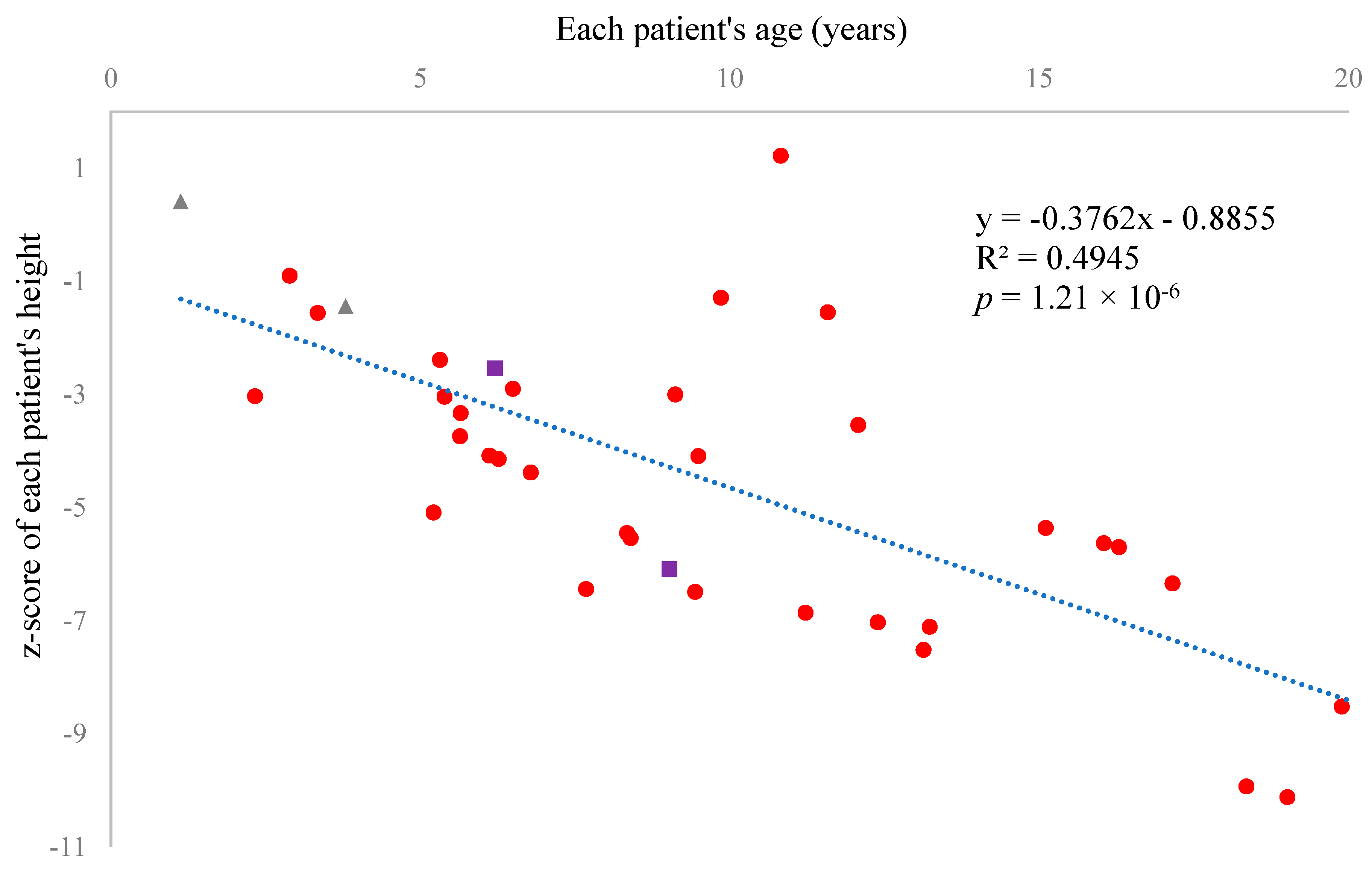
Sixty patients successfully provided us with blood and/or urine specimens. The characteristics of all participants are summarized in Table 1 (also in Table S1, including measured biomarkers). Thirty-three patients (55%) were female, and twenty-seven (45%) were male. Forty-four (73.3%) of the participants were White or Caucasian, five (8.3%) were Black or African American, three (5%) were South Asian, two (3.3%) were Southeast Asian, two (3.3%) were East Asian, and four (6.7%) were other/mixed race. The z-score of each patient’s height showed a significant negative correlation with age, indicating that current therapy is not effective enough to prevent growth impairment in MPS IVA (r = −0.703, p = 1.21 × 10⁻⁶, Figure 1).

2.2. Genotyping

Genetic testing of the GALNS gene revealed 8 homozygotes and 41 compound heterozygotes. In 10 patients, we could detect only one mutation on one allele. One patient could not be genetically tested since we could not obtain her genomic DNA (Table 1 and Table S1). Among the detected mutations, c.448delC (H150Tfs*3), c.633+1G>C, c.946G>A (G316R), and c.1339G>C (D447H) were not listed on the ClinVar website (https://www.ncbi.nlm.nih.gov/clinvar/ (accessed on 17 April 2025)), and we could not find any previously published articles describing these four mutations. The significance of the last two variants was predicted using online bioinformatics tools, MutationTaster (https://www.mutationtaster.org/index.html (accessed on 17 April 2025)) and PolyPhen-2 (http://genetics.bwh.harvard.edu/pph2/ (accessed on 17 April 2025)). Both variants were indicated as “disease-causing” and “probably damaging with a score of 1.000” by MutationTaster and PolyPhen-2, respectively. The remaining two variants were disease-causing mutations since one causes a frameshift that reduces the length of the entire peptide to less than a third of the original protein, and another one disrupts normal splicing by destroying the GT-AG rule [61], the basis of splicing.

2.3. Biomarkers (Collagen Type I, II, NT-ProCNP, and GAGs)—All Raw Data from the Patients Are Listed in Table S1

2.3.1. NT-ProCNP

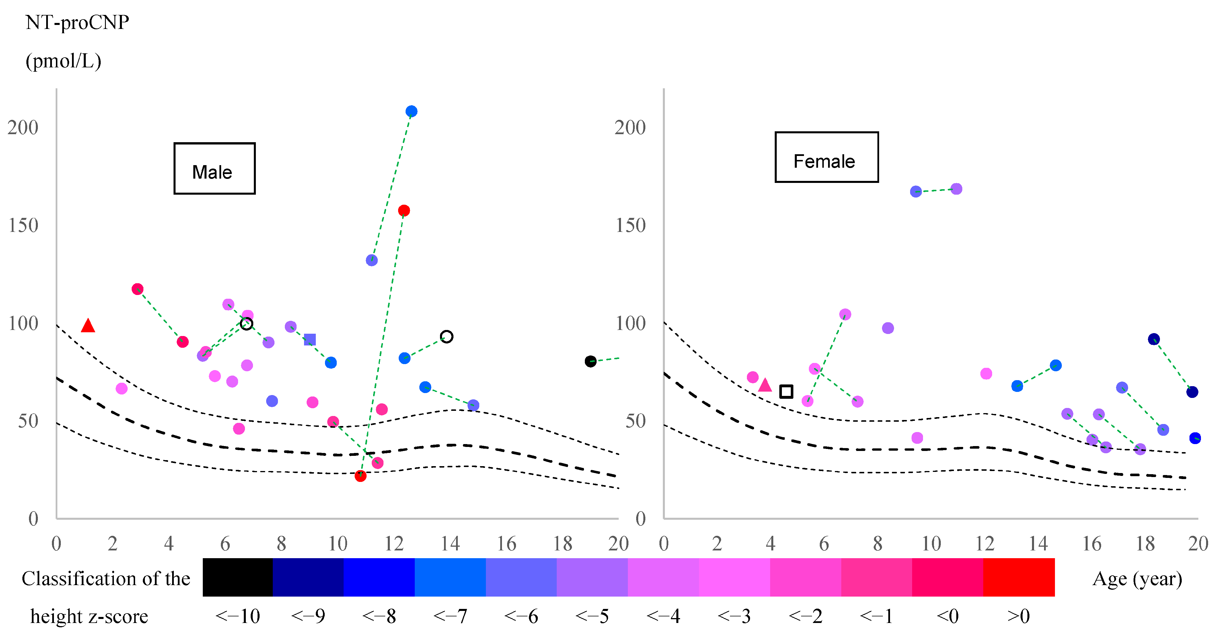
Most pediatric patients, both males and females, had elevated levels above the 95th percentile for healthy controls of both sexes, as established by Olney et al. [45] (Figure 2). The increase was evident just before the age of pubertal growth spurt in healthy subjects. Focusing on patients with two data points at different ages before the age of peak pubertal growth spurt (all of whom had received ERT during these two periods), 5 out of 10 boys and 2 out of 3 girls showed an increase in NT-proCNP levels. Results from adults over 20 years are summarized in Figure S1, showing a significant difference between healthy controls and patients (p = 0.019).
A higher NT-proCNP level correlated with a lower z-score for height, indicating a severe growth impairment, especially between the ages of 8 and 13 years (17 subjects) (Figure 2). Including this age group, the partial correlation coefficient between the NT-proCNP level and the z-score of height was calculated, with age as a controlling factor, revealing a significant negative correlation in patients older than 8 years (Table 2).
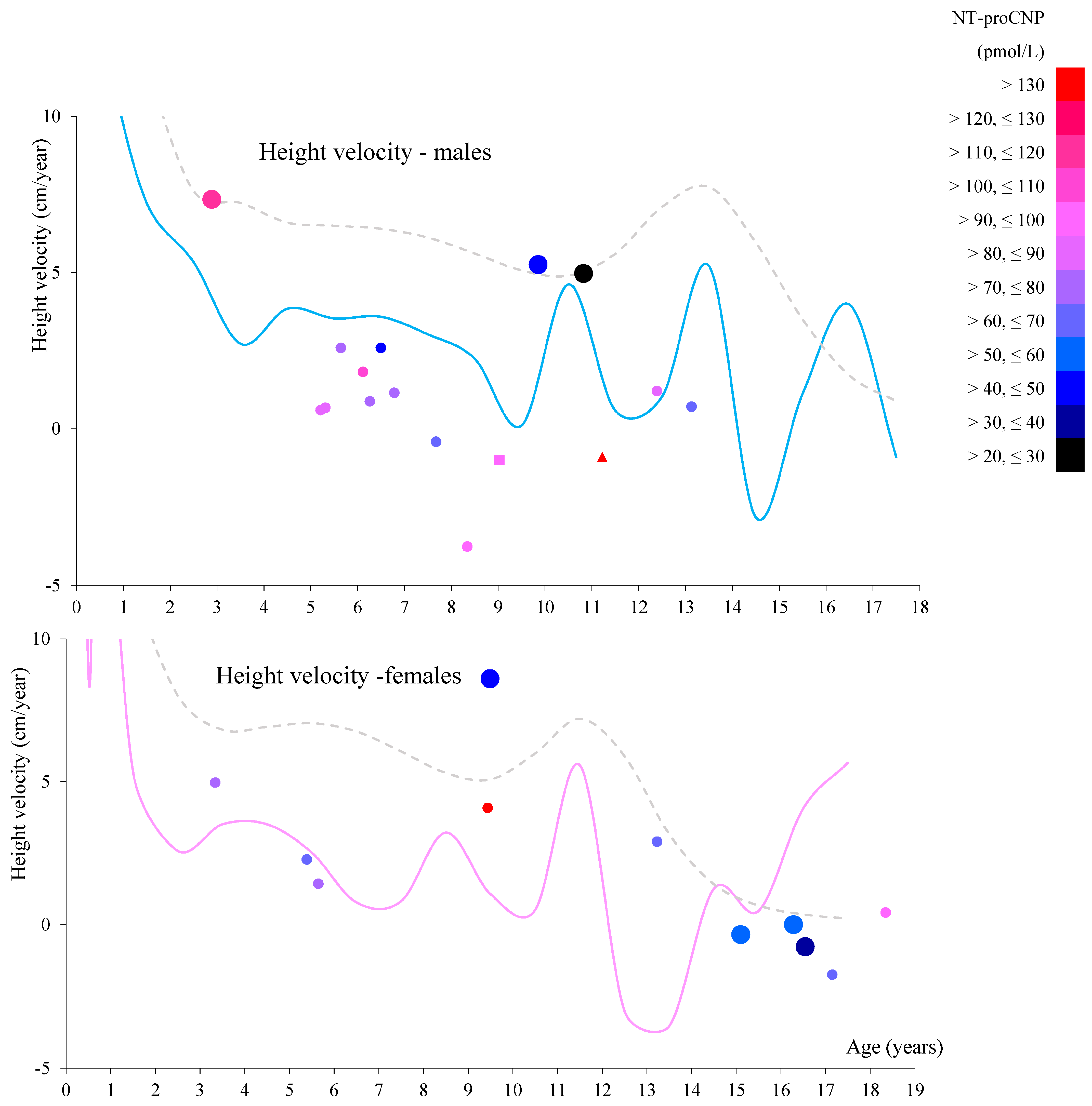
The relationship between each patient’s plasma NT-proCNP level, age, and subsequent height growth rate (i.e., near-future growth rate) is shown in Figure 3. Higher NT-proCNP levels did not lead to a higher growth rate but to a lower one, especially after the age of 8 years. We could not statistically analyze this trend due to the limited data.

2.3.2. Collagen Type I

We found a statistically significant increase in collagen type I in patients older than 20 years (Table 3, Figure S2A). The correlation between serum and plasma was straightforward, as shown in Figure S3, indicating that the comparison between serum from controls and plasma from patients is feasible and reasonable.

2.3.3. Collagen Type II

We found a significant increase in collagen type II in patients in two age groups, between 5 and 10 years and over 20 years (Table 4, Figure S2B). However, the correlation between serum and plasma was unclear, as shown in Figure S4, which complicated the interpretation of the results for the former age group. In addition, we calculated the partial correlation coefficient between collagen type II and NT-proCNP, controlling for age using the entire age group because collagen type II is a marker of chondrocytes [56,57], and CNP is a growth factor for chondrocytes [36]. However, our data showed no correlation (r = 0.042, p = 0.69).

2.3.4. Glycosaminoglycans (GAGs)

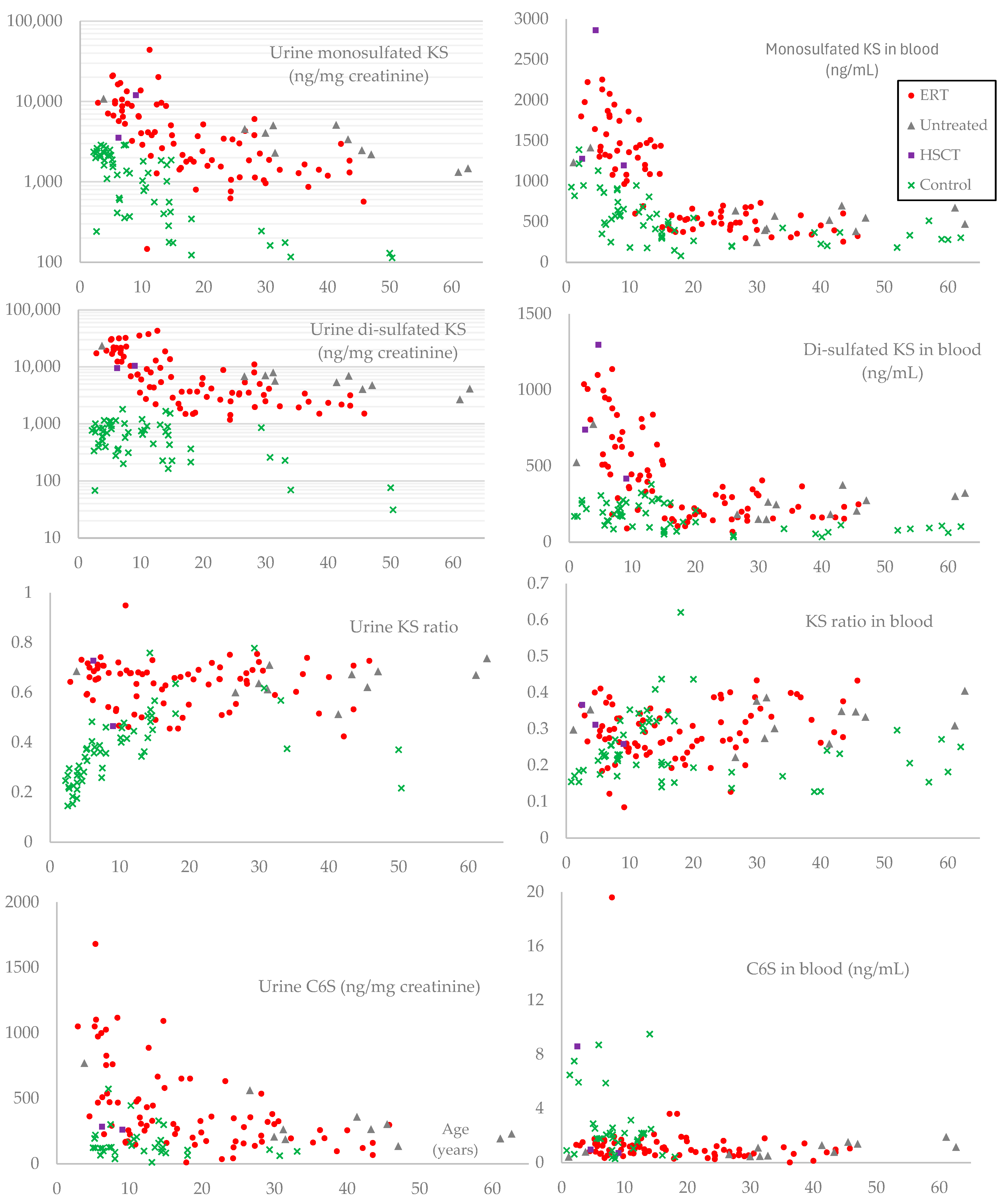
The results of mono-sulfated KS, di-sulfated KS, KS ratio (di-sulfated KS to total KS), and C6S in urine and blood from patients are summarized in Table 5 and Figure 4. Patients had a higher sulfation level in urinary KS than age-matched controls, except for the group aged between 15 and 20 years. Sulfation levels of urinary KS in normal controls increased with age, while the levels in MPS IVA patients remained constant in all age groups. MPS IVA patients had higher sulfation levels in blood KS than age-matched controls up to 10 years. Sulfation levels of blood KS in normal controls increased until 15 years and then declined with age, whereas levels in MPS IVA patients remained relatively constant across all age groups. Overall, sulfation patterns and age-dependent changes differed between urinary KS and blood KS in both control and MPS IVA patient groups, indicating the difference in the origin of urinary and blood KS.
Age-adjusted Pearson partial correlation coefficients (r) or Spearman partial correlation coefficients (ρ) between the z-score of height and each significant GAG (mono- and di-sulfated KS and urinary C6S) were calculated and are summarized in Table 6. Both urinary KS showed a negative correlation with the z-score of height with statistical significance.

2.3.5. Effect of ERT on Each Biomarker in Adult Patients Aged 25–50 Years

Two biomarkers, urinary mono-sulfated KS and di-sulfated KS, significantly decreased in patients treated with ERT, whereas other biomarkers showed no significant differences (Table 7).

2.3.6. Transitions in Skeletal Growth and Biomarkers in Pediatric Patients Younger than 12 Years with More than Two Hospital Visits (Table 8)

All patients who met the criteria described in the Materials and Methods Section received ERT (12 patients) or HSCT (2 patients). Height was categorized into percentiles based on the growth chart constructed from the accumulated data of Morquio A patients without ERT or HSCT [7]. Red-colored cells indicate improvement in the percentile category. Blue-colored cells indicate a worsening of the percentile category. Pink-colored cells indicate an increase of more than 6% in the biomarker value. Light blue-colored cells indicate a decrease of more than 7%. Gray-colored cells indicate almost no change in the biomarker over the two points.
Two of the eleven patients who routinely received ERT worsened in the height percentile category. Three showed improvement, and the remaining six were not affected in their height (Table 8). Five out of nine patients with ERT had decreased NT-proCNP levels in plasma, three had increased levels, and one had the same level. Six out of nine patients with ERT had increased blood mono-sulfated KS levels, and three had decreased levels. Nine out of ten patients with ERT had decreased mono-sulfated KS levels in urine, and one had increased level. Six out of nine patients with ERT had increased blood di-sulfated KS levels, and three had decreased levels. Five out of ten patients with ERT had increased urinary di-sulfated KS levels, and five had decreased levels. No correlation was found in two-point changes (decrease, increase, or no change) between NT-proCNP and urinary KS or blood KS.
Table 8. NT-proCNP and GAG levels in pediatric patients under 12 years of age.
PercentileNT-ProCNPMono-Sulfated KSDi-Sulfated KSKS Ratio
HeightAgeof HeightPlasmaPlasmaUrinePlasmaUrinePlasmaUrine
IDSex(cm)(Year) (pmol/L)(ng/mL)(ng/mg cre)(ng/mL)(ng/mg cre) Treatment
M52F 1.84 HSCT
832.1625th–50thno datano datano datano datano datano datano data
No record2.5 no data1277.0no data739.0no data0.3668no data
91.53.1475th
No record4.6 64.942864.2no data1297.2no data0.3117no data
1046.2175th–90thno datano data3564no data9502no data0.7272
M51M91.52.8950th–75th117.431977.996581005.917,3570.33710.6425ERT
103.34.575th–90th90.391643.971021098.519,2990.40060.7310
M16M85.65.213rd–10th83.291305.020,783509.030,0260.28060.5910ERT
86.26.233rd–10thno datano datano datano datano datano datano data
No record6.77No record99.671818.37679690.517,6110.27520.6964
M15M99.55.3250th–75th85.151378.121,141578.030,9520.29550.5942ERT
100.56.8150th103.812078.68783878.721,6950.29710.7118
M55F96.75.3950th–75th60.131426.96701996.616,9640.41120.7168ERT
99.96.850th–75th104.421310.0no data182.2no data0.1221no data
M50F975.6550th–75th76.662134.810,127949.819,7540.30790.6611ERT
99.37.2650th–75th59.801079.45293627.515,1860.36760.7415
M21M956.1225th–50th109.481328.616,376496.021,6350.27180.5692ERT
97.67.5525th–50th90.061945.913,424834.932,4740.30030.7075
M46F96.66.5150thno datano datano datano datano datano datano dataERT
No record6.92No recordno datano data6476no data12,382no data0.6566
100.68.450th97.351470.73246723.368460.32970.6784
M08M96.47.8625thno datano datano datano datano datano datano dataERT
No record8 no data1377.9no data674.3no data0.3286no data
8.07 HSCT
999.0325th91.531196.012,063418.010,5030.25880.4654
97.610.4210th–25thno datano datano datano datano datano datano data
M22M1008.3425th–50th98.161746.78848626.610,4500.26400.5415ERT
94.69.7810th–25th79.681859.713,776579.135,5140.23750.7205
M19F98.59.4425th–50th167.191081.46616363.073300.25130.5256ERT
104.710.9650th168.591415.34146411.491570.22520.6883
M20F111.19.4975th–90th41.261005.86480353.774160.26020.5337ERT
11610.0675th–90thno datano data2903no data6026no data0.6749
M31M129.59.8690th49.421359.54043445.935380.24700.4667ERT
137.811.4490th–97th28.521760.72116808.544360.31470.6770
Red-colored cells indicate improvement in the percentile category. Blue-colored cells indicate a worsening of the percentile category. Pink-colored cells indicate an increase of more than 6% in the biomarker value. Light blue-colored cells indicate a decrease of more than 7%. Gray-colored cells indicate almost no change in the biomarker over the two points.
Sulfation levels (KS ratio) in the urine of all patients were higher than in the blood. Nine out of ten patients with ERT had increased sulfation levels in urine, and one had the same level. Four out of nine patients with ERT had increased blood sulfation levels, three had decreased levels, and two had the same level.

2.3.7. Correlation Between NT-ProCNP and Other Biomarkers

The correlation between NT-proCNP and other biomarkers was examined in the three pediatric age groups and is summarized in Table S2. Only plasma and urinary C6S showed correlations with statistical significance (p < 0.05) over 13 years of age, suggesting that NT-proCNP can be an independent biomarker from KS.

3. Discussion

In this study, we measured several biomarkers in 60 MPS IVA patients enrolled in the Morquio A natural history program (clinicaltrial.gov: NCT05284006) to evaluate the significance of each biomarker concerning growth failure. Fifty-one patients (85%) received ERT, and two (3.3%) received HSCT. Two (3.3%) pediatric patients had not yet started ERT because they were newly diagnosed. Six (10%) adult patients had never received ERT despite its approval in the United States. Judging from their heights, ERT did not show apparent effects for skeletal dysplasia, including bone growth (Figure 1, Table 8), as indicated by the previous reports [62,63,64,65,66,67]. However, it should be noted that we could not statistically compare with and without ERT in pediatric patients due to the limited number of pediatric patients without ERT, which is a limitation of our research. We identified four novel mutations through genetic testing: one was a deletion resulting in a frameshift, another was a mutation at a splice donor site, and the remaining two were missense mutations predicted to be disease-causing by two bioinformatics tools. In 10 patients, we found only one mutation on one allele. We sequenced only the exons and exon-intron boundaries of the GALNS gene and did not search for large deletions, thereby limiting our genetic testing.
CNP is significantly involved in both vascular homeostasis and bone growth, which is mainly controlled by CNP secreted from vascular endothelial cells [68] and chondrocytes in growth plates [35], respectively. It plays a key role in endochondral ossification, crucial for forming and growing long bones [29]. CNP primarily affects bone growth by signaling through the chondrocyte receptor NPR-B, encoded by the NPR2 gene, which induces cell division and differentiation [38].
Recent research indicated that CNP and NT-proCNP levels are significantly elevated in several skeletal dysplasias (achondroplasia, hypochondroplasia, thanatophoric dysplasia, and Maroteaux type of acromesomelic dysplasia) caused by mutations in the FGFR3 (fibroblast growth factor receptor 3) or NPR2 (natriuretic peptide receptor 2) gene [69]. These genes are closely related to each other and to extracellular CNP, playing a crucial role in cell proliferation and differentiation. Substantial progress has been made in recent years in the study of CNP and their role in skeletal disorders, particularly achondroplasia, which is caused by a gain-of-function mutation in FGFR3 that results in constitutive activation of FGFR3 and the downstream MEK/ERK MAPK pathway [70,71]. The MEK/ERK MAPK pathway is a signaling pathway that regulates various cellular processes, including cell growth, proliferation, and differentiation [72,73,74]. Elevated levels of CNP and NT-proCNP in achondroplasia are thought to be a natural response of the human body to this overactivation of the MEK/ERK MAPK pathway in chondrocytes, which functionally inhibits the effect of CNP on chondrocytes via NPR-B encoded by NPR2 [69,75]. In this paper, we have identified the elevation of NT-proCNP in MPS IVA patients. The gene responsible for MPS IVA is GALNS, which is not closely related to FGFR3 or NPR2. Therefore, this is the first report to identify the elevation of NT-proCNP in patients with a disease unrelated to mutations in the FGFR3 or NPR2 genes.
Recent research has not only identified NT-proCNP as a potential biomarker but also highlighted the therapeutic potential of CNP. The development of drugs such as Vosoritide, a CNP analog approved to induce bone growth in achondroplasia [70,71,76], underscores the potential of CNP as a therapeutic agent against the growth impairment of MPS. This potential has already been demonstrated in MPS IVA and VII mouse models [77,78]. Furthermore, a Phase I/II clinical trial of the CNP analog in MPS IVA and VI patients has been initiated to explore further these therapeutic possibilities (ClinicalTrials.gov ID: NCT05845749) and inform the scientific community about the latest developments in MPS research.
NT-proCNP is a peptide fragment derived from the precursor protein of CNP. The NPPC (natriuretic peptide precursor C) gene expresses a precursor protein known as preproCNP. PreproCNP undergoes proteolytic cleavage to remove the signal peptide and to form proCNP [42]. Furin, one of the proprotein convertases, cleaves NT-proCNP from proCNP in the trans-Golgi network to produce the active CNP [40,41,79,80,81]. After the cleavage of proCNP, active CNP is secreted into the extracellular space via exocytosis [42]. For NT-proCNP, we found no clear mechanism of how it is secreted by cells; however, these processes should occur primarily in neurons, glial cells [30,31,32], vascular endothelial cells [33], and chondrocytes in cartilage [34,35]. The half-life of CNP in the blood is short, about 2–3 min, as NPRs take up CNP, and such a short half-life helps to adapt to frequent changes in circulatory status [41,43,82]. In the case of NT-proCNP, there is no clear pathway to remove it. As a result, the normal concentration ranges of these two molecules in healthy children are significantly different: CNP is 0.5~3 pmol/L, and NT-proCNP is 15~60 pmol/L (74.8~299 pg/mL) [45]. This difference makes NT-proCNP easier to detect and quantify than CNP. Considering that bone growth is a slower and more gradual phenomenon than the adaptation of the circulatory system, NT-proCNP is expected to be a better biomarker than CNP for correlating human skeletal symptoms in MPS. This leads us to select NT-proCNP as a potential surrogate biomarker for future studies.
Our data demonstrated that NT-proCNP was significantly elevated in the plasma of MPS IVA patients in all comparable age groups. In addition, the concentrations in patients negatively correlated with height z-score, the most reliable indicator of skeletal growth failure [83,84]. The source of this elevated NT-proCNP (presumably, CNP as well) remains unknown. The primary sources of these peptides are neurons, glial cells [30,31,32], vascular endothelial cells [33], and chondrocytes in cartilage [34,35], but the exact percentage of production at each site under normal conditions remains uncertain. The CNP production by growth plate chondrocytes is the most critical factor for bone growth, as evidenced by the growth impairment observed in mice where the Nppc gene has been selectively knocked out in chondrocytes [35]. Even when plasma CNP levels were significantly reduced by knocking out the Nppc gene selectively in mouse vascular endothelial cells, Moyes et al. did not observe any growth failure [68].
Based on previous and current results, we propose potential hypotheses (factors) contributing to plasma NT-proCNP elevation in MPS IVA patients.
  • The expression of the NPPC gene is upregulated in MPS IVA patients to compensate for growth impairment or tissue resistance to CNP, as seen in achondroplasia, hypochondroplasia, and thanatophoric dysplasia [69]. A feedback mechanism in patients with growth failure contributes to the higher expression of CNP. This feedback mechanism could involve vascular endothelial cells as a source of excessive CNP and NT-proCNP secretion.
  • CNP and NT-proCNP are leaking from damaged chondrocytes in MPS IVA patients. The secretion or leakage of NT-proCNP from damaged chondrocytes in the growth plates can enter the bloodstream, increasing its concentration in the blood.
  • NT-proCNP clearance is delayed due to a systemic disorder of MPS IVA.
A combination of multiple factors may account for the elevation of NT-proCNP. With age, the contribution factor could vary. Further research is needed to investigate the regulation of CNP expression (at both mRNA and protein levels) and clarify this paradoxical phenomenon.
Focusing on patients who had two data points at different ages prior to peak pubertal growth spurt (all of whom had received ERT during these two periods), NT-proCNP levels increased in 5 of 10 boys and 2 of 3 girls (Figure 2) while urinary mono-sulfated KS was reduced in 9 out of 10 patients, as shown in Table 8. This finding indicates that plasma NT-proCNP is not a pharmacodynamic biomarker like urinary KS prior to the onset of pubertal growth spurt. Therefore, we hypothesize that reduced NT-proCNP can be more directly linked to improving bone growth than reducing total urinary KS. Further studies will confirm whether NT-proCNP is an excellent surrogate biomarker that correlates with clinical improvement in the skeletal system.
Collagen type I is the most abundant collagen in the human body. It is a fibrous protein that forms a significant part of the extracellular matrix in various tissues, including skin, tendons, ligaments, and, importantly, bone [47,48,49]. It provides a scaffold for bone mineralization and forms a fibrous network that serves as the structural framework for bone tissue [50,51,52]. This network supports the deposition of hydroxyapatite crystals, primarily composed of calcium and phosphate [85,86]. The interaction between collagen fibers and these crystals gives bones strength and rigidity. Procollagen Type I N-Terminal Propeptide (P1NP) and C-Terminal Telopeptide of Type I Collagen (CTX-I) are two major peptides well studied in several diseases related to collagen type I metabolism. P1NP reflects collagen synthesis [87], while CTX-I reflects collagen degradation [88]. Elevated levels of these peptides in human blood or urine are associated with diseases or conditions related to increased bone turnover (such as Paget’s disease [89,90], osteoporosis [91], or hyperparathyroidism [92,93]), fibrotic processes (such as liver fibrosis and cirrhosis [94,95]), or cancer (bone metastases [96,97] or multiple myeloma [98]).
Several papers reported that the expression level of mRNA encoding collagen type I increased in chondrocytes from human MPS IVA patients [53,54]. Comprehensive proteomic analysis of 6-week-old mouse femurs also showed that collagen alpha-1 and 2 (Uniprot IDs: P11087 and Q01149) increased in MPS IVA [99]. Therefore, we predicted that patients would present higher concentration levels than the control group. However, only adult patients showed such differences. Other age groups showed lower concentrations in patients. Since human collagen type I has a molecular weight of approximately 300 kDa, much larger than NT-proCNP, surrounded by hydroxyapatite in bone [85,86,100], it could be difficult for collagen type I to escape from bone and cartilage and enter the bloodstream compared to NT-proCNP, thereby preventing higher concentrations in the blood of patients. We should have measured smaller peptides related to collagen type I, as in other diseases described above.
Collagen type II is a specific type of collagen found predominantly in cartilage [55] and is a marker for chondrocytes [56,57]. It is also present in the vitreous humor of the eye and in the nucleus pulposus of intervertebral discs [101]. Like other collagens, type II collagen is a fibrous protein that forms a triple-helical structure, providing tensile strength and structural integrity to tissues [59]. Its primary role in forming and maintaining cartilage makes it crucial for joint health, enabling smooth and pain-free movement [102,103]. Additionally, its presence on the intervertebral discs and the vitreous body of the eye underscores its importance in maintaining flexibility and structural support in these tissues. Blood procollagen II C-terminal propeptide (CPII) and/or urinary C-terminal telopeptide of type II collagen (CTX-II) are currently the two main biomarkers of type II collagen metabolism in osteoarthritis [104,105,106], joint injuries [107], and Kashin–Beck disease (KBD) [108]. CPII reflects the synthesis of type II collagen during cartilage repair and remodeling [109,110,111], while CTX-II is a marker of type II collagen breakdown, reflecting cartilage catabolism [106,112].
De Franceschi et al. and Dvorak-Ewell et al. reported decreased expression of mRNA encoding collagen type II in chondrocytes from human MPS IVA patients, in contrast to collagen type I [53,54]. The same phenomenon was also observed in mouse models of MPS type I and type VII [113,114]. These findings suggest that the decrease in collagen type II may be a potential biomarker for MPS types I, IVA, and VII. In contrast, proteomic analysis of 6-week-old mice revealed a statistically significant increase in collagen alpha-1(II) chain (Uniprot ID: P28481) in the MPS IVA model [99]. This discrepancy in collagen type II expression levels between humans and mice warrants further investigation. Our results showed a statistically significant increase in patients aged 5 to 10 years and over 20 years. This seems to support the results of the latter proteomic analysis [99]. However, due to the poor correlation between the measured concentration in serum and plasma, as shown in Figure S4, it is too early to draw conclusions for patients aged 5 to 10 years.
We also calculated the correlation coefficient between collagen type II and NT-proCNP, controlling for age, to evaluate the effect of excessive CNP expression on chondrocytes in patients. NT-proCNP concentration would indicate the total expression level of CNP in a subject’s body. Collagen type II is a marker of chondrocytes [56,57]. Therefore, elevated NT-proCNP levels could correlate positively with collagen type II levels. However, we found no correlation (r = 0.042, p = 0.69), suggesting that the increased CNP secretion in patients is insufficient to compensate for the damage to chondrocytes in growth plates.
GAG levels are well-established biomarkers for MPS, and the difference between plasma and serum is negligible, making our comparison between controls and patients reasonable [115]. Our results were consistent with the previous reports [22,116]. However, to our knowledge, we could not find any prior report that quantitatively correlates GAG levels with the severity of skeletal symptoms. Therefore, we correlated each GAG with the z-score of height, one of the quantitative indicators of growth failure. Since each GAG level except for plasma C6S tended to decrease with age up to 20 years (Figure 4), we calculated partial correlation coefficients controlling for age, then found a moderate negative correlation in urinary mono- and di-sulfated KS (Table 6). These two biomarkers also showed statistical differences between adult patients with and without ERT (Table 7), making urinary KS a more important prognostic biomarker than C6S in MPS IVA.
We established the KS assay by LC-MS/MS, measuring both mono- and di-sulfated KS in age-matched controls and MPS IVA patients [13,14,18,115]. Our previous studies identified differences in age and sulfation between urine and blood KS in MPS IVA and healthy controls, suggesting the importance of measuring both types of KS in MPS IVA [8,14]. Urinary KS is considered a pharmacodynamic biomarker that does not accurately reflect the therapeutic effect on skeletal symptoms in patients receiving ERT [26]. Hendriksz et al. presumably measured both KSs (mono-sulfated and di-sulfated) and calculated the total KS [117]. In this study, we observed the transition of these two types of KS during ERT or after HSCT in the most critical age group for treatment of skeletal growth, namely, those under 10 years old. Interestingly, a decrease in urinary di-sulfated KS was observed in 5 out of 10 patients, whereas 9 out of 10 patients showed a decrease in mono-sulfated KS (Table 8). Based on this result, we assume that urinary di-sulfated KS may not be a pharmacodynamic biomarker like mono-sulfated KS. Furthermore, blood di-sulfated KS showed more pronounced differences from controls in most age groups (Figure 4). Further investigations are needed to clarify the significance of di-sulfated KS.

4. Materials and Methods

Subjects: MPS IVA patients receiving care at Nemours Children’s Health in Delaware and participating in the natural history program (Non-invasive Functional Assessment and Pathogenesis of Morquio A, clinicaltrial.gov: NCT05284006, NIH funding number: 1R01HD102545-01A1) were enrolled. The diagnosis of MPS IVA was confirmed by deficient enzyme activity of <5% of normal activity measured in plasma, leukocytes, or fibroblasts before enrollment in this clinical trial. Before any study procedures, written informed consent was obtained from all participants. A total of 60 participants formalized their consent for the research study. The z-score of each patient’s height during sample collection was calculated based on the CDC Growth Chart [84]. It was also categorized into percentiles based on the growth chart constructed from the accumulated data of Morquio A patients without ERT or HSCT (hematopoietic stem cell transplantation) [7]. If a patient had two or more height measurements taken at different times, the height growth rate (annual height velocity) was calculated using two data points.
Genotyping: Genomic DNA was extracted from the white blood cells and/or skin fibroblasts. The 14 exons of the GALNS gene were amplified by polymerase chain reaction (PCR) and sequenced using the Sanger method to identify pathogenic mutations. PCR conditions and primer pairs used are summarized in Table S3. Mutations were annotated based on the reference sequence NM_000512.5.
Control Specimens: Serum or urine samples from individuals without skeletal dysplasia at Shimane University Hospital (Japan) were provided as control samples for this research. The age ranged from 0 days to 18 years for serum and from 5 to 33 years for urine. Additionally, blood samples were collected from 7 adults without skeletal dysplasia working in our laboratory, and both plasma and serum were tested to confirm the correlation between serum and plasma for collagen type I and II levels. Written informed consent was obtained from all individuals (IRB# 750932).
ELISA-based method for the measurement of collagen type I, II, and NT-proCNP: The Human Collagen Type I (COL1) ELISA Kit (Cat# EKU03297-96T, BIOMATIK, Kitchener, Canada), the Human Collagen Type II, Col II ELISA Kit (Cat# EKC40379, BIOMATIK), and the NT-proCNP ELISA kit (Cat# BI-20812, BIOMEDICA, Vienna, Austria) were utilized to measure the concentration of each biomarker in patient plasma and serum, control serum from children, and control plasma and serum from adults according to the manufacturer’s instructions. Each kit used a quantitative sandwich enzyme immunoassay technique. The absorbance of each well was measured using FLUOstar Omega (BMG LABTECH, Ortenberg, Germany).
Glycosaminoglycans (GAGs) Analysis by LC-MS/MS: Mono-sulfated KS, di-sulfated KS, and C6S in the plasma and urine samples from patients were measured according to our established method [22,118]. Briefly, 10 μL of each serum, plasma, urine sample, or standard was mixed with 90 μL of 50 mM Tris-hydrochloric acid buffer (pH 7.0) in wells of AcroPrep™Advance 96 Well Filter Plates equipped with ultrafiltration Omega10K membrane filters (PALL Corporation, Port Washington, NY, USA). A cocktail of 30 μL of recombinant chondroitinase ABC and keratanase II (each enzyme at 1 mU/sample) and internal standard solution (5 μg/mL) was added to each well, followed by the addition of 70 μL of 50 mM Tris-hydrochloric acid buffer. After overnight incubation at 37 °C, the filter plate was centrifuged for 20 min at 2500× g. The filtered samples were injected into our 1290 Infinity liquid chromatography system with a 6460 triple quad mass spectrometer (Agilent Technologies, Palo Alto, CA, USA). Levels of GAGs in urine samples were normalized to creatinine, which was measured with a Creatinine (urinary) Colorimetric Assay Kit (Cayman Chemical, Ann Arbor, MI, USA). The serum and urine of control subjects were also measured in the same way.
Effect of ERT on Each Biomarker: We compared the difference in each biomarker between patients treated with ERT and patients who had not been treated with ERT. Since most patients in the latter group were between 25 and 50 years old, we used only this age range.
Transitions in skeletal growth and biomarkers in pediatric patients younger than 12 years with more than two hospital visits: To explore a correlation between significant biomarkers, skeletal development, and treatment (ERT or HSCT) in growing pediatric patients, we summarized the data from patients who provided their first specimen before the age of 10 years in Table 8.
Statistical Analysis: For urinary mono- and di-sulfated KS, the Mann–Whitney U test was used to determine statistical differences between subjects and controls. For other biomarkers, Welch’s t-test was performed. Both tests were calculated with GraphPad Prism version 10.2.3. R-4.4.3 for Windows was used to calculate correlation coefficients or partial correlation coefficients, controlling for age, to reveal the relationship between each factor [119,120]. To evaluate correlations involving urinary mono- or di-sulfated KS, the Spearman correlation coefficient was calculated. For correlations not involving these two biomarkers, the Pearson correlation coefficient was used.
5. Conclusions

In MPS IVA patients, an elevated blood NT-proCNP level is a promising biomarker closely associated with growth failure. Collagens I and II are less promising than other biomarkers, such as urinary and blood KS. In addition, KS should be evaluated in more detail in MPS, as di-sulfated KS may not be a simple pharmacodynamic biomarker in patients receiving ERT.

Supplementary Materials

The following supporting information can be downloaded at: https://www.mdpi.com/article/10.3390/ijms26104940/s1.

Author Contributions

Conceptualization, S.T., methodology, S.T., S.K. and Y.A., software, Y.A., validation, Y.A. and S.K., formal analysis, Y.A. and S.T., investigation, Y.A., S.K., K.K. and A.B.; resources, K.K., A.B. and S.T.; data curation, Y.A., K.K. and A.B.; writing—original draft preparation, Y.A. and S.K.; writing—review and editing, Y.A. and S.T.; visualization, Y.A.; supervision, S.T.; project administration, S.T.; funding acquisition, S.T. All authors have read and agreed to the published version of the manuscript.

Funding

This research was funded by the National Institutes of Health (NIH), project number 1R01HD102545-01A1.

Institutional Review Board Statement

The study was conducted in accordance with the Declaration of Helsinki and approved by the Institutional Review Board of NEMOURS CHILDREN’S HOSPITAL, DELAWARE (750932, approved on 10 February 2021).

Informed Consent Statement

Informed consent was obtained from all subjects involved in the study.

Data Availability Statement

All available data are included in this article.

Acknowledgments

We appreciate all the participants who cooperated in providing their specimens, and all the staff who helped collect the participants’ specimens at Nemours Children’s Hospital in Delaware.

Conflicts of Interest

The authors declare no conflicts of interest.

Abbreviations

The following abbreviations are used in this manuscript:
MPSmucopolysaccharidosis
GAGglycosaminoglycan
CNPC-type natriuretic peptide
NT-proCNPN-terminal pro-C-type natriuretic peptide
KSkeratan sulfate
C6Schondroitin-6-sulfate
GALNSN-acetylgalactosamine-6-sulfate sulfatase
ERTenzyme replacement therapy
NPR-Bnatriuretic peptide receptor B
PKGprotein kinase G
HSCThematopoietic stem cell transplantation
FGFR3fibroblast growth factor receptor 3
NPR2natriuretic peptide receptor 2
NPPCnatriuretic peptide precursor C
P1NPProcollagen Type I N-Terminal Propeptide
CTX-IC-Terminal Telopeptide of Type I Collagen
CPIIprocollagen II C-terminal propeptide
CTX-IIC-terminal telopeptide of type II collagen
KBDKashin–Beck disease
PCRpolymerase chain reaction
NIHNational Institutes of Health

References

  1. Stapleton, M.; Arunkumar, N.; Kubaski, F.; Mason, R.W.; Tadao, O.; Tomatsu, S. Clinical Presentation and Diagnosis of Mucopolysaccharidoses. Mol. Genet. Metab. 2018, 125, 4–17. [Google Scholar] [CrossRef] [PubMed]
  2. Filocamo, M.; Tomanin, R.; Bertola, F.; Morrone, A. Biochemical and Molecular Analysis in Mucopolysaccharidoses: What a Paediatrician Must Know. Ital. J. Pediatr. 2018, 44 (Suppl. S2), 129. [Google Scholar] [CrossRef] [PubMed]
  3. Sawamoto, K.; González, J.V.Á.; Piechnik, M.; Otero, F.J.; Couce, M.L.; Suzuki, Y.; Tomatsu, S. Mucopolysaccharidosis IVA: Diagnosis, Treatment, and Management. Int. J. Mol. Sci. 2020, 21, 1517. [Google Scholar] [CrossRef]
  4. Puckett, Y.; Mallorga-Hernández, A.; Montaño, A.M. Epidemiology of Mucopolysaccharidoses (MPS) in United States: Challenges and Opportunities. Orphanet J. Rare Dis. 2021, 16, 241. [Google Scholar] [CrossRef] [PubMed]
  5. Canda, E.; Yazıcı, H.; Er, E.; Eraslan, C.; Ucar, S.K.; Coker, M. Clinical Presentation and Follow Up of Patients with Mucopolysaccharidosis IVA (Morquio A Disease): Single Center Experience. J. Pediatr. Res. 2018, 5, 28–33. [Google Scholar] [CrossRef]
  6. Jiang, Z.; Byers, S.; Casal, M.L.; Smith, L.J. Failures of Endochondral Ossification in the Mucopolysaccharidoses. Curr. Osteoporosis. Rep. 2020, 18, 759–773. [Google Scholar] [CrossRef]
  7. Montaño, A.M.; Tomatsu, S.; Brusius, A.; Smith, M.; Orii, T. Growth Charts for Patients Affected with Morquio A Disease. Am. J. Med. Genet. A 2008, 146, 1286–1295. [Google Scholar] [CrossRef] [PubMed]
  8. Khan, S.; Alméciga-Díaz, C.J.; Sawamoto, K.; Mackenzie, W.G.; Theroux, M.C.; Pizarro, C.; Mason, R.W.; Orii, T.; Tomatsu, S. Mucopolysaccharidosis IVA and Glycosaminoglycans. Mol. Genet. Metab. 2017, 120, 78–95. [Google Scholar] [CrossRef]
  9. Anderson, C.E.; Crane, J.T.; Harper, H.A.; Hunter, T.W. Morquio’s Disease and Dysplasia Epiphysalis Multiplex. A Study of Epiphyseal Cartilage in Seven Cases. J. Bone Jt. Surg. Am. Vol. 1962, 44, 295–306. [Google Scholar] [CrossRef]
  10. McClure, J.; Smith, P.S.; SorbyAdams, G.; Hopwood, J. The Histological and Ultrastructural Features of the Epiphyseal Plate in Morquio Type a Syndrome Mucopolysaccharidosis Type IVA. Pathology 1986, 18, 217–221. [Google Scholar] [CrossRef]
  11. Lavery, C.; Hendriksz, C. Mortality in Patients with Morquio Syndrome a. JIMD Rep. 2014, 15, 59–66. [Google Scholar] [CrossRef] [PubMed]
  12. Erickson, R.P.; Sandman, R.; Epstein, C.J.; Robertson, W.v.B. Lack of Relationship between Blood and Urine Levels of Glycosaminoglycans and Lysosomal Enzymes. Biochem. Med. 1975, 12, 331–339. [Google Scholar] [CrossRef]
  13. Hintze, J.P.; Tomatsu, S.; Fujii, T.; Montano, A.M.; Yamaguchi, S.; Suzuki, Y.; Fukushi, M.; Ishimaru, T.; Orii, T. Comparison of Liquid Chromatography-Tandem Mass Spectrometry and Sandwich ELISA for Determination of Keratan Sulfate in Plasma and Urine. Biomark. Insights 2011, 6, 69–78. [Google Scholar] [CrossRef]
  14. Shimada, T.; Tomatsu, S.; Mason, R.W.; Yasuda, E.; Mackenzie, W.G.; Hossain, J.; Shibata, Y.; Montano, A.M.; Kubaski, F.; Giugliani, R.; et al. Di-Sulfated Keratan Sulfate as a Novel Biomarker for Mucopolysaccharidosis II, IVA, and IVB. JIMD Rep. 2015, 21, 1–13. [Google Scholar] [CrossRef]
  15. Saville, J.T.; McDermott, B.K.; Fuller, M. Glycosaminoglycan Fragments as a Measure of Disease Burden in the Mucopolysaccharidosis Type I Mouse. Mol. Genet. Metab. 2018, 123, 112–117. [Google Scholar] [CrossRef] [PubMed]
  16. Thonar, E.J.-M.A.; Pachman, L.M.; Lenz, M.E.; Hayford, J.; Lynch, P.; Kuettner, K.E. Age Related Changes in the Concentration of Serum Keratan Sulphate in Children. J. Clin. Chem. Clin. Biochem. 1988, 26, 57–64. [Google Scholar] [CrossRef] [PubMed]
  17. Tomatsu, S.; Dieter, T.; Schwartz, I.V.; Sarmient, P.; Giugliani, R.; Barrera, L.A.; Guelbert, N.; Kremer, R.; Repetto, G.M.; Gutierrez, M.A.; et al. Identification of a Common Mutation in Mucopolysaccharidosis IVA: Correlation among Genotype, Phenotype, and Keratan Sulfate. J. Hum. Genet. 2004, 49, 490–494. [Google Scholar] [CrossRef]
  18. Tomatsu, S.; Montaño, A.M.; Oguma, T.; Dung, V.C.; Oikawa, H.; Carvalho, T.G.; de Gutiérrez, M.L.; Yamaguchi, S.; Suzuki, Y.; Fukushi, M.; et al. Validation of Keratan Sulfate Level in Mucopolysaccharidosis Type IVA by Liquid Chromatography–Tandem Mass Spectrometry. J. Inherit. Metab. Dis. 2010, 33 (Suppl. S3), 35–42. [Google Scholar] [CrossRef]
  19. Tomatsu, S.; Okamura, K.; Maeda, H.; Taketani, T.; Castrillon, S.V.; Gutierrez, M.A.; Nishioka, T.; Fachel, A.A.; Orii, K.O.; Grubb, J.H.; et al. Keratan Sulphate Levels in Mucopolysaccharidoses and Mucolipidoses. J. Inherit. Metab. Dis. 2005, 28, 187–202. [Google Scholar] [CrossRef]
  20. Tomatsu, S.; Yasuda, E.; Patel, P.; Ruhnke, K.; Shimada, T.; Mackenzie, W.G.; Mason, R.; Thacker, M.M.; Theroux, M.; Montaño, A.M.; et al. Morquio A Syndrome: Diagnosis and Current and Future Therapies. Pediatr. Endocrinol. Rev. Per 2014, 12 (Suppl. S1), 141–151. [Google Scholar]
  21. Peracha, H.; Sawamoto, K.; Averill, L.; Kecskemethy, H.; Theroux, M.; Thacker, M.; Nagao, K.; Pizarro, C.; Mackenzie, W.; Kobayashi, H.; et al. Molecular Genetics and Metabolism, Special Edition: Diagnosis, Diagnosis and Prognosis of Mucopolysaccharidosis IVA. Mol. Genet. Metab. 2018, 125, 18–37. [Google Scholar] [CrossRef] [PubMed]
  22. Khan, S.A.; Mason, R.W.; Giugliani, R.; Orii, K.; Fukao, T.; Suzuki, Y.; Yamaguchi, S.; Kobayashi, H.; Orii, T.; Tomatsu, S. Glycosaminoglycans Analysis in Blood and Urine of Patients with Mucopolysaccharidosis. Mol. Genet. Metab. 2018, 125, 44–52. [Google Scholar] [CrossRef]
  23. Kubaski, F.; Suzuki, Y.; Orii, K.; Giugliani, R.; Church, H.J.; Mason, R.W.; Dung, V.C.; Ngoc, C.T.; Yamaguchi, S.; Kobayashi, H.; et al. Glycosaminoglycan Levels in Dried Blood Spots of Patients with Mucopolysaccharidoses and Mucolipidoses. Mol. Genet. Metab. 2017, 120, 247–254. [Google Scholar] [CrossRef] [PubMed]
  24. Harmatz, P.R.; Mengel, E.; Geberhiwot, T.; Muschol, N.; Hendriksz, C.J.; Burton, B.K.; Jameson, E.; Berger, K.I.; Jester, A.; Treadwell, M.; et al. Impact of Elosulfase Alfa in Patients with Morquio A Syndrome Who Have Limited Ambulation: An Open-label, Phase 2 Study. Am. J. Med. Genet. Part. A 2017, 173, 375–383. [Google Scholar] [CrossRef]
  25. Hendriksz, C.J.; Parini, R.; AlSayed, M.D.; Raiman, J.; Giugliani, R.; Villarreal, M.L.S.; Mitchell, J.J.; Burton, B.K.; Guelbert, N.; Stewart, F.; et al. Long-Term Endurance and Safety of Elosulfase Alfa Enzyme Replacement Therapy in Patients with Morquio A Syndrome. Mol. Genet. Metab. 2016, 119, 131–143. [Google Scholar] [CrossRef] [PubMed]
  26. Hendriksz, C.J.; Burton, B.; Fleming, T.R.; Harmatz, P.; Hughes, D.; Jones, S.A.; Lin, S.; Mengel, E.; Scarpa, M.; Valayannopoulos, V.; et al. Efficacy and Safety of Enzyme Replacement Therapy with BMN 110 (Elosulfase Alfa) for Morquio A Syndrome (Mucopolysaccharidosis IVA): A Phase 3 Randomised Placebo-controlled Study. J. Inherit. Metab. Dis. 2014, 37, 979–990. [Google Scholar] [CrossRef]
  27. Moyes, A.J.; Chu, S.M.; Aubdool, A.A.; Dukinfield, M.S.; Margulies, K.B.; Bedi, K.C.; Hodivala-Dilke, K.; Baliga, R.S.; Hobbs, A.J. C-Type Natriuretic Peptide Co-Ordinates Cardiac Structure and Function. Eur. Heart J. 2020, 41, 1006–1020. [Google Scholar] [CrossRef]
  28. Perez-Ternero, C.; Pallier, P.N.; Tremoleda, J.L.; Delogu, A.; Fernandes, C.; Michael-Titus, A.T.; Hobbs, A.J. C-Type Natriuretic Peptide Preserves Central Neurological Function by Maintaining Blood-Brain Barrier Integrity. Front. Mol. Neurosci. 2022, 15, 991112. [Google Scholar] [CrossRef] [PubMed]
  29. Rintz, E.; Węgrzyn, G.; Fujii, T.; Tomatsu, S. Molecular Mechanism of Induction of Bone Growth by the C-Type Natriuretic Peptide. Int. J. Mol. Sci. 2022, 23, 5916. [Google Scholar] [CrossRef]
  30. Sudoh, T.; Kangawa, K.; Minamino, N.; Matsuo, H. A New Natriuretic Peptide in Porcine Brain. Nature 1988, 332, 78–81. [Google Scholar] [CrossRef]
  31. Hodes, A.; Lichtstein, D. Natriuretic Hormones in Brain Function. Front. Endocrinol. 2014, 5, 201. [Google Scholar] [CrossRef]
  32. Korostyshevskaya, I.M.; Maksimov, V.F.; Rudenko, N.S. C-Type Natriuretic Peptide: What, Where and Why? Neurosci. Behav. Physiol. 2016, 46, 888–894. [Google Scholar] [CrossRef]
  33. Suga, S.; Itoh, H.; Komatsu, Y.; Ogawa, Y.; Hama, N.; Yoshimasa, T.; Nakao, K. Cytokine-Induced C-Type Natriuretic Peptide (CNP) Secretion from Vascular Endothelial Cells—Evidence for CNP as a Novel Autocrine/Paracrine Regulator from Endothelial Cells. Endocrinology 1993, 133, 3038–3041. [Google Scholar] [CrossRef] [PubMed]
  34. Galetaki, D.M.; Dauber, A. C-Type Natriuretic Peptide Analogs: Current and Future Therapeutic Applications. Horm. Res. Paediatr. 2024, 98, 51–58. [Google Scholar] [CrossRef] [PubMed]
  35. Nakao, K.; Osawa, K.; Yasoda, A.; Yamanaka, S.; Fujii, T.; Kondo, E.; Koyama, N.; Kanamoto, N.; Miura, M.; Kuwahara, K.; et al. The Local CNP/GC-B System in Growth Plate Is Responsible for Physiological Endochondral Bone Growth. Sci. Rep. 2015, 5, 10554. [Google Scholar] [CrossRef]
  36. Potter, L.R.; Yoder, A.R.; Flora, D.R.; Antos, L.K.; Dickey, D.M. Natriuretic Peptides: Their Structures, Receptors, Physiologic Functions and Therapeutic Applications. In cGMP: Generators, Effectors and Therapeutic Implications; Springer: Berlin/Heidelberg, Germany, 2009; Volume 191, pp. 341–366. [Google Scholar] [CrossRef]
  37. Chikuda, H.; Kugimiya, F.; Hoshi, K.; Ikeda, T.; Ogasawara, T.; Shimoaka, T.; Kawano, H.; Kamekura, S.; Tsuchida, A.; Yokoi, N.; et al. Cyclic GMP-Dependent Protein Kinase II Is a Molecular Switch from Proliferation to Hypertrophic Differentiation of Chondrocytes. Genes Dev. 2004, 18, 2418–2429. [Google Scholar] [CrossRef]
  38. Miyazawa, T.; Ogawa, Y.; Chusho, H.; Yasoda, A.; Tamura, N.; Komatsu, Y.; Pfeifer, A.; Hofmann, F.; Nakao, K. Cyclic GMP-Dependent Protein Kinase II Plays a Critical Role in C-Type Natriuretic Peptide-Mediated Endochondral Ossification. Endocrinology 2002, 143, 3604–3610. [Google Scholar] [CrossRef]
  39. Yasoda, A.; Komatsu, Y.; Chusho, H.; Miyazawa, T.; Ozasa, A.; Miura, M.; Kurihara, T.; Rogi, T.; Tanaka, S.; Suda, M.; et al. Overexpression of CNP in Chondrocytes Rescues Achondroplasia through a MAPK-Dependent Pathway. Nat. Med. 2004, 10, 80–86. [Google Scholar] [CrossRef]
  40. Kone, B.C. Molecular Biology of Natriuretic Peptides and Nitric Oxide Synthases. Cardiovasc. Res. 2001, 51, 429–441. [Google Scholar] [CrossRef]
  41. Mark, P.D.; Goetze, J.P. Encyclopedia of Molecular Pharmacology; Springer International Publishing: Cham, Switzerland, 2022; pp. 499–504. [Google Scholar] [CrossRef]
  42. Wu, C.; Wu, F.; Pan, J.; Morser, J.; Wu, Q. Furin-Mediated Processing of Pro-C-Type Natriuretic Peptide. J. Biol. Chem. 2003, 278, 25847–25852. [Google Scholar] [CrossRef]
  43. Nakagawa, Y.; Nishikimi, T. CNP, the Third Natriuretic Peptide: Its Biology and Significance to the Cardiovascular System. Biology 2022, 11, 986. [Google Scholar] [CrossRef] [PubMed]
  44. Keng, B.M.H.; Gao, F.; Tan, R.S.; Ewe, S.H.; Teo, L.L.Y.; Xie, B.Q.; Goh, G.B.B.; Koh, W.-P.; Koh, A.S. N-Terminal pro C-Type Natriuretic Peptide (NTproCNP) and Myocardial Function in Ageing. PLoS ONE 2018, 13, e0209517. [Google Scholar] [CrossRef]
  45. Olney, R.C.; Permuy, J.W.; Prickett, T.C.R.; Han, J.C.; Espiner, E.A. Amino-terminal Propeptide of C-type Natriuretic Peptide (NTproCNP) Predicts Height Velocity in Healthy Children. Clin. Endocrinol. 2012, 77, 416–422. [Google Scholar] [CrossRef] [PubMed]
  46. Prickett, T.C.R.; Olney, R.C.; Cameron, V.A.; Ellis, M.J.; Richards, A.M.; Espiner, E.A. Impact of Age, Phenotype and Cardio-renal Function on Plasma C-type and B-type Natriuretic Peptide Forms in an Adult Population. Clin. Endocrinol. 2013, 78, 783–789. [Google Scholar] [CrossRef]
  47. Boraschi-Diaz, I.; Wang, J.; Mort, J.S.; Komarova, S.V. Collagen Type I as a Ligand for Receptor-Mediated Signaling. Front. Phys. 2017, 5, 12. [Google Scholar] [CrossRef]
  48. Amirrah, I.N.; Lokanathan, Y.; Zulkiflee, I.; Wee, M.F.M.R.; Motta, A.; Fauzi, M.B. A Comprehensive Review on Collagen Type I Development of Biomaterials for Tissue Engineering: From Biosynthesis to Bioscaffold. Biomedicines 2022, 10, 2307. [Google Scholar] [CrossRef]
  49. Balasubramanian, P.; Prabhakaran, M.P.; Sireesha, M.; Ramakrishna, S. Polymer Composites—Polyolefin Fractionation—Polymeric Peptidomimetics—Collagens. Adv. Polym. Sci. 2012, 251, 173–206. [Google Scholar] [CrossRef]
  50. Stock, S.R. The Mineral–Collagen Interface in Bone. Calcif. Tissue Int. 2015, 97, 262–280. [Google Scholar] [CrossRef]
  51. Vijayalekha, A.; Anandasadagopan, S.K.; Pandurangan, A.K. An Overview of Collagen-Based Composite Scaffold for Bone Tissue Engineering. Appl. Biochem. Biotechnol. 2023, 195, 4617–4636. [Google Scholar] [CrossRef]
  52. Nijsure, M.P.; Kishore, V. Orthopedic Biomaterials, Advances and Applications; Springer: Cham, Switzerland, 2018; pp. 187–224. [Google Scholar] [CrossRef]
  53. Franceschi, L.D.; Roseti, L.; Desando, G.; Facchini, A.; Grigolo, B. A Molecular and Histological Characterization of Cartilage from Patients with Morquio Syndrome. Osteoarthr. Cartil. 2007, 15, 1311–1317. [Google Scholar] [CrossRef]
  54. Dvorak-Ewell, M.; Wendt, D.; Hague, C.; Christianson, T.; Koppaka, V.; Crippen, D.; Kakkis, E.; Vellard, M. Enzyme Replacement in a Human Model of Mucopolysaccharidosis IVA In Vitro and Its Biodistribution in the Cartilage of Wild Type Mice. PLoS ONE 2010, 5, e12194. [Google Scholar] [CrossRef] [PubMed]
  55. Hansen, U. The Collagen Superfamily and Collagenopathies. Biol. Extracell. Matrix 2021, 8, 121–141. [Google Scholar] [CrossRef]
  56. Lefrebvre, V.; de Crombrugghe, B. Toward Understanding S0X9 Function in Chondrocyte Differentiation. Matrix Biol. 1998, 16, 529–540. [Google Scholar] [CrossRef]
  57. Yoon, H.J.; Kim, S.B.; Somaiya, D.; Noh, M.J.; Choi, K.-B.; Lim, C.-L.; Lee, H.-Y.; Lee, Y.-J.; Yi, Y.; Lee, K.H. Type II Collagen and Glycosaminoglycan Expression Induction in Primary Human Chondrocyte by TGF-Β1. BMC Musculoskelet. Disord. 2015, 16, 141. [Google Scholar] [CrossRef]
  58. Kozhemyakina, E.; Lassar, A.B.; Zelzer, E. A Pathway to Bone: Signaling Molecules and Transcription Factors Involved in Chondrocyte Development and Maturation. Development 2015, 142, 817–831. [Google Scholar] [CrossRef] [PubMed]
  59. Engel, J.; Bächinger, H.P. Collagen, Primer in Structure, Processing and Assembly. Top. Curr. Chem. 2005, 247, 7–33. [Google Scholar] [CrossRef]
  60. Bačenková, D.; Trebuňová, M.; Demeterová, J.; Živčák, J. Human Chondrocytes, Metabolism of Articular Cartilage, and Strategies for Application to Tissue Engineering. Int. J. Mol. Sci. 2023, 24, 17096. [Google Scholar] [CrossRef]
  61. Breathnach, R.; Benoist, C.; O’Hare, K.; Gannon, F.; Chambon, P. Ovalbumin Gene: Evidence for a Leader Sequence in MRNA and DNA Sequences at the Exon-Intron Boundaries. Proc. Natl. Acad. Sci. USA 1978, 75, 4853–4857. [Google Scholar] [CrossRef]
  62. Doherty, C.; Stapleton, M.; Piechnik, M.; Mason, R.W.; Mackenzie, W.G.; Yamaguchi, S.; Kobayashi, H.; Suzuki, Y.; Tomatsu, S. Effect of Enzyme Replacement Therapy on the Growth of Patients with Morquio A. J. Hum. Genet. 2019, 64, 625–635. [Google Scholar] [CrossRef]
  63. Cao, J.D.; Wiedemann, A.; Quinaux, T.; Battaglia-Hsu, S.F.; Mainard, L.; Froissart, R.; Bonnemains, C.; Ragot, S.; Leheup, B.; Journeau, P.; et al. 30 Months Follow-up of an Early Enzyme Replacement Therapy in a Severe Morquio A Patient: About One Case. Mol. Genet. Metab. Rep. 2016, 9, 42–45. [Google Scholar] [CrossRef]
  64. Nakamura-Utsunomiya, A.; Nakamae, T.; Kagawa, R.; Karakawa, S.; Sakata, S.; Sakura, F.; Tani, C.; Matsubara, Y.; Ishino, T.; Tajima, G.; et al. A Case Report of a Japanese Boy with Morquio A Syndrome: Effects of Enzyme Replacement Therapy Initiated at the Age of 24 Months. Int. J. Mol. Sci. 2020, 21, 989. [Google Scholar] [CrossRef] [PubMed]
  65. Frigeni, M.; Rodriguez-Buritica, D.F.; Saavedra, H.; Gunther, K.A.; Hillman, P.R.; Balaguru, D.; Northrup, H. The Youngest Pair of Siblings with Mucopolysaccharidosis Type IVA to Receive Enzyme Replacement Therapy to Date: A Case Report. Am. J. Med. Genet. Part A 2021, 185, 3510–3516. [Google Scholar] [CrossRef]
  66. Barak, S.; Anikster, Y.; Sarouk, I.; Stern, E.; Eisenstein, E.; Yissar, T.; Sherr-Lurie, N.; Raas-Rothschild, A.; Guttman, D. Long-Term Outcomes of Early Enzyme Replacement Therapy for Mucopolysaccharidosis IV: Clinical Case Studies of Two Siblings. Diagnostics 2020, 10, 108. [Google Scholar] [CrossRef] [PubMed]
  67. Kılavuz, S.; Basaran, S.; Kor, D.; Bulut, F.D.; Erdem, S.; Ballı, H.T.; Dağkıran, M.; Bisgin, A.; Mungan, H.N.Ö. Morquio A Syndrome and Effect of Enzyme Replacement Therapy in Different Age Groups of Turkish Patients: A Case Series. Orphanet J. Rare Dis. 2021, 16, 144. [Google Scholar] [CrossRef]
  68. Moyes, A.J.; Khambata, R.S.; Villar, I.; Bubb, K.J.; Baliga, R.S.; Lumsden, N.G.; Xiao, F.; Gane, P.J.; Rebstock, A.-S.; Worthington, R.J.; et al. Endothelial C-Type Natriuretic Peptide Maintains Vascular Homeostasis. J. Clin. Investig. 2014, 124, 4039–4051. [Google Scholar] [CrossRef]
  69. Olney, R.C.; Prickett, T.C.R.; Espiner, E.A.; Mackenzie, W.G.; Duker, A.L.; Ditro, C.; Zabel, B.; Hasegawa, T.; Kitoh, H.; Aylsworth, A.S.; et al. C-Type Natriuretic Peptide Plasma Levels Are Elevated in Subjects with Achondroplasia, Hypochondroplasia, and Thanatophoric Dysplasia. J. Clin. Endocrinol. Metab. 2015, 100, E355–E359. [Google Scholar] [CrossRef] [PubMed]
  70. Espiner, E.; Prickett, T.; Olney, R. Plasma C-Type Natriuretic Peptide: Emerging Applications in Disorders of Skeletal Growth. Horm. Res. Paediat. 2019, 90, 345–357. [Google Scholar] [CrossRef]
  71. Galetaki, D.M.; Merchant, N.; Dauber, A. Novel Therapies for Growth Disorders. Eur. J. Pediatr. 2024, 183, 1121–1128. [Google Scholar] [CrossRef]
  72. Meloche, S.; Pouysségur, J. The ERK1/2 Mitogen-Activated Protein Kinase Pathway as a Master Regulator of the G1- to S-Phase Transition. Oncogene 2007, 26, 3227–3239. [Google Scholar] [CrossRef]
  73. Chambard, J.-C.; Lefloch, R.; Pouysségur, J.; Lenormand, P. ERK Implication in Cell Cycle Regulation. Biochim. Biophys. Acta (BBA)-Mol. Cell Res. 2007, 1773, 1299–1310. [Google Scholar] [CrossRef]
  74. Albeck, J.G.; Mills, G.B.; Brugge, J.S. Frequency-Modulated Pulses of ERK Activity Transmit Quantitative Proliferation Signals. Mol. Cell 2013, 49, 249–261. [Google Scholar] [CrossRef]
  75. Klag, K.A.; Horton, W.A. Advances in Treatment of Achondroplasia and Osteoarthritis. Hum. Mol. Genet. 2016, 25, R2–R8. [Google Scholar] [CrossRef]
  76. Duggan, S. Vosoritide: First Approval. Drugs 2021, 81, 2057–2062. [Google Scholar] [CrossRef] [PubMed]
  77. Rintz, E.; Herreño-Pachón, A.M.; Celik, B.; Nidhi, F.; Khan, S.; Benincore-Flórez, E.; Tomatsu, S. Bone Growth Induction in Mucopolysaccharidosis IVA Mouse. Int. J. Mol. Sci. 2023, 24, 9890. [Google Scholar] [CrossRef] [PubMed]
  78. Rintz, E.; Celik, B.; Fnu, N.; Herreño-Pachón, A.M.; Khan, S.; Benincore-Flórez, E.; Tomatsu, S. Molecular Therapy and Nucleic Acid Adeno-Associated Virus-Based Gene Therapy Delivering Combinations of Two Growth-Associated Genes to MPS IVA Mice. Mol. Ther.-Nucleic Acids 2024, 35, 102211. [Google Scholar] [CrossRef] [PubMed]
  79. Takahashi, S.; Nakagawa, T.; Banno, T.; Watanabe, T.; Murakami, K.; Nakayama, K. Localization of Furin to the Trans-Golgi Network and Recycling from the Cell Surface Involves Ser and Tyr Residues within the Cytoplasmic Domain. J. Biol. Chem. 1995, 270, 28397–28401. [Google Scholar] [CrossRef] [PubMed]
  80. Bosshart, H.; Humphrey, J.; Deignan, E.; Davidson, J.; Drazba, J.; Yuan, L.; Oorschot, V.; Peters, P.J.; Bonifacino, J.S. The Cytoplasmic Domain Mediates Localization of Furin to the Trans-Golgi Network En Route to the Endosomal/Lysosomal System. J. Cell Biol. 1994, 126, 1157–1172. [Google Scholar] [CrossRef]
  81. Teuchert, M.; Schäfer, W.; Berghöfer, S.; Hoflack, B.; Klenk, H.-D.; Garten, W. Sorting of Furin at the Trans-Golgi Network. Interaction of the cytoplasmic tail sorting signals with AP-1 Golgi-specific assembly proteins. J. Biol. Chem. 1999, 274, 8199–8207. [Google Scholar] [CrossRef]
  82. Pandey, K.N. Molecular Signaling Mechanisms and Function of Natriuretic Peptide Receptor-A in the Pathophysiology of Cardiovascular Homeostasis. Front. Physiol. 2021, 12, 693099. [Google Scholar] [CrossRef]
  83. WHO Multicentre Growth Reference Study Group. WHO Child Growth Standards Based on Length/Height, Weight and Age. Acta Pædiatrica 2006, 95, 76–85. [Google Scholar] [CrossRef]
  84. Kuczmarski, R.J.; Ogden, C.L.; Guo, S.S.; Grummer-Strawn, L.M.; Flegal, K.M.; Mei, Z.; Wei, R.; Curtin, L.R.; Roche, A.F.; Johnson, C.L. 2000 CDC Growth Charts for the United States: Methods and Development. Vital Health. Stat. 2002, 11, 1–190. [Google Scholar]
  85. Sorokina, L.V.; Shahbazian-Yassar, R.; Shokuhfar, T. Collagen Biomineralization: Pathways, Mechanisms, and Thermodynamics. Emergent Mater. 2021, 4, 1205–1224. [Google Scholar] [CrossRef]
  86. Bakkalci, D.; Micalet, A.; Hosni, R.A.; Moeendarbary, E.; Cheema, U. Associated Changes in Stiffness of Collagen Scaffolds during Osteoblast Mineralisation and Bone Formation. BMC Res. Notes 2022, 15, 310. [Google Scholar] [CrossRef]
  87. Koivula, M.-K.; Risteli, L.; Risteli, J. Measurement of Aminoterminal Propeptide of Type I Procollagen (PINP) in Serum. Clin. Biochem. 2012, 45, 920–927. [Google Scholar] [CrossRef] [PubMed]
  88. Chubb, S.A.P. Measurement of C-Terminal Telopeptide of Type I Collagen (CTX) in Serum. Clin. Biochem. 2012, 45, 928–935. [Google Scholar] [CrossRef] [PubMed]
  89. Singer, F.R.; Bone, H.G.; Hosking, D.J.; Lyles, K.W.; Murad, M.H.; Reid, I.R.; Siris, E.S.; Society, E. Paget’s Disease of Bone: An Endocrine Society Clinical Practice Guideline. J. Clin. Endocrinol. Metab. 2014, 99, 4408–4422. [Google Scholar] [CrossRef]
  90. Shankar, S.; Hosking, D.J. Biochemical Assessment of Paget’s Disease of Bone. J. Bone Miner. Res. 2009, 21, P22–P27. [Google Scholar] [CrossRef] [PubMed]
  91. Kuo, T.-R.; Chen, C.-H. Biomarker for the Clinical Assessment of Osteoporosis: Recent Developments and Future Perspectives. Biomark. Res. 2017, 5, 18. [Google Scholar] [CrossRef]
  92. Rajeev, P.; Movseysan, A.; Baharani, A. Changes in Bone Turnover Markers in Primary Hyperparathyroidism and Response to Surgery. Ann. R. Coll. Surg. Engl. 2017, 99, 559–562. [Google Scholar] [CrossRef]
  93. Iwanowska, M.; Kochman, M.; Szatko, A.; Zgliczyński, W.; Glinicki, P. Bone Disease in Primary Hyperparathyroidism—Changes Occurring in Bone Metabolism and New Potential Treatment Strategies. Int. J. Mol. Sci. 2024, 25, 11639. [Google Scholar] [CrossRef]
  94. Veidal, S.S.; Vassiliadis, E.; Bay-Jensen, A.-C.; Tougas, G.; Vainer, B.; Karsdal, M.A. Procollagen Type I N-Terminal Propeptide (PINP) Is a Marker for Fibrogenesis in Bile Duct Ligation-Induced Fibrosis in Rats. Fibrogenesis Tissue Repair 2010, 3, 5. [Google Scholar] [CrossRef]
  95. Kwon, Y.; Kim, E.S.; Choe, Y.H.; Kim, M.J. Stratification by Non-Invasive Biomarkers of Non-Alcoholic Fatty Liver Disease in Children. Front. Pediatr. 2022, 10, 846273. [Google Scholar] [CrossRef]
  96. Fohr, B.; Dunstan, C.R.; Seibel, M.J. Markers of Bone Remodeling in Metastatic Bone Disease. J. Clin. Endocrinol. Metab. 2003, 88, 5059–5075. [Google Scholar] [CrossRef]
  97. Leeming, D.J.; Koizumi, M.; Byrjalsen, I.; Li, B.; Qvist, P.; Tankó, L.B. The Relative Use of Eight Collagenous and Noncollagenous Markers for Diagnosis of Skeletal Metastases in Breast, Prostate, or Lung Cancer Patients. Cancer Epidemiol. Prev. Biomark. 2006, 15, 32–38. [Google Scholar] [CrossRef]
  98. Terpos, E. Biochemical Markers of Bone Metabolism in Multiple Myeloma. Cancer Treat. Rev. 2006, 32, 15–19. [Google Scholar] [CrossRef]
  99. Álvarez, J.V.; Bravo, S.B.; Chantada-Vázquez, M.P.; Pena, C.; Colón, C.; Tomatsu, S.; Otero-Espinar, F.J.; Couce, M.L. Morquio A Syndrome: Identification of Differential Patterns of Molecular Pathway Interactions in Bone Lesions. Int. J. Mol. Sci. 2024, 25, 3232. [Google Scholar] [CrossRef]
  100. Garnero, P. The Role of Collagen Organization on the Properties of Bone. Calcif. Tissue Int. 2015, 97, 229–240. [Google Scholar] [CrossRef]
  101. Safadi, F.F.; Khurana, J.S. Diagnostic Imaging of Musculoskeletal Diseases, A Systematic Approach; Humana: Louisville, KY, USA, 2009; pp. 1–13. [Google Scholar] [CrossRef]
  102. Goldring, M.B.; Marcu, K.B. Cartilage Homeostasis in Health and Rheumatic Diseases. Arthritis Res. Ther. 2009, 11, 224. [Google Scholar] [CrossRef]
  103. Alcaide-Ruggiero, L.; Molina-Hernández, V.; Granados, M.M.; Domínguez, J.M. Main and Minor Types of Collagens in the Articular Cartilage: The Role of Collagens in Repair Tissue Evaluation in Chondral Defects. Int. J. Mol. Sci. 2021, 22, 13329. [Google Scholar] [CrossRef]
  104. Conrozier, T.; Ferrand, F.; Poole, A.R.; Verret, C.; Mathieu, P.; Ionescu, M.; Vincent, F.; Piperno, M.; Spiegel, A.; Vignon, E. Differences in Biomarkers of Type II Collagen in Atrophic and Hypertrophic Osteoarthritis of the Hip: Implications for the Differing Pathobiologies. Osteoarthr. Cartil. 2007, 15, 462–467. [Google Scholar] [CrossRef]
  105. Arunrukthavon, P.; Heebthamai, D.; Benchasiriluck, P.; Chaluay, S.; Chotanaphuti, T.; Khuangsirikul, S. Can Urinary CTX-II Be a Biomarker for Knee Osteoarthritis? Arthroplasty 2020, 2, 6. [Google Scholar] [CrossRef]
  106. Cheng, H.; Hao, B.; Sun, J.; Yin, M. C-Terminal Cross-Linked Telopeptides of Type II Collagen as Biomarker for Radiological Knee Osteoarthritis: A Meta-Analysis. Cartilage 2020, 11, 512–520. [Google Scholar] [CrossRef]
  107. Nicolini, A.; Mansur, N.; Dreyfuss, J.; Ejnisman, B.; Cohen, M.; Astur, D. Avaliação Do Biomarcador CTX-II Em Pacientes Com Ruptura Do Ligamento Cruzado Anterior: Estudo Piloto. Rev. Bras. Ortop. 2020, 56, 326–332. [Google Scholar] [CrossRef]
  108. Song, Q.Q.; Sun, L.Y.; Li, C.H.; Liu, Y.J.; Cui, S.L.; Liu, Y.Q.; Cao, Y.H.; Pei, J.R.; Wang, Y.; Lian, W.; et al. The Urinary Levels of CTX-II, C2C, PYD, and Helix-II Increased among Adults with KBD: A Cross-Sectional Study. J. Orthop. Surg. Res. 2019, 14, 328. [Google Scholar] [CrossRef]
  109. Cahue, S.; Sharma, L.; Dunlop, D.; Ionescu, M.; Song, J.; Lobanok, T.; King, L.; Poole, A.R. The Ratio of Type II Collagen Breakdown to Synthesis and Its Relationship with the Progression of Knee Osteoarthritis. Osteoarthr. Cartil. 2007, 15, 819–823. [Google Scholar] [CrossRef]
  110. Conrozier, T.; Poole, A.R.; Ferrand, F.; Mathieu, P.; Vincent, F.; Piperno, M.; Verret, C.; Ionescu, M.; Vignon, E. Serum Concentrations of Type II Collagen Biomarkers (C2C, C1, 2C and CPII) Suggest Different Pathophysiologies in Patients with Hip Osteoarthritis. Clin. Exp. Rheumatol. 2008, 26, 430–435. [Google Scholar]
  111. Mullan, R.H.; Matthews, C.; Bresnihan, B.; FitzGerald, O.; King, L.; Poole, A.R.; Fearon, U.; Veale, D.J. Early Changes in Serum Type Ii Collagen Biomarkers Predict Radiographic Progression at One Year in Inflammatory Arthritis Patients after Biologic Therapy. Arthritis Rheum. 2007, 56, 2919–2928. [Google Scholar] [CrossRef]
  112. Bay-Jensen, A.-C.; Tabassi, N.C.; Sondergaard, L.V.; Andersen, T.L.; Dagnaes-Hansen, F.; Garnero, P.; Kassem, M.; Delaissé, J.-M. The Response to Oestrogen Deprivation of the Cartilage Collagen Degradation Marker, CTX-II, Is Unique Compared with Other Markers of Collagen Turnover. Arthritis Res. Ther. 2009, 11, R9. [Google Scholar] [CrossRef]
  113. Heppner, J.M.; Zaucke, F.; Clarke, L.A. Extracellular Matrix Disruption Is an Early Event in the Pathogenesis of Skeletal Disease in Mucopolysaccharidosis I. Mol. Genet. Metab. 2015, 114, 146–155. [Google Scholar] [CrossRef]
  114. Bartolomeo, R.; Cinque, L.; Leonibus, C.D.; Forrester, A.; Salzano, A.C.; Monfregola, J.; Gennaro, E.D.; Nusco, E.; Azario, I.; Lanzara, C.; et al. MTORC1 Hyperactivation Arrests Bone Growth in Lysosomal Storage Disorders by Suppressing Autophagy. J. Clin. Investig. 2017, 127, 3717–3729. [Google Scholar] [CrossRef]
  115. Oguma, T.; Tomatsu, S.; Okazaki, O. Analytical Method for Determination of Disaccharides Derived from Keratan Sulfates in Human Serum and Plasma by High-performance Liquid Chromatography/Turbo-ionspray Ionization Tandem Mass Spectrometry. Biomed. Chromatogr. 2007, 21, 356–362. [Google Scholar] [CrossRef]
  116. Lin, H.; Lee, C.; Lo, Y.; Wang, T.; Huang, S.; Chen, T.; Wang, Y.; Niu, D.; Chuang, C.; Lin, S. The Relationships between Urinary Glycosaminoglycan Levels and Phenotypes of Mucopolysaccharidoses. Mol. Genet. Genom. Med. 2018, 6, 982–992. [Google Scholar] [CrossRef]
  117. Martell, L.A.; Cunico, R.L.; Ohh, J.; Fulkerson, W.; Furneaux, R.; Foehr, E.D. Validation of an LCMS/MS Assay for Detecting Relevant Disaccharides from Keratan Sulfate as a Biomarker for Morquio A Syndrome. Bioanalysis 2011, 3, 1855–1866. [Google Scholar] [CrossRef]
  118. Khan, S.A.; Nidhi, F.N.U.; Amendum, P.C.; Tomatsu, S. Proteoglycans, Methods and Protocols. Methods Mol. Biol. 2023, 2619, 3–24. [Google Scholar] [CrossRef]
  119. Wang, J. Encyclopedia of Systems Biology; Springer: New York, NY, USA, 2013; pp. 1634–1635. [Google Scholar] [CrossRef]
  120. Kim, S. Ppcor: An R Package for a Fast Calculation to Semi-Partial Correlation Coefficients. Commun. Stat. Appl. Methods 2015, 22, 665–674. [Google Scholar] [CrossRef]
Figure 1. The Z-score of each patient’s height under 20 years showed a significant negative correlation with age. Each dot represents the z-score of height when each patient’s height was first recorded during the study period. Red round dots represent the patients who had received enzyme replacement therapy (ERT). Gray triangular dots represent the patients who had never received ERT. Purple square dots represent patients who had received hematopoietic stem cell transplantation (HSCT).
Ijms 26 04940 g001
Figure 2. Correlation between age, NT-proCNP, and height of patients with MPS IVA. Males and females are shown in the left and right figures, respectively. Round dots represent patients treated with enzyme replacement therapy (ERT). Triangle markers represent patients who were never treated with ERT. Square markers represent patients treated with hematopoietic stem cell transplantation (HSCT). Filled markers represent patients for whom height at blood sample collection was recorded. Absence of a color indicates that the corresponding height dataset is missing. The color of each marker represents the z-score of each patient’s height calculated based on the CDC growth chart (https://www.cdc.gov/growthcharts/index.htm (accessed on 15 December 2024)). Black dashed lines show median, 5th and 95th percentiles for healthy controls [45]. When a patient has two data points, each data point is connected by a green dashed line.
Ijms 26 04940 g002
Figure 3. The relationship between each patient’s plasma NT-proCNP level, age, and subsequent height growth rate (height velocity). Height velocity was calculated from two height datasets measured at different times, 7 to 20 months apart. Each dot represents the age of each patient at which the first height was measured (horizontal axis) and the corresponding later height velocity (vertical axis). Round dots represent patients continuously receiving enzyme replacement therapy (ERT) until the second height data were obtained. The square dot represents a patient who had received hematopoietic stem cell transplantation (HSCT) (M08). The triangular dot indicates a patient who never received ERT until his second height data were obtained (M35). Larger round dots represent the attenuated phenotype, while smaller round dots represent the severe phenotype. Each dot is filled with a color corresponding to the plasma NT-proCNP level at the first height measurement. The light blue line shows the 50th percentile for height velocity for male MPS IVA patients, and the pink line shows the 50th percentile for female MPS IVA patients [62]. The gray dotted line shows the 50th percentile for height velocity for healthy males and females.
Ijms 26 04940 g003
Figure 4. Glycosaminoglycan levels in urine and blood. Mono-sulfated KS, di-sulfated KS, KS ratio (di-sulfated KS to total KS), and C6S levels in urine and blood are summarized in scatter plots. The horizontal axes indicate the age (years) of each patient or control. For urinary mono-sulfated and di-sulfated KS levels, the vertical axes are shown on a base-10 logarithmic scale. Round red dots indicate patients who had ever received ERT. Purple squares indicate patients who received HSCT. Gray triangles represent patients who never received ERT or HSCT. Green x marks represent controls. For blood samples, plasma samples were used for patients and serum for controls.
Ijms 26 04940 g004
Table 1. Characteristics of all participants.
Patient HeightAgeGenotype
IDSexRace(Z-Score)(Year)Allele 1Allele 2Treatment
M01FWhite or Caucasian−6.3417.16c.740G>A (G247D)c.901G>T (G301C)ERT
−6.7318.70
M02FChinese 38.57c.953T>G (M318R)c.1567T>G (X523EextX93)ERT
40.00
M03FWhite or Caucasian 24.62c.448delC (H150Tfs*3)c.651_652insG (K218Efs*45)ERT
M04FWhite or Caucasian 42.12c.675dupC (F226Lfs*37)unknownERT
43.51
M05FWhite or Caucasian−9.9318.35c.346G>A (G116S)c.1156C>T (R386C)ERT
−9.7619.78
M06FWhite or Caucasian−8.5219.89c.346G>A (G116S)c.1156C>T (R386C)ERT
21.29
M07MWhite or Caucasian 32.25c.337A>T (I113F)c.1171A>G (M391V)No ERT for 2 years and 8 months
M08MWhite or CaucasianNo record8.00c.122T>A (M41K)c.122T>A (M41K)ERT
−6.099.03 Received HSCT 1 year ago
M10MWhite or Caucasian1.2210.82c.338T>C (I113T)c.1219A>C (N407H)ERT
1.0112.37
M11FWhite or Caucasian−5.3615.11c.421T>A (W141R)unknownERT
−5.5316.55
M12FWhite or Caucasian 26.65c.697G>A (D233N)c.1034T>C (L345P)No ERT for 4 years and 4 months
28.15
M13MBlack or African American−7.5213.13c.251C>A (A84E)c.319G>A (A107T)ERT
−6.7514.84
M14FWhite or Caucasian−5.716.29c.740G>A (G247D)unknownERT
−5.717.83
M15MWhite or Caucasian−2.395.32c.498delC (F167Lfs*32)c.901G>T (G301C)ERT
−3.846.81
M16MFilipino−5.095.21c.228C>A (N76K)c.1480A>G (p.M494V)ERT
No record6.77
M19FBlack or African American−6.499.44c.245C>T (S82L)unknownERT
−5.6410.96
M20FWhite or Caucasian−4.099.49c.498delC (F167Lfs*32)c.1474G>A (A492T)ERT
−3.5310.06
M21MWhite or Caucasian−4.086.12c.651_652insG (K218Efs*45)c.1159G>A (G387S)ERT
−5.227.55
M22MWhite or Caucasian−5.458.34c.651_652insG (K218Efs*45)c.1159G>A (G387S)ERT
−7.249.78
M25MWhite or Caucasian 41.34c.155C>T (P52L)c.337A>T (I113F)Never received ERT
43.28
M26MFilipino−7.0312.39c.93delC (N32Tfs*97)c.946G>A (G316R)ERT
No record13.89
M28FMixed 31.20c.868G>A (G290S)c.868G>A (G290S)Never received ERT
Japanese and Caucasian 32.75
M30FWhite or Caucasian 43.52c.121-12T>Cc.121-12T>CERT
M31MBlack or African American−1.299.86c.935C>G (T312S)c.1520G>T (C507F)ERT
−1.1111.44
M32FWhite or Caucasian 45.50c.121-12T>Cc.121-12T>CNever received ERT
47.03
M33FWhite or Caucasian 29.94c.1012C>T (Q338X)c.1171A>G (M391V)Never received ERT
31.48
M34MWhite or Caucasian 28.17c.121A>T (M41L)c.121A>T (M41L)ERT
29.69
M35MWhite or Caucasian−6.8611.23c.139G>A (G47R)c.1156C>T (R386C)No ERT for 4 years
−7.6912.64
M36FAsian Indian 25.72c.346G>A (G116S)c.346G>A (G116S)ERT
27.27
M37MAsian Indian−10.1219.01c.346G>A (G116S)c.346G>A (G116S)ERT
20.56
M38FWhite or Caucasian−5.6316.04c.1559G>A (W520X)unknownERT
M39FWhite or Caucasian−3.5412.08c.1559G>A (W520X)unknownERT
M41MWhite or Caucasian 35.29c.121A>T (M41L)c.498delC (F167Lfs*32)ERT
36.86
M42FWhite or Caucasian 28.24c.860C>T (S287L)c.1055T>C (L358P)ERT
29.95
M43MWhite or Caucasian 29.04c.121A>T (M41L)c.901G>T (G301C)ERT
30.52
M44FWhite or Caucasian 24.28c.1171A>G (p.M391V)c.502G>A (p.G168R)ERT
25.85
M45FWhite or Caucasian 61.08c.740G>A (G247D)c.761A>G (Y254C)Never received ERT
62.66
M46FWhite or CaucasianNo record6.92c.167C>A (T56N)c.502G>A (G168R)ERT
−5.548.40
M47FSome other race 24.33c.139G>A (G47R)c.466T>C (F156L)ERT
25.78
M48FWhite or Caucasian−7.1113.23c.139G>A (G47R)c.466T>C (F156L)ERT
−7.6614.68
M49MWhite or Caucasian−6.447.68c.1156C>T (R386C)c.1156C>T (R386C)ERT
M50FWhite or Caucasian−3.335.65c.451C>A (P151T)c.477G>A (W159X)ERT
−4.817.26
M51MWhite or Caucasian−0.92.89c.451C>A (P151T)c.477G>A (W159X)ERT
−0.524.50
M52FMultiple RacesNo record2.50c.651_652insG (K218Efs*45)unknownReceived HSCT 11 months ago
No record4.60
−2.546.21
M53FChinese 23.20c.1482+5G>Cc.1498G>T (G500C)No ERT for 13 months
24.37 ERT
M54FWhite or Caucasian 36.26not testednot testedNo ERT for 5 years and 9 months
M55FWhite or Caucasian−3.045.39c.1339G>C (D447H)unknownERT
−4.26.80
M56FWhite or Caucasian−1.443.79c.740G>A (G247D)c.1451C>A (P484H)Never received ERT
M57MWhite or Caucasian0.411.13c.740G>A (G247D)c.1451C>A (P484H)Never received ERT
M58MWhite or Caucasian−4.386.78c.1012C>T (Q338X)c.181C>G (R61G)ERT
M59MWhite or Caucasian−4.146.26c.337A>T (p.I113F)c.901G>T (p.G301C)ERT
M60FSome other race 26.58c.901G>T (G301C)c.1156C>T (R386C)Never received ERT
M63FWhite or Caucasian 22.72c.331C>T (Q111X)c.1365-2A>GERT
M64MBlack or African American−3.745.64c.633+1G>Cc.1558T>C (W520R)ERT
M65MBlack or African American−2.96.50c.633+1G>Cc.1558T>C (W520R)ERT
M66FWhite or Caucasian−1.563.34c.1156C>T (R386C)unknownERT
M67MWhite or Caucasian−1.5511.58c.1171A>G (M391V)c.331C>T (Q111X)ERT
M68MWhite or Caucasian 45.78c.1156C>T (R386C)c.181C>T (R61W)ERT
M69MAsian−3.032.33c.106_111del (p.L36_L37del)c.1201C>T (H401Y)ERT
M71MWhite or Caucasian−39.12c.1219A>C (N407H)unknownERT
Table 2. The partial correlation coefficient between NT-proCNP and the z-score of height, controlling for age, across three age groups.
Age GroupNumber of SubjectsPartial Correlation Coefficientp-Value
≤8 y200.1900.4355
>8, ≤13 y17−0.5680.0217
>13, ≤20 y15−0.8371.865 × 10−4
Table 3. Comparison of collagen type I levels in five age groups.
AgeNo.MeanSDMaximumMinimumMean Agep-ValueSpecimen
Control
≤1 y12803.402803.42803.40.25 Serum
>1, ≤5 y42715.52400.56397.5329.62.51 Serum
>5, ≤10 y61019.0481.22039.0566.07.19 Serum
>10, ≤15 y51572.02123.65734.832.911.98 Serum
>15, ≤20 y1996.10996.1996.115.99 Serum
>20 y776.618.6106.349.339.57 Plasma
MPS IVA
>1, ≤5 y81022.4681.82654.1490.73.050.31Plasma
>5, ≤10 y24679.4469.31630.250.37.380.19Plasma
>10, ≤15 y14752.4398.11722.0381.212.520.48Plasma
>15, ≤20 y11323.9314.81161.667.017.70N/APlasma
>20 y41125.792.1383.024.533.350.0046Plasma
Table 4. Comparison of collagen type II levels in five age groups.
AgeNo.MeanSDMaximumMinimumMean Agep-ValueSpecimen
Control
>1, ≤5 y18129.0392.41324.9911.702.68 Serum
>5, ≤10 y12113.7053.62231.4036.666.97 Serum
>10, ≤15 y785.6478.26253.5716.6612.21 Serum
>15, ≤20 y243.8530.8374.6813.0216.50 Serum
>20 y731.9023.1376.238.9839.57 Plasma
MPS IVA
>1, ≤5 y7105.8074.15240.5829.443.230.55Plasma
>5, ≤10 y21234.54233.64885.5515.937.300.037Plasma
>10, ≤15 y1470.0759.39208.3217.0312.520.67Plasma
>15, ≤20 y11131.71181.03534.1812.7717.700.21Plasma
>20 y4087.15103.81438.586.1033.540.0097Plasma
Table 5. Summary of GAG levels in five age groups.
Age GroupNumber of SamplesMean AgeMono-Sulfated KSDi-Sulfated KSKS Ratio (%)C6SNumber of SamplesMean Age
Control(urine)
>1, ≤5 y183.472128 ± 618663 ± 26323.4 ± 4.6N/A0N/A
>5, ≤10 y146.481380 ± 833768 ± 43936.8 ± 5.6164.7 ± 129.7146.48
>10, ≤15 y1512.75971 ± 574802 ± 41346.5 ± 9.4178.1 ± 104.11512.75
>15, ≤20 y317215 ± 96271 ± 6957.2 ± 4.983.3 ± 17.2317
>20 y637.92157 ± 45255 ± 28348.7 ± 18.688.0 ± 19.8331.03
MPS IVA(urine)
>1, ≤5 y33.739202 ± 1563 **20,081 ± 2603 **68.6 ± 3.6 ***727 ± 282
>5, ≤10 y237.249904 ± 5024 ***18,225 ± 9116 ***63.9 ± 8.3 ***633 ± 398 ***
>10, ≤15 y1512.368143 ± 10,725 ***12,343 ± 11,876 ***65.1 ± 11.1 ***465 ± 247 ***
>15, ≤20 y1117.72340 ± 1178 **3091 ± 1509 **56.7 ± 8.0289 ± 189 **
>20 y3933.562267 ± 1391 ***4033 ± 2286 ***64.4 ± 7.7245 ± 131 ***
Control(serum)
>1, ≤5 y421095 ± 223229 ± 4017.4 ± 1.35.15 ± 2.6742
>5, ≤10 y177.18660 ± 220197 ± 5923.3 ± 3.62.34 ± 2.01177.18
>10, ≤15 y1012.35545 ± 235248 ± 8832.3 ± 5.02.81 ± 2.291012.35
> 15, ≤20 y1015.9322 ± 123118 ± 7327.8 ± 14.70.49 ± 0.06217
>20 y1542.2314 ± 10986 ± 4321.4 ± 7.8N/A0N/A
MPS IVA(plasma)
>1, ≤5 y83.131805 ± 514 *910 ± 229 ***33.7 ± 4.1 ***1.97 ± 2.5383.13
>5, ≤10 y247.381522 ± 369 ***597 ± 258 ***27.2 ± 7.7 *1.97 ± 3.88227.33
>10, ≤15 y1412.521257 ± 306 ***509 ± 181 ***28.4 ± 4.31.12 ± 0.411412.52
>15, ≤20 y1117.7482 ± 90 **166 ± 4525.5 ± 4.61.65 ± 1.041117.7
>20 y4133.35493 ± 131 ***234 ± 79 ***32.1 ± 6.9 ***0.85 ± 0.434033.54
For C6S, the number of available control samples was less than for KS, so the “number of samples” and “mean age” differed from those of KS. GAGs that showed a significant increase in patients are marked with an asterisk (* p < 0.05, ** p < 0.01, *** p < 0.001). No GAGs showed a significant decrease in MPS IVA patients.
Table 6. Pearson or Spearman partial correlation coefficient (r or ρ) between each GAG and the z-score of height of patients under 20 years, controlling for age.
nr or ρp
Urine di-sulfated KS49ρ = −0.6371.13 × 10−6
Urine mono-sulfated KS49ρ = −0.6457.53 × 10−7
Plasma di-sulfated KS52r = −0.0480.740
Plasma mono-sulfated KS52r = −0.1860.192
Urine C6S49r = −0.2820.0517
Table 7. Differences in each biomarker between adult patients aged 25–50 years with and without ERT.
BiomarkerGroupMean AgeNumber of SubjectsMeanSDp-Value
UrineC6SNo ERT37.0482851230.38
ERT33.4921237109
Mono-sulfated KSNo ERT37.048364811460.0023
ERT33.492120051320
Di-sulfated KSNo ERT37.048610212470.0012
ERT33.492135402254
KS ratioNo ERT37.0480.6310.0570.74
ERT33.49210.6410.088
PlasmaC6SNo ERT36.5790.9130.4000.57
ERT33.49210.8140.428
Mono-sulfated KSNo ERT36.5794921340.58
ERT33.1522460135
Di-sulfated KSNo ERT36.57922668.40.89
ERT33.152222184.0
KS ratioNo ERT36.5790.3170.0530.79
ERT33.15220.3240.076
NT-proCNPNo ERT36.57932.56.540.77
ERT33.152231.68.63
Collagen type INo ERT36.579165.4118.80.32
ERT33.1522117.290.8
Collagen type IINo ERT36.57998.784.30.63
ERT33.492179.5116.6
Disclaimer/Publisher’s Note: The statements, opinions and data contained in all publications are solely those of the individual author(s) and contributor(s) and not of MDPI and/or the editor(s). MDPI and/or the editor(s) disclaim responsibility for any injury to people or property resulting from any ideas, methods, instructions or products referred to in the content.