Review

Targeting the IL-23 Receptor Gene: A Promising Approach in Inflammatory Bowel Disease Treatment

by
Ploutarchos Pastras
,
Ioanna Aggeletopoulou
*,
Konstantinos Papantoniou
and
Christos Triantos
Division of Gastroenterology, Department of Internal Medicine, University of Patras, 26504 Patras, Greece
*
Author to whom correspondence should be addressed.
Int. J. Mol. Sci. 2025, 26(10), 4775; https://doi.org/10.3390/ijms26104775
Submission received: 15 April 2025 / Revised: 9 May 2025 / Accepted: 14 May 2025 / Published: 16 May 2025
(This article belongs to the Special Issue Inflammatory Bowel Disease: From Genetics to Treatment)

Abstract

Inflammatory bowel disease (IBD), which includes Crohn’s Disease (CD) and ulcerative colitis (UC), is characterized by chronic inflammation of the gastrointestinal tract. A key component of the inflammatory pathway in IBD is interleukin 23 (IL-23), which promotes the differentiation and maintenance of Th17 cells. These cells are major contributors to intestinal inflammation and the release of pro-inflammatory cytokines. A dysregulated IL-23/Th17 axis can lead to excessive gut inflammation. Notably, IL-23 affects Th17 cell responses differently in UC and CD, fostering IL-17 production in UC and interferon-gamma (IFN-γ) production in CD. Genetic studies have pinpointed specific variants of the IL-23 receptor (IL23R) gene that confer protection against IBD. The R381Q (rs11209026) variant has been linked to a reduced risk of developing both CD and UC. Additionally, other variants, such as G149R (rs76418789) and V362I (rs41313262), inhibit IL23R function by disrupting intracellular trafficking and protein stability. This disruption results in decreased phosphorylation of downstream signal transducers, such as STAT3 and STAT4, and reduced IL23R expression on the cell surface, ultimately dampening the activation of pro-inflammatory pathways. The protective effects of these genetic variants underscore the IL-23/IL23R pathway as a significant therapeutic target in IBD management. Therapies designed to modulate this pathway have the potential to reduce pro-inflammatory cytokine production and enhance anti-inflammatory mechanisms. Ongoing research into the IL23R gene and its variants continues to provide valuable insights, paving the way for more targeted and effective treatments for IBD patients.

Graphical Abstract

1. Introduction

The function of the gastrointestinal tract plays a crucial role in maintaining immune homeostasis. This tract serves a dual role: it preserves tolerance to food antigens, commensal bacteria, and the body’s own antigens, while also triggering an inflammatory response to defend against pathogens [1]. An imbalance between these two functions can lead to gastrointestinal disorders such as inflammatory bowel disease (IBD) [1]. Ulcerative colitis (UC) and Crohn’s disease (CD) are the main clinical forms of IBD, sharing both similarities and differences in their presentation [2]. Despite these differences in pathophysiology, UC and CD have common pathogenetic mechanisms that justify their classification under the umbrella of IBD [3].
The main characteristic of IBD is the lifelong periods of relapse and remission, which are characterized by mucosal inflammation resulting from an inappropriate innate and adaptive immune response [4,5,6]. The interaction between abnormal immune regulation, gut microbiota, environmental factors, and the impaired integrity of the gut barrier promotes the development and progression of IBD [7,8]. However, the pathophysiology of the disease is not fully understood, with several pathogenetic pathways yet to be elucidated [9].
The incidence of IBD is rising in the general population, and its current treatment landscape is multifaceted, complex, and personalized, as available medications address only specific aspects of the disease’s pathophysiology [10]. Consequently, numerous studies have been conducted to investigate the unexplored pathophysiological mechanisms of IBD, with particular emphasis on genetic susceptibility [11]. Several shared genetic loci have been identified between UC and CD, suggesting that treatments targeting these loci could benefit patients across the IBD spectrum [12].
The interleukin-23 receptor (IL23R) gene is among the most promising genetic targets. Extensive genome-wide association studies (GWAS) have revealed that variants in IL23R modify susceptibility to both UC and CD, indicating the vital role of interleukin (IL)-23 signaling in IBD pathogenesis [13]. Building on this genetic insight, this review explores the pathophysiological association between the IL23R gene and IBD, and discusses how this connection may guide the development of targeted therapies. We also present current therapeutic strategies that aim to modulate the IL-23/IL-23R pathway, offering new possibilities for personalized treatment in IBD.

2. The Role of IL-23 and IL-23R in IBD

2.1. IL-23 and IL-23R

IL-23 is a heterodimeric member of the IL-12 family. The IL-12p40 subunit is common to both IL-12 and IL-23, while the other subunits are IL-23p19 for IL-23 and IL-12p35 for IL-12 [14]. IL-23 and IL-12 are cytokines associated with pro-inflammatory reactions, whereas other members of the IL-12 family (IL-27 and IL-35) play an inhibitory role [15]. The IL-23 receptor is a heterodimeric protein composed of two subunits: IL-12Rb1 and IL-23R. The IL-12Rb1 subunit is shared by both the IL-12 receptor and the IL-23 receptor, whereas the IL-23R subunit specifically binds to IL-23p19 of IL-23 [16]. To distinguish the IL-23R heterodimer receptor from the individual subunit chains, in this manuscript, the distinct IL-23 subunit will be referred to as IL23Ra, and the IL-23R heterodimer receptor will be labeled IL23R.

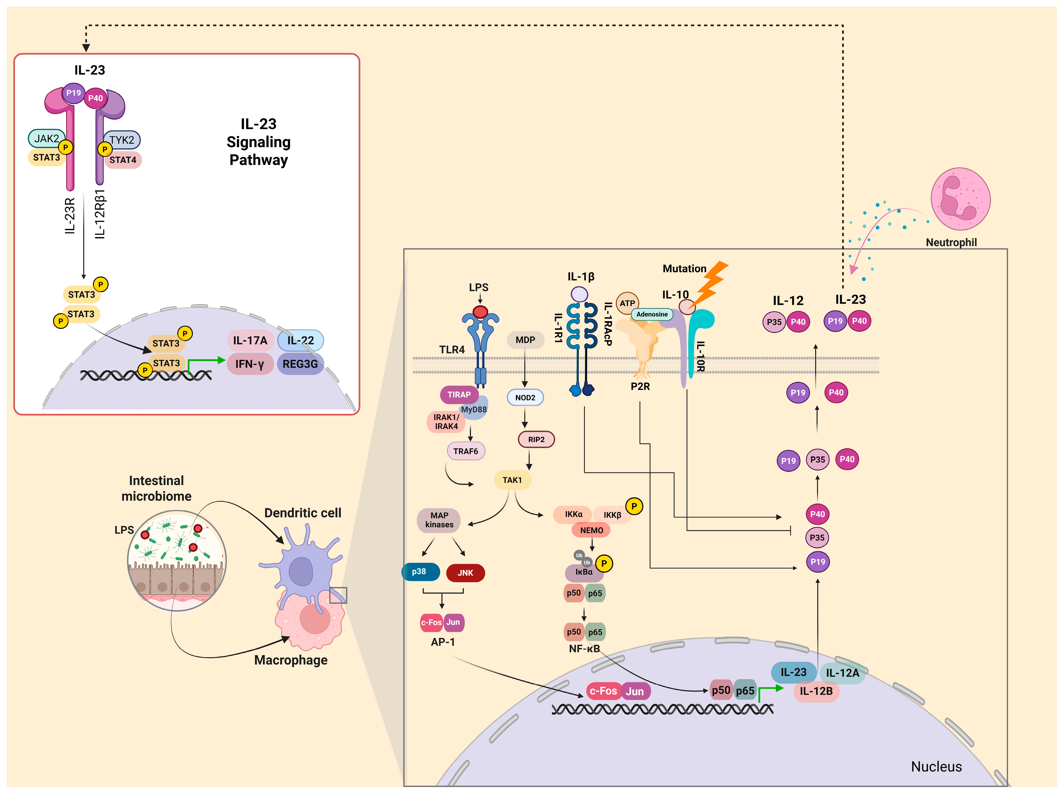
2.2. IL-23 Production

IL-23 production is affected by various factors. Antigen-presenting cells (APCs), particularly macrophages and dendritic cells (DCs), are the primary cellular sources of IL-23 production [17]. These cells increase the creation of IL-23 in the intestine when exposed to gut microbial stimuli. Response to Escherichia coli and Enterococcus faecalis stimulates IL-23 secretion by lamina propria CD14+ CD33+ macrophages [18]. In the presence of intestinal microbiota, CD11c + DCs in the terminal ileum lamina propria constitutively express the IL-23p40 subunit, indicating that IL-23 expression is greater in the healthy mucosa of this region [19]. In the inflamed mucosa of both CD and UC patients, the IL23-p19 subunit is present at increased levels, produced mainly from DCs and CD68+ macrophages [20]. For example, in CD, the severity of macroscopic lesions at endoscopy is positively correlated with IL-23p19 mRNA levels [21]. In CD refractory to TNF antagonism, the IL-23p19 levels in intestinal lamina propria CD14+ macrophages are augmented due to the accumulation of resistance to apoptosis, TNFR2+ IL23+ CD4+ T cells [22].
Molecular mechanisms and cellular signaling play a key role in IL-23 production by APCs. More specifically, when microbial agonist lipopolysaccharide (LPS) binds to Toll-like receptor 4 (TLR4), it promotes the production of IL-12 and IL-23 [23], while bacterial peptidoglycan in vitro stimulates the production of the IL-23p19 subunit over the IL12-p35 subunit [24]. Monocyte-delivered dendritic cells (MoDCs) promote IL-23 in response to both intact Gram-negative and Gram-positive bacteria, in contrast to the activation of DCs via TLR ligands [25]. Additionally, stimulation of cytosolic nucleotide binding oligomerization domain-containing protein 2 (NOD2) receptor in DCs and CD4+ T cells induces the production of IL-23 [26]. There are NOD2 disease-related variants that augment IL-23p19 secretion from DCs through decreased expression of the microRNA (miR-29) when DCs are stimulated by Escherichia coli [27]. Two additional molecular mechanisms contributing to IL-23 production involve extracellular adenosine nucleotides and intracellular metabolism within DCs and macrophages [28,29]. Extracellular adenosine increases IL-23p19 levels through the purinergic receptor P2 when DCs are exposed to Escherichia coli [17]. Macrophage metabolism also affects IL-23 production, as IBD-linked genes encode proteins like the enzyme FAMIN (a purine nucleotide recycling enzyme in macrophages) [30].
It is also crucial to consider factors other than microbial stimulation that affect IL-23 production. IL-10 reduces production via paracrine signaling. Mutations in the IL-10 receptor reduce the expression of IL-23p19 and lead to uncontrolled inflammation [31]. In addition, ATP and IL-1β induce the production of IL-23, and IFN-γ amplifies its creation [32]. Neutrophils are also a significant source of IL-23 in the gut via CXCR1/CXCR2 receptors, which regulate IL-23 through chemokines of the CXCL8 family [33]. A schematic overview of these mechanisms is illustrated in Figure 1, highlighting the interactions between IL-23, immune cell subsets, microbial stimuli, and inflammatory mediators.

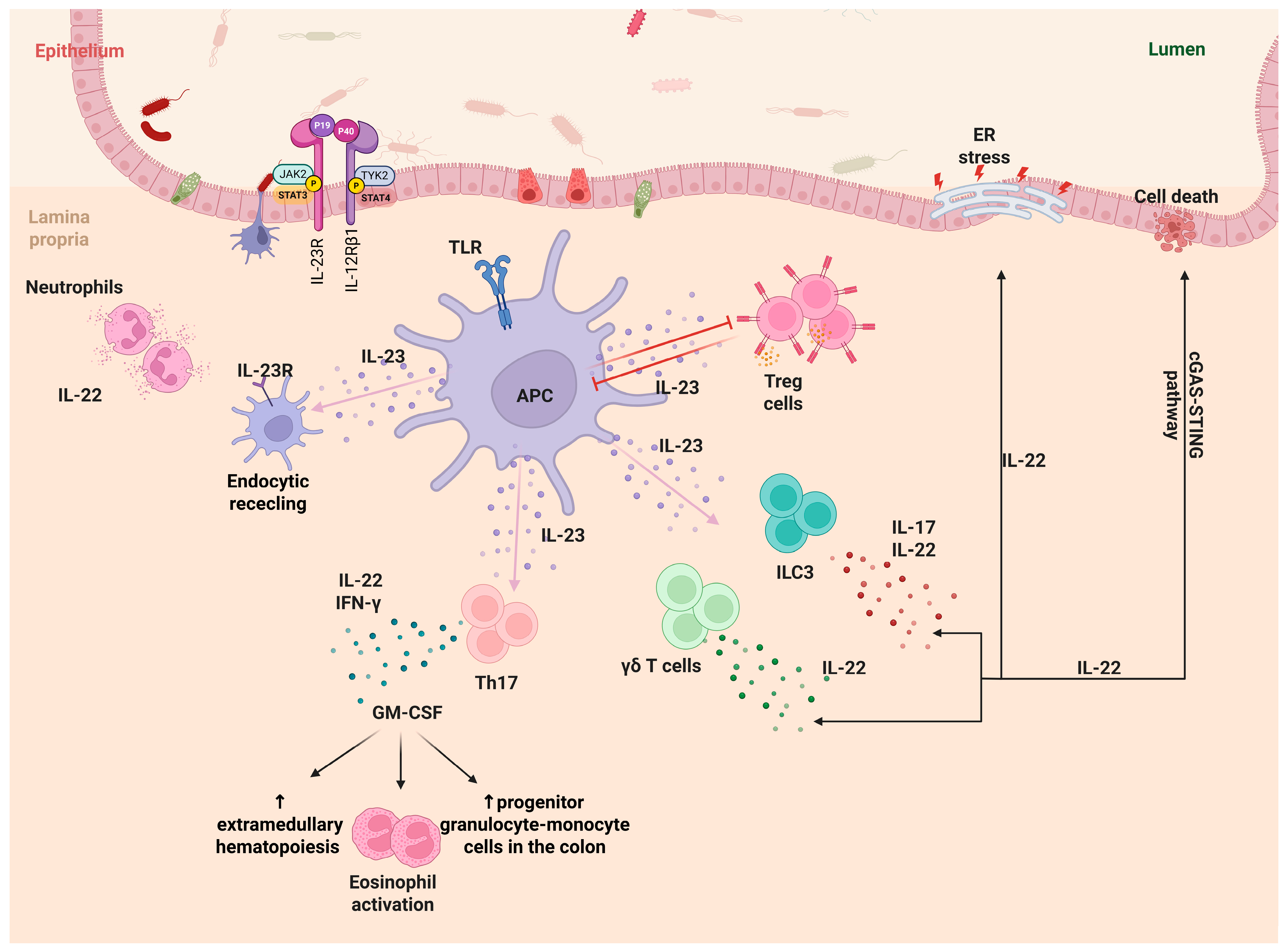
2.3. IL-23 and Its Multifaceted Role in IBD Pathogenesis

IL-23 plays a crucial role in IBD by activating various immune cell types. Its main influence is on Th17 cells, which are located in the intestinal lamina propria and induce the IL-17A production [34]. Th17 cells are largely produced in mice with specific Gram-positive bacteria in their microbiota [35], and the mucosa of IBD-inflamed patients contains more Th17 cells than the mucosa of healthy patients [36]. IL23 does not act alone; it requires IL-6, IL-21, and transforming growth factor β (TGFβ) to differentiate naïve CD4+ T cells into Th17 cells [37]. A combination of these interleukins with low concentrations of TGFβ in vitro enhances the expression of retinoid acid receptor-related orphan receptor γt (RORγt), suppresses the transcription factor Forkhead box P3 (FoxP3), and upregulates IL-23R expression [38]. The interaction between IL-23 and IL-23R promotes further IL-17 production [39], highlighting IL-23’s role in the survival and expansion of Th17 cells, as naïve CD4+ T cells do not express IL-23R [40].
In addition to IL-17, Th17 cells produce granulocyte-macrophage colony-stimulating factor (GM-CSF) and IL-12 when IL-23 is present [41,42]. Additionally, IL-12 and IL-23 can restimulate Th17-polarized CD4+ T cells in vitro, resulting in a decrease in IL-17A and IL-17F and an increase in interferon-gamma (IFNγ) expression when TGFβ is absent [43]. This Th17 phenotype is similar to Th1 cells, indicating the heterogeneity and plasticity of Th17 cells under different circumstances [44]. This heterogeneity could explain the controversial role of IL-17 in IBD development, and the different results observed in UC and CD. A neutralizing human anti-IL-17A antibody (secukinumab) has shown no results in moderate to severe CD, while a human anti-IL17 receptor A monoclonal antibody (brodalumab) improved active disease in CD in a randomized, double-blind clinical trial [45,46]. However, a humanized anti-interferon gamma antibody (fontolizumab) demonstrated clinical activity and safety in patients with moderate to severe CD in a multicenter phase 2 study involving 133 patients [47]. As for UC, a recent study indicated that suppression of the IL-17 signaling pathway may alleviate the progression of UC [48]. Regardless of the differences between disease types, the IL23/Th17 axis can induce inflammatory pathways in IBD. For instance, transferring naïve CD4+ T cells deficient in IL23R expression into mice reduced IL-17+ IFNγ+ cells in the colon [49], and IFNγ+ Il-17+ CD4+ T cells have been observed in the inflamed mucosa of both CD and UC patients but not in controls [50].
Furthermore, IL-23 affects IBD not only through its action on Th17 cells but also by constraining regulatory CD4+ T cells (Tregs) in the intestine. Tregs suppress IL-23 production by intestinal macrophages [49,51]. Tregs express anti-inflammatory cytokines (IL10 and IL-35), TGFβ, and the transcription factor FoxP3 [52]. In experimental models, the transfer of Treg-enriched cells has been shown to prevent colitis [53], and transferring CD4+CD25+ Tregs reduces established colitis [54], demonstrating their anti-inflammatory action. IL-23 inhibits the induction of FoxP3+ CD4+ Tregs in the intestine, blocking the anti-inflammatory effects of Tregs [55]. A recent animal study showed that IL23R signaling impairs the stability and function of Tregs, leading to increased inflammation in IBD patients [56].
IL-23 also significantly influences the activity of innate lymphoid cells (ILC3s). IL-23 stimulation induces ILC3s to produce high levels of IL-17 and IL-22 through STAT3 and STAT5 activation [57,58,59]. Inflamed ileum and colon tissues from CD patients have increased ILC3s responsive to IL-23 compared to healthy, non-inflamed mucosa [60]. Furthermore, an animal study showed that IL-23R on ILCs promotes colitis via IL-22 [61].
In addition to ILC3s, IL-22 is produced by Th17 cells, neutrophilic granulocytes, and γδ T cells in response to IL-23 [62]. Its role in intestinal inflammation is both protective and harmful. On one hand, the lack of IL-22 exacerbates colitis and T cell-mediated colitis in adoptive transfer models, and is necessary for optimal host defense against pathogens [63,64]. On the other hand, IL-22 worsens intestinal inflammation by enhancing endoplasm reticulum (ER) stress in epithelial cells [65,66]. IL-22 inactivation in IBD mouse models significantly reduced colitis levels, correlating with diminished ER stress in the intestinal epithelium [65]. Moreover, IL-22 promotes excessive activation of the cGAS-STING pathway in intestinal mucosa in the absence of the ATG16L1 gene (linked to CD susceptibility), resulting in cell death [66].
Finally, IL-23 influences myeloid cells in several ways. It induces neutrophil production, which may exacerbate established intestinal inflammation [67,68]. In experimental T cell transfer colitis, IL-23 amplifies GM-CSF production by Th17 cells, enhancing extramedullary hematopoiesis, accumulating progenitor granulocyte-monocyte cells in the colon, and activating eosinophils that contribute to tissue damage in colitis [68,69]. It is also important to mention the autocrine effects of IL-23. Some myeloid cells, like macrophages, express IL-23R on their surface. The recruitment of signaling intermediates from the IL-23R complex, such as IL-12Rβ1 and JAK2, along with the endocytic recycling of IL-23R, contributes to the release of pro-inflammatory cytokines by human macrophages, stimulated by IL-23 [70]. Figure 2 illustrates the multifaceted role of IL-23 in IBD pathogenesis, highlighting its interactions with adaptive and innate immune cells and the downstream inflammatory pathways involved.

3. IL-23R Gene in IBD

IL-23R has been shown to influence chronic inflammatory diseases through its expression on T cells and other immune cells in specific individuals [71]. When IL-23 binds to IL23R, Janus kinase 2 (JAK2) is activated via the IL23Ra subunit, leading to subsequent phosphorylation of IL-23R. This is followed by the recruitment and phosphorylation of the signal transducers STAT3 and STAT4 [72]. The phosphorylated STAT3 and STAT4 form homodimers that translocate to the nucleus, where they promote the transcription of pro-inflammatory cytokines in cells expressing IL-23R [73,74]. This transcriptional activity may lead to chronic inflammation through continuous IL-23/IL-23R interaction [75,76]. This signaling pathway is present in IBD, as well as in other chronic inflammatory diseases [77]. Various genome-wide association studies have demonstrated the role of the IL23/IL23R pathway, reporting that variants in the IL-23R gene (located on chromosome 1p31.3) are associated with IBD, mainly playing a protective role by modulating the activity of these signal transducers [78,79]. These variants typically affect the IL-23Ra expression, although they are located in different regions of the IL-23Ra structure. Thus, despite variations in the mutation mechanisms, the functional consequences of these variants are often similar [80].

3.1. R381Q (Arg381Gln, rs11209026)

The R381Q variant (rs11209026) is the most extensively studied protective allele of the IL-23R gene, influencing the IBD pathophysiology [70]. A recent study revealed that this allele was found in ancient European genomes dating back over 14,000 years and may have contributed to increased survival by reducing intestinal inflammation and microbiome dysbiosis [81]. Today, its frequency ranges from 0% to 17%, depending on the population, with approximately 5% observed in Europe [81]. A meta-analysis reported that the rs11209026 polymorphism may be protective against developing both CD and UC [82]. While this allele shows association with UC risk in Caucasian populations, no such association was found in Asian populations [82]. This may reflect either the genetic heterogeneity in different regional and ethnic populations or the limited number of studies in Asia [82].
At the DNA level, the functional single nucleotide polymorphism (SNP) involves a guanine (G) to adenine (A) substitution, resulting in an amino acid change in the protein from arginine (R) to glutamine (Q) at position 381 (R381Q) within the cytoplasmic domain of IL-23R [83]. Owing to its position, this polymorphism potentially affects the surface localization of the receptor and signal transduction [84]. Although rs11209026 is a non-synonymous coding variant located in exon nine, encoding the intracellular tail of the IL-23R, it plays a protective role against IBD susceptibility [85]. The variant location is between the transmembrane domain and the putative JAK2 binding site in the cytoplasmic region of the receptor, potentially reducing protein stability [85]. The replacement of the highly conserved Arg381 with Gln381 can alter interactions between IL-23R and its associated JAK2 kinase, decreasing the cellular response to IL-23. This leads to functional changes in the IL-23R signaling pathway, exerting a protective effect in IBD pathogenesis [85].
At the cellular level, rs11209026 decreases IL-23-dependent IL-17 production. The disease-protective Arg381Gln IL-23R variant leads to a loss of function in primary human CD4+ and CD8+ T cells, resulting in decreased cytokine production through the IL-23/Th17 pathway and enhanced microbial clearance [86]. This phenomenon occurs by a decrease in IL-23-induced STAT3 phosphorylation, resulting in lower secretion of pro-inflammatory cytokines, such as IL-17 and IL-22 in the gut by modulating the duration of the host response [87]. The impaired association between JAK2 proteins and the receptor’s cytoplasmic tail leads to reduced capacity of IL-23R to activate STAT proteins [88]. Thus, the IL-23R rs11209026 variant does not interfere with the Th17 differentiation but attenuates the IL-23-induced Th17 effector functions, specifically IL-17A production [89]. Moreover, this allele correlated to a lower percentage of circulating Th17 cells [90].
Additionally, this SNP exhibits alternative mechanisms of function. Rs11209026 alters IL-23Rα mRNA splicing by decreasing the binding of the splicing enhancer SF2, leading to exon nine skipping. This results in the expression of a soluble IL-23R-encoding mRNA species that dampens the IL-23R signaling and decreases the ability of the host to generate a Th17 phenotype in response to IL-23 stimulation [91]. A study also indicated that this SNP decreases IL-23 receptor recycling and impairs its assembly with Janus kinase and STAT pathway components, thereby modulating macrophage-mediated inflammatory pathways [70].

3.2. G149R (Gly149Arg, rs76418789)

The G149R variant (rs76418789) has been extensively studied in Japan [92], Korea [93], and China [94], as this allele is rare among Europeans and Africans but more prevalent in Asia (approximately ten times greater in East Asians than in Europeans—3.7% vs. 0.34%, respectively) [95]. A recent case–control study in Japan showed a lower risk of UC in patients with the G149R variant [96]. Despite its protective role in IBD, this allele is rare among the general population, with a frequency of less than 2% in healthy individuals [45].
This mutation consists of a functional SNP resulting in an amino acid substitution from glycine (G) to arginine (R) (Gly149Arg) at position 149 (G149R) in exon 4, a highly conserved residue located in the extracellular region of the receptor [97]. The G149R variant is retained in the endoplasmic reticulum (ER) as unfolded polypeptides due to impaired receptor maturation, preventing simultaneous trafficking from the ER to the Golgi apparatus [80]. This phenomenon is also observed in other diseases involving missense mutations, such as those affecting very low-density lipoprotein receptors, cystic fibrosis transmembrane receptors, and amyloid precursor proteins [98,99,100]. However, in this variant, the stability of the protein is not reduced [80]. A potential explanation for the primary retention of the receptor in the ER is that the variant receptor may be cleaved at the extracellular region by convertases in the Golgi, leading to an increase in receptor degradation and the formation of truncated products [101,102]. The effect of rs76418789 on the IL23R signaling, and consequently on IBD pathophysiology, is similar to that of rs11209026. Specifically, IL-23R activation leads to reduced phosphorylation of STAT3 and STAT4 due to lower receptor expression levels on the cell surface [80].

3.3. V362I (Val362Ile, rs41313262)

The V362l variant (rs41313262) is another protective variant of IL-23R for IBD [80]. The epidemiological patterns of this variant in IBD remain poorly characterized, with only limited studies available to date. Notably, the V362I variant showed no association with IBD susceptibility in a Chinese Han population cohort [103]. This mutation consists of a functional SNP that changes an amino acid from valine (V) to isoleucine (I) (Val362Ile) at position 362 in the transmembrane region of the receptor [97]. This variant was identified through targeted re-sequencing, similar to G149R [104]. One of the characteristics of V362I is that it does not exhibit a reduction in the mature/immature receptor ratio, in contrast to R3381Q and G149R. Additionally, it presents a decreased half-life and exhibits lower levels of endogenous receptor expression in V362I homozygous cells [80]. Consequently, it may be characterized by reduced protein stability, similar to R381Q and in contrast to G149R. Furthermore, protein instability may occur due to the location of the V362I substitution. The extra bulk of a methyl group added to the side chain in the transmembrane region of the receptor may disrupt shape complementarity and destabilize the interaction between IL23Ra and IL12Rb1, while still preserving peptide chain rigidity due to branching at the β-carbon, which seems necessary for other transmembrane protein-to-protein interactions [105]. V362I plays a protective role in IBD through the IL-23R signaling pathway in the same manner as G149R [80].

3.4. Other IL-23R-Related Gene Variants

Except for rs11209026, rs76418789, and rs41313262, several other SNPs in the IL-23R gene that may influence IBD have been reported. In contrast to the protective role of the above three variants, the roles of these additional SNPs are either unspecified, protective, or aggravating. The variant rs10889677 is located in the 3′-untranslated domain of the IL-23R gene, leading to enhanced mRNA and protein production due to the loss of microRNA regulation. This dysregulation contributes to sustained IL-23R signaling, which is associated with increased susceptibility and contributes to the chronicity of IBD [106]. A recent meta-analysis of 41 studies confirmed that rs10889677 might be a risk factor for IBD, indicating that this variant is linked to CD risk in Caucasians but not in Asians. In contrast, it is associated with UC risk in Asians but not in Caucasians [82]. The variant rs7517847 shows varied results across IBD subtypes. It may reduce the risk of developing CD [83]. However, it has no association with UC risk, though it may be related to the presence of blood in stool and bowel movements in these patients [107]. Unspecified results are also noted for the rs1004819 variant, as some studies reported no association with UC [107], whereas others identified this polymorphism as the main disease-associated IL-23R variant in German CD patients [108]. Limited research data are available in other variants, such as rs2201841, rs11800503, rs7530511, and rs1884444 [109].

4. IL-23R-Based Treatment Options in IBD

Binding of IL-23 to IL-23R plays a critical and pivotal role in IBD progression, primarily through its effect on the differentiation and maintenance of Th17 cells in the IL-23/Th17 pathway, leading to the production of pro-inflammatory cytokines [13,110]. Due to the specific involvement of IL-23 in IBD pathophysiology, biopharmaceutical agents have recently been developed to more precisely reduce IBD-related inflammation compared to broad-spectrum immunosuppressants [111].
The first biopharmaceutical agent targeting the IL-23 pathway was ustekinumab. It targets the IL-12p40 subunit, which is shared by both IL-12 and IL-23, thereby affecting the activation of both interleukins. Ustekinumab is approved for treating moderate-to-severe CD and UC, even in patients who have not responded to previous biologics therapies [112,113]. However, its effects extend beyond the IL-23 pathway.
More recently, more selective therapeutic agents have been developed that specifically target the IL-23p19 subunit. One such agent is risankizumab, a high-affinity neutralizing antibody that binds to the IL23p19 subunit. It is a human IgG1 kappa antibody containing two mutations in the Fc region (Leu234Ala and Leu235Ala) that reduce Fc gamma receptor interactions [114]. Risankizumab inhibits IL-23 from interacting with the IL-23 receptor and thereby blocks the subsequent inflammatory signaling pathway, without affecting the IL-12 pathway [114]. Early clinical studies have shown effective results in both CD and UC, suggesting that risankizumab could become an important tool in the treatment of moderate-to-severe CD [115,116,117].
Another agent, mirikizumab, is a humanized monoclonal IgG4 antibody that selectively inhibits the IL23p19 subunit. It also blocks interactions with the IL-23R complex, reducing the release of pro-inflammatory cytokines that depend on IL-23 [118]. Recent studies have shown promising results in both CD and UC, indicating its potential effectiveness in the treatment of moderate to severe UC [119,120].
Furthermore, two additional antibodies, guselkumab and brazikumab, are currently under investigation. Both target the IL23p19 subunit [121]. Guselkumab is a humanized monoclonal IgG1 λ antibody that blocks the interactions with the IL-23R complex, thereby reducing cellular signaling and release of pro-inflammatory cytokines in the IL-23 axis [118]. Preliminary data suggest a potential benefit in the treatment of UC and CD [122,123]. Similar encouraging results have also been observed in studies for brazikumab [124].
In addition, scientific research is now targeting the IL23R directly. JNJ-77242113 is an investigational, first-in-class oral peptide designed to selectively block the IL-23 receptor in various immune-mediated inflammatory diseases involving IL-23 signaling pathways, such as psoriasis and IBD, without affecting IL-12 signaling. In a rat model of colitis induced by trinitrobenzene sulfonic acid, JNJ-77242113 attenuated disease parameters [125]. However, research on this oral peptide is limited to the animal experimental level.
While these treatments have been established in adult populations, their use in pediatric IBD remains largely limited to ustekinumab for refractory cases. This preference is based on its demonstrated safety profile comparable to adults, including consistent observations of weight improvement in children with growth failure and low adverse event rates. However, clinical experience with other IL-23 inhibitors in pediatric populations remains limited, highlighting a need for further investigation [126].
For female IBD patients of reproductive age, anti-IL23 biological factors are not contraindicated during pregnancy, in contrast with Janus kinase (JAK) inhibitors and methotrexate [127]. Specifically, clinical studies demonstrate that pregnancy outcomes with ustekinumab treatment show comparable effectiveness and safety profiles to those observed in the general population [128]. This favorable evidence positions IL-23 pathway inhibitors as a viable therapeutic option for managing IBD throughout pregnancy [128].

5. Discussion

The IL-23/IL-23R axis plays a central role in the immunopathogenesis of IBD, primarily through its regulation of the Th17 cell-mediated pro-inflammatory response. Genetic variations in the IL-23R gene significantly influence individual susceptibility to IBD, with specific polymorphisms such as rs11209026 (R381Q), rs76418789 (G149R), and rs41313262 (V362I) offering a protective effect, often by impairing receptor function, reducing stability, or altering signaling efficiency. In contrast, variants like rs10889677, which escape microRNA regulation, contribute to enhanced receptor expression and increased IBD risk. Other variants show population-specific or disease-subtype-specific effects, reflecting the complexity of genetic contributions to IBD. The prevalence of certain variants varies by ethnicity and region. For example, rs76418789 occurs more frequently in Asian populations, while rs11209026 is more common in Caucasian populations. Additional research on IL-23R gene variants is needed to better understand their distribution across different ethnic and regional groups. This knowledge would assist in developing population-specific treatment strategies.
From a therapeutic perspective, these insights have paved the way for the development of targeted biologics. Ustekinumab, initially developed to inhibit both IL-12 and IL-23 pathways, has proven effective in treating moderate-to-severe forms of CD and UC. The emergence of more selective inhibitors such as risankizumab, mirikizumab, guselkumab, and brazikumab, specifically blocking the IL-23p19 subunit, demonstrates the growing precision in IBD therapy. Moreover, novel strategies targeting the IL-23 receptor directly, such as the investigational oral peptide JNJ-77242113, represent a promising frontier in IBD treatment.
The initial clinical trials have demonstrated favorable safety profiles and positive long-term outcomes for IL-23R-targeted therapies. A recent phase 3b randomized trial confirmed that ustekinumab has a favorable risk–benefit profile in Crohn’s disease, with sustained clinical and endoscopic improvements through week 104 [129]. Furthermore, in patients with ulcerative colitis, mirikizumab has shown durable efficacy through 104 weeks, with no safety concerns [130].
Although IL-23R-targeted therapy has only been clinically available for a few years, their use is rapidly expanding mainly as a personalized medicine approach in refractory IBD. Emerging evidence suggests several biomarkers may predict treatment response. In a recent prospective pilot study, higher baseline serum IL-23 levels were associated with clinical and endoscopic response to ustekinumab in patients with CD, whereas fecal calprotectin levels were decreased in responders but not in non-responders [131]. Also, eosinophil reduction was highlighted as a marker for early response to ustekinumab in IBD, but not to adalimumab (anti-Tumor Necrosis Factor) and vedolizumab (anti-integrin) [132].
Most reported variants of the IL-23R gene exhibit a protective role in IBD. While gene editing technologies have not yet been explored as a potential therapeutic approach for IL-23R modification in IBD, related strategies have shown promise in autoimmune conditions. Notably, it has been demonstrated that antisense oligonucleotides have been shown to modify IL-23R splicing through exon nine skipping. This modification increases the production of an IL-23R isoform, which acts as a decoy receptor, attenuates IL-23 signaling and mimics the protective mechanism of the rs11209026 variant, thereby reducing the differentiation of pro-inflammatory Th17 cells [91]. Also, genetically engineered human adipose-delivered mesenchymal stem cells could express a recombinant IL-23 decoy receptor using a lentiviral vector, which could reduce the inflammatory responses in vitro [133]. These innovative approaches represent promising avenues for future targeted therapies in IBD.

6. Conclusions

Genetic and pharmacological findings underscore the IL-23/IL-23R pathway as a crucial determinant in the development and management of IBD. Protective variants in the IL-23R gene provide valuable insights into IBD pathophysiology, offering important clues about disease mechanisms and potential therapeutic targets. Future research into the functional consequences of specific IL-23R variants and ongoing therapeutic innovation will likely enhance personalized approaches to IBD care.

Author Contributions

Conceptualization, I.A. and C.T.; investigation, P.P., I.A. and K.P.; resources, P.P., I.A. and K.P.; data curation, P.P.; writing—original draft preparation, P.P., I.A. and K.P.; writing—review and editing, I.A. and C.T.; visualization, I.A.; supervision, C.T.; project administration, C.T. All authors have read and agreed to the published version of the manuscript.

Funding

This research received no external funding.

Conflicts of Interest

The authors declare no conflicts of interest.

References

  1. Ahluwalia, B.; Magnusson, M.K.; Öhman, L. Mucosal immune system of the gastrointestinal tract: Maintaining balance between the good and the bad. Scand. J. Gastroenterol. 2017, 52, 1185–1193. [Google Scholar] [CrossRef] [PubMed]
  2. Le Berre, C.; Ananthakrishnan, A.N.; Danese, S.; Singh, S.; Peyrin-Biroulet, L. Ulcerative Colitis and Crohn’s Disease Have Similar Burden and Goals for Treatment. Clin. Gastroenterol. Hepatol. 2020, 18, 14–23. [Google Scholar] [CrossRef] [PubMed]
  3. Guan, Q. A Comprehensive Review and Update on the Pathogenesis of Inflammatory Bowel Disease. J. Immunol. Res. 2019, 2019, 7247238. [Google Scholar] [CrossRef]
  4. Gomollón, F.; Dignass, A.; Annese, V.; Tilg, H.; Van Assche, G.; Lindsay, J.O.; Peyrin-Biroulet, L.; Cullen, G.J.; Daperno, M.; Kucharzik, T.; et al. 3rd European Evidence-based Consensus on the Diagnosis and Management of Crohn’s Disease 2016: Part 1: Diagnosis and Medical Management. J. Crohns Colitis 2017, 11, 3–25. [Google Scholar] [CrossRef]
  5. Wallace, K.L.; Zheng, L.B.; Kanazawa, Y.; Shih, D.Q. Immunopathology of inflammatory bowel disease. World J. Gastroenterol. 2014, 20, 6–21. [Google Scholar] [CrossRef] [PubMed]
  6. Xavier, R.J.; Podolsky, D.K. Unravelling the pathogenesis of inflammatory bowel disease. Nature 2007, 448, 427–434. [Google Scholar] [CrossRef] [PubMed]
  7. Xu, X.R.; Liu, C.Q.; Feng, B.S.; Liu, Z.J. Dysregulation of mucosal immune response in pathogenesis of inflammatory bowel disease. World J. Gastroenterol. 2014, 20, 3255–3264. [Google Scholar] [CrossRef]
  8. de Souza, H.S.P.; Fiocchi, C. Immunopathogenesis of IBD: Current state of the art. Nat. Rev. Gastroenterol. Hepatol. 2016, 13, 13–27. [Google Scholar] [CrossRef]
  9. Zhang, Y.Z.; Li, Y.Y. Inflammatory bowel disease: Pathogenesis. World J. Gastroenterol. 2014, 20, 91–99. [Google Scholar] [CrossRef]
  10. Ananthakrishnan, A.N.; Kaplan, G.G.; Ng, S.C. Changing Global Epidemiology of Inflammatory Bowel Diseases: Sustaining Health Care Delivery Into the 21st Century. Clin. Gastroenterol. Hepatol. 2020, 18, 1252–1260. [Google Scholar] [CrossRef]
  11. Park, Y.M.; Ha, E.; Gu, K.N.; Shin, G.Y.; Lee, C.K.; Kim, K.; Kim, H.J. Host Genetic and Gut Microbial Signatures in Familial Inflammatory Bowel Disease. Clin. Transl. Gastroenterol. 2020, 11, e00213. [Google Scholar] [CrossRef] [PubMed]
  12. Jostins, L.; Ripke, S.; Weersma, R.K.; Duerr, R.H.; McGovern, D.P.; Hui, K.Y.; Lee, J.C.; Schumm, L.P.; Sharma, Y.; Anderson, C.A.; et al. Host-microbe interactions have shaped the genetic architecture of inflammatory bowel disease. Nature 2012, 491, 119–124. [Google Scholar] [CrossRef] [PubMed]
  13. Duerr, R.H.; Taylor, K.D.; Brant, S.R.; Rioux, J.D.; Silverberg, M.S.; Daly, M.J.; Steinhart, A.H.; Abraham, C.; Regueiro, M.; Griffiths, A.; et al. A genome-wide association study identifies IL23R as an inflammatory bowel disease gene. Science 2006, 314, 1461–1463. [Google Scholar] [CrossRef] [PubMed]
  14. Kastelein, R.A.; Hunter, C.A.; Cua, D.J. Discovery and Biology of IL-23 and IL-27: Related but Functionally Distinct Regulators of Inflammation. Annu. Rev. Immunol. 2007, 25, 221–242. [Google Scholar] [CrossRef]
  15. Jefremow, A.; Neurath, M.F. All are Equal, Some are More Equal: Targeting IL 12 and 23 in IBD—A Clinical Perspective. ImmunoTargets Ther. 2020, 9, 289. [Google Scholar] [CrossRef]
  16. Lupardus, P.J.; Garcia, K.C. The structure of Interleukin-23 reveals the molecular basis of p40 subunit sharing with IL-12. J. Mol. Biol. 2008, 382, 931. [Google Scholar] [CrossRef]
  17. Schnurr, M.; Toy, T.; Shin, A.; Wagner, M.; Cebon, J.; Maraskovsky, E. Extracellular nucleotide signaling by P2 receptors inhibits IL-12 and enhances IL-23 expression in human dendritic cells: A novel role for the cAMP pathway. Blood 2005, 105, 1582–1589. [Google Scholar] [CrossRef] [PubMed]
  18. Pascal, V.; Pozuelo, M.; Borruel, N.; Casellas, F.; Campos, D.; Santiago, A.; Martinez, X.; Varela, E.; Sarrabayrouse, G.; Machiels, K.; et al. A microbial signature for Crohn’s disease. Gut 2017, 66, 813–822. [Google Scholar] [CrossRef] [PubMed]
  19. Becker, C.; Wirtz, S.; Blessing, M.; Pirhonen, J.; Strand, D.; Bechthold, O.; Frick, J.; Galle, P.R.; Autenrieth, I.; Neurath, M.F. Constitutive p40 promoter activation and IL-23 production in the terminal ileum mediated by dendritic cells. J. Clin. Investig. 2003, 112, 693–706. [Google Scholar] [CrossRef]
  20. Liu, Z.; Yadav, P.K.; Xu, X.; Su, J.; Chen, C.; Tang, M.; Lin, H.; Yu, J.; Qian, J.; Yang, P.C.; et al. The increased expression of IL-23 in inflammatory bowel disease promotes intraepithelial and lamina propria lymphocyte inflammatory responses and cytotoxicity. J. Leukoc. Biol. 2011, 89, 597–606. [Google Scholar] [CrossRef]
  21. Schmidt, C.; Giese, T.; Ludwig, B.; Mueller-Molaian, I.; Marth, T.; Zeuzem, S.; Meurer, S.C.; Stallmach, A. Expression of interleukin-12-related cytokine transcripts in inflammatory bowel disease: Elevated interleukin-23p19 and interleukin-27p28 in Crohn’s disease but not in ulcerative colitis. Inflamm. Bowel Dis. 2005, 11, 16–23. [Google Scholar] [CrossRef] [PubMed]
  22. Schmitt, H.; Billmeier, U.; Dieterich, W.; Rath, T.; Sonnewald, S.; Reid, S.; Hirschmann, S.; Hildner, K.; Waldner, M.J.; Mudter, J.; et al. Expansion of IL-23 receptor bearing TNFR2+ T cells is associated with molecular resistance to anti-TNF therapy in Crohn’s disease. Gut 2019, 68, 814–828. [Google Scholar] [CrossRef]
  23. Verreck, F.A.W.; de Boer, T.; Langenberg, D.M.L.; Hoeve, M.A.; Kramer, M.; Vaisberg, E.; Kastelein, R.; Kolk, A.; de Waal-Malefyt, R.; Ottenhoff, T.H.M. Human IL-23-producing type 1 macrophages promote but IL-10-producing type 2 macrophages subvert immunity to (myco)bacteria. Proc. Natl. Acad. Sci. USA 2004, 101, 4560–4565. [Google Scholar] [CrossRef] [PubMed]
  24. Re, F.; Strominger, J.L. Toll-like receptor 2 (TLR2) and TLR4 differentially activate human dendritic cells. J. Biol. Chem. 2001, 276, 37692–37699. [Google Scholar] [CrossRef] [PubMed]
  25. Veckman, V.; Miettinen, M.; Pirhonen, J.; Sirén, J.; Matikainen, S.; Julkunen, I. Streptococcus pyogenes and Lactobacillus rhamnosus differentially induce maturation and production of Th1-type cytokines and chemokines in human monocyte-derived dendritic cells. J. Leukoc. Biol. 2004, 75, 764–771. [Google Scholar] [CrossRef] [PubMed]
  26. Kamada, N.; Hisamatsu, T.; Okamoto, S.; Chinen, H.; Kobayashi, T.; Sato, T.; Sakurba, A.; Kitazume, M.T.; Sugita, A.; Koganei, K.; et al. Unique CD14 intestinal macrophages contribute to the pathogenesis of Crohn disease via IL-23/IFN-gamma axis. J. Clin. Investig. 2008, 118, 2269–2280. [Google Scholar]
  27. Brain, O.; Owens, B.M.J.; Pichulik, T.; Allan, P.; Khatamzas, E.; Leslie, A.; Steevels, T.; Sharma, S.; Mayer, A.; Catuneanu, A.M.; et al. The intracellular sensor NOD2 induces microRNA-29 expression in human dendritic cells to limit IL-23 release. Immunity 2013, 39, 521–536. [Google Scholar] [CrossRef]
  28. Cekic, C.; Linden, J. Purinergic regulation of the immune system. Nat. Rev. Immunol. 2016, 16, 177–192. [Google Scholar] [CrossRef]
  29. Mogilenko, D.A.; Haas, J.T.; L’homme, L.; Fleury, S.; Quemener, S.; Levavasseur, M.; Becquart, C.; Wartelle, J.; Bogomolova, A.; Pineau, L.; et al. Metabolic and Innate Immune Cues Merge into a Specific Inflammatory Response via the UPR. Cell 2019, 177, 1201–1216.e19. [Google Scholar] [CrossRef]
  30. Cader, M.Z.; de Almeida Rodrigues, R.P.; West, J.A.; Sewell, G.W.; Md-Ibrahim, M.N.; Reikine, S.; Sirago, G.; Unger, L.W.; Iglesias-Romero, A.B.; Ramshorn, K.; et al. FAMIN Is a Multifunctional Purine Enzyme Enabling the Purine Nucleotide Cycle. Cell 2020, 180, 278–295.e23. [Google Scholar] [CrossRef]
  31. Aschenbrenner, D.; Quaranta, M.; Banerjee, S.; Ilott, N.; Jansen, J.; Steere, B.; Chen, Y.H.; Ho, S.; Cox, K.; Arancibia-Carcamo, C.V.; et al. Deconvolution of monocyte responses in inflammatory bowel disease reveals an IL-1 cytokine network that regulates IL-23 in genetic and acquired IL-10 resistance. Gut 2021, 70, 1023–1036. [Google Scholar] [CrossRef] [PubMed]
  32. Glass, C.K.; Natoli, G. Molecular control of activation and priming in macrophages. Nat. Immunol. 2016, 17, 26–33. [Google Scholar] [CrossRef]
  33. Russo, R.C.; Garcia, C.C.; Teixeira, M.M.; Amaral, F.A. The CXCL8/IL-8 chemokine family and its receptors in inflammatory diseases. Expert. Rev. Clin. Immunol. 2014, 10, 593–619. [Google Scholar] [CrossRef] [PubMed]
  34. Ivanov, I.I.; McKenzie, B.S.; Zhou, L.; Tadokoro, C.E.; Lepelley, A.; Lafaille, J.J.; Cua, D.J.; Littman, D.R. The orphan nuclear receptor RORgammat directs the differentiation program of proinflammatory IL-17+ T helper cells. Cell 2006, 126, 1121–1133. [Google Scholar] [CrossRef] [PubMed]
  35. Ivanov, I.I.; Atarashi, K.; Manel, N.; Brodie, E.L.; Shima, T.; Karaoz, U.; Wei, D.; Goldfarb, K.C.; Santee, C.A.; Lynch, S.V.; et al. Induction of intestinal Th17 cells by segmented filamentous bacteria. Cell 2009, 139, 485–498. [Google Scholar] [CrossRef] [PubMed]
  36. Jiang, W.; Su, J.; Zhang, X.; Cheng, X.; Zhou, J.; Shi, R.; Zhang, H. Elevated levels of Th17 cells and Th17-related cytokines are associated with disease activity in patients with inflammatory bowel disease. Inflamm. Res. 2014, 63, 943–950. [Google Scholar] [CrossRef] [PubMed]
  37. Veldhoen, M.; Hocking, R.J.; Atkins, C.J.; Locksley, R.M.; Stockinger, B. TGFbeta in the context of an inflammatory cytokine milieu supports de novo differentiation of IL-17-producing T cells. Immunity 2006, 24, 179–189. [Google Scholar] [CrossRef]
  38. Zhou, L.; Lopes, J.E.; Chong, M.M.W.; Ivanov, I.I.; Min, R.; Victora, G.D.; Shen, Y.; Du, J.; Rubtsov, Y.P.; Rudensky, A.Y.; et al. TGF-beta-induced Foxp3 inhibits T(H)17 cell differentiation by antagonizing RORgammat function. Nature 2008, 453, 236–240. [Google Scholar] [CrossRef]
  39. Zhou, L.; Ivanov, I.I.; Spolski, R.; Min, R.; Shenderov, K.; Egawa, T.; Levy, D.E.; Leonard, W.J.; Littman, D.R. IL-6 programs T(H)-17 cell differentiation by promoting sequential engagement of the IL-21 and IL-23 pathways. Nat. Immunol. 2007, 8, 967–974. [Google Scholar] [CrossRef]
  40. Oppmann, B.; Lesley, R.; Blom, B.; Timans, J.C.; Xu, Y.; Hunte, B.; Vega, F.; Yu, N.; Wang, J.; Singh, K.; et al. Novel p19 protein engages IL-12p40 to form a cytokine, IL-23, with biological activities similar as well as distinct from IL-12. Immunity 2000, 13, 715–725. [Google Scholar] [CrossRef]
  41. El-Behi, M.; Ciric, B.; Dai, H.; Yan, Y.; Cullimore, M.; Safavi, F.; Zhang, G.X.; Dittel, B.N.; Rostami, A. The encephalitogenicity of T(H)17 cells is dependent on IL-1- and IL-23-induced production of the cytokine GM-CSF. Nat. Immunol. 2011, 12, 568–575. [Google Scholar] [CrossRef] [PubMed]
  42. Liang, S.C.; Tan, X.Y.; Luxenberg, D.P.; Karim, R.; Dunussi-Joannopoulos, K.; Collins, M.; Fouser, L.A. Interleukin (IL)-22 and IL-17 are coexpressed by Th17 cells and cooperatively enhance expression of antimicrobial peptides. J. Exp. Med. 2006, 203, 2271–2279. [Google Scholar] [CrossRef] [PubMed]
  43. Lee, Y.K.; Turner, H.; Maynard, C.L.; Oliver, J.R.; Chen, D.; Elson, C.O.; Weaver, C.T. Late developmental plasticity in the T helper 17 lineage. Immunity 2009, 30, 92–107. [Google Scholar] [CrossRef] [PubMed]
  44. Schmitt, H.; Neurath, M.F.; Atreya, R. Role of the IL23/IL17 Pathway in Crohn’s Disease. Front. Immunol. 2021, 12, 622934. [Google Scholar] [CrossRef] [PubMed]
  45. Sewell, G.W.; Kaser, A. Interleukin-23 in the Pathogenesis of Inflammatory Bowel Disease and Implications for Therapeutic Intervention. J. Crohns Colitis. 2022, 16 (Suppl. 2), ii3–ii19. [Google Scholar] [CrossRef] [PubMed]
  46. Targan, S.R.; Feagan, B.; Vermeire, S.; Panaccione, R.; Melmed, G.Y.; Landers, C.; Li, D.; Russell, C.; Newmark, R.; Zhang, N.; et al. A Randomized, Double-Blind, Placebo-Controlled Phase 2 Study of Brodalumab in Patients With Moderate-to-Severe Crohn’s Disease. Am. J. Gastroenterol. 2016, 111, 1599–1607. [Google Scholar] [CrossRef]
  47. Hommes, D.W.; Mikhajlova, T.L.; Stoinov, S.; Stimac, D.; Vucelic, B.; Lonovics, J.; Zakuciova, M.; D’Haens, G.; Van Assche, G.; Ba, S.; et al. Fontolizumab, a humanised anti-interferon gamma antibody, demonstrates safety and clinical activity in patients with moderate to severe Crohn’s disease. Gut 2006, 55, 1131–1137. [Google Scholar] [CrossRef]
  48. Li, O.; Li, X.; He, J. Knockdown of TOP2A suppresses IL-17 signaling pathway and alleviates the progression of ulcerative colitis. Immun. Inflamm. Dis. 2024, 12, e1207. [Google Scholar] [CrossRef]
  49. Ahern, P.P.; Schiering, C.; Buonocore, S.; McGeachy, M.J.; Cua, D.J.; Maloy, K.J.; Powrie, F. Interleukin-23 drives intestinal inflammation through direct activity on T cells. Immunity 2010, 33, 279–288. [Google Scholar] [CrossRef]
  50. Takayama, T.; Kamada, N.; Chinen, H.; Okamoto, S.; Kitazume, M.T.; Chang, J.; Matuzaki, Y.; Suzuki, S.; Sugita, A.; Koganei, K.; et al. Imbalance of NKp44(+)NKp46(-) and NKp44(-)NKp46(+) natural killer cells in the intestinal mucosa of patients with Crohn’s disease. Gastroenterology 2010, 139, 882–892.e3. [Google Scholar] [CrossRef]
  51. Bauché, D.; Joyce-Shaikh, B.; Jain, R.; Grein, J.; Ku, K.S.; Blumenschein, W.M.; Ganal-Vonarburg, S.C.; Wilson, D.C.; McClanahan, T.K.; de Wall Malefyt, R.; et al. LAG3+ Regulatory T Cells Restrain Interleukin-23-Producing CX3CR1+ Gut-Resident Macrophages during Group 3 Innate Lymphoid Cell-Driven Colitis. Immunity 2018, 49, 342.e5–352.e5. [Google Scholar] [CrossRef] [PubMed]
  52. Izcue, A.; Coombes, J.L.; Powrie, F. Regulatory lymphocytes and intestinal inflammation. Annu. Rev. Immunol. 2009, 27, 313–338. [Google Scholar] [CrossRef]
  53. Powrie, F.; Leach, M.W.; Mauze, S.; Caddle, L.B.; Coffman, R.L. Phenotypically distinct subsets of CD4+ T cells induce or protect from chronic intestinal inflammation in C. B-17 scid mice. Int. Immunol. 1993, 5, 1461–1471. [Google Scholar] [CrossRef] [PubMed]
  54. Mottet, C.; Uhlig, H.H.; Powrie, F. Cutting edge: Cure of colitis by CD4+CD25+ regulatory T cells. J. Immunol. 2003, 170, 3939–3943. [Google Scholar] [CrossRef] [PubMed]
  55. Izcue, A.; Hue, S.; Buonocore, S.; Arancibia-Cárcamo, C.V.; Ahern, P.P.; Iwakura, Y.; Maloy, K.J.; Powrie, F. Interleukin-23 restrains regulatory T cell activity to drive T cell-dependent colitis. Immunity 2008, 28, 559–570. [Google Scholar] [CrossRef] [PubMed]
  56. Jacobse, J.; Brown, R.E.; Li, J.; Pilat, J.M.; Pham, L.; Short, S.P.; Peek, C.T.; Rolong, A.; Washington, M.K.; Martinez-Barricarte, R.; et al. Interleukin-23 receptor signaling impairs the stability and function of colonic regulatory T cells. Cell Rep. 2023, 42, 112128. [Google Scholar] [CrossRef]
  57. Longman, R.S.; Diehl, G.E.; Victorio, D.A.; Huh, J.R.; Galan, C.; Miraldi, E.R.; Swaminath, A.; Bonneau, R.; Scherl, E.J.; Littman, D.R. CX3CR1+ mononuclear phagocytes support colitis-associated innate lymphoid cell production of IL-22. J. Exp. Med. 2014, 211, 1571–1583. [Google Scholar] [CrossRef]
  58. Bauché, D.; Joyce-Shaikh, B.; Fong, J.; Villarino, A.V.; Ku, K.S.; Jain, R.; Lee, Y.C.; Annamalai, L.; Yearley, J.H.; Cua, D.J. IL-23 and IL-2 activation of STAT5 is required for optimal IL-22 production in ILC3s during colitis. Sci. Immunol. 2020, 5, eaav1080. [Google Scholar] [CrossRef]
  59. Guo, X.; Qiu, J.; Tu, T.; Yang, X.; Deng, L.; Anders, R.A.; Zhou, L.; Fu, Y.X. Induction of innate lymphoid cell-derived interleukin-22 by the transcription factor STAT3 mediates protection against intestinal infection. Immunity 2014, 40, 25–39. [Google Scholar] [CrossRef]
  60. Geremia, A.; Arancibia-Cárcamo, C.V.; Fleming, M.P.P.; Rust, N.; Singh, B.; Mortensen, N.J.; Travis, S.P.L.; Powrie, F. IL-23-responsive innate lymphoid cells are increased in inflammatory bowel disease. J. Exp. Med. 2011, 208, 1127–1133. [Google Scholar] [CrossRef]
  61. Eken, A.; Singh, A.K.; Treuting, P.M.; Oukka, M. IL-23R+ innate lymphoid cells induce colitis via interleukin-22-dependent mechanism. Mucosal Immunol. 2014, 7, 143–154. [Google Scholar] [CrossRef] [PubMed]
  62. Keir, M.; Yi, T.; Lu, T.; Ghilardi, N. The role of IL-22 in intestinal health and disease. J. Exp. Med. 2020, 217, e20192195. [Google Scholar] [CrossRef] [PubMed]
  63. Zenewicz, L.A.; Yancopoulos, G.D.; Valenzuela, D.M.; Murphy, A.J.; Stevens, S.; Flavell, R.A. Innate and adaptive interleukin-22 protects mice from inflammatory bowel disease. Immunity 2008, 29, 947–957. [Google Scholar] [CrossRef] [PubMed]
  64. Zheng, Y.; Valdez, P.A.; Danilenko, D.M.; Hu, Y.; Sa, S.M.; Gong, Q.; Abbas, A.R.; Modrusan, Z.; Ghilardi, N.; de Sauvage, F.J.; et al. Interleukin-22 mediates early host defense against attaching and effacing bacterial pathogens. Nat. Med. 2008, 14, 282–289. [Google Scholar] [CrossRef] [PubMed]
  65. Powell, N.; Pantazi, E.; Pavlidis, P.; Tsakmaki, A.; Li, K.; Yang, F.; Parker, A.; Pin, C.; Cozzetto, D.; Minns, D.; et al. Interleukin-22 orchestrates a pathological endoplasmic reticulum stress response transcriptional programme in colonic epithelial cells. Gut 2020, 69, 578–590. [Google Scholar] [CrossRef]
  66. Aden, K.; Tran, F.; Ito, G.; Sheibani-Tezerji, R.; Lipinski, S.; Kuiper, J.W.; Tschurtschnthaler, M.; Saveljeva, S.; Bhattacharyya, J.; Hasler, R.; et al. ATG16L1 orchestrates interleukin-22 signaling in the intestinal epithelium via cGAS-STING. J. Exp. Med. 2018, 215, 2868–2886. [Google Scholar] [CrossRef]
  67. Fournier, B.M.; Parkos, C.A. The role of neutrophils during intestinal inflammation. Mucosal Immunol. 2012, 5, 354–366. [Google Scholar] [CrossRef]
  68. Griseri, T.; McKenzie, B.S.; Schiering, C.; Powrie, F. Dysregulated hematopoietic stem and progenitor cell activity promotes interleukin-23-driven chronic intestinal inflammation. Immunity 2012, 37, 1116–1129. [Google Scholar] [CrossRef]
  69. Griseri, T.; Arnold, I.C.; Pearson, C.; Krausgruber, T.; Schiering, C.; Franchini, F.; Schulthess, J.; McKenzie, B.S.; Crocker, P.R.; Powrie, F. Granulocyte Macrophage Colony-Stimulating Factor-Activated Eosinophils Promote Interleukin-23 Driven Chronic Colitis. Immunity 2015, 43, 187–199. [Google Scholar] [CrossRef]
  70. Sun, R.; Hedl, M.; Abraham, C. IL23 induces IL23R recycling and amplifies innate receptor-induced signalling and cytokines in human macrophages, and the IBD-protective IL23R R381Q variant modulates these outcomes. Gut 2020, 69, 264–273. [Google Scholar] [CrossRef]
  71. Chognard, G.; Bellemare, L.; Pelletier, A.N.; Dominguez-Punaro, M.C.; Beauchamp, C.; Guyon, M.J.; Charron, G.; Morin, N.; Sivanesan, D.; Kuchroo, V.; et al. The dichotomous pattern of IL-12r and IL-23R expression elucidates the role of IL-12 and IL-23 in inflammation. PLoS ONE 2014, 9, e89092. [Google Scholar] [CrossRef] [PubMed]
  72. Parham, C.; Chirica, M.; Timans, J.; Vaisberg, E.; Travis, M.; Cheung, J.; Pflanz, S.; Zhang, R.; Singh, K.P.; Vega, F.; et al. A receptor for the heterodimeric cytokine IL-23 is composed of IL-12Rbeta1 and a novel cytokine receptor subunit, IL-23R. J. Immunol. 2002, 168, 5699–5708. [Google Scholar] [CrossRef] [PubMed]
  73. Budarf, M.L.; Labbé, C.; David, G.; Rioux, J.D. GWA studies: Rewriting the story of IBD. Trends Genet. 2009, 25, 137–146. [Google Scholar] [CrossRef]
  74. Ouyang, W.; Valdez, P. IL-22 in mucosal immunity. Mucosal Immunol. 2008, 1, 335–338. [Google Scholar] [CrossRef]
  75. McGeachy, M.J.; Chen, Y.; Tato, C.M.; Laurence, A.; Joyce-Shaikh, B.; Blumenschein, W.M.; McClanahan, T.K.; O’Shea, J.J.; Cua, D.J. The interleukin 23 receptor is essential for the terminal differentiation of interleukin 17-producing effector T helper cells in vivo. Nat. Immunol. 2009, 10, 314–324. [Google Scholar] [CrossRef]
  76. Guo, W.; Luo, C.; Wang, C.; Zhu, Y.; Wang, X.; Gao, X.; Yao, W. Protection against Th17 cells differentiation by an interleukin-23 receptor cytokine-binding homology region. PLoS ONE 2012, 7, e45625. [Google Scholar] [CrossRef] [PubMed]
  77. Abraham, C.; Cho, J. Interleukin-23/Th17 pathways and inflammatory bowel disease. Inflamm. Bowel Dis. 2009, 15, 1090–1100. [Google Scholar] [CrossRef] [PubMed]
  78. Amre, D.K.; Mack, D.; Israel, D.; Morgan, K.; Lambrette, P.; Law, L.; Grimard, G.; Deslandres, C.; Krupoves, A.; Bucionis, V.; et al. Association between genetic variants in the IL-23R gene and early-onset Crohn’s disease: Results from a case-control and family-based study among Canadian children. Am. J. Gastroenterol. 2008, 103, 615–620. [Google Scholar] [CrossRef] [PubMed]
  79. Rioux, J.D.; Xavier, R.J.; Taylor, K.D.; Silverberg, M.S.; Goyette, P.; Huett, A.; Green, T.; Kuballa, P.; Barmada, M.M.; Datta, L.W.; et al. Genome-wide association study identifies new susceptibility loci for Crohn disease and implicates autophagy in disease pathogenesis. Nat. Genet. 2007, 39, 596–604. [Google Scholar] [CrossRef]
  80. Sivanesan, D.; Beauchamp, C.; Quinou, C.; Lee, J.; Lesage, S.; Chemtob, S.; Rioux, J.D.; Michnick, S.W. IL23R (Interleukin 23 Receptor) Variants Protective against Inflammatory Bowel Diseases (IBD) Display Loss of Function due to Impaired Protein Stability and Intracellular Trafficking. J. Biol. Chem. 2016, 291, 8673–8685. [Google Scholar] [CrossRef]
  81. Krause-Kyora, B.; da Silva, N.A.; Kaplan, E.; Kolbe, D.; Archaeological Civilization Disease Consortium; Wohlers, I.; Busch, H.; Ellinghaus, D.; Caliebe, A.; Sezgin, E.; et al. Neolithic introgression of IL23R-related protection against chronic inflammatory bowel diseases in modern Europeans. EBioMedicine 2025, 113, 105591. [Google Scholar] [CrossRef] [PubMed]
  82. Zhu, Y.; Jiang, H.; Chen, Z.; Lu, B.; Li, J.; Shen, X. Genetic association between IL23R rs11209026 and rs10889677 polymorphisms and risk of Crohn’s disease and ulcerative colitis: Evidence from 41 studies. Inflamm. Res. 2020, 69, 87–103. [Google Scholar] [CrossRef] [PubMed]
  83. Ferguson, L.R.; Han, D.Y.; Fraser, A.G.; Huebner, C.; Lam, W.J.; Morgan, A.R. IL23R and IL12B SNPs and Haplotypes Strongly Associate with Crohn’s Disease Risk in a New Zealand Population. Gastroenterol. Res. Pract. 2010, 2010, 539461. [Google Scholar] [CrossRef]
  84. Lerch-Bader, M.; Lundin, C.; Kim, H.; Nilsson, I.; von Heijne, G. Contribution of positively charged flanking residues to the insertion of transmembrane helices into the endoplasmic reticulum. Proc. Natl. Acad. Sci. USA 2008, 105, 4127–4132. [Google Scholar] [CrossRef] [PubMed]
  85. Oliver, J.; Rueda, B.; López-Nevot, M.A.; Gómez-García, M.; Martín, J. Replication of an association between IL23R gene polymorphism with inflammatory bowel disease. Clin. Gastroenterol. Hepatol. 2007, 5, 977–981.e2. [Google Scholar] [CrossRef]
  86. Guan, Q.; Ma, Y.; Aboud, L.; Weiss, C.R.; Qing, G.; Warrington, R.J.; Peng, Z. Targeting IL-23 by employing a p40 peptide-based vaccine ameliorates murine allergic skin and airway inflammation. Clin. Exp. Allergy 2012, 42, 1397–1405. [Google Scholar] [CrossRef]
  87. Pidasheva, S.; Trifari, S.; Phillips, A.; Hackney, J.A.; Ma, Y.; Smith, A.; Sohn, S.J.; Spits, H.; Little, R.D.; Behrens, T.W.; et al. Functional studies on the IBD susceptibility gene IL23R implicate reduced receptor function in the protective genetic variant R381Q. PLoS ONE 2011, 6, e25038. [Google Scholar] [CrossRef]
  88. Sarin, R.; Wu, X.; Abraham, C. Inflammatory disease protective R381Q IL23 receptor polymorphism results in decreased primary CD4+ and CD8+ human T-cell functional responses. Proc. Natl. Acad. Sci. USA 2011, 108, 9560–9565. [Google Scholar] [CrossRef]
  89. Di Meglio, P.; Di Cesare, A.; Laggner, U.; Chu, C.C.; Napolitano, L.; Villanova, F.; Tosi, I.; Capon, F.; Trembath, R.C.; Peris, K.; et al. The IL23R R381Q gene variant protects against immune-mediated diseases by impairing IL-23-induced Th17 effector response in humans. PLoS ONE 2011, 6, e17160. [Google Scholar] [CrossRef]
  90. Abdollahi, E.; Tavasolian, F.; Ghasemi, N.; Mirghanizadeh, S.A.; Azizi, M.; Ghoryani, M.; Samadi, M. Association between lower frequency of R381Q variant (rs11209026) in IL-23 receptor gene and increased risk of recurrent spontaneous abortion (RSA). J. Immunotoxicol. 2015, 12, 317–321. [Google Scholar] [CrossRef]
  91. Yu, R.Y.; Brazaitis, J.; Gallagher, G. The human IL-23 receptor rs11209026 A allele promotes the expression of a soluble IL-23R-encoding mRNA species. J. Immunol. 2015, 194, 1062–1068. [Google Scholar] [CrossRef] [PubMed]
  92. Okamoto, D.; Kawai, Y.; Kakuta, Y.; Naito, T.; Torisu, T.; Hirano, A.; Umero, J.; Fuyuno, Y.; Li, D.; Nakano, T.; et al. Genetic analysis of ulcerative colitis in Japanese individuals using population-specific SNP array. Inflamm. Bowel Dis. 2020, 26, 1177–1187. [Google Scholar] [CrossRef] [PubMed]
  93. Ye, B.D.; Choi, H.; Hong, M.; Yun, W.J.; Low, H.Q.; Haritunians, T.; Kim, K.J.; Park, S.H.; Lee, I.; Bang, S.Y.; et al. Identification of Ten Additional Susceptibility Loci for Ulcerative Colitis Through Immunochip Analysis in Koreans. Inflamm. Bowel Dis. 2016, 22, 13–19. [Google Scholar] [CrossRef] [PubMed]
  94. Bin, C.; Zhirong, Z.; Xiaoqin, W.; Minhu, C.; Mei, L.; Xiang, G.; Baili, C.; Pinjin, H. Contribution of rs11465788 in IL23R gene to Crohn’s disease susceptibility and phenotype in Chinese population. J. Genet. 2009, 88, 191–196. [Google Scholar] [CrossRef]
  95. Ju, Y.S.; Kim, J.I.; Kim, S.; Hong, D.; Park, H.; Shin, J.Y.; Lee, S.; Lee, W.C.; Kim, S.; Yu, S.B.; et al. Extensive genomic and transcriptional diversity identified through massively parallel DNA and RNA sequencing of eighteen Korean individuals. Nat. Genet. 2011, 43, 745–752. [Google Scholar] [CrossRef]
  96. Miyake, Y.; Tanaka, K.; Nagata, C.; Furukawa, S.; Andoh, A.; Yokoyama, T.; Yoshimura, N.; Mori, K.; Ninomiya, T.; Yamamoto, Y.; et al. Case-control study of IL23R rs76418789 polymorphism, smoking, and ulcerative colitis in Japan. Cytokine 2024, 183, 156743. [Google Scholar] [CrossRef]
  97. Momozawa, Y.; Mni, M.; Nakamura, K.; Coppieters, W.; Almer, S.; Amininejad, L.; Cleynen, I.; Colombel, J.F.; de Rijk, P.; Dewit, O.; et al. Resequencing of positional candidates identifies low frequency IL23R coding variants protecting against inflammatory bowel disease. Nat. Genet. 2011, 43, 43–47. [Google Scholar] [CrossRef] [PubMed]
  98. Ali, B.R.; Ben-Rebeh, I.; John, A.; Akawi, N.A.; Milhem, R.M.; Al-Shehhi, N.A.; Al-Ameri, M.M.; Al-Shamisi, S.A.; Al-Gazali, L. Endoplasmic reticulum quality control is involved in the mechanism of endoglin-mediated hereditary haemorrhagic telangiectasia. PLoS ONE 2011, 6, e26206. [Google Scholar] [CrossRef] [PubMed]
  99. Kizhakkedath, P.; Loregger, A.; John, A.; Bleijlevens, B.; Al-Blooshi, A.S.; Al-Hosani, A.H.; Al-Nuaimi, A.M.; Al-Gazali, L.; Zelcer, N.; Ali, B.R. Impaired trafficking of the very low density lipoprotein receptor caused by missense mutations associated with dysequilibrium syndrome. Biochim. Biophys. Acta 2014, 1843, 2871–2877. [Google Scholar] [CrossRef]
  100. Spoerri, L.; Vella, L.J.; Pham, C.L.L.; Barnham, K.J.; Cappai, R. The amyloid precursor protein copper binding domain histidine residues 149 and 151 mediate APP stability and metabolism. J. Biol. Chem. 2012, 287, 26840–26853. [Google Scholar] [CrossRef]
  101. Seidah, N.G.; Sadr, M.S.; Chrétien, M.; Mbikay, M. The multifaceted proprotein convertases: Their unique, redundant, complementary, and opposite functions. J. Biol. Chem. 2013, 288, 21473–21481. [Google Scholar] [CrossRef] [PubMed]
  102. Seidah, N.G. The proprotein convertases, 20 years later. Methods Mol. Biol. 2011, 768, 23–57. [Google Scholar] [PubMed]
  103. Lu, Z.K.; Chen, Z.R.; Zhu, J.Y.; Xu, Y.; Hua, X. Analysis of the association of single nucleotide polymorphisms of interleukin-23 receptor (IL-23R) and inflammatory bowel disease in a Chinese Han cohort. Oncotarget 2016, 7, 67851–67856. [Google Scholar] [CrossRef] [PubMed]
  104. Beaudoin, M.; Goyette, P.; Boucher, G.; Lo, K.S.; Rivas, M.A.; Stevens, C.; Alikashani, A.; Ladouceur, M.; Ellinghaus, D.; Torkvist, L.; et al. Deep resequencing of GWAS loci identifies rare variants in CARD9, IL23R and RNF186 that are associated with ulcerative colitis. PLoS Genet. 2013, 9, e1003723. [Google Scholar] [CrossRef] [PubMed]
  105. Heim, E.N.; Marston, J.L.; Federman, R.S.; Edwards, A.P.B.; Karabadzhak, A.G.; Petti, L.M.; Engelman, D.M.; DiMaio, D. Biologically active LIL proteins built with minimal chemical diversity. Proc. Natl. Acad. Sci. USA 2015, 112, E4717–E4725. [Google Scholar] [CrossRef]
  106. Zwiers, A.; Kraal, L.; van de Pouw Kraan, T.C.T.M.; Wurdinger, T.; Bouma, G.; Kraal, G. Cutting edge: A variant of the IL-23R gene associated with inflammatory bowel disease induces loss of microRNA regulation and enhanced protein production. J. Immunol. 2012, 188, 1573–1577. [Google Scholar] [CrossRef] [PubMed]
  107. Hayatbakhsh, M.M.; Zahedi, M.J.; Shafiepour, M.; Nikpoor, A.R.; Mohammadi, M. IL-23 receptor gene rs7517847 and rs1004819 SNPs in ulcerative colitis. Iran. J. Immunol. 2012, 9, 128–135. [Google Scholar]
  108. Glas, J.; Seiderer, J.; Wetzke, M.; Konrad, A.; Török, H.P.; Schmechel, S.; Tonenchi, L.; Grassl, C.; Dambacher, J.; Pfennig, S.; et al. rs1004819 is the main disease-associated IL23R variant in German Crohn’s disease patients: Combined analysis of IL23R, CARD15, and OCTN1/2 variants. PLoS ONE 2007, 2, e819. [Google Scholar] [CrossRef]
  109. Magyari, L.; Varszegi, D.; Sarlos, P.; Jaromi, L.; Melegh, B.I.; Duga, B.; Kisfali, P.; Kovesdi, E.; Matyas, P.; Szabo, A.; et al. Marked differences of haplotype tagging SNP distribution, linkage, and haplotype profile of IL23 receptor gene in Roma and Hungarian population samples. Cytokine 2014, 65, 148–152. [Google Scholar] [CrossRef]
  110. Cătană, C.S.; Berindan Neagoe, I.; Cozma, V.; Magdaş, C.; Tăbăran, F.; Dumitraşcu, D.L. Contribution of the IL-17/IL-23 axis to the pathogenesis of inflammatory bowel disease. World J. Gastroenterol. 2015, 21, 5823–5830. [Google Scholar] [CrossRef]
  111. Verstockt, B.; Salas, A.; Sands, B.E.; Abraham, C.; Leibovitzh, H.; Neurath, M.F.; Casteeile, N.V. Alimentiv Translational Research Consortium (ATRC) IL-12 and IL-23 pathway inhibition in inflammatory bowel disease. Nat. Rev. Gastroenterol. Hepatol. 2023, 20, 433–446. [Google Scholar] [CrossRef] [PubMed]
  112. D’Amico, F.; Peyrin-Biroulet, L.; Danese, S. Ustekinumab in Crohn’s Disease: New Data for Positioning in Treatment Algorithm. J. Crohns Colitis 2022, 16 (Suppl. 2), ii30–ii41. [Google Scholar] [CrossRef] [PubMed]
  113. Almradi, A.; Hanzel, J.; Sedano, R.; Parker, C.E.; Feagan, B.G.; Ma, C.; Jairath, V. Clinical Trials of IL-12/IL-23 Inhibitors in Inflammatory Bowel Disease. BioDrugs 2020, 34, 713–721. [Google Scholar] [CrossRef] [PubMed]
  114. Pang, Y.; D’Cunha, R.; Winzenborg, I.; Veldman, G.; Pivorunas, V.; Wallace, K. Risankizumab: Mechanism of action, clinical and translational science. Clin. Transl. Sci. 2024, 17, e13706. [Google Scholar] [CrossRef]
  115. Lusetti, F.; D’Amico, F.; Allocca, M.; Furfaro, F.; Zilli, A.; Fiorino, G. Positioning risankizumab in the treatment algorithm of moderate-to-severe Crohn’s disease. Immunotherapy 2024, 16, 581–595. [Google Scholar] [CrossRef]
  116. Caron, B.; Habert, A.; Bonsack, O.; Camara, H.; Jeanbert, E.; Parigi, T.L.; Pagini, T.L.; Radice, S.; Peyrin-Biroulet, L.; Danese, S.; et al. Difficult-to-treat inflammatory bowel disease: Effectiveness and safety of 4th and 5th lines of treatment. United Eur. Gastroenterol. J. 2024, 12, 605–613. [Google Scholar] [CrossRef] [PubMed]
  117. Risankizumab Induction Therapy in Patients With Moderately to Severely Active Ulcerative Colitis: Efficacy and Safety in the Randomized Phase 3 INSPIRE Study. Gastroenterol. Hepatol. 2023, 19 (Suppl. 9), 9–10.
  118. Bourgonje, A.R.; Ungaro, R.C.; Mehandru, S.; Colombel, J.F. Targeting the Interleukin 23 Pathway in Inflammatory Bowel Disease. Gastroenterology 2025, 168, 29–52.e3. [Google Scholar] [CrossRef]
  119. D’Haens, G.; Dubinsky, M.; Kobayashi, T.; Irving, P.M.; Howaldt, S.; Pokrotnieks, J.; Krueger, K.; Laskowski, J.; Li, X.; Lissoos, T.; et al. Mirikizumab as Induction and Maintenance Therapy for Ulcerative Colitis. N. Engl. J. Med. 2023, 388, 2444–2455. [Google Scholar] [CrossRef]
  120. Sands, B.E.; Peyrin-Biroulet, L.; Kierkus, J.; Higgins, P.D.R.; Fischer, M.; Jairath, V.; Hirai, F.; D’Haens, G.; Belin, R.M.; Miller, D.; et al. Efficacy and Safety of Mirikizumab in a Randomized Phase 2 Study of Patients With Crohn’s Disease. Gastroenterology 2022, 162, 495–508. [Google Scholar] [CrossRef]
  121. Massironi, S.; Furfaro, F.; Bencardino, S.; Allocca, M.; Danese, S. Immunity in digestive diseases: New drugs for inflammatory bowel disease treatment—Insights from Phase II and III trials. J. Gastroenterol. 2024, 59, 761. [Google Scholar] [CrossRef] [PubMed]
  122. Danese, S.; Panaccione, R.; Feagan, B.G.; Afzali, A.; Rubin, D.T.; Sands, B.E.; Reinisch, W.; Panes, J.; Sahoo, A.; Terry, N.A.; et al. Efficacy and safety of 48 weeks of guselkumab for patients with Crohn’s disease: Maintenance results from the phase 2, randomised, double-blind GALAXI-1 trial. Lancet Gastroenterol. Hepatol. 2024, 9, 133–146. [Google Scholar] [CrossRef] [PubMed]
  123. Peyrin-Biroulet, L.; Allegretti, J.R.; Rubin, D.T.; Bressler, B.; Germinaro, M.; Huang, K.H.G.; Shipitofsky, N.; Zhang, H.; Wilson, R.; Han, C.; et al. Guselkumab in Patients With Moderately to Severely Active Ulcerative Colitis: QUASAR Phase 2b Induction Study. Gastroenterology 2023, 165, 1443–1457. [Google Scholar] [CrossRef] [PubMed]
  124. Danese, S.; Beaton, A.; Duncan, E.A.; Mercier, A.K.; Neisen, J.; Seth, H.; Zetterstrand, S.; Sands, B.E. Long-term safety of brazikumab in the open-label period of a randomized phase 2a study of patients with Crohn’s disease. BMC Gastroenterol. 2023, 23, 451. [Google Scholar] [CrossRef]
  125. Fourie, A.M.; Cheng, X.; Chang, L.; Greving, C.; Li, X.; Knight, B.; Polidori, D.; Patrick, A.; Bains, T.; Steele, R.; et al. JNJ-77242113, a highly potent, selective peptide targeting the IL-23 receptor, provides robust IL-23 pathway inhibition upon oral dosing in rats and humans. Sci. Rep. 2024, 14, 17515. [Google Scholar] [CrossRef] [PubMed]
  126. Lee, R.B.; Gasparetto, M. Novel pharmacological developments in the management of paediatric inflammatory bowel disease: Time for guideline update—A narrative review. J. Paediatr. Child. Health 2024, 60, 168–175. [Google Scholar] [CrossRef] [PubMed]
  127. Sonnenberg, E.; Siegmund, B. [Pregnancy and breastfeeding in Crohn’s disease]. Dtsch. Med. Wochenschr. 2024, 149, 46–56. [Google Scholar]
  128. Honap, S.; Meade, S.; Ibraheim, H.; Irving, P.M.; Jones, M.P.; Samaan, M.A. Effectiveness and Safety of Ustekinumab in Inflammatory Bowel Disease: A Systematic Review and Meta-Analysis. Dig Dis Sci. 2022, 67, 1018–1035. [Google Scholar] [CrossRef]
  129. Peyrin-Biroulet, L.; Vermeire, S.; D’Heans, G.; Panes, J.; Dignass, A.; Margo, F.; Nazar, M.; Le Bars, M.; Lahaye, M.; Ni, L.; et al. Clinical trial: Clinical and endoscopic outcomes with ustekinumab in patients with Crohn’s disease: Results from the long-term extension period of STARDUST. Aliment. Pharmacol. Ther. 2024, 59, 175–185. [Google Scholar] [CrossRef]
  130. Sands, B.E.; D’Haens, G.; Clemow, D.B.; Irving, P.M.; Johns, J.T.; Gibble, T.H.; Abreu, M.T.; Lee, S.; Hisamatsu, T.; Kobayashi, T.; et al. Two-Year Efficacy and Safety of Mirikizumab Following 104 Weeks of Continuous Treatment for Ulcerative Colitis: Results From the LUCENT-3 Open-Label Extension Study. Inflamm. Bowel Dis. 2024, 30, 2245–2258. [Google Scholar] [CrossRef]
  131. Bertani, L.; Antonioli, L.; Fornili, M.; D’Antogiovanni, V.; Ceccarelli, L.; Carmisciano, L.; Benvenuti, L.; Mumolo, M.G.; Bottari, A.; Pardi, V.; et al. Baseline Assessment of Serum Cytokines Predicts Clinical and Endoscopic Response to Ustekinumab in Patients With Crohn’s Disease: A Prospective Pilot Study. Inflamm. Bowel Dis. 2024, 30, 2449–2456. [Google Scholar] [CrossRef] [PubMed]
  132. Wong, E.C.L.; Dulai, P.S.; Marshall, J.K.; Jairath, V.; Reinisch, W.; Narula, N. Improvement in serum eosinophilia is observed in clinical responders to ustekinumab but not adalimumab in inflammatory bowel disease. J. Crohns Colitis 2025, 19, jjaf006. [Google Scholar] [CrossRef] [PubMed]
  133. Rostami, M.; Haidari, K.; Shahbazi, M. Genetically Engineered Adipose Mesenchymal Stem Cells Using HIV-Based Lentiviral Vectors as Gene Therapy for Autoimmune Diseases. Cell Reprogram. 2018, 20, 337–346. [Google Scholar] [CrossRef] [PubMed]
Figure 1. The IL-23 production and signaling pathway in the context of inflammatory bowel disease pathogenesis. Figure Legend: this figure illustrates the molecular mechanisms underlying IL-23 production and signaling and their central role in the immunopathogenesis of inflammatory bowel disease (IBD). The intestinal microbiome and its interaction with innate immune cells (e.g., macrophages and dendritic cells) are crucial in shaping immune responses. Microbe-derived signals stimulate these cells to produce cytokines, such as IL-23, that influence T cell polarization and perpetuate inflammation in the gut mucosa. IL-23 is produced primarily by antigen-presenting cells such as dendritic cells and macrophages upon stimulation by microbial components. Recognition of pathogen-associated molecular patterns (PAMPs), such as lipopolysaccharides (LPS) and muramyl dipeptide (MDP), occurs through pattern recognition receptors, including TLR4 and NOD2. This activation leads to the recruitment of adaptor molecules (e.g., MyD88, IRAK1/4, and RIP2) and the activation of downstream kinases such as TAK1. The signaling cascade culminates in the activation of transcription factors NF-κB and AP-1, which translocate to the nucleus and induce the expression of pro-inflammatory cytokines, including IL-12 (composed of p35 and p40 subunits) and IL-23 (composed of p19 and p40 subunits). The pathway also integrates input from the purinergic receptor P2R and inhibitory feedback mechanisms mediated by IL-10. However, genetic mutations affecting IL-10 or its receptor can impair this anti-inflammatory signaling, contributing to unchecked IL-23 expression. IL-23 secretion plays a pivotal role in sustaining inflammatory responses, particularly via its effects on Th17 cells and neutrophil recruitment. The IL-23 signaling pathway begins when IL-23 binds to its heterodimeric receptor complex, consisting of IL-23R and IL-12Rβ1. This engagement activates the associated Janus kinases (JAK2 and TYK2), leading to phosphorylation and activation of the transcription factors STAT3 and STAT4. Phosphorylated STAT3 dimerizes and translocates to the nucleus, where it promotes the transcription of pro-inflammatory effector molecules including IL-17A, IL-22, IFN-γ, and REG3G. These cytokines and antimicrobial peptides contribute to mucosal immune defense but also play pathogenic roles in the chronic inflammation observed in IBD. Created using https://BioRender.com (assessed on 14 April 2025). Abbreviations: IL-23, interleukin-23; JAK2, Janus kinase 2; STAT3/STAT4, signal transducer and activator of transcription 3/4; IL-23R, interleukin-23 receptor; IL-12Rβ1, interleukin-12 receptor β1 subunit; IL-17A, interleukin-17A; IL-22, interleukin-22; IFN-γ, interferon-gamma; REG3G, regenerating islet-derived protein 3 gamma; LPS, lipopolysaccharide; TLR4, Toll-like receptor 4; TIRAP, Toll/interleukin-1 receptor domain-containing adapter protein; NF-κB, nuclear factor kappa-light-chain-enhancer of activated B cells; p50/p65, subunits of NF-κB; NOD2, nucleotide-binding oligomerization domain-containing protein 2; TAK1, transforming growth factor beta-activated kinase 1; MDP, muramyl dipeptide; and P2R, purinergic receptor P2Y/P2X family.
Ijms 26 04775 g001
Figure 2. IL-23/IL-23R axis orchestrates innate and adaptive immune responses in the inflamed gut mucosa. Figure Legend: this figure depicts the complex immunological network modulated by IL-23 in the intestinal mucosa, highlighting its effects on both innate and adaptive immune compartments in the context of inflammatory bowel disease (IBD). Upon microbial recognition through Toll-like receptors (TLRs), antigen-presenting cells (APCs), such as dendritic cells and macrophages, secrete IL-23. The cytokine binds to the IL-23 receptor complex, composed of IL-23R and IL-12Rβ1, which is expressed on a variety of immune cells including Th17 cells, γδ T cells, innate lymphoid cells (ILC3), and neutrophils. Ligand-receptor binding activates the JAK2/TYK2-STAT3/STAT4 signaling cascade, resulting in the transcription of effector cytokines and inflammatory mediators. Th17 cells respond to IL-23 by producing IL-22 and IFN-γ, and notably granulocyte-macrophage colony-stimulating factor (GM-CSF), which promotes extramedullary hematopoiesis, eosinophil activation, and the recruitment of granulocyte-monocyte progenitors in the colon, contributing to mucosal inflammation. ILC3 and γδ T cells also respond to IL-23 by secreting IL-17 and IL-22, reinforcing barrier immunity and sustaining inflammation. Neutrophils, through IL-23R expression, engage in endocytic recycling processes and contribute to IL-22–mediated mucosal repair or damage, depending on the inflammatory context. Treg cells are shown to interact bidirectionally with the IL-23 axis. IL-23 exerts an inhibitory effect on Treg-mediated immunosuppression, whereas Tregs may attempt to counterbalance this activation via IL-10 and other regulatory mechanisms. The epithelial barrier is affected by IL-23-induced cytokines, particularly IL-22, which has dual roles: promoting epithelial proliferation and repair, while potentially triggering ER stress and cell death under chronic stimulation. This cellular stress contributes to the release of DNA into the cytoplasm and activation of the cGAS–STING pathway, which further amplifies type I interferon responses and intestinal inflammation. Created in https://BioRender.com (assessed on 14 April 2025). Abbreviations: Treg cells, regulatory T cells; ILC3, group 3 innate lymphoid cells; γδ T cells, gamma delta T cells; JAK2, Janus kinase 2; TYK2, tyrosine kinase 2; P, phosphorylation; STAT3, signal transducer and activator of transcription 3; STAT4, signal transducer and activator of transcription 4; p19, subunit of IL-23; P40, shared subunit of IL-12 and IL-23; IL-23R, interleukin-23 receptor; IL-12Rβ1, interleukin-12 receptor beta 1; TLR, Toll-like receptor; APC, antigen-presenting cell; IL-23, interleukin-23; Th17, T helper 17 cells; IFN-γ, interferon-gamma; ER stress, endoplasmic reticulum stress; cGAS-STING, cyclic GMP-AMP synthase–stimulator of interferon genes pathway; GM-CSF, granulocyte-macrophage colony-stimulating factor.
Ijms 26 04775 g002
Disclaimer/Publisher’s Note: The statements, opinions and data contained in all publications are solely those of the individual author(s) and contributor(s) and not of MDPI and/or the editor(s). MDPI and/or the editor(s) disclaim responsibility for any injury to people or property resulting from any ideas, methods, instructions or products referred to in the content.

