Article

In-Situ Vascular Regeneration by Host Cells of Acellular Human Saphenous Vein Implanted in Porcine Carotid Artery

1
Translational Health Sciences, Bristol Medical School, University of Bristol, Bristol Royal Infirmary, Bristol BS2 8HW, UK
2
Department of Tissue Engineering and Regenerative Medicine, Faculty of Medicine, Universiti Kebangsaan Malaysia, Cheras, Kuala Lumpur 56000, Malaysia
*
Author to whom correspondence should be addressed.
Joint senior authors.
Int. J. Mol. Sci. 2025, 26(10), 4718; https://doi.org/10.3390/ijms26104718
Submission received: 2 April 2025 / Revised: 2 May 2025 / Accepted: 12 May 2025 / Published: 15 May 2025
(This article belongs to the Special Issue Regenerative Medicine: Biomaterials and Stem Cell Research)

Abstract

:
Small vascular graft engineering may help reduce early vein graft failure. We assessed the feasibility, safety, and in vivo vascular regeneration potential of the decellularised human saphenous vein (D-hSV) with and without pre-seeding with porcine endothelial-like cells (ELCs) following grafting in porcine carotid artery (CA). A total of 14 pigs received CA grafting of control D-hSVs (n = 7) or D-hSVs seeded with ELCs (SD-hSV; n = 7). Ultrasound vascular Doppler was undertaken before and after grafting, and at 4 weeks. Outcome measures included patency, intimal thickening (IT), in situ vascular regeneration, endothelial cell (EC) coverage, neo-angiogenesis, mesenchymal–EC transition, and contractile cells. All animals reached the predefined culling point in good health, with no feasibility/safety concerns. Mild graft dilatation occurred at 4 weeks vs. baseline, with no difference between groups. In total, 9/14 grafts (64.3%) remained patent at 4 weeks (4/7 (57.1%) vs. 5/7 (71.4%) in the D-hSV and SD-hSV groups, respectively). IT increased from 17.1 ± 4.7% at baseline to 54.1 ± 12.2% at 4 weeks. Vascular regeneration occurred in all patent grafts with EC coverage, an increase in collagen and elastin, vimentin, SM-MHC-11, and calponin, with no difference between groups. The D-hSV for arterial vascular grafting is feasible and safe and associated with signs of in situ vascular regeneration by host cells at 4 weeks. Pre-seeding with ELCs did not add benefits.

1. Introduction

Severe atherosclerotic disease leads to myocardial infarction (MI) and limb ischemia/amputation globally [1]. These patients benefit from coronary artery (CABG) or peripheral artery bypass grafting (PABG), which involve using predominantly autologous saphenous vein (SV) grafts [2,3]. However, the patency rate of SV grafts (SVGs) is only 50–60% at 10 years for CABG patients, and 25% at 10 years for PABG patients, due to thrombosis and excessive intimal hyperplasia [4,5]. SV harvesting/grafting triggers 76–100% of endothelial cell (EC) damage and loss of viability, depending on the storage and handling conditions [6], predisposing patients to vein graft failure [7]. SVGs are often not available due to their poor quality or previous stripping. Concomitantly, about 20% of all bypass grafts performed during CABG surgery involve using the internal mammary artery, leading to excellent patency rates of 92–95% at 15–20 years [8,9,10]. For PABG patients, however, there are no arterial graft options, with artificial conduits made from expanded polytetrafluoroethylene (ePTFE) [2] or Dacron (polyester fabric), representing the only alternatives to SVGs, despite only 67% and 49% of patency at 2 and 5 years [2], high infection rates, thrombogenicity, pseudo-aneurysms, calcification, and poor suturability. To address these issues, new vascular engineering technologies are being developed using biological or synthetic scaffolds functionalised with cells and/or molecules [11]. Pre-seeding ECs on these scaffolds has been attempted to prevent thrombosis and for nitric oxide release to reduce smooth muscle cell migration within the vascular wall [12] and associated intimal thickening (IT) [13,14]. However, pre-seeding with endothelial colony-forming cells (ECFCs) derived from blood cells has shown controversial results in preclinical studies [15,16], suggesting that cell pre-seeding may not be beneficial. Previously, we showed the feasibility of implanting decellularised human SVs (D-hSVs) in porcine carotid arteries without immunosuppression [17] or a plain thin-layered nanotextile small arterial graft made from biodegradable polymers [18], with evidence of in situ arterialisation by host cells in both cases at 4 weeks post-implant. In addition, we reported a simple method to isolate porcine endothelial-like cells (pELCs) from porcine blood and seed them onto D-hSVs [19]. The aim of the present study was to assess the feasibility and safety of implanting D-hSV grafts with or without pre-seeded pELCs in porcine carotid arteries and the occurrence of signs of in situ vascular regeneration.

2. Results

All animals reached the predefined termination point (29.4 ± 0.4 days) without complications. Nutrition, well-being, and weight gain were within reference intervals. The weight at 4 weeks was 82.9 ± 2.6 kg and 80.0 ± 1.3 kg in the D-hSV and SD-hSV groups, respectively (p = 0.35), representing weight gains of 22.5 ± 1.7% and 26.8 ± 2.0% in the SD-hSV and D-hSV groups, respectively (p = 0.13) (Table A1). All grafts were easy to suture with no evidence of tearing, bleeding, rupture, or haematoma. At 4 weeks, there was no evidence of infection, aneurysm formation, or neoplastic degeneration. Overall, 5/14 grafts (35.7%) were occluded with luminal thrombus. Descriptive data for occluded grafts are shown in Table A2. Staining with Martius scarlet blue (MSB) was undertaken to detect luminal thrombus (Figure A2) as this identifies red blood cells (RBCs), collagen, and fibrin/muscle. Occluded grafts showed blockage by complex thrombus, with RBCs, intertwined with fibrin/muscle cells and collagen (Figure A2c). Patent grafts only showed some RBCs in the lumen, likely deposited at the time of termination (Figure A2d), or thin-mixed RBCs and fibrin patches over the intima. Qualitatively, there were no differences in RBC deposition between SD-hSV or D-hSV grafts (Figure A2a and Figure A2b, respectively).

2.1. Microbiology and Haematology

All grafts were confirmed sterile before grafting, with a lack of anaerobic bacterial growth in the media and with cells seeded onto grafts being mycoplasma-free. Haematology screening was obtained in 10/14 pigs. All variables remained within reference intervals at 4 weeks and did not differ between groups (Table A3). The neutrophil/lymphocyte ratio (NLR) and the leukocyte/monocyte ratio (LMR) did not change over time in both groups.

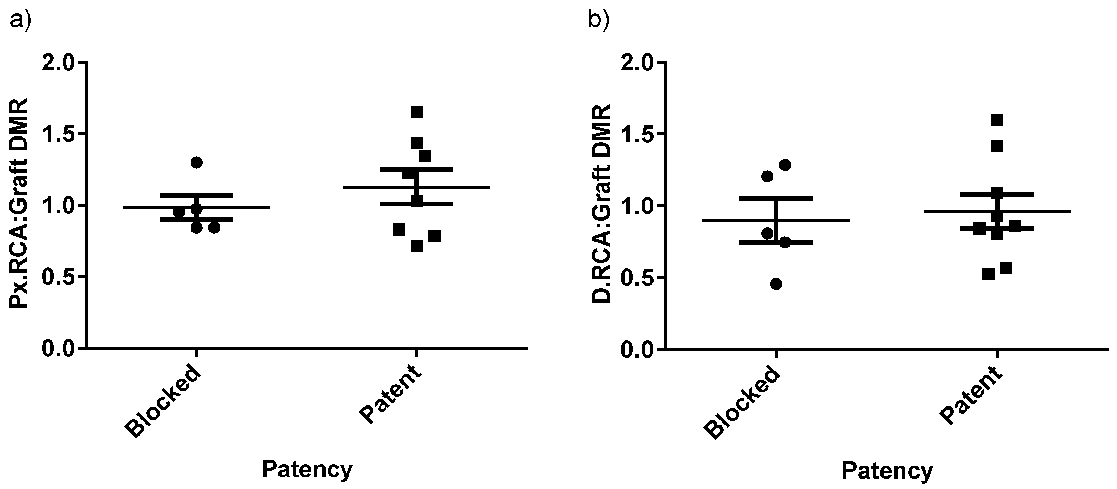
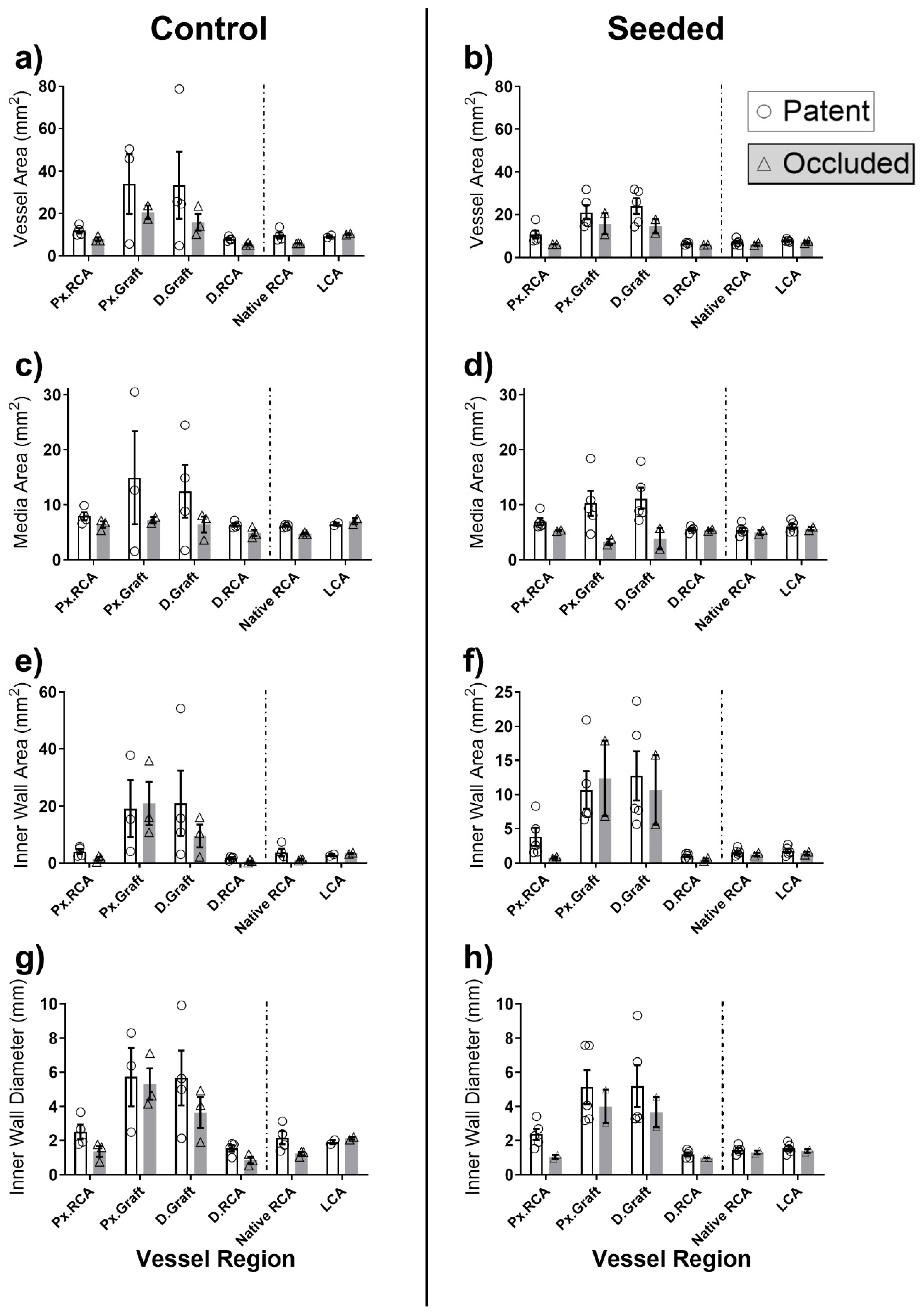
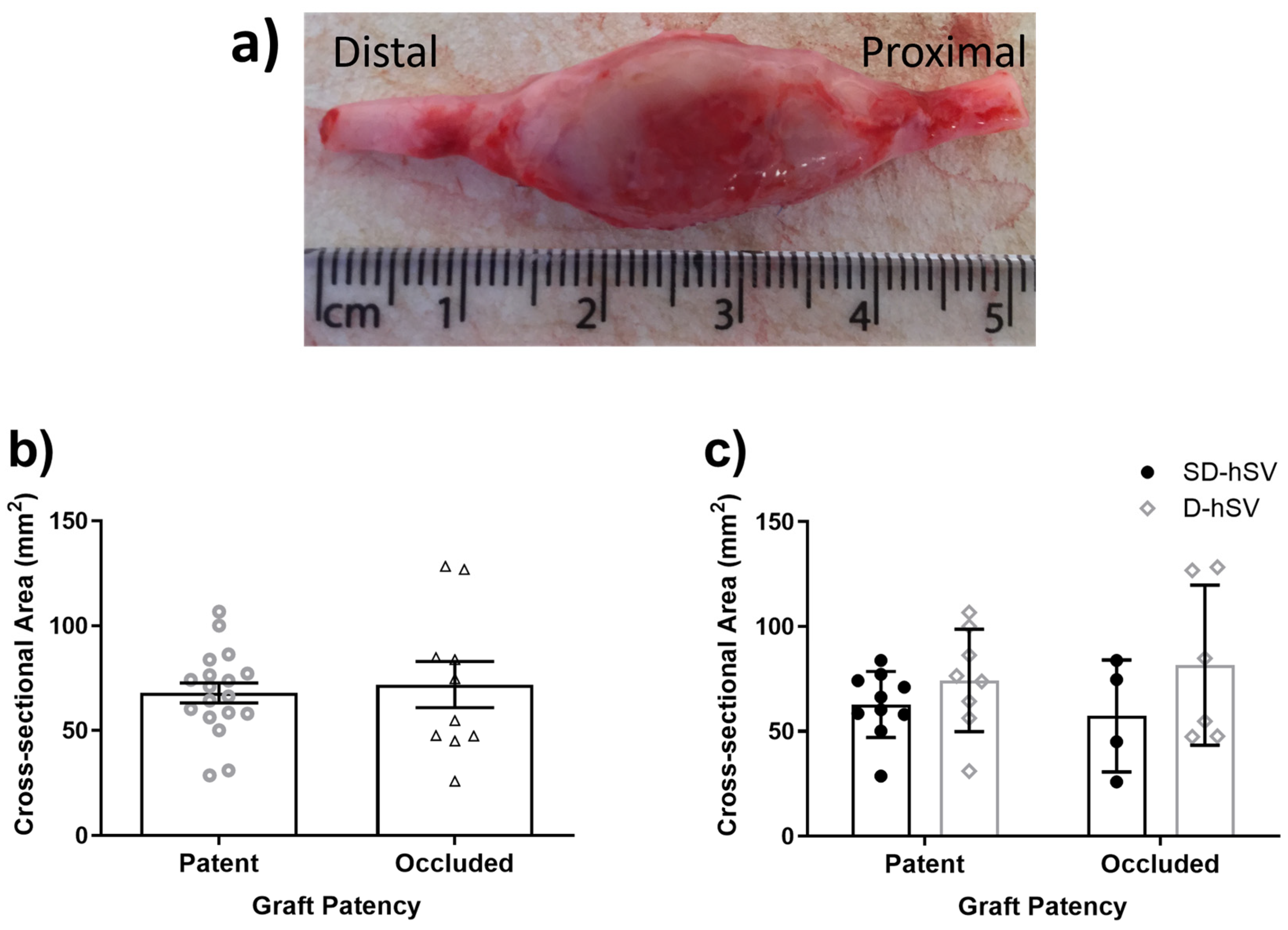
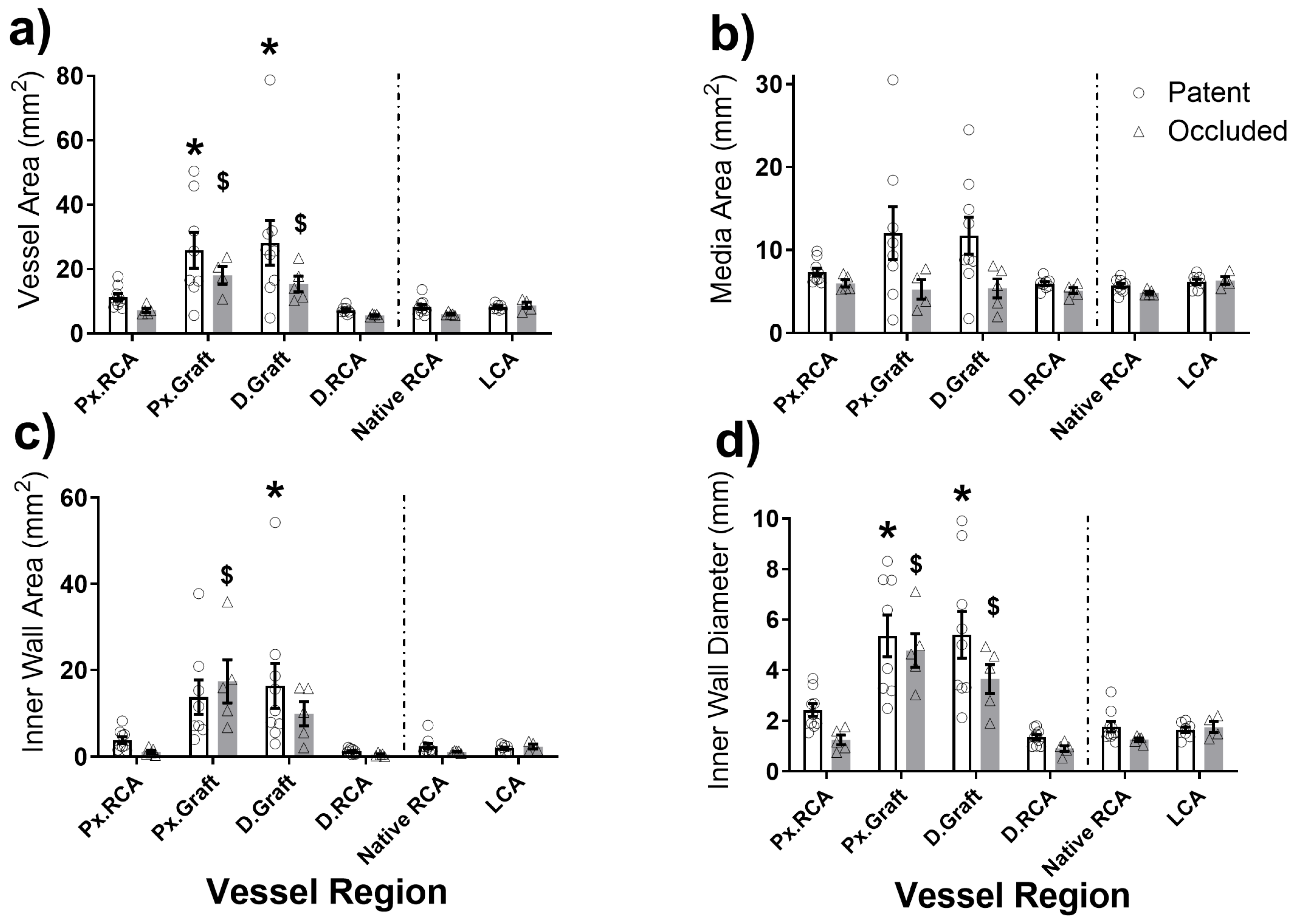
2.2. Graft Patency, Luminal Remodelling, and Intimal Thickening

At 4 weeks, 9/14 (64.3%) of all grafts remained patent with the patency rate of SD-hSV grafts being 71.4% (5/7) vs. 57.1% (4/7) in the D-hSV group (p = 0.58). In vivo ultrasound vascular Doppler (USD) data of the nine patent grafts are shown in Table 1 and Table 2, with no differences between the groups. Similarly, inner wall diameters and the diameter matching ratio (DMR) of patent grafts did not differ between groups, with only the trends for D-hSV grafts showing some outward remodelling (mean DMR > 1), whereas SD-hSV grafts showed remodelling more inwardly (mean DMR < 1). At 4 weeks, the peak blood flow velocity (PBFV) in the proximal (Px.RCA) portion of D-hSVs was significantly reduced compared to post-implant (p < 0.05), while both PBFV and diastolic (DBFV) were significantly reduced in the distal (D.RCA) of SD-hSV grafts (p < 0.05). Other vascular data did not differ, as shown in Figure A3 and Figure A4 and Table A4 and Table A5.
Histological analysis revealed that the average inner diameter of all 14 native RCAs was 1.6 ± 0.1 mm (1.8 ± 0.3 mm and 1.4 ± 0.1 mm for the D-hSV (n = 7) and SD-SV (n = 7) groups, respectively (p = 0.3)). The pre-grafting inner diameter of all 14 D-hSVs was 2.8 ± 0.1 mm with no difference between the groups. Elastin content within pre-grafting RCAs and untouched LCAs at termination was 57.9 ± 3.9% vs. 51.3 ± 4.4% (p > 0.05) (Table A6). As expected, the elastin content of pre-grafting D-hSV grafts (22.7 ± 3.1%), was much lower than the pre-grafting RCA, with no differences between the groups. We have previously shown that the protocol used to derive D-hSVs in vitro is associated with no presence of live residual cells [17]. The pre-grafting coverage with endothelial cells obtained with the ELC seeding of SD-hSV grafts was 61.3 ± 11.3%, although when reassessing after surgery, for the SD-hSV specimens leftover from 5/7 grafts, the luminal surface coverage with endothelial cells was lower at 48.3 ± 8.9%. Of note, the coverage with endothelial cells of RCA segments excised before grafting was 96.7 ± 1.1%. Histological samples at 4 weeks were not fixed under physiological pressure; hence they were subjected to shrinkage, preventing meaningful comparisons with in vivo USD vascular data. Histology confirmed pre-termination USD data, with 9/14 grafts being patent. Intimal thickening was present across the proximal and distal segments of the grafts at 4 weeks (Table 3). This was 54.1 ± 12.2% and 49.2 ± 10.1% for the proximal and distal segments of patent grafts, respectively, with a trend for less intimal thickening between patent D-hSV vs. SD-hSV grafts. No IT was observed at 4 weeks within the native RCA or the untouched left carotid arteries.
Vessel remodelling at 4 weeks is shown in Figure 1a–d. The inner wall diameter of patent grafts increased to 5.4 ± 0.8 mm and 5.4 ± 0.9 mm (proximal and distal graft regions, respectively; both p < 0.05 vs. native RCA) and 4.8 ± 0.7 mm and 3.7 ± 0.6 mm for the proximal and distal regions, respectively, of occluded grafts; both p < 0.05 vs. native RCA (Figure 1d). Data subdivided by control versus seeded vein are shown in Figure A5. At the same time point, the overall diameter of the LCA as an in situ arterial control was 2.0 ± 0.1 mm vs. 1.5 ± 0.1 mm for pigs receiving either D-hSV (n = 4) or SD-hSV (n = 7) grafts, respectively.
A large fibrous capsule was seen surrounding all grafts (Figure A6a) with no differences in size between patent and occluded grafts or between D-hSV and SD-hSV groups (Figure A6b,c; p > 0.05).

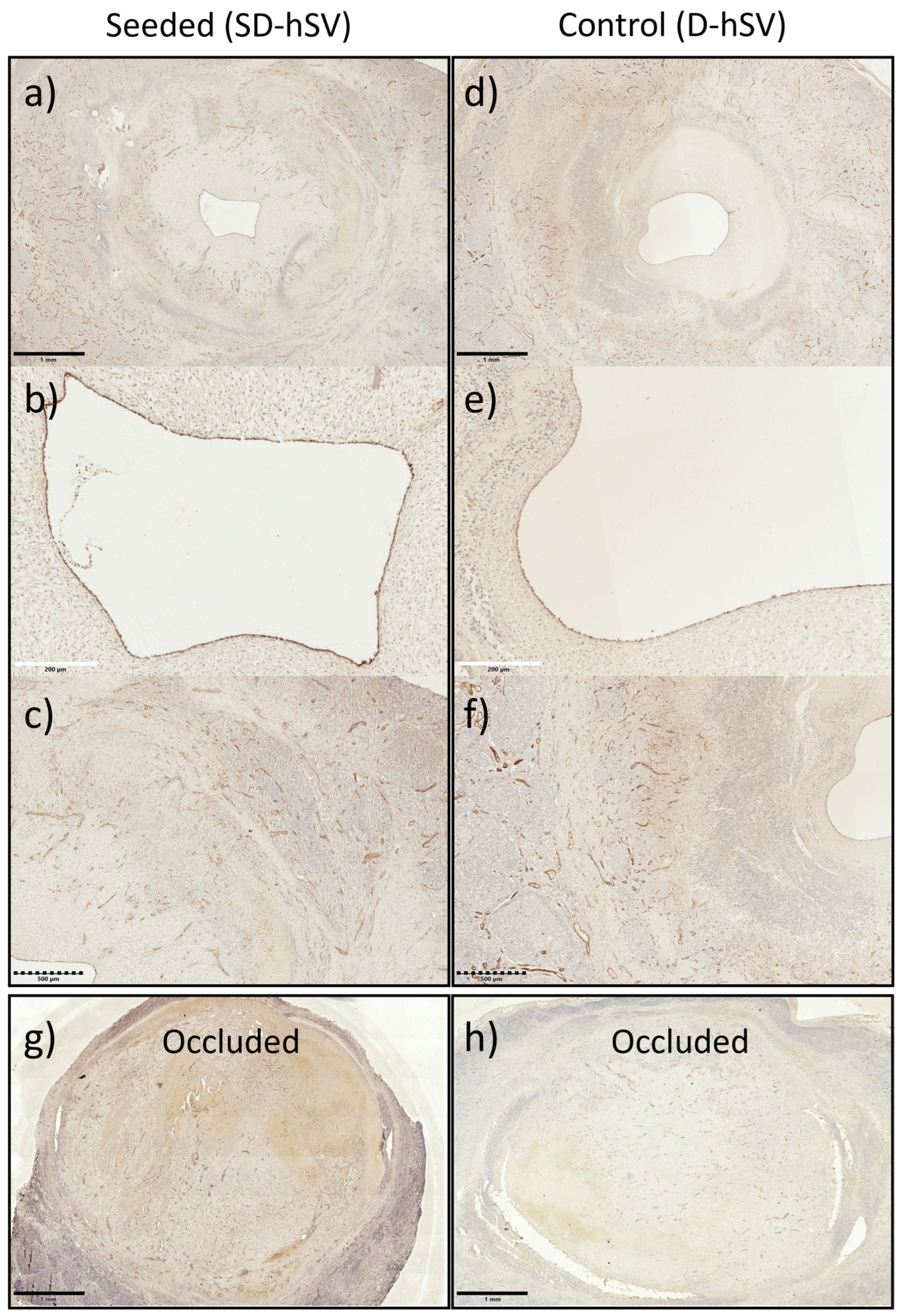
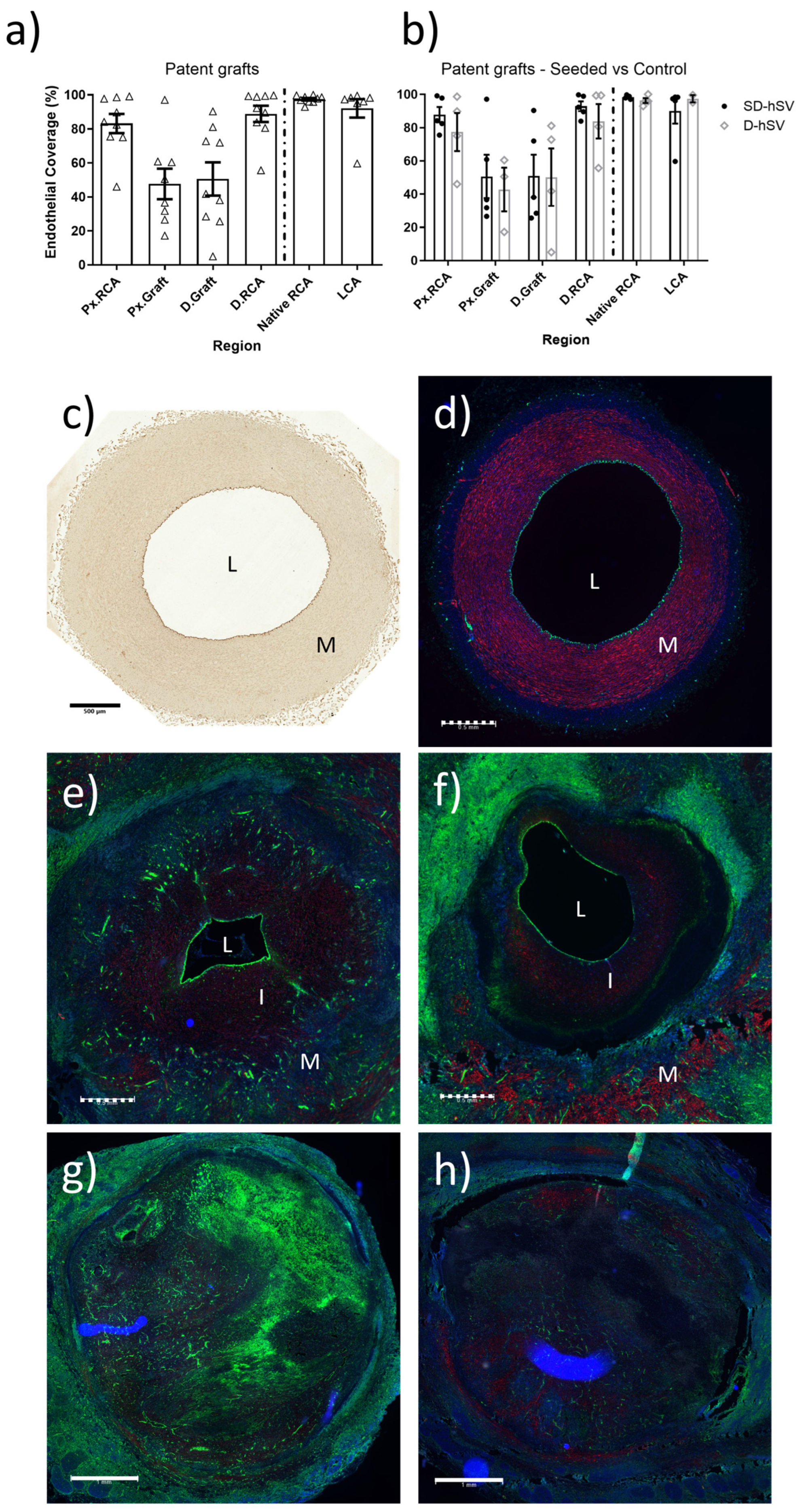
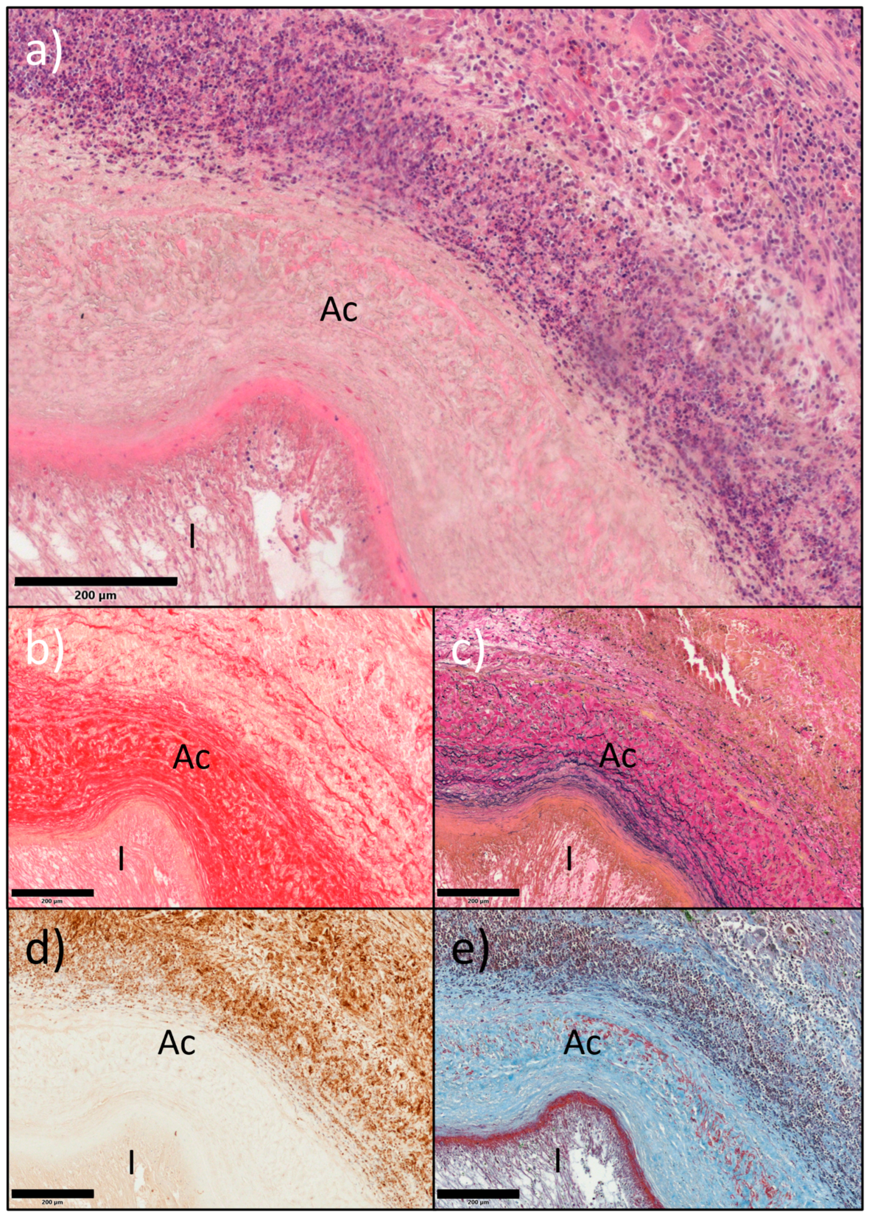
2.3. In Situ Vascular Regeneration by Host Cells

At 4 weeks, all nine patent grafts showed substantial coverage of the luminal surface with CD31-positive endothelial cells (Figure 2). This was higher at the proximal and distal ends of the grafts at the interface with the native RCA and less, but still tangible, in the middle of the grafts (Figure 2a). No difference in endothelial cell coverage was seen between groups (Figure 2b).
H&E staining and nuclear counterstains at 4 weeks showed marked but quantitatively similar intramural cell infiltration in all grafts (11/12 and 8/12 proximal and distal graft regions) across groups and regardless of patent or occluded condition (Figure 3a,d,e). Patent grafts contained large amounts of elastin, collagen, and fibrin (Figure 3b,c,e) and the mesenchymal–endothelial transition marker (vimentin) in the intima and perivascular layers (Figure 3d and Figure 4).
The qualitative evaluation of contractile proteins, calponin, and smooth muscle myosin heavy chain-11 (SM-MHC) indicated the presence of contractile smooth muscle cells in the middle portion of all patent grafts, predominantly within the thickened intima (representative images of an SD-hSV and D-hSV (Figure 4)), with no differences between groups. The presence of these markers was higher in the Px and D. anastomotic regions and very high in the control Px.RCA and D.RCA as expected (Figure 4).
While the elastin content in all grafts was higher at 4 weeks vs. baseline grafts, the abundance was lower than the native porcine RCA or LCA (Figure 5 and Table A6). This difference applied to both patent grafts (2.0 ± 1.4 and 10.1 ± 5.6%; Px. and D.graft, respectively) and occluded grafts (6.2 ± 3.7% and 16.1 ± 7.3%; Px. vs. D.graft, respectively). The elastin content differed across the regions of each graft (p < 0.05) and was consistently higher in the Px.RCA vs. D.RCA (p < 0.05), including when the analysis focused only on patent grafts. Cell seeding did not affect elastin content vs. unseeded controls.
Extensive adventitial neo-angionesis was detected at 4 weeks in all grafts, regardless of patent or occluded and D-hSV or SD-hSV status (Figure A7). Representative examples to demonstrate the co-localisation of DBA (Dolichos Biflorus Agglutinin)-lectin and CD31 in ECs around the patent graft lumens in the same graft are shown in Figure 2e,f and Figure A7a,b,d,e. The sections of the LCA as in situ arterial control at the same time point are shown as positive staining controls for complete endothelium of CD31 and DBA-lectin (Figure 2c and Figure 2d, respectively).

3. Discussion

This study supports the concepts of feasibility, safety, and in situ vascular regeneration when implanting acellular D-hSVs in the pig carotid artery. While there were no feasibility/safety concerns related to suturability, tearing, haematology, aneurysmal, or neoplastic degeneration, 64% of the implanted grafts were patent and showed clear signs of in situ vascular regeneration by host cells. This study also suggests that pre-seeding the acellular graft with pELCs had no beneficial effects.
The 64% patency rate is interesting when considering the xenotransplantation aspect, the lack of immunosuppression, and only 48% of luminal coverage achieved by cell pre-seeding. This may reflect the fact that the human veins were decellularised before implantation and the daily use of 300 mg of Aspirin, a dosage in line with clinical guidelines [20,21]. Noticeably, implanting vein grafts with limited luminal coverage by endothelial cells is typical of the current surgical practice, as the marked loss of endothelial cells is associated with harvesting/handling SVs for CABG/PABG surgery [22], along with reduced cell viability [23], triggering more effective methods of SV harvesting [24] and of endothelial preservation [25]. The lack of immunosuppression in this study is in keeping with the established clinical practice of implanting commercial patches or artificial biological heart valves made from decellularised/acellular bovine pericardium in humans every year [26].
A key finding was the occurrence of signs of in situ vascular regeneration by host cells, including extensive re-endothelialization (expression of CD31 and DBA-lectin), intramural recellularization with mesenchymal–endothelial transition cells (vimentin), smooth muscle cells/SMCs (contractile proteins SM-MHC-11, calponin), and adventitial neo-angiogenesis. This was combined with marked extracellular matrix remodelling (elastin and collagen) and the pulsatility index. The luminal surface coverage with ECs was dense at the proximal and distal parts of the grafts, suggesting migration across the anastomoses from the adjacent RCAs. A large number of ECs were also observed in the mid segment, possibly migrating from the RCAs or resulting from circulating progenitors. While we can be sure that 100% of ECs seen in the control D-hSV group were host cells, this cannot be confirmed for the SD-hSV group, as those grafts had an average pre-seeding coverage of 48% before implantation. Future studies could use cell labelling approaches (e.g., green fluorescent protein (GFP) [27], or implanting male donor cells into female recipients and performing fluorescent in situ hybridization [28]) to determine whether CD31-positive cells at termination are host-derived or residual seeded cells. Quint et al. [27] decellularised tissue-engineered grafts grown from banked porcine smooth muscle cells and then re-endothelialised the obtained extracellular matrix with endothelial progenitor cells (EPCs) or endothelial cells from the intended porcine recipient, achieving 65% lumen coverage before implantation into the porcine carotid artery. In total, 3/3 of their nude grafts were occluded, while 5/5 of the re-endothelialised grafts were patent at 30 days. For 4/5 of these patent grafts, the endothelial coverage was made up of 35% of the seeded GFP-labelled cells, whereas the remaining 65% consisted of host cells. Our study differs markedly from the work by Quint and colleagues regarding graft preparation, and this might explain the different outcome related to the nude graft, with our study showing >50% of nude grafts being patent at 30 days, with extensive re-endothelialization by host cells. Yet, both studies appear to indicate that pre-seeding with ECs may not be beneficial for vascular tissue engineering, as most if not all of the cells present in the graft after in vivo exposure appear to be host cells. This is in keeping with relevant findings by others, suggesting that monocytes seeded onto a mouse venous tissue-engineered graft rapidly disappear within 2 h after implant in vivo [29].
A large amount of transmural SMCs were also observed at 4 weeks with an abundance of SM-MHC-11 and calponin proteins. Again, this was more pronounced at the proximal and distal ends vs. the mid portion of the grafts, although the number of SMCs observed was lower than that seen in the native RCA. While we can speculate that these SMCs migrated across the anastomoses from the native RCA or were derived from circulating progenitors, more work is needed to clarify their origin. Based on their anatomical position within the wall of the graft, it is unlikely that these SMCs could have migrated from the adventitial layer, given that most of them appear to be located within the neointimal region and inside the D-hSV layer (Figure 4). In this same region, there was also an abundance of vimentin, regarded as a mesenchymal–endothelial transition marker. This might indicate that both contractile and synthetic SMCs were in phenotypic flux, explaining why some contractile markers were also present.
Exposing veins to the higher shear stress of arterial circulation (10–70 dyn/cm2 in arteries vs. 1–6 dyn/cm2 in veins [30,31]) and higher partial oxygen pressure leads to intimal thickening [32]. Adaptation to these dramatic changes may be of utmost importance to maintain graft patency. The USD data of this study on the inner wall diameter at 4 weeks showed an occurrence of outward remodelling with a 57% increase in inner wall diameter. This was not uniform, as the inner wall diameter increased by 11% in the proximal segments of patent grafts, decreased by 2.7% in the proximal segments of occluded grafts, and increased by 35% in the distal segments of patent grafts, becoming similar to pre-implanted RCA segments. The increase in inner wall diameter at USD at 4 weeks was reflected by a tangible increase in elastin content. Elastin is needed in arterial vessels to provide the required elasticity/pulsatility during the cardiac cycle and contribute to the PI level, which reflects the resistance in a pulsatile arterial system [33]. In this study, the average PIs for the porcine RCA prior to surgical excision and the LCA at termination were 4.3–4.9 and 2.7–3.9, respectively. Noticeably, implanted grafts showing a PI < 5 soon after implantation remained patent at 4 weeks, while those with a PI > 5 soon after implantation were occluded at 4 weeks, while no differences in the PI were seen between patent D-hSV and SD-hSV grafts (Table 2). These differences might simply reflect baseline differences in D-hSV quality and wall thickness.
Five grafts were occluded at 4 weeks. Early vein graft thrombosis is associated with suboptimal surgical techniques, a poor quality of conduit [34,35], and graft-to-artery mismatch [36] or poor run-off. To minimise the rate of early thrombosis, surgery was undertaken by senior heart surgeons on large pigs (60 kg) to gain good-sized porcine RCAs with good size matching with the human D-hSV grafts. However, the implanted D-hSVs were derived from SV segments discarded at human surgery and therefore not of the ideal quality. This factor might have contributed to the observed 5/14 graft occlusions observed in this study, in keeping with the notion that up to 12% of SV grafts are considered too diseased at CABG surgery, leading to a 50% occlusion rate compared to non-diseased SV grafts [37]. Alternatively, these occlusions might be due to the xenotransplantation aspect of implanting human SV grafts in pigs. However, the decellularization method used has shown no residual viable cells on the D-hSV grafts [17], and 9/14 grafts were patent at 4 weeks despite xenotransplantation. A fibrous capsule was observed at 4 weeks in all grafts with infiltrating inflammatory cells and lymphoid-like nodules. This could indicate the presence of residual human epitopes on the implanted grafts, possibly triggering rejection. However, in a parallel study using autologous porcine venous grafts in the same RCA porcine model, we did not observe this adventitial fibrous capsule (Figure A8). This may suggest that the presence of a fibrous capsule is unlikely in a clinical research setting designed to use autologous D-hSV.
This study has limitations. It might be argued that the sample size was small. However, this was a feasibility/safety pilot study; therefore, with no sample size calculation, we included seven animals in each group, which is in keeping with the large animal sample sizes used by others [27]. In relation to the observed signs of in situ vascular regeneration by host cells, the follow-up period of 4 weeks might be regarded as too short. Keeping the animals for longer might have helped ascertain if the level of in situ vascular regeneration by host cells is time-dependent. In addition, the carotid artery interposition graft model used is not fully relevant to the CABG/PABG surgical practice. In addition, all pigs used in this study were disease-free. Based on the findings from this study, future studies are planned, and for added translational benefit, could potentially additionally use hypertensive or diabetic pig models. Finally, an additional limitation of our study is that we used only female pigs and therefore this study may not account for sex-based differences in vascular biology, immune response, and vascular remodelling typical of male pigs. However, we used only female pigs to minimise intra-experimental variability to increase the robustness of the findings while minimising animal husbandry issues associated with using a mixed gender cohort of animals [38].

4. Materials and Methods

Methods are extensively shown in Appendix A.1. All data underlying this manuscript are included here or in the online Appendix A. Full raw data can be made available on request.

4.1. Ethics

D-hSVs were produced in the lab as by-products from segments of human SVs left over from patients undergoing CABG procedures under approval by the local Research Ethics Committee (Ref. 10/H0170/63). Donors gave generic consent for research use as part of the donation process and are not identifiable. This study conformed to the Declaration of Helsinki principles. The animal procedures were conducted at the MHRA-compliant Translational Biomedical Research Centre (TBRC, Bristol, UK) in line with UK Home Office regulations (Animal Act 1986) and the guidelines from Directive 2010/63/EU under Project Licences 30/2854 and 30/3064, granted by the Home Office after approval by the local Animal Welfare and Ethics Review Body (AWERB). Reporting of research data was in keeping with the ARRIVE (Animals in Research: Reporting In Vivo Experiments) guidelines [39].

4.2. Human Saphenous Vein Decellularisation

Human SVs (n = 10) were collected under sterile conditions, stored in saline at 4 °C, and subsequently decellularised as previously described [17]. D-hSVs were stored in Dulbecco’s phosphate-buffered saline (DPBS; Sigma-Aldrich, Gillingham, UK) at 4 °C until use. Of the 10 veins collected, four were long enough to be divided into two segments and used for two implants in pigs (Table A1), giving a total of 14 grafts implanted.

4.3. Porcine Endothelial-like Cell (ELC) Isolation and Seeding

ELCs were derived from pig blood (n = 3) as previously described [19]. Briefly, peripheral blood mononuclear cells (PBMCs) were isolated from porcine blood using density gradient separation with Ficoll Paque Plus 1077 (density 1.077 g/mL; GE-Healthcare, Hatfield, UK) in Sepmate50 tubes (Stemcell Technologies, Cambridge, UK). Monocytes were allowed to adhere onto culture plates coated with type-I rat tail collagen (Gibco, Waltham, MA, USA) and cultured in ELC media (endothelial cell growth medium-2 Bulletkit (Lonza, Cambridge, UK; CC-3162), including additional foetal bovine serum (FBS, heat inactivated, Gibco) to a final concentration of 11%), until growth of ELCs. ELCs were characterised by a clear cobblestone morphology and the co-expression of the endothelial cell markers CD31, VE-cadherin, and von Willebrand factor via immunocytochemistry. After expansion, cells were cryopreserved in CellBanker 2 (Amsbio, Abingdon, UK) until required. Two weeks prior to surgery, cells were thawed, expanded in 75 cm2 flasks (Corning, Glendale, AZ, USA) at 37 °C/5% CO2 for one week in ELC media. Cell seeding is reported in Appendix A.1.1. Briefly, one week before surgery, ELCs were enzymatically detached from flasks at 37 °C, tested/counted for viability, and pipetted into the lumen of the selected D-hSV segment, with the distal end tied with a 4-0 prolene suture, at 1 × 10⁶ ELC per 1 cm of graft, suspended in 50–100 µL of ELC media. The proximal end was then tied with a suture. Seeded D-hSVs (SD-hSVs) were then placed in 3 mL of ELC media in a 5 mL vented cap tube, on a roller at 1 rpm (37 °C, 5% CO2) for 1.5 h, then the tubes containing tissue were inverted and placed back on the roller to enhance the uniform seeding of cells across the conduit. The following day, the inversion process was repeated, and media replenished. After 96 h, the ties were removed, and the resulting SD-hSVs were placed into 5 mL pre-warmed ELC media under static conditions until implantation. On day 7 post-seeding, small samples from the ends of D-hSVs (n = 7) and SD-hSVs (n = 7) were removed and fixed in 10% (v/v) formalin to be used as pre-grafting controls, while the mid segments were kept in ELC media at 37 °C and taken to the TBRC facility for surgical implantation. The time from the initiation of seeding to surgical grafting was approximately 140 h.

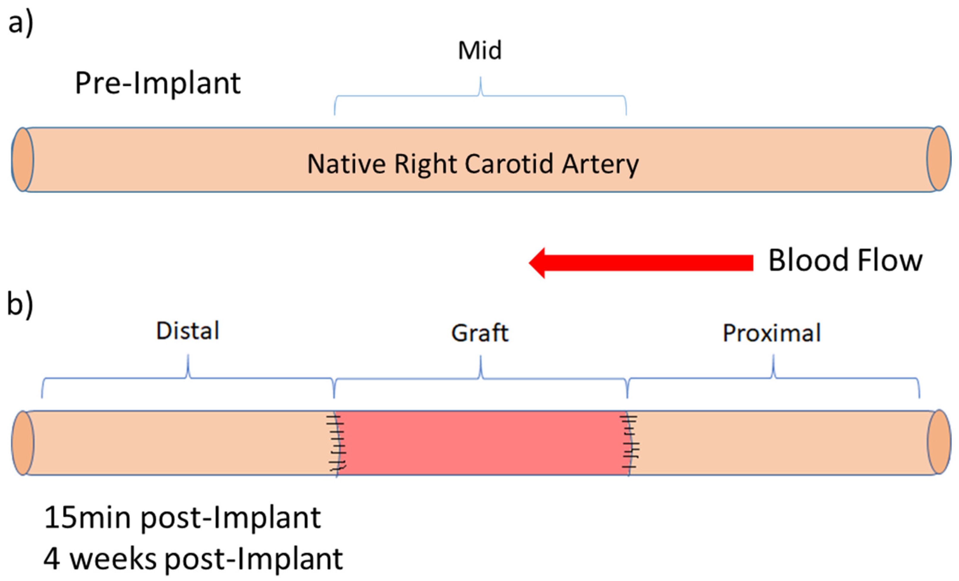
4.4. In Vivo Surgical Grafting and Vascular Doppler Acquisition

D-hSV grafts with (SD-hSV: n = 7) or without (D-hSV: n = 7) pre-seeded ELCs were randomly and blindly implanted in the right carotid artery (RCA) position in female landrace large white pigs (n = 14; weight 61.2 ± 1.0 kg; age 3.9 ± 0.0 months—Table A1) under general anaesthesia (GA), as previously reported [17]. Animals received 75 mg of Aspirin with food daily during the 5 days before surgery, followed by 300 mg of Aspirin daily from day 1 after surgery until termination. No immunosuppression was used. A skin incision was made at the right neck to expose the RCA. Heparin was administered to achieve an activated clotting time >250 s. Following clamping, a 0.5 cm segment of the mid RCA was excised (fixed in 10% (v/v) formalin as histology control), and 1.5–2.0 cm long D-hSV or SD-hSV grafts were interposed using 7-0 Prolene monofilament running sutures. Vascular clamps were removed, and grafts were spread externally with papaverine (30 mg/mL, Torbay Pharmaceuticals, Paignton, UK) to prevent spasm. Ultrasound vascular Doppler (USD) imaging (Edge II ultrasound machine, with HFL38xi 13-6 MHz linear transducer; Fujifilm SonoSite, Bothell, WA, USA) was acquired of the native mid RCA before grafting and of the proximal RCA, implanted grafts, and distal RCA 15 min and 4 weeks after grafting (Figure A1).

4.5. Outcomes Measures

4.5.1. Microbiology

To assess the microbiological contamination of the implanted grafts, 100 µL of remaining media was plated onto sheep blood agar (Oxoid, Basingstoke, UK) and incubated at 37 °C, 5% CO2 for 24 h. Plates were then assessed for visible colonies indicating contamination. In addition, ELC culture media was tested for the presence of mycoplasma using a PCR Mycoplasma Test Kit, following the manufacturer’s protocols (Promocell, Heidelberg, Germany).

4.5.2. Haematology

Four mL of venous blood was collected into vacutainer EDTA K2 tubes (1.8 mg/mL; Becton Dickinson, UK) at baseline and at 4 weeks post-implant and stored at 4 °C. Haematological screening was conducted by a commercial diagnostics lab (Langford Vets Diagnostic Laboratories, Bristol, UK). NLR and LMR were calculated as indicators of systemic inflammation and vein graft failure, respectively [40].

4.5.3. Feasibility/Safety Measures

At surgical implant and 4 weeks, grafts were assessed for suturability, tearing, bleeding, rupture, and haematoma via direct vision and by USD. Histology was then carried out to test for infection, graft occlusion (luminal thrombus formation), excessive dilatation, aneurysm formation, and neoplastic degeneration.

4.5.4. Patency, Intimal Thickening, and Vascular Regeneration

Graft patency was assessed via USD and histology, while intimal thickening (IT) and signs of vascular regeneration were assessed via immunohistochemistry (luminal coverage with endothelial cells (CD31 and DBA–lectin), extracellular matrix remodelling (elastin, collagen), the presence of intramural mesenchymal–endothelial cell transition (vimentin), contractile proteins (SM-MHC-11, calponin) and neo-angiogenesis in the adventitial area (see Methods in Appendix A)). USD measures included DMR as a ratio of the inner diameter of the proximal (Px.)RCA or distal (D.)RCA to the graft to determine mismatch between the graft and native vessel. Graft patency was defined as the persistence of a patent lumen at pre-termination. We also assessed the length of the graft at implant, blood flow velocity (via pulse wave Doppler; PWD), systolic/peak (PBFV) and diastolic (DBFV) flow velocities, and the pulsatility index (PI = (PBFV − DBFV)/Time-averaged flow velocity) as markers of vessel/graft elasticity.

4.6. Statistical Analysis

To test for a statistically significant difference in outcome measures between seeded and non-seeded grafts, a chi-square test was used. Comparisons of Doppler ultrasound data between time points (implant vs. explant), or pig graft outcomes (patent vs. occluded) were performed using an independent samples t-test. When analysing graft data, due to heterogeneity along the h-SVs, each conduit was treated as an individual entity due to the small number of paired samples. Differences between regions were determined using either an ANOVA, or univariate analysis of variance, and post hoc Tukey tests. Differences were deemed significant when the p-value was less than 0.05. IBM SPSS Statistics (Version 26) was used for all analyses.

5. Conclusions

In conclusion, the use of D-hSVs for vascular grafting in arterial position is feasible and safe and is associated with signs of in situ vascular regeneration by host cells at 4 weeks. Pre-seeding with porcine ELCs was not effective. More studies are warranted with a focus on long-term outcomes.

Author Contributions

Conceptualization, A.B., J.J., S.G. and R.A.; Formal Analysis, A.B.; Funding Acquisition, J.J., S.G. and R.A.; Investigation, A.B., V.B., R.A. and N.S.; Methodology, A.B., V.B. and R.A.; Project Administration, R.A.; Supervision, J.J., S.G. and R.A.; Writing—Original Draft, A.B. and R.A.; Writing—Review and Editing, A.B., V.B., N.S., J.J., S.G. and R.A. Co-senior Authorship, S.G. and R.A.; Corresponding Authorship, R.A. All authors have read and agreed to the published version of the manuscript.

Funding

This research was funded by a British Heart Foundation (BHF) Grant (PG/16/104/32652) to Ascione. The TBRC is co-funded by infrastructure grants from the Medical Research Council (MR/L012723/1) and the BHF (IG/14/2/30991).

Institutional Review Board Statement

Human tissue was obtained following approval by the local ethics committee (REC reference number 10/H0170/63). All animals used in this study were carried out under the UK Home Office License PPL/30/2854.

Informed Consent Statement

In line with the content of ethical approval, patient consent was waived due to surplus samples used for this study (instead of being disposed) being anonymized to prevent tracking back the patient.

Data Availability Statement

All datasets presented in this manuscript can be made available upon reasonable request.

Acknowledgments

We thank the staff at the Bristol Translational Biomedical Research Centre/TBRC, a national research facility for large animal and staff at the Wolfson Bioimaging Facility, University of Bristol, for their assistance with fluorescence imaging. We thank Daniel Baz-Lopez for helping with some lab-based work.

Conflicts of Interest

The authors declare no conflicts of interest with respect to the research, authorship, and/or publication of this article.

Abbreviations

The following abbreviations are used in this manuscript:
CACarotid artery
CABGCoronary artery bypass grafting
DBADolichos Biflorus Agglutinin
DBFVDiastolic blood flow velocity
D-hSVDecellularised human saphenous vein
DMRDiameter matching ratio
D.RCADistal right carotid artery
ECsEndothelial cells
ECFCsEndothelial colony-forming cells
ELCsEndothelial-like cells
ITIntimal thickening
LCALeft carotid artery
LMRLeukocyte/monocyte ratio
MSBMartius scarlet blue
NLRNeutrophil/lymphocyte ratio
PABGPeripheral artery bypass grafting
PBFVPeak blood flow velocity
pELCsPorcine endothelial-like cells
PIPulsatility index
PWDPulse wave Doppler
Px.RCAProximal right carotid artery
RBCRed blood cell
RCARight carotid artery
SD-hSVD-hSV seeded with ELC
SMCsSmooth muscle cells
SM-MHCSmooth muscle myosin heavy chain
SVSaphenous vein
SVGsSaphenous vein grafts
USDUltrasound vascular Doppler

Appendix A

Appendix A.1. Expanded Methods

Appendix A.1.1. Cell Seeding of Decellularised Human Saphenous Vein

One week prior to graft surgery, ELCs were enzymatically detached from flasks (TryplE Express, Gibco, Waltham, MA, USA) at 37 °C, and viable cells were counted and seeded into D-hSV as follows: using a blunt-ended needle, 2 mL of ELC media was gently perfused through the lumen of selected D-hSV to check for the presence of valves and to determine the distal end. The distal end was tied with a 4-0 silk suture, and approximately 1 × 10⁶ ELC per 1 cm of graft were pipetted into the lumen, in 50–100 µL of ELC media, and the proximal end was tied to trap the cells within the vein. Seeded D-hSVs (SD-hSV) were rinsed gently in DPBS to remove any cells from the adventitial surface that may have escaped from lumen, before being placed in 3 mL of ELC media in a 5 mL vented cap tube, on a roller at 1 rpm (raised at an angle to prevent media leakage; 37 °C, 5% CO2). After 1.5 h, the veins were inverted in the tube to minimise the pooling of cells at one end of the conduit. The following day, this inversion process was repeated, and media replenished. Approximately 96 h post-seeding, veins were removed from the roller, and ties were cut from the ends. The remaining SD-hSV were placed into 5 mL pre-warmed ELC media in a 6-well plate under static conditions until the day of implant. On the day of surgery, before transport to the surgical facility, the ends of SD-hSV (n = 7) or control D-hSV (D-hSV; n = 7) were removed and fixed in 10% formalin to assess cell seeding success and obtain pre-grafting morphology. Grafts were transported to the TBRC facility, Langford, UK, in ELC media warmed to 37 °C, for surgical implantation. SD-hSV were transported to TBRC on day 7 post-seeding.

Appendix A.1.2. Doppler Assessment of Grafts

After surgical exposure, the HFL38xi 13-6 MHz linear transducer (Fujifilm SonoSite, Bothell, WA, USA) was placed directly onto the perivascular surface of the vessel being imaged to improve image quality. Doppler measurements were taken pre-grafting in the mid region of the native RCA, 15 min post-grafting in the proximal RCA (Px), distal RCA (D), and whole graft regions, and 4 weeks later under general anaesthetic, before termination, in the same regions.
Prior to termination, the left carotid artery (LCA) was exposed and Doppler measurements were taken as an un-grafted control (n = 10). 2D Doppler images were taken at each of the locations, and the inner wall diameter for each location was measured 3–5 times within each image, and the average diameter was calculated for each location. To determine whether there was any mismatch of diameters of grafts and the native RCA, a diameter matching ratio (DMR; ratio of the inner diameter of the Px.RCA or D.RCA to the graft) was calculated. A DMR of one indicates perfect diameter matching, whereas >1 was indicative of vessel narrowing in the graft. In addition, the length of the graft in situ was measured at implantation. PWD was used to obtain traces of blood flow velocity; the systolic/peak (PBFV) and diastolic (DBFV) flow velocities were measured and averaged over two cardiac cycles. The pulsatility index (PI = (PBFV − DBFV)/Time-averaged flow velocity) was obtained from Doppler traces for the elasticity of the vessels at each location, respectively.

Appendix A.1.3. Histological Assessment of Grafts

Following termination at 4 weeks post-implant, excised conduits were divided into the proximal RCA (Px.RCA), proximal graft (Px.Graft), distal graft (D.Graft), and distal RCA (D.RCA) and either fixed in 10% (v/v) formalin (n = 12 pigs) or snap frozen in liquid nitrogen (n = 2 pigs) for histological processing. Tissue sections were cut at 5 μm thickness, and serial sections stained with Elastin Van Gieson (EVG) for elastin content and conduit morphology measurements, and fluorescently dual-stained with the porcine marker of endothelial cell glycocalyx, DBA (Dolichos Biflorus Agglutinin) lectin (Vector Laboratories, 25 µg/mL), and α-smooth muscle actin (smooth muscle cell marker). In addition, paraffin-embedded tissue sections were stained with picrosirius red (PSR) for collagen, or Martius scarlet blue (MSB) for thrombus formation. Sections of patent grafts were additionally immunostained using markers for endothelial cells (CD31), contractile smooth muscle cells (calponin and smooth muscle myosin heavy chain 11), and fibroblasts/mesenchymal cells (vimentin), using a DAB peroxidase substrate. The details of all antibodies used are in Table A7. Images were captured on an O8 Slidescanner (PreciPoint GmbH, Munich, Germany) equipped with a 20× objective. Imaging of fluorescently stained sections was carried out using a Leica DMI6000 inverted epifluorescence microscope equipped with an automatic motorised stage (Leica Microsytems, Wetzlar, Germany). Images of EVG-stained sections of the different regions of the vessels and graft, as described above, were opened in Viewpoint Light (1.0.0.9628v, PreciPoint GmbH, Munich, Germany), and the area and perimeter of the lumen, inner wall, outer wall, and area of fibrous capsule surrounding the graft were measured. The area of the vessel media (outer wall area minus inner wall area), the area of intimal thickening (inner wall area minus lumen area), and the percentage stenosis (intimal thickening area/inner wall area*100) were calculated. Inner diameters of the conduit were calculated from the measured perimeters of the inner wall (diameter = perimeter/π). For measurement of elastin content, images of EVG-stained sections, including regions of interest previously drawn in Viewpoint Light, were opened in image analysis software (ImageJ 1.52v, National Institute of Health, USA). Using left carotid artery images as standards, the pixel intensity of elastin staining was determined, and colour deconvolution based on these values was applied to all subsequent images. This enabled the percentage of elastin-stained tissue within the medial conduit layer to be determined. To determine endothelial lumen coverage, images of DBA-lectin-stained tissue were opened in ImageJ. Lumen perimeter length was measured, where possible, along with the length of lumen covered with DBA-lectin-positive cells, to derive the percentage of luminal surface covered with endothelial cells.

Appendix A.2. Results

Appendix A.2.1. Haematological Screening

Results from baseline samples showed normal levels of leukocytes for 9/10 pigs (11.3–22.8 × 10⁹/L), whereas one control pig had elevated leukocyte levels (26.41 × 10⁹/L). Lymphocyte levels were higher than reference ranges at both baseline and 4-week time points. At baseline sample, control pigs had lower levels of monocyte and basophil levels than those receiving seeded grafts (p < 0.05); however, this difference was not seen at 4 weeks. The baseline leukocyte/monocyte ratio (LMR) was higher in D-hSV vs. SD-hSV (p < 0.05). No changes in immune cell levels were noted in pigs that had occluded grafts at 4 weeks vs. those with patent grafts. The fold-change from baseline to explant could not be calculated for eosinophils or basophils, as some samples did not contain any of these cell types.

Appendix A.2.2. Other Vascular Doppler Results

Doppler assessment showed that the length of implanted grafts ranged from 1.0 to 2.3 cm (mean of 1.70 ± 0.08 cm) with no difference between patent and occluded grafts at 4 weeks (Figure A4). Serial Doppler ultrasound scanning data at pre-implant, immediately post-grafting, and at 4 weeks are shown for inner wall diameter measurements and blood flow velocities in Table 1 and Table 2, respectively. Inner wall diameters (excluding any intimal thickening or thrombus formation) were measured, and the ratio of the Px. or D.RCA to the graft (the DMR) was calculated for grafts that were either patent or occluded after 4 weeks (Figure A3). Native RCA, prior to surgical intervention, had a diameter of 0.45 ± 0.02 cm (n = 13). At the time of implantation, there were no significant differences in the diameters of grafts that remained patent or those that occluded, and the DMR of the native RCA to graft was 1.24 ± 0.08 (n = 13). At the end of the study, there was no difference between the inner wall diameter in either the Px.RCA or graft itself; however, distally of occluded grafts, there was an approximately 35% reduction in diameter compared to those that remained patent (p < 0.01). The distal region was also the only region that significantly changed diameters from the time of implant to explant (~35% increase, p < 0.05), but only in patent grafts. Occluded grafts remained unchanged. Once implanted, the DMRs for all grafts for Px.RCA/graft and D.RCA/graft were 1.07 ± 0.08 and 0.94 ± 0.09, respectively. The DMR was similar between the blocked and patent grafts at the time of implant and explant, and there were no differences in the DMR between grafts at implant and explant. Pulse wave Doppler (PWD) was used to determine mean blood flow velocity data (including pulsatility index—PI). It was not always technically possible to obtain a full dataset for each pig at every time point and region and if, for example, flow was very low or turbulent, then PI values could not be obtained. Baseline native RCA and native LCA (non-surgical age-matched controls) values are also shown as indicators of target values (Table 2). At the time of implant, there were no differences between PWD parameters for grafts that went on to occlude or not, and flow velocities were similar to native RCA pre-implant values. However, Px.RCA and D.RCA systolic and diastolic flow velocities for grafts that occluded were significantly lower than those that stayed patent. At the end of the study, measurements of the PI could not be recorded for occluded grafts. As such, statistical analysis was not performed on these data. The diastolic blood flow velocity in D.RCA for patent grafts was significantly lower (p < 0.01) at the time of explant (31.9 ± 9.5 cm/s), compared to the time of implant (70.2 ± 8.6 cm/s) (Table A5).
Figure A1. In vivo vascular Doppler acquisition sites. (a) Pre-implant at mid native right carotid artery (RCA) location. (b) 15 min post-implant reperfusion, and 4 weeks later, in RCA regions proximal and distal to graft, and within graft.
Ijms 26 04718 g0a1
Figure A2. Luminal thrombus formation. Representative images of thrombus formation in grafts. (a) Seeded and (b) control patent grafts, (c) chronic occluded graft, and (d) small clot within the lumen of the patent graft. Black bar indicates 500 mm. All tissue stained with Martius scarlet blue (MSB) for red blood cells (green/yellow, arrows), fibrin/muscle (red, arrowhead), and collagen (blue).
Ijms 26 04718 g0a2
Figure A3. (a) Proximal RCA (Px.RCA)/graft; (b) distal RCA (D.RCA)/graft diameter matching ratio (vascular Doppler ultrasound data) for individual grafts remaining patent (n = 8) or blocked (n = 5) at 4 weeks.
Ijms 26 04718 g0a3
Figure A4. Graft length at time of implant assessed by vascular Doppler ultrasound, grouped based on whether patent or occluded after 4 weeks in situ.
Ijms 26 04718 g0a4
Figure A5. Histological measurement of vessel area (a,b), medial area (c,d), inner wall area (e,f), and inner wall diameter (g,h) of excised segments across patent (white bars) or occluded (grey bars) grafts; control (a,c,e,g) and seeded (b,d,f,h). Pre-implant native RCA and explant LCA measurements are also included for comparison. RCA—right carotid artery, Px.—proximal, D.—distal, and LCA—left carotid artery as control.
Ijms 26 04718 g0a5
Figure A6. Fibrous capsule in all grafts. At termination, implanted grafts were encased in a fibrous capsule (a). (b) Cross-sectional area of patent and occluded grafts and (c) for seeded (SD-hSV) and control (D-hSV) grafts.
Ijms 26 04718 g0a6
Figure A7. Staining for endothelial cells across the explanted grafts. Representative images of endothelial cell (CD31/PECAM-1) immunohistochemistry staining (brown) of seeded (SD-hSV; (ac,g)) and non-seeded control (D-hSV; (df,h)) grafts that remained patent (af) or occluded (gh). Graft cross-section overview (a,d,g,h), graft lumen (b,e), and perivascular region (c,f). Scale bars represent 1000 μm (black solid), 200 μm (white), and 500 μm (black dashed).
Ijms 26 04718 g0a7
Figure A8. Autologous porcine arteriovenous graft (saphenous vein into carotid artery position) stained with Elastin Van Gieson. Scale bar = 2 mm.
Ijms 26 04718 g0a8
Table A1. Pig identification numbers, ages at implant, weights, human saphenous vein (hSV) used, treatment, and graft outcome following 4 weeks in situ.
Pig IDAge at Implant (Months)Weight at Implant (kg)Weight at Termination (kg)Weight Increase (%)Graft Duration (Days)Vein IDSeeded vs. ControlGraft Outcome
13.7577119.728AControlOccluded
24.0648423.828BControlPatent
33.8607722.128BControlPatent
43.9618023.829CSeededPatent
54.1669026.729CControlPatent
63.9618528.230DControlPatent
74.0648524.730DSeededPatent
84.1678420.227ESeededPatent
93.9637718.227FSeededOccluded
103.9637920.331GSeededOccluded
113.9617518.731HSeededPatent
123.7558031.331ISeededPatent
133.8599034.431JControlOccluded
143.7568332.532IControlOccluded
Table A2. Comparison of control vs. seeded occluded grafts.
Pre-ImplantImplantExplant
ControlSeededControlSeededControlSeeded
Inner Wall Diameter (cm)Mid RCA0.45 ± 0.02
(n = 2)
0.42 ± 0.04
(n = 2)
Px.RCA 0.38 ± 0.06
(n = 3)
0.35 ± 0.00
(n = 2)
0.34 ± 0.07
(n = 3)
0.40 ± 0.03
(n = 2)
Mid RCA/Graft 0.36 ± 0.02
(n = 3)
0.39 ± 0.04
(n = 2)
0.61 ± 0.26
(n = 3)
0.54 ± 0.27
(n = 2)
D.RCA 0.33 ± 0.04
(n = 3)
0.32 ± 0.13
(n = 2)
0.26 ± 0.05
(n = 3)
0.37 ± 0.03
(n = 2)
LCA 0.34
(n = 1)
0.51 ± 0.04
(n = 2)
Diameter Matching Index (DMI)Proximal RCA/Graft Diameter 1.03 ± 0.14
(n = 3)
0.91 ± 0.07
(n = 2)
0.67 ± 0.16
(n = 3)
0.96 ± 0.42
(n = 2)
Distal RCA/Graft Diameter 0.92 ± 0.14
(n = 3)
0.88 ± 0.42
(n = 2)
0.52 ± 0.13
(n = 3)
0.88 ± 0.39
(n = 2)
PBFV (cm/s)Mid RCA95.9 ± 31.2
(n = 2)
154.2 ± 13.6
(n = 2)
Px.RCA 24.2 ± 0.2
(n = 2)
38.1 ± 7.7
(n = 2)
5.7 ± 5.7
(n = 2)
22.9 ± 15.1
(n = 2)
Mid RCA/Graft 164.0 ± 130.4
(n = 2)
77.8 ± 31.0
(n = 2)
0.0 ± 0.0
(n = 3)
1.0 ± 1.0
(n = 2)
D.RCA 80.5 ± 72.1
(n = 2)
55.4 ± 22.2
(n = 2)
0.0 ± 0.0
(n = 2)
15.5
(n = 1)
Mid LCA 193.0
(n = 1)
154.4 ± 18.0
(n = 2)
DBFV (cm/s)Mid RCA34.0 ± 9.7
(n = 2)
37.2 ± 3.1
(n = 2)
Px.RCA 7.2 ± 0.9
(n = 2)
11.0 ± 2.4
(n = 2)
1.2 ± 1.2
(n = 2)
10.2 ± 7.1
(n = 2)
Mid RCA/Graft 64.9 ± 59.0
(n = 2)
14.0 ± 3.2
(n = 2)
0.0 ± 0.0
(n = 3)
0.7 ± 0.7
(n = 2)
D.RCA 40.1 ± 37.1
(n = 2)
7.7 ± 2.0
(n = 2)
0.0 ± 0.0
(n = 2)
3.7
(n = 1)
Mid LCA 71.8
(n = 1)
33.2 ± 9.9
(n = 2)
Pulsatility IndexMid RCA3.00 ± 1.34
(n = 2)
6.78 ± 1.49
(n = 2)
Px.RCA 15.67 ± 11.53
(n = 2)
10.00 ± 5.30
(n = 2)
ndnd
Mid RCA/Graft 6.31 ± 3.80
(n = 2)
8.54 ± 6.26
(n = 2)
nd2.67
(n = 1)
D.RCA 4.29 ± 2.70
(n = 2)
2.40 ± 1.38
(n = 2)
nd4.27
(n = 1)
Mid LCA 1.70
(n = 1)
3.21 ± 0.52
(n = 2)
Vascular Doppler 2D and flow measurements of conduits that occluded, obtained pre-grafting, immediately post-grafting, and at explant after 4 weeks. Means ± SEM. n = number of pigs imaged.
Table A3. Immune cells within the blood at baseline (pre-implant) and 4 weeks post-grafting at time of explant (mean ± SEM).
Leukocytes
(×109/L)
Neutrophils
(×109/L)
Lymphocytes
(×109/L)
NLRMonocytes
(×109/L)
LMREosinophils
(×109/L)
Basophils
(×109/L)
Ref. Ranges [41]11.3–22.83.1–9.64.6–10-0.3–1.2-0–0.90–0.5
BaselinePatent (n = 4)17.2 ± 0.95.3 ± 0.510.7 ± 0.90.5 ± 0.10.7 ± 0.120.2 ± 4.00.4 ± 0.10.1 ± 0.0
Occluded (n = 7)18.0 ± 3.16.9 ± 2.49.5 ± 0.60.7 ± 0.20.7 ± 0.114.6 ± 0.90.7 ± 0.30.1 ± 0.1
D-hSV (n = 4)15.5 ± 1.44.2 ± 0.410.3 ± 1.50.4 ± 0.10.4 ± 0.1 *25.0 ± 5.6 *0.5 ± 0.20.0 ± 0.0 *
SD-hSV (n = 7)18.7 ± 1.56.9 ± 1.210.3 ± 0.60.7 ± 0.10.8 ± 0.114.3 ± 1.50.5 ± 0.20.1 ± 0.0
ExplantPatent (n = 4)17.3 ± 0.94.4 ± 0.511.4 ± 0.80.4 ± 0.10.8 ± 0.117.8 ± 3.10.5 ± 0.10.1 ± 0.0
Occluded (n = 7)17.3 ± 1.86.0 ± 1.110.0 ± 0.40.6 ± 0.10.6 ± 0.219.1 ± 5.10.5 ± 0.30.1 ± 0.0
D-hSV (n = 4)16.4 ± 1.64.1 ± 0.111.1 ± 1.50.4 ± 0.00.5 ± 0.122.3 ± 4.40.5 ± 0.20.0 ± 0.0
SD-hSV (n = 7)17.8 ± 0.95.5 ± 0.810.8 ± 0.40.5 ± 0.10.8 ± 0.116.0 ± 3.00.5 ± 0.20.1 ± 0.0
Explant/Baseline RatioPatent (n = 4)1.0 ± 0.10.9 ± 0.11.1 ± 0.10.8 ± 0.11.3 ± 0.21.0 ± 0.1n/dn/d
Occluded (n = 7)1.0 ± 0.11.0 ± 0.21.1 ± 0.01.0 ± 0.21.0 ± 0.21.3 ± 0.30.5 ± 0.20.5 ± 0.3
D-hSV (n = 4)1.1 ± 0.01.0 ± 0.11.1 ± 0.00.9 ± 0.11.3 ± 0.31.1 ± 0.4n/dn/d
SD-hSV (n = 7)1.0 ± 0.10.9 ± 0.11.1 ± 0.10.9 ± 0.11.1 ± 0.11.1 ± 0.11.0 ± 0.20.6 ± 0.2
NLR; neutrophil/lymphocyte ratio, LMR; leukocyte/monocyte ratio. * p < 0.05; control (D-hSV) vs. seeded (SD-hSV) grafts at baseline (pre-implant).
Table A4. Doppler measurements over time of inner wall diameters and diameter matching index of patent vs. occluded grafts.
Region Pre-Implant15 min Post-Implant4 Weeks
PatentOccludedPatentOccludedPatentOccluded
Inner Wall Diameter (cm)Mid RCA0.46 ± 0.03
(n = 9)
0.43 ± 0.02
(n = 4)
Px.RCA 0.38 ± 0.03
(n = 8)
0.37 ± 0.03
(n = 5)
0.42 ± 0.03
(n = 9)
0.36 ± 0.04
(n = 5)
Graft 0.37 ± 0.03
(n = 9)
0.37 ± 0.02
(n = 5)
0.58 ± 0.11
(n = 9)
0.58 ± 0.17
(n = 5)
D.RCA 0.34 ± 0.04 a
(n = 9)
0.33 ± 0.05
(n = 5)
0.46 ± 0.02 **,a
(n = 9)
0.30 ± 0.04 **
(n = 5)
Mid LCA 0.52 ± 0.02
(n = 7)
0.45 ± 0.06
(n = 3)
Diameter Matching Ratio (DMR)Px.RCA/Graft 1.13 ± 0.12
(n = 8)
0.98 ± 0.08
(n = 5)
1.12 ± 0.31
(n = 9)
0.78 ± 0.17
(n = 5)
D.RCA/Graft 0.96 ± 0.12
(n = 9)
0.90 ± 0.15
(n = 5)
1.19 ± 0.31
(n = 9)
0.67 ± 0.17
(n = 5)
Means ± SEM. n = number of pigs imaged. ** p < 0.01, patent vs. occluded; a p < 0.05, implant vs. explant.
Table A5. Doppler flow measurements over time of patent vs. occluded grafts.
Region Pre-Implant15 min Post-Implant4 Weeks
PatentOccludedPatentOccludedPatentOccluded
Peak Blood Flow Velocity (cm/s)Mid RCA164.6 ± 22.2
(n = 9)
125.0 ± 21.8
(n = 4)
Px.RCA 63.2 ± 9.9 *
(n = 9)
31.1 ± 5.1 *
(n = 4)
50.0 ± 7.4
(n = 9)
14.3 ± 8.3
(n = 4)
Graft 162.1 ± 38.2
(n = 9)
120.9 ± 60.1
(n = 4)
226.0 ± 56.2
(n = 9)
0.4 ± 0.4
(n = 5)
D.RCA 185.2 ± 22.3 *
(n = 9)
67.9 ± 31.7 *
(n = 4)
114.1 ± 34.1
(n = 9)
5.2 ± 5.2
(n = 3)
Mid LCA 153.7 ± 16.2
(n = 7)
167.2 ± 16.5
(n = 3)
Diastolic Blood Flow Velocity (cm/s)Mid RCA53.9 ± 15.9
(n = 9)
35.6 ± 4.3
(n = 4)
Px.RCA 26.8 ± 4.4 **
(n = 9)
9.1 ± 1.5 **
(n = 4)
18.9 ± 2.4
(n = 9)
5.7 ± 3.9
(n = 4)
Graft 54.5 ± 12.3
(n = 9)
39.5 ± 28.2
(n = 4)
76.6 ± 20.9
(n = 9)
0.3 ± 0.3
(n = 5)
D.RCA 70.2 ± 8.6 *,b
(n = 9)
23.9 ± 17.8 *
(n = 4)
31.9 ± 9.5 b
(n = 9)
1.2 ± 1.2
(n = 3)
Mid LCA 36.9 ± 5.3
(n = 7)
46.1 ± 14.1
(n = 3)
Pulsatility IndexMid RCA4.32 ± 0.66
(n = 8)
4.89 ± 1.36
(n = 4)
Px.RCA 1.82 ± 0.33
(n = 8)
12.84 ± 5.43
(n = 4)
5.42 ± 2.63
(n = 9)
n/d
Graft 2.24 ± 0.32
(n = 8)
7.42 ± 3.06
(n = 4)
3.13 ± 0.39
(n = 9)
n/d
D.RCA 1.88 ± 0.21
(n = 8)
3.34 ± 1.35
(n = 4)
15.40 ± 12.84
(n = 9)
n/d
Mid LCA 3.89 ± 0.59
(n = 7)
2.70 ± 0.58
(n = 3)
Means ± SEM. n = number of pigs imaged. * p < 0.05 and ** p < 0.01 indicate significance between patent and occluded grafts; b p < 0.01 indicate significance between conduit at implant vs. explant. Statistical analysis was not carried out for occluded grafts at explant.
Table A6. Elastin content (%; mean ± SEM) across the grafted artery immediately at pre-implant, and at 4 weeks post-grafting.
Pre-ImplantImplant4 Weeks
TreatmentPatencyNative RCA aGraft bPx. RCA cPx. Graft dD. Graft eD. RCA fLCA g
All57.9 ± 3.9 b,d,e
(n = 14)
22.7 ± 3.1 a,c,d,f,g
(n = 11)
69.4 ± 4.1 b,d,e,f,g
(n = 13)
3.4 ± 1.6 a,b,c,f,g
(n = 12)
12.2 ± 4.3 a,c,f,g
(n = 14)
51.2 ± 3.7 b,c,d,e
(n = 14)
51.3 ± 4.4 b,c,d,e
(n = 10)
Patent59.1 ± 5.7 b,d,e
(n = 9)
16.8 ± 2.1 a,c,f,g
(n = 7)
73.2 ± 5.4 b,d,e,f,g
(n = 9)
2.0 ± 1.4 a,c,f,g
(n = 8)
10.1 ± 5.6 a,c,f,g
(n = 9)
51.8 ± 4.9 b,c,d,e
(n = 9)
48.1 ± 4.3 b,c,d,e
(n = 7)
Occluded55.7 ± 4.8 d,e
(n = 5)
33.0 ± 4.2 c
(n = 4)
61.0 ± 2.7 b,d,e
(n = 4)
6.2 ± 3.7 a,c,f,g
(n = 4)
16.1 ± 7.3 a,c,f,g
(n = 5)
50.1 ± 6.0 d,e
(n = 5)
58.7 ± 10.8 d,e
(n = 3)
PatentD-hSV60.5 ± 9.8 b,d,e
(n = 4)
13.3 ± 1.0 a,c
(n = 2)
70.0 ± 8.8 b,d,e
(n = 4)
0.9 ± 0.8 a,c,f,g
(n = 3)
3.9 ± 3.6 a,c,f,g
(n = 4)
43.9 ± 6.2 d,e
(n = 4)
50.4 ± 3.0 d,e
(n = 2)
SD-hSV58.0 ± 7.6 b,d,e
(n = 5)
18.2 ± 2.7 a,c,f
(n = 5)
75.7 ± 7.5 b,d,e
(n = 5)
2.7 ± 2.2 a,c,f,g
(n = 5)
15.1 ± 9.5 a,c,f,g
(n = 5)
58.2 ± 6.5 b,d,e
(n = 5)
47.1 ± 6.1 d,e
(n = 5)
OccludedD-hSV51.0 ± 6.9
(n = 3)
36.1 ± 7.6
(n = 2)
58.5 ± 5.7
(n = 2)
3.9 ± 3.5
(n = 2)
17.0 ± 11.7
(n = 3)
42.3 ± 5.3
(n = 3)
42.8
(n = 1)
SD-hSV62.7 ± 1.6
(n = 2)
30.0 ± 5.2
(n = 2)
63.5 ± 0.7
(n = 2)
8.5 ± 7.7
(n = 2)
14.7 ± 11.2
(n = 2)
61.8 ± 6.9
(n = 2)
66.7 ± 12.7
(n = 2)
Data divided into all grafts, those that were occluded or patent, and those that were seeded (SD-hSV) or non-seeded (D-hSV). RCA—right carotid artery, LCA—left carotid artery, px.—proximal, and D.—distal. n = number of pigs. Significant difference (p < 0.05) from native RCA a, graft b, Px. RCA c, Px. graft d, D. graft e, D. RCA f, and LCA g.
Table A7. Antibodies used for immunocytochemical staining.
Primary AntibodyCat. No.;
Supplier
Working
Concentration
Secondary AntibodyCat. No.;
Supplier
Working
Concentration
Anti-PECAM-1/CD31Ab28364;
Abcam
0.45 μg/mLGoat anti-rabbit IgG, biotinylatedB7389;
Sigma-Aldrich
3.5 μg/mL
Anti-calponin 1NBP1-87029;
Novus Biologicals
0.2 μg/mL
Anti-smooth muscle myosin heavy chain 11AB53219;
Abcam
1.08 μg/mL
Rabbit IgG Isotype ControlAb172730;
Abcam
0.45 μg/mL
Rabbit IgG Isotype Control10500C;
Invitrogen
0.45–1.08 μg/mL
Anti-vimentinAb8979;
Abcam
5 μg/mLGoat anti-mouse IgG (H+L), biotinylatedBA-9200;
Vector Labs
7.5 μg/mL
Mouse IgG Isotype Control10400C;
Invitrogen
5 μg/mL
DBA-Lectin $$B-1035;
Vector Laboratories
25 μg/mLAlexa-Fluor 488
Streptavidin
S32354; ThermoFisher Scientific10 μg/mL
Anti-SMA-Cy3C6198;
Sigma
2.5 μg/mLn/an/an/a
$$ DBA-lectin not a primary antibody but follows the same immunostaining protocols.

References

  1. Bauersachs, R.; Zeymer, U.; Briere, J.B.; Marre, C.; Bowrin, K.; Huelsebeck, M. Burden of Coronary Artery Disease and Peripheral Artery Disease: A Literature Review. Cardiovasc. Ther. 2019, 2019, 8295054. [Google Scholar] [CrossRef] [PubMed]
  2. Klinkert, P.; Post, P.N.; Breslau, P.J.; van Bockel, J.H. Saphenous vein versus PTFE for above-knee femoropopliteal bypass. A review of the literature. Eur. J. Vasc. Endovasc. Surg. 2004, 27, 357–362. [Google Scholar] [CrossRef] [PubMed]
  3. Bowdish, M.E.; D’Agostino, R.S.; Thourani, V.H.; Desai, N.; Shahian, D.M.; Fernandez, F.G.; Badhwar, V. The Society of Thoracic Surgeons Adult Cardiac Surgery Database: 2020 Update on Outcomes and Research. Ann. Thorac. Surg. 2020, 109, 1646–1655. [Google Scholar] [CrossRef]
  4. Harskamp, R.E.; Lopes, R.D.; Baisden, C.E.; de Winter, R.J.; Alexander, J.H. Saphenous vein graft failure after coronary artery bypass surgery: Pathophysiology, management, and future directions. Ann. Surg. 2013, 257, 824–833. [Google Scholar] [CrossRef] [PubMed]
  5. Shah, D.M.; Darling, R.C., 3rd; Chang, B.B.; Fitzgerald, K.M.; Paty, P.S.; Leather, R.P. Long-term results of in situ saphenous vein bypass. Analysis of 2058 cases. Ann. Surg. 1995, 222, 438–446, discussion 446–438. [Google Scholar] [CrossRef]
  6. Thatte, H.S.; Biswas, K.S.; Najjar, S.F.; Birjiniuk, V.; Crittenden, M.D.; Michel, T.; Khuri, S.F. Multi-photon microscopic evaluation of saphenous vein endothelium and its preservation with a new solution, GALA. Ann. Thorac. Surg. 2003, 75, 1145–1152, discussion 1152. [Google Scholar] [CrossRef]
  7. Abendschein, D.R.; Recchia, D.; Meng, Y.Y.; Oltrona, L.; Wickline, S.A.; Eisenberg, P.R. Inhibition of thrombin attenuates stenosis after arterial injury in minipigs. J. Am. Coll. Cardiol. 1996, 28, 1849–1855. [Google Scholar] [CrossRef]
  8. Cosgrove, D.M.; Loop, F.D.; Lytle, B.W.; Gill, C.C.; Golding, L.A.; Gibson, C.; Stewart, R.W.; Taylor, P.C.; Goormastic, M. Predictors of reoperation after myocardial revascularization. J. Thorac. Cardiovasc. Surg. 1986, 92, 811–821. [Google Scholar] [CrossRef] [PubMed]
  9. Loop, F.D.; Lytle, B.W.; Cosgrove, D.M.; Stewart, R.W.; Goormastic, M.; Williams, G.W.; Golding, L.A.; Gill, C.C.; Taylor, P.C.; Sheldon, W.C.; et al. Influence of the internal-mammary-artery graft on 10-year survival and other cardiac events. N. Engl. J. Med. 1986, 314, 1–6. [Google Scholar] [CrossRef]
  10. Lytle, B.W.; Blackstone, E.H.; Loop, F.D.; Houghtaling, P.L.; Arnold, J.H.; Akhrass, R.; McCarthy, P.M.; Cosgrove, D.M. Two internal thoracic artery grafts are better than one. J. Thorac. Cardiovasc. Surg. 1999, 117, 855–872. [Google Scholar] [CrossRef]
  11. Jouda, H.; Larrea Murillo, L.; Wang, T. Current Progress in Vascular Engineering and Its Clinical Applications. Cells 2022, 11, 493. [Google Scholar] [CrossRef]
  12. Pearson, J.D. Normal endothelial cell function. Lupus 2000, 9, 183–188. [Google Scholar] [CrossRef] [PubMed]
  13. Verrier, E.D.; Boyle, E.M., Jr. Endothelial cell injury in cardiovascular surgery. Ann. Thorac. Surg. 1996, 62, 915–922. [Google Scholar] [CrossRef]
  14. Allaire, E.; Clowes, A.W. Endothelial cell injury in cardiovascular surgery: The intimal hyperplastic response. Ann. Thorac. Surg. 1997, 63, 582–591. [Google Scholar] [CrossRef]
  15. Paschalaki, K.E.; Randi, A.M. Recent Advances in Endothelial Colony Forming Cells Toward Their Use in Clinical Translation. Front. Med. 2018, 5, 295. [Google Scholar] [CrossRef] [PubMed]
  16. Medina, R.J.; Barber, C.L.; Sabatier, F.; Dignat-George, F.; Melero-Martin, J.M.; Khosrotehrani, K.; Ohneda, O.; Randi, A.M.; Chan, J.K.Y.; Yamaguchi, T.; et al. Endothelial Progenitors: A Consensus Statement on Nomenclature. Stem Cells Transl. Med. 2017, 6, 1316–1320. [Google Scholar] [CrossRef]
  17. Sulaiman, N.S.; Bond, A.R.; Bruno, V.D.; Joseph, J.; Johnson, J.L.; Suleiman, M.S.; George, S.J.; Ascione, R. Effective decellularisation of human saphenous veins for biocompatible arterial tissue engineering applications: Bench optimisation and feasibility in vivo testing. J. Tissue Eng. 2021, 12, 2041731420987529. [Google Scholar] [CrossRef] [PubMed]
  18. Joseph, J.; Domenico Bruno, V.; Sulaiman, N.; Ward, A.; Johnson, T.W.; Baby, H.M.; Kerala Varma, P.; Jose, R.; Nair, S.V.; Menon, D.; et al. A novel small diameter nanotextile arterial graft is associated with surgical feasibility and safety and increased transmural endothelial ingrowth in pig. J. Nanobiotechnol. 2022, 20, 71. [Google Scholar] [CrossRef]
  19. Bond, A.; Bruno, V.; Johnson, J.; George, S.; Ascione, R. Development and Preliminary Testing of Porcine Blood-Derived Endothelial-like Cells for Vascular Tissue Engineering Applications: Protocol Optimisation and Seeding of Decellularised Human Saphenous Veins. Int. J. Mol. Sci. 2022, 23, 6633. [Google Scholar] [CrossRef]
  20. Solo, K.; Lavi, S.; Kabali, C.; Levine, G.N.; Kulik, A.; John-Baptiste, A.A.; Fremes, S.E.; Martin, J.; Eikelboom, J.W.; Ruel, M.; et al. Antithrombotic treatment after coronary artery bypass graft surgery: Systematic review and network meta-analysis. BMJ 2019, 367, l5476. [Google Scholar] [CrossRef]
  21. Xenogiannis, I.; Zenati, M.; Bhatt, D.L.; Rao, S.V.; Rodes-Cabau, J.; Goldman, S.; Shunk, K.A.; Mavromatis, K.; Banerjee, S.; Alaswad, K.; et al. Saphenous Vein Graft Failure: From Pathophysiology to Prevention and Treatment Strategies. Circulation 2021, 144, 728–745. [Google Scholar] [CrossRef] [PubMed]
  22. Hinokiyama, K.; Valen, G.; Tokuno, S.; Vedin, J.B.; Vaage, J. Vein graft harvesting induces inflammation and impairs vessel reactivity. Ann. Thorac. Surg. 2006, 82, 1458–1464. [Google Scholar] [CrossRef]
  23. Alrawi, S.J.; Raju, R.; Alshkaki, G.; Acinapura, A.J.; Cunningham, J.N., Jr. Saphenous vein endothelial cell viability: A comparative study of endoscopic and open saphenectomy for coronary artery bypass grafting. JSLS 2001, 5, 37–45. [Google Scholar]
  24. Samano, N.; Souza, D.; Pinheiro, B.B.; Kopjar, T.; Dashwood, M. Twenty-Five Years of No-Touch Saphenous Vein Harvesting for Coronary Artery Bypass Grafting: Structural Observations and Impact on Graft Performance. Braz. J. Cardiovasc. Surg. 2020, 35, 91–99. [Google Scholar] [CrossRef] [PubMed]
  25. Tsui, J.C.; Souza, D.S.; Filbey, D.; Bomfim, V.; Dashwood, M.R. Preserved endothelial integrity and nitric oxide synthase in saphenous vein grafts harvested by a ’no-touch’ technique. Br. J. Surg. 2001, 88, 1209–1215. [Google Scholar] [CrossRef]
  26. Vahanian, A.; Beyersdorf, F.; Praz, F.; Milojevic, M.; Baldus, S.; Bauersachs, J.; Capodanno, D.; Conradi, L.; De Bonis, M.; De Paulis, R.; et al. 2021 ESC/EACTS Guidelines for the management of valvular heart disease. Eur. Heart J. 2022, 43, 561–632. [Google Scholar] [CrossRef]
  27. Quint, C.; Kondo, Y.; Manson, R.J.; Lawson, J.H.; Dardik, A.; Niklason, L.E. Decellularized tissue-engineered blood vessel as an arterial conduit. Proc. Natl. Acad. Sci. USA 2011, 108, 9214–9219. [Google Scholar] [CrossRef]
  28. Unger, E.R.; Sung, J.H.; Manivel, J.C.; Chenggis, M.L.; Blazar, B.R.; Krivit, W. Male donor-derived cells in the brains of female sex-mismatched bone marrow transplant recipients: A Y-chromosome specific in situ hybridization study. J. Neuropathol. Exp. Neurol. 1993, 52, 460–470. [Google Scholar] [CrossRef] [PubMed]
  29. Harrington, J.K.; Chahboune, H.; Criscione, J.M.; Li, A.Y.; Hibino, N.; Yi, T.; Villalona, G.A.; Kobsa, S.; Meijas, D.; Duncan, D.R.; et al. Determining the fate of seeded cells in venous tissue-engineered vascular grafts using serial MRI. FASEB J. 2011, 25, 4150–4161. [Google Scholar] [CrossRef]
  30. Malek, A.M.; Alper, S.L.; Izumo, S. Hemodynamic shear stress and its role in atherosclerosis. JAMA 1999, 282, 2035–2042. [Google Scholar] [CrossRef]
  31. Nerem, R.M.; Alexander, R.W.; Chappell, D.C.; Medford, R.M.; Varner, S.E.; Taylor, W.R. The study of the influence of flow on vascular endothelial biology. Am. J. Med. Sci. 1998, 316, 169–175. [Google Scholar] [CrossRef]
  32. Joddar, B.; Firstenberg, M.S.; Reen, R.K.; Varadharaj, S.; Khan, M.; Childers, R.C.; Zweier, J.L.; Gooch, K.J. Arterial levels of oxygen stimulate intimal hyperplasia in human saphenous veins via a ROS-dependent mechanism. PLoS ONE 2015, 10, e0120301. [Google Scholar] [CrossRef]
  33. Rafati, M.; Havaee, E.; Moladoust, H.; Sehhati, M. Appraisal of different ultrasonography indices in patients with carotid artery atherosclerosis. EXCLI J. 2017, 16, 727–741. [Google Scholar] [CrossRef] [PubMed]
  34. Goldman, S.; Zadina, K.; Moritz, T.; Ovitt, T.; Sethi, G.; Copeland, J.G.; Thottapurathu, L.; Krasnicka, B.; Ellis, N.; Anderson, R.J.; et al. Long-term patency of saphenous vein and left internal mammary artery grafts after coronary artery bypass surgery: Results from a Department of Veterans Affairs Cooperative Study. J. Am. Coll. Cardiol. 2004, 44, 2149–2156. [Google Scholar] [CrossRef] [PubMed]
  35. Task Force Members; Windecker, S.; Kolh, P.; Alfonso, F.; Collet, J.P.; Cremer, J.; Falk, V.; Filippatos, G.; Hamm, C.; Head, S.J.; et al. 2014 ESC/EACTS Guidelines on myocardial revascularization: The Task Force on Myocardial Revascularization of the European Society of Cardiology (ESC) and the European Association for Cardio-Thoracic Surgery (EACTS)Developed with the special contribution of the European Association of Percutaneous Cardiovascular Interventions (EAPCI). Eur. Heart J. 2014, 35, 2541–2619. [Google Scholar] [CrossRef]
  36. Yamane, Y.; Uchida, N.; Okubo, S.; Morimoto, H.; Mukai, S. Impact of the size mismatch between saphenous vein graft and coronary artery on graft patency. Gen. Thorac. Cardiovasc. Surg. 2017, 65, 25–31. [Google Scholar] [CrossRef]
  37. Motwani, J.G.; Topol, E.J. Aortocoronary saphenous vein graft disease: Pathogenesis, predisposition, and prevention. Circulation 1998, 97, 916–931. [Google Scholar] [CrossRef]
  38. Rydhmer, L.; Zamaratskaia, G.; Andersson, H.K.; Algers, B.; Guillemet, R.; Lundström, K. Aggressive and sexual behaviour of growing and finishing pigs reared in groups, without castration. Acta Agric. Scand. Sect. A—Anim. Sci. 2006, 56, 109–119. [Google Scholar] [CrossRef]
  39. Kilkenny, C.; Browne, W.J.; Cuthi, I.; Emerson, M.; Altman, D.G. Improving bioscience research reporting: The ARRIVE guidelines for reporting animal research. Vet. Clin. Pathol. 2012, 41, 27–31. [Google Scholar] [CrossRef]
  40. Turak, O.; Ozcan, F.; Isleyen, A.; Tok, D.; Sokmen, E.; Buyukkaya, E.; Aydogdu, S.; Akpek, M.; Kaya, M.G. Usefulness of the neutrophil-to-lymphocyte ratio to predict bare-metal stent restenosis. Am. J. Cardiol. 2012, 110, 1405–1410. [Google Scholar] [CrossRef]
  41. Fielder, S.E. Hematologic Reference Ranges. In The Merck Veterinary Manual; Merck & Co., Inc.: Rahway, NJ, USA, 2015. [Google Scholar]
Figure 1. Histological measurement of (a) vessel area, (b) medial area, (c) inner wall area, and (d) inner wall diameter of segments across patent (white bars) or occluded (grey bars) grafts (all grafts included—not split by pre-seeding). Pre-implant native RCA and explant LCA measurements are also included for comparison. RCA—right carotid artery, Px.—proximal, D.—distal, LCA—left carotid artery as control. p < 0.05 vs. Native RCA for patent (*) and occluded ($) grafts.
Ijms 26 04718 g001
Figure 2. Coverage of luminal surface with endothelial cells (ECs; %) of regions along vascular conduit of (a) all patent grafts and (b) patent grafts subdivided by seeded (SD-hSV) or control (D-hSV). (c) CD31 (dark brown) immunohistochemistry and (d) DBA (Dolichos Biflorus Agglutinin)-lectin (green) staining of a left carotid artery showing ECs in the lumen. (eh) Representative images of DBA-lectin-stained SD-hSV (e,g) and D-hSV (f,h) graft sections. Representative patent (e,f) and occluded (g,h) grafts are shown. DBA-lectin (green), α-smooth muscle actin (red), and DAPI (nuclei, blue). L—Lumen, I—intimal thickening, and M—media. Scale bars represent 500 μm (black bar; (c), and white dashed line; (df)) and 1000 μm (white solid line).
Ijms 26 04718 g002
Figure 3. Adjacent sections of a region of an SD-hSV, patent at termination, containing an acellular area (Ac), stained for (a) H&E (nuclei, purple), (b) picrosirius red (collagen, red), (c) elastin van Gieson (elastin, purple), (d) immunohistochemistry of vimentin (brown, nuclei, blue), and (e) Martius scarlet blue (MSB) (fibrin, red; collagen, blue; nuclei, brown; red blood cells, green). I—intimal thickening. Scale bars represent 200 μm.
Ijms 26 04718 g003
Figure 4. Representative immunohistochemical staining of unseeded control (D-hSV and cell-seeded (SD-hSV) grafts). Immunohistochemical staining (marker of interest, brown) of the contractile smooth muscle cell markers calponin and smooth muscle myosin heavy chain 11 (SM-MHC) and vimentin (mesenchymal–endothelial transition cells), at regions along an explanted seeded (SD-hSV) and unseeded (D-hSV) graft and in an un-grafted left carotid artery (LCA) control. D—distal; Px—proximal. Scale bars are 1 mm (black) or 2 mm (red).
Ijms 26 04718 g004
Figure 5. Elastin content (%) across graft regions of (a) patent vs. occluded and (b) control (D-hSV) vs. ELC-seeded (SD-hSV) patent grafts. RCA—right carotid artery, LCA—left carotid artery, Px.—proximal, and D.—distal. Significance levels: a—p < 0.01 compared to native RCA patent; b—p < 0.01 compared to native RCA occluded; a’—p < 0.05 SD-hSV compared to native RCA.
Ijms 26 04718 g005
Table 1. Inner wall diameters and diameter matching index of control vs. seeded patent grafts.
Region Pre-Implant15 min Post-Implant4 Weeks Post-Implant
D-hSV
(n = 4)
SD-hSV
(n = 5)
D-hSV SD-hSV D-hSV SD-hSV
Inner Wall Diameter (cm)Mid RCA0.48 ± 0.050.44 ± 0.03
Px.RCA 0.37 ± 0.03
(n = 4)
0.39 ± 0.04
(n = 4)
0.44 ± 0.05
(n = 4)
0.40 ± 0.03
(n = 5)
Graft 0.36 ± 0.04
(n = 4)
0.38 ± 0.04
(n = 5)
0.45 ± 0.16
(n = 4)
0.68 ± 0.16
(n = 5)
D.RCA 0.29 ± 0.05
(n = 4)
0.38 ± 0.05
(n = 5)
0.45 ± 0.05
(n = 4)
0.47 ± 0.02
(n = 5)
Mid LCA 0.53 ± 0.02
(n = 2)
0.51 ± 0.02
(n = 5)
Diameter Matching Ratio (DMR)Px.RCA/Graft 1.08 ± 0.14
(n = 4)
1.18 ± 0.22
(n = 4)
1.52 ± 0.62
(n = 4)
0.81 ± 0.25
(n = 5)
D.RCA/Graft 0.79 ± 0.08
(n = 4)
1.10 ± 0.19
(n = 5)
1.51 ± 0.60
(n = 4)
0.92 ± 0.29
(n = 5)
Vascular Doppler data. Means ± SEM. n = number of pigs imaged. D-hSV: control grafts; SD-hSV: seeded grafts.
Table 2. Blood flow measurements in control vs. seeded patent grafts by vascular Doppler.
Region Pre-Implant15 min Post-Implant4 Weeks Post-Implant
D-hSV SD-hSV D-hSV SD-hSV D-hSV SD-hSV
Peak Blood Flow Velocity (cm/s)Mid RCA151.0 ± 16.9
(n = 4)
175.5 ± 39.2
(n = 5)
Px.RCA 81.1 ± 11.7 a
(n = 4)
48.9 ± 12.6
(n = 5)
43.3 ± 7.1 a
(n = 4)
55.4 ± 12.3
(n = 5)
Graft 184.9 ± 66.5
(n = 4)
143.8 ± 49.2
(n = 5)
225.7 ± 70.2
(n = 4)
226.3 ± 91.7
(n = 5)
D.RCA 186.6 ± 33.8
(n = 4)
184.1 ± 33.2 a
(n = 5)
151.6 ± 75.9
(n = 4)
84.1 ± 15.2 a
(n = 5)
Mid LCA 163.6 ± 47.8
(n = 2)
149.7 ± 17.5
(n = 5)
Diastolic Blood Flow Velocity (cm/s)Mid RCA35.0 ± 5.9
(n = 4)
69.1 ± 27.6
(n = 5)
Px.RCA 33.9 ± 6.6
(n = 4)
21.0 ± 5.0
(n = 5)
18.1 ± 2.1
(n = 4)
19.6 ± 4.2
(n = 5)
Graft 59.9 ± 23.0
(n = 4)
50.2 ± 14.6
(n = 5)
79.6 ± 27.0
(n = 4)
74.1 ± 33.6
(n = 5)
D.RCA 77.9 ± 14.2
(n = 4)
64.0 ± 11.2 a
(n = 5)
42.9 ± 20.7
(n = 4)
23.1 ± 5.1 a
(n = 5)
Mid LCA 37.5 ± 12.4
(n = 2)
36.6 ± 6.6
(n = 5)
Pulsatility IndexMid RCA3.04 ± 0.70 *
(n = 4)
5.61 ± 0.66 *
(n = 4)
Px.RCA 1.59 ± 0.53
(n = 4)
2.04 ± 0.43
(n = 4)
8.28 ± 5.99
(n = 4)
3.12 ± 0.61
(n = 5)
Graft 2.25 ± 0.28
(n = 4)
2.23 ± 0.64
(n = 4)
2.75 ± 0.39
(n = 4)
3.43 ± 0.64
(n = 5)
D.RCA 1.55 ± 0.25
(n = 4)
2.22 ± 0.26
(n = 4)
2.30 ± 0.36
(n = 4)
25.88 ± 23.06
(n = 5)
Mid LCA 3.72 ± 0.05
(n = 2)
3.96 ± 0.85
(n = 5)
Means ± SEM. n = number of pigs imaged. * p < 0.05 indicates significance between control and seeded grafts; a p < 0.05 indicates significance between conduit at implant vs. explant. D-hSV: control grafts; SD-hSV: seeded grafts.
Table 3. Degree of intimal thickening in vein grafts prior to implant, and at the proximal and distal grafts at explant.
D-hSVProximal GraftDistal Graft
All (n = 14)15.8 ± 3.5%71.4 ± 9.7%67.1 ± 9.2%
Patent (n = 9)17.1 ± 4.7%54.1 ± 12.2%49.2 ± 10.1%
Occluded (n = 5)12.8 ± 4.7% a99.0 ± 0.8%99.4 ± 0.6%
Control
n = 7
All 63.4 ± 17.6%60.2 ± 15.7%
Patent 28.5 ± 18.4%31.2 ± 14.2%
Occluded 98.3 ± 1.1%98.9 ± 1.1%
Seeded
n = 7
All 78.2 ± 10.4%74.1 ± 10.3%
Patent 69.5 ± 12.6%63.7 ± 11.3%
Occluded 100.0 ± 0.0%100.0 ± 0.0%
a: n = 4 for occluded D-hSV; mean ± SEM.
Disclaimer/Publisher’s Note: The statements, opinions and data contained in all publications are solely those of the individual author(s) and contributor(s) and not of MDPI and/or the editor(s). MDPI and/or the editor(s) disclaim responsibility for any injury to people or property resulting from any ideas, methods, instructions or products referred to in the content.

