Article

Bioavailability for the Improved Therapeutic Profile of trans-Dehydrocrotonin Incorporated into a Copaiba Oil Self-Nanoemulsifying Drug Delivery System: Formulation, Physicochemical Characterizations, and Antioxidant In Vitro Effect

by
José Robério de Oliveira Netto
1,
Natália Pignataro Corrêa
2,
Leonardo Bruno Aragão de Araujo
1,
Weslley de Souza Paiva
3,
Hugo Alexandre Oliveira Rocha
1,3,
Waldenice de Alencar Morais Lima
4,
José Heriberto Oliveira do Nascimento
5,
Daniel Charles dos Santos Macedo
6,
Nereide Stela Santos-Magalhães
6,
Valdir Florêncio da Veiga Júnior
7 and
Maria Aparecida Medeiros Maciel
1,2,*
1
Post Graduate Program in Biotechnology (REDE RENORBIO), Federal University of Rio Grande do Norte, Natal 59072-970, RN, Brazil
2
Post Graduate Program in Biotechnology, Potiguar University, Campus Salgado Filho, Natal 59075-000, RN, Brazil
3
Department of Biochemistry, Federal University of Rio Grande do Norte, Natal 59072-970, RN, Brazil
4
Department of Pharmacy, Federal University of Rio Grande do Norte, Natal 59072-970, RN, Brazil
5
Post Graduate Program in Chemical Engineering, Federal University of Rio Grande do Norte, Natal 59072-970, RN, Brazil
6
Institute Keizo-Asami (LIKA), Federal University of Pernambuco, Recife 50670-901, PE, Brazil
7
IME-Chemistry Section, Military Institute of Engineering, Rio de Janeiro 22290-270, RJ, Brazil
*
Author to whom correspondence should be addressed.
Int. J. Mol. Sci. 2025, 26(10), 4469; https://doi.org/10.3390/ijms26104469
Submission received: 13 December 2024 / Revised: 22 January 2025 / Accepted: 26 January 2025 / Published: 8 May 2025

Abstract

Croton cajucara Benth and Copaifera reticulata Ducke are prominent species in the traditional medicine of the Amazon region of Brazil. Copaifera species produce oil resin rich in bioactive diterpenes, and C. cajucara is a prolific producer of the diterpene 19-nor-clerodane trans-dehydrocrotonin (t-DCTN). This research aimed to develop a self-nanoemulsion drug delivery system (SNEDDS) by using copaiba oil resin (C. reticulata) as a carrier for t-DCTN. A stable SNEDDS single-phase nanoemulsion comprising Tween 80 (7%, w/w) and copaiba oil (0.5%, w/w) afforded a fine oil-in-water carrier system (SNEDDS-CO). The dropwise solubilization of t-DCTN (1 mg) into SNEDDS-CO resulted in the nanoformulation called SNEDDS-CO-DCTN. Transmission electron microscopy (TEM) analysis revealed spherical nanodevices, while particle size, polydispersity index (PDI), and zeta potential measurements indicated small nanodroplets (about 10 nm), uniformly distributed (between 0.1 and 0.2) and negatively charged for both systems. The in vitro kinetic of t-DCTN-loaded (SNEDDS-CO-DCTN) analyzed by using simulated conditions of the gastrointestinal microenvironment, as perspective for oral drug delivery, showed a controlled release profile, and corresponded to the Fickian diffusion model. The in vitro antioxidant activity of the samples (t-DCTN, SNEDDS-CO, and SNEDDS-CO-DCTN) was confirmed through total antioxidant capacity (TAC), reducing power, copper ion chelation, and hydroxyl radical scavenging assays. The antioxidant activity of SNEDDS-CO-DCTN which contained 1 mg of t-DCTN per mL−1 of the carrier SNEDDS-CO was similar or even better when compared to the unload t-DCTN solubilized in DMSO (10 mg mL−1). The SNEDDS formulations herein described were successfully obtained under moderated and controlled conditions, exhibiting effective physicochemical data and release characteristics with huge bioaccessibility for co-loading copaiba oil and t-DCTN. The novel colloidal system SNEDDS-CO-DCTN is a potential antioxidant nanoproduct and, from now on, is available for further pharmacological investigations.

1. Introduction

Croton cajucara Benth (Euphorbiaceae) is well known from ancient times (about 3000 years) and occurs exclusively in the Amazon region of Brazil, where it is widely used in folk medicine. The strong scientific validations of the medicinal properties of C. cajucara enabled this plant to be part of the Brazilian public health program named SUS (Brazil Unified Health System). The stem bark of C. cajucara is popularly used as tea or pills to treat several diseases, including diabetes, diarrhea, stomach ache, fever, hepatitis, and malaria [1,2]. The diterpene 19-nor-clerodane trans-dehydrocrotonin (t-DCTN, Figure 1) is the major constituent present in the bark of this Croton, and represents one of the most investigated clerodane diterpenoid types in the current literature [3]. This bioactive natural product, along with extracts and fractions of C. cajucara, shows a huge variety of pharmacological activities, such as anti-inflammatory [4,5,6], antinociceptive [4,5,6], antiulcerogenic [7], antitumor [8], antiestrogenic [9], hypoglycemic [10,11,12,13], hypolipidemic [10,11,12,13], antigenotoxic [14,15], antimutagenic [16], antitrypanosomal [17,18,19], antileishmanial [17,18], and cardiovascular protective properties [20].
Generally, clerodane diterpenoids are a widespread class of special phytometabolites found in a diversity of plant species from various families and also in organisms from other taxonomic groups. These substances have attracted interest in recent years due to their notable biological activities. The distribution, chemotaxonomic significance, chemical structures, synthesis for new derivative compounds, and biological activities of clerodane diterpenes have shown potential for their advances in modern biotechnological applications [21,22,23,24,25]. As recently reported by Lima et al. (2024) in a review paper, nanobiotechnological investigations of C. cajucara are highlighted in the National Institute of Industrial Property of Brazil (INPI), and among its patents stand out SNEDDS (Self-Nanoemulsion Drug Delivery System) formulations whose oil phase contains the food oil soybean (Glycine max), aiming to load t-DCTN in low concentration (1 mg–5 mg) for oral pharmacological applications [3]. Since the main pharmacological activity of C. cajucara was correlated to t-DCTN, the aim of this work was to prepare, characterize, and evaluate the in vivo antioxidant effect of the novel formulation called SNEDDS-CO-DCTN, in which t-DCTN (1 mg) was co-encapsulated with the oil resin (1%, w/w) from Copaifera reticulata Ducke. Despite the therapeutic importance of copaiba oil (CO), its bioavailability on a large scale is limited due the low solubility in water and dosage control of this oil extracted from Copaifera multijuga Hayne, Copaifera reticulata Ducke, Copaifera langsdorffii Desf., Copaifera officinalis L., Copaifera paupera Herzog, and also from unidentified Copaifera species [26,27,28,29,30,31,32,33,34]. Additionally, some studies associated copaiba oil with adverse reactions [27,28,29]. In this present work, the antioxidant potential of t-DCTN co-encapsulated with copaiba oil resin (SNEDDS-CO-DCTN) was evidenced in the tests of TAC-assay, reducing power, chelating activity of copper ions, and hydroxyl radical scavenging. The aqueous compatibility of these renowned natural products (t-DCTN and CO) co-loaded into a SNEDDS system, in lower concentration, was confirmed by physicochemical analysis such as particle size, PDI, and zeta potential, and in vitro kinetic analysis.

Colloidal Delivery Systems in Improving Drug Bioavailability

Oral drug administration is preferred in conventional therapy of some diseases, being the first form researched for the development of new drugs. However, the lower solubility of drugs in aqueous media and their lack of stability in the first-pass metabolism are the primary limitations in the development of oral medicinal products [35,36,37,38,39,40]. In addition, the pre-systemic metabolism interferes, reducing the drug concentration before it reaches the site of action, demanding elevation of the drug therapeutic dosage. To solve these old problems and create safe bioproducts, novel tools and advanced materials have become available in nanobiotechnology [41,42,43,44]. Indeed, in the last 20 years, nanobiotechnology has been employed in the search for more effective drugs, especially with regard to the assisted delivery (prolonged and controlled) of drugs, and also to allow a decrease in doses, reducing toxicity risks and/or adverse side effects. In this perspective, standing out are colloidal delivery systems such as microemulsion and nanoemulsion, as well as their advanced formulations:—self-microemulsion drug delivery system (SMEDDS) and self-nanoemulsion drug delivery system (SNEDDS)—which increase the drug bioavailability and contribute to the enhancement of drug long-term stability, permeability, and therapeutic functions, affording effectiveness to dissolve and load polar and nonpolar bioactive compounds, maintaining them in a molecular dispersion by controlled and sustained release action [35,36,37,38,41,42,45,46,47,48,49].
The stability of classical colloidal systems (micro- and nano-emulsions) is governed by the physico-chemical properties of components and also the balance of the molecular interactions between the nanodomains of the colloidal system [50,51,52,53,54,55]. The preparation of nanoemulsions requires mechanical energy or energy generated by the chemical potential inherent to the components. High-energy emulsification (dispersion) affords fine nanoemulsions by using mechanical energy through high shear stress, and is generally used in industrial operations through high-pressure homogenizers or ultrasonic generators, which make it possible to control the size and distribution of droplets. Low-energy emulsification (condensation) basically requires the chemical energy stored in the system itself for the formation of emulsions with very small droplets, and goes through the phase transition during the emulsification process, which can occur at constant temperature while varying the composition, or by keeping the composition constant and varying the temperature [45,53,56,57,58,59,60]. Meanwhile, the method of phase inversion temperature (PIT) is based on the change in solubility of nonionic surfactants with the change in temperature [61].
Self-nanoemulsifying colloidal systems can be prepared by using small amounts of non-ionic surfactants (lower than 12%, w/v); these show (i) different patterns in physical and rheological properties with decreases in droplet size, and suitability for oral ingestion [29,41,42,45,46,47,62]; (ii) resistance to water dilution and pH variation [42,45,47,48]; (iii) less surface tension between the oil and water molecules, raising the formulation stability and easing penetration, due to very fine particle size, favoring the droplet agglomeration, reducing the possibility of creaming or sedimentation [42,46,63]; (iv) ability to effectively solve problems of low drug solubility and oral absorption, and reduce the instability of some colloidal polar systems (O/W) [29,42,45,46,47,62,63]; (v) ability to prevent the high instability of numerous organic substances in the gastrointestinal tract and facilitate intestinal lymphatic transport, conferring advantages over blood absorption such as transport of orally administered drugs through the intestinal lymph, preventing the pre-systemic hepatic metabolism, increasing the drug concentration in the systemic circulation, and increasing the possibility of drug distribution at specific sites to lymphatic organs [41,42,45,46,63].
In this scenario, the novel nanoformulation SNEDDS-CO-DCTN, containing 1 mg of t-DCTN per mL−1 of the carrier system (SNEDDS-CO), stands out as prospective for oral drug delivery through several pharmacological investigations, and also is available to be largely evaluated on some biological studies, such as cellular culture medium, molecular environment at general organism levels, searching for the drug mechanism of action, aiming at to understanding the interplay between physiological parameters, and the intrinsic properties of the bioactive 19-nor-clerodane trans-dehydrocrotonin.

2. Results and Discussion

The authenticity of copaiba oil resin (Copaifera reticulata Ducke) was confirmed by our standardized chromatographic methodology previously reported for some Copaifera spices [27,29,64,65,66,67] whose constituents were analyzed as their methyl ester derivative compounds. The identification of the chemical components was carried out by comparing the information obtained in the chromatographic analysis with information available in the Wiley Mass Spectral Databases. Therefore, the analysis was performed by the detection of methyl esters, as derived compounds of the in natura oil resin of C. reticulata Ducke (CO). The chromatographic and spectrometric analysis enabled the identification of some sesquiterpene (55.62%) and diterpene (35.47%) constituents analyzed as the CO-methyl-ester derivative compounds. The major identified sesquiterpenes comprise β-bisabolene (20.56%) and α-bergamotene (17.53%), and the minor compounds comprise β-selinene (5.07%), α-selinene (4.34%), β-sesquifelandrene (2.05%), β-farnesene (1.81%), β-caryophyllene (1.23%), and α-yllangene (1.16%). For the diterpene, kaurenoic acid (13.95%), danialic acid (12.42%), cativic acid (6.88%), ent-kaurenol (1.71%), and pinifolic acid (traces) were found.
For copaiba oil studies, although most articles did not detail the investigated species, C. reticulata is the most frequently reported [68]. The pharmacological properties of the Copaifera genus (Leguminosae-Caesalpinoideae) are related to terpenoid compounds such as sesquiterpenes and diterpenes. In this genus, the main identified sesquiterpenes are β-caryophyllene, caryophyllene oxide, α-copaene, α-humulene, τ-muurolene, β-bisabolene, and β-bisabolol, and the highlighted diterpenes are kaurenol, kaurenoic acid, copalic acid, agathic acid, and hardwiickic acid. It is known that (i) β-bisabolene has anti-inflammatory and analgesic proprieties and β-caryophyllene is described as anticancer, anti-inflammatory, and antimicrobial agent [27,65,68,69,70,71,72,73,74,75,76,77,78]; (ii) α-bergamotene attracts ectoparasites such as Melittobia digitata (wasps), serving as a trapping strategy, warding off pests [79], and acts as a bioactive compound, acts reducing inflammatory cytokines [80]; (iii) β-salinene has been found as a major compound in several bioactive herbal oils and shows diversity in both the qualitative and quantitative makeup of some medicinal species which exhibit anti-inflammatory, analgesic, and antipyretic activity [81,82,83]; and (iv) kaurenoic acid has been described as a multifunctional substance that exhibits a broad spectrum of biological activities such as diuretic, vasorelaxant, anti-asthmatic, antispasmodic, hypoglycemic, analgesic, anti-inflammatory, anticancer, antimicrobial, and neuroprotective [84,85,86].

2.1. Phytochemistry Analysis of Croton cajucara Benth

The hydroalcoholic extract (85 g, 8.5%) from the stem bark of Croton cajucara (1 kg) was prepared by using maceration procedure with extraction solvent containing a mixture of EtOH:H2O (8:2). The classical chromatography method, under a silica gel (70–230 mesh) condition and elution with mixtures of hexane: EtOAc (9:0–0:1), afforded 25 fractions (Figure 2).
The authenticity of the plant material was proven by the isolation and spectroscopic characterization of t-DCTN through infrared spectra (IR) and the nuclear magnetic resonance (1H NMR) spectrum. As a natural product from herbal species, it was isolated in good yield (10.86 g, 1,17%), as a colorless crystal, and was considered satisfactory by comparison with our previous results [87,88]. The analysis from thin-layer chromatography (TLC), melting point (136–137 °C), and 1H NMR indicate that t-DCTN showed satisfactory purity.

Spectral Data of the 19-nor-Clerodane trans-Dehydrocrotonin

IR ν max cm−1 (CHCl3): 3120, 2959, 2859, 1748, 1666, 1434, 1504, 1285, 873.(IR spectra available on Supplementary Material).
The infrared (IR) spectra show the presence of a characteristic α,β-unsaturated ketone (1666 cm−1) and a furyl moiety (1504 and 873 cm−1), and the absorption at 1748 cm−1 is related to the presence of a lactone carbonyl group, 1434 cm−1 to methylene groups (CH2), and 1285 cm−1 to C-O.
1H NMR (CDCl3, 600 MHz, δ ppm, J Hz): H1a δ 2.19 (1H, dd, 15.61, J = 13.80); H1e δ 2.54 (dd, J = 15.61 and 2.80); H3 δ 5.89 (1H, m, J = 1.27 and 1.19); H5 δ 3.18 (1H, m, J = 12.50, 10.70, 3.35, 1.20, and 1.19); H10 δ1.81 (1H, ddd, J = 13.80, 10.70, and 2.80); H11A δ 2.42 (1H, dd, J = 13.95 and 8.62); H11B δ 2.37 (1H, dd, J = 13.95 and 8.65); H12 δ 5.43 (1H, ddd, J = 8.65, 8.62, and 0.72); H14 δ 6.41 (1H, br dd, J = 1.83 and 0.89); H15 δ 7.45 (1H, dd, J = 1.83 and 1.66); H16 δ 7.46 (1H ddd, J = 1.66, 0.89, and 0.72); Me17 δ 1.16 (3H, d, J = 6.80); Me18 δ 1.97 (3H, dd, J = 1.27 and 1.20).
The analysis of the 1H NMR spectrum (chemical shifts δ in ppm) indicated the presence of a secondary methyl group (Me-17) at δ 1.16 (3H, d, J = 6.80 Hz), and a vinyl methyl group at δ 1.97 (3H, dd, J = 1.27, and 1.20 Hz), confirming the presence of a methyl group attached to sp2 carbon (Me-18). The β-substituted furyl group is evidenced at δ 6.41 (1H, br dd, J = 1.83, and 0.89 Hz), 7.45 (1H, dd, J = 1.83, and 1.66 Hz) and 7.46 (1H, ddd, J = 1.66, 0.89, and 0.72 Hz). Hydrogens at position 1 were assigned to be axial and equatorial, respectively, δ 2.19 (1H, dd, J = 15.61, and 13.80 Hz) and δ 2.54 (1H, dd, J = 15.61, and 2.80 Hz), attributed based in their J values. The hydrogen attached to sp2 carbon (H3) shows a multiplet at 5.89 (1H, m, J = 1.27, and 1.19 Hz). The coupling constant between the hydrogens H5 and H10 (J = 10.70 Hz) confirms the trans-junction of the decalin system. The coupling constants of the hydrogens H12 and H11 (CH2 methylene group) allowed the unambiguous assignments at δ 2.42 (1H, dd, J = 13.95, and 8.62 Hz), δ 2.37 (1H, dd, J = 13.95, and 8.65 Hz) and δ 5.43 (1H, ddd, J = 8.65, 8.62, and 0.72 Hz). 1H NMR spectra is available on Supplementary Material. The IR and 1H NMR data are consistent with the literature [87,88,89].
Diterpenes have a basic skeleton of 20 carbon atoms and are biogenetically derived from geranyl geranyl pyrophosphate, which results from the head–tail chaining of four isoprene units [90]. Clerodane-type diterpenes originate from the labdanus skeleton by concerted rearrangement, involving consecutive hydride and methyl migrations. Indeed, trans-clerodane compounds are obtained by migration of methyl 19 (Me-19) and cis-clerodanes by migration of Me-18 [21].
The use of prefixes indicating modifications in the skeleton in relation to the basic structure is quite common, such as ent, seco, nor. The prefix ent is used to indicate inversion in all chiral centers, seco shows the breakage of some bond in the ring skeleton, and nor indicates a missing carbon on the basic skeleton structure. A nor-diterpene will have 19 carbon atoms and not 20 as would be expected; bisnor, trinor and tetranor indicate the loss of two, three and four carbon atoms, respectively [21,25]. Natural sources of nor-diterpenes exhibit diverse pharmacological activities, including anti-inflammatory, anti-tumor, and antimicrobial effects. Therefore, extensive attention has been drawn toward research on nor-diterpenes.
Diterpenes of the clerodane type are widely found in plants belonging to the genus Teucrium (Labiatae), followed by the genera Ajuga, Scutellaria, Clerodendrum, and Croton [25,91]. The species Croton schiedeanus Schlecht, Croton sonderianus Müll. Arg., and Croton cajucara Benth are the most representative, due to the abundant occurrence of clerodane [24,25,91]. Currently, the 19-nor-clerodane trans-dehydrocrotonin was correlated with most of the therapeutic indications of C. cajucara, and because of the various studies carried out with this compound, it has become the most representative biologically active nor-clerodane diterpene [3,91,92,93,94,95,96].

2.2. SNEDDS-Copaiba Oil Colloidal Formulation, Characterization, and Stability Analysis

A stable single-phase SNEDDS system containing copaiba oil (Copaifera reticulata Ducke) was produced by using a ternary phase diagram based on determination of the maximum solubility of the active matter (surfactant) in the aqueous and oil phases, by means of mass titrations. The colloidal-based system prepared from the ternary phase diagram (Figure 3) comprising Tween 80 (7%, w/w) as surfactant, an oil phase (1%, w/w) containing a mixture of copaiba oil and sunflower oil (1:1), and water (92%, w/w) afforded a fine oil-in-water (O/W) colloidal system called SNEDDS-CO. Under mild agitation and temperature condition (55 °C to 65 °C), the mixture of surfactant and oil phase was diluted dropwise with double-distilled water, aiming at the formation of a homogeneous and transparent colloidal SNEDDS-carrier system.
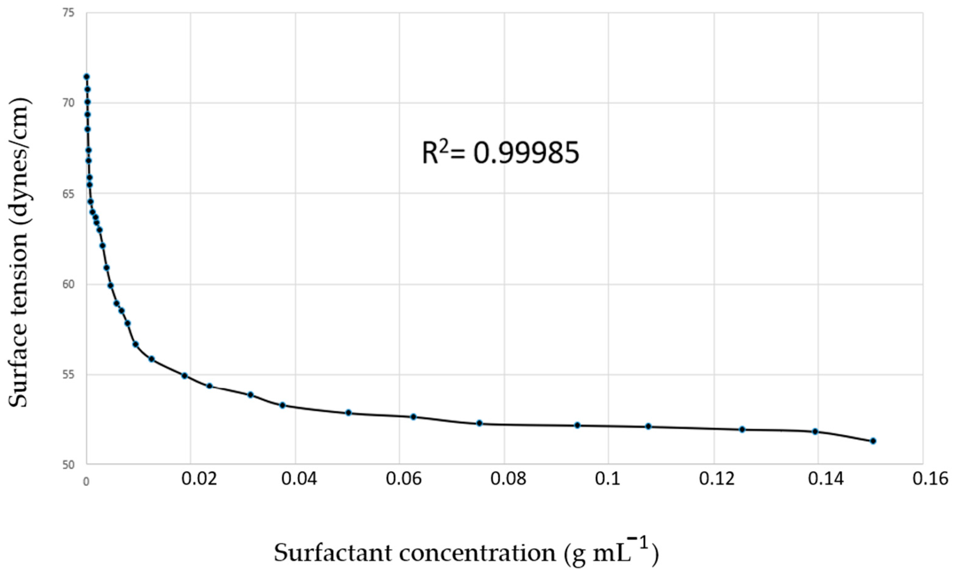
The surface tension (Figure 4) was 51.29 dynes/cm at 25 °C, showing the cohesion strength between the molecules on the surface of the SNEDDS-CO system. From these data, it was possible to determine the critical micellar concentration (CMC) as 8.315 × 10⁻³ g mL−1, which is the surfactant optimal concentration of micelle formation.
Surface forces arise from short-range intermolecular interactions and manifest themselves as longer-range repulsive or attractive forces, and the surface tension, or surface energy is associated with increasing the surface area. Therefore, surface tension is the thermodynamic key property in the interfacial phenomenon resulting from interactions in the region between two different phases (a solid and a liquid, or a gas and a liquid), and also from cohesion forces between the molecules of a given liquid (in this case, resulting from the imbalance of these forces). The asymmetry of the cohesion forces gives rise to the surface energy, which in turn can be evaluated by quantifying the surface tension [97,98,99].
Surfactants, due to their amphiphilic nature, adsorb on interfaces or surface regions in the form of monomers, reducing interfacial tension. This phenomenon is responsible for most of the surfactant properties and structural characteristics of micelles. When small amounts of surfactant are added to water, one part is dissolved as a monomer and another part forms a monolayer at the air–water interface. The molecules of the monolayer remain in equilibrium with the monomers, and each monomer concentration corresponds to a characteristic surface tension. When the concentration of monomers reaches a critical value that determines saturation at the interface, the process of spontaneous formation of molecular aggregates (formation of micelles) is triggered [99,100,101].
The experimental correlation between CMC and the surface properties of surfactants is evaluated, aiming to understand the micellar aggregate formation, which decreases the free energy of the system by reducing the interaction between the hydrophobic groups and the surfactant. Therefore, the CMC provides optimal measurement of surfactant concentration, ensuring the system’s stability. Above the CMC, the concentration of the surfactant results in more micelle formation, but on the other hand, it reduces the free energy of the system. So, below the CMC, the surfactant molecules remain in the form of monomers [102,103,104,105,106,107].
The refractive index of SNEDDS-CO was measured as 1.490, indicating the speed at which light propagates through the system. No deviations were observed in polarized light, which suggests a homogeneous distribution of the components in the formulation. This system was also homogeneous when observed through a light microscope with the phase contrast mode.
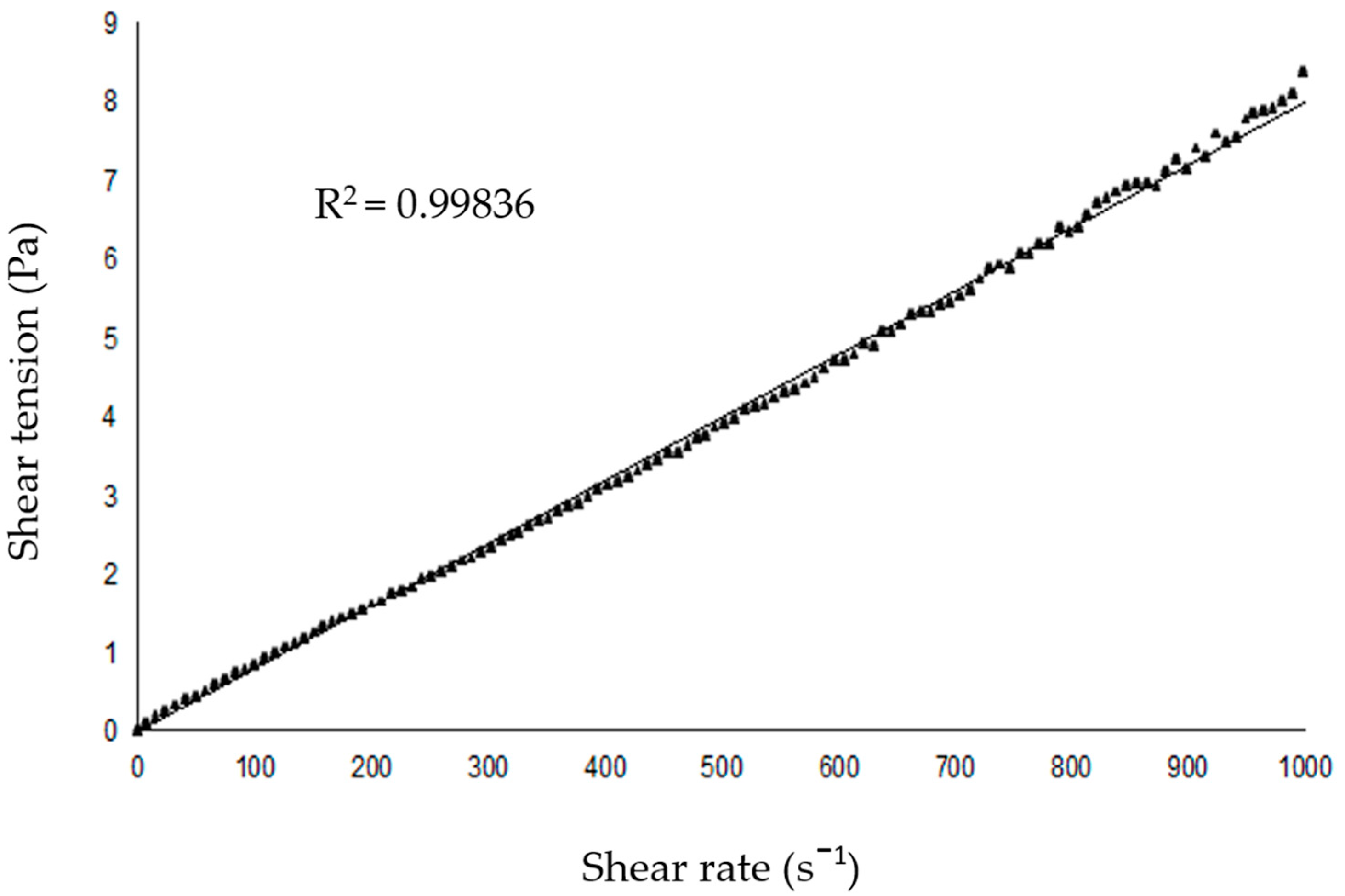
The viscosity of the SNEDDS-CO carrier system was 8 × 10⁻³ cP, which indicates a low resistance to the flow of the emulsion (Figure 5).

2.2.1. Transmission Electron Microscopy Analysis of the SNEDDS-CO System

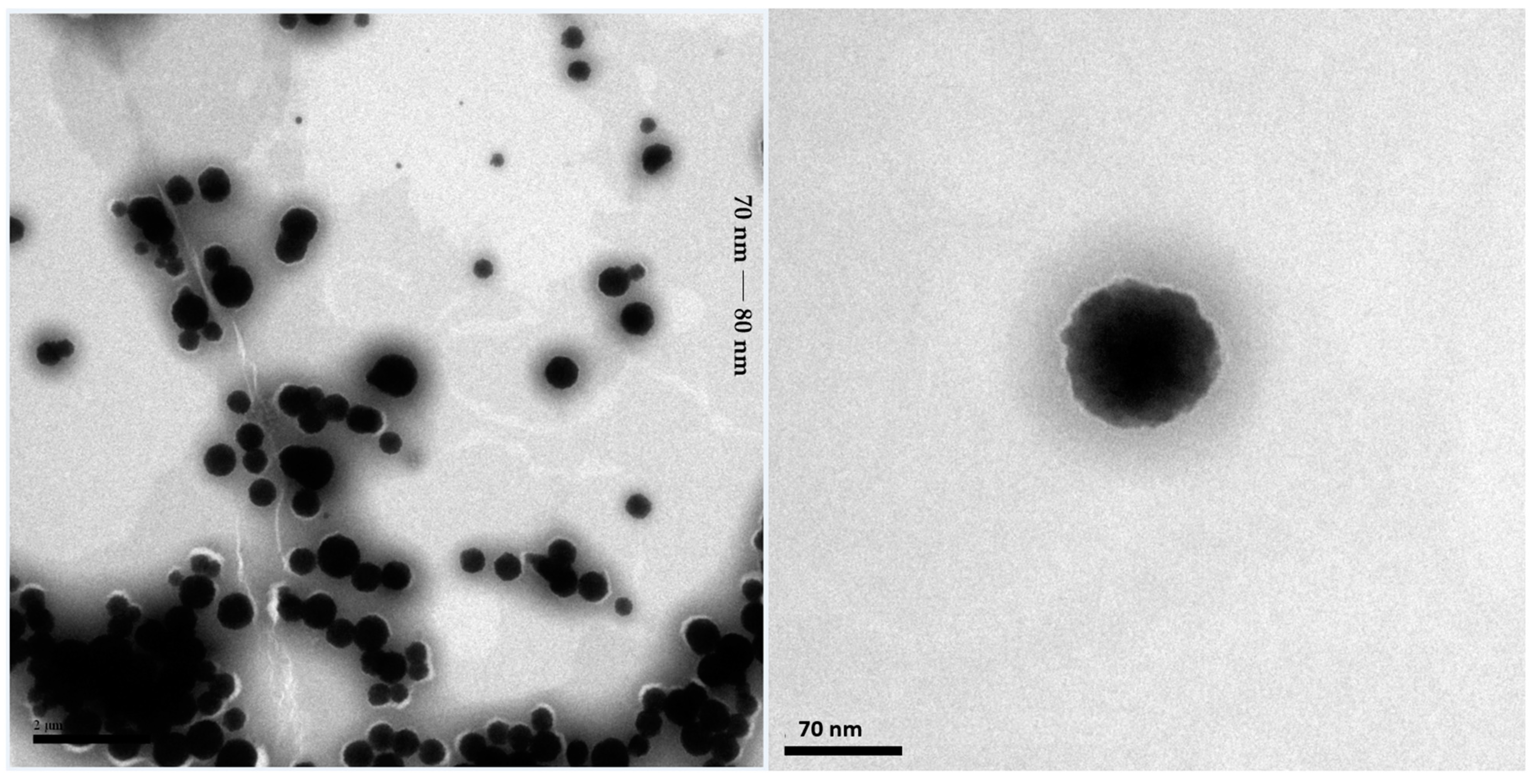
The transmission electron microscopy (TEM) analysis of the SNEDDS-CO system, performed aiming to elucidate the morphology and size of micelles, showed the presence of spherical nanoparticles with an average size between 70 nm and 80 nm (Figure 6).
Similar TEM analysis was observed for a SNEDDS system containing coconut oil, Tween 80, and citral as a bioactive, with a higher contrast for the oily cores and a lower contrast for the outer part of the micelle, which could be correlated with the surfactant [108].
Generally, nanoparticles that showed a relative increase in droplet size when compared to the value obtained in the nanoparticle analyzer measurement (dynamic light diffusion/DLS) are justified by the wide difference conditions for TEM analysis, such as non-dilution and the need for drying, that cause modifications [109,110,111].
In the case of the SNEDDS-CO, the mean DLS value was 11.66 nm. Therefore, the increase in TEM analysis was approximately 70%.
In the study developed by Filippov et al. (2023), a detailed analysis was carried out on the discrepancies in the size measurements of nanoparticles obtained by TEM and DLS, identifying percentage deviations that can exceed 200%. The authors associated these differences with factors intrinsic and extrinsic to the samples, such as particle concentration, contrast, surface charge, and morphology [112].
Mondéjar-López et al. (2022) developed chitosan nanoparticles loaded with thymoquinone as preservatives, and observed DLS with sizes ranging between 48.6 nm and 65 nm, while TEM analysis revealed particles with sizes close to 20 nm [113].
The capabilities and limitations of different nanoparticle characterization techniques, including TEM, SEM, AFM, and DLS, were evaluated by Eaton et al. (2017), who observed that the main difference between the methods lies in the distinct environments in which the nanoparticles are analyzed. DLS, for example, measures the polydisperse index and estimates the size of the particles based on the calculation of the hydrodynamic radius, determined by Brownian motion. However, this technique is sensitive to the interaction of particles with other components of the formulation and also to the presence of artifacts or aggregates, which can make it difficult to interpret the results. TEM, on the other hand, provides data dependent on both the type of material and the state of the sample (solid, liquid, dry, or frozen), which makes the direct correlation between the two techniques a huge challenge, since each method reflects specific aspects of the nanoparticles [114].
The physicochemical properties of emulsions depend not only on the component composition of the systems, but also on the methods of their preparation aiming at achieving stable solutions [51,54,115,116].
Since polar colloidal systems are largely used to solubilize hydrophobic substances, using copaiba (CO) oil as the oil phase of a given SNEDDS-CO carrier system could be an optimal choice for its co-encapsulating along with the bioactive terpenoids, such as trans-dehydrocrotonin (t-DCTN). Indeed, aiming to release this biologic compound contributing to the bioavailability of this 19-nor-clerodane diterpene-type into a polar formulation for oral use, the copaiba oil, together with the surfactant phase, facilitated the dissolution of t-DCTN, which, in its free form, does not solubilize in aqueous medium. In this sense, the SNEDDS-OC carrier system allows the controlled release of both CO and t-DCTN, enabling its lower and prolonged therapeutic action. Specifically, the lower concentration of t-DCTN (1 mg) was quantitatively solubilized by mechanical stirring under heating (40 °C to 55 °C) with 1 mL dropwise of SNEDDS-CO. Quantification was performed by spectrophotometer in the UV–visible region using the wavelength (λmax 238 nm). The calibration curve was constructed based on the relationship between concentration versus absorbance with R2 = 0.9998, showing that the sample was quantitatively solubilized. Therefore, the loading effectiveness of CO and t-DCTN co-encapsulation afforded the nanoproduct called SNEDDS-CO-DCTN (1 mg mL−1).
Particle size [dynamic light diffusion/DLS, Ø (nm)], polydispersity index (PDI), and zeta potential (ζ mV)) analysis showed, respectively, mean values of 11.66 nm, 0.17, and −3.85 for the carrier system SNEDDS-CO, and 11.29 nm, 0.10, and −3.44 for the nanoproduct SNEDDS-CO-DCTN (Table 1), indicating that there was a small and uniform distribution of the nanodroplets in both systems.
The zeta potential of SNEDDS-CO-DCTN could be a modular small number due the presence of nonionic surfactant (Tween 80) fixed around the oil droplet. So, due the solvation effect at the polar head group of the surfactant and also by the presence of carboxylic acid groups of some acidic molecules from copaiba oil, as well as the carbonyl groups of the chemical structure of t-DCTN, it is possible that these SNEDDS systems stabilized through steric rather than electrostatic effects.
The choice of emulsifiers to stabilize emulsions is carried out by using HLB (hydrophilic–lipophilic balance) data, and the effect of surfactant type is determined by the characteristic scale of HLB, as well as the surfactant amount and emulsification methodology [30,31,32,33,34,39,51,117]. Focusing in our previous studies caried out with copaiba oil (Copaifera langsdorffii Desf.) loaded into SNEDDS-type formulations composed of Tween 80 (20%), no co-surfactant, and an oil phase (1%) containing copaiba oil mixed (1:1) with coconut oil or sunflower oil, it we found lower data for particle size and zeta potential, such as 5.87 nm and −1.240 (ζ mV) for SNEDDS-CO/coconut oil, and 6.94 nm and −2.460 (ζ mV) for SNEDDS-CO/sunflower oil [29].
In the other hand, the study developed by Nogueira Barradas et al. (2023) addresses the elaboration of a classical nanoemulsion based on C. officinalis oil, finding particle diameters ranging from 33.03 ± 1.18 nm to 43.98 ± 4.23 nm, measured by using the DLS method. It was observed that the increase in diameter was intrinsically related to the increase in oil concentration. In order to reduce the discrepancies between the values obtained by TEM and DLS, the authors used the negative staining technique, with phosphotungstic acid as the coloring agent, which helped in the identification of the film created by the surfactant, and promoted an amorphous protection around the particles, favoring their visualization and also preventing collapse and coalescence in the drying stage. So, TEM analyses revealed particles with pseudospherical/oval morphology, with dimensions close to the values obtained by DLS [118].
Venturini et al. (2015) performed particle diameter measurements using the DLS technique, for a formulation based on copaiba oil (unidentified species) co-encapsulated with imiquimod, obtaining, for two proposed formulations, values of 206 ± 6 nm and 177 ± 2 nm. TEM analysis revealed particles with conserved morphology and spherical appearance, being in agreement with the DLS measurements [119].
Carvalho et al. (2022) used a copaiba oil-based nanocarrier system for encapsulation of docetaxel, aiming at to optimize its solubility and bioavailability in anticancer therapy. The DLS particle diameter values were 221.5 ± 2.5 nm (docetaxel-loaded) and 192.6 ± 2.3 nm (docetaxel without encapsulation). TEM analysis of docetaxel-loaded revealed spherical particles with no morphological changes, with dimensions ~200 nm [120].

2.2.2. In Vitro Release Kinetic of trans-Dehydrocrotonin Loaded into the SNEDDS-CO System

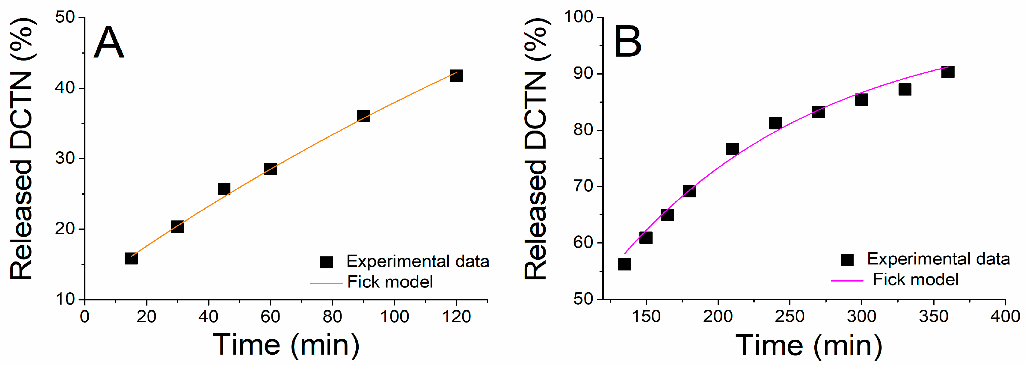
The in vitro release of trans-dehydrocrotonin co-encapsulated with copaiba oil (Copaifera reticulata Ducke) was determined in two mediums simulating physiological conditions such as (i) simulated gastric fluid (SGF, pH = 1.2) during 120 min., and (ii) in the simulated intestinal fluid (SIF, pH = 6.8), continued for up to 360 min., by using the dialysis method (Figure 7). This assay is very important to predict the oral bioperformance of the drug delivery system under the human gastrointestinal microenvironment [121].
In the first stage (SGF), the release of t-DCTN exhibited a two-phase pattern. The initial phase featured a rapid onset release (burst effect) of 25.68 ± 0.02% of t-DCTN at 45 min., which can be attributed to the weakly bound or absorbed drug (t-DCTN) incorporated in the surface of the carrier system SNEDDS-CO. The second phase is characterized by a more controlled drug release profile, which can be attributed to the diffusion of the t-DCTN through the SNEDDS-CO-DCTN formulation, reaching a maximum cumulative release of 41.76 ± 0.01% at 120 min. In the second stage, SIF is released initially at 56.23 ± 0.03% of t-DCTN at 135 min., where the sustained profile is maintained until of end of the experiment, with a maximum cumulative release of 90.33 ± 0.01% at 360 min. As evidenced (Figure 7), the process of drug release diffusion was more favored at the intestinal medium (pH 6.8), when compared to the gastric one (pH 1.2), which is possibly associated with t-DCTN enhanced solubility at higher values of pH. Phenomena such this present one were observed by Arriagada et al. (2020) and Estime et al. (2010) for other pharmaceuticals [122,123].
The release kinetic data were adjusted by the non-linear regression by using some mathematical models, but the equations from the Fickian kinetic model provided the best correlation coefficient (R2 = 0.98), as shown in Figure 8. This result indicated that the diffusion was considered the main release mechanism. The kinetic parameters calculated from the Fickian model were k = 0.00355 ± 0.00007 min−1 for SGF, and k = 0.00694 ± 0.00036 min−1 for SIF.
In our previous work, the release of trans-dehydrocrotonin loaded at the highest dose (10 mg) into a vesicular delivery system (liposome formulation) also showed a controlled drug profile by diffusion, in agreement with the Fickian kinetic model. In this case, the t-DCTN nanoformulation employed a simulated condition for parenteral application (7.4 phosphate buffer). Indeed, there was improvement in t-DCTN antitumor activity against Sarcoma 180, and also a reduction in the drug hepatotoxicity risk in Swiss mice. Thus was evidenced an important advance for enabling t-DCTN nanoformulations to be used in the nanotherapy of cancer [124,125].
In this present work, trans-dehydrocrotonin loaded into the colloidal SNEDDS-CO carrier system (1 mg mL−1) was bioaccessible to perform, for the first time, simulated conditions for its oral delivery. So, aiming at a preliminary application of the formulation, SNEDDS-CO-DCTN effectiveness was evaluated on in vitro antioxidant assays.
Despite the SNEDDS drug delivery system’s advantages, and hundreds of in vivo studies carried out in several animal models, whose results showed the improvement of the drugs’ solubility, and the SNEDDS formulation’s being able to bypass lymphatic transport with promising results, this type of colloidal system faces limitations in the sense that there are not many pharmacokinetic studies carried out in humans, using SNEDDS-type systems administered orally. Therefore, the results of this present work are promising for oral t-DCTN load into a stable SNEDDS formulation.

2.3. In Vitro Antioxidant Analysis of trans-Dehydrocrotonin Loaded into the SNEDDS-CO System

2.3.1. Determination of Total Antioxidant Capacity

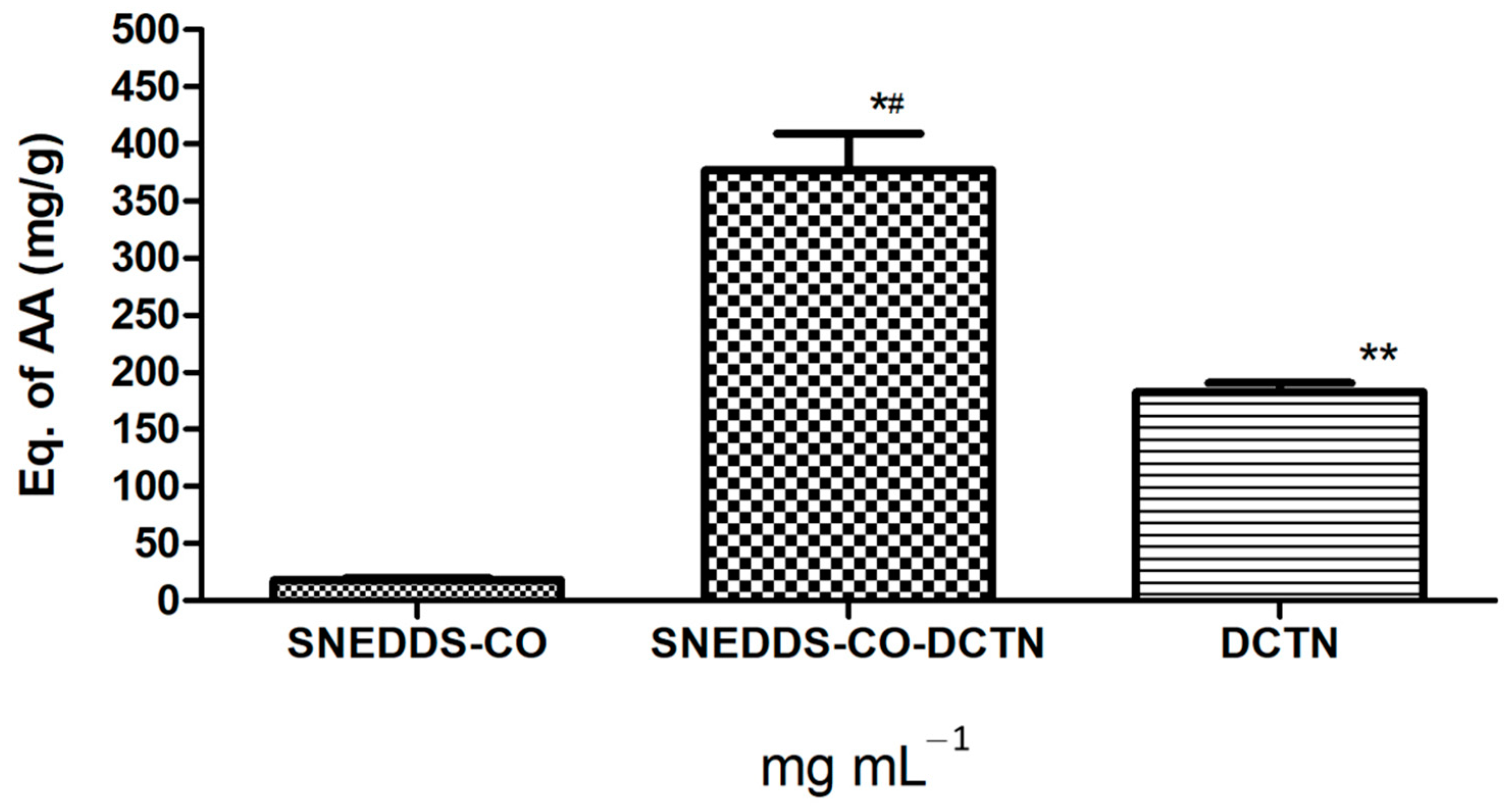
In this assay, SNEDDS-CO-DCTN formulation (t-DCTN, 1 mg mL−1) and the unload t-DCTN (10 mg) solubilized in DMSO, showed TAC activity strongly higher than the result observed for the SNEDDS-CO carrier system (Figure 9).
In this assay, the ascorbic acid has the ability to transfer electrons and bind to metal ions, providing protection against oxidation in aqueous media within cells. Indeed, when antioxidant defenses are weakened, the body cells and tissues become more prone to develop dysfunction and/or disease [126]. Then, the maintenance of adequate antioxidant levels is essential to manage or even prevent a great number of disease conditions. In this sense, the measurement of TAC (total antioxidant capacity) of biological samples is a useful tool for investigating biological fluids and monitoring several diseases associated with oxidative stress, such as type 2 diabetes [127], chronic hepatitis C [128,129], cardiovascular and inflammatory bowel diseases [130]. This result reinforces the therapeutic potential of t-DCTN previously evaluated in in vivo models for hypoglycemic and hypolipidemic activities [10,11,12,13], and also its cardiovascular protective property [20].

2.3.2. Antioxidant Capacity via Reducing Power

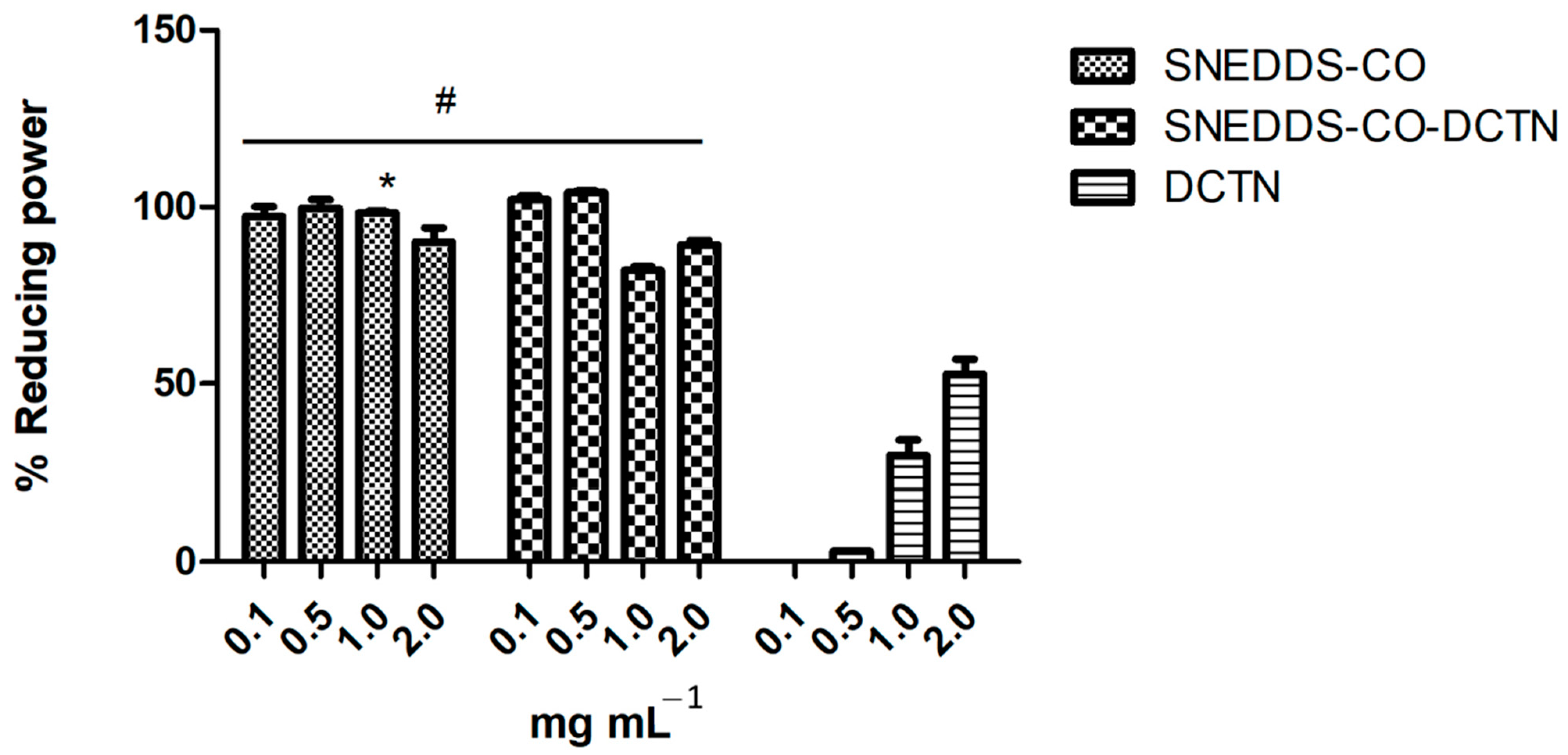
In this assay, the samples (SNEDDS-CO, SNEDDS-CO-DCTN, and t-DCTN-DMSO solution) were evaluated at different concentrations (0.1, 0.5, 1.0, and 2.0 mg mL−1). For the SNEDDS-CO sample, the reduced power is over 94%. Specifically, 100% reducing activity was observed at the 0.5 mg mL−1, 98.5% at 1.0 mg mL−1, 97.33% at 0.1 mg mL−1, and 94.0% at 2.0 mg mL−1 (Figure 10). For the SNEDDS-CO-DCTN sample, the reducing power was observed above 80%—specifically, 99.33% at the concentration of 0.1 mg mL−1, 99.66% at 0.5 mg mL−1, 82% at 1 mg mL−1, and 89.33% at 2 mg mL−1 (Figure 10). These results were compared with trans-dehydrocrotonin solubilized in DMSO (10 mg mL−1), and it was possible to evidence the bioavailability improvement of t-DCTN loaded into the SNEDDS-CO system (Figure 10).
Many antioxidants are proven to undergo an electron-transfer mechanism upon exerting their antioxidant functions. Therefore, reducing power is a measure of a substance’s ability to transfer electrons in a neutral condition [131,132]. In this test, the SNEDDS-CO system showed greater capacity to donate electrons at all concentrations with no statistical difference compared with the product SNEDDS-CO-DCTN at 0.1 mg mL−1 (99.33%) and 0.5 mg mL−1 (99.66%). These data indicate that the SNEDDS formulations showed high reducing activity, which can be associated with an antioxidant property via free radical neutralization.

2.3.3. Copper Ion Chelation Method

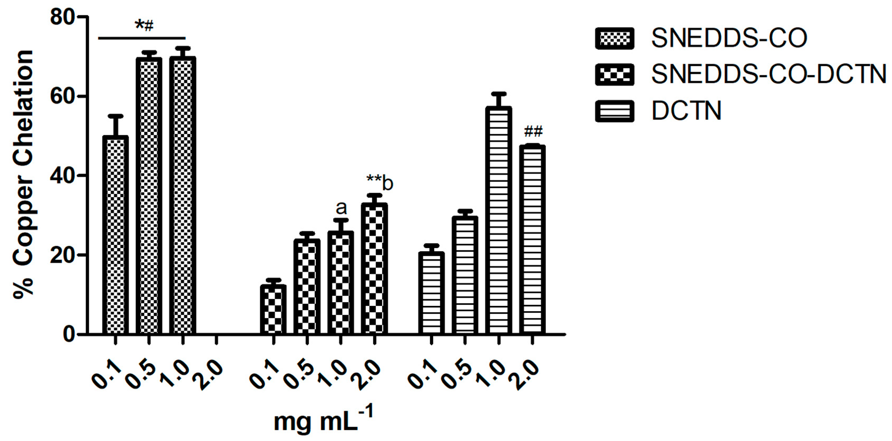
In this assay, the SNEDDS-CO-DCTN showed low antioxidant activity at all tested concentrations: 8.66% (0.1 mg mL−1), 23.66% (0.5 mg mL−1), 25.66% (1 mg mL−1), and 31.50% (2 mg mL−1). Comparatively, SNEDDS-CO at 0.1 mg mL−1 (49.66%), 0.5 mg mL−1 (69.33%), and 1 mg mL−1 (69.33%) showed higher potential activity (Figure 11). On the other hand, the t-DCTN-DMSO solution showed great result, at 1 mg mL−1.
The induction of oxidative stress is an imbalance between the production of radical species and the antioxidant defense systems, causing oxidative damage to proteins, lipids, and DNA [133]. In this sense, ligand antioxidant properties complexed by selected metals may significantly affect the free radical neutralization [134,135]. Considering the results of the carrier system at 1 mg mL−1 and also the t-DCTN-DMSO solution at 1 mg mL−1 (Figure 10), these samples also could be extensively applied on in vivo models associated with oxidative stress such as type 2 diabetes [127] and chronic hepatitis [129,130].

2.3.4. Hydroxyl Radical Sequestration Capability

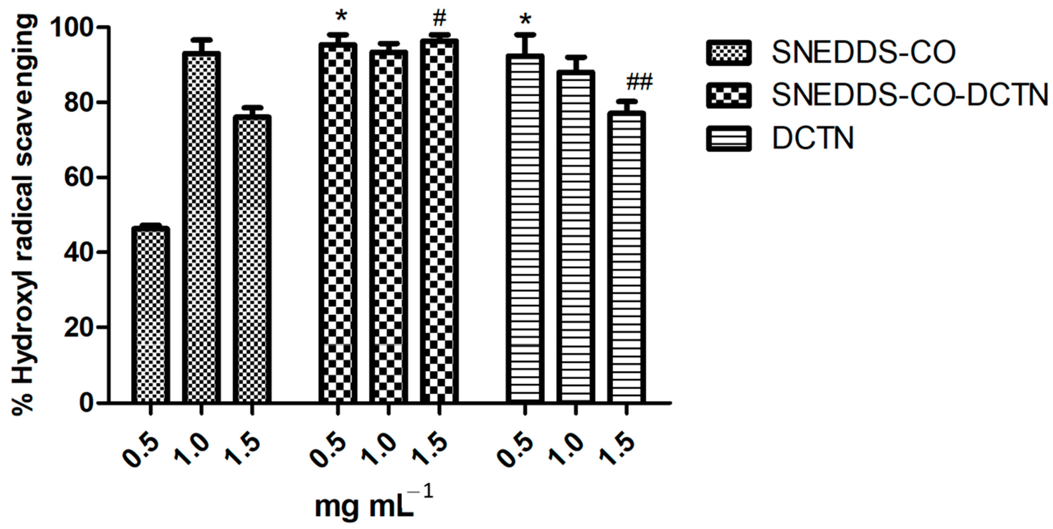
In this test, the SNEDDS-CO system at concentrations 1.0 mg mL−1 and 1.5 mg mL−1 showed 94% and 76% of antioxidant capacity, respectively (Figure 12). On the other hand, the highest results were noted for SNEDDS-CO-DCTN and t-DCTN-DMSO solution at all tested concentrations (0.5, 1.0, and 1.5 mg mL−1).
It is important to highlight that copaiba oil (0.5%) from Copaifera langsdorffii Desf., mixed with coconut oil (0.5%) or with sunflower oil (0.5%), was previously loaded into SNEDDS systems and then assayed for its antioxidant property. The reducing power and hydroxyl radical (OH)-formation inhibition previously observed for the SNEDDS-CO/coconut oil system, were, respectively, 46.48% and 84.11%, and for the SNEDDS-CO/sunflower system, were, respectively, 52.46% and 74.48% [29].
Since OH causes oxidative DNA damage that compromises the integrity and function of cell membranes and reacts with almost all organic biomolecules found in living organisms [131,132], certainly, these SNEDDS systems based on copaiba oil from Copaifera langsdorffii Desf. and Copaifera reticulata Ducke become important targets to combat cell lipoperoxidation.

3. Materials and Methods

The herbal resource copaiba oil (Copaifera reticulata Ducke), the stem bark of Croton cajucara Benth (Euphorbiaceae), and the sunflower oil were commercially obtained. Copaiba oil and C. cajucara samples were purchased in traditional markets specialized in natural products located in the Amazon region of Brazil, respectively at Manaus and Belém cities. Complying with the requirements of the New Brazilian Biodiversity Law (Law 13.123/2015), this work was registered in the National Management System of Genetic Heritage and Associated Traditional Knowledge (SISGEN) under number AFF8812.
The solvents acetone (Me2CO), chloroform (CHCl3), ethanol (EtOH), ethyl acetate (AcOEt), hexane, and methanol (MeOH), all of analytical grade, as well as all other used chemicals, such as ammonium molybdate, ascorbic acid, copper II sulfate pentahydrate, ethylene-di-amine-tetra-acetic acid (EDTA), ferric chloride, ferrous sulfate heptahydrate (FeSO4·7H2O), ferrous sulphate II, gallic acid, nitroblue tetrazolium (NBT), phosphate buffer, potassium ferricyanide, pyrocatechol violet, riboflavin, sodium phosphate, sodium salicylate, sulfuric acid, trichloroacetic acid, and Tween 80, were purchased from Sigma Aldrich Inc. (St. Louis, MO, USA) or Merck (Darmstadt, Germany).
The melting points were determined with a Kofler (Jasco DIP-370) apparatus, and they have not been corrected. IR spectra (CHCl3) were found with a Perkin–Elmer FT-16PC spectrophotometer, and UV spectra (MeOH) with GBC UV/VIS 911A CG or Thermo Electron Corporation/Nicolet Evolution 100) instruments (London, UK and Boston, MA, USA). The NMR 1H and all 2D experiments were recorded on a Bruker–Avance spectrometer (600 MHz for 1H) (Bruker, Millerica, MA, USA). The deuterated solvent (CDCl3) used in NMR spectroscopy was from Merck brands.

3.1. Chromatography Analysis of Copaifera reticulata Ducke

Characterization of copaiba oil (CO) after its chemical esterification was performed using gas chromatography high resolution analyses with flame ionization detection (GC-FID) and coupled with mass spectrometry: Trace CG Ultra, Thermo Scientific (Eindhoven, The Netherlands) and mass spectrometry detector (DSQ II, Thermo Scientific) (Eindhoven, The Netherlands) with quadrupolanalyser and auto-injetor (AI 3000, Thermo Scientific) (Eindhoven, The Netherlands). Mass spectra were obtained by electron impact (70 eV), from 40 to 400 u.m.a. The CO sample (5.0 mg) was derivatized in situ using trimethylsilildiazomethane (TMSD), converting the diterpene carboxylic acids into the corresponding methyl esthers. Split injections (1:20) were performed in DB-1 dimethylpolisiloxane (25 m × 0.25 mm × 0.25 μm) and zebron ZB-5ms (Phenomenex-20 m × 0.18 mm × 0.18 μm) columns, using He as carrier gas at 2 mL min−1. Oven temperature was programmed from 120 °C to 150 °C at 3 °C.min−1, followed by another heating ramp until 290 °C, at 15 °C min−1. Detector and injector temperatures were set at 300 °C and 270 °C, respectively. Two standard mixtures were injected at this same condition: a homologous series of linear hydrocarbons from tridecane to heptadecane (C13 to C17), and a mixture containing the sesquiterpenes caryophyllene, humulene, and caryophyllene oxide. The homologous series of hydrocarbons were applied to obtain the Linear Retention Index (LRI) of the copaiba oil constituents, and then compared with the literature. The mixture of these three sesquiterpenes, very common constituents from copaiba oils, was used to correct the LRI obtained and compare them with literature data. Mass spectrometry (MS) experiments were useful to confirm the identification of the sesquiterpenes by comparing their mass spectra with an automatic database (NIST) and also to obtain the diterpene methyl esther mass spectra and compare all of them with mass spectra data from previously isolated substances from copaiba oils that were stored, and to compose a personal data library. The applied methodology is accordance with our previous reported studies [29,133,134].

3.2. Chromatography Analysis of Croton cajucara Benth

Stem bark (1 kg) of C. cajucara was dried at 37 °C in an oven with controlled temperature and constant renewal of air for four days, and its coarse milling was performed using an SM 300 cutting mill (Grupo Imetal, Santa Catarina, Brazil) operating at 3000 rpm and equipped with a 2 mm grid. The pulverized plant was kept in an air-tight container until before being used. The maceration extraction ratio of 1:10 (w/v) was applied to prepare the hydroalcoholic extract (EtOH/distilled water).
Classical column chromatography was performed with silica gel (70–230 mesh) as adsorbent, and Thin Layer Chromatography (TLC) was carried out using silica gel 60 H and, revelation was employed with sulfuric acid/methanol (1:1) reagent. TLC was carried out on 0.25 mm layers of silica gel PF 254 (Merck). TLC also was revealed by UV-Vis radiation at wavelength of 254 and 356 nm. The hydroalcoholic EtOH/H2O (8:2) extract, after solvent reduction (85 g, 8.5%), was submitted to a chromatographic procedure and afforded 25 fractions eluted with hexane or mixtures of hexane/EtOAc at different ratios of increasing polarity (Figure 2).
The non-polar fractions (F1, F2, and F3) were eluted with hexane, and fractions F4 to F25 were eluted with mixtures of hexane/EtOAc (9:1 to 5:5), and then submitted to recrystallization or submitted to another chromatographic fractionation and then recrystallization, affording 10.86 g (0.17%) of trans-dehydrocrotonin (t-DCTN). For the TLC analysis mobile phase, mixtures of hexane/EtOAc t (8:2, 7:3, and 6:4) were applied, and revelation was employed with sulfuric acid/methanol (1:1) reagent. Additionally, TLC was also revealed using UV-Vis radiation at wavelength of 254 nm and 356 nm.
The structural elucidation of t-DCTN was analyzed by spectroscopic method in the infrared (IR) region, and also proton nuclear magnetic resonance spectra (1H NMR).

3.3. SNEDDS-Copaiba Oil Colloidal Formulation

The ternary phase diagram was constructed using the surfactant mass titration methodology into the aqueous and oily phases in order to obtain a polar O/W nanoemulsion region. The phase diagram was prepared with Tween 80 as a surfactant, copaiba oil (Copaifera reticulata Ducke) and sunflower oil (Helianthus annuus) as the oil phase, and doublydistilled water. The surfactant was mixed with the oil phase (weight ratios) respectively: 9:1, 8:2, 7:3, 6:4, 5:5, 4:6, 3:7, 2:8, and 1:9. All mixtures were diluted dropwise with distilled water (w/v), and the nanoemulsion region of the copaiba oil (CO) colloidal system was produced by using the mechanical stirring Vortex (IKA, Staufen, Germany) at ambient temperature.
The colloidal-based system was composed of 7% surfactant (Tween 80), 1% vegetable oil (a mixture of copaiba oil and sunflower oil, at a 1:1 ratio), and 92% doubly-distilled water, under heating (55 °C to 65 °C). After the physicochemical characterizations by using polarized light microscopy, pH, conductivity, refractive index, droplet size, rheological behavior, and surface tension analysis, the formulation called SNEDDS-CO containing 1% of oil phase (0.5% of CO and 0.5% of sunflower oil) was characterized as SNEDDS-type system.
The pH was measured by using a pre-calibrated pH meter PG-2000 (Gehaka, São Paulo, SP, Brazil) at 25 °C. The electrical conductivity of the samples was measured using a DM-32 conductivity meter (Digicrom Analytical, Campo Grande, SP, Brazil) with a cell constant of 0.11 cm−1. The measurements were performed at 25 °C.
The refractive index of the carrier SNEDDS-CO system was determined using Abbe’s refractometer (Bellingham plus Stanley Limited, England) at 25 °C, and the rheological property was determined using an oscillatory Haake Mars rheometer (Thermo Fisher Scientific, Karlsruhe, Germany, cup Z43 DIN 53018 and rotor Z41 DIN 53018). The temperature was kept at 25 °C using a thermostatic bath. Analyses were carried out by applying a shear rate sweep from 0 to 103 s−1.
The surface tension of SNEDDS-CO was carried out using a SensaDyne tensiometer (model QC-6000, Milwaukee, WI, USA) employing the maximum pressure bubble technique, using nitrogen as gas phase. The results of the surface tension assay, expressed in mN m−1 (or dynes cm−1), were analyzed with SensaDyne tensiometer software, version 1.21.
The transmission electron microscopy (TEM) analysis was performed with high-resolution JEM-1230 JEOL equipment to identify nanoparticle images. The analysis was carried out with an acceleration voltage of 300 kV and a magnification range of 50 to 800 thousand times. In order to observe the morphology of the SNEDDS-CO carrier system, a drop of this solution was introduced into a 200-mesh copper grid, and then a drop of 5% phosphotungistic acid was added to this same grid as a contrast agent. And then, it was left to rest for 4 h, and then, the grid was analyzed via TEM. The descriptive methodology for particle size, polydispersity index, and zeta potential analysis for both SNEDDS-CO and the nanoproduct SNEDDS-CO-DCTN are described below (4.5).

3.4. Loading of trans-Dehydrocrotonin and Effectiveness of the Solubility in the SNEDDS-CO System

The lower concentration of t-DCTN (1 mg) was solubilized dropwise with 1 mL of SNEDDS-CO by mechanical stirring under heating (40 °C to 55 °C), affording the nanoproduct called SNEDDS-CO-DCTN (1 mg mL−1). The loading effectiveness of t-DCTN was evaluated by the minimum and maximum solubility method. Quantification was performed by spectrophotometer (Thermo Electron Corporation/Nicolet Evolution 100) (London, UK) in the UV–visible region using wavelength (λ) at 200 to 400 nm for determining the λmax (observed at 238 nm). To obtain the calibration curve, the correlation of t-DCTN concentration versus absorbance was analyzed using four samples. To determine the minimum and maximum solubility of t-DCTN in the SNEDDS-CO carrier system, two masses (1 mg mL−1 and 5 mg mL−1) were used, and mechanical stirring under heating (40 °C to 55 °C) was applied. After cooling at room temperature, the samples were vortexed for 10 min, followed by stirring in ultrasound for 10 min at room temperature. After equilibration, samples were filtered, and diluted as required with distilled water. Then, the solutions were analyzed in the UV-Vis spectrophotometer at a wavelength of 238 nm.

3.5. Particle Size, Polydispersity Index, and Zeta Potential Analysis

The physicochemical characterizations of the nanoproduct SNEDDS-CO-DCTN were conducted through analysis of particle size (Ø), polydispersity index (PDI), and zeta potential (ζ), following the methodology previously [124,125], and the obtained data were compared with those observed for SNEDDS-CO.
Micelle dispersion was measured using photon correlation spectroscopy with a Zetasizer Nano-ZS90 (Malvern, Worcestershire, UK). For Ø and PDI analyses, 100 µL of the formulations were diluted in 900 µL of purified water. Measurements were performed at 25 °C with a fixed 90° scattering angle, and results were reported as the mean hydrodynamic diameter of the micelles (nm). Zeta potential (ζ) was measured after diluting 50 µL of the formulations in 950 µL of purified water. Surface charge (mV) was evaluated using the Zetasizer Nano-ZS90 (Malvern, Worcestershire, UK).

3.6. In Vitro Release Kinetics

The in vitro release kinetics of t-DCTN loaded into the nanoproduct SNEDDS-CO-DCTN were analyzed using the dialysis technique under sink conditions in simulated gastric fluid (SGF, pH = 1.2) and in simulated intestinal fluid (SIF, pH = 6.8) at 37 °C. An aliquot (1 mL) of the formulation was withdrawn and placed in a dialysis membrane (Viskase Lombard, USA), which were sealed and suspended in the simulated buffered medium (100 mL). The system was maintained under stirring (100 rpm) and heating (37 °C). Aliquots of 1 mL of the release fluid were collected (replaced by 1 mL of fresh buffer) at predetermined times over 120 min for SGF and continued for up to 360 min for SIF. The amount of t-DCTN released as a function of time was determined spectrophotometrically at 238 nm using a standard curve in concentrations ranging from 1 to 20 µg mL−1. t-DCTN release kinetics were performed in triplicate, and the values were expressed as mean ± standard deviation for the concentration in percentage of cumulative drug released.
The release profile was evaluated by non-linear regression for the mathematical correlation to Fickian’s law, uni-exponential using the following equation:
Mt/M = 1 − be−kt
where Mt and M are the amounts of drug released at a given time t, and the total drug released from SNEDDS-CO-DCTN system, respectively; k is the rate constant associated with the diffusion coefficient of the drug loaded [124,125].

3.7. Antioxidant Activity Assays

To evaluate the antioxidant activities, in vitro antioxidant assays applied to the SNEDDS formulations (SNEDDS-CO and SNEDDS-CO-DCTN) and t-DCTN solubilized in DMSO (10 mg mL−1) were performed by determination of total antioxidant capacity (TAC), reducing power, copper ion chelation, and hydroxyl radical scavenging, according to the methodologies previously described [135,136,137]. The tests were performed in triplicate.

3.7.1. Determination of Total Antioxidant Capacity (TAC)

Briefly, the solution (1 mL) containing the samples, ammonium molybdate (4 mM), sodium phosphate (28 mM), and sulfuric acid (0.6 M) were added into a tube, stirred, and incubated (100 °C, 90 min.). The tubes were cooled and were read at the 695 nm wavelength. The standard used was ascorbic acid (AA) and the results were expressed as AA equivalent per gram of sample [135,136].

3.7.2. Reducing Power

Briefly, phosphate buffer (0.2 M, pH 6.6) containing potassium ferricyanide (1%) was mixed with samples in different concentrations (0.05 to 2 mg mL−1) to a final volume of 1.0 mL. After 20 min., at 50 °C, the reaction was stopped with 10% trichloroacetic acid (TCA). One milliliter of ferric chloride (0.1% w/v) in distilled water was added to the mixture, and the absorbance was measured at 700 nm [135]. The results were expressed as percentage activity of 0.1 mg/mL ascorbic acid (standard), which corresponded to 100% activity.

3.7.3. Copper Chelation

The test was performed in 96-well microplates with a reaction mixture containing different concentrations of samples (0.1 to 2 mg mL−1), copper II sulfate pentahydrate (50 mg mL−1), and pyrocatechol violet (4 mM). All wells were homogenized with the aid of a micropipette, and the solution absorbance was measured at 632 nm [137]. The ability of the samples in chelating the copper ion was calculated using the following equation:
Chelating effect (%) = (Absorbance of blank) − (Absorbance of the sample)/(Absorbance of the blank) × 100

3.7.4. Hydroxyl Radical Scavenging Assay

The OH radical scavenging activity of samples was investigated using Fenton’s reaction (Fe2 + + H2O2 - > Fe3 + + OH + OH). The data were expressed as the inhibition rate. For OH production, the samples (at different concentrations) were added to 3 mL sodium phosphate buffer (150 mM, pH 7.4), which contained 10 mM FeSO4.7H2O (ferrous sulfate heptahydrate), 10 mM EDTA 2 mM sodium salicylate, and 30% H2O2. In the control, sodium phosphate buffer replaced H2O2. After treatment of 37 °C for 1 h, OH radicals were detected by monitoring absorbance at 510 nm using a microplate reader. Gallic acid was used as a positive control [136].

4. Conclusions

The authenticity of copaiba oil resin (Copaifera reticulata Ducke) was confirmed by GC-MS and standards, identifying sesquiterpenes (55.62%) and diterpene (35.47%) as the major components. Key sesquiterpenes included β-bisabolene and α-bergamotene, while diterpenic acids such as kaurenoic, danielic, cativic, and pinifolic acids were also detected. The diterpene 19-nor-clerodane trans-dehydrocrotonin (t-DCTN) was isolated from Croton cajucara and fully characterized through FTIR and ¹H NMR spectroscopy.
A stable single-phase colloidal system was developed based on a ternary phase diagram consisting of Tween 80 as a surfactant, an oil phase comprising a 1:1 mixture of copaiba oil and sunflower oil, and water (92%). This formulation produced a fine oil-in-water nanoemulsion designated as the SNEDDS-CO carrier system. The bioactive t-DCTN (1 mg) was loaded in the carrier SNEDDS-CO under mechanical stirring and heating, yielding the nanoproduct SNEDDS-CO-DCTN (1 mg mL−1). The physicochemical properties and in vitro kinetic profiles of SNEDDS-CO-DCTN were thoroughly characterized, demonstrating sustained t-DCTN release, with a maximum cumulative release of 90.33 ± 0.01% over 360 min, simulating conditions for oral delivery system.
Both the SNEDDS-CO and SNEDDS-CO-DCTN formulations exhibited significant antioxidant activity across all in vitro assays. SNEDDS-CO-DCTN showed superior results in total antioxidant capacity (TAC), reducing power, and hydroxyl radical scavenging activity. Meanwhile, SNEDDS-CO demonstrated greater efficacy in copper ion chelating activity.
These findings highlight the effective bioaccessibility of t-DCTN co-loaded with copaiba oil resin (Copaifera reticulata Ducke) and t-DCTN into the SNEDDS-based system and are available for further in vitro and in vivo models for cancer nanotherapy investigations.

5. Patent

De Oliveira Netto, J. R.; Corrêa, N. P.; De Araujo, L.B.A.; Paiva, W. De S.; Rocha, H. A. De O.; Morais LIMA, W. De A.; Daniel Macedo, C. Dos S.; Santos Magalhães N. S.; Do Nascimento, J. H. O.; Da Veiga Junior, V. F.; Maciel, M. A. M. Formulação Antioxidante com Potencial Imunomodulador contendo Bioativos de Copaifera reticulata Ducke (Óleo Resina de Copaíba) e Croton cajucara Benth (trans-Desidrocrotonina). 10 April 2024. Brazil, INPI Patent BR102024020730-0 (Instituto Nacional de Propriedade Intelectual).

Supplementary Materials

The following supporting information can be downloaded at: https://www.mdpi.com/article/10.3390/ijms26104469/s1.

Author Contributions

Conceptualization: M.A.M.M., V.F.d.V.J. and H.A.O.R.; Methodology: M.A.M.M., N.S.S.-M., H.A.O.R., W.d.A.M.L., V.F.d.V.J. and J.H.O.d.N.; Formal analysis: all authors were involved; Writing: M.A.M.M., W.d.A.M.L., W.d.S.P. and J.R.d.O.N.; Original draft preparation: J.R.d.O.N., W.d.A.M.L. and M.A.M.M.; Writing/review and editing: M.A.M.M., W.d.A.M.L. and J.R.d.O.N.; Visualization: all authors were involved; Supervision: M.A.M.M., N.S.S.-M., H.A.O.R. and V.F.d.V.J.; Project administration: M.A.M.M.; Funding acquisition: CNPq and CAPES. All authors have read and agreed to the published version of the manuscript.

Funding

This research received no external funding.

Institutional Review Board Statement

Not applicable.

Informed Consent Statement

Not applicable.

Data Availability Statement

No new data were created or analyzed in this study. Data sharing is not applicable to this article.

Acknowledgments

The authors thank the National Council for Scientific and Technological Development (CNPq) and the Coordination for the Improvement of Higher Education Personnel (CAPES), from Brazil, for research grant concerning to student fellowship and scientific productivity fellowship.

Conflicts of Interest

The authors declare no conflicts of interest.

References

  1. Maciel, M.A.M.; Pinto, A.C.; Arruda, A.C.; Pamplona, S.G.S.R.; Vanderline, F.A.; Lapa, A.J.; Echevarria, A.; Grynberg, N.F.; Côlus, I.M.S.; Farias, R.A.F.; et al. Ethnopharmacology, phytochemistry and pharmacology: A successful combination in the study of Croton cajucara. J. Ethnopharmacol. 2000, 70, 41–55. [Google Scholar] [CrossRef] [PubMed]
  2. Maciel, M.A.M.; Pinto, A.C.; Veiga Junior, V.F.; Grynberg, N.F.; Echevarria, A. Medicinal plants: The need for multidisciplinary scientific studies. Quim. Nova 2002, 25, 429–438. [Google Scholar] [CrossRef]
  3. Lima, L.R.; Da Silva Júnior, F.L.; Rufino Arcanjo, D.D.; Maciel, M.A.M. Croton cajucara: Patents and nanotechnological advances. Recent. Pat. Nanotechnol. 2024, 18, 389–394. [Google Scholar] [CrossRef] [PubMed]
  4. Carvalho, J.C.T.; Silva, M.F.C.; Maciel, M.A.M.; Pinto, A.C.; Nunes, D.S.; Lima, R.M.; Bastos, J.K.; Sarti, S.J. Investigation of anti-inflammatory and antinociceptive activities of trans-dehydrocrotonin, a 19-nor-clerodane diterpene from Croton cajucara. Part 1. Planta Med. 1996, 62, 402–404. [Google Scholar] [CrossRef]
  5. Khan, M.T.H.; Ather, A.; Pinto, A.C.; Maciel, M.A.M. Potential benefits of the 19-nor-clerodane trans-dehydrocrotonin on the central nervous system. Rev. Bras. Farmacogn. 2009, 9, 7–13. [Google Scholar] [CrossRef]
  6. Perazzo, F.F.; Carvalho, J.C.T.; Rodrigues, M.; Morais, E.K.L.; Maciel, M.A.M. Comparative anti-inflammatory and antinociceptive effects of terpenoids and an aqueous extract obtained from Croton cajucara Benth. Rev. Bras. Farmacogn. 2007, 17, 521–528. [Google Scholar] [CrossRef]
  7. Hiruma-Lima, C.A.; Spadari-Bratfisch, R.C.; Grassi-Kassisse, D.M.; Souza-Brito, A.R.M. Antiulcerogenic mechanisms of dehydrocrotonin, a diterpene lactone obtained from Croton cajucara. Planta Med. 1999, 65, 325–330. [Google Scholar] [CrossRef]
  8. Rodríguez, J.A.; Hiruma-Lima, C.A.; Souza-Brito, A.R.M. Antiulcer activity and subacute toxicity of trans-dehydrocrotonin from Croton cajucara. Hum. Exp. Toxicol. 2004, 23, 455–461. [Google Scholar] [CrossRef]
  9. Luna Costa, A.M.; Silva, J.C.; Campos, A.R.; Rao, V.S.; Maciel, M.A.; Pinto, A.C. Antioestrogenic effect of trans-dehydrocrotonin, a nor-clerodane diterpene from Croton cajucara Benth. in rats. Phytother. Res. 1999, 13, 689–691. [Google Scholar] [CrossRef]
  10. Farias, R.A.F.; Rao, V.S.N.; Viana, C.S.B.; Silveira, E.R.; Maciel, M.A.M.; Pinto, A.C. Hypoglycemic effect of trans-dehydrocrotonin, a nor-clerodane diterpene from Croton cajucara. Planta Med. 1997, 63, 558–560. [Google Scholar] [CrossRef]
  11. Silva, R.M.; Santos, F.A.; Rao, V.S.N.; Maciel, M.A.M.; Pinto, A.C. The lipid-lowering effect of trans-dehydrocrotonin, a clerodane diterpene from Croton cajucara Benth. in mice fed on high-fat diet. J. Pharm. Pharmacol. 2001, 53, 535–539. [Google Scholar] [CrossRef] [PubMed]
  12. Silva, R.M.; Santos, F.A.; Rao, V.S.N.; Maciel, M.A.M.; Pinto, A.C. Blood glucose- and triglyceride-lowering effect of trans-dehydrocrotonin, a diterpene from Croton cajucara Benth, in rats. Diabetes Obes. Metab. 2001, 3, 452–456. [Google Scholar] [CrossRef] [PubMed]
  13. Silva, R.M.; Santos, F.A.; Maciel, M.A.M.; Pinto, A.C.; Rao, V.S.N. Effect of trans-dehydrocrotonin, a 19-nor-clerodane diterpene from Croton cajucara on experimental hypertriglyceridaemia and hypercholesterolaemia induced by triton WR 1339 (tyloxapol) in mice. Planta Med. 2001, 67, 763–765. [Google Scholar] [CrossRef] [PubMed]
  14. Agner, A.R.; Maciel, M.A.M.; Pinto, A.C.; Cólus, I.M.S. Antigenotoxicity of trans-dehydrocrotonin, a clerodane diterpene from Croton cajucara. Planta Med. 2001, 67, 815–819. [Google Scholar] [CrossRef]
  15. Agner, A.R.; Maciel, M.A.M.; Pinto, A.C.; Pamplona, S.G.S.R.; Cólus, I.M.S. Investigation of genotoxicity activity of trans-dehydrocrotonin, a clerodane diterpene from Croton cajucara. Teratog. Carcinog. Mutagen. 1999, 19, 377–384. [Google Scholar] [CrossRef]
  16. Santos, F.V.; Mesquita, S.F.P.; Faria, M.J.S.S.; Poersh, A.; Maciel, M.A.M.; Pinto, A.C.; Morimoto, H.K.; Cólus, I.M.S. Absence of mutagenicity in somatic and germ cells of mice submitted to subchronic treatment with an extract of Croton cajucara Benth (Euphorbiaceae). Genet. Mol. Biol. 2011, 29, 159–165. [Google Scholar] [CrossRef]
  17. Lima, G.; Machado, G.; Maciel, M.M.; Echevarria, A. Antitrypanosomal and antileishmanial effects of the hydroalcoholic extract of Croton cajucara benth and its 19-nor-clerodane chromatographic fractions. Pharmacogn. Mag. 2021, 17, 302–306. [Google Scholar] [CrossRef]
  18. Lima, G.S.; Castro-Pinto, D.B.; Machado, G.C.; Maciel, M.A.M.; Echevarria, A. Antileishmanial activity and trypanothione reductase effects of terpenes from the Amazonian species Croton cajucara Benth (Euphorbiaceae). Phytomedicine 2015, 22, 1133–1137. [Google Scholar] [CrossRef]
  19. Campos, M.C.O.; Salomão, K.; Castro-Pinto, D.B.; Leon, L.L.; Barbosa, H.S.; Maciel, M.A.M.; De Castro, S. Croton cajucara crude extract and isolated terpenes: Activity on Trypanosoma cruzi. Parasitol. Res. 2010, 107, 1193–1204. [Google Scholar] [CrossRef]
  20. Silva, R.M.; Oliveira, F.M.; Cunha, K.M.A.; Maia, J.L.; Maciel, M.A.M.; Pinto, A.C.; Nascimento, N.R.F.; Santos, F.A.; Rao, V.S.N. Cardiovascular effects of trans-dehydrocrotonin, a diterpene from Croton cajucara in rats. Vascul. Pharmacol. 2005, 43, 11–18. [Google Scholar] [CrossRef]
  21. Zeng, N.; Zhang, Q.; Yao, Q.; Fu, G.; Su, W.; Wang, W.; Li, B. A Comprehensive Review of the Classification, Sources, Phytochemistry, and Pharmacology of Norditerpenes. Molecules 2024, 29, 60. [Google Scholar] [CrossRef] [PubMed]
  22. Esteves-Souza, A.; Pissinate, K.; Maciel, M.A.M.; Echevarria, A. Synthesis of new trans-dehydrocrotonin nitrogenated derivatives and their cytotoxic and DNA-Topoisomerase I Inhibition Activities. J. Braz. Chem. Soc. 2018, 29, 133–139. [Google Scholar] [CrossRef]
  23. Li, R.; Morris-Natschke, S.L.; Lee, K.H. Clerodane diterpenes: Sources, structures, and biological activities. Nat. Prod. Rep. 2016, 33, 1166–1226. [Google Scholar] [CrossRef] [PubMed]
  24. Salatino, A.; Salatino, F.; Negri, G. Traditional uses, chemistry and pharmacology of Croton species (Euphorbiaceae). J. Braz. Chem. Soc. 2007, 18, 11–33. [Google Scholar] [CrossRef]
  25. Maciel, M.A.M.; Cortez, J.K.P.C.; Gomes, F.E.S. The genus Croton and relevant aspects of clerodane diterpenes. Rev. Fitos 2006, 2, 54–73. [Google Scholar] [CrossRef]
  26. Pinto, E.P.; Menezes, R.P.; Tavares, W.S.; Ferreira, A.M.; De Sousa, F.F.O.; Da Silva, G.A.; Zamora, R.R.M.; Araújo, R.S.; De Souza, T.M. Copaiba essential oil loaded-nanocapsule film as potential candidate for treating skin disorders: Preparations, characterization, and antibacterial properties. Int. J. Pharm. 2023, 633, 122608. [Google Scholar] [CrossRef]
  27. De Medeiros, M.L.; Araújo-Filho, I.; Soares, C.D.; Rossi, C.G.F.T.; De Mello Salgueiro, C.C.; Ramalho, H.M.M.; Xavier Júnior, F.H.; Veiga Júnior, V.F.; Maciel, M.A.M. Copaiba oil loaded into self-nanoemulsifying drug delivery system enriched with powdered coconut water as a strategy for therapeutic enhancement of skin wound healing. In Applications and Industrialisation of Nanotechnology; Ahmed, W., Maciel, M.A.M., Eds.; One Central Press: Manchester, UK, 2022; pp. 1–25. [Google Scholar]
  28. De Medeiros, M.L.; De Oliveira Netto, J.R.; Xavier Júnior, F.H.; Veiga Júnior, V.F.; Maciel, M.A.M. Copaiba oil as a natural product challenge in the chemistry, pharmacological and biotecnological fields. In Applications and Industrialisation of Nanotechnology; Ahmed, W., Maciel, M.A.M., Eds.; One Central Press: Manchester, UK, 2022; pp. 220–262. [Google Scholar]
  29. Emerenciano, D.P.; Baracho, B.B.D.; Medeiros, M.L.; Rocha, H.A.O.; Xavier-Júnior, F.H.; Veiga-Júnior, V.F.; Maciel, M.A.M. Physicochemical characterizations and antioxidant property of copaiba oil loaded into SNEDDS systems. J. Braz. Chem. Soc. 2019, 30, 234–246. [Google Scholar] [CrossRef]
  30. Lucca, L.G.; De Matos, S.P.; Kreutz, T.; Teixeira, H.F.; Veiga-Júnior, V.F.; De Araújo, B.V.; Limberger, R.P.; Koester, L.S. Anti-inflammatory effect from a hydrogel containing nanoemulsified copaiba oil (Copaifera multijuga Hayne). AAPS PharmSciTech. 2018, 19, 522–530. [Google Scholar] [CrossRef]
  31. Oliveira Neves, J.K.; Apolinário, A.C.; Saraiva, K.L.A.; Da Silva, D.T.C.; Araújo Reis, M.Y.F.; Damasceno, B.P.G.L.; Pessoa Junior, A.; Galvão, M.A.M.; Soares, L.A.L.; Veiga-Júnior, V.F.; et al. Microemulsions containing Copaifera multijuga Hayne oil-resin: Challengs to acieve na efficient system for β-caryophyllene delivery. Ind. Crop. Prod. 2018, 111, 185–192. [Google Scholar] [CrossRef]
  32. Rodrigues, I.A.; Ramos, A.S.; Falcão, D.Q.; Ferreira, J.L.P.; Basso, S.L.; Silva, J.R.A.; Amaral, A.C.F. Development of nanoemulsions to enhance the antileishmanial activity of Copaifera paupera oleoresins. Biomed. Res. Int. 2018, 9, 9781724. [Google Scholar] [CrossRef]
  33. Dias, D.O.; Colombo, M.; Kelmann, R.G.; Kaiser, S.; Lucca, L.G.; Teixeira, H.F.; Limberger, R.P.; Veiga Junior, V.F.; Koester, L.S. Optimization of copaiba oil-based nanoemulsions obtained by different preparation methods. Ind. Crops. Prod. 2014, 59, 154–162. [Google Scholar] [CrossRef]
  34. Xavier Junior, F.H.; Huang, N.; Vachon, J.J.; Rehder, V.L.; Egito, E.S.; Vauthier, C. Match of solubility parameters between oil and surfactants as a rational approach for the formulation of microemulsion with a high dispersed volume of copaiba oil and low surfactant content. Pharm. Res. 2016, 33, 3031–3043. [Google Scholar] [CrossRef] [PubMed]
  35. Ames-Sibin, A.P.; Chagas, A.C.; Ferreira, S.B.S.; Mandim, F.; Finimundy, T.C.; Calhelha, R.C.; Peralta, R.M.; Sá-Nakanishi, A.B.; Bracht, L.; Bruschi, M.L.; et al. Characterization and bioactivity of copaiba essential oil carried in a self-nanoemulsifying drug delivery system. J. Drug Deliv. Sci. Technol. 2024, 91, 105206. [Google Scholar] [CrossRef]
  36. Lee, S.-M.; Lee, J.-G.; Yun, T.-H.; Cho, J.-H.; Kim, K.-S. Enhanced stability and improved oral absorption of enzalutamide with self-nanoemulsifying drug delivery system. Int. J. Mol. Sci. 2024, 25, 1197. [Google Scholar] [CrossRef]
  37. Mohite, P.; Sule, S.; Pawar, A.; Alharbi, H.M.; Maitra, S.; Subramaniyan, V.; Kumarasamy, V.; Uti, D.E.; Ogbu, C.O.; Oodo, S.I.; et al. Development and characterization of a self-nano emulsifying drug delivery system (SNEDDS) for Ornidazole to improve solubility and oral bioavailability of BCS class II drugs. Sci. Rep. 2024, 14, 27724. [Google Scholar] [CrossRef]
  38. Morakul, B.; Teeranachaideekul, V.; Limwikrant, W.; Junyaprasert, V.B. Dissolution and antioxidant potential of apigenin self-nanoemulsifying drug delivery system (SNEDDS) for oral delivery. Sci. Rep. 2024, 14, 8851. [Google Scholar] [CrossRef]
  39. Maciel, M.A.M.; Calado, P.F.; Pereira, J.D.S.; Medeiros, M.I.T. Sistemas Carreadores Coloidais e Aplicações na Odontologia. In Contribuições Científicas em Odontologia: Pesquisas, Práticas e Novos Paradigmas; Casais, P.M., Lins, L.S.S., Eds.; AMPLLA: Campina Grande, Brasil, 2022; Volume 1, pp. 477–517. [Google Scholar]
  40. Alqahtani, M.S.; Kazi, M.; Alsenaidy, M.A.; Ahmad, M.Z. Advances in oral drug delivery. Front. Pharmacol. 2021, 12, 618411. [Google Scholar] [CrossRef]
  41. Buya, A.B.; Beloqui, A.; Memvanga, P.B.; Préat, V. Self-nano-emulsifying drug-delivery systems: From the development to the current applications and challenges in oral drug delivery. Pharmaceutics 2020, 12, 1194. [Google Scholar] [CrossRef]
  42. Rani, S.; Rana, R.; Saraogi, G.K.; Kumar, V.; Gupta, U. Self-emulsifying oral lipid drug delivery systems: Advances and challenges. AAPS PharmSciTech. 2019, 20, 129. [Google Scholar] [CrossRef]
  43. Zhuo, Y.; Zhao, Y.-G.; Zhang, Y. Enhancing drug solubility, bioavailability, and targeted therapeutic applications through magnetic nanoparticles. Molecules 2024, 29, 4854. [Google Scholar] [CrossRef]
  44. Haripriyaa, M.; Suthindhiran, K. Pharmacokinetics of nanoparticles: Current knowledge, future directions and its implications in drug delivery. Futur. J. Pharm. Sci. 2023, 9, 113. [Google Scholar] [CrossRef]
  45. Saikia, P.; Das, A.; Chutia, A. Nano tech for better drugs: Exploring the advantages of self-nano emulsifying drug delivery systems. Int. J. Pharm. Sci. Nanotechnol. 2024, 2, 1734–1752. [Google Scholar] [CrossRef]
  46. Salawi, A. Self-emulsifying drug delivery systems: A novel approach to deliver drugs. Drug Deliv. 2022, 29, 1811–1823. [Google Scholar] [CrossRef] [PubMed]
  47. Kazi, M.; Al-Swairi, M.; Ahmad, A.; Raish, M.; Alanazi, F.K.; Badran, M.M.; Khan, A.A.; Alanazi, A.M.; Hussain, M.D. Evaluation of Self-Nanoemulsifying Drug Delivery Systems (SNEDDS) for Poorly Water-Soluble Talinolol: Preparation, in vitro and in vivo Assessment. Front. Pharmacol. 2019, 10, 459. [Google Scholar] [CrossRef]
  48. Dokania, S.; Joshi, A.K. Self-microemulsifying drug delivery system (SMEDDS): Challenges and road ahead. Drug Deliv. 2015, 22, 675–690. [Google Scholar] [CrossRef]
  49. Sinde, A.; Shinde, P.; Deshmukh, A. A complete review on self-nanoemulsifying drug delivery system. Int. J. Anal. Exp. Modal Anal. 2021, 13, 1459–1473. [Google Scholar]
  50. Tenjarla, S. Microemulsions: An overview and pharmaceutical applications. Crit. Rev. Ther. Drug. Carrier. Syst. 1999, 16, 461–521. [Google Scholar] [CrossRef]
  51. Nikolaev, B.; Yakovleva, L.; Fedorov, V.; Li, H.; Gao, H.; Shevtsov, M. Nano- and microemulsions in biomedicine: From theory to practice. Pharmaceutics 2023, 15, 1989. [Google Scholar] [CrossRef]
  52. Almeida, A.R.; Morais, W.A.; Oliveira, N.D.; Silva, W.C.G.; Gomes, A.P.B.; Espindola, L.S.; Araujo, M.O.; Araujo, R.M.; Albernaz, L.C.; De Sousa, D.P.; et al. Nanoemulsions and solid microparticles containing pentyl cinnamate to control Aedes aegypti. Int. J. Mol. Sci. 2023, 24, 12141. [Google Scholar] [CrossRef]
  53. Xavier Júnior, F.H.; Silva, K.G.H.; Farias, I.E.G.; Morais, A.R.V.; Alencar, E.N.; Araujo, I.B.; Oliveira, A.G.; Egito, E.S.T. Prospective study for the development of emulsion systems containing natural oil products. J. Drug. Deliv. Sci. Technol. 2012, 22, 367–372. [Google Scholar] [CrossRef]
  54. Sutradhar, K.B.; Amin, M.D.L. Nanoemulsions: Increasing possibilities in drug delivery. Eur. J. Nanomed. 2013, 5, 97–110. [Google Scholar] [CrossRef]
  55. Arab, D.; Kantzas, A.; Bryant, S.L. Nanoparticle stabilized oil in water emulsions: A critical review. J. Pet. Sci. Eng. 2018, 163, 217–242. [Google Scholar] [CrossRef]
  56. Salager, J.-L.; Marquez, R.; Rodón, M.; Bullón, J.; Graciaa, A. Review on some confusion produced by the bicontinuous microemulsion terminology and its domains microcurvature: A simple spatiotemporal model at optimum formulation of surfactant-oil-water systems. ACS Omega 2023, 8, 9040–9057. [Google Scholar] [CrossRef] [PubMed]
  57. Jaiswal, M.; Dudhe, R.; Sharma, P.K. Nanoemulsion: An advanced mode of drug delivery system. 3 Biotech 2015, 5, 123–127. [Google Scholar] [CrossRef]
  58. Solans, C.; Izquierdo, P.; Nolla, J.; Azemar, N.; Celma, M.J.G. Nano-emulsions. Curr. Opin. Colloid Interface Sci. 2005, 10, 102–110. [Google Scholar] [CrossRef]
  59. Kupikowska-Stobba, B.; Kasprzak, M. Fabrication of nanoparticles for bone regeneration: New insight into applications of nanoemulsion technology. J. Mater. Chem. B. 2021, 9, 5221–5244. [Google Scholar] [CrossRef]
  60. Wilson, R.J.; Li, Y.; Yang, G.; Zhao, C. Nanoemulsion for drug delivery. Particuology 2022, 64, 85–97. [Google Scholar] [CrossRef]
  61. Friberg, S.; Corkery, R.; Blute, I. Phase inversion temperature (PIT) emulsification process. J. Chem. Eng. Data 2011, 56, 4282–4290. [Google Scholar] [CrossRef]
  62. Zhang, L.; Zhang, L.; Zhang, M.; Pang, Y.; Li, Z.; Zhao, A.; Feng, J. Self-emulsifying drug delivery system and the applications in herbal drugs. Drug Deliv. 2015, 22, 475–486. [Google Scholar] [CrossRef]
  63. Yan, B.; Ma, Y.; Guo, J.; Wang, Y. Self-microemulsifying delivery system for improving bioavailability of water insoluble drugs. J. Nanopart. Res. 2020, 22, 18. [Google Scholar] [CrossRef]
  64. Telles, L.O.; Silva, B.S.; Paulino, A.M.B.; Mendonça, S.T.; Sinhorin, V.D.G.; Lima, M.C.F.; Junior, V.F.V.; Andrighetti, C.R.; Nascimento, A.F.; Bomfim, G.F.; et al. Copaiba oleoresin presents anti-obesogenic effect and mitigates inflammation and redox imbalance in adipose tissue. Acta Amaz. 2022, 52, 331–338. [Google Scholar] [CrossRef]
  65. Veiga Junior, V.F.; Rosas, E.C.; Carvalho, M.V.; Henriques, M.G.; Pinto, A.C. Chemical composition and anti-inflammatory activity of copaiba oils from Copaifera cearensis Huber ex Ducke, Copaifera reticulata Ducke and Copaifera multijuga Hayne-a comparative study. J. Ethnopharmacol. 2007, 112, 248–254. [Google Scholar] [CrossRef] [PubMed]
  66. Veiga Junior, V.F.; Zunino, L.; Patitucci, M.L.; Pinto, A.C.; Calixto, J.B. The inhibition of paw oedema formation caused by the oil of Copaifera multijuga Hayne and its fractions. J. Pharm. Pharmacol. 2006, 58, 1405–1410. [Google Scholar] [CrossRef] [PubMed]
  67. Veiga Junior, V.F.; Pinto, A.C.; De Lima, H.C. The essential oil composition of Copaifera trapezifolia Hayne leaves. J. Essent. Oil Res. 2006, 18, 430–431. [Google Scholar] [CrossRef]
  68. Frazão, D.R.; Cruz, J.N.; Santana de Oliveira, M.; Baia-da-Silva, D.C.; Nazário, R.M.F.; Rodrigues, M.F.L.; Saito, M.T.; Souza-Rodrigues, R.D.; Lima, R.R. Evaluation of the biological activities of Copaiba (Copaifera spp): A comprehensive review based on scientometric analysis. Front. Pharmacol. 2023, 14, 1215437. [Google Scholar] [CrossRef]
  69. Mauro, M.; Da Silva, R.M.; De Campos, M.L.; Bauermeister, A.; Lopes, N.P.; Moraes, N.V. In vitro metabolism of copalic and kaurenoic acids in rat and human liver microsomes. Quim Nova 2021, 44, 700–708. [Google Scholar] [CrossRef]
  70. Símaro, G.V.; Lemos, M.; Mangabeira da Silva, J.J.; Ribeiro, V.P.; Arruda, C.; Schneider, A.H.; Wanderley, C.W.S.; Carneiro, L.J.; Mariano, R.L.; Ambrósio, S.R.; et al. Antinociceptive and anti-inflammatory activities of Copaifera pubiflora Benth oleoresin and its major metabolite ent-hardwickiic acid. J. Ethnopharmacol. 2021, 271, 113883. [Google Scholar] [CrossRef]
  71. Couto, R.S.D.; Rodrigues, M.F.S.D.; Ferreira, L.S.; Diniz, I.M.A.; Silva, F.S.; Lopez, T.C.C.; Lima, R.R.; Marques, M.M. Evaluation of resin-based material containing copaiba oleoresin (Copaifera reticulata Ducke): Biological effects on the human dental pulp stem cells. Biomolecules 2020, 10, 972. [Google Scholar] [CrossRef]
  72. Moraes, T.S.; Leandro, L.F.; Santiago, M.B.; De Oliveira Silva, L.; Bianchi, T.C.; Veneziani, R.C.S.; Ambrósio, S.R.; Ramos, S.B.; Bastos, J.K.; Martins, C.H.G. Assessment of the antibacterial, antivirulence, and action mechanism of Copaifera pubiflora oleoresin and isolated compounds against oral bacteria. Biomed. Pharmacother. 2020, 129, 110467. [Google Scholar] [CrossRef]
  73. Arruda, C.; Aldana Mejía, J.A.; Ribeiro, V.P.; Gambeta Borges, C.H.; Gomes Martins, C.H.; Sola Veneziani, R.C.; Ambrósio, S.R.; Bastos, J.K. Occurrence, chemical composition, biological activities and analytical methods on Copaifera genus: A review. Biomed. Pharmacother. 2019, 109, 1–20. [Google Scholar] [CrossRef]
  74. Senedese, J.M.; Rinaldi-Neto, F.; Furtado, R.A.; Nicollela, H.D.; De Souza, L.D.R.; Ribeiro, A.B.; Ferreira, L.S.; Magalhães, G.M.; Carlos, I.Z.; Da Silva, J.J.M.; et al. Chemopreventive role of Copaifera reticulata Ducke oleoresin in colon carcinogenesis. Biomed. Pharmacother. 2019, 111, 331–337. [Google Scholar] [CrossRef] [PubMed]
  75. Abrão, F.; Alves, J.A.; Andrade, G.; De Oliveira, P.F.; Ambrósio, S.R.; Veneziani, R.C.S.; Tavares, D.C.; Bastos, J.K.; Martins, C.H.G. Antibacterial effect of Copaifera duckei Dwyer oleoresin and its main diterpenes against oral pathogens and their cytotoxic effect. Front. Microbiol. 2018, 9, 201. [Google Scholar] [CrossRef] [PubMed]
  76. Da Trindade, R.; Da Silva, J.K.; Setzer, W.N. Copaifera of the neotropics: A review of the phytochemistry and pharmacology. Int. J. Mol. Sci. 2018, 19, 1511. [Google Scholar] [CrossRef]
  77. Souza, M.G.M.; Leandro, L.F.; Moraes, T.S.; Abrão, F.; Veneziani, R.C.S.; Ambrosio, S.R.; Martins, C.H.G. ent-Copalic acid antibacterial and anti-biofilm properties against Actinomyces naeslundii and Peptostreptococcus anaerobius. Anaerobe 2018, 52, 43–49. [Google Scholar] [CrossRef]
  78. Veiga Junior, V.F.; Pinto, A.C. The Copaifera L. genus. Quim. Nova 2002, 25, 273–286. [Google Scholar] [CrossRef]
  79. Yin, J.L.; Wong, W.S. Production of santalenes and bergamotene in nicotiana tabacum plants. PLoS ONE 2019, 14, e0203249. [Google Scholar] [CrossRef]
  80. Li, H.; Ge, Y.; Zhou, Y.; Zhang, X.; Zhang, J.; Fu, Q. Evaluation of the chemical composition, antioxidant and anti-inflammatory activities of distillate and residue fractions of sweet basil essential oil. J. Food. Sci. Technol. 2017, 54, 1882–1890. [Google Scholar] [CrossRef]
  81. Bardají, D.K.; Da Silva, J.J.; Bianchi, T.C.; De Souza Eugênio, D.; De Oliveira, P.F.; Leandro, L.F.; Rogez, H.L.; Venezianni, R.C.; Ambrosio, S.R.; Tavares, D.C.; et al. Copaifera reticulata oleoresin: Chemical characterization and antibacterial properties against oral pathogens. Anaerobe 2016, 40, 18–27. [Google Scholar] [CrossRef]
  82. Chandra, M.; Prakash, O.; Kumar, R.; Bachheti, R.K.; Bhushan, B.; Kumar, M.; Pant, A.K. β-Selinene-Rich Essential Oils from the Parts of Callicarpa macrophylla and Their Antioxidant and Pharmacological Activities. Medicines 2017, 4, 52. [Google Scholar] [CrossRef]
  83. Singh, A.K.; Chanotiya, C.S.; Yadav, A.; Kalra, A. Volatile of Callicarpa macropylla: A rich source of selineneisomer. Nat. Prod. Commun. 2010, 5, 269–272. [Google Scholar]
  84. Borghi, S.M.; Mizokami, S.S.; Carvalho, T.T.; Rasquel-Oliveira, F.S.; Ferraz, C.R.; Fattori, V.; Hayashida, T.H.; Peron, J.P.S.; Camilios-Neto, D.; Ambrosio, S.R.; et al. The diterpene from Sphagneticola trilobata (L.) Pruski, kaurenoic acid, reduces lipopolysaccharide-induced peritonitis and pain in mice. J. Ethnopharmacol. 2021, 273, 113980. [Google Scholar] [CrossRef] [PubMed]
  85. Moreti, D.L.C.; Leandro, L.F.; Da Silva Moraes, T.; Moreira, M.R.; Sola Veneziani, R.C.; Ambrosio, S.R.; Gomes, B.P.; Martins, C.H.G. Mikania glomerata sprengel extract and its major compound ent-kaurenoic acid display activity against bacteria present in endodontic infections. Anaerobe 2017, 47, 201–208. [Google Scholar] [CrossRef] [PubMed]
  86. Cunha, K.M.A.; Silveira, E.R.; Santos, F.A.; Rao, V.S. Effects of kaurenoic acid, a bioactive diterpene on embryo implantation and pregnancy outcome in mice. Curr. Top. Toxicol. 2011, 7, 89–94. [Google Scholar]
  87. Maciel, M.A.M.; Pinto, A.C.; Kaiser, C.R. NMR and structure review of some natural furoclerodanes. Magn. Reson. Chem. 2003, 41, 278–282. [Google Scholar] [CrossRef]
  88. Maciel, M.A.M.; Pinto, A.C.; Brabo, S.N.; Silva, M.N. Terpenoids from Croton cajucara. Phytochemistry 1998, 49, 823–828. [Google Scholar] [CrossRef]
  89. Soares, B.A.; Firme, C.L.; Maciel, M.A.M.; Kaiser, C.R.; Schilling, E.; Bortoluzzi, A.J. Experimental and NMR theoretical methodology applied to geometric analysis of the bioactive clerodane trans-dehydrocrotonin. J. Braz. Chem. Soc. 2014, 25, 629–638. [Google Scholar] [CrossRef]
  90. Eisenreich, W.; Rohdich, F.; Bacher, A. Deoxyxylulose phosphate pathway to terpenoids. Trends Plant Sci. 2001, 6, 78–84. [Google Scholar] [CrossRef]
  91. Meerts, P.; Hano, C. Book Review: Teucrium Species: Biology and Applications; Stanković, M., Ed.; Springer Nature: Cham, Switzerland, 2020; ISBN 978-3-030-52158-5. [Google Scholar] [CrossRef]
  92. Ahmed, W.; Maciel, M.A.M. (Eds.) Applications and Industrialisation of Nanotechnology; One Central Press: Manchester, UK, 2022. [Google Scholar]
  93. Guerrero Pabón, M.F.; Ortiz, S.A.P.; Puebla Ibáñez, D.P. Vascular interactions of Croton Schiedeanus major flavonoids in isolated aortic rings from wistar rats. Vitae 2021, 28, 343923. [Google Scholar] [CrossRef]
  94. Vieira, G.I.A.; Bittencourt, C.B.; Andrade, I.M. Prospecção científica e tecnológica da espécie Croton sonderianus Muell. Arg. Cad. Prospecç. 2024, 17, 225–240. [Google Scholar] [CrossRef]
  95. Maciel, M.A.M.; Dantas, T.N.C.; Câmara, J.K.P.; Pinto, A.C.; Veiga Jr, V.F.; Kaiser, C.R.; Pereira, N.A.; Carneiro, C.M.T.S.; Vanderline, F.A.; Lapa, A.J.; et al. Pharmacological and biochemical profiling of lead compounds from traditional remedies: The case of Croton cajucara. In Advances in Phytomedicine; Khan, M.T.H., Ather, A., Eds.; Elsevier: Amsterdam, The Netherlands, 2006; Volume 2, pp. 225–253. [Google Scholar]
  96. Maciel, M.A.M.; Gomes, F.E.S.; Soares, B.A.; Grynberg, N.F.; Echevarria, A.; Cólus, I.M.S.; Kaiser, C.; Morais, W.A.; Magalhães, N.S.S. Biological Effectiveness and Recent Advancing of Natural Products on the Discovery of Anticancer Agents, In Bioactive Phytochemicals: Perspectives for Modern Medicine; Khan, M.T., Ather, A., Eds.; Elsevier: Amsterdam, The Netherlands, 2014; pp. 239–293. [Google Scholar]
  97. Varghese, N.; Sykes, T.C.; Quetzeri-Santiago, M.A.; Castrejón-Pita, A.A.; Castrejón-Pita, J.R. Effect of Surfactants on the Splashing Dynamics of Drops Impacting Smooth Substrates. Langmuir 2024, 40, 8781–8790. [Google Scholar] [CrossRef]
  98. Dos Santos, R.G. Surface Tension and Its Derivative Properties. In Fundamentals of Surface Thermodynamics; Dos Santos, R.G., Ed.; Springer Nature: Cham, Switzerland, 2024; pp. 73–87. [Google Scholar] [CrossRef]
  99. Rossi, C.G.F.T.; Dantas, T.N.C.; Dantas Neto, A.A.; Maciel, M.A.M. Microemulsões: Uma abordagem básica e perspectivas para aplicabilidade industrial. Rev. Univ. Rural Sér. Ciênc. Exatas Terra 2007, 26, 45. [Google Scholar]
  100. Ghosh, S.; Ray, A.; Pramanik, N. Self-assembly of surfactants: An overview on general aspects of amphiphiles. Biophys. Chem. 2020, 265, 106429. [Google Scholar] [CrossRef] [PubMed]
  101. Rossi, C.G.F.T.; Dantas, T.N.C.; Dantas Neto, A.A.; Maciel, M.A.M. Tensoativos: Uma abordagem básica e perspectivas Para aplicabilidade industrial. Rev. Univ. Rural Sér. Ciênc. Exatas Terra 2006, 25, 73–85. [Google Scholar]
  102. Lindman, B.; Medronho, B.; Karlström, G. Clouding of nonionic surfactants. Curr. Opin. Colloid Interface Sci. 2016, 22, 23–29. [Google Scholar] [CrossRef]
  103. Mudwadkar, A.; Sonawane, G.; Patil, T. Thermodynamic of clouding behavior of non-ionic surfactant, polyoxyethylene (10) cetyl ether (Brij-56) with and without non-polar additives. Chem. Sci. Rev. Lett. 2014, 3, 962–969. [Google Scholar]
  104. Kupikowska-Stobba, B.; Domagała, J.; Kasprzak, M.M. Critical review of techniques for food emulsion characterization. Appl. Sci. 2024, 14, 1069. [Google Scholar] [CrossRef]
  105. Fischer, P.; Windhab, E.J. Rheology of food materials. Curr. Opin. Colloid Interface Sci. 2011, 16, 36–40. [Google Scholar] [CrossRef]
  106. Zhou, B.; Drusch, S.; Hogan, S.A. Confined flow behavior under high shear rates and stability of oil/water high internal phase emulsions (HIPEs) stabilized by whey protein isolate: Role of protein concentration and pH. Food Res. Int. 2022, 160, 111674. [Google Scholar] [CrossRef]
  107. Bioucas, F.E.B.; Koller, T.M.; Fröba, A.P. Effective thermal conductivity, effective viscosity, and particle diffusion coefficient of microemulsions consisting of water, n-decane, and a non-ionic surfactant in different regions of the phase diagram. Int. J. Heat Mass Transf. 2024, 232, 125901. [Google Scholar] [CrossRef]
  108. Izham, M.N.M.; Hussin, Y.; Aziz, M.N.M.; Yeap, S.K.; Rahman, H.S.; Masarudin, M.J.; Mohamad, N.E.; Abdullah, R.; Alitheen, N.B. Preparation and Characterization of self-nanoemulsifying drug delivery system loaded with citraland its antiproliferative effect on colorectal cells in vitro. Nanomaterials 2019, 9, 1028. [Google Scholar] [CrossRef]
  109. Moraes, A.R.D.P.; Tavares, G.D.; Rocha, F.J.S.; Paula, E.; Giorgio, S. Effects of nanoemulsions prepared with essential oils of copaiba- and andiroba against Leishmania infantum and Leishmania amazonensis infections. Exp. Parasitol. 2018, 187, 12–21. [Google Scholar] [CrossRef] [PubMed]
  110. Bahloul, B.; Lassoued, M.A.; Seguin, J.; Lai-Kuen, R.; Dhotel, H.; Sfar, S.; Mignet, N. Self-emulsifying drug delivery system developed by the HLB-RSM approach: Characterization by transmission electron microscopy and pharmacokinetic study. Int. J. Pharm. 2015, 487, 56–63. [Google Scholar] [CrossRef]
  111. Klang, V.; Matsko, N.B.; Valenta, C.; Hofer, F. Electron microscopy of nanoemulsions: An essential tool for characterisation and stability assessment. Micron 2012, 43, 85–103. [Google Scholar] [CrossRef] [PubMed]
  112. Filippov, S.K.; Khusnutdinov, R.; Murmiliuk, A.; Inam, W.; Zakharova, L.Y.; Zhang, H.; Khutoryanskiy, V.V. Dynamic light scattering and transmission electron microscopy in drug delivery: A roadmap for correct characterization of nanoparticles and interpretation of results. Mater. Horiz. 2023, 10, 5354–5370. [Google Scholar] [CrossRef]
  113. Mondéjar-López, M.; López-Jiménez, A.J.; Martínez, J.C.G.; Ahrazem, O.; Gómez-Gómez, L.; Niza, E. Thymoquinone-Loaded Chitosan Nanoparticles as Natural Preservative Agent in Cosmetic Products. Int. J. Mol. Sci. 2022, 23, 898. [Google Scholar] [CrossRef]
  114. Eaton, P.; Quaresma, P.; Soares, C.; Neves, C.; De Almeida, M.P.; Pereira, E.; West, P. A direct comparison of experimental methods to measure dimensions of synthetic nanoparticles. Ultramicroscopy 2017, 182, 179–190. [Google Scholar] [CrossRef]
  115. Zhao, T.; Maniglio, D.; Wang, K.; Chen, B.; Motta, A.; Migliaresi, C. Design and optimization of self-nanoemulsifying formulations for lipophilic drugs. Nanotechnology 2015, 26, 125102. [Google Scholar] [CrossRef]
  116. Niknam, S.M.; Escudero, I.; Benito, J.M. Formulation and preparation of water-in-oil-in-water emulsions loaded with a phenolic-rich inner aqueous phase by application of high energy emulsification methods. Foods 2020, 9, 1411. [Google Scholar] [CrossRef]
  117. Ravera, F.; Dziza, K.; Santini, E.; Cristofolini, L.; Liggieri, L. Emulsification and emulsion stability: The role of the interfacial properties. Adv. Colloid Interface Sci. 2021, 288, 102344. [Google Scholar] [CrossRef]
  118. Nogueira Barradas, T.; Araujo Cardoso, S.; De Castro Grimaldi, P.; Lohan-Codeço, M.; Escorsim Machado, D.; Medina de Mattos, R.; Eurico Nasciutti, L.; Palumbo Jr, A. Development, characterization and evidence of anti-endometriotic activity of phytocannabinoid-rich nanoemulsions. Int. J. Pharm. 2023, 643, 123049. [Google Scholar] [CrossRef]
  119. Venturini, C.G.; Bruinsmann, F.A.; Contri, R.V.; Fonseca, F.N.; Frank, L.A.; D’Amore, C.M.; Raffin, R.P.; Buffon, A.; Pohlmann, A.R.; Guterres, S.S. Co-encapsulation of imiquimod and copaiba oil in novel nanostructured systems: Promising formulations against skin carcinoma. Eur. J. Pharm. Sci. 2015, 79, 36–43. [Google Scholar] [CrossRef] [PubMed]
  120. Carvalho, F.V.D.; Ribeiro, L.N.D.M.; Moura, L.D.D.; Rodrigues Da Silva, G.H.; Mitsutake, H.; Mendonça, T.C.; Geronimo, G.; Breitkreitz, M.C.; De Paula, E. Docetaxel loaded in copaiba oil-nanostructured lipid carriers as a promising DDS for breast cancer treatment. Molecules 2022, 27, 8838. [Google Scholar] [CrossRef] [PubMed]
  121. Piazzini, V.; Landucci, E.; Urru, M.; Chiarugi, C.; Pellegrini-Giampietro, D.E.; Bilia, A.R.; Bergonzi, M.C. Enhanced dissolution, permeation and oral bioavailability of aripiprazole mixed micelles: In vitro and in vivo evaluation. Int. J. Pharm. 2020, 583, 119361. [Google Scholar] [CrossRef]
  122. Arriagada, F.; Günter, G.; Morales, J. Nanoantioxidant–based silica particles as flavonoid carrier for drug delivery applications. Pharmaceutics 2020, 2, 302. [Google Scholar] [CrossRef]
  123. Estime, N.; Teychené, S.; Autret, J.-M.; Biscans, B. Influence of pH, temperature and impurities on the solubility of an active pharmaceutical ingredient (API). Int. J. Chem. React. Eng. 2010, 8, 1–14. [Google Scholar] [CrossRef]
  124. Lapenda, T.L.S.; Morais, W.A.; Almeida, F.J.F.; Ferraz, M.S.; Lira, M.C.B.; Santos, N.P.S.; Maciel, M.A.M.; Santos-Magalhães, N.S. Encapsulation of trans-dehydrocrotonin in liposomes: An enhancement of the antitumor activity. J. Biomed. Nanotechnol. 2013, 9, 499–510. [Google Scholar] [CrossRef]
  125. Morais, W.A.; Barros Neto, B.; Cavalcanti, I.M.F.; Xavier Júnior, F.H.; Santos-Magalhães, N.S.; Maciel, M.A.M. Coencapsulation of trans-dehydrocrotonin and trans-dehydrocrotonin:hydroxypropyl-β-cyclodextrin into microparticles. J. Braz. Chem. Soc. 2017, 28, 1494–1505. [Google Scholar] [CrossRef]
  126. Tran, M.T.; Gomez, S.V.; Alenicheva, V.; Remcho, V.T. A paper-based assay for the determination of total antioxidant capacity in human serum samples. Biosensors 2024, 14, 559. [Google Scholar] [CrossRef]
  127. Bhatti, J.S.; Sehrawat, A.; Mishra, J.; Sidhu, I.S.; Navik, U.; Khullar, N.; Kumar, S.; Bhatti, G.K.; Reddy, P.H. Oxidative stress in the pathophysiology of type 2 diabetes and related complications: Current therapeutics strategies and future perspectives. Free Radic. Biol. Med. 2022, 184, 114–134. [Google Scholar] [CrossRef]
  128. Allameh, A.; Niayesh-Mehr, R.; Aliarab, A.; Sebastiani, G.; Pantopoulos, K. Oxidative stress in liver pathophysiology and disease. Antioxidants 2023, 12, 1653. [Google Scholar] [CrossRef]
  129. Wang, J.-H.; Lee, S.-B.; Lee, D.-S.; Son, C.-G. Total antioxidant capacity in HBV carriers, a promising biomarker for evaluating hepatic fibrosis: A pilot study. Antioxidants 2021, 10, 77. [Google Scholar] [CrossRef] [PubMed]
  130. Krzystek-Korpacka, M.; Kempiński, R.; Bromke, M.A.; Neubauer, K. Oxidative stress markers in inflammatory bowel diseases: Systematic review. Diagnostics 2020, 10, 601. [Google Scholar] [CrossRef] [PubMed]
  131. Halliwell, B.; Adhikary, A.; Dingfelder, M.; Dizdaroglu, M. Hydroxyl radical is a significant player in oxidative DNA damage in vivo. Chem. Soc. Rev. 2021, 50, 8355–8360. [Google Scholar] [CrossRef]
  132. Souza-Barbosa, P.C.; Medeiros, R.S.; Sampaio, P.T.B.; Vieira, G.; Wiedemann, L.S.M.; Veiga Junior, V.F. Influence of abiotic factors on the chemical composition of copaiba oil (Copaifera multijuga Hayne): Soil composition, seasonality and diameter at breast height. J. Braz. Chem. Soc. 2012, 23, 1823–1833. [Google Scholar] [CrossRef]
  133. Barylski, M.; Kowalczyk, E.; Banach, M.; Ciećwierz, J.; Pawlicki, L.; Kowalski, J. Plasma total antioxidant activity in comparison with plasma NO and VEGF levels in patients with metabolic syndrome. Angiology 2009, 60, 87–92. [Google Scholar] [CrossRef]
  134. Barreto Júnior, A.G.; Biscaia Junior, E.C.; Veiga Junior, V.F.; Pinto, A.C.; Carvalhaes, S.F.; Maciel, M.A.M. Cromatografia de troca-iônica aplicada ao isolamento da fração ácida do óleo de copaíba (Copaifera multijuga) e da sacaca (Croton cajucara). Quim. Nova 2005, 28, 719–722. [Google Scholar] [CrossRef]
  135. Juan, C.A.; Pérez de la Lastra, J.M.; Plou, F.J.; Pérez-Lebeña, E. The Chemistry of reactive oxygen species (ros) revisited: Outlining their role in biological macromolecules (DNA, lipids and proteins) and induced pathologies. Int. J. Mol. Sci. 2021, 22, 4642. [Google Scholar] [CrossRef]
  136. Melo-Silveira, R.F.; Fidelis, G.P.; Costa, M.S.; Telles, C.B.; Dantas-Santos, N.; Oliveira Elias, S.; Ribeiro, V.B.; Barth, A.L.; Macedo, A.J.; Leite, E.L.; et al. In vitro antioxidant, anticoagulant and antimicrobial activity and in inhibition of cancer cell proliferation by xylan extracted from corn cobs. Int. J. Mol. Sci. 2012, 13, 409–426. [Google Scholar] [CrossRef]
  137. Melo, K.R.; Camara, R.B.G.; Queiroz, M.F.; Vidal, A.A.J.; Lima, C.R.; Melo-Silveira, R.F.; Almeida-Lima, J.; Rocha, H.A.O. Evaluation of sulfated polysaccharides from the brown seaweed Dictyopteris justii as antioxidant agents and as inhibitors of the formation of calcium oxalate crystals. Molecules 2013, 18, 14543–14563. [Google Scholar] [CrossRef]
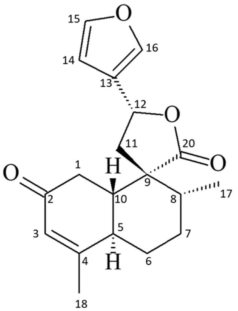
Figure 1. Chemical structure of the 19-nor-clerodane trans-dehydrocrotonin (t-DCTN).
Ijms 26 04469 g001
Figure 2. Phytochemical approach for isolation of the trans-dehydrocrotonin (t-DCTN). EtOH = Ethanol; H2O = Water; EtOAc = Ethyl Acetate; Me2CO = acetone.
Ijms 26 04469 g002
Figure 3. Ternary phase diagram of the SNEDDS-CO system. Surfactant = Tween 80 (7%, w/w), oil phase (1%, w/w) containing a mixture of copaiba oil and sunflower oil (1:1), and water (92%, w/w); SNEDDS-CO = oil-in-water (O/W) colloidal carrier system. The homogeneous and transparent colloidal system is shown in the region above the curvature line.
Ijms 26 04469 g003
Figure 4. Surface tension of SNEDDS-CO system. SNEDDS-CO = oil-in-water (O/W) colloidal carrier system, containing copaiba oil (0.5%, w/w).
Ijms 26 04469 g004
Figure 5. Representative graph of rheological behavior of the SNEDDS-CO system. SNEDDS-CO = oil-in-water (O/W) colloidal carrier system, containing copaiba oil (0.5%, w/w).
Ijms 26 04469 g005
Figure 6. Transmission electron microscopy images of the SNEDDS-CO system. SNEDDS-CO = oil-in-water (O/W) colloidal carrier system, containing copaiba oil (0.5%, w/w).
Ijms 26 04469 g006
Figure 7. In vitro release profile of t-DCTN (percentage released) as a function of time (in minutes) from the SNEDDS-CO-DCTN formulation in the simulated gastric fluid (SGF, pH = 1.2) and simulated intestinal fluid (SIF, pH = 6.8). The drug release diffusion was more favored at the intestinal medium (pH 6.8).
Ijms 26 04469 g007
Figure 8. Adjustment of the in vitro release profile of t-DCTN from the SNEDDS-CO-DCTN formulation (squares) to Fickian’s kinetic model (lines): (A) (SGF, simulated gastric fluid) and (B) (SIF, simulated intestinal fluid).
Ijms 26 04469 g008
Figure 9. Determination of total antioxidant capacity equivalent to ascorbic acid (AA) mg/g of the SNEDDS systems and the unload t-DCTN. * and ** mean the statistical difference between the samples related to SNEDDS-CO system (p < 0.001). # means the statistical difference between SNEDDS-CO-DCTN system and t-DCTN-DMSO solution (p < 0.001). SNEDDS-CO = oil-in-water (O/W) colloidal carrier system, containing copaiba oil (0.5%, w/w). SNEDDS-CO-DCTN containing 1 mg of t-DCTN per mL of the carrier SNEDDS-CO. DCTN = solution containing 10 mg of t-DCTN per mL of DMSO.
Ijms 26 04469 g009
Figure 10. Antioxidant capacity via reducing power of SNEDDS systems and the unload t-DCTN. * means the statistical difference between SNEDDS-CO and SNEDDS-CO-DCTN systems at 1.0 mg mL−1 (p < 0.001). # means the statistical difference of both SNEDDS-CO and SNEDDS-CO-DCTN at the same concentrations by comparing with t-DCTN-DMSO solution (p < 0.001). SNEDDS-CO = oil-in-water (O/W) colloidal carrier system, containing copaiba oil (0.5%, w/w). SNEDDS-CO-DCTN containing 1 mg of t-DCTN per mL of the carrier SNEDDS-CO. DCTN = solution containing 10 mg of t-DCTN per mL of DMSO.
Ijms 26 04469 g010
Figure 11. Antioxidant capacity via copper ion chelation of SNEDDS systems and the unload t-DCTN. * means the statistical difference between SNEDDS-CO and SNEDDS-CO-DCTN systems, at 0.1 to 1.0 mg mL−1 (p < 0.001). ** means the statistical difference between SNEDDS-CO-DCTN and SNEDDS-CO systems, at 2 mg mL−1 (p < 0.001). # means the statistical difference between SNEDDS-CO system and t-DCTN-DMSO solution, at 0.1 to 1.0 mg mL−1 (p < 0.001). ## means the statistical difference between t-DCTN-DMSO solution and SNEDDS-CO system, at 2 mg mL−1 (p < 0.001). a, b mean the statistical difference between SNEDDS-CO-DCTN and t-DCTN-DMSO solution, at 1 mg mL−1 and 2 mg mL−1, respectively (p < 0.001). SNEDDS-CO = oil-in-water (O/W) colloidal carrier system, containing copaiba oil (0.5%, w/w). SNEDDS-CO-DCTN containing 1 mg of t-DCTN per mL of the carrier SNEDDS-CO. DCTN = solution containing 10 mg of t-DCTN per mL of DMSO.
Ijms 26 04469 g011
Figure 12. Hydroxyl radical sequestration capability of SNEDDS systems and the unload t-DCTN. * means the statistical difference of SNEDDS-CO-DCTN system and t-DCTN-DMSO solution by comparing with SNEDDS-CO system, at 0.5 mg mL−1 (p < 0.001). # means the statistical difference between SNEDDS-CO-DCTN and SNEDDS-CO systems, at 1.5 mg mL−1 (p < 0.001). ## means the statistical difference between SNEDDS-CO-DCTN and t-DCTN-DMSO solution, at 1.5 mg mL−1 (p < 0.01). SNEDDS-CO = oil-in-water (O/W) colloidal carrier system, containing copaiba oil (0.5%, w/w). SNEDDS-CO-DCTN containing 1 mg of t-DCTN per mL of the carrier SNEDDS-CO. DCTN = solution containing 10 mg of t-DCTN per mL of DMSO.
Ijms 26 04469 g012
Table 1. Particle size, PDI, and zeta potential analysis of the SNEDDS systems.
Triplicate AnalysisØ (nm)PDIζ (mV)
A11.150.083−3.60
SNEDDS-CO-DCTNB11.310.107−3.63
C11.430.130−3.11
D11.460.135−4.13
SNEDDS-COE11.710.188−3.39
F11.810.200−4.04
Ø (nm) = particle size (dynamic light diffusion/DLS); PDI = polydispersity index; ζ = zeta potential; SNEDDS-CO = oil-in-water (O/W) colloidal carrier system, containing copaiba oil (0.5%, w/w); SNEDDS-CO-DCTN containing 1 mg of t-DCTN per mL of SNEDDS-CO.
Disclaimer/Publisher’s Note: The statements, opinions and data contained in all publications are solely those of the individual author(s) and contributor(s) and not of MDPI and/or the editor(s). MDPI and/or the editor(s) disclaim responsibility for any injury to people or property resulting from any ideas, methods, instructions or products referred to in the content.