Analysis of Cellular Stress Assay Parameters and Intracellular ATP in Platelets: Comparison of Platelet Preparation Methods
Abstract
1. Introduction
2. Results
2.1. Comparison of CSA Parameters in Platelets Isolated with and without Optiprep
2.2. Correlation of CSA and ATP Content in Platelets Isolated with and without Optiprep Methods
3. Discussion
4. Materials and Methods
4.1. Study Design, Period and Settings
4.2. Study Participants
4.3. Blood Sample Collection and Platelet Isolation
4.4. Cellular Stress Assay (CSA)
4.5. Measurement of Intracellular ATP
4.6. Statistical Analyses
5. Conclusions
Author Contributions
Funding
Institutional Review Board Statement
Informed Consent Statement
Data Availability Statement
Acknowledgments
Conflicts of Interest
References
- van der Meijden, P.E.J.; Heemskerk, J.W.M. Platelet biology and functions: New concepts and clinical perspectives. Nat. Rev. Cardiol. 2019, 16, 166–179. [Google Scholar] [CrossRef]
- Holinstat, M. Normal platelet function. Cancer Metastasis Rev. 2017, 36, 195–198. [Google Scholar] [CrossRef] [PubMed]
- Hayashi, T.; Tanaka, S.; Hori, Y.; Hirayama, F.; Sato, E.F.; Inoue, M. Role of mitochondria in the maintenance of platelet function during in vitro storage. Transfus. Med. 2011, 21, 166–174. [Google Scholar] [CrossRef] [PubMed]
- Baccarelli, A.A.; Byun, H.M. Platelet mitochondrial DNA methylation: A potential new marker of cardiovascular disease. Clin. Epigenet. 2015, 7, 44. [Google Scholar] [CrossRef] [PubMed]
- Wang, L.; Wu, Q.; Fan, Z.; Xie, R.; Wang, Z.; Lu, Y. Platelet mitochondrial dysfunction and the correlation with human diseases. Biochem. Soc. Trans. 2017, 45, 1213–1223. [Google Scholar] [CrossRef] [PubMed]
- Aibibula, M.; Naseem, K.M.; Sturmey, R.G. Glucose metabolism and metabolic flexibility in blood platelets. J. Thromb. Haemost. 2018, 16, 2300–2314. [Google Scholar] [CrossRef] [PubMed]
- Prakhya, K.S.; Vekaria, H.; Coenen, D.M.; Omali, L.; Lykins, J.; Joshi, S.; Alfar, H.R.; Wang, Q.J.; Sullivan, P.; Whiteheart, S.W. Platelet glycogenolysis is important for energy production and function. Platelets 2023, 34, 2222184. [Google Scholar] [CrossRef] [PubMed]
- Doery, J.C.G.; Hirsh, J.; Cooper, I. Energy Metabolism in Human Platelets: Interrelationship between Glycolysis and Oxidative Metabolism. Blood 1970, 36, 159–168. [Google Scholar] [CrossRef] [PubMed]
- McDowell, R.E.; Aulak, K.S.; Almoushref, A.; Melillo, C.A.; Brauer, B.E.; Newman, J.E.; Tonelli, A.R.; Dweik, R.A. Platelet glycolytic metabolism correlates with hemodynamic severity in pulmonary arterial hypertension. Am. J. Physiol. Lung Cell. Mol. Physiol. 2020, 318, L562–L569. [Google Scholar] [CrossRef] [PubMed]
- Thon, J.N.; Italiano, J.E. Platelets: Production, Morphology and Ultrastructure. Handb. Exp. Pharmacol. 2012, 210, 3–22. [Google Scholar]
- Chacko, B.K.; Smith, M.R.; Johnson, M.S.; Benavides, G.; Culp, M.L.; Pilli, J.; Shiva, S.; Uppal, K.; Go, Y.-M.; Jones, D.P.; et al. Mitochondria in precision medicine; linking bioenergetics and metabolomics in platelets. Redox Biol. 2019, 22, 101165. [Google Scholar] [CrossRef] [PubMed]
- Ravi, S.; Chacko, B.; Sawada, H.; Kramer, P.A.; Johnson, M.S.; Benavides, G.A.; O’Donnell, V.; Marques, M.B.; Darley-Usmar, V.M. Metabolic Plasticity in Resting and Thrombin Activated Platelets. PLoS ONE 2015, 10, e0123597. [Google Scholar] [CrossRef] [PubMed]
- Myhill, S.; Booth, N.E.; McLaren-Howard, J. Chronic fatigue syndrome and mitochondrial dysfunction. Int. J. Clin. Exp. Med. 2009, 2, 1–16. [Google Scholar] [PubMed]
- Suomalainen, A.; Battersby, B.J. Mitochondrial diseases: The contribution of organelle stress responses to pathology. Nat. Rev. Mol. Cell Biol. 2018, 19, 77–92. [Google Scholar] [CrossRef] [PubMed]
- Hill, B.G.; Benavides, G.A.; Lancaster, J.R., Jr.; Ballinger, S.; Dell’Italia, L.; Zhang, J.; Darley-Usmar, V.M. Integration of cellular bioenergetics with mitochondrial quality control and autophagy. Biol. Chem. 2012, 393, 1485–1512. [Google Scholar] [CrossRef] [PubMed]
- Chacko, B.K.; Kramer, P.A.; Ravi, S.; Benavides, G.A.; Mitchell, T.; Dranka, B.; Ferrick, D.; Singal, A.K.; Ballinger, S.W.; Bailey, S.M.; et al. The Bioenergetic Health Index: A new concept in mitochondrial translational research. Clin. Sci. 2014, 127, 367–373. [Google Scholar] [CrossRef]
- Hill, B.G.; Dranka, B.P.; Zou, L.; Chatham, J.C.; Darley-Usmar, V.M. Importance of the bioenergetic reserve capacity in response to cardiomyocyte stress induced by 4-hydroxynonenal. Biochem. J. 2009, 424, 99–107. [Google Scholar] [CrossRef] [PubMed]
- Tessema, B.; Haag, J.; Sack, U.; König, B. The Determination of Mitochondrial Mass Is a Prerequisite for Accurate Assessment of Peripheral Blood Mononuclear Cells’ Oxidative Metabolism. Int. J. Mol. Sci. 2023, 24, 14824. [Google Scholar] [CrossRef] [PubMed]
- Tessema, B.; Riemer, J.; Sack, U.; König, B. Cellular Stress Assay in Peripheral Blood Mononuclear Cells: Factors Influencing Its Results. Int. J. Mol. Sci. 2022, 23, 13118. [Google Scholar] [CrossRef] [PubMed]
- Van der Heijden, W.A.; van de Wijer, L.; Jaeger, M.; Grintjes, K.; Netea, M.G.; Urbanus, R.T.; van Crevel, R.; van den Heuvel, L.P.; Brink, M.; Rodenburg, R.J.; et al. Long-term treated HIV infection is associated with platelet mitochondrial dysfunction. Sci. Rep. 2021, 11, 6246. [Google Scholar] [CrossRef] [PubMed]
- Yasseen, B.A.; Elkhodiry, A.A.; El-Messiery, R.M.; El-Sayed, H.; Elbenhawi, M.W.; Kamel, A.G.; Gad, S.A.; Zidan, M.; Hamza, M.S.; Al-Ansary, M.; et al. Platelets’ morphology, metabolic profile, exocytosis, and heterotypic aggregation with leukocytes in relation to severity and mortality of COVID-19-patients. Front. Immunol. 2022, 13, 1022401. [Google Scholar] [CrossRef] [PubMed]
- Malinow, A.M.; Schuh, R.A.; Alyamani, O.; Kim, J.; Bharadwaj, S.; Crimmins, S.D.; Galey, J.L.; Fiskum, G.; Polster, B.M. Platelets in preeclamptic pregnancies fail to exhibit the decrease in mitochondrial oxygen consumption rate seen in normal pregnancies. Biosci. Rep. 2018, 38, BSR20180286. [Google Scholar] [CrossRef] [PubMed]
- Calton, E.K.; Keane, K.N.; Soares, M.J.; Rowlands, J.; Newsholme, P. Prevailing vitamin D status influences mitochondrial and glycolytic bioenergetics in peripheral blood mononuclear cells obtained from adults. Redox Biol. 2016, 10, 243–250. [Google Scholar] [CrossRef]
- Hintzpeter, B.; Mensink, G.B.M.; Thierfelder, W.; Müller, M.J.; Scheidt-Nave, C. Vitamin D status and health correlates among German adults. Eur. J. Clin. Nutr. 2008, 62, 1079–1089. [Google Scholar] [CrossRef] [PubMed]
- Petruș, A.; Lighezan, D.; Dănilă, M.; Duicu, O.; Sturza, A.; Muntean, D.; Ioniță, I. Assessment of Platelet Respiration as Emerging Biomarker of Disease. Physiol. Res. 2019, 68, 347–363. [Google Scholar] [CrossRef]
- Weiss, L.; MacLeod, H.; Comer, S.P.; Cullivan, S.; Szklanna, P.B.; Áinle, F.N.; Kevane, B.; Maguire, P.B. An optimized protocol to isolate quiescent washed platelets from human whole blood and generate platelet releasate under clinical conditions. STAR Protoc. 2023, 4, 102150. [Google Scholar] [CrossRef] [PubMed]
- Graham, J. Isolation of Human Platelets (Thrombocytes). Sci. World J. 2002, 2, 1607–1609. [Google Scholar] [CrossRef] [PubMed][Green Version]
- Wrzyszcz, A.; Urbaniak, J.; Sapa, A.; Woźniak, M. An efficient method for isolation of representative and contamination-free population of blood platelets for proteomic studies. Platelets 2017, 28, 43–53. [Google Scholar] [CrossRef]


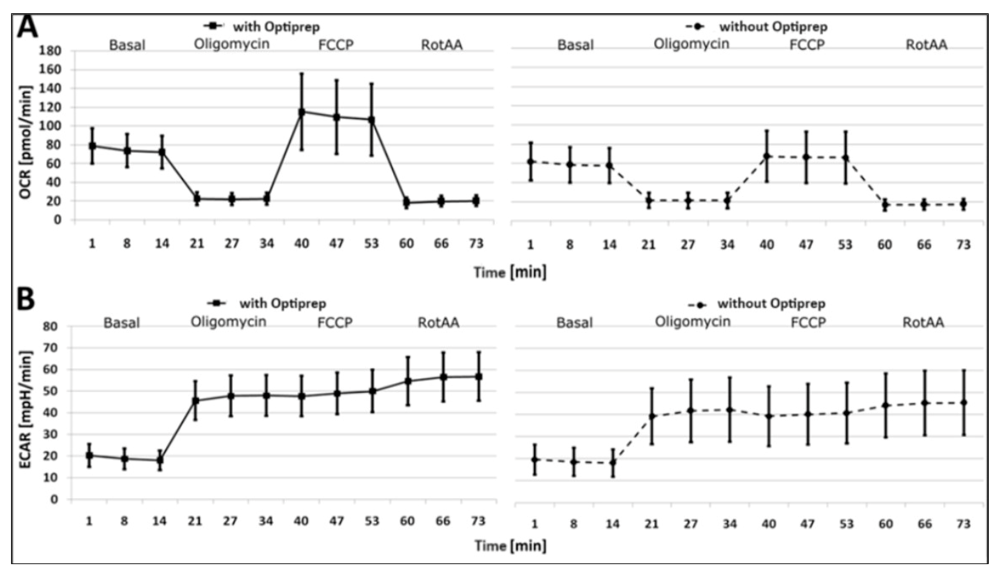
| Parameter | With Optiprep | Without Optiprep | ||||
|---|---|---|---|---|---|---|
| Mean (SD) | 95% CI | Shapiro-Wilk (W) | Mean (SD) | 95% CI | Shapiro-Wilk (W) | |
| Basal respiration (pmol/min) | 51.8 (15.6) | 47.5–56.2 | 0.968 | 41.1 (16.4) | 37.4–44.8 | 0.982 |
| basal OCR (pmol/min) | 72.0 (17.5) | 67.1–76.8 | 0.971 | 57.6 (18.3) | 53.5–61.8 | 0.984 |
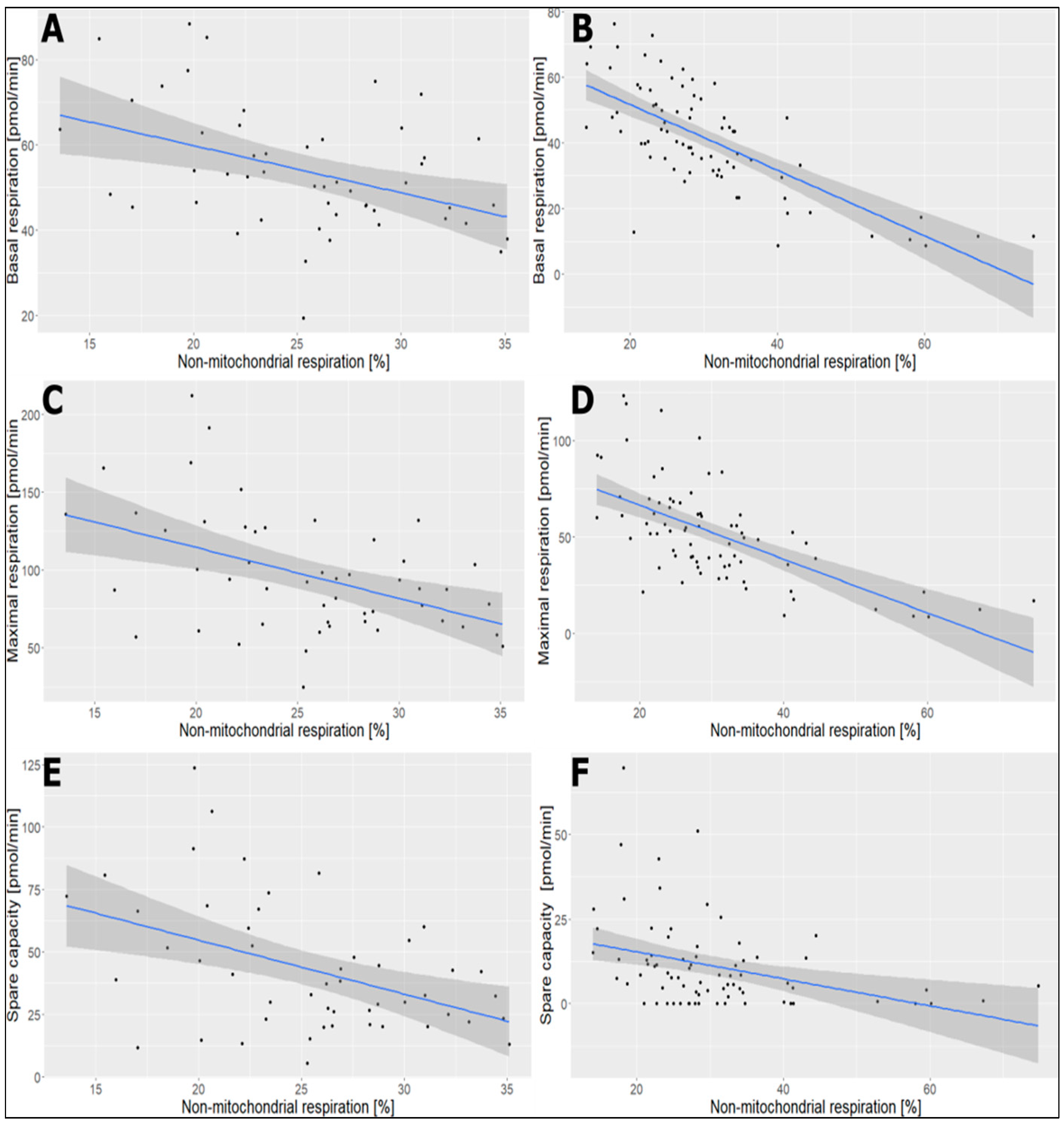
| non-mitochondrial respiration (pmol/min) | 18.1 (5.5) | 16.6–19.7 | 0.970 | 16.5 (5.6) | 15.3–17.8 | 0.956 |
| non-mitochondrial respiration (%) | 25.4 (5.4) | 23.9–26.9 | 0.982 | 30.5 (11.8) | 27.9–33.2 | 0.859 |
| coupling efficiency (%) | 93.0 (6.0) | 91.3–94.7 | 0.740 | 89.6 (13.2) | 86.6–92.6 | 0.894 |
| proton leak (pmol/min) | 3.7 (2.3) | 3.1–4.3 | 0.954 | 4.5 (5.4) | 3.3–5.7 | 0.793 |
| proton leak (%) | 7.2 (5.8) | 5.6–8.8 | 0.699 | 10.9 (12.4) | 8.1–13.7 | 0.817 |
| maximal respiration (pmol/min) | 97.0 (38.7) | 86.2–108.0 | 0.943 | 51.6 (25.8) | 45.8–57.5 | 0.962 |
| spare capacity (pmol/min) | 43.1 (26.3) | 35.8–50.4 | 0.919 | 11.1 (13.2) | 8.2–14.1 | 0.777 |
| spare capacity (%) | 75.6 (32.5) | 66.5–84.6 | 0.955 | 25.3 (26.5) | 19.3–31.3 | 0.823 |
| bioenergetic health index (BHI) | 1.5 (0.4) | 1.4–1.6 | 0.955 | 0.7 (0.6) | 0.6–0.9 | 0.898 |
| basal ECAR (mpH/min) | 18.0 (4.5) | 16.7–19.2 | 0.980 | 18.0 (6.2) | 16.6–19.3 | 0.972 |
| ECAR Oligomycin (mpH/min) | 48.0 (9.4) | 45.4–50.6 | 0.982 | 42.2 (14.6) | 38.9–45.5 | 0.956 |
| maximal ECAR (FCCP) (mpH/min) | 47.7 (9.3) | 45.1–50.3 | 0.993 | 39.2 (13.6) | 36.2–42.3 | 0.964 |
| Parameter | Cohens d * | Interpretation |
|---|---|---|
| basal respiration [pmol/min] | 0.81 | strong |
| basal OCR [pmol/min] | 0.80 | medium |
| non-mitochondrial respiration [pmol/min] | 0.29 | small |
| non-mitochondrial respiration [%] | 0.53 | medium |
| coupling efficiency [%] | 0.31 | small |
| proton leak [pmol/min] | 0.18 | no |
| proton leak [%] | 0.37 | small |
| maximal respiration [pmol/min] | 1.43 | strong |
| spare capacity [pmol/min] | 1.64 | strong |
| spare capacity [%] | 1.73 | strong |
| bioenergetic health index (BHI) | 1.41 | strong |
| basal ECAR [mpH/min] | 0.00 | no |
| ECAR Oligomycin [mpH/min] | 0.45 | small |
| maximal ECAR (FCCP) [mpH/min] | 0.70 | medium |
Disclaimer/Publisher’s Note: The statements, opinions and data contained in all publications are solely those of the individual author(s) and contributor(s) and not of MDPI and/or the editor(s). MDPI and/or the editor(s) disclaim responsibility for any injury to people or property resulting from any ideas, methods, instructions or products referred to in the content. |
© 2024 by the authors. Licensee MDPI, Basel, Switzerland. This article is an open access article distributed under the terms and conditions of the Creative Commons Attribution (CC BY) license (https://creativecommons.org/licenses/by/4.0/).
Share and Cite
Tessema, B.; Haag, J.; Sack, U.; König, B. Analysis of Cellular Stress Assay Parameters and Intracellular ATP in Platelets: Comparison of Platelet Preparation Methods. Int. J. Mol. Sci. 2024, 25, 4885. https://doi.org/10.3390/ijms25094885
Tessema B, Haag J, Sack U, König B. Analysis of Cellular Stress Assay Parameters and Intracellular ATP in Platelets: Comparison of Platelet Preparation Methods. International Journal of Molecular Sciences. 2024; 25(9):4885. https://doi.org/10.3390/ijms25094885
Chicago/Turabian StyleTessema, Belay, Janine Haag, Ulrich Sack, and Brigitte König. 2024. "Analysis of Cellular Stress Assay Parameters and Intracellular ATP in Platelets: Comparison of Platelet Preparation Methods" International Journal of Molecular Sciences 25, no. 9: 4885. https://doi.org/10.3390/ijms25094885
APA StyleTessema, B., Haag, J., Sack, U., & König, B. (2024). Analysis of Cellular Stress Assay Parameters and Intracellular ATP in Platelets: Comparison of Platelet Preparation Methods. International Journal of Molecular Sciences, 25(9), 4885. https://doi.org/10.3390/ijms25094885


