Review

Local Production of Acute Phase Proteins: A Defense Reaction of Cancer Cells to Injury with Focus on Fibrinogen

by
Péter Hamar
Institute of Translational Medicine, Semmelweis University Budapest, Tűzoltó u 37–47, 1094 Budapest, Hungary
Int. J. Mol. Sci. 2024, 25(6), 3435; https://doi.org/10.3390/ijms25063435
Submission received: 12 January 2024 / Revised: 12 March 2024 / Accepted: 15 March 2024 / Published: 19 March 2024
(This article belongs to the Special Issue Molecular Research in Triple-Negative Breast Cancer)

Abstract

:
This review is intended to demonstrate that the local production of acute phase proteins (termed local acute phase response (lAPR)) and especially fibrin/fibrinogen (FN) is a defense mechanism of cancer cells to therapy, and inhibition of the lAPR can augment the effectiveness of cancer therapy. Previously we detected a lAPR accompanying tumor cell death during the treatment of triple-negative breast cancer (TNBC) with modulated electro-hyperthermia (mEHT) in mice. We observed a similar lAPR in in hypoxic mouse kidneys. In both models, production of FN chains was predominant among the locally produced acute phase proteins. The production and extracellular release of FN into the tumor microenvironment is a known method of self-defense in tumor cells. We propose that the lAPR is a new, novel cellular defense mechanism like the heat shock response (HSR). In this review, we demonstrate a potential synergism between FN inhibition and mEHT in cancer treatment, suggesting that the effectiveness of mEHT and chemotherapy can be enhanced by inhibiting the HSR and/or the lAPR. Non-anticoagulant inhibition of FN offers potential new therapeutic options for cancer treatment.

1. Introduction of the Topic

The heat shock response (HSR) is a well-described general and ancient stress response of cells to various types of stress. The acute phase response (APR) is a part of the immune response to infections and tissue damage. Proteins whose plasma concentration is changed by at least 25% in response to pro-inflammatory stimuli are termed acute phase proteins (APPs) [1]. They have a role in restoring homeostasis after inflammation [2]. The generally held concept is that APPs are produced in the liver; these are triggered mainly by the inflammatory interleukin-6 (IL-6) (as well as IL-1, IL-8 and TNF-α), and secreted into the blood. However, APPs are also synthesized in other organs. Thus, APPs are a part of local defense responses and can contribute to repair mechanisms [3]. We demonstrated by multiplex analysis a complex local production of several APPs in different tissues upon stress, such as electromagnetic heating of tumor cells [4], acute hypoxic [5] or chronic fibrotic injury of the kidney [6]. Thus, we propose that the local APP production is an organized, ancient cellular stress response, like the much-better-characterized HSR [7]. The local APR (lAPR) can be an important mechanism to protect cells from stress similarly to the HSR [8]. While APPs are traditionally known for their systemic roles in the acute phase response, there is increasing recognition that they can also exert canonical and non-canonical functions in local tissue defense. Although, besides hepatocytes, inflammatory cells are thought to be the key sources of systemic acute phase proteins, other cells such as stromal cells, adipocytes, endothelial cells [9] and cancer cells [10] have been described to produce acute phase proteins. APPs can modulate inflammatory signaling pathways [11]. For example, they may interact with cell surface receptors or soluble mediators to regulate the intensity and duration of the inflammatory response [9]. By fine-tuning inflammation, APPs can help prevent excessive tissue damage while still promoting effective host defense. Furthermore, APPs can also modulate immune cell function and communication within the tissue. They may influence the activation, differentiation, and function of immune cells, such as macrophages [12,13], neutrophils [14], and dendritic cells [15]. By shaping the immune response at the local level, APPs help coordinate effective host defense while minimizing collateral tissue damage. Finally, APPs play roles in tissue repair and remodeling processes beyond their traditional roles in inflammation. For instance, some acute phase proteins, such as fibronectin and tenascin, are involved in cell adhesion, migration, and extracellular matrix remodeling [16]. By promoting tissue integrity and repair, these proteins contribute to the restoration of tissue homeostasis following injury or infection.
An association between acute-phase proteins (APPs) and cancer has long been established and many cancerous cells produce APPs. The biological significance of the lAPR in cancer, and the benefit of cancer cells from these proteins remains largely unknown. Recent data revealed that some APPs, including α1-antitrypsin, are able to enhance cancer cell resistance to anticancer drug-induced apoptosis [10].
Inhibition of the HSR can support cancer therapies as demonstrated by us [8] and others [17,18]. Similarly to the HSR inhibition, inhibition of the local APR may be beneficial in cancer treatment [4,19].

2. Background: Description of the Model Used in the Reviewed Experiments

2.1. Breast Cancer (BC)

Breast cancer (BC) is one of the most frequent cancer types among women worldwide. Triple-negative breast cancer (TNBC) is a highly aggressive BC type with very poor survival due to the lack of targeted therapy. The most used mouse TNBC models utilize cell lines derived from mouse mammary carcinoma cell line 410.4, isolated from a single spontaneous tumor in Balb/c mice. The cell lines 4T1 and 4T07 are the most aggressive and invasive sub-clones [19]. Inoculation of these syngeneic cell lines creates isografts in Balb/c mice. Thus, immune mechanisms can be investigated under conditions very similar to those of human TNBC [8].

2.2. Modulated Electro-Hyperthermia (mEHT)

Modulated electro-hyperthermia (mEHT) is a newly emerging adjuvant cancer treatment used in human oncology, with strong cancer inhibitory effect in monotherapy in our mouse model. During mEHT, a focused electromagnetic field (EMF) is generated within the tumor by applying capacitive radiofrequency. Selective energy absorption by the tumor is the consequence of its elevated oxidative glycolysis (Warburg effect) and conductivity. The EMF induces cell death by thermal and non-thermal effects. Capacitive energy delivery and frequency modulation enable the application of non-thermal effects. We have developed the rodent mEHT device to enable accurate, reproducible, standard, and effective treatment of TNBC in the inguinal region of mice [8]. Selective energy absorption enabled to warm up the tumor by +2.5 °C—a great advantage over former hyperthermia methods, where the temperature difference between the tumor and surrounding tissues was only +1 °C. Thus, the thermal and non-thermal effects amplify each, other leading to effective tumor cell killing. The device is also useful for local delivery of therapeutic agents by using thermo-sensitive liposomes (TSL).

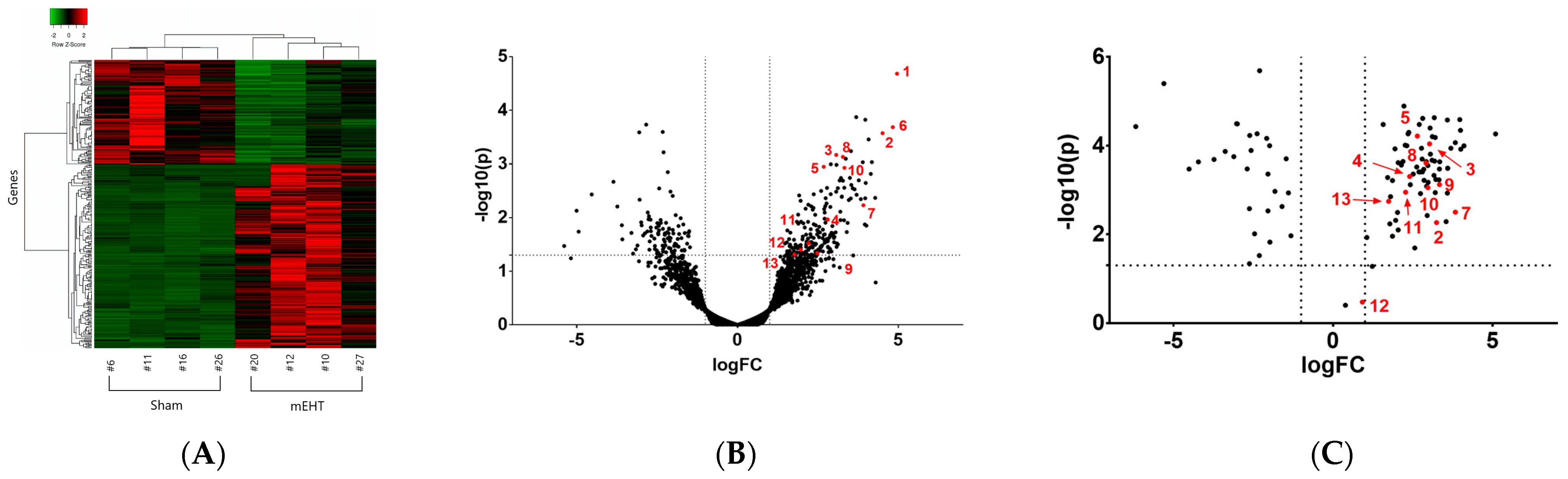
3. Experimental Evidence Demonstrating Local Acute Phase Protein and Fibrinogen (FN) Production in Cancer Treated with Modulated Electro-Hyperthermia

We have demonstrated that mEHT-induced tumor cell death is located in the core of treated tumors. The necrotic area was cleaved caspase-3 (cC3) positive, suggesting apoptotic cell death. We observed a strong HSP-70 staining in the treated tumors around the dead area, suggesting induction of the heat-shock response (HSR) by mEHT [8].
Multiplex analysis of mEHT-treated TNBC [4,8] and hypoxic [5] or fibrotic [6] kidney samples allowed us to identify a proteomic response dominated by APPs as detailed below (Figure 1). Comparison of the raw data from our four papers analyzing tissue/cell stress with multiplex methods revealed that several APPs were upregulated in more than one model. Furthermore, real-time polymerase chain reaction (RT-PCR) detection of mRNA and cell culture studies confirmed that the APPs were produced locally by the tumor or renal cells, confirming that APPs were produced by the stressed cells themselves and not by the liver.
Contrary to most previous studies, where multiplex RNA (next-generation sequencing, NGS) results were verified by few, individual data (using, e.g., qPCR), we verified our NGS data by another multiplex method: Nano-String (NS) nCounter® (NanoString Technologies, Seattle, WA, USA), using the bar-coding of each RNA in the sample. We selected 134 target genes from the NGS data to create a custom NanoString gene panel. Nano-String verified 104 target genes with the same direction of change as detected by NGS (Figure 1C). Furthermore, we performed mass spectrometry (MS) as a multiplex method to verify gene expression at the protein level.

The Local Acute Phase Response (lAPR)

APPs are plasma proteins synthesized in the liver whose concentrations increase (or decrease) by 25% or more during inflammation [20]. This definition is based on the electrophoretic separation of plasma proteins, which demonstrates elevation of the alpha1 and alpha2 fractions [21]. APPs and the APR in general are stably conserved in evolution [22]. These proteins play a crucial role in the innate immune response and the body’s defense mechanisms, and serve to protect tissues during infection, inflammation and/or tissue damage [23]. The production of APP’s maintains homeostasis and tissue repair, but also represents a central component of the organism’s defense strategy, especially in the context of innate immunity [21].
In contrast to the systemic APR, where the APPs are produced by the liver, proteins of the lAPR are produced in tissues in response to injury or inflammation. Unlike APPs produced in the liver, which circulate throughout the bloodstream, local APPs are primarily synthesized at the site of injury or inflammation. They help regulate the inflammatory response and promote tissue repair.
The background of the identified APPs and their known contribution to cancer progression are summarized in a recent review [11]. The lAPR identified in our screens are all major APPs: all 3 FN chains (FGA, FGB, FGG), haptoglobin (HP), complement component 4B (C4B) and several protease inhibitors: inter-alpha-trypsin inhibitor (I alpha I) family members (light chain (AMBP) and heavy chains: ITIH-1,3,4), clade A (extracellular) serpins (especially A1 (alpha-1 antitrypsin) A3B (alpha-1 antichymotrypsin), and A3K (kallikrein inhibitor).
The following is true for all of these proteins: (1) they are APPs; (2) the main site of their synthesis is the hepatocyte, where besides basal expression, inflammation upregulates their synthesis during APR; (3) extra-hepatic production has been demonstrated already; (4) they are well preserved throughout evolution (ancient proteins) found in many lower species; (5) they are involved in cellular stress response (defensive).
Indeed, several previous publications support our observation that local APP production is an ancient defense mechanism. Although these scarce and isolated findings demonstrate local production of some APPs, but the coordinated, complex local APR has not been described before. A proteomic analysis of the urine revealed APPs (FGA, FGG, HP, ITI4, SERPINA1) [14] as biomarkers for prostate cancer without further analysis.

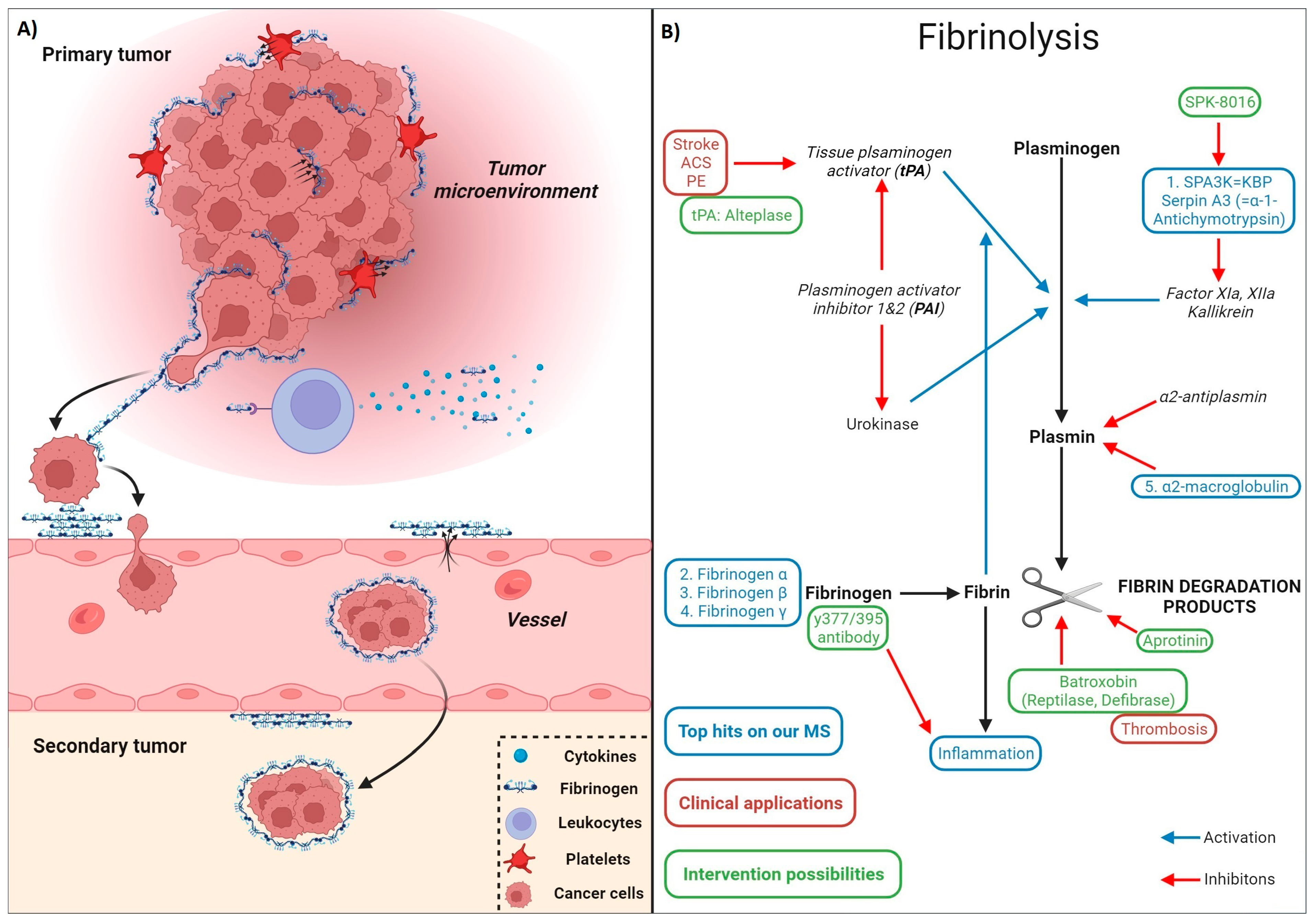
4. Fibrin(ogen) (FN)

4.1. The Structure of FN

FN is a complex protein composed of two sets of disulfide-bridged polypeptide chains: 2 Aα (alpha), 2 Bβ (beta), and 2 γ (gamma) chains [24]. Each chain has distinct roles in the process of blood clotting and FN formation: Aα chains play a crucial role in the polymerization process of fibrinogen. They contain binding sites that interact with platelets and other molecules involved in clot formation. Additionally, Aα chains contribute to the mechanical properties of the fibrin clot, influencing its stability and structure. Bβ chains also participate in polymerization of fibrinogen, contributing to the formation of the fibrin clot. They contain sites that are involved in interactions between fibrin molecules, helping to link them together during clot formation. Gamma chains are important for the overall structure of fibrinogen. They contribute to the stability and integrity of the molecule. Although not directly involved in fibrin clot formation, they play a critical role in maintaining the overall structure of fibrinogen, which influences its function in clotting. The fibrinogen-like domain is found in all three types of fibrinogen chains, and contributes to the assembly of fibrinogen molecules into a larger, functional fibrin polymer with specific binding sites for fibrinogen, allowing fibrinogen molecules to connect and form a mesh-like structure.
The leukocyte integrin, αMβ2 (Mac-1), is a high affinity receptor for fibrin(ogen) on stimulated monocytes and neutrophils that is important for fibrin(ogen)–leukocyte interactions that contributes to the inflammatory response. This domain binds the CD11b/CD18 complement receptor-3 (CR-3, Mac-1) leading to macrophage activation [25]. The Mac-1-binding site within the fibrinogen D domain contains two peptide sequences, γ190–202 and γ377–395, designated as P1 and P2 [24].

4.2. The Physiological (Canonical and Non-Canonical) Functions of Fibrin(ogen) (FN) in the Blood and in the Extracellular Matrix (ECM)

Fibrinogen is a glycoprotein that is involved in blood clotting and is synthesized in the liver. However, it can accumulate in injured tissues and contribute to local inflammation and tissue repair. Fibrinogens are soluble proteins in the blood. Fibrinogen chains (Aα, Bβ, γ) are present in the human blood at concentrations of 1.5–4 g/L, thus being one of the most abundant plasma proteins (7–8%) after albumin [26].
The physiological functions of FN are: in addition to providing a scaffold for blood clotting, FN is important for the assembly of (extracellular) matrices to enhance host defense [27]. Upon passing out of the bloodstream, fibrinogens become unsolvable and precipitate in the ECM. The fibrin mesh formed in the ECM serves as a provisional matrix essential for tissue repair [9]. Increased production of matrix molecules such as fibrinogen and protease inhibitors during the APR serve as a general mechanism to promote tissue repair (Table 1).
Proteins such as fibrinogen (fibrinogen-related proteins) comprise a collection of extracellular molecules, each containing a conserved fibrinogen-like globe: a common and ancient domain that has evolved to play key roles in tissue repair. The ancestral function of fibrinogen domain-containing molecules was defense [28]. Fibrinogen-related proteins are upregulated during tissue repair [29]. Thus, cancer cells stressed by hyperthermia or chemotherapy can produce fibrinogen locally as a mechanism of defense and repair.
Over time, as organisms evolved and developed more complex systems, fibrinogen and its related molecules may have acquired additional functions beyond basic clotting and defense. In vertebrates, fibrinogen-related proteins play roles not only in hemostasis but also in inflammation, immune regulation, and tissue repair [29]. Specific, non-coagulatory functions of fibrinogen include stimulation of angiogenesis, keratinocyte and fibroblast migration and proliferation, matrix remodeling and the recruitment and activation of immune cells [29]. Understanding the ancestral functions of fibrinogen domain-containing molecules provides insights into their diverse roles in modern organisms, which include not only coagulation but also aspects of immunity and tissue homeostasis.

4.3. Documented Roles of Fibrinogens in Non-Hematologic Disease

While fibrinogen is primarily known for its crucial role in blood clotting and coagulation, the involvement of fibrinogen in non-hematologic diseases has been widely documented.

4.3.1. Fibrin(ogen) Plays a Role in Inflammatory Conditions

However, precipitated fibrin is also inflammatory. Fibrin- and interferon-induced genes are inflammatory [30]. FN has been implicated in pathological processes including renal diseases and cancer. FN-deficient mice (Fg−/−) [31] were protected from endotoxemia [32], renal fibrosis [33] and ischemia-reperfusion injury [34]. In fibrinogen KO mice nerve injury was reduced. Fibrin precipitated in the ECM activates local inflammatory cells (e.g., microglia in the brain) but may also induce plaque formation directly in neurodegenerative diseases.
Fibrinogen and its downstream degradation products (D-dimer and other fibrin degradation products) are widely used as diagnostic markers in inflammatory conditions, including COVID-19 [28].
Fibrinogen has been associated with the development and progression of atherosclerosis and elevated fibrinogen levels were demonstrated to be a predictor of cardiovascular disease in the Framingham study [35].
In patients with rheumatoid arthritis serum fibrinogen was elevated and correlated with markers of inflammation [36]. Also, in inflammatory bowel disease, increased fibrinogen levels were demonstrated to be a predictive biomarker of disease activity [37].
Elevated fibrinogen levels are associated with insulin resistance and may contribute to the increased cardiovascular risk in individuals with type 2 diabetes [38]. Diabetes is associated with increased plasma fibrinogen especially in patients with albuminuria [38]. Fibrinogen levels are often elevated in obesity. This may contribute to the pro-inflammatory state associated with obesity and its complications. Obese patients show increased fibrinogen levels and associated with insulin resistance and metabolic syndrome [39].
FN appears extracellularly in the brain in neurodegenerative diseases such as Alzheimer’s dementia (AD) or progressive multiple sclerosis (MS) [40,41], where fibrinogen deposition is linked to plaque formation [42]. Increased vascular permeability is associated with worse prognosis in these diseases. It has been demonstrated that fibrinogen is causative in the neurodegenerative process [40]. Furthermore, fibrinogen has been documented to be deposited in different brain diseases as a neuropathological hallmark. The causative role of FN is supported by the observation that FN KO mice are rescued from cognitive impairment (Alzheimer’s disease).
Elevated fibrinogen levels are linked to inflammation, exacerbations, severity, and mortality in chronic obstructive pulmonary disease [43].
Taken together FN and its extracellular deposition has a proinflammatory role in several diseases.

4.3.2. Documented Roles of Fibrin(ogen) in Cancer Progression

Molecular mechanisms induced by extracellular fibrin deposition and their relevance in cancer progression are summarized in Table 1 and Figure 2A. Coagulation factors have been linked with malignancy for over 100 years, and high plasma fibrinogen has been associated with cancer progression. Fibrin(ogen) (FN) may enter the tumor stroma from the circulation as evidenced by FN deposition in the vicinity of blood vessels in tumors [44]. The therapeutic removal of such FN deposition has been demonstrated to enhance therapy delivery to the cancer [45].
However, FN can be produced by cancer cells [46], such as BC cells [27]. The locally produced FN binds to and surrounds cancer cells, forming a protective structure. Furthermore, by interacting with endothelial cells, FN contributes to the extravasation of cancer cells [26]. A hallmark of breast carcinoma is the local synthesis and deposition of fibrinogen (precipitation without cleavage by thrombin to fibrin) [47]. FN in the ECM augments the innate immune response to tissue injury or cancer [47]. FN deposition is a predominant component in breast tumor stroma [47]. In fibrinogen KO mice, the carcinogenesis gene-network was downregulated [30]. In our studies, all three FN chains were within the top upregulated genes on all three multiplex screens in TNBC and renal tissue. Our multiplex data do not distinguish between fibrinogen and fibrin and do not indicate the involvement of thrombin or other coagulation-related factors.
From the identified panel of APPs in our studies, most data on cancer progression are available on FN. As reviewed by us previously [48], the data are conflicting regarding beneficial or deleterious effects of FN on cancer progression. FN is important for the assembly of (extracellular) matrices to enhance host defense [27]. High plasma FN has been associated with cancer progression. FN produced by BC cells [27] binds to and surrounds cancer cells, forms a protective structure, and contributes to extravasation of BC cells [26]. A hallmark of BC is the local synthesis and deposition of FN (precipitation without conversion to fibrin) making FN a predominant component of BC stroma [47]. Furthermore, FN augments the innate immune response to cancer [47].
Table 1. Molecular mechanisms induced by extracellular fibrin deposition and their relevance in cancer progression. Abbreviations: FN: fibrin, ECM: extracellular matrix, PS: phosphatydil-serine, Fgα−/−: fibrinogen alpha chain knockout, NK-cell: natural killer cell, αMβ2: integrin receptor, EC: endothelial cell.
FN Function/Molecular MechanismFN in Cancer ProgressionExperimental EvidenceReference
FN filtered from the circulation (FN deposition in the vicinity of blood vessels in tumors)Compact stroma hinders therapy deliveryRemoval of the compact stroma enhances therapy delivery[44,45,49]
Enhancement of cancer cell extravasationLactadherin—by competing with FN for PS binding sites may delay tumor progression[26,50]
FN stimulates ECM formationFN scaffold in the TME → adhesion, migration, invasion and metastasis of cancer cells FN is a predominant component of the BC stroma. FN KO mice (Fgα−/−) were protected against hematogenous metastasis[26,27,51,52]
FN binds to + surrounds cancer cells → protective structure from treatments and immune systemFN interaction with platelets via β3-integrins facilitates the protection of tumor cells from NK-cell cytotoxicity, permitting escape from host immune surveillance[26,27,30]
FN is important for the assembly of ECM to enhance host defenseFN contributes to the tumor favorable TMEIn fibrinogen KO mice, the carcinogenesis gene-network was downregulated[27,53]
FN supports cancer cell growth, survival and proliferationPrimary colon cancer development diminished in Fgα−/− mice [54,55]
FN net: growth factor reservoirFN contributes to the proinflammatory TME to favor tumor progressionInteraction with αMβ2 induce leukocyte adhesion to ECs + proinflammatory cytokine production[47,55]

4.3.3. Fibrinolysis

Fibrinolysis pathways and their drugability is demonstrated on Figure 2B. Besides FN upregulation, fibrinolysis inhibitor serpins (A1, A3) were also upregulated in our multiplex screens. Thus, removing FN from the tumor microenvironment (TME) by plasmin may have a synergistic potential with anti-cancer therapies including mEHT. On the other hand, plasmin may have pro-cancer effects by releasing cancer-related growth factors and promoting angiogenesis [56]. Thus, modulating FN production may have a therapeutic potential.
Furthermore, serine protease inhibitors (extracellular clade A serpins) are produced in the liver and different cancer cells. Serpins participate in fibrinolysis, invasion, and migration and thus metastasis of cancer cells, and their expression is associated with cancer progression and poor prognosis [57], supporting their role in cancer cell protection. Besides FN upregulation, fibrinolysis inhibitors Serpin A3 (alpha-1-Antichymotrypsin) and alpha-2-macroglobulin were also upregulated in both the tumor and the renal samples. Thus, these data by us and others support a central role of local fibrinogen production and deposition in the TME of breast cancer.
Figure 2. (A) Fibrin(ogen) surrounding tumor cells produced by the cancer cells themselves or platelets in the TME aids the formation of protective tumor microenvironment. The deposited fibrin may stimulate proinflammatory processes in the TME by recruiting and activating leucocytes. Fibrin filtered from permeable blood vessels and deposited around them aid the vascular entrance of potentially metastatic cells [26]. (B) Drugability of the fibrinolysis pathways. Top upregulated proteins (blue) in our mouse model were related to fibrin(ogen) deposition and degradation. (tPA: tissue plasminogen activator [58], PAI: plasminogen activator inhibitor, SPA3K: serine protease inhibitor (Serpin)-A3 = alpha-1-anitchymotrypsin). Current possibilities for intervention (green labels) and diseases where these interventions are used clinically (green letters) are indicated. (ACS: acute coronary syndrome, PE: pulmonary embolism, tPA: tissue plasminogen activator [59]: direct thrombin inhibitor (dabigatran), SPK: Spa3K inhibitor developed by Spark Therapeutics [60]. The figures were created with Biorender.com.
Ijms 25 03435 g002

4.3.4. Fibrin Induces a Tumor-Favorable Microenvironment (TME)

A TME includes blood vessels, immune cells, fibroblasts, and extracellular matrix components. FN contributes to the tumor-favorable TME as follows: (1) Stimulating angiogenesis that increases oxygen and nutrient supply to the tumor [53]. Tumor hypoxia-induced VEGF production by the cancer cells increases vascular permeability and consequent fibrin leakage which directly stimulates angiogenesis [55]. However, FN may be directly produced by the cancer cells as suggested by our multiplex studies. (2) Stimulating extracellular matrix formation: Fibrin forms a scaffold in the TME that can support the adhesion and migration of tumor cells, facilitating their invasion into surrounding tissues. The ECM acts as a barrier to protect cancer from treatments and supports tumor progression [52]. (3) The FN net can act as a reservoir for growth factors and cytokines that support cell growth via a proinflammatory TME. (4) FN can interact with various cells in the microenvironment through signaling pathways. This interaction may support the survival and proliferation of tumor cells or promote their ability to evade the immune system.
Although FN is a component of the ECM, it is often not discussed in papers on ECM or TME for a few reasons: Fibrin is a temporary component of the ECM. Fibrin, unlike collagen or fibronectin, is not associated with the mature tissue structure, but as a somewhat temporary repair stage ECM component [61]. It’s formed during blood clotting in response to injury and wound healing. In the context of tumors, while fibrin may be present due to ongoing vascular permeability or injury, or synthetized locally by the cancer cells, it might not remain in the ECM for extended periods, unlike other ECM components like collagen or fibronectin [62]. Research on the ECM often emphasizes permanent structural components such as collagens, proteoglycans, and glycoproteins. Mainly fibroblasts produce the matrix and regulate matrix remodeling, but in cancer, the tumor matrix also originates from cancer cells [63]. An enriched distribution of fibrin in tumor tissues obtained from high-grade non-small cell lung cancer was demonstrated, and extracellular fibrin was associated with cancer progression, cancer stemness and chemotherapy resistance [64]. Thus, despite its relative under-representation, understanding the role of FN in the ECM is crucial, especially in contexts such as wound healing [65], inflammation, and tumor progression [64], where its presence and transient nature can significantly influence cell behavior and tissue dynamics [66].

4.3.5. Fibrin Deposition-Induced Chronic Inflammation of the Tumor Microenvironment (TME)

Inflammation predisposes to the development of cancer and promotes all stages of tumorigenesis [67]. The TME is a complex and structured mixture characterized by abnormal angiogenesis, chronic inflammation, and dysregulated extracellular matrix (ECM) remodeling, collectively contributing to an immunosuppressive milieu [68]. Chronic inflammation can contribute to cancer progression. Inflammation creates a microenvironment that supports the survival and growth of cancer cells, promotes angiogenesis, and suppresses the immune system’s ability to eliminate cancer cells. Furthermore, an important component of the tumor extracellular matrix is fibrin, which is abundant near tumor vessels [45].

4.3.6. Drugability of Fibrinogen

There are drugs approved and in development targeting fibrinogen for thrombolytic (Abciximab) and non-thrombolytic purposes. Abciximab (ReoPro) inhibits the procoagulant properties (moiety: y400–411) of FN.
As stated above (in the chapter: “The structure of fibrinogen”) the Mac-1-binding site is responsible for the proinflammatory properties of FN. The Mac-1-binding site within the fibrinogen D domain contains two peptide sequences, γ190–202 and γ377–395, designated as P1 and P2. These epitopes differ from the epitopes responsible for the procoagulant function [25]. A FN mutant where only seven amino acids (390–396) are deleted in the inflammatory domain functions under normal hemostasis. For therapeutic application, a first in class antibody (Ab) was demonstrated to recognize only the inflammatory domain of FN—the Ab is targeting a cryptic inflammatory domain of FN exposed only in precipitated fibrin not in soluble fibrinogen [69]. Such an Ab would selectively block the pro-inflammatory function of FN without jeopardizing coagulation. The therapeutic effects have been demonstrated in MS/AD mouse models [41]. The antibody has now been humanized, GMP manufactured and tested in primates, and the first in human studies began in 2023. However, deletion of the minimal recognition sequence, γ390–395, was not sufficient to ablate Mac-1 binding, because mutants were still effective in supporting adhesion of Mac-1-expressing cells probably due to a second sequence in the P1 segment between γ228 and γ253 that contributed to the binding activity [24].
A further possibility of targeted removal of fibrin deposited in tumors would be the targeted delivery of fibrinolytic enzymes to the tumor. In one study tissue plasminogen activator (tPA) was given systemically by intraperitoneal application [45]. However, such a systemic treatment may have bleeding side effects. A more sophisticated application was demonstrated in a recent paper by Ting Mei et al. They demonstrated that redox-active nanoparticles were able to deliver their cargo into solid tumors. Delivery of tPA via these redox-active nanoparticles was able to degrade fibrin in the tumor [44]. A similar approach would be to deliver tPA (Alteplase) or batroxabin (Defibrase) into the tumor via lyso-thermosensitive liposomes (LTSL) and local heating of the tumor [70].

5. Summary and Future Perspectives

In conclusion, the presently prevailing concept of the APR may only be part of the truth. There is the well-known systemic acute-phase response, where, in response to tissue injury and consequent inflammation, locally produced inflammatory cytokines (IL-6, IL-1b, etc.) induce acute-phase protein (APP) production by the liver. However, there is also a local APR (lAPR) at the cellular level: a coordinated local production of several acute-phase proteins with the aim of protecting the cells from environmental stress, like the also well-described heat shock response. The HSR exerts its protection within the cell, whereas the lAPR is an extracellular protective mechanism. A future perspective includes the possible utilization of this novel stress response for diagnostic and therapeutic purposes. In cancer, the cancer cells produce APPs to protect themselves from conventional (chemotherapy, radiotherapy) or adjuvant (heat therapy) treatments. Among the identified lAPR proteins fibrinogen appears to be of particular interest as a major component of the TME protecting tumor cells from injury. Local, targeted inhibition of the production of these APPs in the tumor may substantially enhance the anti-tumor effects of currently available anti-cancer treatments. Thus, the potential clinical implications and future directions of research into local acute phase protein production as a therapeutic target in cancer treatment lie primarily in combination therapies: targeting the local acute phase protein production in addition to other therapeutic modalities, such as chemotherapy, radiation therapy, or immunotherapy, may enhance treatment efficacy through synergistic effects. By combining treatments that target different aspects of tumor biology and the immune microenvironment, there lies the potential to overcome resistance mechanisms and improve overall response rates in cancer patients. Furthermore, better understanding of the pathophysiological roles of locally produced APPs may help in the development of targeted immunomodulation: Understanding how APPs are produced and function within the TME could provide insights into ways to modulate local immune responses to better fight cancer. By targeting specific APPs involved in promoting immunosuppression or tumor growth, strategies can be developed to enhance antitumor immune response and improve the efficacy of cancer immunotherapy.

Funding

This research was funded by (1) the Hungarian Research Fund (OTKA) to Peter Hamar: Ministry for Innovation and Technology from the source of the National Research, Development and Innovation Fund, OTKA_K 145998 (2) the Semmelweis Science and Innovation Fund: Semmelweis NKFIH-OTKA Transitional Support (STIA-OTKA-2022), Péter Hamar was a recipient of the Merit Price of Semmelweis University (2021–23 and 2012–18).

Institutional Review Board Statement

Not applicable.

Informed Consent Statement

Not applicable.

Data Availability Statement

Not applicable.

Acknowledgments

The author would like to acknowledge the people participating in the experiments leading to this hypothesis. In alphabetical order: Lea Danics, Tamás Kaucsár, Csaba Kécsán-Révész, Beáta Róka, Csaba Schvarcz, Gábor Szénási, Pál Tod, Aloss Kenan, Bokhari Syeda Mahak Zahra, Leroy Viana Pedro Hernrique, Nino Giunashvili. Furthermore, I would like to acknowledge the help of Zoltán Koós, Dániel Bócsi for technical assistance in the submission of the present review. The author wants to express his sincere gratitude for the thorough language editing performed by Gábor Szénási.

Conflicts of Interest

The author declares no conflicts of interest.

Abbreviations

A1Malpha-1-microglobulin
a2-MGalpha-2-macroglobulin
AMBPbikunin precursor
ACSacute coronary syndrome
AKIacute kidney injury
APPacute-phase proteins
APRacute-phase response
AT-IIIantithrombin-3
CKDchronic kidney disease
CPBcarboxypeptidase-B
CRCcolorectal cancer
DVTdeep vein thrombosis
ECMextracellular matrix
EMFfocused electromagnetic field
FNfibrin(ogen)
GSglomerular sclerosis
HIF-1hypoxia-inducible factor-1
Hphaptoglobin
HSRheat shock response
IalphaI/ITIinter-alpha-trypsin inhibitor
ILinterleukin
LMWHlow-molecular-weight heparin
LPSendotoxin/lipopolysaccharide
mEHTmodulated electro-hyperthermia
MSmass spectrometry
NGSnext-generation sequencing
NSCLCnon-small-cell lung cancer
PAIplasminogen activator inhibitor
PCRpolymerase chain reaction
PEpulmonary embolism
Pproperidin
PTX3pentraxin-related gene
RT-PCRreal-time polymerase chain reaction
SERPINserine protease inhibitor
SPA3Kserine protease inhibitor (Serpin)-A3 = alpha-1-anitchymotrypsin
SPKSpa3K inhibitor
TAFIthrombin-activatable fibrinolysis inhibitor
TECtubular epithelial cell
TGF-betatransforming growth factor beta
TMEtumor microenvironment
TNBCtriple-negative breast cancer
TNF-alphatumor necrosis factor-alpha
tPAtissue plasminogen activator
TSLthermo-sensitive liposome

References

  1. Gabay, C.; Kushner, I. Acute-phase proteins and other systemic responses to inflammation. N. Engl. J. Med. 1999, 340, 448–454. [Google Scholar] [CrossRef]
  2. Moshage, H. Cytokines and the hepatic acute phase response. J. Pathol. 1997, 181, 257–266. [Google Scholar] [CrossRef]
  3. Schrodl, W.; Buchler, R.; Wendler, S.; Reinhold, P.; Muckova, P.; Reindl, J.; Rhode, H. Acute phase proteins as promising biomarkers: Perspectives and limitations for human and veterinary medicine. Proteom. Clin. Appl. 2016, 10, 1077–1092. [Google Scholar] [CrossRef] [PubMed]
  4. Schvarcz, C.A.; Danics, L.; Krenacs, T.; Viana, P.; Beres, R.; Vancsik, T.; Nagy, A.; Gyenesei, A.; Kun, J.; Fonovic, M.; et al. Modulated Electro-Hyperthermia Induces a Prominent Local Stress Response and Growth Inhibition in Mouse Breast Cancer Isografts. Cancers 2021, 13, 1744. [Google Scholar] [CrossRef] [PubMed]
  5. Roka, B.; Tod, P.; Kaucsar, T.; Vizovisek, M.; Vidmar, R.; Turk, B.; Fonovic, M.; Szenasi, G.; Hamar, P. The Acute Phase Response Is a Prominent Renal Proteome Change in Sepsis in Mice. Int. J. Mol. Sci. 2019, 21, 200. [Google Scholar] [CrossRef] [PubMed]
  6. Bukosza, E.N.; Kornauth, C.; Hummel, K.; Schachner, H.; Huttary, N.; Krieger, S.; Nobauer, K.; Oszwald, A.; Razzazi Fazeli, E.; Kratochwill, K.; et al. ECM Characterization Reveals a Massive Activation of Acute Phase Response during FSGS. Int. J. Mol. Sci. 2020, 21, 2095. [Google Scholar] [CrossRef] [PubMed]
  7. Kaucsar, T.; Bodor, C.; Godo, M.; Szalay, C.; Revesz, C.; Nemeth, Z.; Mozes, M.; Szenasi, G.; Rosivall, L.; Soti, C.; et al. LPS-induced delayed preconditioning is mediated by Hsp90 and involves the heat shock response in mouse kidney. PLoS ONE 2014, 9, e92004. [Google Scholar] [CrossRef]
  8. Danics, L.; Schvarcz, C.A.; Viana, P.; Vancsik, T.; Krenacs, T.; Benyo, Z.; Kaucsar, T.; Hamar, P. Exhaustion of Protective Heat Shock Response Induces Significant Tumor Damage by Apoptosis after Modulated Electro-Hyperthermia Treatment of Triple Negative Breast Cancer Isografts in Mice. Cancers 2020, 12, 2581. [Google Scholar] [CrossRef]
  9. Mantovani, A.; Garlanda, C. Humoral Innate Immunity and Acute-Phase Proteins. N. Engl. J. Med. 2023, 388, 439–452. [Google Scholar] [CrossRef]
  10. Janciauskiene, S.; Wrenger, S.; Gunzel, S.; Grunding, A.R.; Golpon, H.; Welte, T. Potential Roles of Acute Phase Proteins in Cancer: Why Do Cancer Cells Produce or Take Up Exogenous Acute Phase Protein Alpha1-Antitrypsin? Front. Oncol. 2021, 11, 622076. [Google Scholar] [CrossRef]
  11. Serrano, I.; Luque, A.; Aran, J.M. Exploring the Immunomodulatory Moonlighting Activities of Acute Phase Proteins for Tolerogenic Dendritic Cell Generation. Front. Immunol. 2018, 9, 892. [Google Scholar] [CrossRef] [PubMed]
  12. Lu, X.; Huang, Y.; Zhou, M.; Guo, Y.; Zhou, Y.; Wang, R.; Jin, W.; Wen, C.; Zhang, Y.; Tang, Y. Artesunate attenuates serum amyloid A-induced M1 macrophage differentiation through the promotion of PHGDH. Int. Immunopharmacol. 2024, 127, 111462. [Google Scholar] [CrossRef] [PubMed]
  13. Zhang, S.; Rao, G.; Heimberger, A.; Li, S. Fibrinogen-like protein 2: Its biological function across cell types and the potential to serve as an immunotherapy target for brain tumors. Cytokine Growth Factor Rev. 2023, 69, 73–79. [Google Scholar] [CrossRef]
  14. Shephard, E.G.; Anderson, R.; Strachan, A.F.; Kuhn, S.H.; De Beer, F.C. CRP and neutrophils: Functional effects and complex uptake. Clin. Exp. Immunol. 1986, 63, 718–727. [Google Scholar]
  15. Jimenez, R.V.; Wright, T.T.; Jones, N.R.; Wu, J.; Gibson, A.W.; Szalai, A.J. C-Reactive Protein Impairs Dendritic Cell Development, Maturation, and Function: Implications for Peripheral Tolerance. Front. Immunol. 2018, 9, 372. [Google Scholar] [CrossRef] [PubMed]
  16. Tremble, P.; Chiquet-Ehrismann, R.; Werb, Z. The extracellular matrix ligands fibronectin and tenascin collaborate in regulating collagenase gene expression in fibroblasts. Mol. Biol. Cell 1994, 5, 439–453. [Google Scholar] [CrossRef]
  17. Ban, H.S.; Han, T.S.; Hur, K.; Cho, H.S. Epigenetic Alterations of Heat Shock Proteins (HSPs) in Cancer. Int. J. Mol. Sci. 2019, 20, 4758. [Google Scholar] [CrossRef]
  18. Asea, A.; Ara, G.; Teicher, B.A.; Stevenson, M.A.; Calderwood, S.K. Effects of the flavonoid drug quercetin on the response of human prostate tumours to hyperthermia in vitro and in vivo. Int. J. Hyperth. 2001, 17, 347–356. [Google Scholar] [CrossRef]
  19. Le, M.T.; Hamar, P.; Guo, C.; Basar, E.; Perdigao-Henriques, R.; Balaj, L.; Lieberman, J. miR-200-containing extracellular vesicles promote breast cancer cell metastasis. J. Clin. Investig. 2014, 124, 5109–5128. [Google Scholar] [CrossRef]
  20. Ackermann, M.R. Chapter 3—Inflammation and Healing1. In Pathologic Basis of Veterinary Disease, 6th ed.; Zachary, J.F., Ed.; Mosby: Amsterdam, The Netherlands, 2017; pp. 73–131.e2. [Google Scholar]
  21. Ehlting, C.; Wolf, S.D.; Bode, J.G. Acute-phase protein synthesis: A key feature of innate immune functions of the liver. Biol. Chem. 2021, 402, 1129–1145. [Google Scholar] [CrossRef]
  22. Sabina, J.; Tobias, W.; Ravi, M. Acute Phase Proteins: Structure and Function Relationship. In Acute Phase Proteins; Francisco, V., Ed.; IntechOpen: Rijeka, Croatia, 2011; p. 2. [Google Scholar]
  23. Pepys, M.B. Acute Phase Proteins. In Encyclopedia of Immunology, 2nd ed.; Delves, P.J., Ed.; Elsevier: Oxford, UK, 1998; pp. 18–20. [Google Scholar]
  24. Mosesson, M.W. Fibrinogen and fibrin structure and functions. J. Thromb. Haemost. 2005, 3, 1894–1904. [Google Scholar] [CrossRef]
  25. Davalos, D.; Ryu, J.K.; Merlini, M.; Baeten, K.M.; Le Moan, N.; Petersen, M.A.; Deerinck, T.J.; Smirnoff, D.S.; Bedard, C.; Hakozaki, H.; et al. Fibrinogen-induced perivascular microglial clustering is required for the development of axonal damage in neuroinflammation. Nat. Commun. 2012, 3, 1227. [Google Scholar] [CrossRef]
  26. Vilar, R.; Fish, R.J.; Casini, A.; Neerman-Arbez, M. Fibrin(ogen) in human disease: Both friend and foe. Haematologica 2020, 105, 284–296. [Google Scholar] [CrossRef]
  27. Simpson-Haidaris, P.J.; Rybarczyk, B. Tumors and fibrinogen. The role of fibrinogen as an extracellular matrix protein. Ann. N. Y. Acad. Sci. 2001, 936, 406–425. [Google Scholar] [CrossRef]
  28. Bottazzi, B.; Doni, A.; Garlanda, C.; Mantovani, A. An integrated view of humoral innate immunity: Pentraxins as a paradigm. Annu. Rev. Immunol. 2010, 28, 157–183. [Google Scholar] [CrossRef]
  29. Zuliani-Alvarez, L.; Midwood, K.S. Fibrinogen-Related Proteins in Tissue Repair: How a Unique Domain with a Common Structure Controls Diverse Aspects of Wound Healing. Adv. Wound Care 2015, 4, 273–285. [Google Scholar] [CrossRef] [PubMed]
  30. Mendiola, A.S.; Yan, Z.; Dixit, K.; Johnson, J.R.; Bouhaddou, M.; Meyer-Franke, A.; Shin, M.G.; Yong, Y.; Agrawal, A.; MacDonald, E.; et al. Defining blood-induced microglia functions in neurodegeneration through multiomic profiling. Nat. Immunol. 2023, 24, 1173–1187. [Google Scholar] [CrossRef] [PubMed]
  31. Degen, J.L.; Drew, A.F.; Palumbo, J.S.; Kombrinck, K.W.; Bezerra, J.A.; Danton, M.J.; Holmbäck, K.; Suh, T.T. Genetic manipulation of fibrinogen and fibrinolysis in mice. Ann. N. Y. Acad. Sci. 2001, 936, 276–290. [Google Scholar] [CrossRef] [PubMed]
  32. Cruz-Topete, D.; Iwaki, T.; Ploplis, V.; Castellino, F. Delayed inflammatory responses to endotoxin in fibrinogen-deficient mice. J. Pathol. 2006, 210, 325–333. [Google Scholar]
  33. Sörensen, I.; Susnik, N.; Inhester, T.; Degen, J.L.; Melk, A.; Haller, H.; Schmitt, R. Fibrinogen, acting as a mitogen for tubulointerstitial fibroblasts, promotes renal fibrosis. Kidney Int. 2011, 80, 1035–1044. [Google Scholar] [CrossRef] [PubMed]
  34. Petzelbauer, P.; Zacharowski, P.A.; Miyazaki, Y.; Friedl, P.; Wickenhauser, G.; Castellino, F.J.; Gröger, M.; Wolff, K.; Zacharowski, K. The fibrin-derived peptide Bbeta15-42 protects the myocardium against ischemia-reperfusion injury. Nat. Med. 2005, 11, 298–304. [Google Scholar] [CrossRef]
  35. Kannel, W.B.; Wolf, P.A.; Castelli, W.P.; D’Agostino, R.B. Fibrinogen and Risk of Cardiovascular Disease: The Framingham Study. JAMA 1987, 258, 1183–1186. [Google Scholar] [CrossRef]
  36. Rooney, T.; Scherzer, R.; Shigenaga, J.K.; Graf, J.; Imboden, J.B.; Grunfeld, C. Levels of plasma fibrinogen are elevated in well-controlled rheumatoid arthritis. Rheumatology 2011, 50, 1458–1465. [Google Scholar] [CrossRef]
  37. Chen, X.F.; Zhao, Y.; Guo, Y.; Huang, Z.M.; Huang, X.L. Predictive value of fibrinogen in identifying inflammatory bowel disease in active stage. BMC Gastroenterol. 2021, 21, 472. [Google Scholar] [CrossRef]
  38. Martín-Timón, I.; Sevillano-Collantes, C.; Segura-Galindo, A.; Del Cañizo-Gómez, F.J. Type 2 diabetes and cardiovascular disease: Have all risk factors the same strength? World J. Diabetes 2014, 5, 444–470. [Google Scholar] [CrossRef]
  39. Solá, E.; Vayá, A.; Simó, M.; Hernández-Mijares, A.; Morillas, C.; España, F.; Estellés, A.; Corella, D. Fibrinogen, plasma viscosity and blood viscosity in obesity. Relationship with insulin resistance. Clin. Hemorheol. Microcirc. 2007, 37, 309–318. [Google Scholar]
  40. Petersen, M.A.; Ryu, J.K.; Akassoglou, K. Fibrinogen in neurological diseases: Mechanisms, imaging and therapeutics. Nat. Rev. Neurosci. 2018, 19, 283–301. [Google Scholar] [CrossRef] [PubMed]
  41. Akassoglou, K. The immunology of blood: Connecting the dots at the neurovascular interface. Nat. Immunol. 2020, 21, 710–712. [Google Scholar] [CrossRef] [PubMed]
  42. Merlini, M.; Rafalski, V.A.; Rios Coronado, P.E.; Gill, T.M.; Ellisman, M.; Muthukumar, G.; Subramanian, K.S.; Ryu, J.K.; Syme, C.A.; Davalos, D.; et al. Fibrinogen Induces Microglia-Mediated Spine Elimination and Cognitive Impairment in an Alzheimer’s Disease Model. Neuron 2019, 101, 1099–1108.e1096. [Google Scholar] [CrossRef] [PubMed]
  43. Sun, W.; Cao, Z.; Ma, Y.; Wang, J.; Zhang, L.; Luo, Z. Fibrinogen, a Promising Marker to Evaluate Severity and Prognosis of Acute Exacerbation of Chronic Obstructive Pulmonary Disease: A Retrospective Observational Study. Int. J. Chron. Obs. Pulmon Dis. 2022, 17, 1299–1310. [Google Scholar] [CrossRef] [PubMed]
  44. Mei, T.; Shashni, B.; Maeda, H.; Nagasaki, Y. Fibrinolytic tissue plasminogen activator installed redox-active nanoparticles (t-PA@iRNP) for cancer therapy. Biomaterials 2020, 259, 120290. [Google Scholar] [CrossRef]
  45. Zhang, B.; Jiang, T.; She, X.; Shen, S.; Wang, S.; Deng, J.; Shi, W.; Mei, H.; Hu, Y.; Pang, Z.; et al. Fibrin degradation by rtPA enhances the delivery of nanotherapeutics to A549 tumors in nude mice. Biomaterials 2016, 96, 63–71. [Google Scholar] [CrossRef]
  46. Sahni, A.; Simpson-Haidaris, P.J.; Sahni, S.K.; Vaday, G.G.; Francis, C.W. Fibrinogen synthesized by cancer cells augments the proliferative effect of fibroblast growth factor-2 (FGF-2). J. Thromb. Haemost. 2008, 6, 176–183. [Google Scholar] [CrossRef]
  47. Rybarczyk, B.J.; Simpson-Haidaris, P.J. Fibrinogen assembly, secretion, and deposition into extracellular matrix by MCF-7 human breast carcinoma cells. Cancer Res. 2000, 60, 2033–2039. [Google Scholar]
  48. Hamar, P. A New Role of Acute Phase Proteins: Local Production Is an Ancient, General Stress-Response System of Mammalian Cells. Int. J. Mol. Sci. 2022, 23, 2972. [Google Scholar] [CrossRef] [PubMed]
  49. Nagy, J.A.; Brown, L.F.; Senger, D.R.; Lanir, N.; Van de Water, L.; Dvorak, A.M.; Dvorak, H.F. Pathogenesis of tumor stroma generation: A critical role for leaky blood vessels and fibrin deposition. Biochim. Biophys. Acta 1989, 948, 305–326. [Google Scholar] [CrossRef] [PubMed]
  50. Jing, H.; Wu, X.; Xiang, M.; Wang, C.; Novakovic, V.A.; Shi, J. Microparticle Phosphatidylserine Mediates Coagulation: Involvement in Tumor Progression and Metastasis. Cancers 2023, 15, 1957. [Google Scholar] [CrossRef] [PubMed]
  51. Dvorak, H.F.; Senger, D.R.; Dvorak, A.M. Fibrin as a component of the tumor stroma: Origins and biological significance. Cancer Metastasis Rev. 1983, 2, 41–73. [Google Scholar] [CrossRef] [PubMed]
  52. Yuan, Z.; Li, Y.; Zhang, S.; Wang, X.; Dou, H.; Yu, X.; Zhang, Z.; Yang, S.; Xiao, M. Extracellular matrix remodeling in tumor progression and immune escape: From mechanisms to treatments. Mol. Cancer 2023, 22, 48. [Google Scholar] [CrossRef] [PubMed]
  53. Hadjipanayi, E.; Kuhn, P.H.; Moog, P.; Bauer, A.T.; Kuekrek, H.; Mirzoyan, L.; Hummel, A.; Kirchhoff, K.; Salgin, B.; Isenburg, S.; et al. The Fibrin Matrix Regulates Angiogenic Responses within the Hemostatic Microenvironment through Biochemical Control. PLoS ONE 2015, 10, e0135618. [Google Scholar] [CrossRef] [PubMed]
  54. Adams, G.N.; Rosenfeldt, L.; Frederick, M.; Miller, W.; Waltz, D.; Kombrinck, K.; McElhinney, K.E.; Flick, M.J.; Monia, B.P.; Revenko, A.S.; et al. Colon Cancer Growth and Dissemination Relies upon Thrombin, Stromal PAR-1, and Fibrinogen. Cancer Res. 2015, 75, 4235–4243. [Google Scholar] [CrossRef]
  55. Gupta, M.K.; Qin, R.Y. Mechanism and its regulation of tumor-induced angiogenesis. World J. Gastroenterol. 2003, 9, 1144–1155. [Google Scholar] [CrossRef]
  56. Lin, H.; Xu, L.; Yu, S.; Hong, W.; Huang, M.; Xu, P. Therapeutics targeting the fibrinolytic system. Exp. Mol. Med. 2020, 52, 367–379. [Google Scholar] [CrossRef]
  57. Kryvalap, Y.; Czyzyk, J. The Role of Proteases and Serpin Protease Inhibitors in beta-Cell Biology and Diabetes. Biomolecules 2022, 12, 67. [Google Scholar] [CrossRef]
  58. Baskin, J.L.; Pui, C.H.; Reiss, U.; Wilimas, J.A.; Metzger, M.L.; Ribeiro, R.C.; Howard, S.C. Management of occlusion and thrombosis associated with long-term indwelling central venous catheters. Lancet 2009, 374, 159–169. [Google Scholar] [CrossRef]
  59. Gomez-Outes, A.; Terleira-Fernandez, A.I.; Calvo-Rojas, G.; Suarez-Gea, M.L.; Vargas-Castrillon, E. Dabigatran, Rivaroxaban, or Apixaban versus Warfarin in Patients with Nonvalvular Atrial Fibrillation: A Systematic Review and Meta-Analysis of Subgroups. Thrombosis 2013, 2013, 640723. [Google Scholar] [CrossRef]
  60. Therapeutics, S. Hemophilia a Inhibitor. Available online: https://sparktx.com/pipelines/spk-8016-hemophilia-a-inhibitor-market/ (accessed on 6 November 2018).
  61. Reddy, R.; Reddy, N. Biomimetic approaches for tissue engineering. J. Biomater. Sci. Polym. Ed. 2018, 29, 1667–1685. [Google Scholar] [CrossRef] [PubMed]
  62. Peltanova, B.; Raudenska, M.; Masarik, M. Effect of tumor microenvironment on pathogenesis of the head and neck squamous cell carcinoma: A systematic review. Mol. Cancer 2019, 18, 63. [Google Scholar] [CrossRef] [PubMed]
  63. Popova, N.V.; Jucker, M. The Functional Role of Extracellular Matrix Proteins in Cancer. Cancers 2022, 14, 238. [Google Scholar] [CrossRef] [PubMed]
  64. Li, G.; Cai, J.; Xie, J.; Dai, Y. Extracellular fibrin promotes non-small cell lung cancer progression through integrin beta1/PTEN/AKT signaling. Open Life Sci. 2023, 18, 20220716. [Google Scholar] [CrossRef] [PubMed]
  65. Laurens, N.; Koolwijk, P.; de Maat, M.P. Fibrin structure and wound healing. J. Thromb. Haemost. 2006, 4, 932–939. [Google Scholar] [CrossRef] [PubMed]
  66. Huang, J.; Zhang, L.; Wan, D.; Zhou, L.; Zheng, S.; Lin, S.; Qiao, Y. Extracellular matrix and its therapeutic potential for cancer treatment. Signal Transduct. Target. Ther. 2021, 6, 153. [Google Scholar] [CrossRef]
  67. Greten, F.R.; Grivennikov, S.I. Inflammation and Cancer: Triggers, Mechanisms, and Consequences. Immunity 2019, 51, 27–41. [Google Scholar] [CrossRef]
  68. Chen, C.; Wang, Z.; Ding, Y.; Qin, Y. Tumor microenvironment-mediated immune evasion in hepatocellular carcinoma. Front. Immunol. 2023, 14, 1133308. [Google Scholar] [CrossRef] [PubMed]
  69. Ryu, J.K.; Rafalski, V.A.; Meyer-Franke, A.; Adams, R.A.; Poda, S.B.; Rios Coronado, P.E.; Pedersen, L.; Menon, V.; Baeten, K.M.; Sikorski, S.L.; et al. Fibrin-targeting immunotherapy protects against neuroinflammation and neurodegeneration. Nat. Immunol. 2018, 19, 1212–1223. [Google Scholar] [CrossRef] [PubMed]
  70. Aloss, K.; Hamar, P. Recent Preclinical and Clinical Progress in Liposomal Doxorubicin. Pharmaceutics 2023, 15, 893. [Google Scholar] [CrossRef] [PubMed]
Figure 1. Multiplex analysis of mEHT treated TNBC samples. (A) Next generation sequencing (NGS) results of mEHT- vs. sham-treated tumors (heat map: red: up-, green: down-regulated genes). (B) Volcano plot of the NGS data: the most upregulated proteins were acute phase proteins (red dots and numbers). (C) NanoString verified acute phase protein upregulation [4].
Ijms 25 03435 g001
Disclaimer/Publisher’s Note: The statements, opinions and data contained in all publications are solely those of the individual author(s) and contributor(s) and not of MDPI and/or the editor(s). MDPI and/or the editor(s) disclaim responsibility for any injury to people or property resulting from any ideas, methods, instructions or products referred to in the content.

