Article

METACASPASE8 (MC8) Is a Crucial Protein in the LSD1-Dependent Cell Death Pathway in Response to Ultraviolet Stress

by
Maciej Jerzy Bernacki
1,2,
Anna Rusaczonek
3,
Kinga Gołębiewska
2,
Agata Barbara Majewska-Fala
2,
Weronika Czarnocka
3 and
Stanisław Mariusz Karpiński
2,*
1
Institute of Technology and Life Sciences—National Research Institute, Falenty, Al. Hrabska 3, 05-090 Raszyn, Poland
2
Department of Plant Genetics, Breeding and Biotechnology, Institute of Biology, Warsaw University of Life Sciences, Nowoursynowska Street 159, 02-776 Warsaw, Poland
3
Department of Botany, Institute of Biology, Warsaw University of Life Sciences, Nowoursynowska 159, 02-776 Warsaw, Poland
*
Author to whom correspondence should be addressed.
Int. J. Mol. Sci. 2024, 25(6), 3195; https://doi.org/10.3390/ijms25063195
Submission received: 16 January 2024 / Revised: 5 March 2024 / Accepted: 6 March 2024 / Published: 11 March 2024

Abstract

:
LESION-SIMULATING DISEASE1 (LSD1) is one of the well-known cell death regulatory proteins in Arabidopsis thaliana. The lsd1 mutant exhibits runaway cell death (RCD) in response to various biotic and abiotic stresses. The phenotype of the lsd1 mutant strongly depends on two other proteins, ENHANCED DISEASE SUSCEPTIBILITY 1 (EDS1) and PHYTOALEXIN-DEFICIENT 4 (PAD4) as well as on the synthesis/metabolism/signaling of salicylic acid (SA) and reactive oxygen species (ROS). However, the most interesting aspect of the lsd1 mutant is its conditional-dependent RCD phenotype, and thus, the defined role and function of LSD1 in the suppression of EDS1 and PAD4 in controlled laboratory conditions is different in comparison to a multivariable field environment. Analysis of the lsd1 mutant transcriptome in ambient laboratory and field conditions indicated that there were some candidate genes and proteins that might be involved in the regulation of the lsd1 conditional-dependent RCD phenotype. One of them is METACASPASE 8 (AT1G16420). This type II metacaspase was described as a cell death-positive regulator induced by UV-C irradiation and ROS accumulation. In the double mc8/lsd1 mutant, we discovered reversion of the lsd1 RCD phenotype in response to UV radiation applied in controlled laboratory conditions. This cell death deregulation observed in the lsd1 mutant was reverted like in double mutants of lsd1/eds1 and lsd1/pad4. To summarize, in this work, we demonstrated that MC8 is positively involved in EDS1 and PAD4 conditional-dependent regulation of cell death when LSD1 function is suppressed in Arabidopsis thaliana. Thus, we identified a new protein compound of the conditional LSD1-EDS1-PAD4 regulatory hub. We proposed a working model of MC8 involvement in the regulation of cell death and we postulated that MC8 is a crucial protein in this regulatory pathway.

1. Introduction

Because of their sessile nature, plants cannot avoid environmental stresses by changing their place of inhabitance. Therefore, in natural environments, plants are constantly exposed to biotic and abiotic stress simultaneously, such as various pathogens, excess/deficiency of light, UV irradiation, drought, cold, heat, or salinity. Throughout the course of evolution, plants have developed many molecular and physiological mechanisms that enable them to simultaneously optimize acclimation and defense responses to variable and adverse environmental conditions [1,2,3]. One of the mechanisms crucial in plants response to stress is cell death (CD). CD is a molecular and physiological process that leads to the selective death of some cells, i.e., mesophyll cells, thus triggering a beneficial immune defense and acclimatory response in other cells [4,5]. In this regard, CD is a specified and highly organized process of the cells’ self-elimination. It plays a crucial role in plant development [6,7], immune defense [8] and acclimatory responses [1,2]. From this point of view, CD is not only the ultimate end of the cell life cycle, but most importantly, it maintains cellular homeostasis in organs and in the whole plant during unfavorable environmental conditions.
Knowledge of the molecular, physiological and genetic mechanisms of plant CD at different levels of complexity (cellular and organismal) was facilitated by the identification of different Arabidopsis thaliana mutants exhibiting CD deregulation [9,10,11]. Some of the best-known conditional CD regulators in plants are LESION-SIMULATING DISEASE 1 protein (LSD1), ENHANCED DISEASE SUSCEPTIBILITY 1 (EDS1) and PHYTOALEXIN-DEFICIENT 4 (PAD4). The dysfunctional CD phenotype of the lsd1 mutant has been broadly studied. This mutant exhibits a runaway cell death (RCD) phenotype which is manifested by the inability to restrict CD propagation if it has been initiated by an external stimulus [3,8,12]. It has been shown that RCD can be induced in the lsd1 mutant by the following stress factors: high light and photorespiration [13,14], root hypoxia [1,15], drought [16,17], cold [18], UV radiation [16,19] or biotic stresses [20,21]. However, the lsd1 RCD phenotype is dependent on growing conditions and was not observed when plants were grown in multivariable field conditions [16,22]. Based on many studies, LSD1 is considered a conditional suppressor of CD which is positively regulated by EDS1 and PAD4 and integrates various signaling pathways in response to both biotic and abiotic stresses [3].
Initially, the RCD phenotype of the lsd1 mutant was linked to the accumulation of superoxide ions produced by plasma membrane-bound NADPH oxidase [8] and only after the other reaction oxygen species (ROS) forms were identified to be involved in RCD phenotype elicitation [13,14,18,23,24]. Since in the lsd1 mutant the initial level of antioxidative enzyme activity is lower than in the wild type [23,25], LSD1 is regarded as a positive regulator of the enzymatic antioxidant machinery. Another CD-related molecule, which is excessively accumulated in lsd1, is salicylic acid (SA) [16,22,23]. It has been found that the artificial blocking of SA accumulation in the lsd1 mutant prevents RCD induction; therefore, it was proposed that SA accumulation, controlled by LSD1, is essential in triggering CD in response to stress [19].
Dysfunctional overaccumulation of SA in the lsd1 mutant is caused by the fact that LSD1 can physically interact with proteins involved in SA signaling. It was shown that LSD1 interacts with ENHANCED DISEASE SUSCEPTIBILITY 1 (EDS1), while EDS1 forms complexes with PHYTOALEXIN-DEFICIENT 4 (PAD4) [26,27]. Both EDS1 and PAD4 possess triacylglycerol lipase domains that were originally described as components of gene-mediated and basal disease resistance [28,29,30,31]. EDS1 and PAD4 are crucial for RCD propagation in the lsd1 mutant, because in the eds1/lsd1 and pad4/lsd1 double mutants, the RCD was inhibited regardless of the stimulus type [21,22,23,32]. Therefore, LSD1 is considered a negative regulator of EDS1- and PAD4-dependent pathways that lead to RCD [3].
From a molecular perspective, an LSD1 protein contains three zinc (Zn)-finger domains that are responsible for DNA/protein binding [33]. LSD1 was proven to be a transcriptional regulator and a scaffold protein [27]. The Zn-finger motifs in LSD1 belong to the C2C2 class that is also present in GaTa1-type transcription factors containing the conserved consensus sequence CxxCRxxLMYxxGaSxVxCxxC [33,34]. Using yeast two-hybrid (Y2H) assay, additional ten putative LSD1-interacting proteins were found, from which one of them was METACASPASE1 (MC1), a positive CD regulator [35].
The METACASAPASES family is interesting in the context of cell death studies. Arabidopsis thaliana contains nine METACASAPASES proteins, 1 to 9, that are divided into two groups [36]. Type I metacaspases (MC1, MC2 and MC3) contain zinc-finger domains, while type II (MC4-MC9) do not [36,37]. This family contains both positive and negative CD regulators. While type I metacaspases’ role is relatively well understood [35], the function of the type II subfamily is still largely unknown. In the context of the LSD1-dependent cell death regulation pathway, METACASPASE 8 (MC8) seems to be especially interesting. Its expression level is strongly up-regulated in response to UV-C [38], pathogens [39] and methyl viologen [38]. It was also shown that the recessive mutants in MC8 exhibit higher resistance to UV-C and ROS treatment [38]. Therefore, we postulated a hypothesis that MC8 is involved in the propagation of RCD in lsd1 plants and we decided to explore the mutual interdependence of these two proteins in CD regulation.

2. Results

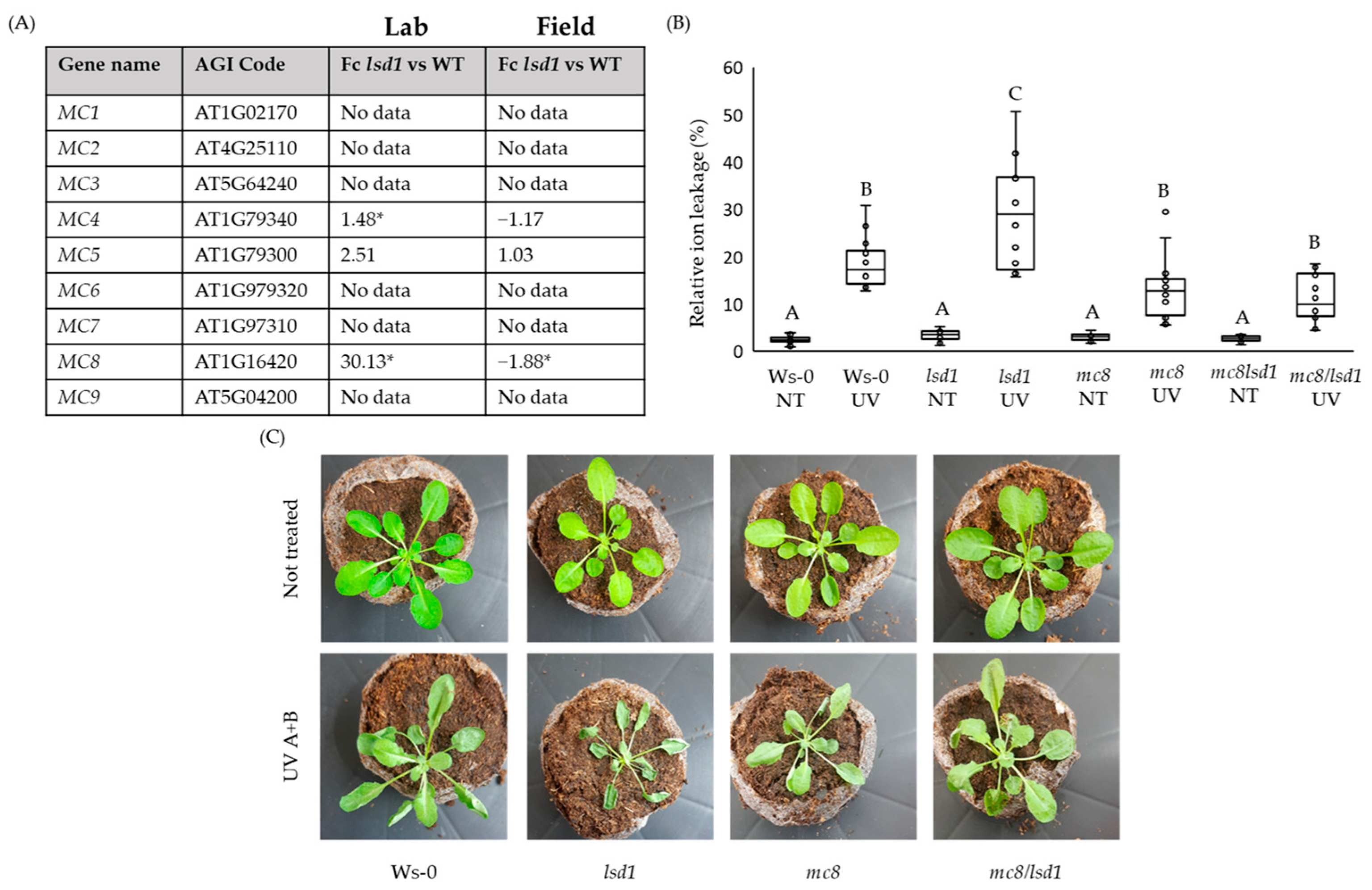
2.1. MC8 Is Important in the LSD1-Dependent Cell Death Pathway

Because proteins belonging to the METACASPASE family are known to be involved in cell death regulation [35,36,40], we decided to search for MC genes within microarray data published in one of our previous articles [16]. Having compared the fold changes in MC genes’ expression level in the lsd1 mutant and wild-type plants, we observed that only the level of MC4 and MC8 were significantly up-regulated (Figure 1A). Interestingly, the expression level of MC8 was antagonistically regulated in different growing conditions, an ambient laboratory or natural field (Figure 1A). In addition, in eds1/lsd1 and pad4/lsd1 double mutants, which do not exhibit the specific RCD phenotype, the MC8 fold change was not detected (Supplementary Table S1). Data from microarray experiments were confirmed using a real-time PCR (Supplementary Figure S1). Interestingly, in double lsd1/eds1 and lsd1/pad4 mutants that reverted the lsd1 RCD phenotype to the wild type, there were not any significant changes in the MC8 transcript level (Supplementary Table S1). This finding together with the reverted cell death in the double mc8/lsd1 mutant in response to UV irradiation (Figure 1B) and the plant phenotype (Figure 1C and Supplementary Figure S2) allowed us to conclude that MC8 was an important component in the LSD1-dependent conditional cell death regulatory hub. Moreover, we proved that in Arabidopsis thaliana wild-type plants, MC8 expression was strongly up-regulated in response to a combined UV-A + UV-B irradiation episode, while in response to heat or high light stress, there were no differences compared to plants growing in controlled conditions (Supplementary Figure S3).

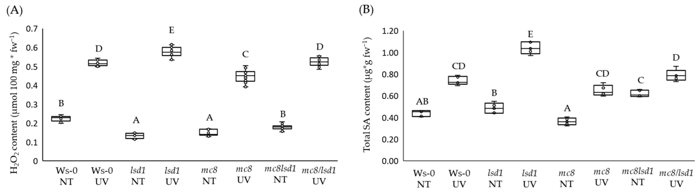
2.2. MC8 Affects LSD1-Dependent Foliar ROS and SA Levels

In control conditions, we observed a significantly lower foliar H2O2 level in lsd1 and mc8 mutants compared to wild-type plants. The concentration of foliar H2O2 in the double mc8/lsd1 mutant did not differ significantly from the wild type. After the UV irradiation episode, we observed the highest foliar H2O2 content in the lsd1 mutant, while in mc8, it was lower than in the wild-type plants. The double mc8/lsd1 mutant did not differ from the wild type in terms of H2O2 content, while the mc8 mutant demonstrated the lowest H2O2 content after UV stress (Figure 2A). Salicylic acid is an important cell death signaling molecule and the RCD phenotype of lsd1 is related to high foliar SA content [19,20]. Because of this fact, we performed an analysis of SA content in foliar tissues of tested Arabidopsis thaliana mutants. In control conditions, the lsd1 mutant did not differ much from the wild type, while the mc8 plants exhibited an insignificantly lower SA content. After UV stress, foliar SA levels were strongly increased in lsd1, but in mc8 and in double lsd1/mc8 mutants, they were increased similarly like in the wild-type plants (Figure 2B).

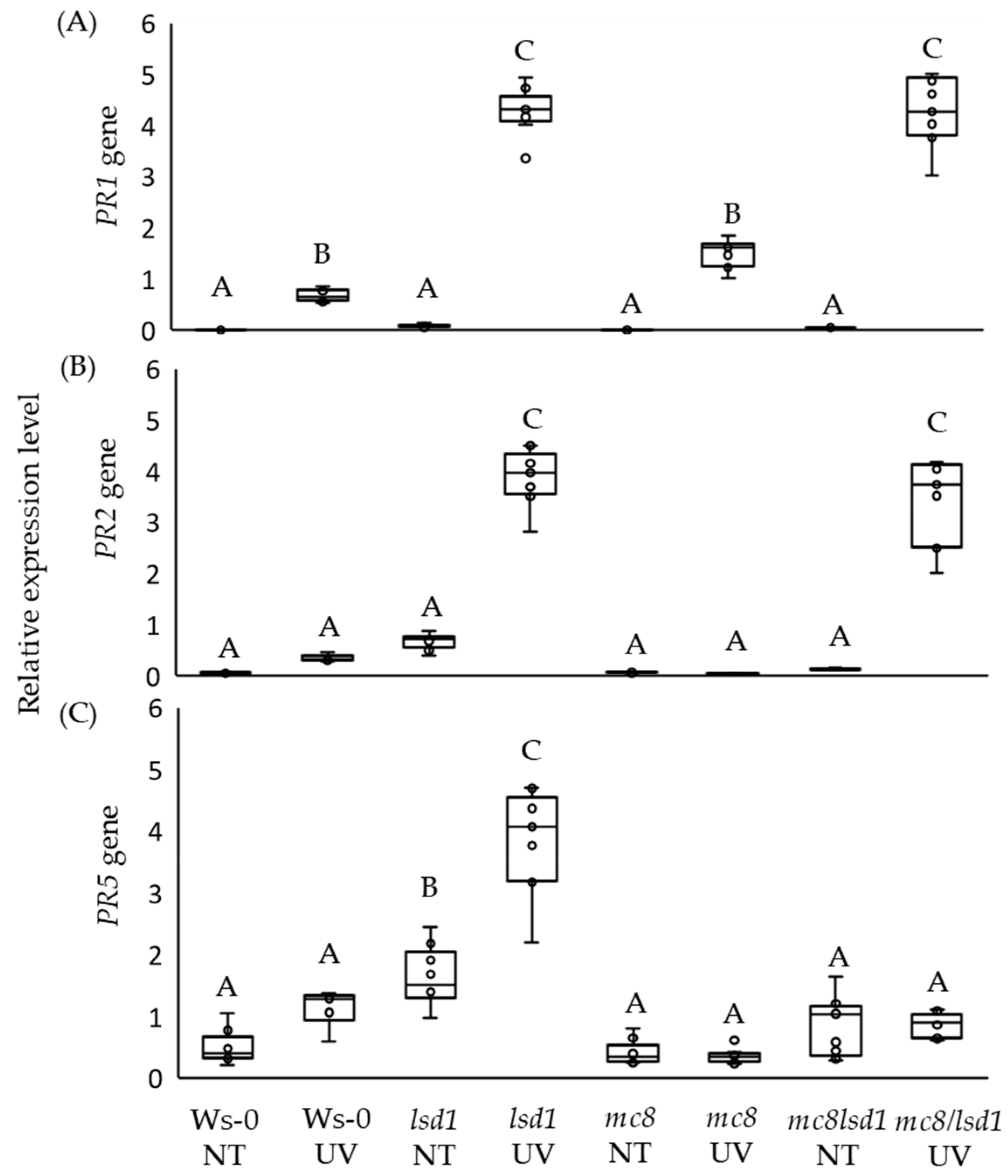
2.3. High PR5 Gene Expression Level in lsd1 Is Reverted by the Mutation in MC8

PR genes’ expression is strongly up-regulated in the lsd1 mutant [19] and their expression is related to SA levels in plant tissues [41,42]. Therefore, we decided to check the expression of PR1, PR2 and PR5 in all lines being investigated in this study. In control conditions, we found similarly low expression levels of PR1 and PR2 in all tested genotypes. The PR5 expression level was differentiated, and its expression was significantly higher in the lsd1 mutant in control conditions in comparison to the wild type and other tested mutants (Figure 3A–C). After UV irradiation, PR1 expression was higher in all tested genotypes when compared to control conditions. However, the PR1 transcript was drastically increased in lsd1 and mc8/lsd1 mutants (Figure 3A). Upon UV stress, the PR2 expression level was significantly higher in lsd1 and mc8/lsd1 mutants (Figure 3B). However, the expression level of PR5 after UV stress was significantly higher only in the lsd1 mutant when compared to the wild type (Figure 3C).

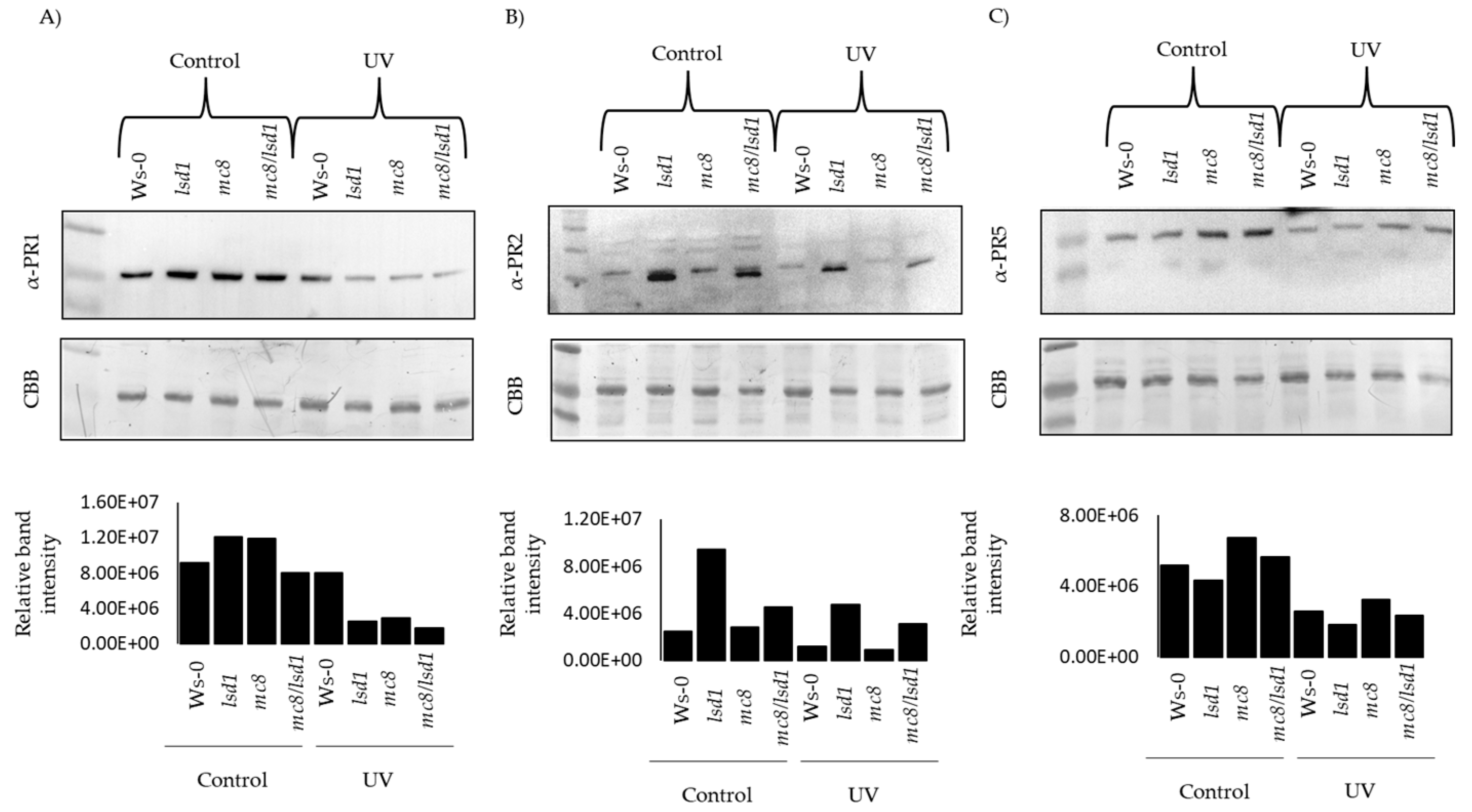
2.4. PR Proteins Are Degraded in Response to UV Stress

Despite the fact that we found a very strong up-regulation of PR1 gene expression in lsd1 and mc8/lsd1 mutants in stress conditions (Figure 3A), we did not observe a higher PR1 protein level. In fact, the PR1 protein level was similar in wild type plants and decreased in other tested genotypes after UV stress, in comparison to control conditions (Figure 4A). Even though the expression of the PR2 gene was strongly up-regulated in lsd1 and mc8/lsd1 mutants in response to UV irradiation, in lsd1 and in mc8/lsd1, the level of the PR2 protein decreased (Figure 4B). The PR5 protein was present in all of the tested genotypes; however, the higher amount of this protein was found in mc8 and lsd1/mc8 mutants in control conditions. The level of PR5 protein decreased in response to stress in all of the tested genotypes (Figure 4C). The experiment was performed in two independent biological replicates (Supplementary Figure S4).

2.5. Prediction of MC8 Interaction
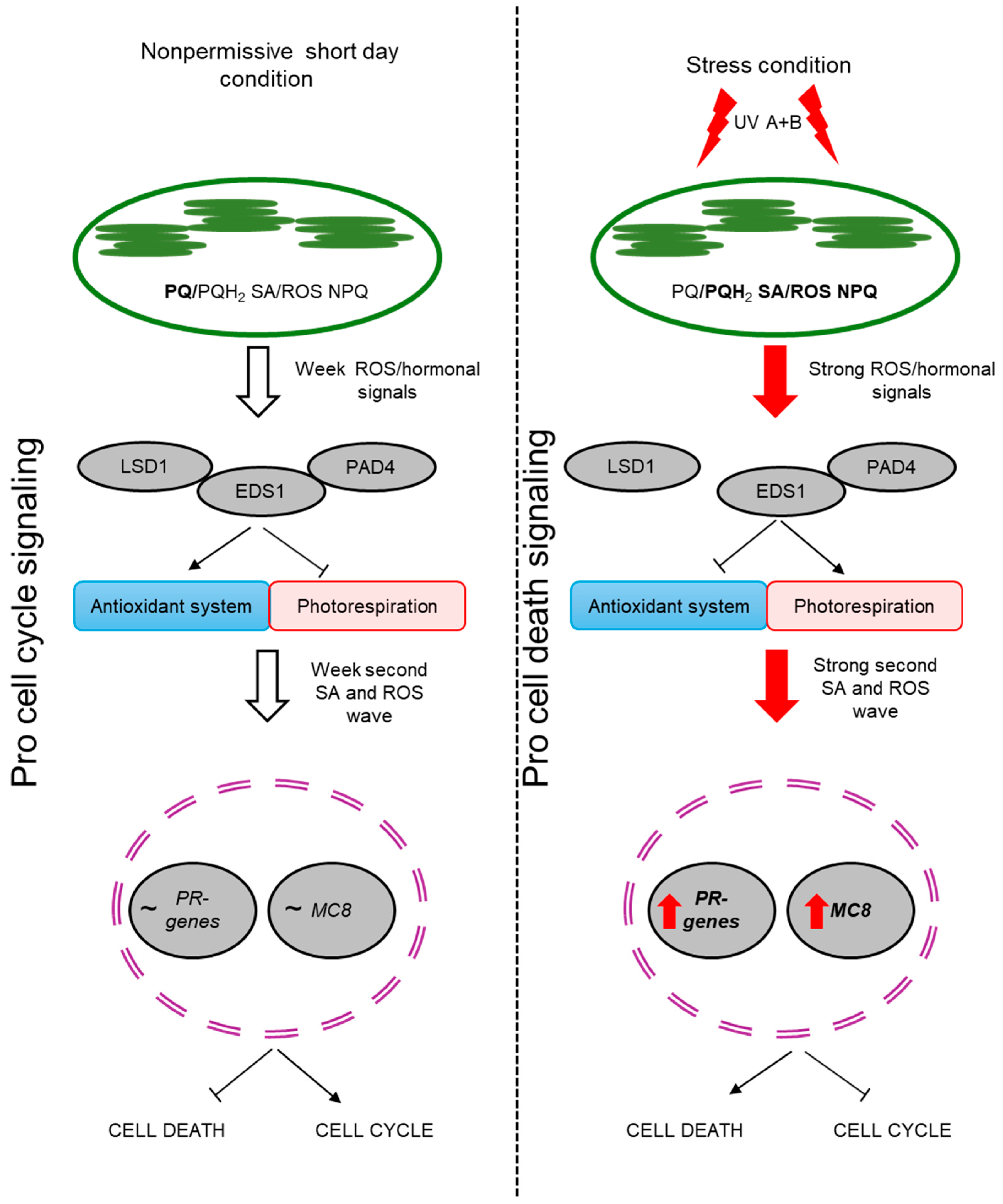
The protein–protein interaction of LSD1 is well known, and there is no information about any LSD1-MC8 interaction [27]. There is no prediction of the potential interaction of MC8 with LSD1, EDS1 and PAD4 nor of any other proteins which were previously described as LSD1, EDS1 and PAD4 interactors. This indicates the lack of protein–protein interaction with the above-mentioned CD-regulating proteins. Moreover, it shows that the analyzed pathway is dependent on hormones and ROS, which are regulated by LSD1/EDS1/PAD4 proteins (Supplementary Figure S5 and Supplementary Table S2).

3. Discussion

Plants are constantly exposed to various types of environmental stresses and because of that, they have developed many mechanisms and pathways to mitigate the effects of unfavorable conditions. One of the mechanisms activated during plant response to stress is programed cell death (PCD), a very old evolutionary mechanism present in all multicellular living organisms [43,44,45]. The role of PCD in plant response to stresses is basically the elimination of affected and/or older cells in order to induce higher tolerance and better acclimation in the other cells and maintenance of cellular homeostasis within an organism [46,47]. It is well known that this process is under conditional control of the LSD1 protein as well as EDS1 and PAD4, which are negative and positive cell death regulators, respectively [16,22,23,26,48]. Reversal of the lsd1 RCD phenotype is nothing new. This effect was obtained by mutation in EDS1 or PAD4 genes in the lsd1 mutant background, because these proteins physically interact with each other, forming a trimmer [26,27]. When there is no LSD1 protein present (lsd1 mutant) or it is at a lower level (in field conditions), the EDS1 and PAD4 proteins are active, and when there is only EDS1 (lsd1/pad4 mutant) present, it cannot induce a PR pathway alone [21,22,23,49]. Another way to revert the lsd1 phenotype is to disable the chloroplast signal recognition particle cpSRP 43 protein (encoded by the CAO gene) [32]. CAO is involved in the regulation of light-harvesting antenna size and non-photochemical quenching of absorbed energy in excess by photosystem II [50]. In the lsd1/cao double mutant, there is a reversion of the RCD phenotype due to its reduced ability to absorb light energy [3,32]. Thus, LSD1 is a negative regulator of PCD and photorespiration and a positive regulator of the antioxidant system [3]. Another piece of evidence to support chloroplast retrograde signaling mediated by cytoplasmic LSD1/EDS1/PAD4 proteins is presented in the lsd1/ex1 mutant which did not exhibit the RCD phenotype [51]. EXECUTER1 (EX1) is involved in singlet oxygen chloroplast retrograde signaling. ROS do not act alone in plant cells and the relation between SA and ROS in plant stress response is well known [22]. Because of this, deregulation in SA synthesis/metabolism also leads to reversion of the lsd1 phenotype in mutants such as lsd1/sid2 or in transgenic plants like lsd1/NahG [19,20]. Ours results and those of previous studies strongly suggest that a unified genetic and molecular system for the regulation of biotic and abiotic stress responses and cross-tolerance has evolved in plants [52]. Ethylene is also involved in conditional regulation by LSD1/EDS1/PAD4 PCD signaling, since impairment of this signaling pathway in the ein2/lsd1 double mutant also leads to reversion of the RCD lsd1 phenotype [2]. Interestingly, the lsd1 mutant, when grown in field conditions, did not differ from the wild type in size, visually or in seed yield [16,22]. In ambient laboratory conditions, lsd1 mutant plants are much smaller and produce much fewer seeds (fourfold lower seed yield) than the wild-type or double eds1/lsd1 and pad4/lsd1 plants. While all currently known ways to revert the lsd1 RCD phenotype are related to changes in ROS, SA or ethylene signaling pathways or to LSD1-interactor proteins, in this study, we found a new protein, MC8, which, according to the current knowledge [38,53] and bioinformatic prediction (Supplementary Figure S4 and Supplementary Table S2), does not interact with LSD1 and is not involved in SA, ROS and ET synthesis/metabolism, which suggests its functions as a signal receiver (ROS/hormonal) and as the final enforcer of LSD1-dependent cell death. During the analysis of our previously performed microarray [16], in the context of genes from the METACASPASE family, we found a significant positive fold change in MC8 in laboratory growing conditions (inducing RCD) and a significant negative fold change in field conditions (not inducing RCD). Moreover, in double lsd1/eds1 and lsd1/pad4 mutants with well-known reversion of the lsd1 RCD phenotype [22,23], there was a non-positive fold change in MC8, which indicates that MC8 acts upstream of the LSD1/EDS1/PAD4 trimer. These results were confirmed by studies on the mc8/lsd1 mutant. A double mutant exhibited a completely reverted RCD phenotype. This is probably caused by the fact that MC8 transcription is ROS-dependent, which was experimentally demonstrated by exogenous H2O2 treatment and by the induction ROS synthesis by of UV-C stress [22,38]. In conclusion, we can assume that MC8 is an important protein in the conditional LSD1/EDS1/PAD4-dependent cell death regulatory pathway. MC8 most probably is not related to SA and ROS synthesis or metabolism since in the lsd1/mc8 double mutant, we found lower or similar foliar levels of SA and H2O2 to those observed in wild-type plants. It is probably involved in the very early stage of metacaspases cascades of PCD and/or the inhibition of the second burst of the ROS/SA [44,54] wave from cell organelles undergoing PCD, which was observed in the RCD of the lsd1 mutant [23] or generally while PCD progressed in wild-type plants [44,55]. Inhibition of gene expression from the PR family was previously proposed as one of the reasons for the inhibition of RCD in lsd1/eds1, pad4/lsd1, sid2/lsd1, etc. [19,49]. However, in the lsd1/mc8 mutant, EDS1 and PAD4 were not inhibited by LSD1. This led to the induction of PR1 and PR2 expression in lsd1/mc8 on a similar level as in the lsd1 mutant. The exception is PR5 and its higher expression, which was observed only in the lsd1 mutant but not in lsd1/mc8. It could be caused by the fact that SA and ROS levels were slightly, but statistically significantly higher in lsd1 compared to lsd1/mc8. There are some alternatives to the SA-related signaling pathway [56] and there is a reverse correlation between H2O2 steady-state concentration and PR5 gene expression [57]. However, the number of PR proteins appear to be different to the PR genes’ transcription level. In general, in all of the mutants and in the wild-type plants, 24 h after UV-A + B stress, we found lower levels of PR1, PR2 and PR5. This is opposite to biotic stress, where the number of PR proteins were higher after pathogen inoculation [58]. This indicates that in response to abiotic stress (UV stress), a PR gene family is regulated differently on transcription and translation levels. However, this requires further research. Based on the current knowledge and on our results, we propose a model of LSD1 and MC8 interdependence in plant PCD regulation in response to UV-A + B irradiation (Figure 5). The UV episode is comprehended mostly by chloroplasts and provokes changes in the quantum-redox status of the photosynthetic electron transport chain components [3,59,60,61,62]. It leads to ROS overproduction [63], which consequently leads to increased synthesis of SA [3]. Both ROS and phytohormones act as a signal for LSD1 inhibition, which leads to EDS1 and PAD4 increasing in activity and then to the induction of cell death. Nevertheless, in other cells, LSD1 is induced, thus inhibiting EDS1 and PAD4 and preventing PCD spreading. Meanwhile, in the lsd1 mutant, there is no LSD1, thus EDS1 and PAD4 are hyperactive and act as inhibitors of the antioxidant system [3] and are important in SA synthesis [64,65]. This altogether leads to RCD induction [66]. However, in lsd1/mc8, all ROS/SA/ET signaling is still deregulated because of the lack of functional LSD1 and the lack of LSD1/EDS1/PAD4 trimers, thus EDS1 and PAD4 should induce the RCD phenotype, but such phenotype in lsd1/mc8 is not observed after UV stress. This is probably because MC8 is a receiver of the above-mentioned signals and MC8 acts upstream of all of the above-described proteins and thus is the ultimate executor of the run-away cell death process.

4. Materials and Methods

4.1. Plant Material

In this study, Arabidopsis thaliana mutants, lsd1, mc8 and mc8/lsd1, and wild-type plants (Ws-0) were used. All used mutants were of Wassilewskija (Ws-0) background. Wild-type and lsd1 seeds were already available in our lab, while mc8 seeds were provided by Dr. Patrick Gallois (Faculty of Biology, Medicine and Health, University of Manchester, Manchester, UK). In order to obtain a double mutant, we crossed lsd1 and mc8. A double mutant was obtained via selection in T3 generation. Correctness of the crossing was checked by PCR (Figure S6) and by RT-PCR (Figure S1). All primers used in the study are attached in the Supplementary Material (Table S3).

4.2. Growing Conditions

For all experiments described in this study, plants were grown in a walk-in-type growing chamber (Siemens, München, Germany) under the following conditions: 8/16 h photoperiod, photosynthetic photon flux density of 80 μmol photons m−2·s−1, air humidity of 50% and day/night temperature of 20/18 °C.

4.3. Stress Induction

For the determination of different stress impacts on MC8 expression levels, wild-type plants were treated with heat, light and UV-A + B using the following methods. For UV A + B stress application, the UV 500 Crosslinker (Hoefer Pharmacia Biotech, San Francisco, CA, USA) was used. It was equipped with three UV-B lamps (type G8T5E, Sankyo Denki, peak wavelength 306 nm) and two UV-A lamps (type TL8WBLB, Philips, Tokyo, Japan, peak wavelength 365 nm). Arabidopsis thaliana plants were exposed to a single irradiation dose of 1500 mJ·cm−2. Light treatment was performed using a white LED panel with the emission of white light (with 1500 μmol photons m−2·s−1) (Photon System Instrument, Brno, Czech Republic) for 2 h. For heat stress treatment, plants were incubated in 40 °C for two hours in the laboratory incubator MOV-212s (Philips, Tokyo, Japan). All analyses described in this study were performed 24 h after stress application. After the stress episode, plants were put back in the growing chamber in the same conditions as they were grown in before. For future experiments, UV A + B were chosen, and wild-type plants and all mutants used in this study were exposed to a single irradiation dose of 1500 mJ·cm−2.

4.4. Ion Leakage Measurement

Ion leakage was determined as described before [19,44].

4.5. Determination of H2O2 and SA Contents

The concentration of hydrogen peroxide (H2O2) was assessed according to the method described before [67], with slight adjustments. A total of 50–100 mg of frozen tissue was homogenized in a TissueLyser LT (Qiagen, Venlo, The Netherlands) for 5 min at 50 Hz and 4 °C, using 300 µL of cold 0.1% trichloroacetic acid (TCA), then centrifuged at 13,000 rpm for 15 min. The resulting supernatant was combined with 10 mM potassium phosphate buffer (pH 7.0) and 1 M potassium iodide (KI) at a 1:1:2 (v:v:v) ratio. The absorbance was measured at 390 nm using a microplate reader, Multiscan GO (Thermo Scientific, Waltham, MA, USA), and the H2O2 concentration was determined using an appropriate standard curve. Results were quantified and expressed as micromoles of H2O2 per 100 mg of fresh weight.
The determination of salicylic acid (SA) followed the protocol described before [68], with the utilization of 2-methoxybenzoic acid (oANI) and 3-hydroxybenzoic acid (pHBA) as internal standards. Salicylic acid was separated using a Luna 5uC18(2)100A150x4.6mm column (Phenomenex, Torrance, CA, USA) at 30 °C for 15 min, employing a Shimadzu HPLC System (Shimadzu, Kyoto, Japan). A low-pressure gradient system was used, utilizing a 20 mM phosphate buffer (pH 2.5; adjusted with 8 M HCl) and acetonitrile (75:25; v/v) at a flow rate of 1 mL per minute. Results were quantified and expressed as micrograms of SA per gram of fresh weight.

4.6. RNA Isolation, cDNA Synthesis and RT-PCR Analysis

RNA was extracted from frozen tissue previously stored in −80 °C. For RNA isolation, the GeneMaTRIX Universal RNA Purification Kit (EURX, Gdańsk, Poland) with an additional step of on-column DNase digestion was used. RNA concentration and quality were assessed using a spectrometer (Eppendorf, Hamburg, Germany). RNA quality was controlled by electrophoretic separation in 1% agarose gel. cDNa synthesis was performed for equimolar RNA amounts of each sample using a High Capacity cDNa Reverse Transcription Kit (Thermo Fisher Scientific). qPCRs were performed in three technical repetitions for each of the three biological replicates using the Power SYBR Green PCR Master Mix and the aBI 7500 Fast Real-Time PCR System (Thermo Fisher Scientific, Waltham, MA, USA). Two reference genes were used: 5-FORMYLTETRAHYDROFOLATE CYCLOLIGASE (5-FCL, AT5G13050) and PROTEIN PHOSPHATASE 2A SUBUNIT A2 (PP2AA2, aT3G25800).

4.7. Protein Extraction and Western Blot Analysis

Total plant protein extraction was performed as previously described [69], with modifications. Proteins were extracted from 100 mg of ground leaf tissue. The powder was resuspended in 500 μL of 2× Leammli buffer (4% SDS, 20% glycerol, 0.12 M Tris-HCl pH 7.0, 0.02% bromophenol blue and 0.7 M β-mercaptoethanol) and incubated for 10 min at 95 °C, followed by incubation for 10 min in ice [70]. The resuspended samples were centrifuged at 12,000× g for 5 min at 4 °C and the supernatants were used for further steps. Total protein concentration was determined using the RC-DC protein assay kit II (Bio-Rad, Hercules, CA, USA, 5000122). A total of 50 μg of total protein extract was used for 12% SDS-PAGE. Next, the proteins were electrotransferred to an Immobilon P PVDF membrane (Merck, Darmstadt, Germany) using the Trans-Blot Turbo Transfer System (Bio-Rad). The membrane was blocked in 2% skim milk in TBS-T buffer (1× Tris-buffered Saline, 0.1% Tween-20) for at least 1 h and incubated with primary antibodies (diluted at a ratio of 1:10,000, 2% skim milk in TBS-T buffer)—PR1 (AS10 687, Agrisera, Vännäs, Sweden) for 1 h at room temperature, and PR2 (AS12 2366, Agrisera) and PR5 (AS12 2373, Arisera) overnight at 4 °C. Incubation with the goat anti-rabbit horseradish peroxidase-conjugated secondary antibodies (Thermofisher, QG221919; diluted at a ratio of 1:10,000) was performed for 1 h at room temperature. Protein bands were immunodetected using SuperSignal West Dura Extended Duration Substrate (Thermo Scientific, 34075) according to the manufacturer’s recommendations, visualized with the ChemiDoc XRS+ System (Bio-Rad) and analyzed with ImageLab Software 5.2.1 (Bio-Rad). Total protein staining of membranes was conducted as described previously [71].

Supplementary Materials

The supporting information can be downloaded at: https://www.mdpi.com/article/10.3390/ijms25063195/s1.

Author Contributions

M.J.B. formed the hypothesis, planned the experiments, wrote the manuscript and was involved in mutans crossing, elative electrolyte leakage measurement, RNA isolation, cDNA synthesis and real-time PCR analysis. A.R. was involved in SA and H2O2 determination. K.G. was involved in proteomic analysis. A.B.M.-F. performed the experiment for determination of MC8 levels in response to abiotic stresses. W.C. was involved in drafting the manuscript, and S.M.K. was involved in formulating the hypothesis, planning the experiments and drafting the manuscript. All authors have read and agreed to the published version of the manuscript.

Funding

This study was funded by the “Miniatura 4” project (2020/04/X/NZ3/00829) granted to Maciej Jerzy Bernacki by the National Science Centre and the “OPUS 15” project (UMO-2018/29/B/NZ3/01198) granted to Stanisław Karpiński by the National Science Centre.

Institutional Review Board Statement

Not applicable.

Informed Consent Statement

Not applicable.

Data Availability Statement

All of the data generated or analyzed during this study are included in the published article and its Supplementary Files.

Acknowledgments

We would like to thank Patrick Gallois for providing the mc8 mutant seeds and for his help and advice during genotyping. We would like to express our gratitude to Aleksandra Lechańska for proofreading as well.

Conflicts of Interest

The authors declare that they have no competing interests.

References

  1. Mühlenbock, P.; Plaszczyca, M.; Plaszczyca, M.; Mellerowicz, E.; Karpinski, S. Lysigenous Aerenchyma Formation in Arabidopsis Is Controlled by LESION SIMULATING DISEASE1. Plant Cell 2007, 19, 3819–3830. [Google Scholar] [CrossRef] [PubMed]
  2. Mühlenbock, P.; Szechynska-Hebda, M.; Plaszczyca, M.; Baudo, M.; Mateo, A.; Mullineaux, P.M.; Parker, J.E.; Karpinska, B.; Karpinski, S. Chloroplast Signaling and LESION SIMULATING DISEASE1 Regulate Crosstalk between Light Acclimation and Immunity in Arabidopsis. Plant Cell 2008, 20, 2339–2356. [Google Scholar] [CrossRef] [PubMed]
  3. Karpiński, S.; Szechyńska-Hebda, M.; Wituszyńska, W.; Burdiak, P. Light Acclimation, Retrograde Signalling, Cell Death and Immune Defences in Plants. Plant Cell Environ. 2013, 36, 736–744. [Google Scholar] [CrossRef]
  4. Szechyńska-Hebda, M.; Kruk, J.; Górecka, M.; Karpińska, B.; Karpiński, S. Evidence for Light Wavelength-Specific Photoelectrophysiological Signaling and Memory of Excess Light Episodes in Arabidopsis. Plant Cell 2010, 22, 2201–2218. [Google Scholar] [CrossRef] [PubMed]
  5. Spoel, S.H.; Dong, X. How Do Plants Achieve Immunity? Defence without Specialized Immune Cells. Nat. Rev. Immunol. 2012, 12, 89–100. [Google Scholar] [CrossRef]
  6. Fukuda, H. Programmed Cell Death of Tracheary Elements as a Paradigm in Plants. Plant Mol. Biol. 2000, 44, 245–253. [Google Scholar] [CrossRef]
  7. Domínguez, F.; Cejudo, F.J. Programmed Cell Death (PCD): An Essential Process of Cereal Seed Development and Germination. Front Plant Sci 2014, 5, 366. [Google Scholar] [CrossRef]
  8. Jabs, T.; Dietrich, R.A.; Dangl, J.L. Initiation of Runaway Cell Death in an Arabidopsis Mutant by Extracellular Superoxide. Science 1996, 273, 1853–1856. [Google Scholar] [CrossRef]
  9. Lorrain, S.; Vailleau, F.; Balagué, C.; Roby, D. Lesion Mimic Mutants: Keys for Deciphering Cell Death and Defense Pathways in Plants? Trends Plant Sci. 2003, 8, 263–271. [Google Scholar] [CrossRef]
  10. Vandenabeele, S.; Vanderauwera, S.; Vuylsteke, M.; Rombauts, S.; Langebartels, C.; Seidlitz, H.K.; Zabeau, M.; Van Montagu, M.; Inzé, D.; Van Breusegem, F. Catalase Deficiency Drastically Affects Gene Expression Induced by High Light in Arabidopsis Thaliana. Plant J. 2004, 39, 45–58. [Google Scholar] [CrossRef]
  11. Dietrich, R.A.; Delaney, T.P.; Uknes, S.J.; Ward, E.R.; Ryals, J.A.; Dangl, J.L. Arabidopsis Mutants Simulating Disease Resistance Response. Cell 1994, 77, 565–577. [Google Scholar] [CrossRef]
  12. Wituszyńska, W.; Karpiński, S. Programmed Cell Death as a Response to High Light, UV and Drought Stress in Plants. In Abiotic Stress—Plant Responses and Applications in Agriculture; InTech: Rijeka, Croatia; Shanghai, China, 2013. [Google Scholar] [CrossRef]
  13. Mateo, A.; Funck, D.; Mühlenbock, P.; Kular, B.; Mullineaux, P.M.; Karpinski, S. Controlled Levels of Salicylic Acid Are Required for Optimal Photosynthesis and Redox Homeostasis. J. Exp. Bot. 2006, 57, 1795–1807. [Google Scholar] [CrossRef]
  14. Chai, T.; Zhou, J.; Liu, J.; Xing, D. LSD1 and HY5 Antagonistically Regulate Red Light Induced-Programmed Cell Death in Arabidopsis. Front. Plant Sci. 2015, 6, 292. [Google Scholar] [CrossRef] [PubMed]
  15. Bernacki, M.J.; Mielecki, J.; Antczak, A.; Drożdżek, M.; Witoń, D.; Dąbrowska-Bronk, J.; Gawroński, P.; Burdiak, P.; Marchwicka, M.; Rusaczonek, A.; et al. Biotechnological Potential of the Stress Response and Plant Cell Death Regulators Proteins in the Biofuel Industry. Cells 2023, 12, 2018. [Google Scholar] [CrossRef] [PubMed]
  16. Wituszyńska, W.; Ślesak, I.; Vanderauwera, S.; Szechyńska-Hebda, M.; Kornaś, A.; Kelen, K.V.D.; Mühlenbock, P.; Karpińska, B.; Maćkowski, S.; Breusegem, F.V.; et al. LESION SIMULATING DISEASE1, ENHANCED DISEASE SUSCEPTIBILITY1, and PHYTOALEXIN DEFICIENT4 Conditionally Regulate Cellular Signaling Homeostasis, Photosynthesis, Water Use Efficiency, and Seed Yield in Arabidopsis. Plant Physiol. 2013, 161, 1795–1805. [Google Scholar] [CrossRef] [PubMed]
  17. Szechyńska-Hebda, M.; Czarnocka, W.; Hebda, M.; Karpiński, S. PAD4, LSD1 and EDS1 Regulate Drought Tolerance, Plant Biomass Production, and Cell Wall Properties. Plant Cell Rep. 2016, 35, 527–539. [Google Scholar] [CrossRef] [PubMed]
  18. Huang, X.; Li, Y.; Zhang, X.; Zuo, J.; Yang, S. The Arabidopsis LSD1 Gene Plays an Important Role in the Regulation of Low Temperature-Dependent Cell Death. New Phytol. 2010, 187, 301–312. [Google Scholar] [CrossRef] [PubMed]
  19. Bernacki, M.J.; Rusaczonek, A.; Czarnocka, W.; Karpiński, S. Salicylic Acid Accumulation Controlled by LSD1 Is Essential in Triggering Cell Death in Response to Abiotic Stress. Cells 2021, 10, 962. [Google Scholar] [CrossRef] [PubMed]
  20. Aviv, D.H.; Rustérucci, C.; Iii, B.F.H.; Dietrich, R.A.; Parker, J.E.; Dangl, J.L. Runaway Cell Death, but Not Basal Disease Resistance, in Lsd1 Is SA- and NIM1/NPR1-Dependent. Plant J. 2002, 29, 381–391. [Google Scholar] [CrossRef] [PubMed]
  21. Rustérucci, C.; Aviv, D.H.; Holt, B.F.; Dangl, J.L.; Parker, J.E. The Disease Resistance Signaling Components EDS1 and PAD4 Are Essential Regulators of the Cell Death Pathway Controlled by LSD1 in Arabidopsis. Plant Cell 2001, 13, 2211–2224. [Google Scholar] [CrossRef]
  22. Bernacki, M.J.; Czarnocka, W.; Rusaczonek, A.; Witoń, D.; Kęska, S.; Czyż, J.; Szechyńska-Hebda, M.; Karpiński, S. LSD1, EDS1 and PAD4-dependent conditional correlation among salicylic acid, hydrogen peroxide, water use efficiency, and seed yield in Arabidopsis thaliana. Physiol. Plant. 2018, 165, 369–382. [Google Scholar] [CrossRef]
  23. Wituszyńska, W.; Szechyńska-Hebda, M.; Sobczak, M.; Rusaczonek, A.; Kozłowska-Makulska, A.; Witoń, D.; Karpiński, S. Lesion Simulating Disease 1 and Enhanced Disease Susceptibility 1 Differentially Regulate UV-C-Induced Photooxidative Stress Signalling and Programmed Cell Death in Arabidopsis Thaliana. Plant Cell Environ. 2015, 38, 315–330. [Google Scholar] [CrossRef]
  24. Li, Y.; Chen, L.; Mu, J.; Zuo, J. LESION SIMULATING DISEASE1 Interacts with Catalases to Regulate Hypersensitive Cell Death in Arabidopsis1[C][W]. Plant Physiol. 2013, 163, 1059–1070. [Google Scholar] [CrossRef]
  25. Kliebenstein, D.J.; Dietrich, R.A.; Martin, A.C.; Last, R.L.; Dangl, J.L. LSD1 Regulates Salicylic Acid Induction of Copper Zinc Superoxide Dismutase in Arabidopsis Thaliana. Mol. Plant. Microbe. Interact. 1999, 12, 1022–1026. [Google Scholar] [CrossRef]
  26. Feys, B.J.; Moisan, L.J.; Newman, M.-A.; Parker, J.E. Direct Interaction between the Arabidopsis Disease Resistance Signaling Proteins, EDS1 and PAD4. EMBO J. 2001, 20, 5400–5411. [Google Scholar] [CrossRef]
  27. Czarnocka, W.; Van Der Kelen, K.; Willems, P.; Szechyńska-Hebda, M.; Shahnejat-Bushehri, S.; Balazadeh, S.; Rusaczonek, A.; Mueller-Roeber, B.; Van Breusegem, F.; Karpiński, S. The Dual Role of LESION SIMULATING DISEASE 1 as a Condition-Dependent Scaffold Protein and Transcription Regulator. Plant Cell Environ. 2017, 40, 2644–2662. [Google Scholar] [CrossRef]
  28. Parker, J.E.; Holub, E.B.; Frost, L.N.; Falk, A.; Gunn, N.D.; Daniels, M.J. Characterization of Eds1, a Mutation in Arabidopsis Suppressing Resistance to Peronospora Parasitica Specified by Several Different RPP Genes. Plant Cell 1996, 8, 2033–2046. [Google Scholar] [CrossRef] [PubMed]
  29. Glazebrook, J.; Rogers, E.E.; Ausubel, F.M. Isolation of Arabidopsis Mutants with Enhanced Disease Susceptibility by Direct Screening. Genetics 1996, 143, 973–982. [Google Scholar] [CrossRef] [PubMed]
  30. Falk, A.; Feys, B.J.; Frost, L.N.; Jones, J.D.G.; Daniels, M.J.; Parker, J.E. EDS1, an Essential Component of R Gene-Mediated Disease Resistance in Arabidopsis Has Homology to Eukaryotic Lipases. Proc. Natl. Acad. Sci. USA 1999, 96, 3292–3297. [Google Scholar] [CrossRef] [PubMed]
  31. Jirage, D.; Tootle, T.L.; Reuber, T.L.; Frost, L.N.; Feys, B.J.; Parker, J.E.; Ausubel, F.M.; Glazebrook, J. Arabidopsis Thaliana PAD4 Encodes a Lipase-like Gene That Is Important for Salicylic Acid Signaling. Proc. Natl. Acad. Sci. USA 1999, 96, 13583–13588. [Google Scholar] [CrossRef] [PubMed]
  32. Mateo, A.; Mühlenbock, P.; Rustérucci, C.; Chang, C.C.-C.; Miszalski, Z.; Karpinska, B.; Parker, J.E.; Mullineaux, P.M.; Karpinski, S. LESION SIMULATING DISEASE 1 Is Required for Acclimation to Conditions That Promote Excess Excitation Energy. Plant Physiol. 2004, 136, 2818–2830. [Google Scholar] [CrossRef]
  33. Dietrich, R.A.; Richberg, M.H.; Schmidt, R.; Dean, C.; Dangl, J.L. A Novel Zinc Finger Protein Is Encoded by the Arabidopsis LSD1 Gene and Functions as a Negative Regulator of Plant Cell Death. Cell 1997, 88, 685–694. [Google Scholar] [CrossRef]
  34. Takatsuji, H. Zinc-Finger Transcription Factors in Plants. CMLS Cell. Mol. Life Sci. 1998, 54, 582–596. [Google Scholar] [CrossRef]
  35. Coll, N.S.; Vercammen, D.; Smidler, A.; Clover, C.; Van Breusegem, F.; Dangl, J.L.; Epple, P. Arabidopsis Type I Metacaspases Control Cell Death. Science 2010, 330, 1393–1397. [Google Scholar] [CrossRef]
  36. Fagundes, D.; Bohn, B.; Cabreira, C.; Leipelt, F.; Dias, N.; Bodanese-Zanettini, M.H.; Cagliari, A. Caspases in Plants: Metacaspase Gene Family in Plant Stress Responses. Funct. Integr. Genom. 2015, 15, 639–649. [Google Scholar] [CrossRef]
  37. Vercammen, D.; van de Cotte, B.; De Jaeger, G.; Eeckhout, D.; Casteels, P.; Vandepoele, K.; Vandenberghe, I.; Van Beeumen, J.; Inzé, D.; Van Breusegem, F. Type II Metacaspases Atmc4 and Atmc9 of Arabidopsis Thaliana Cleave Substrates after Arginine and Lysine. J. Biol. Chem. 2004, 279, 45329–45336. [Google Scholar] [CrossRef] [PubMed]
  38. He, R.; Drury, G.E.; Rotari, V.I.; Gordon, A.; Willer, M.; Farzaneh, T.; Woltering, E.J.; Gallois, P. Metacaspase-8 Modulates Programmed Cell Death Induced by Ultraviolet Light and H2O2 in Arabidopsis. J. Biol. Chem. 2008, 283, 774–783. [Google Scholar] [CrossRef] [PubMed]
  39. Kwon, S.I.; Hwang, D.J. Expression Analysis of the Metacaspase Gene Family in Arabidopsis. J. Plant Biol. 2013, 56, 391–398. [Google Scholar] [CrossRef]
  40. Tsiatsiani, L.; Van Breusegem, F.; Gallois, P.; Zavialov, A.; Lam, E.; Bozhkov, P.V. Metacaspases. Cell Death Differ. 2011, 18, 1279–1288. [Google Scholar] [CrossRef]
  41. Glazebrook, J. Genes Controlling Expression of Defense Responses in Arabidopsis—2001 Status. Curr. Opin. Plant Biol. 2001, 4, 301–308. [Google Scholar] [CrossRef] [PubMed]
  42. Thomma, B.P.; Penninckx, I.A.; Cammue, B.P.; Broekaert, W.F. The Complexity of Disease Signaling in Arabidopsis. Curr. Opin. Immunol. 2001, 13, 63–68. [Google Scholar] [CrossRef]
  43. Burke, R.; Schwarze, J.; Sherwood, O.L.; Jnaid, Y.; McCabe, P.F.; Kacprzyk, J. Stressed to Death: The Role of Transcription Factors in Plant Programmed Cell Death Induced by Abiotic and Biotic Stimuli. Front. Plant Sci. 2020, 11, 1235. [Google Scholar] [CrossRef]
  44. Bernacki, M.J.; Czarnocka, W.; Zaborowska, M.; Różańska, E.; Labudda, M.; Rusaczonek, A.; Witoń, D.; Karpiński, S. EDS1-Dependent Cell Death and the Antioxidant System in Arabidopsis Leaves Is Deregulated by the Mammalian Bax. Cells 2020, 9, 2454. [Google Scholar] [CrossRef]
  45. Lacomme, C.; Cruz, S.S. Bax-Induced Cell Death in Tobacco Is Similar to the Hypersensitive Response. Proc. Natl. Acad. Sci. USA 1999, 96, 7956–7961. [Google Scholar] [CrossRef]
  46. Danon, A.; Delorme, V.; Mailhac, N.; Gallois, P. Plant Programmed Cell Death: A Common Way to Die. Plant Physiol. Biochem. 2000, 38, 647–655. [Google Scholar] [CrossRef]
  47. Williams, B.; Dickman, M. Plant Programmed Cell Death: Can’t Live with It; Can’t Live without It. Mol. Plant Pathol. 2008, 9, 531–544. [Google Scholar] [CrossRef] [PubMed]
  48. Aarts, N.; Metz, M.; Holub, E.; Staskawicz, B.J.; Daniels, M.J.; Parker, J.E. Different Requirements for EDS1 and NDR1 by Disease Resistance Genes Define at Least Two R Gene-Mediated Signaling Pathways in Arabidopsis. Proc. Natl. Acad. Sci. USA 1998, 95, 10306–10311. [Google Scholar] [CrossRef] [PubMed]
  49. Cui, H.; Gobbato, E.; Kracher, B.; Qiu, J.; Bautor, J.; Parker, J.E. A Core Function of EDS1 with PAD4 Is to Protect the Salicylic Acid Defense Sector in Arabidopsis Immunity. New Phytol. 2017, 213, 1802–1817. [Google Scholar] [CrossRef] [PubMed]
  50. Tanaka, R.; Koshino, Y.; Sawa, S.; Ishiguro, S.; Okada, K.; Tanaka, A. Overexpression of Chlorophyllide a Oxygenase (CAO) Enlarges the Antenna Size of Photosystem II in Arabidopsis Thaliana. Plant J. 2001, 26, 365–373. [Google Scholar] [CrossRef] [PubMed]
  51. Lv, R.; Li, Z.; Li, M.; Dogra, V.; Lv, S.; Liu, R.; Lee, K.P.; Kim, C. Uncoupled Expression of Nuclear and Plastid Photosynthesis-Associated Genes Contributes to Cell Death in a Lesion Mimic Mutant. Plant Cell 2019, 31, 210–230. [Google Scholar] [CrossRef] [PubMed]
  52. Szechyńska-Hebda, M.; Ghalami, R.Z.; Kamran, M.; Van Breusegem, F.; Karpiński, S. To Be or Not to Be? Are Reactive Oxygen Species, Antioxidants, and Stress Signalling Universal Determinants of Life or Death? Cells 2022, 11, 4105. [Google Scholar] [CrossRef] [PubMed]
  53. Ahn, G.; Jung, I.J.; Cha, J.-Y.; Jeong, S.Y.; Shin, G.-I.; Ji, M.G.; Kim, M.G.; Lee, S.Y.; Kim, W.-Y. Phytochrome B Positively Regulates Red Light-Mediated ER Stress Response in Arabidopsis. Front. Plant Sci. 2022, 13, 846294. [Google Scholar] [CrossRef] [PubMed]
  54. Fichman, Y.; Myers, R.J.; Grant, D.G.; Mittler, R. Plasmodesmata-Localized Proteins and ROS Orchestrate Light-Induced Rapid Systemic Signaling in Arabidopsis. Sci. Signal. 2021, 14, eabf0322. [Google Scholar] [CrossRef] [PubMed]
  55. Kawai-Yamada, M.; Jin, L.; Yoshinaga, K.; Hirata, A.; Uchimiya, H. Mammalian Bax-Induced Plant Cell Death Can Be down-Regulated by Overexpression of Arabidopsis Bax Inhibitor-1 (AtBI-1). Proc. Natl. Acad. Sci. USA 2001, 98, 12295–12300. [Google Scholar] [CrossRef] [PubMed]
  56. Nawrath, C.; Métraux, J.-P. Salicylic Acid Induction–Deficient Mutants of Arabidopsis Express PR-2 and PR-5 and Accumulate High Levels of Camalexin after Pathogen Inoculation. Plant Cell 1999, 11, 1393–1404. [Google Scholar] [CrossRef] [PubMed]
  57. Kalbina, I.; Strid, Å. Supplementary Ultraviolet-B Irradiation Reveals Differences in Stress Responses between Arabidopsis Thaliana Ecotypes. Plant Cell Environ. 2006, 29, 754–763. [Google Scholar] [CrossRef] [PubMed]
  58. Sahoo, A.; Satapathy, K.B.; Panigrahi, G.K. Ectopic Expression of Disease Resistance Protein Promotes Resistance against Pathogen Infection and Drought Stress in Arabidopsis. Physiol. Mol. Plant Pathol. 2023, 124, 101949. [Google Scholar] [CrossRef]
  59. Czégény, G.; Mátai, A.; Hideg, É. UV-B Effects on Leaves—Oxidative Stress and Acclimation in Controlled Environments. Plant Sci. 2016, 248, 57–63. [Google Scholar] [CrossRef]
  60. Li, H.; Li, Y.; Deng, H.; Sun, X.; Wang, A.; Tang, X.; Gao, Y.; Zhang, N.; Wang, L.; Yang, S.; et al. Tomato UV-B Receptor SlUVR8 Mediates Plant Acclimation to UV-B Radiation and Enhances Fruit Chloroplast Development via Regulating SlGLK2. Sci. Rep. 2018, 8, 6097. [Google Scholar] [CrossRef]
  61. Nasibova, A.N. UV-B Radiation Effects on Electron-Transport Reactions in Biomaterials. Adv. Biol. Earth Sci. 2022, 7, 13–18. [Google Scholar]
  62. Piccini, C.; Cai, G.; Dias, M.C.; Romi, M.; Longo, R.; Cantini, C. UV-B Radiation Affects Photosynthesis-Related Processes of Two Italian Olea Europaea (L.) Varieties Differently. Plants 2020, 9, 1712. [Google Scholar] [CrossRef]
  63. Mittler, R.; Vanderauwera, S.; Suzuki, N.; Miller, G.; Tognetti, V.B.; Vandepoele, K.; Gollery, M.; Shulaev, V.; Van Breusegem, F. ROS Signaling: The New Wave? Trends Plant Sci. 2011, 16, 300–309. [Google Scholar] [CrossRef]
  64. Shah, J. The Salicylic Acid Loop in Plant Defense. Curr. Opin. Plant Biol. 2003, 6, 365–371. [Google Scholar] [CrossRef] [PubMed]
  65. Straus, M.R.; Rietz, S.; Ver Loren van Themaat, E.; Bartsch, M.; Parker, J.E. Salicylic Acid Antagonism of EDS1-Driven Cell Death Is Important for Immune and Oxidative Stress Responses in Arabidopsis. Plant J. 2010, 62, 628–640. [Google Scholar] [CrossRef]
  66. Mur, L.A.J.; Kenton, P.; Lloyd, A.J.; Ougham, H.; Prats, E. The Hypersensitive Response; the Centenary Is upon Us but How Much Do We Know? J. Exp. Bot. 2008, 59, 501–520. [Google Scholar] [CrossRef] [PubMed]
  67. Velikova, V.; Sharkey, T.D.; Loreto, F. Stabilization of Thylakoid Membranes in Isoprene-Emitting Plants Reduces Formation of Reactive Oxygen Species. Plant Signal. Behav. 2012, 7, 139–141. [Google Scholar] [CrossRef]
  68. Meuwly, P.; Métraux, J.P. Ortho-Anisic Acid as Internal Standard for the Simultaneous Quantitation of Salicylic Acid and Its Putative Biosynthetic Precursors in Cucumber Leaves. Anal. Biochem. 1993, 214, 500–505. [Google Scholar] [CrossRef]
  69. Górecka, M.; Lewandowska, M.; Dąbrowska-Bronk, J.; Białasek, M.; Barczak-Brzyżek, A.; Kulasek, M.; Mielecki, J.; Kozłowska-Makulska, A.; Gawroński, P.; Karpiński, S. Photosystem II 22kDa Protein Level—A Prerequisite for Excess Light-Inducible Memory, Cross-Tolerance to UV-C and Regulation of Electrical Signalling. Plant Cell Environ. 2020, 43, 649–661. [Google Scholar] [CrossRef]
  70. Laemmli, U.K. Cleavage of Structural Proteins during the Assembly of the Head of Bacteriophage T4. Nature 1970, 227, 680–685. [Google Scholar] [CrossRef] [PubMed]
  71. Goldman, A.; Harper, S.; Speicher, D.W. Detection of Proteins on Blot Membranes. Curr. Protoc. Protein Sci. 2016, 86, 10.8.1–10.8.11. [Google Scholar] [CrossRef]
Figure 1. The role of METACASPASE 8 in conditional LSD1-dependent cell death regulation. (A) Analysis of transcriptomic data [16] in the context of expression changes in genes encoding METACACASPES family proteins, and in lsd1 mutant grown in ambient laboratory conditions (Lab) or in natural field conditions (Field), significant changes are marked with asterisks. FC—fold change. (B) Level of measured foliar ion leakage (manifesting the cell death level) for plants grown in control conditions and exposed to episode of UV-A + UV-B irradiation. Within a subgraph, values sharing the same letters are not significantly different from each other (p > 0.001) (n = 10–15). (C) Pictures of Arabidopsis thaliana rosettes from control conditions (upper row) and after UV-A + UV-B irradiation incidents (bottom row).
Ijms 25 03195 g001
Figure 2. Foliar (A) H2O2 and (B) salicylic acid levels in control conditions and 24 h after UV irradiation. Within a subgraph, values sharing common letters are not significantly different from each other (p > 0.001) (n = 6).
Ijms 25 03195 g002
Figure 3. PR genes’ relative expression levels in non-stress ambient laboratory conditions and in response to UV irradiation episode. (A) PR1, (B) PR2 and (C) PR5 expression levels in tested mutants in control conditions and 24 h after UV irradiation. Within a subgraph, values sharing common letters are not significantly different from each other (p > 0.001) (n = 9).
Ijms 25 03195 g003
Figure 4. Level of PR proteins in response to UV irradiation. Western blot analysis of (A) PR1, (B) PR2 and (C) PR5 proteins in all tested genotypes in control conditions and 24 h after UV irradiation. The graphs show relative intensity of the bands (presented on gels) for each tested protein. As a loading control, CBB staining was used.
Ijms 25 03195 g004
Figure 5. Proposed/hypothetical model of LSD1 and MC8 interdependence in plant cell death regulation in response to UV-A + B irradiation. We propose that MC8 is more important in the cell death pathway dependent on LSD1 than the proposed EDS1 and PAD4 proteins (there are more details in the last paragraph of the discussion). The arrow is the inducing action; The blunt-headed arrow is braking; The red arrow next to the gene names is increased expression; The tilde sign is an expression without change.
Ijms 25 03195 g005
Disclaimer/Publisher’s Note: The statements, opinions and data contained in all publications are solely those of the individual author(s) and contributor(s) and not of MDPI and/or the editor(s). MDPI and/or the editor(s) disclaim responsibility for any injury to people or property resulting from any ideas, methods, instructions or products referred to in the content.

