Article

Association of MDM2 Overexpression in Ameloblastomas with MDM2 Amplification and BRAFV600E Expression

by
Konstantinos I. Tosios
1,*,
Eleni-Marina Kalogirou
2 and
Ioannis G. Koutlas
3
1
Department of Oral Pathology & Medicine and Hospital Dentistry, School of Dentistry, National and Kapodistrian University of Athens, 11527 Athens, Greece
2
Faculty of Health and Rehabilitation Sciences, Metropolitan College, 15125 Athens, Greece
3
Division of Oral Pathology, School of Dentistry, University of Minnesota, Minneapolis, MN 55455, USA
*
Author to whom correspondence should be addressed.
Int. J. Mol. Sci. 2024, 25(4), 2238; https://doi.org/10.3390/ijms25042238
Submission received: 31 December 2023 / Revised: 1 February 2024 / Accepted: 9 February 2024 / Published: 13 February 2024
(This article belongs to the Special Issue Molecular Basis of Odontogenic Tumors and Odontogenic Cysts)

Abstract

:
Ameloblastoma is a rare tumor but represents the most common odontogenic neoplasm. It is localized in the jaws and, although it is a benign, slow-growing tumor, it has an aggressive local behavior and high recurrence rate. Therefore, alternative treatment options or complementary to surgery have been evaluated, with the most promising one among them being a targeted therapy with the v-Raf murine sarcoma viral oncogene homologue B (BRAF), as in ameloblastoma the activating mutation V600E in BRAF is common. Studies in other tumors have shown that the synchronous inhibition of BRAF and human murine double minute 2 homologue (MDM2 or HDM2) protein is more effective than BRAF monotherapy, particularly in the presence of wild type p53 (WTp53). To investigate the MDM2 protein expression and gene amplification in ameloblastoma, in association with BRAFV600E and p53 expression. Forty-four cases of ameloblastoma fixed in 10% buffered formalin and embedded in paraffin were examined for MDM2 overexpression and BRAFV600E and p53 expression by immunohistochemistry, and for MDM2 ploidy with fluorescence in situ hybridization. Sixteen of forty-four (36.36%) cases of ameloblastoma showed MDM2 overexpression. Seven of sixteen MDM2-positive ameloblastomas (43.75%) were BRAFV600E positive and fifteen of sixteen MDM2-positive ameloblastomas (93.75%) were p53 negative. All MDM2 overexpressing tumors did not show copy number alterations for MDM2. Overexpression of MDM2 in ameloblastomas is not associated with MDM2 amplification, but most probably with MAPK activation and WTp53 expression. Further verification of those findings could form the basis for the use of MDM2 expression as a marker of MAPK activation in ameloblastomas and the trial of dual BRAF/MDM2 inhibition in the management of MDM2-overexpressing/BRAFV600E-positive/WTp53 ameloblastomas.

1. Introduction

Ameloblastoma is a rare benign epithelial odontogenic neoplasm, i.e., a tumor originating from the tooth-forming epithelium. It is one of the most common odontogenic tumors and the commonest odontogenic neoplasm [1,2,3]. It is usually diagnosed in patients in the third to fifth decade of life, shows a slight predilection for males, and is preferentially localized in the mandible, with the angle of the mandible and the molar region being the most commonly affected regions [3,4]. Based on clinical, radiographic, and pathologic features, three types of ameloblastoma are described: the conventional solid/multicystic and the unicystic ameloblastoma are intraosseous tumors, whereas the peripheral ameloblastoma is an extraosseous tumor that develops in the gingiva. The conventional solid/multicystic ameloblastoma and the peripheral ameloblastoma are the most prevalent and the rarest types, respectively [3,4]. Clinically, conventional solid/multicystic ameloblastoma manifests among other symptoms and signs with swelling of the involved jaw region, mobility and displacement of teeth with consequent malocclusion, facial asymmetry, neurological signs, and bone fracture. Radiographically, it presents as a multilocular or unilocular radiolucency that may cause extensive bone destruction [3]. The diagnostic pathological feature of ameloblastoma is its resemblance to odontogenic epithelium, as it is composed centrally by cells resembling the stellate reticulum epithelium and peripherally by cylindrical, palisading cells with reverse nuclear polarity, resembling the ameloblasts. Pathological subtypes include follicular, plexiform, desmoplastic, basal cell, granular cell, and acanthomatous patterns, whereas the unilocular type is categorized into luminal, intraluminal, and mural subtypes, based on the localization of the tumor [5].
Although ameloblastoma is a benign, slow-growing tumor, it has an aggressive local behavior and high recurrence rate after conservative surgical techniques, such as enucleation alone or in combination with curettage, cryotherapy, cauterization, Carnoy solution, etc. [4,6,7]. On the other hand, radical surgical management techniques, such as segmental resection with wide bone margins or marginal resection, are associated with a lower recurrence rate, but also with severe complications and considerable decline in the patients’ quality of life [4,7,8]. Recurrences usually appear 2–5 years after treatment but may develop even 20 years later [4]. Furthermore, ameloblastomas extending into vital anatomical compartments, such as the base of the skull, orbit, and parapharyngeal space, pose additional difficulties in treatment, may be non-amenable to radical resection, and may even be life-threatening [9]. Therefore, there is a need for conservative treatment options, which are alternatives to or complementary to surgery.
Ameloblastomas commonly show the activating mutation V600E in the v-Raf murine sarcoma viral oncogene homologue B (BRAFV600E) located on chromosome 7q34; BRAF is a key member of the mitogen-activated protein kinase (MAPK) signaling pathway, which plays a crucial role in cell growth and proliferation [10,11,12,13,14,15,16,17]. This finding prompted the clinical trial of BRAF inhibitors (BRAFi) as a targeted therapy in this tumor. BRAFi vemurafenib and dabrafenib have shown promising results in the management of BRAFV600E-positive ameloblastomas [18,19,20,21,22,23] or as neoadjuvant therapy [24,25,26]. In the largest series reported to date, 19 patients with BRAFV600E-positive ameloblastomas treated with dabrafenib ± trametinib achieved complete radiological response; one showed complete clinical response, and in ten of them residual tumor enucleation was possible with near complete or partial response [26]. However, the genetic heterogeneity of ameloblastoma, as well as variable responses and development of tolerance in targeted therapies [23], may limit the clinical success of such strategies.
Ameloblastomas commonly express the human murine double minute 2 homologue (MDM2 or HDM2) protein [27,28,29,30,31,32,33]. MDM2 is located on chromosome 12q14.3–q15 and encodes the MDM2 E3 ubiquitin ligase [34,35]. MDM2 regulates cell growth and differentiation by binding ubiquitin to other proteins, such as the oncosuppressor proteins p53, pRB, and p14ARF [36,37,38,39,40], and FOXO3a transcription factor [41,42]. Its main function is to negatively regulate the wild-type p53 (WTp53) transcriptional activity [34,36,37,38,39,40,43,44] through its overexpression [45]. This results in p53-dependent arrest of cell proliferation and apoptosis [46], and it increases genomic instability [47]. MDM2 overexpression is commonly associated with MDM2 gene amplification in several solid tumors, such as liposarcomas, osteosarcomas, breast and esophageal carcinomas, brain tumors, and neuroblastomas [40,46,47,48,49], while in cutaneous melanomas and hematological malignancies MDM2 overexpression is seen in the absence of MDM2 amplification [49,50,51,52]. Blocking of MDM2 expression, downregulation of MDM2 activity, or interference in the MDM2-p53 complex may reconstitute p53 function in cell cycle progression control and apoptosis promotion [49,52]. Therefore, MDM2 inhibitors have been tested in preclinical studies and some of them are under clinical investigation for solid tumors and hematological malignancies [49]. In some studies, synchronous inhibition of MDM2 and BRAF is more effective than BRAF monotherapy in BRAFV600E-positive melanomas [53,54,55].
In view of the complimentary roles of BRAFi and MDM2i in the treatment of various tumors, the objective of this study was to investigate MDM2 protein expression and gene amplification in ameloblastoma, in association with BRAFV600E and p53 expression.

2. Results

Forty-four patients with ameloblastoma were included in this study; twenty-three were males and twenty-one females. The age range was 6–82 years and the mean age 42.6 ± 18.7 years. Thirty-five lesions were localized in the mandible and eight in the maxilla (mandible to maxilla ratio 4.3:1). Forty-one ameloblastomas were conventional solid/multicystic and three were unicystic. Solid ameloblastomas were follicular in twenty-seven cases, plexiform in six cases, basaloid in six cases, and acanthomatous in two cases. Unicystic ameloblastomas were of the mural subtype, all of them showing a follicular growth pattern. Squamous metaplasia, cystic degeneration, and granular cells were seen in five, ten, and one of the follicular ameloblastomas, respectively.

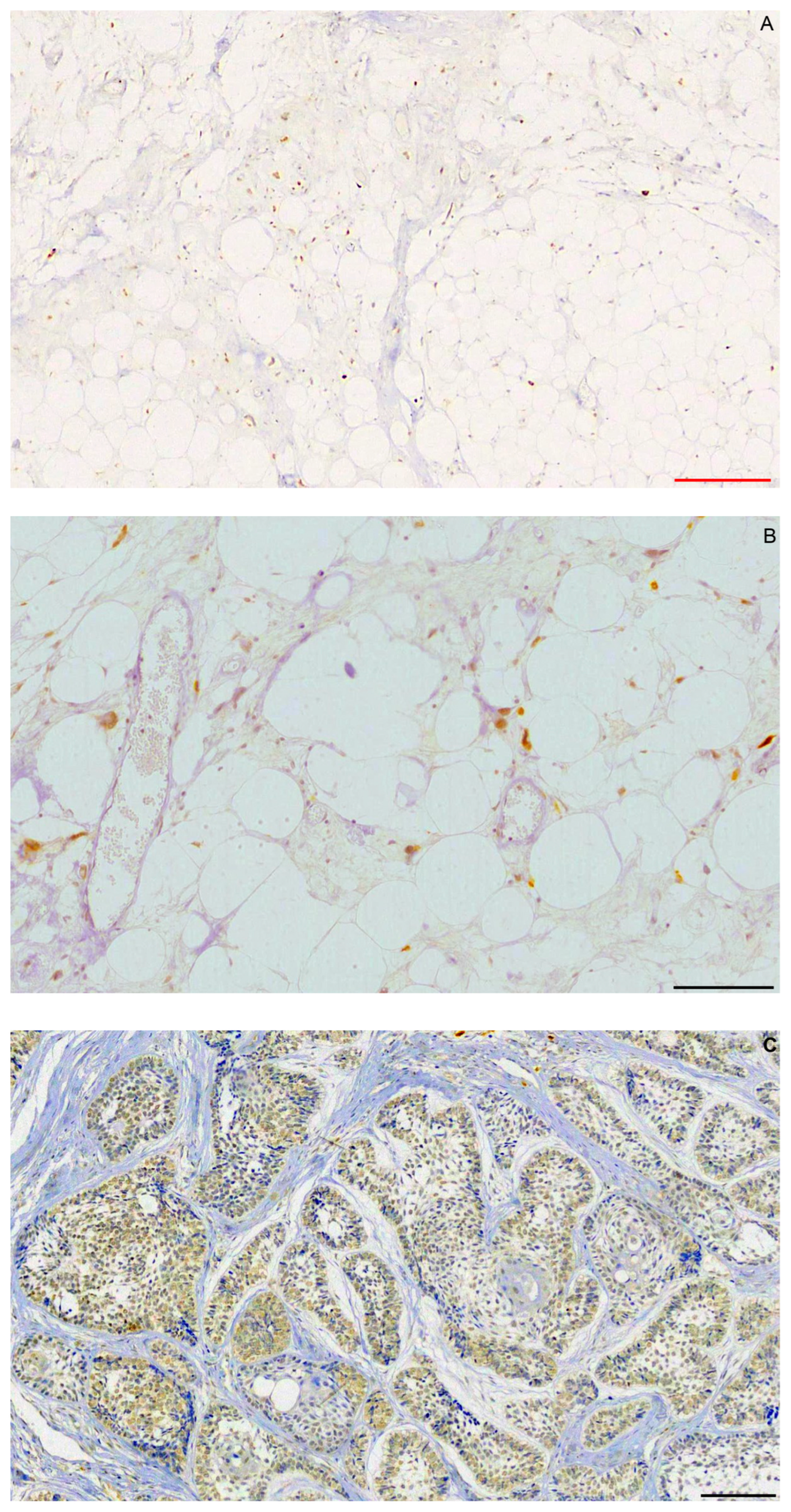
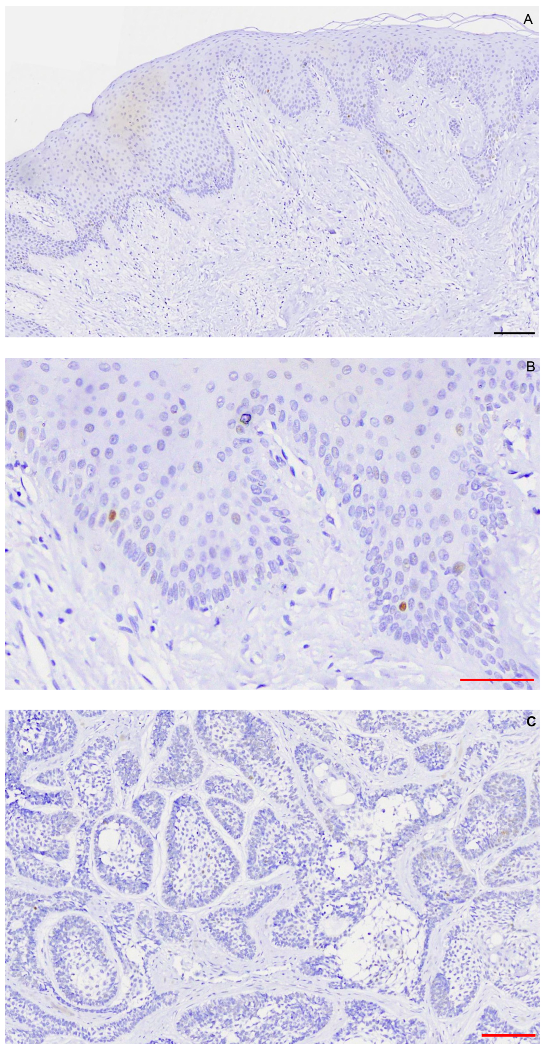
2.1. Immunohistochemistry
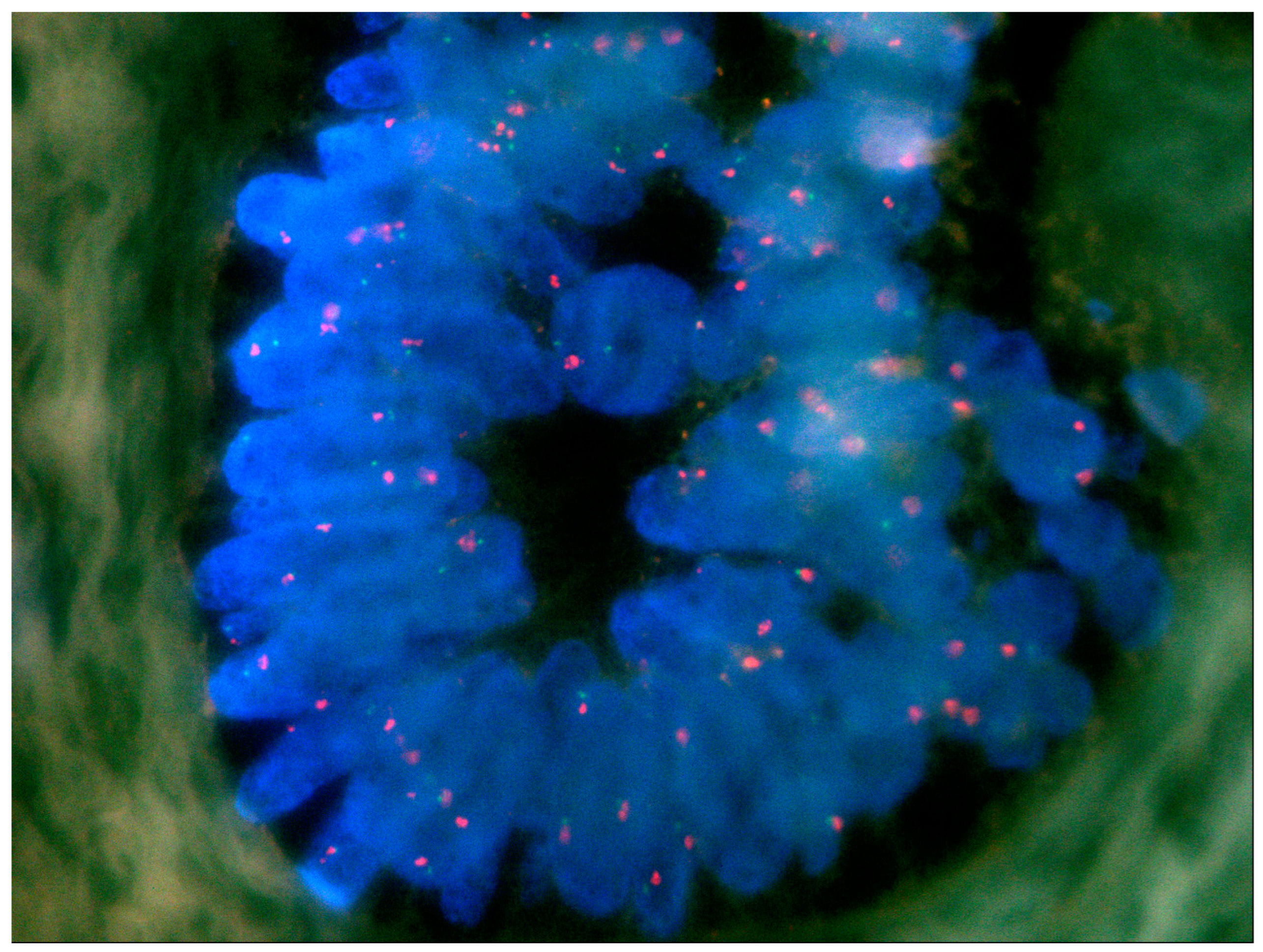
Liposarcomas showed grade 3+ MDM2 immunostaining (Figure 1A,Β). In ameloblastomas, staining was 0 in 28 cases (63.63%), 2+ in 2 cases, and 3+ in 14 cases (Figure 1C). Therefore, all 16 (36.36%) MDM2-positive cases showed overexpression. No difference was observed in immunostaining between peripheral, ameloblast-like, and central, stellate reticulum-like cells. Squamous or granular cells did not react for MDM2. There was no statistically significant association (p < 0.05) between MDM2 expression and age or gender of the patients.
Melanoma showed cytoplasmic, homogenous, and intense BRAFV600E immunostaining in most tumor cells (Figure 2A,B). Seven of sixteen MDM2-positive ameloblastomas (43.75%) were BRAFV600E positive (Figure 2C), two showed weak/ambiguous staining and were considered as negative, and seven were BRAFV600E negative. All seven positive cases were from the mandible, representing seven of ten mandibular ameloblastomas and five of seven negative cases from the maxilla, and representing five of six maxillary ameloblastomas. Almost all (14/16) were of the follicular subtype.
Specimens of normal oral mucosa showed nuclear, homogenous, and intense p53 immunostaining in rare cells of the basal cell layer (Figure 3A,B). Fifteen of sixteen MDM2-positive ameloblastomas (93.75%) were p53 negative and one was p53 positive (1+) (Figure 3C). Seven of sixteen ameloblastomas (43.75%) presented an MDM2-positive/BRAFV600E-positive/WTp53 phenotype. Figure 1C, Figure 2C and Figure 3C are from the same follicular ameloblastoma.
Table 1 shows the main clinical, histopathological, and immunohistochemical features of the 16 MDM2-positive ameloblastomas.

2.2. FISH

FISH was performed in the 16 MDM2-positive ameloblastomas. No copy number alterations for MDM2 were identified in all tumors examined (Figure 4).

3. Discussion

Herein, we aimed to investigate MDM2 protein expression and gene amplification in ameloblastoma, in association with BRAFV600E and p53 expression. The main finding of the present study is that ameloblastomas overexpressing MDM2 did not show MDM2 amplification, as accessed by FISH, whereas some of them expressed BRAFV600E in the presence of WTp53, as was shown by immunohistochemistry.
The immunohistochemical expression of MDM2 has been studied in ameloblastomas with inconsistent results. Carvalhais et al. [27] noticed weak nuclear reaction in eight of thirteen (61.53%) ameloblastomas. Sandra et al. [28] detected MDM2 in 33 of 34 (88%) and 32 of 34 (86%) ameloblastomas by immunohistochemistry and western blotting, respectively. In a study conducted by Kumamoto et al. [29], all 46 ameloblastomas expressed MDM2. Sharifi-Sistani et al. [30] found MDM2 expression in 31 of 39 (79.48%) ameloblastomas, Krishna et al. [31] in 33 of 36 (91.6%) ameloblastomas, and Singh et al. [32] in 18 of 20 (90%) of conventional solid/multicystic ameloblastomas and 12 of 20 (60%) unicystic ameloblastomas. Finally, Udeabor et al. [33] reported MDM2 expression in three of twenty-eight (10.7%) ameloblastomas. The varying results on MDM2 immunoexpression among different studies may be attributed to technical parameters [47], but overall MDM2 expression was recorded in 181 of 226 ameloblastomas tested. In this study, MDM2 positivity, defined as nuclear immunostaining [56,57] that is homogenous and clear [58,59], was seen in 16 of 44 cases (36.36%).
In accordance with previous reports, MDM2-positive cells were seen in all cell layers [27], squamous or granular cells were MDM2 negative [31], and no difference was observed between follicular and plexiform ameloblastomas [27]. Other studies have reported more intense MDM2 expression in the peripheral cells of ameloblastoma [28,29,32] and variation among the histopathological types [29,30,31,32], findings that were not confirmed in the present material.
MDM2 overexpression is considered suggestive of MDM2 amplification in various malignant tumors, such as bladder carcinoma, melanoma, and liposarcoma [51,56,57], but it has not been evaluated in ameloblastomas. In this study, all MDM2-positive cases showed MDM2 overexpression, whereas in previous studies none [28,33], 38.46% [27], or 61.54% [30] of the tumors examined showed immunohistochemical expression consistent with overexpression. However, none of the 16 MDM2-overexpressing tumors showed MDM2 amplification by FISH. Those findings are in line with previous studies [10,12,13,60,61] that by employing various molecular techniques, e.g., microarrays, RNA-sequencing, Sanger sequencing, and polymerase chain reaction (PCR), highlighted the differential expression and/or mutations of other molecular markers that do not predict MDM2 mutations in ameloblastoma.
MDM2 overexpression in the absence of MDM2 amplification has been reported in malignant melanoma [50,51,52], Burkitt lymphoma [62], carcinoma of the breast [63,64], carcinoma of the bladder [65], and soft tissue sarcomas [66]. It may be attributed to alternative splicing or increased transcription of MDM2, with the latter being the leading cause [34,67,68]. Increased MDM2 transcription may be stimulated by the MAPK and transforming growth factor-β (TGF-β) pathways, as MDM2 is a transcriptional target of both pathways [34,69].
MDM2 transcription may be activated by binding on its P2 promoter, located in the first intron of the molecule, by Activator protein 1 (AP-1) and E26 transformation-specific or E-twenty-six (ETS) transcription factors that are downstream molecules of the BRAF pathway [34]. The MAPK pathway may be constitutively activated by BRAFV600E mutation [51] that is frequently detected in ameloblastomas [10,11,12,13,14,15,17]. Seven of sixteen MDM2-positive cases in the present study were shown by immunohistochemistry to be positive for BRAFV600E (43.75%), compared with the reported 46% to 82% positivity for this marker in other studies [10,11,12,13,14,15,17], and most of them were in the mandible [10,17]. Although the gold standards for detecting BRAFV600E mutation are PCR and DNA sequencing, immunohistochemistry with VE1 antibody, as applied in the present study, shows high concordance with molecular techniques [12,70]. TGF-β upregulates MDM2 expression via the interaction of Smad2 and Smad3 with the P2 promoter located in the first intron of MDM2 [34]. In ameloblastomas, low expression of TGF-β1 and functional pSmad2/3 and Smad4 proteins do not indicate a critical role for the TGF-β pathway [71]. Those findings support the suggestion that MAPK activation through BRAFV600E mutation may be the main cause of MDM2 overexpression in BRAFV600E-positive tumors. As for BRAFV600E-negative cases that overexpress MDM2, it should be noticed that MAPK-activating mutations other than BRAFV600E have been identified in ameloblastomas [16], and in some ameloblastomas the TGF-β pathway may be activated [71]. Further evaluation of MDM2 overexpression/MAPK activation association could show its possible utility as a marker of MAPK activation.
In ameloblastoma, TP53 mutations are rare [29,72,73]; p53 is mostly normal WTp53 [28], and there is heavy suppression of p53 [60]. In accordance with those studies, the present one showed that p53 was not expressed in most ameloblastomas with MDM2 overexpression, indicating p53 suppression.
In cutaneous melanomas that overexpress MDM2 without MDM2 amplification and which have normal p53, inhibition of MDM2 may reconstitute WTp53 action in tumor cells and suppress tumor growth [52]. Furthermore, MDM2i may act as a complement to BRAFi in malignant neoplasms that overexpress MDM2, are BRAFV600E positive, and express WTp53. Dual BRAF/MDM2 inhibition suppressed the viability of WTp53 melanoma cells in vitro and WTp53 melanoma growth in vivo [54], and in cell cultures of cutaneous melanoma [74,75,76] and colon carcinoma [77] this led to restoration of p53 function, possibly promoting apoptosis and suppressing tumor growth. In mice xenografted with RKO colon carcinoma cell inhibitors of BRAF and MDM4, a nuclear protein structurally homologous to MDM2 that interacts with both p53 and MDM2, this treatment managed to shrink the tumor by 80%, when the response to each drug tested separately was 23% and 24%, respectively [77]. Furthermore, dual inhibition helped overrun tolerance to BRAFi, an adverse effect attributed to reactivation of the MAPK pathway [22,53,54]. In BRAFV600E-positive ameloblastomas which were unresectable due to multiple recurrences and lung metastases, monotherapy with BRAFi dabrafenib [19,21] or vemurafenib [20], neoadjuvant treatment with dabrafenib [24,25,26], or dual BRAF/MEK inhibition with dabrafenib and trametinib [18,22,23,26] showed good response without severe toxicity. The addition of MDM2i such as nutlins, which disrupt the MDM2-p53 interaction by competing with p53 for binding to the MDM2 protein, could augment the therapeutic outcome and possibly overcome tolerance to BRAFi [23].
A limitation of the present study is that BRAFV600E and p53 expression were not examined in MDM2-negative ameloblastomas of our sample, as the investigation was focused on MDM2-overexpressing ameloblastomas. Therefore, the association of BRAFV600E and p53 expression with MDM2 status cannot be concluded, although for the latter it is expected, based on the available literature, that the tumors would be p53 negative.

4. Materials and Methods

The cohort consisted of 44 biopsies of ameloblastoma fixed in 10% buffered formalin and embedded in paraffin (FFPE). Histopathologic diagnosis in each case was confirmed by microscopic examination of 5 μm thick FFPE tissue sections stained with hematoxylin and eosin by all researchers according to the World Health Organization diagnostic criteria for ameloblastoma [5]. Relevant clinical information was retrieved from the pathology request forms, tabulated, and anonymized. The study was approved by the Ethics Committee of the Dental School, National and Kapodistrian University of Athens (#302), and the Institutional Review Board of the University of Minnesota (IRB #1604E86681), and was conducted in accordance with the principles of the Declaration of Helsinki.

4.1. Immunohistochemistry

Immunohistochemistry was performed on 5 μm thick FFPE tissue sections utilizing the OptiView DAB IHC Detection Kit (Ventana Medical Systems Inc., Tucson, AZ, USA). Staining was performed in the fully automated VENTANA BenchMark ULTRA Slide Staining System (Ventana Medical Systems Inc., Tucson, AZ, USA) with mouse anti-human monoclonal antibodies against MDM2 (1:50, Clone IF2, Invitrogen Corporation, Camarillo, CA, USA) that recognize an epitope between amino acids 26–169 of human MDM2; recombinant mouse monoclonal antibody to BRAF-mutated V600E (1:50, clone VE1, (1:50, clone VE1, Abcam, Amsterdam, Netherlands); and mouse anti-human monoclonal antibodies against p53 (1:300, clone DO7, Biogenex, Fremond, CA, USA). The staining reaction was visualized with 0.2% 3,3′-diaminobezidine solution solubilized with OptiView DAB (Ventana Medical Systems Inc., Tucson, AZ, USA). For antigen retrieval, sections were treated in 97 °C for 25 min with ULTRA Cell Conditioning Solution (ULTRA CC1, Ventana, Roche Diagnostic GmbH, Manheim, Germany) for all antibodies.
MDM2, BRAFV600E, and p53 were evaluated in one representative 5 μm FFPE tissue section from each tumor, digitized with a semi-automated system with Intel Pentium V, Digital Camera Sony 1600 × 1200, and Microscope Olympus CX-31 hardware features, and the software Windows XP/Windows XP/NIS-Elements Software AR v3.0, Nikon Corp, Tokyo, Japan. The 5 μm FFPE tissue section of each sample was scanned, resulting in a digital image of 20x original magnification. Scoring for MDM2 was performed semi-quantitatively based on the pattern of nuclear staining with the following scale: 0 < 5%; 1+ = 5–20%; 2+ = 21–50%; and 3+ > 50% [57]; overexpression was defined as ≥2+ [51,56,57]. Scoring for BRAFV600E was based on the intensity of cytoplasmic staining, with the following scale: 0, negative; 1+, weak; 2+, moderate; and 3+, strong [78], and the threshold for p53 positivity was 5%.
Positive controls were sections from two cases of atypical lipomatous tumor of the thigh for MDM2, two cases of oral mucosa for p53, and one case of BRAFV600E-positive cutaneous melanoma for VE1. For negative controls, the primary antibodies were substituted with Negative Control-monoclonal (Ventana, Medical Systems Inc., Tucson, AZ, USA).

4.2. FISH

FISH was performed on 5 μm thick FFPE tissue sections with the commercially available ZytoLight-FISH tissue implementation kit and ZytoLight-FISH SPEC MDM2/CEN12 Dual Color Probe (ZytoVision® GmbH, Bremerhaven, Germany). This is a direct labeling technique optimized for use with FFPE tissue sections, with ready-to-use fluorescence-labeled polynucleotide probes: a green one targeting the chromosomal region of the human MDM2, and an orange one targeting DNA sequences of centromeric alpha-satellites of chromosome 12 (CEN12). Alpha-satellite sequences of CEN12 served as an internal control and as a measure for DNA integrity. Sections were examined with an oil-immersion ×100 lens and proper fluorescence filters (green-labeled polynucleotides: excitation at 503 nm and emission at 528 nm, orange-labeled polynucleotides: excitation at 547 nm and emission at 572 nm). Amplification of the MDM2 gene locus was defined as an MDM2/CEN12 signal ratio ≥2 in >10% of the total number of cells or as clustering of multiple copies of green signals [79,80]. In ameloblastomas, CEN12 was expected to be euploid, as chromosomal copy number variations for this tumor are rare and do not include chromosome 12 [73,81,82,83,84]. Therefore, no external controls were necessary. Fifty interphase nuclei from different areas of the FISH slides were evaluated in each case.

4.3. Statistical Analysis

Statistical analysis was performed with the SPSS, v25.0 Software for Windows (SPSS Inc., Chicago, IL, USA). Associations between the MDM2 immunohistochemistry results and patients’ demographic characteristics were investigated with the Chi Square Test and, when expected frequency was <5, with the Fisher Exact Test. The level of statistical significance was set at p-value (p) < 0.05.

5. Conclusions

In conclusion, overexpression of MDM2 in ameloblastomas is not associated with MDM2 amplification, but most probably with MAPK activation and WTp53 expression. Further verification of those findings could form the basis for the use of MDM2 expression as a marker of MAPK activation in ameloblastomas and the trial of dual BRAF/MDM2 inhibition in the management of MDM2-overexpressing/BRAFV600E-positive/WTp53 ameloblastomas.

Author Contributions

Conceptualization, K.I.T.; methodology, K.I.T.; software, K.I.T. and E.-M.K.; validation, K.I.T. and I.G.K.; formal analysis, K.I.T. and I.G.K.; investigation, K.I.T. and I.G.K.; resources, K.I.T. and I.G.K.; data curation, K.I.T., E.-M.K. and I.G.K.; visualization, K.I.T. and E.-M.K.; writing—original draft preparation, K.I.T.; writing—review and editing, K.I.T., E.-M.K. and I.G.K. All authors have read and agreed to the published version of the manuscript.

Funding

This research received no external funding.

Institutional Review Board Statement

The study was conducted in accordance with the Declaration of Helsinki and was approved by the Ethics Committee of the School of Dentistry, National and Kapodistrian University of Athens (#302), and the Institutional Review Board of the University of Minnesota (IRB #1604E86681).

Informed Consent Statement

Not applicable.

Data Availability Statement

The original contributions of the study are included in the article. Further inquiries can be directed to the corresponding author.

Acknowledgments

We appreciate the excellent technical assistance of Georgios Vilaras.

Conflicts of Interest

The authors declare no conflicts of interest.

References

  1. Avelar, R.L.; Primo, B.T.; Pinheiro-Nogueira, C.B.; Studart-Soares, E.C.; de Oliveira, R.B.; Romulo de Medeiros, J.; Hernandez, P.A. Worldwide incidence of odontogenic tumors. J. Craniofac. Surg. 2011, 22, 2118–2123. [Google Scholar] [CrossRef]
  2. Siriwardena, B.; Crane, H.; O’Neill, N.; Abdelkarim, R.; Brierley, D.J.; Franklin, C.D.; Farthing, P.M.; Speight, P.M.; Hunter, K.D. Odontogenic tumors and lesions treated in a single specialist oral and maxillofacial pathology unit in the United Kingdom in 1992–2016. Oral Surg. Oral Med. Oral Pathol. Oral Radiol. 2019, 127, 151–166. [Google Scholar] [CrossRef]
  3. da Silva, L.A.M.; Filho, S.R.C.; Saraiva, M.J.D.; Maia, C.R.; Santos, C.; Santos, P.P.A. Clinical, Radiographic and Histopathological Analysis of Craniopharyngiomas and Ameloblastomas: A Systematic Review. Head Neck Pathol. 2022, 16, 1195–1222. [Google Scholar] [CrossRef]
  4. Boffano, P.; Cavarra, F.; Tricarico, G.; Masu, L.; Brucoli, M.; Ruslin, M.; Forouzanfar, T.; Ridwan-Pramana, A.; Rodriguez-Santamarta, T.; Rui Ranz, M.; et al. The epidemiology and management of ameloblastomas: A European multicenter study. J. Craniomaxillofac. Surg. 2021, 49, 1107–1112. [Google Scholar] [CrossRef]
  5. El-Naggar, A.K.; Chan, J.K.C.; Grandis, J.R.; Takata, T.; Slootweg, P.J. Odontogenic and maxillofacial bone tumors. In WHO Classification of Head and Neck Tumours, 4th ed.; IARC: Lyon, France, 2017; pp. 215–219. [Google Scholar]
  6. McClary, A.C.; West, R.B.; Pollack, J.R.; Fischbein, N.J.; Holsinger, C.F.; Sunwoo, J.; Colevas, A.D.; Sirjani, D. Ameloblastoma: A clinical review and trends in management. Eur. Arch. Otorhinolaryngol. 2015, 273, 1649–1661. [Google Scholar] [CrossRef] [PubMed]
  7. Hendra, F.N.; Helder, M.N.; Ruslin, M.; Van Cann, E.M.; Forouzanfar, T. A network meta-analysis assessing the effectiveness of various radical and conservative surgical approaches regarding recurrence in treating solid/multicystic ameloblastomas. Sci. Rep. 2023, 13, 8445. [Google Scholar] [CrossRef]
  8. Laborde, A.; Nicot, R.; Wojcik, T.; Ferri, J.; Raoul, G. Ameloblastoma of the jaws: Management and recurrence rate. Eur. Ann. Otorhinolaryngol. Head Neck Dis. 2017, 134, 7–11. [Google Scholar] [CrossRef] [PubMed]
  9. Armocida, D.; Berra, L.V.; Pucci, R.; Battisti, A.; Della Monaca, M.; Valentini, V.; Santoro, A. Ameloblastoma and Intracranial Involvement: The Current Challenge of the Radical Surgical Treatment. Comprehensive Review of the Literature and Institution experience. J. Maxillofac. Oral. Surg. 2022, 21, 34–43. [Google Scholar] [CrossRef]
  10. Sweeney, R.T.; McClary, A.C.; Myers, B.R.; Biscocho, J.; Neahring, L.; Kwei, K.A.; Qu, K.; Gong, X.; Ng, T.; Jones, C.D.; et al. Identification of recurrent SMO and BRAF mutations in ameloblastomas. Nat. Genet. 2014, 46, 722–725. [Google Scholar] [CrossRef] [PubMed]
  11. Gultekin, S.E.; Aziz, R.; Heydt, C.; Senguven, B.; Zoller, J.; Safi, A.F.; Kreppel, M.; Buettner, R. The landscape of genetic alterations in ameloblastomas relates to clinical features. Virchows Arch. 2018, 472, 807–814. [Google Scholar] [CrossRef]
  12. Brown, N.A.; Rolland, D.; McHugh, J.B.; Weigelin, H.C.; Zhao, L.; Lim, M.S.; Elenitoba-Johnson, K.S.; Betz, B.L. Activating FGFR2-RAS-BRAF mutations in ameloblastoma. Clin. Cancer Res. 2014, 20, 5517–5526. [Google Scholar] [CrossRef]
  13. Kurppa, K.J.; Caton, J.; Morgan, P.R.; Ristimaki, A.; Ruhin, B.; Kellokoski, J.; Elenius, K.; Heikinheimo, K. High frequency of BRAF V600E mutations in ameloblastoma. J. Pathol. 2014, 232, 492–498. [Google Scholar] [CrossRef]
  14. Soltani, M.; Tabatabaiefar, M.A.; Mohsenifar, Z.; Pourreza, M.R.; Moridnia, A.; Shariati, L.; Razavi, S.M. Genetic study of the BRAF gene reveals new variants and high frequency of the V600E mutation among Iranian ameloblastoma patients. J. Oral. Pathol. Med. 2018, 47, 86–90. [Google Scholar] [CrossRef]
  15. Diniz, M.G.; Gomes, C.C.; Guimaraes, B.V.; Castro, W.H.; Lacerda, J.C.; Cardoso, S.V.; de Faria, P.R.; Dias, F.L.; Eisenberg, A.L.; Loyola, A.M.; et al. Assessment of BRAFV600E and SMOF412E mutations in epithelial odontogenic tumours. Tumour Biol. 2015, 36, 5649–5653. [Google Scholar] [CrossRef]
  16. Oh, K.Y.; Kim, J.H.; Cho, S.D.; Yoon, H.J.; Lee, J.I.; Hong, S.D. BRAF V600E and previously unidentified KRAS G12C mutations in odontogenic tumors may affect MAPK activation differently depending on tumor type. Genes Chromosomes Cancer 2022, 61, 481–490. [Google Scholar] [CrossRef] [PubMed]
  17. do Canto, A.M.; da Silva Marcelino, B.M.R.; Schussel, J.L.; Wastner, B.F.; Sassi, L.M.; Correa, L.; de Freitas, R.R.; Hasseus, B.; Kjeller, G.; Junior, C.A.L.; et al. Immunohistochemical analysis of BRAF V600E mutation in ameloblastomas. Clin. Oral. Investig. 2019, 23, 779–784. [Google Scholar] [CrossRef]
  18. Kaye, F.J.; Ivey, A.M.; Drane, W.E.; Mendenhall, W.M.; Allan, R.W. Clinical and radiographic response with combined BRAF-targeted therapy in stage 4 ameloblastoma. J. Natl. Cancer Inst. 2015, 107, 378. [Google Scholar] [CrossRef] [PubMed]
  19. Faden, D.L.; Algazi, A. Durable treatment of ameloblastoma with single agent BRAFi Re: Clinical and radiographic response with combined BRAF-targeted therapy in stage 4 ameloblastoma. J. Natl. Cancer Inst. 2017, 109, djw190. [Google Scholar] [CrossRef] [PubMed]
  20. Broudic-Guibert, M.; Blay, J.Y.; Vazquez, L.; Evrard, A.; Karanian, M.; Taieb, S.; Hoog-Labouret, N.; Oukhatar, C.M.A.; Boustany-Grenier, R.; Arnaud, A. Persistent response to vemurafenib in metastatic ameloblastoma with BRAF mutation: A case report. J. Med. Case Rep. 2019, 13, 245. [Google Scholar] [CrossRef]
  21. Fernandes, G.S.; Girardi, D.M.; Bernardes, J.P.G.; Fonseca, F.P.; Fregnani, E.R. Clinical benefit and radiological response with BRAF inhibitor in a patient with recurrent ameloblastoma harboring V600E mutation. BMC Cancer 2018, 18, 887. [Google Scholar] [CrossRef]
  22. Brunet, M.; Khalifa, E.; Italiano, A. Enabling Precision Medicine for Rare Head and Neck Tumors: The Example of BRAF/MEK Targeting in Patients with Metastatic Ameloblastoma. Front. Oncol. 2019, 9, 1204. [Google Scholar] [CrossRef] [PubMed]
  23. Abramson, Z.; Dayton, O.L.; Drane, W.E.; Mendenhall, W.M.; Kaye, F.J. Managing stage 4 ameloblastoma with dual BRAF/MEK inhibition: A case report with 8-year clinical follow-up. Oral Oncol. 2022, 128, 105854. [Google Scholar] [CrossRef] [PubMed]
  24. Hirschhorn, A.; Campino, G.A.; Vered, M.; Greenberg, G.; Yacobi, R.; Yahalom, R.; Barshack, I.; Toren, A.; Amariglio, N.; Rechavi, G. Upfront rational therapy in BRAF V600E mutated pediatric ameloblastoma promotes ad integrum mandibular regeneration. J. Tissue Eng. Regen. Med. 2021, 15, 1155–1161. [Google Scholar] [CrossRef] [PubMed]
  25. Tan, S.; Pollack, J.R.; Kaplan, M.J.; Colevas, A.D.; West, R.B. BRAF inhibitor treatment of primary BRAF-mutant ameloblastoma with pathologic assessment of response. Oral Surg. Oral Med. Oral Pathol. Oral Radiol. 2016, 122, e5–e7. [Google Scholar] [CrossRef] [PubMed]
  26. Grynberg, S.; Vered, M.; Shapira-Frommer, R.; Asher, N.; Ben-Betzalel, G.; Stoff, R.; Steinberg, Y.; Amariglio, N.; Greenberg, G.; Barshack, I.; et al. Neoadjuvant BRAF Targeted Therapy for Ameloblastoma of the Mandible: An Organ Preservation Approach. J. Natl. Cancer Inst. 2023, djad232. [Google Scholar] [CrossRef]
  27. Carvalhais, J.; Aguiar, M.; Araujo, V.; Araujo, N.; Gomez, R. p53 and MDM2 expression in odontogenic cysts and tumours. Oral Dis. 1999, 5, 218–222. [Google Scholar] [CrossRef]
  28. Sandra, F.; Nakamura, N.; Kanematsu, T.; Hirata, M.; Ohishi, M. The role of MDM2 in the proliferative activity of ameloblastoma. Oral Oncol. 2002, 38, 153–157. [Google Scholar] [CrossRef]
  29. Kumamoto, H.; Izutsu, T.; Ohki, K.; Takahashi, N.; Ooya, K. p53 gene status and expression of p53, MDM2, and p14 proteins in ameloblastomas. J. Oral Pathol. Med. 2004, 33, 292–299. [Google Scholar] [CrossRef]
  30. Sharifi-Sistani, N.; Zartab, H.; Babakoohi, S.; Saghravanian, N.; Jamshidi, S.; Esmaili, H.; Mohtasham, N.; Zamanzadeh, M.; Abbaszadeh-Bidokhty, H. Immunohistochemical comparison of the expression of p53 and MDM2 proteins in ameloblastomas and keratocystic odontogenic tumors. J. Craniofac. Surg. 2011, 22, 1652–1656. [Google Scholar] [CrossRef]
  31. Krishna, A.; Kaveri, H.; Naveen Kumar, R.K.; Kumaraswamy, K.L.; Shylaja, S.; Murthy, S. Overexpression of MDM2 protein in ameloblastomas as compared to adenomatoid odontogenic tumor. J. Cancer Res. Ther. 2012, 8, 232–237. [Google Scholar] [CrossRef]
  32. Singh, A.; Jain, A.; Shetty, D.C.; Rathore, A.S.; Juneja, S. Immunohistochemical expression of p53 and murine double minute 2 protein in odontogenic keratocyst versus variants of ameloblastoma. J. Cancer Res. Ther. 2020, 16, 521–529. [Google Scholar] [CrossRef]
  33. Udeabor, S.E.; Adisa, A.O.; Lawal, A.O.; Barbeck, M.; Booms, P.; Sader, R.A.; Ghanaati, S. PTCH-1 and MDM2 expression in ameloblastoma from a West African sub-population: Implication for chemotherapeutics. Pan Afr. Med. J. 2015, 20, 140. [Google Scholar] [CrossRef] [PubMed]
  34. Manfredi, J.J. The Mdm2-p53 relationship evolves: Mdm2 swings both ways as an oncogene and a tumor suppressor. Genes Dev. 2010, 24, 1580–1589. [Google Scholar] [CrossRef] [PubMed]
  35. Fakharzadeh, S.S.; Trusko, S.P.; George, D.L. Tumorigenic potential associated with enhanced expression of a gene that is amplified in a mouse tumor cell line. EMBO J. 1991, 10, 1565–1569. [Google Scholar] [CrossRef] [PubMed]
  36. Haupt, Y.; Maya, R.; Kazaz, A.; Oren, M. Mdm2 promotes the rapid degradation of p53. Nature 1997, 387, 296–299. [Google Scholar] [CrossRef] [PubMed]
  37. Honda, R.; Tanaka, H.; Yasuda, H. Oncoprotein MDM2 is a ubiquitin ligase E3 for tumor suppressor p53. FEBS Lett. 1997, 420, 25–27. [Google Scholar] [CrossRef] [PubMed]
  38. Kubbutat, M.H.; Jones, S.N.; Vousden, K.H. Regulation of p53 stability by Mdm2. Nature 1997, 387, 299–303. [Google Scholar] [CrossRef] [PubMed]
  39. Lehman, J.A.; Mayo, L.D. Integration of DNA damage and repair with murine double-minute 2 (Mdm2) in tumorigenesis. Int. J. Mol. Sci. 2012, 13, 16373–16386. [Google Scholar] [CrossRef] [PubMed]
  40. Juven-Gershon, T.; Oren, M. Mdm2: The ups and downs. Mol. Med. 1999, 5, 71–83. [Google Scholar] [CrossRef]
  41. Fu, W.; Ma, Q.; Chen, L.; Li, P.; Zhang, M.; Ramamoorthy, S.; Nawaz, Z.; Shimojima, T.; Wang, H.; Yang, Y.; et al. MDM2 acts downstream of p53 as an E3 ligase to promote FOXO ubiquitination and degradation. J. Biol. Chem. 2009, 284, 13987–14000. [Google Scholar] [CrossRef]
  42. Yang, J.Y.; Zong, C.S.; Xia, W.; Yamaguchi, H.; Ding, Q.; Xie, X.; Lang, J.Y.; Lai, C.C.; Chang, C.J.; Huang, W.C.; et al. ERK promotes tumorigenesis by inhibiting FOXO3a via MDM2-mediated degradation. Nat. Cell Biol. 2008, 10, 138–148. [Google Scholar] [CrossRef]
  43. Levine, A.J.; Oren, M. The first 30 years of p53: Growing ever more complex. Nat. Rev. Cancer 2009, 9, 749–758. [Google Scholar] [CrossRef]
  44. Levav-Cohen, Y.; Goldberg, Z.; Tan, K.H.; Alsheich-Bartok, O.; Zuckerman, V.; Haupt, S.; Haupt, Y. The p53-Mdm2 Loop: A Critical Juncture of Stress Response. In Mutant p53 and MDM2 in Cancer; Deb, S.P., Deb, S., Eds.; Springer: Dordrecht, The Netherland, 2014; Volume 85, pp. 161–186. [Google Scholar]
  45. Vogelstein, B.; Lane, D.; Levine, A.J. Surfing the p53 network. Nature 2000, 408, 307–310. [Google Scholar] [CrossRef]
  46. Pilotti, S.; Della Torre, G.; Lavarino, C.; Di Palma, S.; Sozzi, G.; Minoletti, F.; Rao, S.; Pasquini, G.; Azzarelli, A.; Rilke, F.; et al. Distinct mdm2/p53 expression patterns in liposarcoma subgroups: Implications for different pathogenetic mechanisms. J. Pathol. 1997, 181, 14–24. [Google Scholar] [CrossRef]
  47. Eischen, C.M.; Lozano, G. The Mdm network and its regulation of p53 activities: A rheostat of cancer risk. Hum. Mutat. 2014, 35, 728–737. [Google Scholar] [CrossRef] [PubMed]
  48. Ware, P.L.; Snow, A.N.; Gvalani, M.; Pettenati, M.J.; Qasem, S.A. MDM2 copy numbers in well-differentiated and dedifferentiated liposarcoma: Characterizing progression to high-grade tumors. Am. J. Clin. Pathol. 2014, 141, 334–341. [Google Scholar] [CrossRef] [PubMed]
  49. Tisato, V.; Voltan, R.; Gonelli, A.; Secchiero, P.; Zauli, G. MDM2/X inhibitors under clinical evaluation: Perspectives for the management of hematological malignancies and pediatric cancer. J. Hematol. Oncol. 2017, 10, 133. [Google Scholar] [CrossRef] [PubMed]
  50. Gembarska, A.; Luciani, F.; Fedele, C.; Russell, E.A.; Dewaele, M.; Villar, S.; Zwolinska, A.; Haupt, S.; de Lange, J.; Yip, D.; et al. MDM4 is a key therapeutic target in cutaneous melanoma. Nat. Med. 2012, 18, 1239–1247. [Google Scholar] [CrossRef] [PubMed]
  51. Polsky, D.; Bastian, B.C.; Hazan, C.; Melzer, K.; Pack, J.; Houghton, A.; Busam, K.; Cordon-Cardo, C.; Osman, I. HDM2 protein overexpression, but not gene amplification, is related to tumorigenesis of cutaneous melanoma. Cancer Res. 2001, 61, 7642–7646. [Google Scholar] [PubMed]
  52. Lu, M.; Breyssens, H.; Salter, V.; Zhong, S.; Hu, Y.; Baer, C.; Ratnayaka, I.; Sullivan, A.; Brown, N.R.; Endicott, J.; et al. Restoring p53 function in human melanoma cells by inhibiting MDM2 and cyclin B1/CDK1-phosphorylated nuclear iASPP. Cancer Cell 2013, 23, 618–633. [Google Scholar] [CrossRef] [PubMed]
  53. Thiel, A.; Ristimaki, A. Toward a Molecular Classification of Colorectal Cancer: The Role of BRAF. Front. Oncol. 2013, 3, 281. [Google Scholar] [CrossRef] [PubMed]
  54. Ji, Z.; Njauw, C.N.; Taylor, M.; Neel, V.; Flaherty, K.T.; Tsao, H. p53 rescue through HDM2 antagonism suppresses melanoma growth and potentiates MEK inhibition. J. Investig. Dermatol. 2012, 132, 356–364. [Google Scholar] [CrossRef] [PubMed]
  55. Patel, A.; Garcia, L.F.; Mannella, V.; Gammon, L.; Borg, T.M.; Maffucci, T.; Scatolini, M.; Chiorino, G.; Vergani, E.; Rodolfo, M.; et al. Targeting p63 Upregulation Abrogates Resistance to MAPK Inhibitors in Melanoma. Cancer Res. 2020, 80, 2676–2688. [Google Scholar] [CrossRef] [PubMed]
  56. Weaver, J.; Goldblum, J.R.; Turner, S.; Tubbs, R.R.; Wang, W.L.; Lazar, A.J.; Rubin, B.P. Detection of MDM2 gene amplification or protein expression distinguishes sclerosing mesenteritis and retroperitoneal fibrosis from inflammatory well-differentiated liposarcoma. Mod. Pathol. 2009, 22, 66–70. [Google Scholar] [CrossRef] [PubMed]
  57. Pfister, C.; Larue, H.; Moore, L.; Lacombe, L.; Veilleux, C.; Tetu, B.; Meyer, F.; Fradet, Y. Tumorigenic pathways in low-stage bladder cancer based on p53, MDM2 and p21 phenotypes. Int. J. Cancer 2000, 89, 100–104. [Google Scholar] [CrossRef]
  58. Lianes, P.; Orlow, I.; Zhang, Z.F.; Oliva, M.R.; Sarkis, A.S.; Reuter, V.E.; Cordon-Cardo, C. Altered patterns of MDM2 and TP53 expression in human bladder cancer. J. Natl. Cancer Inst. 1994, 86, 1325–1330. [Google Scholar] [CrossRef] [PubMed]
  59. Schmitz-Drager, B.J.; Kushima, M.; Goebell, P.; Jax, T.W.; Gerharz, C.D.; Bultel, H.; Schulz, W.A.; Ebert, T.; Ackermann, R. p53 and MDM2 in the development and progression of bladder cancer. Eur. Urol. 1997, 32, 487–493. [Google Scholar]
  60. Hu, S. Unraveling the Trasncriptome of Odontogenic Tumors. PhD Thesis, The University of North Carolina at Chapel Hill, Chapel Hill, NC, USA, 2016. [Google Scholar]
  61. Diniz, M.G.; Duarte, A.P.; Villacis, R.A.; Guimaraes, B.V.A.; Duarte, L.C.P.; Rogatto, S.R.; Gomez, R.S.; Gomes, C.C. Rare copy number alterations and copy-neutral loss of heterozygosity revealed in ameloblastomas by high-density whole-genome microarray analysis. J. Oral. Pathol. Med. 2017, 46, 371–376. [Google Scholar] [CrossRef]
  62. Capoulade, C.; Bressac-de Paillerets, B.; Lefrere, I.; Ronsin, M.; Feunteun, J.; Tursz, T.; Wiels, J. Overexpression of MDM2, due to enhanced translation, results in inactivation of wild-type p53 in Burkitt’s lymphoma cells. Oncogene 1998, 16, 1603–1610. [Google Scholar] [CrossRef]
  63. Bueso-Ramos, C.E.; Manshouri, T.; Haidar, M.A.; Yang, Y.; McCown, P.; Ordonez, N.; Glassman, A.; Sneige, N.; Albitar, M. Abnormal expression of MDM-2 in breast carcinomas. Breast Cancer Res. Treat. 1996, 37, 179–188. [Google Scholar] [CrossRef] [PubMed]
  64. Rathinavelu, P.; Malave, A.; Raney, S.R.; Hurst, J.; Roberson, C.T.; Rathinavelu, A. Expression of mdm-2 oncoprotein in the primary and metastatic sites of mammary tumor (GI-101) implanted athymic nude mice. Cancer Biochem. Biophys. 1999, 17, 133–146. [Google Scholar]
  65. Shiina, H.; Igawa, M.; Shigeno, K.; Yamasaki, Y.; Urakami, S.; Yoneda, T.; Wada, Y.; Honda, S.; Nagasaki, M. Clinical significance of mdm2 and p53 expression in bladder cancer. A comparison with cell proliferation and apoptosis. Oncology 1999, 56, 239–247. [Google Scholar] [CrossRef]
  66. Cordon-Cardo, C.; Latres, E.; Drobnjak, M.; Oliva, M.R.; Pollack, D.; Woodruff, J.M.; Marechal, V.; Chen, J.; Brennan, M.F.; Levine, A.J. Molecular abnormalities of mdm2 and p53 genes in adult soft tissue sarcomas. Cancer Res. 1994, 54, 794–799. [Google Scholar]
  67. Landers, J.E.; Cassel, S.L.; George, D.L. Translational enhancement of mdm2 oncogene expression in human tumor cells containing a stabilized wild-type p53 protein. Cancer Res. 1997, 57, 3562–3568. [Google Scholar]
  68. Landers, J.E.; Haines, D.S.; Strauss, J.F., 3rd; George, D.L. Enhanced translation: A novel mechanism of mdm2 oncogene overexpression identified in human tumor cells. Oncogene 1994, 9, 2745–2750. [Google Scholar] [PubMed]
  69. Yam, C.H.; Siu, W.Y.; Arooz, T.; Chiu, C.H.; Lau, A.; Wang, X.Q.; Poon, R.Y. MDM2 and MDMX inhibit the transcriptional activity of ectopically expressed SMAD proteins. Cancer Res. 1999, 59, 5075–5078. [Google Scholar] [PubMed]
  70. Martins-de-Barros, A.V.; Anjos, R.S.D.; Silva, C.C.G.; Silva, E.; Araujo, F.; Carvalho, M.V. Diagnostic accuracy of immunohistochemistry compared with molecular tests for detection of BRAF V600E mutation in ameloblastomas: Systematic review and meta-analysis. J. Oral. Pathol. Med. 2022, 51, 223–230. [Google Scholar] [CrossRef] [PubMed]
  71. Karathanasi, V.; Tosios, K.I.; Nikitakis, N.G.; Piperi, E.; Koutlas, I.; Trimis, G.; Sklavounou, A. TGF-beta1, Smad-2/-3, Smad-1/-5/-8, and Smad-4 signaling factors are expressed in ameloblastomas, adenomatoid odontogenic tumors, and calcifying cystic odontogenic tumors: An immunohistochemical study. J. Oral Pathol. Med. 2013, 42, 415–423. [Google Scholar] [CrossRef] [PubMed]
  72. Shibata, T.; Nakata, D.; Chiba, I.; Yamashita, T.; Abiko, Y.; Tada, M.; Moriuchi, T. Detection of TP53 mutation in ameloblastoma by the use of a yeast functional assay. J. Oral Pathol. Med. 2002, 31, 534–538. [Google Scholar] [CrossRef] [PubMed]
  73. Bartels, S.; Adisa, A.; Aladelusi, T.; Lemound, J.; Stucki-Koch, A.; Hussein, S.; Kreipe, H.; Hartmann, C.; Lehmann, U.; Hussein, K. Molecular defects in BRAF wild-type ameloblastomas and craniopharyngiomas-differences in mutation profiles in epithelial-derived oropharyngeal neoplasms. Virchows Arch. 2018, 472, 1055–1059. [Google Scholar] [CrossRef] [PubMed]
  74. Vassilev, L.T. Small-molecule antagonists of p53-MDM2 binding: Research tools and potential therapeutics. Cell Cycle 2004, 3, 419–421. [Google Scholar] [CrossRef]
  75. Vassilev, L.T.; Vu, B.T.; Graves, B.; Carvajal, D.; Podlaski, F.; Filipovic, Z.; Kong, N.; Kammlott, U.; Lukacs, C.; Klein, C.; et al. In vivo activation of the p53 pathway by small-molecule antagonists of MDM2. Science 2004, 303, 844–848. [Google Scholar] [CrossRef]
  76. Ji, Z.; Kumar, R.; Taylor, M.; Rajadurai, A.; Marzuka-Alcala, A.; Chen, Y.E.; Njauw, C.N.; Flaherty, K.; Jonsson, G.; Tsao, H. Vemurafenib synergizes with nutlin-3 to deplete survivin and suppresses melanoma viability and tumor growth. Clin. Cancer Res. 2013, 19, 4383–4391. [Google Scholar] [CrossRef]
  77. Saiki, A.Y.; Caenepeel, S.; Yu, D.; Lofgren, J.A.; Osgood, T.; Robertson, R.; Canon, J.; Su, C.; Jones, A.; Zhao, X.; et al. MDM2 antagonists synergize broadly and robustly with compounds targeting fundamental oncogenic signaling pathways. Oncotarget 2014, 5, 2030–2043. [Google Scholar] [CrossRef] [PubMed]
  78. Chang, J.Y.F.; Lu, P.H.; Tseng, C.H.; Wang, Y.P.; Lee, J.J.; Chiang, C.P. Factors affecting the accuracy of anti-BRAF V600E immunohistochemistry results in ameloblastomas. J. Oral. Pathol. Med. 2023, 52, 342–350. [Google Scholar] [CrossRef] [PubMed]
  79. Cappuzzo, F.; Hirsch, F.R.; Rossi, E.; Bartolini, S.; Ceresoli, G.L.; Bemis, L.; Haney, J.; Witta, S.; Danenberg, K.; Domenichini, I.; et al. Epidermal growth factor receptor gene and protein and gefitinib sensitivity in non-small-cell lung cancer. J. Natl. Cancer Inst. 2005, 97, 643–655. [Google Scholar] [CrossRef]
  80. Weaver, J.; Downs-Kelly, E.; Goldblum, J.R.; Turner, S.; Kulkarni, S.; Tubbs, R.R.; Rubin, B.P.; Skacel, M. Fluorescence in situ hybridization for MDM2 gene amplification as a diagnostic tool in lipomatous neoplasms. Mod. Pathol. 2008, 21, 943–949. [Google Scholar] [CrossRef] [PubMed]
  81. Barril, N.; Oliveira, P.R.; Tajara, E.H. Monosomy 22 and del(10)(p12) in an ameloblastoma previously diagnosed as an adenoid cystic carcinoma of the salivary gland. Cancer Genet. Cytogenet. 1996, 91, 74–76. [Google Scholar] [CrossRef]
  82. Jaaskelainen, K.; Jee, K.J.; Leivo, I.; Saloniemi, I.; Knuutila, S.; Heikinheimo, K. Cell proliferation and chromosomal changes in human ameloblastoma. Cancer Genet. Cytogenet. 2002, 136, 31–37. [Google Scholar] [CrossRef]
  83. Stenman, G.; Sandros, J.; Mark, J.; Happonen, R.P. Observations by G-banding in benign odontogenic tumors. Cancer Genet. Cytogenet. 1986, 19, 253–259. [Google Scholar] [CrossRef]
  84. Toida, M.; Balazs, M.; Treszl, A.; Rakosy, Z.; Kato, K.; Yamazaki, Y.; Matsui, T.; Suwa, T.; Hatakeyama, D.; Makita, H.; et al. Analysis of ameloblastomas by comparative genomic hybridization and fluorescence in situ hybridization. Cancer Genet. Cytogenet. 2005, 159, 99–104. [Google Scholar] [CrossRef] [PubMed]
Figure 1. MDM2 strong (3+) nuclear immunostaining in (A,B) atypical lipomatous tumor and (C) follicular ameloblastoma. Scalebars: red = 50 μm, black = 100 μm.
Ijms 25 02238 g001
Figure 2. BRAFV600E cytoplasmic immunostaining in (A,B) cutaneous melanoma and (C) follicular ameloblastoma. Scalebars: red = 50 μm, black = 100 μm.
Ijms 25 02238 g002
Figure 3. Scattered p53 nuclear immunostaining in gingival epithelium (A,B) and (C) in follicular ameloblastoma. Scalebars: red = 50 μm, black = 100 μm.
Ijms 25 02238 g003
Figure 4. Fluorescence in situ hybridization (FISH) for MDM2 in a follicular ameloblastoma (ZytoLight-FISH tissue implementation kit). The orange signals represent the probe and the green the control probe. The presence of two orange and two green hybridized signals is representative of euploidy.
Ijms 25 02238 g004
Table 1. Main clinical, histopathological, and immunohistochemical features of 16 MDM2-positive ameloblastomas.
GenderAgeJawHistological SubtypeMDM2BRAFV600Ep53
M52maxillareticular31-
M33mandiblefollicular31-
F37mandiblefollicular31+
M45maxillareticular31-
F55maxillafollicular30-
M61maxillafollicular31-
M46mandiblefollicular20-
M49mandiblefollicular20-
M24mandiblefollicular30-
F77mandiblefollicular31-
M67mandiblefollicular31-
F31mandiblefollicular30-
F55mandiblefollicular30-
M43maxillafollicular30-
F41maxillafollicular20-
M38mandiblefollicular30-
Disclaimer/Publisher’s Note: The statements, opinions and data contained in all publications are solely those of the individual author(s) and contributor(s) and not of MDPI and/or the editor(s). MDPI and/or the editor(s) disclaim responsibility for any injury to people or property resulting from any ideas, methods, instructions or products referred to in the content.

