Article

Associations between Cerebrovascular Function and the Expression of Genes Related to Endothelial Function in Hormonal Migraine

by
Jemima S. A. Dzator
1,2,*,
Robert A. Smith
3,
Kirsten G. Coupland
1,4,
Peter R. C. Howe
1,5,6,† and
Lyn R. Griffiths
3,†
1
School of Biomedical Sciences and Pharmacy, University of Newcastle, Newcastle, NSW 2308, Australia
2
School of Pharmacy and Medical Sciences, Griffith University, Gold Coast, QLD 4222, Australia
3
Genomics Research Centre, Centre for Genomics and Personalised Health, Queensland University of Technology, Brisbane, QLD 4059, Australia
4
Hunter Medical Research Institute, New Lambton Heights, NSW 2305, Australia
5
Adelaide Medical School, University of Adelaide, Adelaide, SA 5005, Australia
6
Centre for Health Research, University of Southern Queensland, Raceview, QLD 4350, Australia
*
Author to whom correspondence should be addressed.
These authors contributed equally to this work.
Int. J. Mol. Sci. 2024, 25(3), 1694; https://doi.org/10.3390/ijms25031694
Submission received: 11 December 2023 / Revised: 23 January 2024 / Accepted: 26 January 2024 / Published: 30 January 2024
(This article belongs to the Special Issue Genetic and Molecular Mechanisms in Primary Headache and Treatment)

Abstract

:
There is evidence to suggest that hormonal migraine is associated with altered cerebrovascular function. We aimed to investigate whether the expression of genes related to endothelial function in venous blood (1) might influence cerebrovascular function, (2) differs between hormonal migraineur and non-migraineur women, and (3) changes following resveratrol supplementation. This study utilised data obtained from 87 women (59 hormonal migraineurs and 28 controls) where RNA from venous blood was used to quantify gene expression and transcranial Doppler ultrasound was used to evaluate cerebrovascular function. Spearman’s correlation analyses were performed between gene expression, cerebrovascular function, and migraine-related disability. We compared the expression of genes associated with endothelial function between migraineurs and non-migraineurs, and between resveratrol and placebo. The expression of several genes related to endothelial function was associated with alterations in cerebrovascular function. Notably, the expression of CALCA was associated with increased neurovascular coupling capacity (p = 0.013), and both CALCA (p = 0.035) and VEGF (p = 0.014) expression were associated with increased cerebral blood flow velocity in the overall study population. Additionally, VCAM1 expression correlated with decreased pulsatility index (a measure of cerebral arterial stiffness) (p = 0.009) and headache impact test-6 scores (p = 0.007) in the migraineurs. No significant differences in gene expression were observed between migraineurs and controls, or between placebo and resveratrol treatments in migraineurs. Thus, altering the expression of genes related to endothelial function may improve cerebrovascular function and decrease migraine-related disability.

1. Introduction

Hormonal migraine describes migraines that occur within a few days of the onset of menstruation. Estimated to afflict one in two women with migraine, hormonal migraines are triggered by the rapid decrease of estrogen concentrations that occur prior to the onset of menstruation [1,2]. As estrogen contributes to healthy vascular function, it is possible that underlying deficits of cerebrovascular function in hormonal migraineurs are exacerbated by the rapid decrease of estrogen [3,4]. Previously, we reported cerebrovascular function to be poorer in hormonal migraineurs than in controls, as evidenced by lower neurovascular coupling capacity and lower resting blood flow velocity in the middle cerebral artery [5]. Furthermore, Tietjen et al. reported that migraineurs (particularly premenopausal migraineurs) have endothelial dysfunction, as evidenced by elevated concentrations of inflammatory markers such as C-reactive protein and lower nitrate and nitrite levels compared to controls; this may reflect the involvement of cerebrovascular dysfunction in the pathophysiology of hormonal migraine [6]. Building on these findings, we previously investigated whether supplementing the diet with resveratrol (a phytoestrogen with vasoactive properties) for three months could improve cerebrovascular function in hormonal migraineurs [7,8]. We reported some modest alterations in cerebrovascular function following resveratrol supplementation that might have resulted from altered expression of endothelial genes of interest in hormonal migraineurs, but this remains to be directly examined [7].
In addition to the roles that hormonal fluctuations and vascular function may play in the pathogenesis of hormonal migraine, recent evidence suggests that some women may be genetically predisposed to hormonal migraine. Please note that in this present study, we use the term “hormonal migraine” rather than “menstrual migraine” to describe our participants, as our enrolment criteria utilised a slightly modified version of the criteria for menstrual migraine as defined by the International Classification of Headache Disorders-3. In a study conducted by Rodriguez-Acevedo et al. [9], two SNPs, rs3093664 and rs9371601 in tumour necrosis factor (TNF) and synaptic nuclear envelope protein-1 (SYNE 1) genes respectively, were found to be significantly associated with menstrual migraine. Although they found no differences in gene expression between migraineurs and non-migraineur controls, they observed correlations between the expressions of TNF, SYNE 1, estrogen receptor 1, and progesterone receptor genes which differed between follicular and luteal phases of the menstrual cycle. Some of these correlations in gene expression were also found to be weaker or absent in controls.
There is evidence that both genetic factors and reduced cerebrovascular function are associated with hormonal migraine; however, it is not currently clear whether these deficits in cerebrovascular function in hormonal migraineurs are related to expression of genes associated with endothelial function. It remains to be investigated whether the expression of genes related to endothelial function is associated with altered cerebrovascular function in hormonal migraineurs, or whether there are differences in the expression of genes related to endothelial function between hormonal migraineurs and controls. Furthermore, no previous study has investigated whether resveratrol supplementation can alter the expression of genes related to endothelial function in hormonal migraineurs. Therefore, the main aims of this post-hoc study are to investigate whether (1) the expression of genes related to endothelial function in venous blood is associated with cerebrovascular function, (2) the expression of genes related to endothelial function in venous blood differs between hormonal migraineurs and controls, and (3) resveratrol supplementation can alter the expression of genes related to endothelial function in hormonal migraineurs.

2. Results

Baseline characteristics of participants can be seen in Table 1. There were no significant differences in baseline characteristics between hormonal migraineurs and controls.
Aim 1. 
Associations between gene expression, cerebrovascular function, migraine-related disability, and quality of life.
Higher expression of CALCA was associated with higher neurovascular coupling capacity during the 1-Back (Spearman’s correlation coefficient = 0.259; p = 0.020) and 2-Back (Spearman’s correlation coefficient = 0.280; p = 0.013) tasks, and higher mean blood flow velocity (Spearman’s correlation coefficient = 0.233; p = 0.035) in the left middle cerebral artery when baseline correlation analyses were performed without taking the menstrual phase of the sample into account (see Figure 1). Additionally, higher expression of VEGF was associated with mean blood flow velocity in the left middle cerebral artery (Spearman’s correlation coefficient = 0.270; p = 0.014) in the total population, and increased expression of IL1B was associated with a higher Migraine Disability Assessment score (Spearman’s correlation coefficient = 0.329; p = 0.012) (see Supplementary Figure S1).
Several additional correlations were identified when analysed according to the menstrual phase of the sample (see Supplementary Tables S1–S3). In the follicular phase, higher CALCA expression was associated with higher mean blood flow velocity (Spearman’s correlation coefficient = 0.397; p = 0.050) and higher neurovascular coupling capacity during the 1-Back task (Spearman’s correlation coefficient = 0.450; p = 0.021) in the right middle cerebral artery. Additionally, higher expression of VCAM1 was found to be associated with lower migraine severity (Spearman’s correlation coefficient = −0.559; p = 0.010) and headache impact test-6 scores (Spearman’s correlation coefficient = −0.596; p = 0.007).
In the luteal phase, higher NOS3 expression was associated with higher mean blood flow velocity (Spearman’s correlation coefficient = 0.615; p = 0.044) in the left middle cerebral artery. Higher VCAM1 expression was associated with a lower pulsatility index (Spearman’s correlation coefficient = −0.743; p = 0.009) in the right middle cerebral artery.
Mid-cycle, VEGF expression correlated with resting mean blood flow velocity (Spearman’s correlation coefficient = 0.421; p = 0.036) in the left middle cerebral artery, and ICAM1 expression correlated with the pulsatility index (Spearman’s correlation coefficient = 0.419; p = 0.030). After correcting for multiple comparisons, there were no significant correlations between the expression of genes related to endothelial function and measures of cerebrovascular function, migraine-related disability, or migraine-related quality of life, regardless of whether phase was taken into account.
Aims 2 and 3. 
Gene expression between hormonal migraineurs and controls, and between baseline and resveratrol-treated samples.
No significant differences in gene expression were detected between hormonal migraineurs and controls (see Supplementary Figure S2), nor between any of the treatment phases for the active treatment samples in hormonal migraineurs (see Supplementary Figure S3). Given that Rodriguez-Acevedo et al. previously observed that the different phases of the menstrual cycle can affect expression relationships between genes [9], we also undertook an analysis taking account of the phase of the menstrual cycle in which the samples were collected, i.e., luteal, follicular, or mid-phase. This analysis also showed no significant difference for any gene between controls and hormonal migraineurs.

3. Discussion

In this post-hoc analysis, we found that the expression of several genes known to be related to vascular endothelial function was associated with variations in cerebrovascular function, migraine-related disability, and migraine-related quality of life in hormonal migraineurs. The expressions of CALCA and VEGF were generally associated with increased cerebral blood flow velocity and neurovascular coupling capacity regardless of menstrual phase or migraine status (i.e., hormonal migraineur or control). In terms of migraine-related disability, an association between IL1B expression and the Migraine Disability Assessment was found in hormonal migraineurs, regardless of menstrual phase. We also found associations between the expression of CALCA, EDN1, IL6, NOS3, VCAM1, and VEGF and measures of cerebrovascular function and migraine-related disability when menstrual phase (i.e., follicular, mid-cycle, or luteal) was accounted for. To our knowledge, this is the first study to report on associations between the expression of genes related to endothelial vascular function and measures of cerebrovascular function or migraine-related quality of life in hormonal migraineurs.
The associations between CALCA gene expression and cerebrovascular function are of particular interest as the CALCA gene encodes calcitonin gene-related peptide (CGRP), which is a potent vasodilator and inflammatory mediator that is thought to be involved in migraine pathophysiology [10,11,12]. Additionally, studies have shown CGRP antagonists to be beneficial in reducing the number of monthly migraine days [13,14]. In our total study population, we found higher CALCA expression to be associated with higher resting cerebral blood flow velocity, particularly in the follicular menstrual phase. In our study population of hormonal migraineurs and controls, we also found that increasing expression of CALCA was associated with increasing neurovascular coupling capacity during a cognitive task. Interestingly, a previous study reported an association between increased CGRP levels and decreased cerebral blood flow velocity. Visocnik et al. reported that infusion of 30 mcg of αCGRP decreased mean blood flow velocity in the middle cerebral artery (p < 0.001) and posterior cerebral artery (p < 0.001) in 20 healthy subjects, reflecting evidence of CGRP-mediated vasodilation [15]. The relationship between cerebrovascular function and CALCA expression seen in our study may be a result of feedback mechanisms signalling for increased CALCA expression to counteract higher resting cerebral blood pressure. A few studies have investigated associations between CGRP concentrations and blood pressure, with mixed findings [16,17]. Masuda et al. found plasma CGRP concentrations to be higher in hypertensive patients than controls, whereas Schifter et al. reported no difference in CGRP concentrations between those with hypertension and controls [16,17]. However, when hypertensive participants and controls were analysed as one group, Schifter et al. reported CGRP concentrations to be associated with systolic and diastolic blood pressure [16]. This is of interest as blood pressure can influence blood flow velocity. Blood pressure, a form of potential energy, is converted into blood flow velocity (a form of kinetic energy) [18]. In this present study, we found no difference in blood pressure between hormonal migraineurs and controls. However, it is possible that our findings result from our data being collected in the interictal period, and data collected during a migraine attack may show a difference.
Previous studies suggest that blood pressure may change across the menstrual cycle, particularly around the onset of menstruation [19,20]. However, again, there are mixed findings, with some reporting an increase in blood pressure and others reporting a decrease in blood pressure around the onset of menstruation [19,20,21]. Perhaps changes in blood pressure across the menstrual cycle may influence CGRP expression.
There is conflicting evidence regarding whether migraine is associated with hypertension [22,23]. In a systematic review, Wang et al. were unable to conclude whether migraine is associated with hypertension but reported that diastolic blood pressure in migraine tended to be higher than controls [22]. Additionally, in a cross-sectional study of 1134 participants, Zhang et al. found migraine and severe headaches to be associated with hypertension [23]. An association of migraine with high blood pressure may explain why antihypertensive medications such as beta blockers (i.e., propranolol) are widely and effectively used as migraine prophylaxis [24]. In a recently published systematic review and meta-analysis of 60 comparisons, migraine monthly headache days were reduced with the use of the following antihypertensive medications: alpha-blockers, angiotensin-converting enzyme inhibitors, angiotensin receptor blockers, beta blockers, and calcium channel blocks, compared to placebo [25].
We also found higher VEGF expression to be associated with higher cerebral blood flow velocity in our population of hormonal migraineurs and controls, regardless of menstrual phase. VEGF is involved in blood vessel proliferation and is induced by hypoxia to promote the growth of blood vessels in hypoxic cells and tissues [26]. VEGF has been shown to increase endothelial nitric oxide synthase, and therefore nitric oxide production [27]. When accounting for menstrual phase, we found higher VEGF expression during mid-cycle to be associated with higher neurovascular coupling capacity (cerebrovascular responsiveness to cognitive stimulation) and cerebral blood flow velocity. Perhaps, similarly to CALCA, increased expression of VEGF may be a regulatory response to increased resting cerebral blood flow, to stabilise overall pressure. Previous studies have shown VEGF inhibitor therapy to be associated with hypertension [28,29]. The incidence of hypertension with VEGF inhibitor therapy has been said to range from 25–90%, with almost all patients experiencing an absolute increase in their blood pressure compared to baseline [28,29]. The VEGF expression levels seen in our results may thus be counteracting increased blood pressure from the observed CALCA expression levels.
Another gene of particular interest is VCAM1, which facilitates the adhesion of leukocytes to endothelial cells, is involved in the inflammatory response, and is a biomarker of endothelial dysfunction [30]. A previous study reported an association between increased levels of VCAM1 and arterial stiffness [31,32]. Additionally, VCAM1 has been utilised as a marker of arterial stiffness [30,33]. Surprisingly, our baseline correlation analyses revealed a negative correlation between VCAM1 expression in the luteal phase and stiffness in the right middle cerebral artery. However, it is important to note that previous studies measured serum VCAM1 levels rather than VCAM1 mRNA levels in blood cells. Additionally, expression of VCAM1 negatively correlated with migraine-related disability (headache impact test-6 scores), as well as migraine severity in the follicular phase.
Some studies suggest that increased levels of VCAM1 may have a protective effect on cardiovascular disease risk. In a longitudinal study, Kunutsor et al. found plasma VCAM1 levels to be inversely associated with cardiovascular disease [34]. Ulyanova et al. suggested that VCAM1 may dampen the release of progenitor cells from bone marrow, and with VCAM1 deletion may come an elevation in the release of immature B-cells and progenitor cells from the bone marrow into the peripheral blood [35]. In a study by Huse et al., it was found that rituximab, which decreases B-cell levels, administered at baseline and day 15 significantly improved endothelial function from baseline and reduced systemic inflammation [36]. Reduced systemic inflammation as a result of decreased B-cell levels may be involved in minimising cardiovascular disease outcomes, as inflammation plays a key role in the development of cardiovascular disease [34,37]. Thus, increased expression of VCAM1 in peripheral leukocytes, as seen in our results, might represent reduced inflammatory potential, which would be consistent with lower arterial stiffness. As highlighted previously, our baseline correlation analyses did not remain significant after correction for multiple comparisons.
Analysis of the qPCR results indicated that there were no significant differences in gene expression between hormonal migraineurs and non-migraineur controls. This is consistent with previous studies, including that of Rodriguez-Acevedo et al., which found no difference in TNF expression levels between menstrual migraineurs and controls [9]. Likewise, Mueller et al. studied levels of IL1B, IL6, and TNFα in the urine of hormonal migraineurs and controls, and found no difference between them, though a sub-set of individuals with no detectable levels of TNFα was identified in their study [38]. While no studies are available specifically addressing the expression of the remaining genes in hormonal migraine, Ibrahimi et al. determined that the significant increase in dermal blood flow response mediated by the product of CALCA, calcitonin gene-related peptide, in the menstrual cycle was lost in migraineurs, though no gene expression results were available [39]. The absence of differences in gene expression between hormonal migraineurs and controls in our study may imply that, if any of the genes in our study are involved in hormonal migraine attacks, the effect is not mediated by a steady-state difference in expression and may be mediated by changes in the way the gene pathways function at different points in the menstrual cycle, as in the altered CALCA response noted by Ibrahimi et al., but this remains to be directly examined. Alternatively, our small sample size may have affected our ability to identify any significant differences in gene expression between hormonal migraineurs and controls or identify any changes in gene expression following resveratrol supplementation compared to placebo. Future studies should include expanded cohort sizes and, wherever possible, attempt to collect samples in the same phase of the menstrual cycle or, ideally, at all stages, so that any altered response at different times can be effectively detected.
Whilst this study is the first to investigate associations between cerebrovascular function and genes related to endothelial function in hormonal migraineurs, there are a few limitations. Firstly, given that our samples were incubated at room temperature for two to 48 h, it is possible that the gene expression profile changed with increased incubation time. However, longer incubation times could potentially be advantageous, as a previous study indicated higher total RNA yield with increased incubation time [40]. Secondly, our use of only one reference gene, GAPDH, may reduce the robustness and precision of our findings, as natural variations in our samples may be unaccounted for. Nonetheless, previous studies have shown GAPDH to be a stable reference gene for gene expression analysis in human blood [41,42]. Thirdly, it is possible that the accuracy of RNA concentration measurements obtained using the Nanodrop spectrophotometer may have been affected by potential residual DNA contamination following DNase I treatment. However, it is important to note that the impact of DNA contamination on expression results is likely minimal, as we used relative quantitation methods. Moreover, the use of intron-spanning primers ensured that the PCR fragments derived from RNA and genomic DNA would be different, and the genomic DNA fragment would be too large to be replicated. Fourthly, our study only investigated associations between measures of cerebrovascular function and the expression of genes related to endothelial function, and therefore does not provide information on how systemic vascular function may be linked with the expression of genes associated with endothelial function. Further studies are needed to evaluate whether systemic vascular function is associated with the expression of genes related to endothelial function. Finally, whilst our study provides information on the expression of genes associated with endothelial function, it is important to note that gene expression levels may not accurately reflect the actual level of protein. Therefore, future studies should consider using function analysis techniques such as Western Blot analysis to validate actual protein expression levels.

4. Methods and Materials

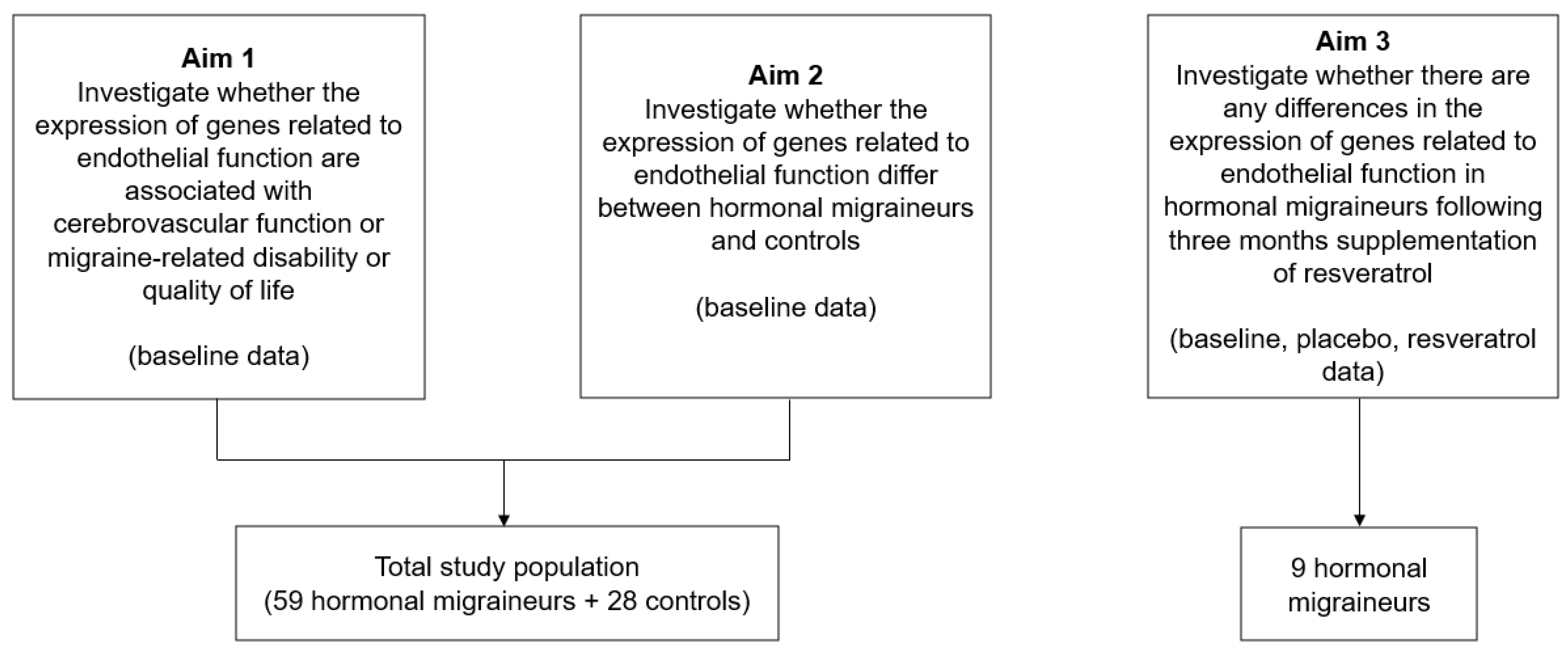
4.1. Study Design and Population

This study comprised data obtained from 87 women (59 hormonal migraineurs and 28 controls) in two studies (a cross-sectional clinical trial [5] and a randomised, double-blind, placebo-controlled, crossover intervention trial [43]) recruited by the Clinical Nutrition Research Centre at the University of Newcastle (see Figure 2). The cross-sectional clinical trial comprised both hormonal migraineurs and controls, whereas the crossover intervention trial included nine hormonal migraineur participants only. Participants in both studies were considered to have hormonal migraine if they fulfilled the criteria for migraines as defined by the International Classification for Headache Disorders [44] and if they suffered from migraines that occurred ±3 days from the onset of their period (or during ovulation for the cross-sectional study) in their three previous menstrual cycles. The inclusion and exclusion criteria for both studies have been described in detail elsewhere [5,43]. Briefly, participants in the placebo-controlled, crossover intervention trial were randomised to take resveratrol (150 mg/day) or placebo capsules twice daily for three months before crossing over to the other treatment arm for another three months. Data were collected from these participants at three time points (month-0 (baseline), month-3, and month-6) [43]. Participants in the cross-sectional clinical trial visited the research centre once and underwent testing to assess their cerebrovascular function, migraine-related disability, and migraine-related quality of life at one time-point [5].
The protocols for both studies were approved by the University of Newcastle’s Human Research Ethics Committee (H-2018-0167 and H-2019-0416) and registered with the Australian and New Zealand Clinical Trials Registry (ACTRN12618001230246 and ACTRN12620000180910). Approval for work conducted at the Queensland University of Technology (QUT) was obtained from the QUT Human Research Ethics Committee (Approval 2000000759). Written informed consent was obtained for all participants from both studies prior to commencement of the trial.

4.2. Study Outcomes

Outcomes for both studies were measured when the participant was one-hour fasted, migraine-free, and not menstruating. Study outcomes included cerebrovascular function, migraine-related disability, migraine-related quality of life, and gene expression.
  • Cerebrovascular Function
Cerebrovascular function was measured using transcranial Doppler ultrasound. Each participant was fitted with a transcranial Doppler headpiece with one probe fixed to the left and right sides of the temporal region for the bilateral measurement of cerebral blood flow velocity. Cerebral blood flow velocity was measured in the middle cerebral artery for both studies, and in the posterior cerebral artery for the resveratrol intervention study [5,43]. Cerebral blood flow velocity was measured at rest and during both hypercapnic stimulation (inhalation of carbogen gas (95% O2 and 5% CO2) for three minutes) and cognitive stimulation (neurovascular coupling). The cognitive tests that were completed by the participants have been described in detail elsewhere [5,43]. Cerebrovascular responsiveness to hypercapnia and neurovascular coupling capacity were calculated using the peak percentage increase in cerebral blood flow velocity during hypercapnic stimulation and during cognitive stimulation, respectively.
b.
Migraine Disability and Quality of Life
Migraine-related disability was measured using the Headache Impact Test-6™ and Migraine Disability Assessment [45,46]. Migraine-related quality of life was measured using the Migraine-Specific Quality of Life Questionnaire (version 2.1) [47]. Further details about these questionnaires have been described elsewhere [5].
c.
Gene Expression Analysis
Approximately 2.5 mL of venous blood was collected in a 9 mL PAXgene Blood RNA tube (QIAGEN, Clayton, VIC, Australia) by a commercial pathology service at the University of Newcastle, Australia. After a minimum of 2 h (but no more than 48 h) at room temperature to ensure blood cell lysis, the collected blood samples were frozen at −80 degrees Celsius until shipment with dry ice to Queensland University of Technology for gene expression analysis.
Analysis was limited to the following ten genes related to endothelial function and whose proteins are thought to be altered in migraine [48,49,50,51,52,53,54,55,56,57]: calcitonin-related polypeptide alpha (CALCA), endothelin 1 (EDN1), hypoxia-inducible factor 1, alpha (HIF1A), intracellular adhesion molecule 1 (ICAM1), interleukin 1 beta (IL1B), interleukin 6 beta 2 (IL6), nitric oxide synthase 3 (NOS3), tumour necrosis factor (TNF), vascular cell adhesion molecule 1 (VCAM1) and vascular endothelial growth factor A (VEGFA). GAPDH was utilised as an endogenous control gene to produce −ΔCt and ΔΔCt values.
RNA was extracted using a PAXgene Blood RNA Kit (QIAGEN, Clayton, VIC, Australia) and automated using a QIAcube extraction instrument (QIAGEN), according to manufacturer instructions. Once extracted, RNA was quantitated using a Nanodrop spectrophotometer (ThermoFisher Scientific, Waltham, MA, USA), and transcribed to cDNA using the Super Script IV First-Strand Synthesis System (ThermoFisher Scientific), according to manufacturer instructions.
Gene expression analysis was undertaken using qPCR with the following final reaction concentrations: 0.83× GoTaq qPCR Master Mix (Promega Corporation, Madison, WI, USA), forward and reverse primers at 0.83 μM, and cDNA equivalent to 30 ng of RNA input per reaction, in a total reaction volume of 12 μL. Thermal cycling and data acquisition was undertaken using a QuantStudio 6 Flex instrument (ThermoFisher Scientific), with thermal cycling parameters of 95 °C for three minutes, followed by 40 cycles of 95 °C for 20 s, 60 °C for one minute, and 72 °C for one minute. Intron-spanning primers for ten specified genes were designed for this study, to attempt to amplify as many gene isoforms as possible, and are outlined in Table 2.

4.3. Statistical Analysis

Cerebral blood flow velocity basal and peak values were determined using Table Curve 2D Version 5.01 (SYSTAT Software Incorporated, San Jose, CA, USA 2002). Neurovascular coupling and cerebrovascular responsiveness to hypercapnia were estimated using the smooth data spline estimation function with 10% and 20% Loess curve fitting, respectively.
Gene expression data were analysed using SPSS Release 27.0.1.0, (IBM Corporation, Armonk, NY, USA). qPCR data were analysed using t-tests or One Way ANOVA, as appropriate for the comparison, using both ΔCt (difference in threshold cycle between the housekeeping gene and gene of interest) and 2−ΔΔCt values. We calculated 2−ΔΔCt using the average ΔCt values for a sample’s phase of collection. The α value was set to 0.05.
Baseline correlation analyses were performed to identify correlations between cerebrovascular function, migraine-related disability, and migraine-related quality of life with −ΔCt. Spearman’s correlations were used for all analyses, as data were not normally distributed. The α value was set to 0.05 for the baseline correlation analyses. The Benjamini–Hochberg method was applied to each gene to account for multiple comparisons with a false discovery rate level of 0.1.

5. Conclusions

In conclusion, several genes related to endothelial function were found to be associated with decreased cerebral arterial stiffness, increased cerebral blood flow velocity, and decreased migraine-related disability or migraine severity in our total study population of hormonal migraineurs and controls, although findings did not remain significant following corrections for multiple comparisons. Based on our findings, it is important that future intervention studies investigate whether improvements of migraine symptomatology and cerebrovascular function are associated with alterations of genes that are related to endothelial function.

Supplementary Materials

The following supporting information can be downloaded at: https://www.mdpi.com/article/10.3390/ijms25031694/s1.

Author Contributions

Conceptualization, P.R.C.H.; methodology, J.S.A.D., L.R.G., K.G.C., P.R.C.H. and R.A.S.; data collection, J.S.A.D., L.R.G., K.G.C., P.R.C.H. and R.A.S.; formal analysis, J.S.A.D., L.R.G., K.G.C., P.R.C.H. and R.A.S.; writing—original draft preparation, J.S.A.D., L.R.G., K.G.C., P.R.C.H. and R.A.S.; writing—review and editing, J.S.A.D., L.R.G., K.G.C., P.R.C.H. and R.A.S.; funding acquisition, P.R.C.H. All authors have read and agreed to the published version of the manuscript.

Funding

This research was supported by an Australian Government Research Training Scholarship awarded to J.D. The test products and partial financial support for the study were provided by Evolva SA (Basel, Switzerland).

Institutional Review Board Statement

The study was conducted in accordance with the Declaration of Helsinki and approved by the University of Newcastle Human Research Ethics Committee and Queensland University of Technology’s Human Research Ethics Committee.

Informed Consent Statement

Informed consent was obtained from all subjects involved in the study.

Data Availability Statement

The data presented in this study are available on request from the corresponding author.

Acknowledgments

We would like to recognise Rachel Wong for her involvement in the study concept and design. Special thanks to Natasha Baker for her assistance with the trial management.

Conflicts of Interest

The authors declare no conflicts of interest.

References

  1. Sacco, S.; Ricci, S.; Degan, D.; Carolei, A. Migraine in women: The role of hormones and their impact on vascular diseases. J. Headache Pain 2012, 13, 177–189. [Google Scholar] [CrossRef] [PubMed]
  2. Macgregor, E.A. Menstrual migraine: Therapeutic approaches. Ther. Adv. Neurol. Disord. 2009, 2, 327–336. [Google Scholar] [CrossRef] [PubMed]
  3. Krause, D.N.; Duckles, S.P.; Pelligrino, D.A. Influence of sex steroid hormones on cerebrovascular function. J. Appl. Physiol. 2006, 101, 1252–1261. [Google Scholar] [CrossRef]
  4. Novella, S.; Dantas, A.P.; Segarra, G.; Medina, P.; Hermenegildo, C. Vascular Aging in Women: Is Estrogen the Fountain of Youth? Front. Physiol. 2012, 3, 165. [Google Scholar] [CrossRef]
  5. Dzator, J.S.A.; Howe, P.R.C.; Griffiths, L.R.; Coupland, K.G.; Wong, R.H.X. Cerebrovascular Function in Hormonal Migraine: An Exploratory Study. Front. Neurol. 2021, 12, 694980. [Google Scholar] [CrossRef]
  6. Tietjen, G.E.; Herial, N.A.; White, L.; Utley, C.; Kosmyna, J.M.; Khuder, S.A. Migraine and biomarkers of endothelial activation in young women. Stroke 2009, 40, 2977–2982. [Google Scholar] [CrossRef] [PubMed]
  7. Dzator, J.S.A.; Coupland, K.G.; Howe, P.R.C. Exploring the Effects of Resveratrol Supplementation on Cerebrovascular Function in Hormonal Migraineurs: A Pilot Study. IBRO Neurosci. Rep. 2023, in press. [CrossRef] [PubMed]
  8. Li, H.; Xia, N.; Hasselwander, S.; Daiber, A. Resveratrol and Vascular Function. Int. J. Mol. Sci. 2019, 20, 2155. [Google Scholar] [CrossRef]
  9. Rodriguez-Acevedo, A.J.; Smith, R.A.; Roy, B.; Sutherland, H.; Lea, R.A.; Frith, A.; MacGregor, E.A.; Griffiths, L.R. Genetic association and gene expression studies suggest that genetic variants in the SYNE1 and TNF genes are related to menstrual migraine. J. Headache Pain 2014, 15, 62. [Google Scholar] [CrossRef] [PubMed]
  10. Wattiez, A.S.; Sowers, L.P.; Russo, A.F. Calcitonin gene-related peptide (CGRP): Role in migraine pathophysiology and therapeutic targeting. Expert. Opin. Ther. Targets 2020, 24, 91–100. [Google Scholar] [CrossRef] [PubMed]
  11. Russell, F.A.; King, R.; Smillie, S.J.; Kodji, X.; Brain, S.D. Calcitonin gene-related peptide: Physiology and pathophysiology. Physiol. Rev. 2014, 94, 1099–1142. [Google Scholar] [CrossRef]
  12. Fila, M.; Sobczuk, A.; Pawlowska, E.; Blasiak, J. Epigenetic Connection of the Calcitonin Gene-Related Peptide and Its Potential in Migraine. Int. J. Mol. Sci. 2022, 23, 6151. [Google Scholar] [CrossRef] [PubMed]
  13. Rivera-Mancilla, E.; Villalón, C.M.; MaassenVanDenBrink, A. CGRP inhibitors for migraine prophylaxis: A safety review. Expert. Opin. Drug Saf. 2020, 19, 1237–1250. [Google Scholar] [CrossRef] [PubMed]
  14. Urits, I.; Jones, M.R.; Gress, K.; Charipova, K.; Fiocchi, J.; Kaye, A.D.; Viswanath, O. CGRP Antagonists for the Treatment of Chronic Migraines: A Comprehensive Review. Curr. Pain Headache Rep. 2019, 23, 29. [Google Scholar] [CrossRef] [PubMed]
  15. Visočnik, D.; Žvan, B.; Zaletel, M.; Zupan, M. αCGRP-Induced Changes in Cerebral and Systemic Circulation; A TCD Study. Front. Neurol. 2020, 11, 578103. [Google Scholar] [CrossRef]
  16. Schifter, S.; Krusell, L.R.; Sehested, J. Normal Serum Levels of Calcitonin Gene-Related Peptide (CGRP) in Mild to Moderate Essential Hypertension. Am. J. Hypertens. 1991, 4, 565–569. [Google Scholar] [CrossRef] [PubMed]
  17. Masuda, A.; Shimamoto, K.; Mori, Y.; Nakagawa, M.; Ura, N.; Iimura, O. Plasma calcitonin gene-related peptide levels in patients with various hypertensive diseases. J. Hypertens. 1992, 10, 1499–1504. [Google Scholar] [CrossRef]
  18. Rijnberg, F.M.; Hazekamp, M.G.; Wentzel, J.J.; de Koning, P.J.H.; Westenberg, J.J.M.; Jongbloed, M.R.M.; Blom, N.A.; Roest, A.A.W. Energetics of Blood Flow in Cardiovascular Disease: Concept and Clinical Implications of Adverse Energetics in Patients with a Fontan Circulation. Circulation 2018, 137, 2393–2407. [Google Scholar] [CrossRef]
  19. Dunne, F.P.; Barry, D.G.; Ferriss, J.B.; Grealy, G.; Murphy, D. Changes in blood pressure during the normal menstrual cycle. Clin. Sci. 1991, 81, 515–518. [Google Scholar] [CrossRef]
  20. Choi, H.M.; Stebbins, C.L.; Nho, H.; Kim, M.S.; Chang, M.J.; Kim, J.K. Effects of Ovarian Cycle on Hemodynamic Responses during Dynamic Exercise in Sedentary Women. Korean J. Physiol. Pharmacol. 2013, 17, 499–503. [Google Scholar] [CrossRef]
  21. Greenberg, G.; Imeson, J.D.; Thompson, S.G.; Meade, T.W. Blood pressure and the menstrual cycle. Br. J. Obs. Gynaecol. 1985, 92, 1010–1014. [Google Scholar] [CrossRef]
  22. Wang, Y.F.; Wang, S.J. Hypertension and Migraine: Time to Revisit the Evidence. Curr. Pain Headache Rep. 2021, 25, 58. [Google Scholar] [CrossRef]
  23. Zhang, J.; Mao, Y.; Li, Y.; Zhao, K.; Xie, Q.; Wang, K.; Shi, J. Association between migraine or severe headache and hypertension among US adults: A cross-sectional study. Nutr. Metab. Cardiovasc. Dis. 2023, 33, 350–358. [Google Scholar] [CrossRef]
  24. Jackson, J.L.; Kuriyama, A.; Kuwatsuka, Y.; Nickoloff, S.; Storch, D.; Jackson, W.; Zhang, Z.J.; Hayashino, Y. Beta-blockers for the prevention of headache in adults, a systematic review and meta-analysis. PLoS ONE 2019, 14, e0212785. [Google Scholar] [CrossRef]
  25. Carcel, C.; Haghdoost, F.; Shen, J.; Nanda, P.; Bai, Y.; Atkins, E.; Torii-Yoshimura, T.; Clough, A.J.; Davies, L.; Cordato, D.; et al. The effect of blood pressure lowering medications on the prevention of episodic migraine: A systematic review and meta-analysis. Cephalalgia 2023, 43, 03331024231183166. [Google Scholar] [CrossRef]
  26. Niu, G.; Chen, X. Vascular endothelial growth factor as an anti-angiogenic target for cancer therapy. Curr. Drug Targets 2010, 11, 1000–1017. [Google Scholar] [CrossRef]
  27. Jozkowicz, A.; Cooke, J.P.; Guevara, I.; Huk, I.; Funovics, P.; Pachinger, O.; Weidinger, F.; Dulak, J. Genetic augmentation of nitric oxide synthase increases the vascular generation of VEGF. Cardiovasc. Res. 2001, 51, 773–783. [Google Scholar] [CrossRef] [PubMed]
  28. Mäki-Petäjä, K.M.; McGeoch, A.; Yang, L.L.; Hubsch, A.; McEniery, C.M.; Meyer, P.A.R.; Mir, F.; Gajendragadkar, P.; Ramenatte, N.; Anandappa, G.; et al. Mechanisms Underlying Vascular Endothelial Growth Factor Receptor Inhibition-Induced Hypertension: The HYPAZ Trial. Hypertension 2021, 77, 1591–1599. [Google Scholar] [CrossRef] [PubMed]
  29. Pandey, A.K.; Singhi, E.K.; Arroyo, J.P.; Ikizler, T.A.; Gould, E.R.; Brown, J.; Beckman, J.A.; Harrison, D.G.; Moslehi, J. Mechanisms of VEGF (Vascular Endothelial Growth Factor) Inhibitor-Associated Hypertension and Vascular Disease. Hypertension 2018, 71, e1–e8. [Google Scholar] [CrossRef] [PubMed]
  30. Troncoso, M.F.; Ortiz-Quintero, J.; Garrido-Moreno, V.; Sanhueza-Olivares, F.; Guerrero-Moncayo, A.; Chiong, M.; Castro, P.F.; García, L.; Gabrielli, L.; Corbalán, R.; et al. VCAM-1 as a predictor biomarker in cardiovascular disease. Biochim. Et Biophys. Acta (BBA)—Mol. Basis Dis. 2021, 1867, 166170. [Google Scholar] [CrossRef] [PubMed]
  31. de Faria, A.P.; Ritter, A.M.; Sabbatini, A.R.; Corrêa, N.B.; Brunelli, V.; Modolo, R.; Moreno, H. Deregulation of Soluble Adhesion Molecules in Resistant Hypertension and Its Role in Cardiovascular Remodeling. Circ. J. 2016, 80, 1196–1201. [Google Scholar] [CrossRef]
  32. Kilic, I.D.; Findikoglu, G.; Alihanoglu, Y.I.; Yildiz, B.S.; Uslu, S.; Rota, S.; Evrengul, H. Circulating adhesion molecules and arterial stiffness. Cardiovasc. J. Afr. 2015, 26, 21–24. [Google Scholar] [CrossRef] [PubMed]
  33. Szeghy, R.E.; Stute, N.L.; Province, V.M.; Augenreich, M.A.; Stickford, J.L.; Stickford, A.S.L.; Ratchford, S.M. Six-month longitudinal tracking of arterial stiffness and blood pressure in young adults following SARS-CoV-2 infection. J. Appl. Physiol. 2022, 132, 1297–1309. [Google Scholar] [CrossRef]
  34. Kunutsor, S.K.; Bakker, S.J.L.; Dullaart, R.P.F. Soluble Vascular Cell Adhesion Molecules May be Protective of Future Cardiovascular Disease Risk: Findings from the PREVEND Prospective Cohort Study. J. Atheroscler. Thromb. 2017, 24, 804–818. [Google Scholar] [CrossRef]
  35. Ulyanova, T.; Scott, L.M.; Priestley, G.V.; Jiang, Y.; Nakamoto, B.; Koni, P.A.; Papayannopoulou, T. VCAM-1 expression in adult hematopoietic and nonhematopoietic cells is controlled by tissue-inductive signals and reflects their developmental origin. Blood 2005, 106, 86–94. [Google Scholar] [CrossRef] [PubMed]
  36. Hsue, P.Y.; Scherzer, R.; Grunfeld, C.; Imboden, J.; Wu, Y.; Del Puerto, G.; Nitta, E.; Shigenaga, J.; Schnell Heringer, A.; Ganz, P.; et al. Depletion of B-cells with rituximab improves endothelial function and reduces inflammation among individuals with rheumatoid arthritis. J. Am. Heart Assoc. 2014, 3, e001267. [Google Scholar] [CrossRef] [PubMed]
  37. Henein, M.Y.; Vancheri, S.; Longo, G.; Vancheri, F. The Role of Inflammation in Cardiovascular Disease. Int. J. Mol. Sci. 2022, 23, 12906. [Google Scholar] [CrossRef] [PubMed]
  38. Mueller, L.; Gupta, A.K.; Stein, T.P. Deficiency of tumor necrosis factor alpha in a subclass of menstrual migraineurs. Headache 2001, 41, 129–137. [Google Scholar] [CrossRef]
  39. Ibrahimi, K.; Vermeersch, S.; Frederiks, P.; Geldhof, V.; Draulans, C.; Buntinx, L.; Lesaffre, E.; MaassenVanDenBrink, A.; de Hoon, J. The influence of migraine and female hormones on capsaicin-induced dermal blood flow. Cephalalgia 2017, 37, 1164–1172. [Google Scholar] [CrossRef] [PubMed]
  40. Wang, J.; Robinson, J.F.; Khan, H.M.R.; Carter, D.E.; McKinney, J.; Miskie, B.A.; Hegele, R.A. Optimizing RNA extraction yield from whole blood for microarray gene expression analysis. Clin. Biochem. 2004, 37, 741–744. [Google Scholar] [CrossRef]
  41. Brkljacić, J.; Tanić, N.; Milutinović, D.V.; Elaković, I.; Jovanović, S.M.; Perisić, T.; Dundjerski, J.; Matić, G. Validation of endogenous controls for gene expression studies in peripheral lymphocytes from war veterans with and without PTSD. BMC Mol. Biol. 2010, 11, 26. [Google Scholar] [CrossRef]
  42. Xiao, J.; Li, X.; Liu, J.; Fan, X.; Lei, H.; Li, C. Identification of reference genes in blood before and after entering the plateau for SYBR green RT-qPCR studies. PeerJ 2017, 5, e3726. [Google Scholar] [CrossRef] [PubMed]
  43. Dzator, J.S.A.; Howe, P.R.C.; Coupland, K.G.; Wong, R.H.X. A Randomised, Double-Blind, Placebo-Controlled Crossover Trial of Resveratrol Supplementation for Prophylaxis of Hormonal Migraine. Nutrients 2022, 14, 1763. [Google Scholar] [CrossRef]
  44. Olesen, J. Headache Classification Committee of the International Headache Society (IHS) The International Classification of Headache Disorders, 3rd edition. Cephalalgia 2018, 38, 1–211. [Google Scholar] [CrossRef]
  45. Yang, M.; Rendas-Baum, R.; Varon, S.F.; Kosinski, M. Validation of the Headache Impact Test (HIT-6™) across episodic and chronic migraine. Cephalalgia 2011, 31, 357–367. [Google Scholar] [CrossRef]
  46. Stewart, W.F.; Lipton, R.B.; Kolodner, K.B.; Sawyer, J.; Lee, C.; Liberman, J.N. Validity of the Migraine Disability Assessment (MIDAS) score in comparison to a diary-based measure in a population sample of migraine sufferers. Pain 2000, 88, 41–52. [Google Scholar] [CrossRef]
  47. Martin, B.C.; Pathak, D.S.; Sharfman, M.I.; Adelman, J.U.; Taylor, F.; Kwong, W.J.; Jhingran, P. Validity and reliability of the migraine-specific quality of life questionnaire (MSQ Version 2.1). Headache 2000, 40, 204–215. [Google Scholar] [CrossRef]
  48. Rodríguez-Osorio, X.; Sobrino, T.; Brea, D.; Martínez, F.; Castillo, J.; Leira, R. Endothelial progenitor cells: A new key for endothelial dysfunction in migraine. Neurology 2012, 79, 474–479. [Google Scholar] [CrossRef]
  49. Fidan, I.; Yüksel, S.; Ýmir, T.; İrkeç, C.; Aksakal, F.N. The importance of cytokines, chemokines and nitric oxide in pathophysiology of migraine. J. Neuroimmunol. 2006, 171, 184–188. [Google Scholar] [CrossRef]
  50. Kursun, O.; Yemisci, M.; van den Maagdenberg, A.M.J.M.; Karatas, H. Migraine and neuroinflammation: The inflammasome perspective. J. Headache Pain 2021, 22, 55. [Google Scholar] [CrossRef] [PubMed]
  51. Mason, B.N.; Russo, A.F. Vascular Contributions to Migraine: Time to Revisit? Front. Cell. Neurosci. 2018, 12, 233. [Google Scholar] [CrossRef]
  52. Sabri, M.R.; Dehghan, B.; Yaghini, O.; Nasiri, J.; Mansourian, M.; Khalifehsoltani, S. Endothelial dysfunction state in migraine headache and neutrally mediated syncope in children and young adults. J. Res. Med. Sci. 2015, 20, 771–776. [Google Scholar] [CrossRef] [PubMed]
  53. Huang, X.; Zhang, X.; Zhao, D.X.; Yin, J.; Hu, G.; Evans, C.E.; Zhao, Y.Y. Endothelial Hypoxia-Inducible Factor-1α Is Required for Vascular Repair and Resolution of Inflammatory Lung Injury through Forkhead Box Protein M1. Am. J. Pathol. 2019, 189, 1664–1679. [Google Scholar] [CrossRef] [PubMed]
  54. Zhang, H.; Park, Y.; Wu, J.; Chen, X.; Lee, S.; Yang, J.; Dellsperger, K.C.; Zhang, C. Role of TNF-α in vascular dysfunction. Clin. Sci. 2009, 116, 219–230. [Google Scholar] [CrossRef]
  55. Galea, J.; Armstrong, J.; Gadsdon, P.; Holden, H.; Francis, S.E.; Holt, C.M. Interleukin-1β in Coronary Arteries of Patients with Ischemic Heart Disease. Arterioscler. Thromb. Vasc. Biol. 1996, 16, 1000–1006. [Google Scholar] [CrossRef]
  56. Böhm, F.; Pernow, J. The importance of endothelin-1 for vascular dysfunction in cardiovascular disease. Cardiovasc. Res. 2007, 76, 8–18. [Google Scholar] [CrossRef]
  57. Didion, S.P. Cellular and Oxidative Mechanisms Associated with Interleukin-6 Signaling in the Vasculature. Int. J. Mol. Sci. 2017, 18, 2563. [Google Scholar] [CrossRef]
Figure 1. Correlations between CALCA, VEGF, and measures of cerebrovascular function in the left middle cerebral artery. (A) Spearman correlation between CALCA (−ΔCt) and neurovascular coupling capacity during the 1-back task, (B) Spearman correlation between CALCA (−ΔCt) and neurovascular coupling capacity during the 2-back task, (C) Spearman correlation between CALCA (−ΔCt) and mean blood flow velocity, and (D) Spearman correlation between VEGF (−ΔCt) and mean blood flow velocity.
Ijms 25 01694 g001
Figure 2. Outline of the aims and study population.
Ijms 25 01694 g002
Table 1. Baseline Participant Characteristics.
Hormonal Migraineurs (N = 59)Controls
(N = 28)
p Value
Age (years) a38.63 + 1.0535.89 + 1.830.208
Body mass index (kg/m2) a25.58 + 0.6325.12 + 1.180.443
Systolic blood pressure (mmHg) b116.35 + 1.52114.61 + 2.520.536
Diastolic blood pressure (mmHg) b70.75 + 1.1167.68 + 1.560.116
a Mann–Whitney U b Independent t-test.
Table 2. Primers used in qPCR analysis.
Primer NamePrimer Sequence (5′-3′)TranscriptExon
CALCA ForwardTCAGCATCTTGGTCCTGTTGNM_001741.32
CALCA ReverseCTGCACATAGTCCTGCACCANM_001741.33
EDN1 ForwardTGGGAAAAAGTGTATTTATCAGCANM_001955.54
EDN1 ReverseTTTGACGCTGTTTCTCATGGNM_001955.55
HIF1A ForwardGCTTGGTGCTGATTTGTGAANM_001530.46
HIF1A ReverseTTCTGGCTCATATCCCATCANM_001530.47
ICAM1 ForwardCTTGAGGGCACCTACCTCTGNM_000201.36
ICAM1 ReverseCATTATGACTGCGGCTGCTANM_000201.37
IL1B ForwardCTGTCCTGCGTGTTGAAAGANM_000576.36
IL1B ReverseACTGGGCAGACTCAAATTCCNM_000576.37
IL6 ForwardGGCTGAAAAAGATGGATGCTNM_000600.53
IL6 ReverseGCTCTGGCTTGTTCCTCACTNM_000600.54
NOS3 ForwardTGTCTGCATGGACCTGGATANM_000603.510
NOS3 ReverseCACGATGGTGACTTTGGCTANM_000603.511
VCAM1 ForwardATGGAATTCGAACCCAAACANM_001078.46
VCAM1 ReverseCCTGGCTCAAGCATGTCATANM_001078.47
VEGF ForwardCCCACTGAGGAGTCCAACATNM_001025366.33
VEGF ReverseTGCATTCACATTTGTTGTGCNM_001025366.34
TNF ForwardGACAAGCCTGTAGCCCATGTNM_000594.43
TNF ReverseGAGGTACAGGCCCTCTGATGNM_000594.44
GAPDH ForwardCGACCACTTTGTCAAGCTCANM_002046.78
GAPDH ReverseGGTGGTCCAGGGGTCTTACTNM_002046.79
Disclaimer/Publisher’s Note: The statements, opinions and data contained in all publications are solely those of the individual author(s) and contributor(s) and not of MDPI and/or the editor(s). MDPI and/or the editor(s) disclaim responsibility for any injury to people or property resulting from any ideas, methods, instructions or products referred to in the content.

