Review

Microbiota–Gastric Cancer Interactions and the Potential Influence of Nutritional Therapies

by
Pauline Raoul
1,
Valeria Maccauro
2,
Marco Cintoni
1,3,
Emidio Scarpellini
4,
Gianluca Ianiro
5,6,
Antonio Gasbarrini
3,5,6,
Maria Cristina Mele
1,3,6 and
Emanuele Rinninella
1,3,6,*
1
Clinical Nutrition Unit, Department of Medical and Abdominal Surgery and Endocrine-Metabolic Sciences, Fondazione Policlinico Universitario Agostino Gemelli IRCCS, 00168 Rome, Italy
2
School of Specialization in Internal Medicine, Catholic University of the Sacred Heart, 00168 Rome, Italy
3
Research and Training Center in Human Nutrition, Catholic University of the Sacred Heart, 00168 Rome, Italy
4
Translationeel Onderzoek van Gastro-Enterologische Aandoeningen (T.A.R.G.I.D.), Gasthuisberg University 11 Hospital, KU Leuven, Herestraat 49, 3000 Leuven, Belgium
5
Digestive Disease Center (CEMAD), Department of Medical and Abdominal Surgery and Endocrine-Metabolic Sciences, Fondazione Policlinico Universitario Agostino Gemelli IRCCS, 00168 Rome, Italy
6
Department of Translational Medicine and Surgery, Catholic University of the Sacred Heart, 00168 Rome, Italy
*
Author to whom correspondence should be addressed.
Int. J. Mol. Sci. 2024, 25(3), 1679; https://doi.org/10.3390/ijms25031679
Submission received: 25 December 2023 / Revised: 16 January 2024 / Accepted: 23 January 2024 / Published: 30 January 2024
(This article belongs to the Special Issue Microbiota and Cancer 3.0)

Abstract

:
Gastric cancer (GC) is one of the most common causes of cancer deaths, and GC treatments represent a large area of research. Although initially regarded as a sterile organ and unsuitable for microbial communities, the discovery of Helicobacter pylori made us realize that some microbes can colonize the stomach. In recent years, growing interest in gastric bacteria has expanded to the gut microbiota and, more recently, to the oral microbiota. Indeed, the oral–gastric–gut microbiota axis may play a crucial role in maintaining homeostasis, while changes in microbiota composition in GC patients can influence clinical outcomes. On the one hand, the microbiota and its metabolites may significantly influence the progression of GC, while anti-GC treatments such as gastrectomy and chemotherapy may significantly impact the oral–gastric–gut microbiota axis of GC patients. In this context, the role of nutritional therapies, including diet, prebiotics, and probiotics, in treating GC should not be underestimated. Wit this review, we aim to highlight the main role of the gastric, oral, and gut microbiota in GC onset and progression, representing potential future biomarkers for early GC detection and a target for efficient nutritional therapies during the course of GC.

1. Introduction

Gastric cancer (GC) is the third leading cause of cancer-related mortality worldwide [1]; corresponding to the fifth highest incidence in the world, it is estimated to cause over one million new cases worldwide each year [2]. Five-year GC survival rates are estimated at almost 30% in most western countries due to late-stage diagnosis [3]. GC is initiated insidiously, and the early symptoms are atypical. Thus, most patients have already progressed to advanced stages upon diagnosis, making their treatment challenging. GC is a multifactorial disease affected by various genetic and environmental factors, including Helicobacter pylori (H. pylori) infection, lifestyle, socioeconomic factors, dietary behavior, and aging [4].
Until recently, the gastric environment was considered sterile, probably due to increased acidity, but emerging data have revealed that there is a broad range of microorganisms in the stomach with a density of 10 to 1000 colony-forming units/g. The gastric microbiota comprises bacteria ingested mainly through the respiratory tract and secondarily from the intestine by transpyloric biliary reflux. The predominant phyla in the gastric mucosa consist of acidity-resisting bacteria such as Actinomycetota, Bacillota, Bacteroidota, and Pseudomonadota, among more than one hundred types, the most important of which is H. pylori [5]. Since 1994 it has been universally accepted that H. pylori is the main driver of the precancerous cascade and the most important etiological factor for GC, estimated between to be involved in between 74.7% and 90% of new non-cardia GC cases [6].
Furthermore, recent studies have focused not only on the gastric microbiota but also on the oral microbiota. Indeed, the oral microbiota exhibits the second highest level of alpha diversity following that of the gut, including over 700 species of bacteria, over 100 species of fungi, and protozoa. The latter include Entamoeba gingivalis and Trichomonas tenax. Phyla such as Actinomycetota, Bacillota, Pseudomonadota, Bacteroidota, and Saccharibacteria are well represented in healthy oral microbiota. An imbalanced microbiota, known as dysbiosis, is associated with intraoral diseases such as dental caries, gingivitis, and periodontitis, as well as with different neoplasms such as gastrointestinal cancers [7,8].
Finally, in recent years, the association between the gut microbiota and GC has gradually attracted researchers’ attention. The gut microbiota may be involved in carcinogenesis, mainly by modulating immune responses, and, consequently, in the mechanisms of cancer treatment, potentially affecting cancer treatment responses [9].
Concomitantly to GC treatment like surgery or chemotherapy, interest is increasing in nutritional therapy to improve patients’ nutritional status, adherence to therapies, and quality of life. European nutritional guidelines recommended nutritional support for GC patients undergoing surgery, as well as those with unresectable disease; surgery may be performed in combination with oral, enteral, and parenteral nutrition [10].
The objective of this review is to highlight the main role of the gastric, oral, and gut microbiota in GC onset and progression, representing potential future biomarkers for early GC detection and a target for efficient nutritional therapies during the course of GC.

2. Gastric Microbiota and Gastric Carcinogenesis

2.1. Mutual Relationships between Chronic Gastric Inflammatory Response to H. pylori and Gastric Microbiota Dysbiosis

A normal gastric microbiota is composed of Pseudomonadota (H. pylori belongs to this phylum), Bacillota, Actinomycetota, Bacteroides, and Fusobacteria, which are the most abundant phyla. H. pylori has inhibitory effects on the colonization of other bacteria, harboring a significantly lower diversity of such bacteria in the stomach. On the contrary, the gastric microbiota in H. pylori-negative patients was found to be predominated by other bacterial strains, including Pseudomonadota (52.6%), Bacillota (26.4%), Bacteroidota (12%), and Actinomycetota (6.4%); the most common genera included Gemella, Prevotella, and Streptococcus [11].
H. pylori has direct oncogenic potential in GC due to its oncoprotein cytotoxin-associated gene A (CagA), which destabilizes cellular junctions by disrupting the E-cadherin–β-catenin complex and activates proliferating pathways as an ERK-MAP kinase cascade, leading to cellular morphological transition to the “hummingbird” phenotype [12]. In addition, H. pylori can facilitate gastric cells’ oncogenic transformation via its vacuolating cytotoxin A (VacA), which interferes with normal autophagy, thereby promoting abnormal cell survival [13].
Given that H. pylori also creates a premalignant environment of atrophy and intestinal metaplasia, the subsequent alteration in the gastric microbiota seems to play a crucial role in gastric tumorigenesis itself. Consequently, successful H. pylori eradication seems to be essential for the restoration of a balanced gastric microbiota and the prevention of gastric carcinogenesis—at least in the primary stages [14]. It has also been noted that the activity of gastritis demonstrates a close relationship with either H. pylori or with other pathogenic phyla, as it increases the abundance of Bacteroidota, Bacillota, or Pseudomonadota, favoring GC-related dysbiosis [15]. H. pylori may modify the gastric environment, paving the way for the growth of a dysbiotic gastric bacterial community. In this regard, H. pylori eradication may reverse gastric dysbiosis to a similar level in uninfected patients, exerting beneficial effects on the gut microbiota and achieving increased probiotic and putative downregulation of drug resistance. More specifically, successful H. pylori eradication was reported to significantly inhibit dysbiosis, and treatment failure was associated with an increased dysbiosis rate comparable to that of active H. pylori infection [16]. However, the exact background of the interaction between sustained inflammation and genotoxicity damage has still not been evaluated [15]. In a recent study, a possible explanation of this mutual relationship was evidenced: chronic gastric inflammatory response to H. pylori may modify the gastric environment, mainly by reducing gastric acidity, leading to the overgrowth of a dysbiotic gastric bacterial community. In this way, successful H. pylori eradication has been shown to significantly reverse gastric dysbiosis to a similar level as in uninfected patients, although the dysbiosis rate of such patients remained higher than the non-H. pylori control. Nonetheless, treatment failure was associated with an increased dysbiosis rate comparable to that of active H. pylori. More intense dysbiosis was also found to be associated with progress from gastritis to atrophy and GC [16].
In antral-predominant gastritis, the production of gastric acid is increased, which is associated with a high risk of the development of duodenal ulcer disease, whereas it is protective against GC development. In contrast, corpus-predominant gastritis leads to reduced production of gastric acid and may lead to atrophic gastritis, a condition associated with an increased risk of developing GC. H. pylori exerts a direct inflammatory effect on the mucosal surface of the stomach, variably affecting the production of mucin, which, in turn, affects the composition of the nearby microbiota, resulting in gastric dysbiosis. A reduced capacity for gastric acid secretion allows for the survival and proliferation of other microbes that are normally killed by acidic environments. This may be the backbone of GC development, given that during the last stage of gastric malignancy, oral or intestinal-type bacteria are predominantly discovered, which is not observed in premalignant conditions (e.g., chronic gastritis and atrophy) where H. pylori abundance is evident. Whether this phenomenon is due to tumor-related mucin-type differentiation, possibly resulting in GC-related microbiota, must be elucidated [17].

2.2. Close Association between Gastric Microbiota Dysbiosis and GC

A study examined the composition of the microbiota of the gastric mucosa in intestinal-type GC, suggesting that bacterial diversity decreased at the genus level as patients progressed from superficial gastritis to intestinal metaplasia and GC. The authors also described an increased abundance of Lactobacillus coleohominis and Lachnospiraceae in GC patients, indicating the presence of microbial dysbiosis in gastric carcinogenesis [18]. Another cohort study confirmed the hypothesis of procarcinogenic gastric dysbiosis, underlining that Parvimonas micra, Dialister pneumosintes, Slackia exigua, Peptostreptococcus stomatis, Prevotella intermedia, Fusobacterium nucleatum, Prevotella oris, and Catonella morbi were significantly enriched in the GC microbiota compared to precancerous stages [19]. In parallel, a Portuguese study reported that the GC microbiota demonstrated reduced microbial diversity; reduced Helicobacter abundance; and over-representation of bacterial genera belonging to intestinal commensals of Pseudomonadota, Bacillota, and Actinomycetota phyla. Among Pseudomonadota, an overgrowth of the genera Phyllobacterium and Achromobacter and the families Xanthomonadaceae and Enterobacteriaceae was reported. Additionally, Lactobacillaceae family member, Clostridium and Rhodococcus were also significantly more abundant in gastric carcinoma, whereas Helicobacter, Neisseria, Prevotella, and Streptococcus were most abundant in the microbiota of patients with chronic gastritis. In addition to oral cavity bacterial strains, intestinal mucosal commensal, such as Citrobacter, Clostridium, Lactobacillaceae family, Achromobacter, and Rhodococcus, were reported to be significantly increased in GC samples compared with chronic gastritis samples. This observation is compatible with the hypothesis that during carcinogenesis, changes in the stomach’s mucosa that lead to decreased acid secretion allow for the growth of bacteria with nitrosative functions, exerting a major genotoxic potential. In these bacteria, the nitrate reductase enzyme, whose function is degradation of nitrate into nitrite and nitrite into nitric oxide, appears to be overexpressed, resulting in gastric cell DNA damage [20]. A case–control study conducted by Gunathilake showed that patients with GC had higher relative abundances of Helicobacteraceae, Propionibacteriaceae, and Prevotellaceae than healthy subjects at the family level, with overall relative abundances of Helicobacter, Propionibacterium, and Prevotella at the genus level, whereas the relative abundance of Lactococcus lactis (L. lactis) was higher in the healthy controls than in infected patients. Moreover, the Shannon index was significantly higher in the controls than in the patients, as H. pylori-positive carriers were found to have higher abundances of Spirochetes, Acidobacteria, and non-Helicobacter Pseudomonadota and comparatively lower abundances of Actinomycetota, Bacteroidota, and Bacillota phyla than uninfected subjects. P. acnes can enhance GC development by producing proinflammatory cytokines such as interleukin (IL)-15, whereas one cytoplasmic fraction of L. lactis has been reported to exert an antiproliferative effect on a human stomach cancer cell line, thereby protecting against carcinogenesis; the G0/G1 cell cycle arrest induced by such bacteria was associated with an increase in p53 and p21 expression, a reduction in cyclin D1 expression, and retinoblastoma protein phosphorylation, thereby inducing apoptosis. Thus, it can be suggested that if there is a bacterial species that can promote GC occurrence, the eradication of this bacterium is useful in decreasing GC incidence [21]. Other bacterial genera reported to be increased in GC patients compared to healthy controls include Enterococcus, Lactobacillaceae family, Carnobacterium, Glutaminibacter, and Fusobacterium [22].
Thus, the development of GC may be H. pylori-independent, since H. pylori colonization decreases in later steps of carcinogenesis, whereas other gastric microbial strains predominate, as the gastric microbial community profiles in cancer were reported to significantly differ from those of non-cancerous stomachs. Genera that were more consistently enriched in the GC microbiota belong to intestinal strains, including Lactobacillaceae family, Streptococcaceae, Staphylococcus, Clostridium, and Fusobacterium nucleatum [23].
Gastric microbiota balance is extremely complex, and the specific mechanisms causing the occurrence and development of GC remain unknown. The growth of conditional microbiota may inhibit the growth of pathogenic microbes such as H. pylori or other pathogenic bacteria such as bacteria with nitrosative functions. Future studies focusing on GC pathogenesis should evaluate H. pylori strains, host genetic characteristics, microbiota composition, and environmental factors of the host. Indeed, the mechanisms of microbe–microbe interactions and microbe–host interactions causing GC are multifactorial. Compared with non-GC patients, GC patients have lower alpha diversities and a higher abundances of H. pylori [5] but also an overabundance of bacteria that differ in GC patients depending on environmental factors such as the living conditions of patients. Thus, although H. pylori is the main trigger of histopathological changes in GC, its relationships with non-H. pylori are also involved in the development of GC. Future studies should investigate whether conditional oral or gastric microbiota can serve as hallmarks of GC.

2.3. Potential Implications of Other Specific Gastric Bacteria in GC

2.3.1. Fusobacterium nucleatum

Interestingly, Fusobacterium nucleatum can act directly on host cells affecting the expression of cancer marker genes, thereby promoting the occurrence of cancer, and can indirectly secrete endotoxins to inhibit the immune function of the body and generate an inflammatory microenvironment. Fusobacterium nucleatum can activate the nuclear factor-kappa B (NF-κB) pathway to stimulate the production and release of inflammatory cytokines, such as IL-1β, IL-6, IL-8, and tumor necrosis factor (TNF), thereby creating a proinflammatory microenvironment that favors tumor development [24,25]. Moreover, the adhesion of Fusobacterium nucleatum from FadA to the E-cadherin of intestinal epithelial cells drives the activation of the Wnt/β-catenin pathway to promote the proliferation of tumor cells [26]. Fusobacterium nucleatum has also been reported to be more abundant in the microbiota of GC patients compared with that of non-tumoral controls, and its detection has been associated with GC risk, patient age, tumor size, and decreased survival [27]. Its carcinogenetic mechanism is not fully understood due to its impact on the microenvironment and metabolic function, as well as the deregulation of actin dynamics and changes in cancer cell motility; however, it is accepted that the carcinogenic potential is explicated in the later phases when H. pylori is no longer present [28]. Hsieh et al. studied the bacterial species associated with gastric epithelium in 11 GC patients and found that Fusobacterium nucleatum was abundantly enriched in GC patients and that the gastric microbes of most GC patients differed from those of non-cancerous gastric disease patients. Analysis of the operating characteristic curve showed that the sensitivity of Fusobacterium nucleatum combined with Clostridium colicanis and Fusobacterium canifelinum in the diagnosis of GC was 100%, and the specificity was about 70% [29].

2.3.2. Lactobacillaceae Family

Lactic acid bacteria, including Streptococcus, Lactobacillaceae family, Bifidobacterium, and Lactococcus, are implicated in carcinogenesis by increasing the amount of toxic N-nitroso compounds and reactive oxygen species (ROS), which, in turn, promote deoxyribonucleic acid (DNA) damage, and by fostering HIF-1-mediated epithelial–mesenchymal transition; then, these bacterial strains can promote the colonization of other carcinogenic pathobionts by inducing immunotolerance. Importantly, lactate itself can act as a fuel source for cancer cell metabolism and can create an immunosuppressive tumor microenvironment by mediating M2-like polarization of tumor-associated macrophages and enhancing the concentration of immune-escaping factors, such as vascular endothelial growth factor and arginase 1. Finally, lactate inhibits T-cell and natural killer cell function and increases the amount of myeloid-derived suppressor cells, which can further suppress natural killer cell cytotoxicity [30,31,32]. The Lactobacillaceae family was reported to act as a carcinogenic factor by producing lactic acid, which may serve as an energy source for tumor cells and stimulate tumor angiogenesis [33]. The Lactobacillaceae family can also enhance the effect of H. pylori on human monocyte-derived dendritic cells, leading to dendritic cell maturation and induction, exacerbating the H. pylori-mediated inflammatory response, and promoting gastric carcinogenesis [34].

2.3.3. Nitrate Reductase Bacteria

Bacteria implicated in the metabolism of dietary nitrates and nitrites are also considered as potential contributing factors to gastric malignant transformation by increasing the intragastric concentration of nitrite and N-nitroso compounds such as N-nitrosamines and N-nitrosamides [20]. Indeed, N-nitroso compounds are implicated in gastric carcinogenesis, as they generate adducts with DNA, such as O6-methylguanine, which lead to direct DNA damage, and alter the normal intracellular methylation processes, thereby producing epigenetic mutations in some oncogenes [35].
The GC microbiota has been reported to express an increased representation of nitrate reductases, with Citrobacter, Achromobacter, Clostridium, Campylobacter, Deinococcus, Sulfurospirillum, and Phyllobacterium representing ascendant species. In another study, Nitrospirae was reported to be present in all patients with GC but completely absent in patients with chronic gastritis [36,37].

3. Oral Microbiota and GC

The oral microbiota is composed of the microorganisms found in the human oral cavity. Oral cavity bacterial translocation in stomach tissues may produce toxic metabolites and inflammation that can directly damage host cells or interfere with host signaling pathways engaged in cell turnover and survival, thereby increasing the risk of gastric malignant transformation. The overproduction of reactive oxygen species by lactic acid bacteria, as well as N-nitroso compounds, can damage DNA, promote tumor growth and metastasis, and inhibit tumor apoptosis, favoring carcinogenesis. Furthermore, the excessive inflammatory response caused by oral microbiota overabundance can lead to oncogene activation, mutation, DNA damage, cell proliferation, tumor invasion, migration, metastasis, and angiogenesis [38]. A comparative analysis of the gastric microbiota from stomach biopsies of GC patients with dyspeptic controls reported a predominance of Bacillota, mainly of oral origin (Streptococcus, Lactobacillaceae family, and different Clostridiales, such as Veillonella) and a lower abundance of H. pylori. Among the Bacteroidota phylum, Prevotella was the dominant species, and all five classes of Pseudomonadota (Alpha-, Beta-, Gamma-, Delta-, and Epsilonproteobacteria), with Neisseria and Haemophilus as the most dominant genera, were reported to be most present in GC subjects [39]. Wang et al. conducted a case–control study to analyze the difference between GC and chronic gastritis patients and found that the bacterial load in the gastric mucosa was 6.9 × 10⁸ per gram of tissue, with a marked increase in GC cases compared to chronic gastritis controls. Surprisingly, no significant difference was observed between the diversity index of the GC patients and that of patients with chronic gastritis, but an enrichment of five bacterial genera (Lactobacillaceae family, Escherichia-Shigella, Nitrospirae, Burkholderia fungorum, and Lachnospiraceae) was found in GC [37]. Hu et al. performed another case–control analysis of oral microbiota, comparing GC patients with healthy controls, and observed that the tongue coatings of patients with GC were significantly thicker than those of healthy controls, with a complex reduced α-diversity in the first group. These findings may promote tongue coating as a potential non-invasive diagnostic tool for early gastric tumors. The authors assessed a significant abundance of Pseudomonadota and Actinomycetota in GC patients compared with controls, possibly due to lower levels of Neisseria and Haemophilus. Fusobacterium and Porphyromonas, which contribute to periodontal disease, were also less common in the GC group, as well as Prevotella, Veillonella, Neisseria, Lactococcus, and Streptococcus. On the other hand, Prevotella, Streptococcus, Actinomyces, Veillonella, and Leptotrichia were more abundant in thick-coated GC patients [40]. Another study examined the total bacterial profile of saliva and plaque samples from 50 subjects, including 37 individuals with GC and 13 controls, and evidenced differences in the biomass, species richness, and species diversity between GC patients and normal human subjects not only in the saliva but also in the dental plaque, with a complex overabundance of Prevotella spp. and Aggregatibacter spp. in the oral cavity of GC patients. The authors reported an increased presence of Prevotella, Porphyromonas, Fusobacterium, Tannerella, Streptococcus, and Actinobacillus in plaque samples of GC patients compared with controls, whereas periodontal pathogens such as Wolinella and Actinomyces were more common in GC saliva samples, suggesting a relationship between GC and periodontal disease.
This fact might suggest a new potential method for screening suspected GC patients via oral microbiota examination, as periodontal infection can lead to chronic systemic inflammation, which, in turn, represents a risk factor for GC onset [41].
An overabundance of Corynebacterium and Streptococcus, as well as a reduction in Haemophilus, Neisseria, Parvimonas, Peptostreptococcus, Porphyromonas, and Prevotella, was observed in the oral cavity of GC patients, suggesting that these bacterial strains migrate into the stomach, resulting in dysbiotic overgrowth of pro-inflammatory strains. At the genus level, Prevotella, Neisseria, Veillonella, Haemophilus, Porphyromonas, Streptococcus, Fusobacterium, and Rothia constitute more than 70% of the salivary microbiota for each histological stage of GC. In particular, pathways involved in isoleucine and valine biosynthesis were highly expressed by the salivary microbiota of GC patients compared to the non-malignant stages, suggesting that alterations in such a metabolism are involved in carcinogenesis [42]. A predisposition to developing GC was also correlated with a different composition of the oral microbiota. An analysis of 16S rRNA genes in the tongue coating microbiota of 57 subjects with GC showed a higher relative abundance of Bacillota and a lower relative abundance of Bacteroidota compared with 80 healthy controls. In particular, a predominance of the genera Streptomyces, Streptococcus, Veillonella, and Abiotrophia was reported in GC patients. At the genus level, GC patients have a higher abundance of Streptomyces, Streptococcus, Veillonella, and Abiotrophia. In the oral cavity, Streptococcus can induce alcohol oxidization to acetaldehyde, a group I human carcinogen. Other Gram-negative bacteria in the oral cavity, such as Porphyromonas, Prevotella, Atopobium, Ruminococcaceae, Stomatobaculum, Candidatus Saccharimonas, Lachnospiraceae uncultured, Oribacterium, Eubacterium nodatum group, Erysipelotrichaceae, and Neisseria were found to be inversely associated with risk of GC [43]. In another study, a reduction in Tenericutes, M. Orale, E. Yurii, and Cutibacterium and increased presence of BetaPseudomonadota, Neisseriales, Neisseriaceae, N. mucosa, and P. pleuritidis were evidenced in subjects at high risk of GC [44]. Bacteria in the oral cavity can also interact with H. Pylori, favoring its overgrowth in the mouth and its migration to the stomach, where it can exert its carcinogenic role; Candida albicans, Fusobacterium nucleatum, Porphyromonas gingivalis, and Streptococcus mutans are the most involved strains in this process [45]. Compared with patients affected by other gastric diseases, GC subjects showed major abundance of oropharyngeal commensals such as Streptococcus, Bifidobacterium, Escherichia, Pseudomonas, Neisseria, Staphylococcus, Lactobacillaceae family, Veillonella, Klebsiella, Alloprevotella, Aggregatibacter, Porphyromonas endodontalis, and Bacillus [46]. A recent study by Guo et al. revealed strong co-excluding relationships between the Helicobacter genus and multiple potential oral genera in advanced gastric lesions, including Fusobacterium, Neisseria, Prevotella, Veillonella, and Rothia [16,47].
Specific oral bacterial taxa can be used as GC microbial signatures; among them, Peptostreptococcus stomatis, Streptococcus anginosus, Parvimonas micra, Slackia exigua, and Dialister pneumosintes, as well as Clostridium colicanis, Fusobacterium canifelinum, F. nucleatum, Lactobacillus gasseri, and Lactobacillus reuteri, are the most studied strains that may become diagnostics tool for early GC detection in gastric mucosa bioptic samples.
However, in clinical practice, it does not seem feasible to replace traditional screening or diagnostic methods with a gastric microbial examination due to the invasiveness of bioptic sample collection. In this regard, the identification of specific cancer-associated bacterial strains in saliva samples could be used as a diagnostic tool for early GC detection and as a biomarker to predict the overall response rate to pharmacological interventions, avoiding invasive biopsies. Consequently, new strategies to analyze the microbiota composition should be evaluated for screening of both asymptomatic patients at high risk of GC and individual response to treatment; liquid microbial biopsy, consisting of the identification of bacterial circulating DNA, which is similar to genomic material in primary neoplastic tissue, is among such promising techniques [48].

4. Gut Microbiota and GC

Recent studies reported an association between intestinal microbiota strains—such as Clostridium ASF356, Lactobacillus ASF361, Prevotella copri, and Bacillus ASF519—and GC [34]. For example, analyses of fecal samples from 10 GC patients revealed that Bacteroides were the most important bacteria, followed by Blautia, Veillonella, and Sartrella [49]. In another fecal sample analysis including 20 GC patients and 22 healthy controls, an overabundance of Shigella, Clostridium perfringens, and Clostridium, as well as the depletion of Bacteroides and Bifidobacterium, characterized the cancer-related microbiota [50]. Recently, Enterobacteriaceae was also identified as a pivotal microbiota strain in all GC subtypes. Moreover, a lower level of intestinal microbiota diversity was correlated with advanced tumor stages in diffuse GC [51]. A recent study sampled paired tumor tissues and fecal samples from 1043 GC and gastritis individuals and found that the relative abundance of Streptococcus anginosus was significantly increased in both GC tumor tissues and feces, suggesting that fecal Streptococcus anginosus combined with Streptococcus constellatus is an accurate and sensitive biomarker for GC [52].
In summary, oral, gastric, and gut dysbiosis may play a pivotal role in gastric carcinogenesis, as summarized in Table 1. The overgrowth of the microbiota mentioned above may partially contribute to the “point of no return” of carcinogenesis after H. pylori eradication, suggesting that the modification of the whole microbiota—with a consequent increase in bacterial diversity—is likely beneficial [36].

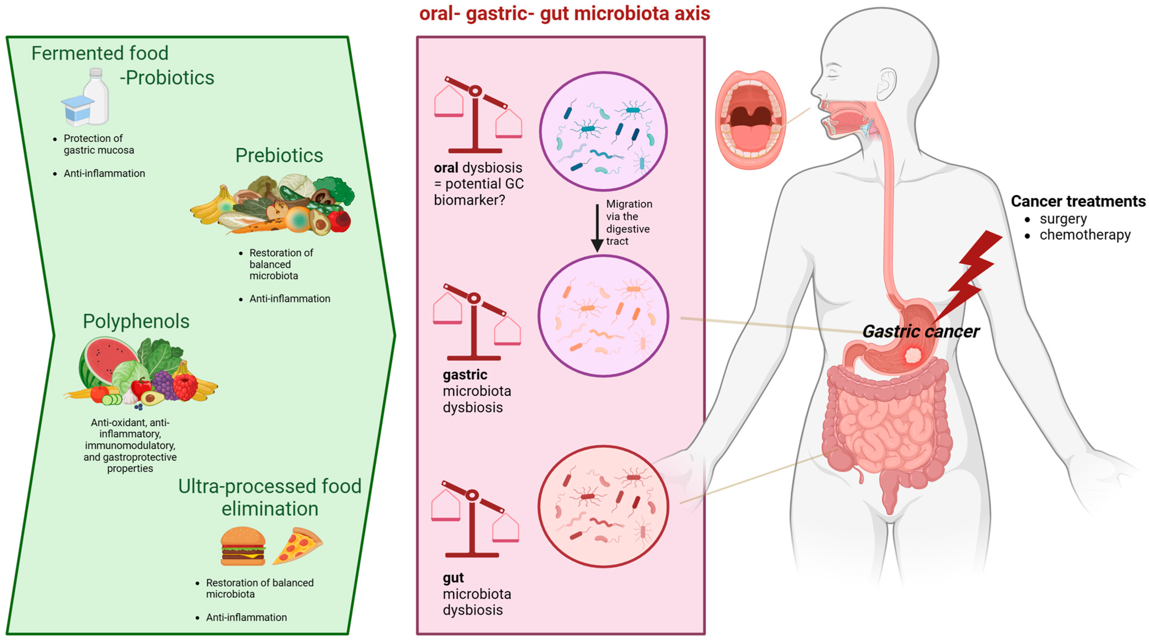
5. Diet: A Potential Therapy to Restore Microbiota Dysbiosis Associated with GC?

Nutritional therapies should be studied thoroughly to delineate their effectiveness in the rebalancing of human microbiota and reducing the progression of carcinogenesis.

5.1. Role of SCFAs

In cancer patients, the oral microbiota can transit into the intestinal lumen, where it may be related to increased pH and decreased acid secretion, which, in turn, reduce the efficiency of the gastric barrier, facilitating the orofecal transit of bacteria. A reduction in short-chain fatty acid (SCFAs) producer microbes as a consequence of oral dysbiosis in the gut microbiota may be a contributing factor in GC development and progression. In fact, under normal conditions, SCFAs exert a protective effect on the cell cycle, apoptosis, and immune response by modulating cellular pathways (Akt/mTOR and MEK/ERK signaling pathways) and transcription factors (downregulation of NF-κB), as well as epigenetic regulation (inhibition of histone deacetylase inhibitor (HDAC)–histone deacetylases activity, DNA methylation, histone phosphorylation, and methylation) [53]. In particular, in GC cells, butyrate produces beneficial alterations in the proliferation of apoptosis-related genes, decreasing the expression of focal adhesion kinase and increasing the expression of DAPK1/2, which induces apoptosis. Furthermore, it can stimulate the p53-p21 pathways, leading to cell cycle arrest and apoptosis of cancerous cells, suggesting a potential adjuvant role of these SCFAs in chemotherapy for GC [54]. In addition, a study conducted by Yuan-Linag et al. revealed a depletion of pathways (acetyl-CoA fermentation to butanoate II, L-glutamate degradation V, and 4-aminobutanoate degradation V) associated with SCFA production in GC patients compared with healthy controls, indicating the existence of a more inflammatory microenvironment and dysbiotic microbial communities in GC mucosal samples [47]. Another study demonstrated that the SCFA concentration can be measured in peripheric plasma samples; plasma concentrations of most SCFAs were found to be lower in patients with GC than in gastritis patients. Moreover, other intermediates of tricarboxylic acid were demonstrated to differ in the cancer population relative to non-cancer controls, suggesting them as potential GC diagnostic markers in peripheral blood [55]. In a recent case–control study conducted by Nouri et al., a reduced concentration of SCFAs was observed in GC patients compared with healthy controls, which was correlated with oral microbiota dysbiosis, suggesting that alterations in the composition of the oral microbiota can contribute to a reduction in SCFAs. In particular, the authors observed a negative correlation between the carcinogenic Streptococcus, Abiotrophia, and Leuconostoc strains and the concentration of total SCFAs, which, in turn, may contribute to the promotion of the proinflammatory TNFAIP8 and IL-6/STAT3 pathways, favoring cancer onset and progression. Therefore, the authors suggested that SCFA administration may represent an early intervention and targeted treatment against GC, although more evidence is needed before testing this hypothesis in clinical practice [56]. In another study, it was observed that butyric acid produced by Porphyromonas gingivalis exerted an inhibiting effect on H. pylori growth and proliferation in the oral cavity, suggesting that the bactericidal properties of this SCFA make it suitable for use as a preventive therapy against H. pylori translocation in the stomach and, therefore, against GC genesis. Conversely, butyric acid has been shown to exhibit an antimicrobial effect on Campylobacter species, Escherichia coli, and Staphylococcus aureus, which have also been associated with GC onset and progression [57].
Furthermore, SCFAs represent potential adjuvant chemotherapeutic agents. Indeed, a recent study of a combination treatment with butyrate and cisplatin showed an increased apoptosis rate in GC cell lines and an in vivo xenograft tumor model, as well as reduced cell migration and invasion [58].
SCFA composition is also influenced by surgical resection of the stomach. A recent study analyzing the 16SrRNA gene sequence in stool samples of 20 GC patients after subtotal gastrectomy demonstrated a general reduction in beneficial SCFA production after surgery. The latter may further increase the permeability of the intestinal mucosal barrier and negatively affect postoperative recovery. This is probably due to an overall increase in Bacteroidota and a decrease in Pseudomonadota and Actinomycetota and, at the genus level, higher abundances of Streptococcus, Escherichia/Shigella, Akkermansia, Verrucomicrobia, Dialister, Prevotella, and Veillonella in the gut [50]. A recent study conducted by Castano-Rodriguez et al. observed an increased richness and phylogenetic diversity but not Shannon’s diversity in GC patients when compared to dyspeptic controls. Among the bacterial taxa, Lactococcus, Veillonella, Fusobacterium, and Leptotrichia were reported to be increased only in GC samples. The authors also highlighted that the bacterial carbohydrate metabolism was enriched in GC patients and; as carbohydrate digestion and absorption are partly responsible for SCFA production, this suggested that alteration in energy metabolism may be directly involved in carcinogenesis [33]. Hu et al. reported a complex depletion of pathways (acetyl-CoA fermentation to butanoate II, L-glutamate degradation V, and 4-aminobutanoate degradation V) associated with SCFA production in GC, indicating the existence of a more inflammatory microenvironment and dysbiotic microbial communities in GC [47].

5.2. Prebiotics

Prebiotics—including inulin, fructo-oligosaccharide (FOS), and galacto-oligosaccharides (GOS)—are fibers promoting the growth of specific groups of anaerobic colonic indigenous bacteria; they are undigestible by endogenous enzymes in the small intestine but are actively fermented by colonic bacteria, selectively promoting the growth of beneficial bacteria [59]. To date, although scientific interest in the role of prebiotics in the course of cancer is growing, original studies remain lacking. One recent interesting study showed that some carrot compounds, such as acetylenic oxylipins like falcarinol and falcarinol, can inhibit cell growth in different cancers, such as breast cancer, colon cancer, and lymphoid leukemia. Nevertheless, the protective effect against cancer seems to be due to intrinsic anti-inflammatory and proapoptotic properties rather than a modulatory effect on microbiota [60,61,62]. The effect of inulin consumption, a natural dietary soluble fiber consisting of a mixture of oligo- and/or polysaccharides should be studied, given its prebiotic potential, technological properties, and beneficial effects on the gut microbiota [63].
After gastrectomy for GC, a short course of enteral nutrition (EN) is recommended as support to preserve intestinal structure and function and enhance intestinally mediated immunity. However, postoperative complications such as diarrhea can negatively affect the overall recovery of postoperative patients with GC [64]. Diarrhea is a common complication of EN, which affects recovery and prolongs the length of hospital stay. Considering that intestinal flora imbalance is associated with a reduction in protective Bifidobacteria and an increase in the counts of pathogens, administration of fiber prebiotics may represent a potential treatment for EN-induced diarrhea. A recent study of 120 GC patients divided into three groups (fiber-free nutrition formula (FF group, n = 40), fiber-enriched nutrition formula (FE group, n = 40), and fiber- and probiotic-enriched nutrition formula (FEP group, n = 40)) demonstrated that the combination of fiber and probiotics was significantly effective in treating EN-associated diarrhea in such patients. Fiber can be metabolized by intestinal flora and produce SCFAs, which are the preferred source of energy for colonic cells to improve gut barrier integrity and function. Moreover, some fermentation of fiber in the colon may favor the selective growth of beneficial bacteria such as Bifidobacteria and Lactobacillaceae family [65]. Furthermore, in animal models, perioperative immunopurified with supplementation of SCFAs has been proposed to reinforce the strength of gastrointestinal anastomosis, thereby reducing the risk of surgical-site infection [66].

5.3. Polyphenols

The positive effect of polyphenols may be related to the induction of apoptosis and cell cycle arrest, as well as to the inhibition of proinflammatory mediators [67]. Polyphenols act as antioxidant, anti-inflammatory, immunomodulatory, and gastroprotective agents by interacting with oral, gastric, and intestinal bacteria in a beneficial way [68].
A recent meta-analysis conducted by Ma et al. demonstrated that supplementation with polyphenol can significantly improve the production of health-promoting bacterial species like Bifidobacterium and Lactobacillaceae family and suppress the production of harmful or undesirable bacterial species like Clostridium and Escherichia coli [69]. Among polyphenols, the most important studied compounds are green tea, resveratrol, curcumin, and quercetin [70,71,72,73,74]. In particular, promising preclinical studies have been conducted to demonstrate the antiproliferative, antimetastatic, and chemotherapeutic roles of resveratrol in inhibiting GC growth, either directly or indirectly by increasing the amount of beneficial Ruminococcus, Akkermansia, Dehalobacterium, and Anerostipes in the gut. However, more evidence is needed before these compounds can be recommended as effective anti-GC adjuvant therapy in clinical practice [75,76,77,78,79].
Conversely, tryptophan, a dietary compound processed by Bacillota (Clostridium sporogenes, Ruminococcus gnavus, and Lactobacillaceae family) has been shown to play an essential role in the suppression of anticancer immune responses, in enhancing ROS production and DNA damage, and in increasing the malignant properties of cancer tissues, leading to tumor spread [80]. In conclusion, even though preclinical studies and one American clinical trial evaluating the effect of eicosapentaenoic acid’s role in liver metastases in colorectal cancer have shown promising results, demonstrating the effective anticancer role of polyphenols, at present, these compounds are not used as antitumoral therapies in clinical practice to modulate microbiota [81].

5.4. Fermented Foods and Probiotics

Probiotics are the living organisms in our gut that contribute to healthy conditions. They are found in fermented foods such as kefir, sauerkraut, yogurt, and kimchi. Natural yogurt, sweetened yogurt, and mature cheese are the most consumed among fermented dairy foods. L. casei is used in milk fermentation processes to produce yogurt and cheeses, and it has been associated with important health-promoting benefits, such as regulation of the intestinal microbiota, tumor inhibition, proapoptotic and antiproliferative effects, and the production of bioactive peptides in fermented milk. Among fermented foods, yogurt can be beneficial with respect to gastrointestinal diseases by acting positively on the gut microbiota, balancing inflammation and dysbiosis; however, a recent meta-analysis of 16 international studies found no association between yogurt intake and GC risk, suggesting that its potential beneficial probiotic role in GC development can be achieved only with much higher levels of intake [82,83]. Importantly, the role of lactic acid bacteria in GC seems to be bimodal. In vitro and in vivo evidence has focused on the role of lactic acid bacteria in the production of ROS, causing DNA damage and harmful N-nitroso compounds, which are implicated in mutagenesis, angiogenesis, protooncogene expression, and inhibition of apoptosis. Lactate, a lactic acid bacteria metabolite, is also a fuel for cancer cell survival [84].
After gastrectomy, gastric acid in the gastrointestinal tract is almost suppressed, causing modifications in the environment and the composition of the gut microbiota. Increased migration of pathogens can damage the intestinal mucosal immune barrier and provoke intestinal and systemic inflammation, which, in turn, delays the patient’s postoperative recovery. Therefore, it has been hypothesized that probiotic administration may positively affect the composition of the gut microbiota, thereby rebalancing gastrectomy-induced dysbiosis and favoring the recovery of the patient [85]. Dietary enrichment with probiotics (Bifidobacterium spp., Lactobacillaceae family, and Streptococcus spp.) in a murine model resulted in a decrease in the polyamine concentration, in association with anticancer effects, by fostering gastric cancer cell apoptosis [86]. In a recent randomized double-blind trial, oral supplementation with probiotic C. butyricum, an anaerobic Gram-positive bacillus, after gastrectomy for GC resulted in a reduced inflammatory response in terms of leucocyte and neutrophil count and the expression of proinflammatory cytokines such as IL-1b, IL-6, and TNF-a, as well as in amelioration of immune and nutritional status in terms of immunoglobulin, lymphocyte, albumin, and total protein levels. These results are probably due to the increased production of SCFAs, such as acetic acid, propionic acid, butyric acid, and isobutyric acid, facilitating postoperative recovery and decreasing the risk of complications. After C. butyricum administration, an overabundance of beneficial intestinal bacteria, such as Bacteroides, Faecalibacterium, and Gemmiger, with a concomitant reduction in pathogenic Streptococcus, Desulfovibrio, and Actinomyces, was also reported [87]. Another study analyzing the effect of probiotics (B. infantis, L. acidophilus, Enterococcus faecalis, and Bacillus cereus) after gastrectomy demonstrated a decrease in inflammatory response and an improvement in the immune index, with an overall promotion of postoperative recovery [88]. Moreover, a recent preclinical study suggested that probiotic administration (e.g., of butyrate-producing bacteria and Bifidobacterium) after endoscopic GC resection can reduce the risk of tumor recurrence [89]. Another important consideration is that different methods of digestive tract reconstruction produce variable effects on the microbial composition. 16S rRNA analysis revealed that fecal microbiota transplantation in gastrectomized mice with the fecal microbiota of Roux-en-Y reconstruction patients can reverse dysbacteriosis triggered by radical gastrectomy and elevate the relative abundance of some SCFA-producing bacteria, thereby ameliorating postoperative nutritional status, colitis, and overall recovery of GC subjects. A 16SrRNA analysis of fecal bacteria of RY patients revealed an overabundance of Bacteroides, Clostridium, and Ruminococcus, which are noted SCFA producers [90].

5.5. Dietary Patterns

Imbalanced dietary habits with increased consumption of smoked, salty foods and reduced intake of fiber, vegetables, and fruits have been associated with an increased risk of gastric carcinogenesis [91]. As food additives in processed meats, the consumption of nitrates, nitrites, and N-nitrosodimethylamine was reported to result in in an increased risk of GC [92]. N-nitroso compounds can be derived by the diet (essentially processed meat and smoked fish) or by endogenous synthesis from pathogenic bacterial strains such as Veillonella, Staphylococcus, Neisseria, Clostridium, Haemophilus, Lactobacillaceae family, and Nitrospirae; such compounds have been demonstrated to be overabundant in GC patients, favoring the initiation and progression of carcinogenesis [93]. Recent evidence suggests a potential carcinogenic role of lipids, cholesterol, and bile acids in different types of neoplasms, as they contribute to inflammation-mediated tumor growth through oncogenic activation and negatively interact with the host microbiota, favoring procarcinogenic dysbiosis. Nevertheless, specific studies on their role in GC are still lacking [94].
A recent scoping review evidenced that a more versatile high-fat diet was associated with a higher rate of intestinal microbial dysbiosis, as well as with increased GC risk, as it led to an overabundance of Lactobacillaceae family reaching the stomach. Conversely, a high intake of fruit, vegetables, dairy products, and seafood presented a protective microbial signature and an inverse correlation with GC [95].
Even though considerable evidence have confirmed that Western dietary habits are correlated with worse prognoses in different types of cancer, no study has focused on the specific effects of diet on the oral microbiota in gastric cancer development and progression [96].
Nutritional strategies such as fasting and caloric restriction have been evaluated as adjuvant treatments in different cancers, as it was observed that they can protectively modulate systemic metabolism. In an ongoing clinical trial (NCT01642953), researchers are restricting patient diets after gastric cancer surgery to assess whether fasting promotes recovery and reduces mortality and adverse events. However, the role of such dietary habits with respect to gastric and intestinal microbiota composition still needs to be elucidated [97,98].

6. Conclusions

This review highlights that the oral microbiota is a pivotal influencer in terms of tumor development and response to gastric anticancer treatments. Although a large body of preclinical evidence has been released, the exact mechanism whereby the altered oral microbiota contributes to carcinogenesis remains unknown. The oral microbiota, as well as gastric and/or gut bacteria, can be used a potential marker for the diagnosis and prognosis of GC. Moreover, the adjustment of microbiota composition using efficient dietary strategies may improve clinical GC patient outcomes. Further studies on the microbiota in GC patients could allow us to understand the mechanisms of GC pathogenesis and propose promising oral–gastric gut microbiota interventions to optimize GC treatments, including the use of prebiotics, probiotics, and nutritional interventions. In this context, Figure 1 illustrates nutritional interventions such as prebiotics, fermented food, probiotics, or other dietary strategies that may regulate the composition and balance of the oral–gastric–gut microbiota environment. For these reasons, more attention should be paid to the effect of nutritional interventions on the microbiota of GC patients during the course of treatment.

Author Contributions

Conceptualization, E.R.; methodology, V.M.; software, M.C.; validation, E.R., M.C.M. and A.G.; investigation, V.M. and E.S.; writing—original draft preparation, V.M. and P.R.; writing—review and editing, P.R. and E.R.; visualization, G.I.; supervision, E.R. All authors have read and agreed to the published version of the manuscript.

Funding

This research received no external funding.

Institutional Review Board Statement

Not applicable.

Informed Consent Statement

Not applicable.

Data Availability Statement

Not applicable.

Conflicts of Interest

The authors declare no conflicts of interest.

References

  1. Bray, F.; Ferlay, J.; Soerjomataram, I.; Siegel, R.L.; Torre, L.A.; Jemal, A. Global cancer statistics 2018, GLOBOCAN estimates of incidence and mortality worldwide for 36 cancers in 185 countries. CA Cancer J. Clin. 2020, 70, 313. [Google Scholar] [CrossRef] [PubMed]
  2. Petryszyn, P.; Chapelle, N.; Matysiak-Budnik, T. GC: Where are we heading? Dig. Dis. 2020, 38, 280–285. [Google Scholar] [CrossRef] [PubMed]
  3. Allemani, C.; Matsuda, T.; Di Carlo, V.; Harewood, R.; Matz, M.; Nikšić, M.; Hood, M. Global surveillance of trends in cancer survival 2000–14 (CONCORD-3): Analysis of individual records for 37,513,025 patients diagnosed with one of 18 cancers from 322 population-based registries in 71 countries. Lancet 2018, 391, 1023–1075. [Google Scholar] [CrossRef]
  4. Binato, R.; Santos, E.C.; Boroni, M.; Demachki, S.; Assumpção, P.; Abdelhay, E. A common molecular signature of intestinal-type gastric carcinoma indicates processes related to gastric carcinogenesis. Oncotarget 2018, 9, 7359–7371. [Google Scholar] [CrossRef]
  5. Yu, G.; Torres, J.; Hu, N.; Medrano-Guzman, R.; Herrera-Goepfert, R.; Humphrys, M.S.; Wang, L.; Wang, C.; Ding, T.; Ravel, J.; et al. Molecular Characterization of the Human Stomach Microbiota in GC Patients. Front. Cell. Infect. Microbiol. 2017, 7, 302. [Google Scholar] [CrossRef] [PubMed]
  6. de Martel, C.; Georges, D.; Bray, F.; Ferlay, J.; Clifford, G.M. Global burden of cancer attributable to infections in 2018, a worldwide incidence analysis. Lancet Glob. Health 2020, 8, e180–e190. [Google Scholar] [CrossRef] [PubMed]
  7. Caselli, E.; Fabbri, C.; D’accolti, M.; Soffritti, I.; Bassi, C.; Mazzacane, S.; Franchi, M. Defining the oral microbiome by whole-genome sequencing and resistome analysis: The complexity of the healthy picture. BMC Microbiol. 2020, 20, 120. [Google Scholar] [CrossRef] [PubMed]
  8. Park, S.-Y.; Hwang, B.-O.; Lim, M.; Ok, S.-H.; Lee, S.-K.; Chun, K.-S.; Park, K.-K.; Hu, Y.; Chung, W.-Y.; Song, N.-Y. Oral-gut microbiome axis in gastrointestinal disease and cancer. Cancers 2021, 13, 2124. [Google Scholar] [CrossRef]
  9. Chen, C.; Chen, L.; Lin, L.; Jin, D.; Du, Y.; Lyu, J. Research progress on gut microbiota in patients with gastric cancer, esophageal cancer, and small intestine cancer. Appl. Microbiol. Biotechnol. 2021, 105, 4415–4425. [Google Scholar] [CrossRef]
  10. Muscaritoli, M.; Arends, J.; Bachmann, P.; Baracos, V.; Barthelemy, N.; Bertz, H.; Bozzetti, F.; Hütterer, E.; Isenring, E.; Kaasa, S.; et al. ESPEN practical guideline: Clinical Nutrition in cancer. Clin. Nutr. 2021, 40, 2898–2913. [Google Scholar] [CrossRef]
  11. Llorca, L.; Pérez-Pérez, G.; Urruzuno, P.; Martinez, M.J.; Iizumi, T.; Gao, Z.; Sohn, J.; Chung, J.; Cox, L.; Simón-Soro, A.; et al. Characterization of the Gastric Microbiota in a Pediatric Population According to Helicobacter pylori Status. Pediatr. Infect. Dis. J. 2017, 36, 173–178. [Google Scholar] [CrossRef]
  12. Toh, J.W.T.; Wilson, R.B. Pathways of gastric carcinogenesis, Helicobacter pylori virulence and interactions with antioxidant systems, vitamin C and phytochemicals. Int. J. Mol. Sci. 2020, 21, 6451. [Google Scholar] [CrossRef]
  13. Li, Q.; Liu, J.; Gong, Y.; Yuan, Y. Serum VacA antibody is associated with risks of peptic ulcer and gastric cancer: A meta-analysis. Microb. Pathog. 2016, 99, 220–228. [Google Scholar] [CrossRef] [PubMed]
  14. Liatsos, C.; Papaefthymiou, A.; Kyriakos, N.; Galanopoulos, M.; Doulberis, M.; Giakoumis, M.; Petridou, E.; Mavrogiannis, C.; Rokkas, T.; Kountouras, J. Helicobacter pylori, gastric microbiota and GC relationship: Unrolling the tangle. World J. Gastrointest. Oncol. 2022, 14, 959. [Google Scholar] [CrossRef]
  15. Pereira-Marques, J.; Ferreira, R.M.; Pinto-Ribeiro, I.; Figueiredo, C. Helicobacter pylori Infection, the Gastric Microbiome and GC. Adv. Exp. Med. Biol. 2019, 1149, 195–210. [Google Scholar]
  16. Guo, Y.; Zhang, Y.; Gerhard, M.; Gao, J.J.; Mejias-Luque, R.; Zhang, L.; Vieth, M.; Ma, J.-L.; Bajbouj, M.; Suchanek, S.; et al. Effect of Helicobacter pylori on gastrointestinal microbiota: A population-based study in Linqu, a high-risk area of GC. Gut 2020, 69, 1598–1607. [Google Scholar] [CrossRef] [PubMed]
  17. Vogtmann, E.; Goedert, J.J. Epidemiologic studies of the human microbiome and cancer. Br. J. Cancer 2016, 114, 237–242. [Google Scholar] [CrossRef] [PubMed]
  18. Aviles-Jimenez, F.; Vazquez-Jimenez, F.; Medrano-Guzman, R.; Mantilla, A.; Torres, J. Stomach microbiota composition varies between patients with non-atrophic gastritis and patients with intestinal type of gastric cancer. Sci. Rep. 2014, 4, 4202. [Google Scholar] [CrossRef] [PubMed]
  19. Coker, O.O.; Dai, Z.; Nie, Y.; Zhao, G.; Cao, L.; Nakatsu, G.; Wu, W.K.; Wong, S.H.; Chen, Z.; Sung, J.J.Y.; et al. Mucosal microbiome dysbiosis in gastric carcinogenesis. Gut 2018, 67, 1024–1032. [Google Scholar] [CrossRef] [PubMed]
  20. Ferreira, R.M.; Pereira-Marques, J.; Pinto-Ribeiro, I.; Costa, J.L.; Carneiro, F.; Machado, J.C.; Figueiredo, C. Gastric microbial community profiling reveals a dysbiotic cancer-associated microbiota. Gut 2018, 67, 226–236. [Google Scholar] [CrossRef]
  21. Gunathilake, M.N.; Lee, J.; Choi, I.J.; Kim, Y.I.; Ahn, Y.; Park, C.; Kim, J. Association between the relative abundance of gastric microbiota and the risk of GC: A case-control study. Sci. Rep. 2019, 9, 13589. [Google Scholar] [CrossRef]
  22. Gantuya, B.; El Serag, H.B.; Matsumoto, T.; Ajami, N.J.; Uchida, T.; Oyuntsetseg, K.; Bolor, D.; Yamaoka, Y. Gastric mucosal microbiota in a Mongolian population with GC and precursor conditions. Aliment. Pharmacol. Ther. 2020, 51, 770–780. [Google Scholar] [CrossRef] [PubMed]
  23. Rajilic-Stojanovic, M.; Figueiredo, C.; Smet, A.; Hansen, R.; Kupcinskas, J.; Rokkas, T.; Andersen, L.; Machado, J.C.; Ianiro, G.; Gasbarrini, A.; et al. Systematic review: Gastric microbiota in health and disease. Aliment. Pharmacol. Ther. 2020, 51, 582–602. [Google Scholar] [CrossRef] [PubMed]
  24. Brennan, C.A.; Garrett, W.S. Gut microbiota, inflammation, and colorectal cancer. Annu. Rev. Microbiol. 2016, 70, 395–411. [Google Scholar] [CrossRef] [PubMed]
  25. Kostic, A.D.; Chun, E.; Robertson, L.; Glickman, J.N.; Gallini, C.A.; Michaud, M.; Clancy, T.E.; Chung, D.C.; Lochhead, P.; Hold, G.L.; et al. Fusobacterium nucleatum potentiates intestinal tumorigenesis and modulates the tumor-immune microenvironment. Cell Host Microbe 2013, 14, 207–215. [Google Scholar] [CrossRef] [PubMed]
  26. Rubinstein, M.R.; Wang, X.; Liu, W.; Hao, Y.; Cai, G.; Han, Y.W. Fusobacterium nucleatum promotes colorectal carcinogenesis by modulating E-cadherin/β-catenin signaling via its FadA adhesin. Cell Host Microbe 2013, 14, 195–206. [Google Scholar] [CrossRef] [PubMed]
  27. Nie, S.; Wang, A.; Yuan, Y. Comparison of clinicopathological parameters, prognosis, micro-ecological environment and metabolic function of Gastric Cancer with or without Fusobacterium sp. Infect. J. Cancer 2021, 12, 1023–1032. [Google Scholar] [CrossRef] [PubMed]
  28. Hsieh, Y.Y.; Tung, S.Y.; Pan, H.Y.; Chang, T.S.; Wei, K.L.; Chen, W.M.; Deng, Y.-F.; Lu, C.-K.; Lai, Y.-H.; Wu, C.-S.; et al. Fusobacterium nucleatum colonization is associated with decreased survival of Helicobacter pylori-positive gastric cancer patients. World J. Gastroenterol. 2021, 27, 7311–7323. [Google Scholar] [CrossRef]
  29. Hsieh, Y.Y.; Tung, S.Y.; Pan, H.Y.; Yen, C.W.; Xu, H.W.; Lin, Y.J.; Deng, Y.-F.; Hsu, W.-T.; Wu, C.-S.; Li, C. Increased abundance of clostridium and fusobacterium in gastric microbiota of patients with GC in Taiwan. Sci. Rep. 2018, 8, 158. [Google Scholar] [CrossRef]
  30. San-Millán, I.; Brooks, G.A. Reexamining cancer metabolism: Lactate production for carcinogenesis could be the purpose and explanation of the Warburg Effect. Carcinogenesis 2017, 38, 119–133. [Google Scholar] [CrossRef]
  31. Voss, D.M.; Spina, R.; Carter, D.L.; Lim, K.S.; Jeffery, C.J.; Bar, E.E. Disruption of the monocarboxylate transporter-4-basigin interaction inhibits the hypoxic response, proliferation, and tumor progression. Sci. Rep. 2017, 7, 4292. [Google Scholar] [CrossRef]
  32. Brand, A.; Singer, K.; Koehl, G.E.; Kolitzus, M.; Schoenhammer, G.; Thiel, A.; Matos, C.; Bruss, C.; Klobuch, S.; Peter, K.; et al. LDHA-Associated Lactic Acid Production Blunts Tumor Immunosurveillance by T and NK Cells. Cell Metab. 2016, 24, 657–671. [Google Scholar] [CrossRef]
  33. Castaño-Rodríguez, N.; Goh, K.L.; Fock, K.M.; Mitchell, H.M.; Kaakoush, N.O. Dysbiosis of the microbiome in gastric carcinogenesis. Sci. Rep. 2017, 7, 15957. [Google Scholar] [CrossRef]
  34. Lertpiriyapong, K.; Whary, M.T.; Muthupalani, S.; Lofgren, J.L.; Gamazon, E.R.; Feng, Y.; Ge, Z.; Wang, T.C.; Fox, J.G. Gastric colonisation with a restricted commensal microbiota replicates the promotion of neoplastic lesions by diverse intestinal microbiota in the Helicobacter pylori INS-GAS mouse model of gastric carcinogenesis. Gut 2014, 63, 54–63. [Google Scholar] [CrossRef]
  35. Chen, Y.X.; He, L.L.; Xiang, X.P.; Shen, J.; Qi, H.Y. O6-methylguanine DNA methyltransferase is upregulated in malignant transformation of gastric epithelial cells via its gene promoter DNA hypomethylation. World J. Gastrointest. Oncol. 2022, 14, 664–677. [Google Scholar] [CrossRef]
  36. Thorell, K.; Bengtsson-Palme, J.; Liu, O.H.; Palacios Gonzales, R.V.; Nookaew, I.; Rabeneck, L.; Paszat, L.; Graham, D.Y.; Nielsen, J.; Lundin, S.B.; et al. In Vivo Analysis of the Viable Microbiota and Helicobacter pylori Transcriptome in Gastric Infection and Early Stages of Carcinogenesis. Infect. Immun. 2017, 85, e00031-17. [Google Scholar] [CrossRef] [PubMed]
  37. Wang, L.; Zhou, J.; Xin, Y.; Geng, C.; Tian, Z.; Yu, X.; Dong, Q. Bacterial overgrowth and diversification of microbiota in GC. Eur. J. Gastroenterol. Hepatol. 2016, 28, 261–266. [Google Scholar] [CrossRef] [PubMed]
  38. Li, Y.; Huang, X.; Tong, D.; Jiang, C.; Zhu, X.; Wei, Z.; Jin, C. Relationships among microbiota, gastric cancer, and immunotherapy. Front. Microbiol. 2022, 13, 987763. [Google Scholar] [CrossRef] [PubMed]
  39. Dicksved, J.; Lindberg, M.; Rosenquist, M.; Enroth, H.; Jansson, J.K.; Engstrand, L. Molecular characterization of the stomach microbiota in patients with gastric cancer and in controls. J. Med. Microbiol. 2009, 58 Pt 4, 509–516. [Google Scholar] [CrossRef] [PubMed]
  40. Hu, J.; Han, S.; Chen, Y.; Ji, Z. Variations of Tongue Coating Microbiota in Patients with Gastric Cancer. Biomed. Res. Int. 2015, 2015, 173729. [Google Scholar] [CrossRef] [PubMed]
  41. Sun, J.H.; Li, X.L.; Yin, J.; Li, Y.H.; Hou, B.X.; Zhang, Z. A screening method for gastric cancer by oral microbiome detection. Oncol. Rep. 2018, 39, 2217–2224. [Google Scholar] [CrossRef] [PubMed]
  42. Huang, K.; Gao, X.; Wu, L.; Yan, B.; Wang, Z.; Zhang, X.; Peng, L.; Yu, J.; Sun, G.; Yang, Y. Salivary microbiota for GC prediction: An exploratory study. Front. Cell. Infect. Microbiol. 2021, 11, 640309. [Google Scholar] [CrossRef] [PubMed]
  43. Wu, J.; Xu, S.; Xiang, C.; Cao, Q.; Li, Q.; Huang, J.; Shi, L.; Zhang, J.; Zhan, Z. Tongue coating microbiota community and risk effect on GC. J. Cancer 2018, 9, 4039–4048. [Google Scholar] [CrossRef] [PubMed]
  44. Yang, Y.; Long, J.; Wang, C.; Blot, W.; Pei, Z.; Shu, X.; Wu, F.; Rothman, N.; Wu, J.; Lan, Q.; et al. Prospective study of oral microbiome and GC risk among Asian, African American and European American populations. Int. J. Cancer 2022, 150, 916–927. [Google Scholar] [CrossRef]
  45. Nomura, R.; Kadota, T.; Ogaya, Y.; Matayoshi, S.; Iwashita, N.; Okawa, R.; Nakano, K. Contribution of streptococcus mutans to Helicobacter pylori colonisation in oral cavity and gastric tissue. Sci. Rep. 2020, 10, 12540. [Google Scholar] [CrossRef]
  46. Chen, C.C.; Liou, J.M.; Lee, Y.C.; Hong, T.C.; El-Omar, E.M.; Wu, M.S. The interplay between helicobacter pylori and gastrointestinal microbiota. Gut Microbes 2021, 13, 1909459. [Google Scholar] [CrossRef]
  47. Hu, Y.L.; Pang, W.; Huang, Y.; Zhang, Y.; Zhang, C.J. The Gastric Microbiome Is Perturbed in Advanced Gastric Adenocarcinoma Identified Through Shotgun Metagenomics. Front. Cell. Infect. Microbiol. 2018, 8, 433. [Google Scholar] [CrossRef]
  48. Barra, W.F.; Sarquis, D.P.; Khayat, A.S.; Khayat, B.C.M.; Demachki, S.; Anaissi, A.K.M.; Ishak, G.; Santos, N.P.C.; dos Santos, S.E.B.; Burbano, R.R.; et al. Gastric Cancer Microbiome. Pathobiology 2021, 88, 156–169. [Google Scholar] [CrossRef]
  49. Wang, D.; Li, Y.; Zhong, H.; Ding, Q.; Lin, Y.; Tang, S.; Zong, Y.; Wang, Q.; Zhang, X.; Yang, H.; et al. Alterations in the human gut microbiome associated with Helicobacter pylori infection. FEBS Open Bio 2019, 9, 1552–1560. [Google Scholar] [CrossRef]
  50. Liang, W.; Yang, Y.; Wang, H.; Wang, H.; Yu, X.; Lu, Y.; Shen, S.; Teng, L. Gut microbiota shifts in patients with GC in perioperative period. Medicine 2019, 98, e16626. [Google Scholar] [CrossRef]
  51. Sarhadi, V.; Mathew, B.; Kokkola, A.; Karla, T.; Tikkanen, M.; Rautelin, H.; Lahti, L.; Puolakkainen, P.; Knuutila, S. Gut microbiota of patients with different subtypes of GC and gastrointestinal stromal tumors. Gut Pathog. 2021, 13, 11. [Google Scholar] [CrossRef] [PubMed]
  52. Zhou, C.B.; Pan, S.Y.; Jin, P.; Deng, J.W.; Xue, J.H.; Ma, X.Y.; Xie, Y.-H.; Cao, H.; Liu, Q.; Xie, W.-F.; et al. Fecal Signatures of Streptococcus anginosus and Streptococcus constellatus for Noninvasive Screening and Early Warning of GC. Gastroenterology 2022, 162, 1933–1947.E18. [Google Scholar] [CrossRef] [PubMed]
  53. Feng, F.-B.; Qiu, H.-Y. Effects of Artesunate on chondrocyte proliferation, apoptosis and autophagy through the PI3K/AKT/mTOR signaling pathway in rat models with rheumatoid arthritis. Biomed. Pharmacother. 2018, 102, 1209–1220. [Google Scholar] [CrossRef] [PubMed]
  54. Chen, G.; Ran, X.; Li, B.; Li, Y.; He, D.; Huang, B.; Fu, S.; Liu, J.; Wang, W. Sodium butyrate inhibits inflammation and maintains epithelium barrier integrity in a TNBS-induced inflammatory bowel disease mice model. eBioMedicine 2018, 30, 317–325. [Google Scholar] [CrossRef] [PubMed]
  55. Kim, Y.L.; Lee, W.; Chung, S.H.; Yu, B.M.; Lee, Y.C.; Hong, J. Metabolic alterations of short-chain fatty acids and TCA cycle intermediates in human plasma from patients with gastric cancer. Life Sci. 2022, 309, 121010. [Google Scholar] [CrossRef] [PubMed]
  56. Nouri, Z.; Choi, S.W.; Choi, I.J.; Ryu, K.W.; Woo, S.M.; Park, S.J.; Lee, W.J.; Choi, W.; Jung, Y.-S.; Myung, S.-K.; et al. Exploring Connections between Oral Microbiota, Short-Chain Fatty Acids, and Specific Cancer Types: A Study of Oral Cancer, Head and Neck Cancer, Pancreatic Cancer, and Gastric Cancer. Cancers 2023, 15, 2898. [Google Scholar] [CrossRef] [PubMed]
  57. Yonezawa, H.; Osaki, T.; Hanawa, T.; Kurata, S.; Zaman, C.; Woo, T.D.H.; Takahashi, M.; Matsubara, S.; Kawakami, H.; Ochiai, K.; et al. Destructive effects of butyrate on the cell envelope of Helicobacter pylori. J. Med. Microbiol. 2012, 61, 582–589. [Google Scholar] [CrossRef] [PubMed]
  58. Li, Y.; He, P.; Liu, Y.; Qi, M.; Dong, W. Combining sodium butyrate with cisplatin increases the apoptosis of gastric cancer in vivo and in vitro via the mitochondrial apoptosis pathway. Front. Pharmacol. 2021, 2, 708093. [Google Scholar] [CrossRef]
  59. Sanders, M.E.; Merenstein, D.J.; Reid, G.; Gibson, G.R.; Rastall, R.A. Probiotics and prebiotics in intestinal health and disease: From biology to the clinic. Nat. Rev. Gastroenterol. Hepatol. 2019, 16, 605–616. [Google Scholar] [CrossRef]
  60. Hansen, S.L.; Purup, S.; Christensen, L.P. Bioactivity of falcarinol and the influence of processing and storage on its content in carrots (Daucus carota L.). J. Sci. Food Agric. 2003, 83, 1010–1017. [Google Scholar] [CrossRef]
  61. Young, J.F.; Duthie, S.J.; Milne, L.; Christensen, L.P.; Duthie, G.G.; Bestwick, C.S. Biphasic effect of falcarinol on CaCo-2 cell proliferation, DNA damage, and apoptosis. J. Agric. Food Chem. 2007, 55, 618–623. [Google Scholar] [CrossRef]
  62. Zaini, R.G.; Brandt, K.; Clench, M.R.; Le Maitre, C.L. Effects of bioactive compounds from carrots (Daucus carota L.), polyacetylenes, beta-carotene and lutein on human lymphoid leukaemia cells. Anticancer Agents Med. Chem. 2012, 12, 640–652. [Google Scholar] [CrossRef]
  63. Tarifa, M.C.; Piqueras, C.M.; Genovese, D.B.; Brugnoni, L.I. Microencapsulation of Lactobacillus casei and Lactobacillus rhamnosus in pectin and pectin-inulin microgel particles: Effect on bacterial survival under storage conditions. Int. J. Biol. Macromol. 2021, 179, 457–465. [Google Scholar] [CrossRef]
  64. Weimann, A.; Braga, M.; Harsanyi, L.; Laviano, A.; Ljungqvist, O.; Soeters, P.; Jauch, K.; Kemen, M.; Hiesmayr, J.; Horbach, T.; et al. ESPEN guidelines on enteral nutrition: Surgery including organ transplantation. Clin. Nutr. 2006, 25, 224–244. [Google Scholar] [CrossRef]
  65. Zhao, R.; Wang, Y.; Huang, Y.; Cui, Y.; Xia, L.; Rao, Z.; Zhou, Y.; Wu, X. Effects of fiber and probiotics on diarrhea associated with enteral nutrition in gastric cancer patients: A prospective randomized and controlled trial. Medicine 2017, 96, e8418. [Google Scholar] [CrossRef]
  66. Bloemen, J.G.; Schreinemacher, M.H.; de Bruine, A.P.; Buurman, W.A.; Bouvy, N.D.; Dejong, C.H. Butyrate enemas improve intestinal anastomotic strength in a rat model. Dis. Colon. Rectum. 2010, 53, 1069–1075. [Google Scholar] [CrossRef]
  67. Larrosa, M.; Luceri, C.; Vivoli, E.; Pagliuca, C.; Lodovici, M.; Moneti, G.; Dolara, P. Polyphenol metabolites from colonic microbiota exert anti-inflammatory activity on different inflammation models. Mol. Nutr. Food Res. 2009, 53, 1044–1054. [Google Scholar] [CrossRef] [PubMed]
  68. Cueva, C.; Gil-Sánchez, I.; Ayuda-Durán, B.; González-Manzano, S.; González-Paramás, A.M.; Santos-Buelga, C.; Bartolomé, B.; Moreno-Arribas, M.V. An Integrated View of the Effects of Wine Polyphenols and Their Relevant Metabolites on Gut and Host Health. Molecules 2017, 22, 99. [Google Scholar] [CrossRef] [PubMed]
  69. Ma, G.; Yanting, C. Polyphenol supplementation benefits human health via gut microbiota: A systematic review via meta-analysis. J. Funct. Foods 2020, 66, 103829. [Google Scholar] [CrossRef]
  70. Afzal, M.; Safer, A.M.; Menon, M. Green Tea Polyphenols and Their Potential Role in Health and Disease. Inflammopharmacology 2015, 23, 151–161. [Google Scholar] [CrossRef] [PubMed]
  71. Wang, L.Y.; Zhao, S.; Lv, G.J.; Ma, X.J.; Zhang, J.B. Mechanisms of Resveratrol in the Prevention and Treatment of Gastrointestinal Cancer. World J. Clin. Cases 2020, 8, 2425–2437. [Google Scholar] [CrossRef]
  72. Kunnumakkara, A.B.; Anand, P.; Aggarwal, B.B. Curcumin Inhibits Proliferation, Invasion, Angiogenesis and Metastasis of Different Cancers through Interaction with Multiple Cell Signaling Proteins. Cancer Lett. 2008, 269, 199–225. [Google Scholar] [CrossRef]
  73. Davoodvandi, A.; Shabani Varkani, M.; Clark, C.C.T.; Jafarnejad, S. Quercetin as an Anticancer Agent: Focus on Esophageal Cancer. J. Food Biochem. 2020, 44, e13374. [Google Scholar] [CrossRef]
  74. Chiu, H.F.; Venkatakrishnan, K.; Golovinskaia, O.; Wang, C.K. Gastroprotective Effects of Polyphenols against Various Gastro-Intestinal Disorders: A Mini-Review with Special Focus on Clinical Evidence. Molecules 2021, 26, 2090. [Google Scholar] [CrossRef]
  75. Kim, S.; Kim, W.; Kim, D.H.; Jang, J.H.; Kim, S.J.; Park, S.A.; Hahn, H.; Han, B.W.; Na, H.-K.; Chun, K.-S.; et al. Resveratrol suppresses gastric cancer cell proliferation and survival through inhibition of PIM-1 kinase activity. Arch. Biochem. Biophys. 2020, 689, 108413. [Google Scholar] [CrossRef]
  76. Yang, Z.; Xie, Q.; Chen, Z.; Ni, H.; Xia, L.; Zhao, Q.; Chen, Z.; Chen, P. Resveratrol suppresses the invasion and migration of human gastric cancer cells via inhibition of malat1-mediated epithelial-to-mesenchymal transition. Exp. Ther. Med. 2019, 17, 1569–1578. [Google Scholar] [CrossRef] [PubMed]
  77. Yin, L.; Zhang, R.; Hu, Y.; Li, W.; Wang, M.; Liang, Z.; Sun, Z.; Ji, R.; Xu, W.; Qian, H. Gastric-cancer-derived mesenchymal stem cells: A promising target for resveratrol in the suppression of gas- tric cancer metastasis. Hum. Cell 2020, 33, 652–662. [Google Scholar] [CrossRef] [PubMed]
  78. Xu, Q.H.; Xiao, Y.; Li, X.Q.; Fan, L.; Zhou, C.C.; Cheng, L.; Jiang, Z.-D.; Wang, G.-H. Resveratrol counteracts hypoxia-induced gastric cancer invasion and EMT through hedge- hog pathway suppression. Anti-Cancer Agents Med. Chem. 2020, 20, 1105–1114. [Google Scholar] [CrossRef]
  79. Ren, M.; Zhou, X.; Gu, M.; Jiao, W.; Yu, M.; Wang, Y.; Liu, S.; Yang, J.; Ji, F. Resveratrol synergizes with cis- platin in antineoplastic effects against AGS gastric cancer cells by inducing endoplasmic reticulum stress-mediated apoptosis and G2/M phase arrest. Oncol. Rep. 2020, 44, 1605–1615. [Google Scholar] [CrossRef] [PubMed]
  80. Pilotte, L.; Larrieu, P.; Stroobant, V.; Colau, D.; Dolusic, E.; Frédérick, R.; De Plaen, E.; Uyttenhove, C.; Wouters, J.; Masereel, B.; et al. Reversal of tumoral immune resistance by inhibition of tryptophan 2, 3-dioxygenase. Proc. Natl. Acad. Sci. USA 2012, 109, 2497–2502. [Google Scholar] [CrossRef] [PubMed]
  81. Hull, M.A. Biospecimen Collection for: Prebiotic Effect of Eicosapentaenoic Acid Treatment for Colorectal Cancer Liver Metastases. 2021; Report No.: NCT04682665. Available online: https://classic.clinicaltrials.gov/ct2/show/NCT04682665 (accessed on 1 December 2023).
  82. Stiemsma, L.T.; Nakamura, R.E.; Nguyen, J.G.; Michels, K.B. Does Consumption of Fermented Foods Modify the Human Gut Microbiota? J. Nutr. 2020, 150, 1680–1692. [Google Scholar] [CrossRef]
  83. Collatuzzo, G.; Negri, E.; Pelucchi, C.; Bonzi, R.; Turati, F.; Rabkin, C.S.; Liao, L.M.; Sinha, R.; Palli, D.; Ferraroni, M.; et al. Yoghurt Intake and Gastric Cancer: A Pooled Analysis of 16 Studies of the StoP Consortium. Nutrients 2023, 15, 1877. [Google Scholar] [CrossRef]
  84. Vinasco, K.; Mitchell, H.M.; Kaakoush, N.O.; Castaño-Rodríguez, N. Microbial carcinogenesis: Lactic acid bacteria in gastric cancer. Biochim. Biophys. Acta Rev. Cancer 2019, 1872, 188309. [Google Scholar] [CrossRef]
  85. Erawijantari, P.P.; Mizutani, S.; Shiroma, H.; Shiba, S.; Nakajima, T.; Sakamoto, T.; Saito, Y.; Fukuda, S.; Yachida, S.; Yamada, T. Influence of gastrectomy for gastric cancer treatment on faecal microbiome and metabolome profiles. Gut 2020, 69, 1404–1415. [Google Scholar] [CrossRef]
  86. Linsalata, M.; Russo, F.; Notarnicola, M.; Guerra, V.; Cavallini, A.; Clemente, C.; Messa, C. Effects of genistein on the polyamine metabolism and cell growth in DLD-1 human colon cancer cells. Nutr. Cancer 2005, 52, 84–93. [Google Scholar] [CrossRef]
  87. Cao, W.; Zheng, C.; Xu, X.; Jin, R.; Huang, F.; Shi, M.; He, Z.; Luo, Y.; Liu, L.; Liu, Z.; et al. Clostridium butyricum potentially improves inflammation and immunity through alteration of the microbiota and metabolism of gastric cancer patients after gastrectomy. Front. Immunol. 2022, 13, 1076245. [Google Scholar] [CrossRef]
  88. Zheng, C.; Chen, T.; Wang, Y.; Gao, Y.; Kong, Y.; Liu, Z.; Deng, X. A randomised trial of probiotics to reduce severity of physiological and microbial disorders induced by partial gastrectomy for patients with gastric cancer. J Cancer 2019, 10, 568–576. [Google Scholar] [CrossRef] [PubMed]
  89. Arai, J.; Niikura, R.; Hayakawa, Y.; Kawahara, T.; Honda, T.; Hasatani, K.; Yoshida, N.; Nishida, T.; Sumiyoshi, T.; Kiyotoki, S.; et al. Use of antibiotics and probiotics reduces the risk of metachronous gastric cancer after endoscopic resection. Biology 2021, 10, 455. [Google Scholar] [CrossRef] [PubMed]
  90. Yao, Y.; Sun, S.; Gu, J.; Ni, H.; Zhong, K.; Xu, Q.; Zhou, D.; Wang, X.; Gao, L.; Zhu, X. Roux-en-Y reconstruction alleviates radical gastrectomy-induced colitis via down-regulation of the butyrate/NLRP3 signaling pathway. eBioMedicine 2022, 86, 104347. [Google Scholar] [CrossRef] [PubMed]
  91. Karimi, P.; Islami, F.; Anandasabapathy, S.; Freedman, N.D.; Kamangar, F. Gastric cancer: Descriptive epidemiology, risk factors, screening, and prevention. Cancer Epidemiol. Biomark. Prev. 2014, 23, 700–713. [Google Scholar] [CrossRef] [PubMed]
  92. Song, P.; Wu, L.; Guan, W. Dietary Nitrates, Nitrites, and Nitrosamines Intake and the Risk of Gastric Cancer: A Meta-Analysis. Nutrients 2015, 7, 9872–9895. [Google Scholar] [CrossRef] [PubMed]
  93. Jo, H.J.; Kim, J.; Kim, N.; Park, J.H.; Nam, R.H.; Seok, Y.-J.; Kim, Y.; Kim, J.S.; Kim, J.M.; Lee, D.H.; et al. Analysis of gastric microbiota by pyrosequencing: Minor role of bacteria other than Helicobacter pylori in the gastric carcinogenesis. Helicobacter 2016, 21, 364–374. [Google Scholar] [CrossRef]
  94. Chattopadhyay, I.; Gundamaraju, R.; Jha, N.K.; Gupta, P.K.; Dey, A.; Mandal, C.C.; Ford, B.M. Interplay between dysbiosis of gut microbiome, lipid metabolism, and tumorigenesis: Can gut dysbiosis stand as a prognostic marker in cancer? Dis. Markers 2022, 2022, 2941248. [Google Scholar] [CrossRef]
  95. Thrastardottir, T.O.; Copeland, V.J.; Constantinou, C. The Association Between the Gut Microbiome, Nutritional Habits, Antibiotics, and Gastric Cancer: A Scoping Review. Curr. Nutr. Rep. 2022, 11, 19–38. [Google Scholar] [CrossRef]
  96. Rinninella, E.; Mele, M.C.; Cintoni, M.; Raoul, P.; Ianiro, G.; Salerno, L.; Pozzo, C.; Bria, E.; Muscaritoli, M.; Molfino, A.; et al. The facts about food after cancer diagnosis: A systematic review of prospective cohort studies. Nutrients 2020, 12, 2345. [Google Scholar] [CrossRef] [PubMed]
  97. Vernieri, C.; Fucà, G.; Ligorio, F.; Huber, V.; Vingiani, A.; Iannelli, F.; Raimondi, A.; Rinchai, D.; Frigè, G.; Belfiore, A.; et al. Fasting-mimicking diet is safe and reshapes metabolism and antitumor immunity in patients with cancer. Cancer Discov. 2022, 12, 90–107. [Google Scholar] [CrossRef] [PubMed]
  98. Hur, H. Prospective Clinical Study for Early Recovery after Gastric Cancer Surgery. 2012; Report No.: NCT01642953. Available online: https://classic.clinicaltrials.gov/ct2/show/NCT01642953 (accessed on 1 December 2023).
Figure 1. Potential nutritional interventions that can positively influence the oral–gastric–gut microbiota during gastric cancer treatment.
Ijms 25 01679 g001
Table 1. Microbiota compositional changes and potential effects on GC carcinogenesis mechanisms.
Microbiota Compositional ChangesMechanisms of CarcinogenesisReferences
Oral microbiotaVeillonella
Neisseria
Haemophilus
Porphyromonas
↑ Bacillota
↓ Bacteroides
Periodontal infection → gastric chronic inflammation and dysbiosis
↑ N-nitroso compounds
[40,41,42,43]
Streptococcus↑ Induction of alcohol to be oxidized to acetaldehyde, a group I human carcinogen[45]
Candida albicans, Fusobacterium nucleatum, Porphyromonas gingivalis, and Streptococcus mutans↑ Interaction H. pylori → ↑ H. pylori survival in the unsuitable environment of the mouth, fostering H. pylori migration to the stomach and exertion of its carcinogenic role[45]
Gastric microbiotaH. pylori
↑ Gastric dysbiosis
Oncoprotein cytotoxin-associated gene A
Destabilization of cellular junctions via disruption of the E-cadherin-β-catenin complex
Activation of proliferating pathways such as the ERK-MAP kinase cascade
Oncogenic transformation of gastric cells via vacuolation of cytotoxin A
Promotion of abnormal cell survival
Inflammatory effect on the gastric mucosal surface of the stomach with altered production of mucin
[12,13,14,15,16]
↑ Nitrate reductase bacteria↑ Nitrate reductase activity
↑ Proinflammatory activity
[36,37]
Streptococcus, Lactobacillaceae family, and Lactococcus↑ N-nitroso compounds
↑ Reactive oxygen species
Promotion of DNA damage
[30,31,32,33]
Lactococcus lactis
↓ Antiproliferative activity in a human stomach cancer cell line
↑ Cell apoptosis
[21]
Fusobacterium nucleatum
↓ Bacterial diversity
↑ NF-κB, IL-1β, IL-6, IL-8, TNF, and FadA[22,24,25,26,27,28]
Gut microbiota↑ Enterobacteriaceae
Bifidobacterium
↓ Bacteroides
Shigella
Clostridium perfringens
↓ Bacterial diversity
↓ Production of SCFAs and their
protective effects on the cell cycle, apoptosis, and immune stimulation, impacting the Akt/mTOR and MEK/ERK signaling pathways
↓ Inhibition of NF-κB
[50,51,52,53]
Abbreviations: ↑, increase; ↓, decrease; Akt/mTOR, protein kinase B/mammalian target of rapamycin; DNA, deoxyribonucleic acid; ERK-MAP, extracellular signal-regulated kinase–mitogen-activated protein kinase; H. pylori, Helicobacter pylori; NF-κB, nuclear factor kappa-light-chain-enhancer of activated B cells; IL, interleukin; SCFAs, short-chain fatty acids; TNF, tumor necrosis factor.
Disclaimer/Publisher’s Note: The statements, opinions and data contained in all publications are solely those of the individual author(s) and contributor(s) and not of MDPI and/or the editor(s). MDPI and/or the editor(s) disclaim responsibility for any injury to people or property resulting from any ideas, methods, instructions or products referred to in the content.

