Article

Network-Based Bioinformatics Highlights Broad Importance of Human Milk Hyaluronan

Department of Pediatrics, University of Oklahoma Health Sciences Center, Oklahoma City, OK 73104, USA
*
Author to whom correspondence should be addressed.
Int. J. Mol. Sci. 2024, 25(23), 12679; https://doi.org/10.3390/ijms252312679
Submission received: 27 October 2024 / Revised: 20 November 2024 / Accepted: 25 November 2024 / Published: 26 November 2024

Abstract

Human milk (HM) is rich in bioactive factors promoting postnatal small intestinal development and maturation of the microbiome. HM is also protective against necrotizing enterocolitis (NEC), a devastating inflammatory condition predominantly affecting preterm infants. The HM glycosaminoglycan, hyaluronan (HA), is present at high levels in colostrum and early milk. Our group has demonstrated that HA with a molecular weight of 35 kDa (HA35) promotes maturation of the murine neonatal intestine and protects against two distinct models of NEC. However, the molecular mechanisms underpinning HA35-induced changes in the developing ileum are unclear. CD-1 mouse pups were treated with HA35 or vehicle control daily, from P7 to P14, and we used network and functional analyses of bulk RNA-seq ileal transcriptomes to further characterize molecular mechanisms through which HA35 likely influences intestinal maturation. HA35-treated pups separated well by principal component analysis, and cell deconvolution revealed increases in stromal, Paneth, and mature enterocyte and progenitor cells in HA35-treated pups. Gene set enrichment and pathway analyses demonstrated upregulation in key processes related to antioxidant and growth pathways, such as nuclear factor erythroid 2-related factor-mediated oxidative stress response, hypoxia inducible factor-1 alpha, mechanistic target of rapamycin, and downregulation of apoptotic signaling. Collectively, pro-growth and differentiation signals induced by HA35 may present novel mechanisms by which this HM bioactive factor may protect against NEC.

1. Introduction

Nutrients within human milk (HM) spur growth and development during infancy. Bioactive components within HM, such as lactoferrin, immunoglobulin, and HM oligosaccharides [1], provide healthy building blocks for colonization and maintenance of the gut microbiome and maturation of intestinal immune cells. Infants on an exclusively HM diet are exposed to 50% fewer enteric infections compared with infants fed formula [2,3]. Importantly, protection offered by HM extends to necrotizing enterocolitis (NEC) [4,5,6,7,8,9], a life-threatening intestinal inflammatory disorder afflicting primarily premature infants. The etiology of NEC is complex and multifactorial, and risk factors beyond prematurity, including antibiotic use [10], dysbiosis [11], and formula feeding [12], contribute to increased incidence and severity of the disease. Studies have indicated preterm infants exposed to even a partial bovine-based formula diet are susceptible to increased NEC morbidity and mortality [13], while very low birthweight infants fed a diet of at least 50% HM within the 2 weeks following birth are six times less likely to develop NEC [14]. Much of the protection afforded by HM feeding has been attributed to bioactive factors, but how NEC risk is altered in association with isolated HM bioactive factors, rather than the composite, remains incompletely understood.
Unlike bovine-based infant formula [15], HM is rich in hyaluronan (HA), an ubiquitous and unique, non-sulfated glycosaminoglycan (GAG) with repeating linear residues of D-glucuronic acid and N-acetyl-D-glucosamine [16]. A primary component of the extracellular matrix (ECM), HA is produced endogenously by membrane-bound HA synthases (HASs) 1–3 [17]. The molecular weight, mode of administration, and local tissue physiology largely dictate the pro- or anti-inflammatory properties of HA [18,19]. In general, high molecular weight (HMW, >800 kDa) HA synthesized by HAS enhances tissue hydration, has antioxidant properties, and inhibits endothelial cell growth [20]. Oligo (<10 kDa), low MW (LMW, 10–300 kDa), and medium MW (MMW, 300–800 kDa) HA, produced either via enzymatic or oxidative degradation [20], induce tissue healing and repair through activation of the innate immune system, induce growth factor secretion, and stimulate angiogenesis [20,21,22,23,24,25,26,27,28].
Concentrations of HA peak in colostrum and early HM during the first weeks after birth, suggesting a critical role for exogenous HA during intestinal development postnatally [29]. As the proximal small intestine lacks the enzymes required to degrade HA [30], HM HA likely reaches the distal small intestine intact. In animal models of colitis, oral provision of HA, either purified from HM or produced biosynthetically at an average MW of 35 kDa (HA35), protects against bacterial-induced disease through increased antimicrobial β-defensin and tight junction protein expression within the epithelium [31,32,33,34]. Notably, our group has also demonstrated HA35 is protective against in vitro mechanical injury of intestinal epithelial monolayers [35], as well as in two distinct animal models of NEC [36,37].
Most recently, we demonstrated that HA35 promotes maturation of the murine neonatal small intestine, increasing villous length, crypt depth, and secretory cell abundance within the ileal epithelium [38]. Initial functional analyses of the ileal transcriptome indicated enrichment of pathways associated with growth, proliferation, and survival, including human epidermal growth factor receptor 2 (HER2), extracellular signal-regulated kinase/mitogen-activated protein kinase (ERK/MAPK), mechanistic target of rapamycin (mTOR), and phosphatidylinositol 3-kinase/protein kinase B (PI3K/Akt) signaling. Here, we aim to utilize additional functional and network analyses to further characterize the anti-inflammatory and pro-growth ileal transcriptome induced by HA35 supplementation during the neonatal period. As NEC remains a therapeutically intractable disease, our goal of identifying mechanisms mediating HA35’s protective and maturational effects on the ileum could provide novel therapeutic targets for the disease.

2. Results

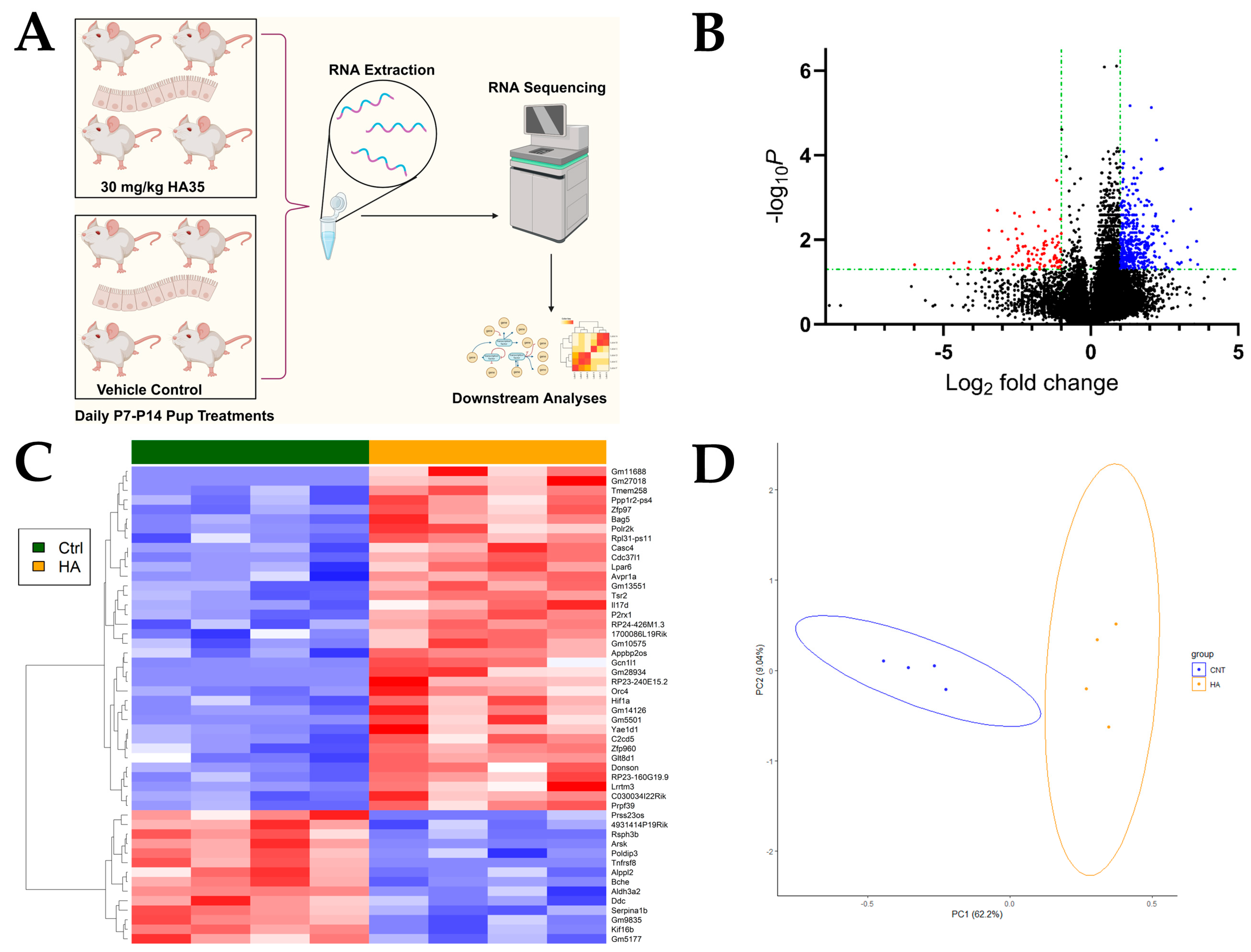
2.1. Oral HA35 Induces a Unique Transcriptional Response Within the Developing Ileum

Mouse pups were treated daily with 30 mg/kg body weight HA35 or vehicle control from postnatal day (P)7 to P14 (Figure 1A), a developmental timepoint corresponding with human intestinal development at 22–23 weeks’ gestation and a period of heightened susceptibility to NEC [39]. Bulk RNA-sequencing was performed on the distal ileum of pups at P14. From a total of 43,548 genes and using a p-value < 0.05 and |log2FoldChange| > 1, treatment with HA35 resulted in significant alterations in gene expression, with 249 differentially expressed genes (DEGs; 203 upregulated, 46 downregulated; Figure 1B). Dimensionality reduction in DEGs, the top 50 of which are illustrated in Figure 1C, from HA35-treated and control mice using principal component analysis (PCA) indicates pups cluster well by treatment (PC1, 62.2%; PC2, 9.04%; combined 71.24% variation explained; Figure 1D).

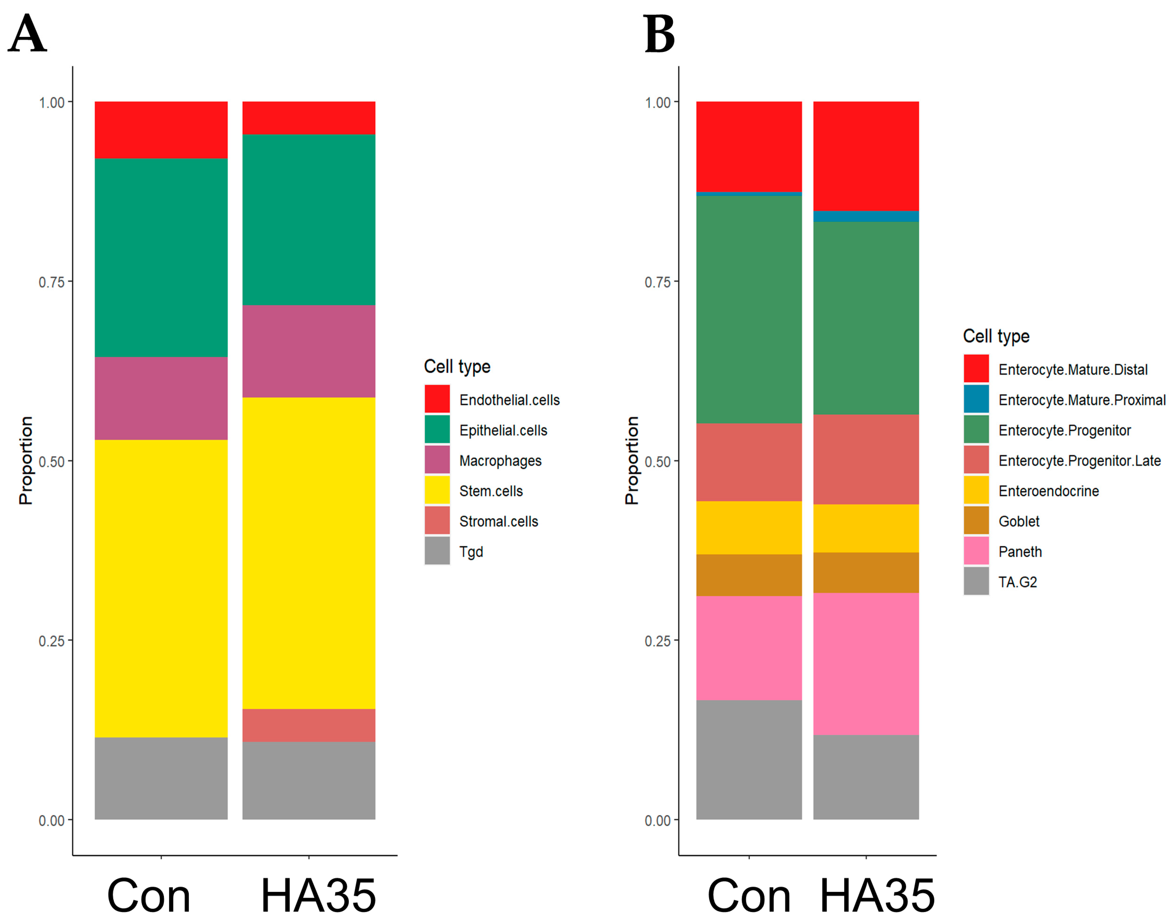
2.2. HA35 Induces Trend Toward Differentiation of Stromal, Mature Enterocytes, and Paneth Cells

To evaluate the potential of HA35 to induce cell-type abundance changes in the distal ileum of developing pups, cell counts for general small intestine (Figure 2A; Figure S1) and intestinal epithelial-specific cell types (Figure 2B; Figure S2) were estimated using cellular deconvolution and gene expression profiles derived from the Mouse Cell Atlas scRNA-seq reference dataset [40]. Treatment with HA35 trended toward an enrichment in stromal cells, while endothelial and epithelial cell populations were expected to diminish in the developing ileum. In addition, Paneth, late enterocyte progenitor, mature proximal enterocyte, and mature distal enterocyte cell types within the intestinal epithelium showed indications of enrichment, potentially drawing from the reduced pool of cycling transit-amplifying progenitors (TA.G2). Altogether, HA35 may bolster differentiated secretory and absorptive cell types, as well as those within the mesenchymal compartment of the developing intestine.

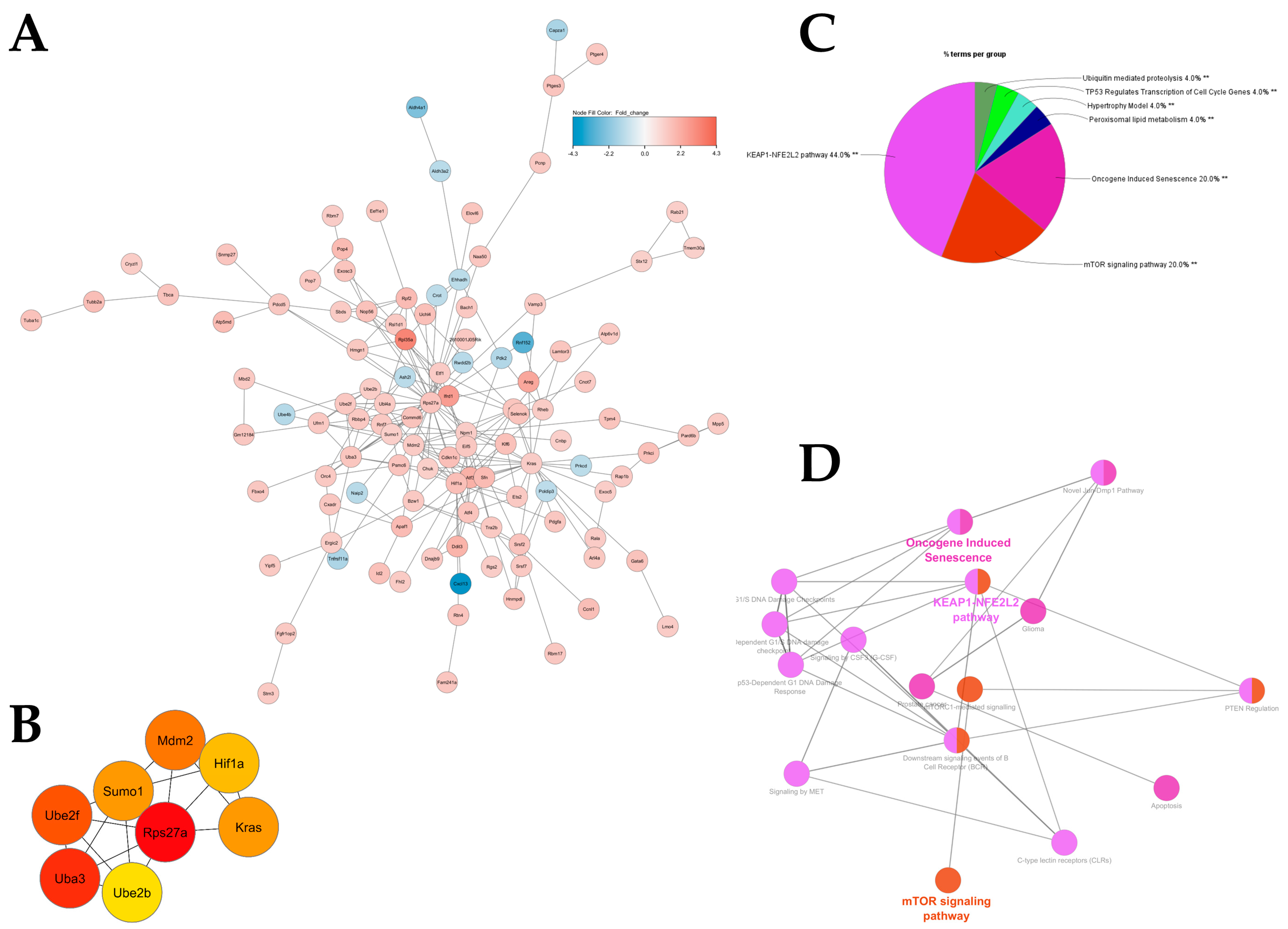
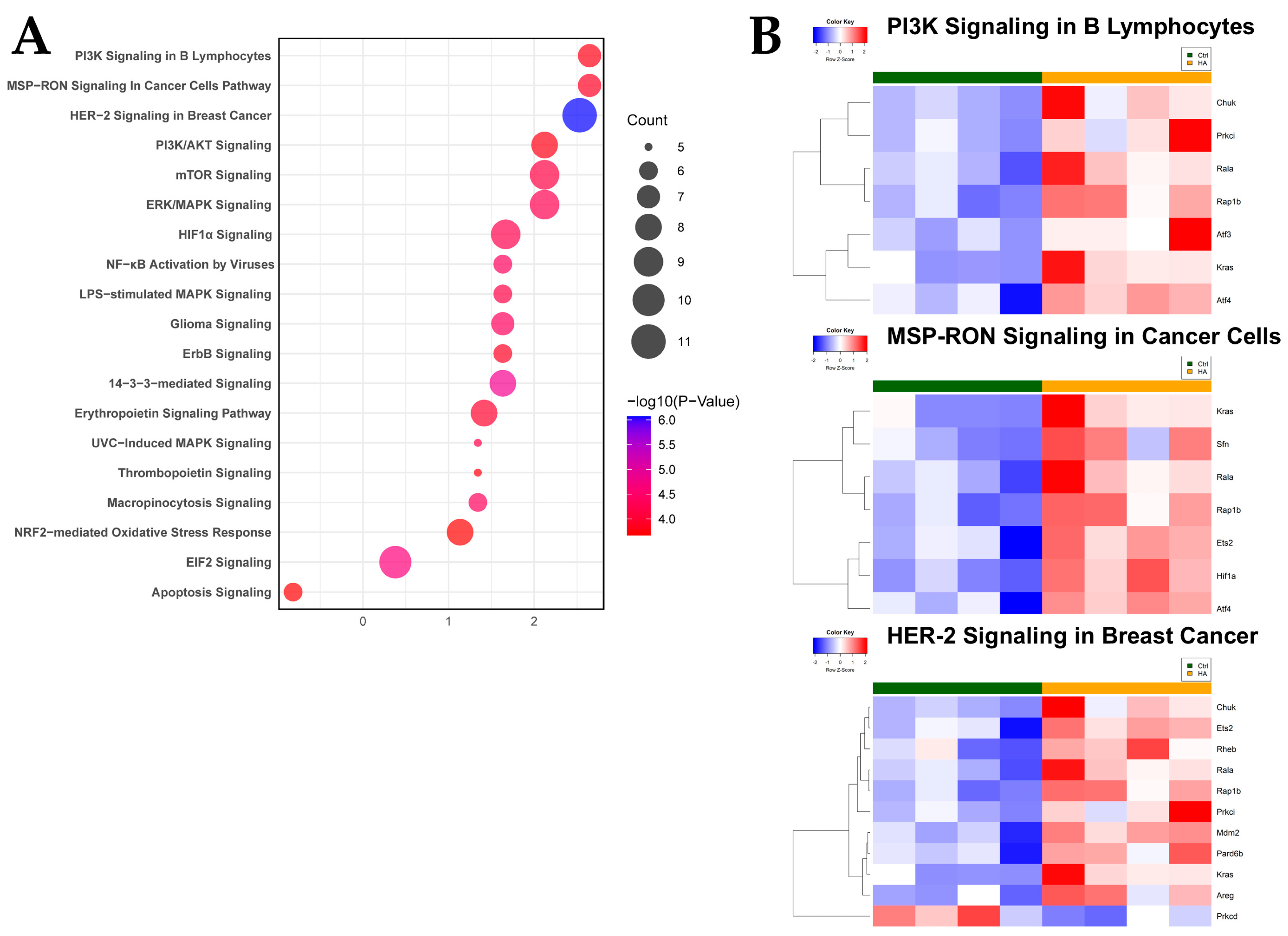
2.3. Antioxidant and Growth Signaling Characterize Functional Intestinal Response to HA35

When comparing gene expression altered by HA35 treatment, we utilized DEGs to construct a protein–protein interaction (PPI) network, where nodes indicate discrete genes and edges represent connections between and among genes. The largest subnetwork (Figure 3A) was utilized for functional hub gene analysis, identifying Rps27a (ribosomal protein S27a), Uba3 (ubiquitin like modifier activating enzyme 3), Ube2f (ubiquitin conjugating enzyme E2 F), Mdm2 (mouse double minute 2), Sumo1 (small ubiquitin like modifier 1), Kras, Hif1α (hypoxia inducible factor alpha), and Ube2b as genes driving connections within the PPI network (Figure 3B). The largest subnetwork was subjected to functional annotation using ClueGO, indicating enrichment of antioxidant (kelch-like ECH-associated protein 1-nuclear factor erythroid 2-like 2 [KEAP1-NFE2L2]) and growth (mechanistic target of rapamycin [mTOR]) pathways (Figure 3C) with HA35 treatment. The largest functional network based upon ClueGO analysis is depicted in Figure 3D.
To further evaluate the functional canonical signaling pathways involved in the intestinal response to HA35, DEGs were uploaded to Ingenuity Pathway Analysis (IPA) for enrichment analysis. Interestingly, pathways associated with growth and antioxidant response were enriched in HA35-treated pups, including PI3K/Akt, MSP-RON (macrophage stimulating protein-RON), HER-2, mTOR, ERK/MAPK, 14-3-3-mediated, HIF-1α, EIF2 (eukaryotic initiation factor 2), ErbB (erythroblastic leukemia viral oncogene homologue) signaling, and NRF2-mediated oxidative stress response (Figure 4A; Figure S3), while apoptosis signaling was predicted to be downregulated. Heatmaps for expression of genes driving the three most highly enriched pathways associated with HA35 treatment, PI3K, MSP-RON, and HER-2 signaling, are provided in Figure 4B.
Finally, we performed gene set enrichment analysis (GSEA) to further explore functional effects of HA35 treatment on the developing murine intestine. GSEA revealed significant associations of epithelial mesenchymal transition, MYC targets, E2F targets, genes downregulated in response to UV radiation, unfolded protein response, protein secretion, and G2M checkpoint gene sets (Figure 5) with HA35 treatment. Core enrichment genes overlapping in multiple gene sets include genes associated with stromal cell and ECM expansion (e.g., Ccn2 [cellular communication network factor 2], Has2 [hyaluronic acid synthase 2], Col1a1 [collagen type I alpha 1 chain], etc.), cell proliferation (e.g., Pcna [proliferating cell nuclear antigen], Mki67 [marker of proliferation Ki-67], Met, Kit, Myc, Egfr [epidermal growth factor receptor], etc.), cell growth and survival (e.g., Hif1a, Igfr2 [insulin-like growth factor 2 receptor], Igfbp2/3 [insulin-like growth factor binding protein 2 and 3], Vegfa [vascular endothelial growth factor alpha], Mapk14, etc.), ubiquitination (e.g., Ube2e1, Usp1 [ubiquitin specific peptidase 1], etc.), differentiation (e.g., Wnt5a [Wnt family member 5A], Notch2 [notch receptor 2], etc.), and eukaryotic translational initiation factor genes (e.g., Eif4e, etc.). Overall, our data suggest HA35 promotes intestinal development during early neonatal life through a variety of pro-growth signaling pathways. In addition, the unexpected enrichment of pathways associated with antioxidant response and predicted reduction in apoptosis signaling may provide physiological protection against events predisposing preterm infants to NEC.

3. Discussion

NEC, a leading cause of morbidity and mortality in the neonatal period, is characterized by severe intestinal inflammation, resulting in abdominal distension, necrosis, and in severe cases, intestinal perforation, multi-organ failure, and death [41,42]. While the precise etiology of the disease remains unclear, NEC pathogenesis is multifactorial and incorporates intestinal immaturity [43], a dysregulated immune system [44], and an altered gut microbiome [45]. NEC onset typically occurs 2–3 weeks following birth [46], providing a crucial window of opportunity for clinical prevention. HM, particularly mother’s own milk (MOM), is thought to play a key role in protecting preterm infants from NEC through promoting maturation of the intestinal epithelial barrier, reducing intestinal permeability, and fostering development of a favorable microbiome [6,47,48,49,50,51].
Our group has published extensively on the protective effects of the HM HA mimic [32], HA35, both in vitro and in vivo. In the former [36], HA35 accelerates restitution and healing from a mechanical wound via cell proliferation at the wound edge and migration to the site of injury, while in the latter, HA35 reduces mortality, severity of intestinal injury, and systemic inflammation, in part through preservation of intestinal barrier function [36,37]. Importantly, in the unchallenged gut of postnatal mice, HA35 promotes intestinal development and maturation, increasing goblet and Paneth cell numbers, and deepening and lengthening crypts and villi, respectively [38]. Here, we sought to elucidate potential mechanisms by which HA35 induces maturational changes within the developing postnatal intestine.

3.1. Role of HA35 in Differentiation of the Intestinal Epithelium

The intestinal epithelium is spatially organized into villi for maximal nutritional absorption. Proliferative intestinal stem cells (ISCs) migrate from the crypt base toward the villi tips [52]. Fully differentiated enterocytes at villi tips undergo apoptosis and are shed into the lumen [53], generating a cycle of epithelial regeneration that renews the entire villus population within a matter of days. The critical barrier function of the intestine is maintained by expression of tight junctions, regulating the passage of luminal contents into the mucosa, as well as multiple layers of mechanical and chemical defense. Goblet cells secrete mucus, preventing direct microbial adherence to the epithelium [54]. Paneth cells chemically deter microbes via the release of antimicrobial peptides (AMPs) [55], and embedded immune cells within the submucosa functionally mature following stimulation by bacteria [56].
The intestinal epithelium of preterm infants is developmentally immature. Compared with those of infants born at term, goblet cells of preterm infants have a reduced capacity to generate mucins, resulting in a thinner mucus lining and increased susceptibility to bacterial translocation of the epithelium [43]. Preterm enterocytes secrete less detoxifying alkaline phosphatase, and Paneth cells produce less lysozyme and defensins, altogether allowing potential pathogens direct access to the single-cell barrier, with limited ability to combat bacterial toxins or prevent mucosal invasion.
Endogenous HA receptor binding during postnatal development induces Lgr5+ ISC proliferation, expansion of Paneth cell numbers, and enhanced crypt fission, with pericryptal macrophages implicated as the potential source of this signaling [57]. However, the role of HA35 in postnatal small intestinal development has not been as well-studied. Cell deconvolution of our bulk sequencing data predicts an increase in stromal cells and decreases in endothelial and epithelial cells. HA35-mediated expansion of the stromal cell lineage likely contributes to epithelial differentiation and maturation [58], enabling critical defense of the postnatal intestinal barrier. Within the epithelium, deconvolution predicted an increase in mature enterocytes, late enterocyte progenitors, and Paneth cells, and a reduction in enterocyte progenitors and cycling TA cells. Overall, the shift in cellular composition following HA35 treatment appears to align with increased maturation and enterocyte differentiation, and largely substantiates our prior in vivo immunohistochemical (IHC) studies indicating increased goblet and Paneth cells in these same animals [38]. Further, the increase in differentiated cell types likely draws from the reduced pool of cycling TA cells, the latter of which is associated with an increase in the ratio of secretory to absorptive enterocytes within the epithelium [59]. Interestingly, TA cell abundance represents a balance in capacity for cell differentiation or regeneration, and treatment with HA35 appears to shift the balance toward differentiation. Altogether, these observations indicate HA35 likely accelerates epithelial maturation and expansion of the stromal cell compartment without substantially altering immune cell abundances.

3.2. HA35 as an Inducer of the PI3K/Akt/mTOR Signaling Cascade

Signal transduction pathways associated with growth and epithelial proliferation are likely to influence NEC pathogenesis [60,61,62,63,64]. PI3K/Akt/mTOR signaling, fundamental to key cellular processes including protein synthesis and secretion, nutrient uptake, and glycolytic metabolism [65], is essential to meet the needs of a rapidly growing and differentiating intestine during the neonatal period [66]. Growth factors, cytokines, and toll-like receptor (TLR) ligands initiate mTOR activation, primarily through PI3K/Akt or ERK phosphorylation and inactivation of the repressor, tuberous sclerosis complex 1/2 (TSC1/2) [67]. Through integration of extracellular and intracellular signals, mTOR plays essential roles in cell growth, proliferation, differentiation, migration, and survival [68]. Importantly, the PI3K/Akt/mTOR signaling axis, when significantly disrupted, is often embryonically lethal. Global or conditional knockout of mTORC1 (mTOR complex 1), a key component of the mTOR pathway, results in intestinal epithelial defects, including ileal villus blunting and reduced numbers of Paneth and goblet cells [69], underscoring the importance of the pathway to intestinal development. Additionally, mTOR activation is vital for functional intestinal epithelial repair, demonstrated in models of ischemia-reperfusion injury, radiation-induced damage, and colitis [70].
The ability of HA to activate PI3K/Akt/mTOR signaling has been reported in monocytes, chondrocytes, and rod photoreceptors of the eye [71,72,73], providing plausibility for relevance of this pathway within the neonatal intestine. GSEA, IPA, and network analyses (Figure 3, Figure 4 and Figure 5) indicated treatment with HA35 activated PI3K/Akt/mTOR signaling within the developing murine ileum, supporting our prior IHC, quantitative polymerase chain reaction (qPCR), and Western blot indications of mTORC1 activation in these pups [38]. Enrichment of the PI3K/Akt/mTOR signaling axis suggests a mechanism by which HA35 supports robust intestinal development, potentially reducing susceptibility to complications like NEC in preterm infants.

3.3. Activation of HIF-1α and VEGF Signaling Through HA35

In the gut, HIF-1α has dual roles in intestinal development and prevention of hypoxic injury, such as that observed during NEC pathogenesis [74]. During gut development, activation of HIF-1α upregulates VEGF, promoting creation of new vasculature to ensure adequate oxygen delivery to rapidly expanding intestinal tissues [75]. Developmental angiogenesis is crucial in supporting epithelial cell growth, differentiation, and the formation of a functional intestinal barrier.
In the context of intestinal injury, HIF-1α activation is critical for initiating repair processes [76]. Upregulation of VEGF leads to enhanced angiogenesis, increasing the flow of blood to damaged tissues. Delivery of fresh nutrients and oxygen by new vasculature spurs regenerative processes, reduces further ischemic damage, and supports integrity of the intestinal barrier by inducing epithelial expression of tight junction proteins and antimicrobial peptides. HIF-1α also plays a protective role by preventing excessive mucosal apoptosis, further facilitating recovery of the intestinal epithelium.
Activation of ileal HIF-1α and VEGF signaling by HA35 correlates with similar processes in cancer biology, where overproduction of HA generates a positive feedback loop of increased metabolic demands, a shift toward glycolysis and PI3K/Akt-mediated glucose uptake, activation of HIF-1α, and a boost in endogenous HA synthesis [57,77,78]. This metabolic reprogramming also fuels upregulation of VEGF, promoting angiogenesis to enhance nutrient and oxygen delivery, facilitating both cancer progression and maintenance of cancer stem cell properties. The sum of these processes results in rapid cellular proliferation in increasingly hypoxic conditions. However, within the developing ileum, activation of HIF-1α and VEGF likely induces endothelial development, potentially protecting against mucosal injuries involving splanchnic hypoperfusion [79].

3.4. HA35 and the NRF2-Mediated Response to Oxidative Stress

The KEAP1-NRF2 pathway plays a crucial role in regulating response to oxidative stress through activation of a molecular system tuned to perturbations in cellular redox balance [80], orchestrating a network of inducible proteins in defense of homeostasis and preservation of gut integrity [81,82]. Under oxidative conditions, NRF2 dissociates from KEAP1 and translocates to the nucleus, where the protein binds antioxidant response elements (AREs), activating enzymes such as glutathione peroxidase and superoxide dismutase [83]. These antioxidant enzymes neutralize reactive oxygen species (ROS), minimizing oxidative damage within intestinal tissues, a process inherent to NEC pathogenesis [84]. Additionally, NRF2 activation reduces inflammation through inhibition of NF-κB, induction of epithelial tight junction protein (e.g., zonula occludens 1 [ZO-1], occludin) expression, and modulation of T-cell function [82,85,86]. Fragmentation of HMW HA by ROS is reportedly anti-inflammatory [22], and HA35 inhibits ROS production by neutrophils [87]. Activation of the anti-inflammatory KEAP1-NRF2 pathway by HA has been broadly reported in a number of tissues (e.g., [88]), and our functional enrichment analyses support extrapolation of these effects to HA35 within the developing neonatal ileum, implicating a role for HM HA35 in enhancing intestinal resilience, preventing oxidative damage, and mitigating risk of NEC.
Intriguingly, our network analyses revealed strong connections among the KEAP1-NFE2L2 pathway, oncogene-induced senescence, and mTOR signaling. Activating transcription factor 4 (Atf4) is strongly upregulated within the ileum of HA35-treated pups (Tables S1 and S2). A stress-induced transcription factor [89], ATF4 mediates crosstalk between the KEAP1-NRF2 and mTOR pathways, highlighting a potentially broader role for HA35 in regulating cellular adaptation to stress, promoting repair and survival, and hastening intestinal development, potentially contributing to prevention of NEC.

3.5. MSP-RON and Epithelial-Mesenchymal Transition (EMT) Signaling

Mucosal tissue damage is the primary injury incurred during NEC. Epithelial repair requires cell migration through EMT [90]. However, enterocytes within the NEC intestine suffer impaired migratory capacity due to abnormal induction of autophagy [91]. The MSP-RON signaling pathway is a critical developmental pathway within the epithelium, and complete disruption of RON is embryonically lethal [92]. MSP-RON directs EMT [93] and has been shown to play a role in modulating inflammation and epithelial repair in the gut [94]. Activated by MSP, RON, a receptor tyrosine kinase primarily expressed in epithelial cells, attenuates inflammation by inhibiting nitric oxide and pro-inflammatory cytokine production through PI3K/Akt and RAS/ERK/MAPK pathways [95]. High RON expression within the epithelium suggests a crucial role in maintaining tissue integrity and repair rather than direct modulation of immune responses. Interestingly, single nucleotide polymorphism variants such as 689C within the MSP gene, reducing MSP levels, are associated with an increased risk of intestinal inflammatory diseases and impaired wound healing [96].
Our results indicate HA35 treatment enriches the MSP-RON pathway in the terminal ileum, and parallels similar findings in the lung. In the latter tissue, LMW HA interacts with receptor for hyaluronan-mediated mobility (RHAMM) and RON to enhance mucociliary function and clearance of pathogens [97]. Whether such interactions among HA35, RHAMM, and RON occur in the developing neonatal intestinal epithelium or contribute to repair of wounds incurred during NEC is not yet clear.

3.6. The Role of HA35 as a Developmentally Appropriate Oncofetal Signal

Oncogenes play a significant role in embryonic, fetal, and adult tissues, promoting rampant proliferation of cells during development or repair [98]. While most commonly associated with carcinogenic and metastatic processes, oncogene activity during healthy development is tightly regulated to promote maximal proliferation within the bounds of healthy tissue growth. Fetal stem cell programs can be induced within the intestine via a variety of extrinsic factors [99], and we propose oral supplementation with HA35 through provision of HM is one such extrinsic factor. Within our dataset, multiple oncogenes and proto-oncogenes (e.g., Kras, Mdm2 [a key regulator of the tumor suppressor, p53], Rala [RAS-like proto-oncogene], Lmo4 [LIM domain only 4]), and oncogenic pathways (e.g., HER-2, 14-3-3, MSP-RON, ErbB signaling) are upregulated in the ileum of mouse pups treated with HA35, and the collective effect of these synchronized cues induces maturation and development of the neonatal murine intestine [38].
The content of GAGs and HA, in particular, within term and preterm milk declines over the first six months of breastfeeding [100] from a peak in colostrum shortly after birth [29]. Interestingly, as with other GAGs [101], HA35 content is significantly elevated in the HM provided to preterm compared with term infants (unpublished data). We posit that HA35 serves as an oncofetal signal within the infant intestine, and its maximal content in preterm colostrum is consistent with an essential role in development and maturation of the underdeveloped preterm intestine. As with other instances of oncogene expression within healthy tissue, provision of the oncofetal HA35 is tightly regulated via maternal mammary gland production and subsequent feeding events. Importantly, the duration of maximal HA35 content within preterm HM overlaps with the period of enhanced susceptibility to NEC [46].

4. Materials and Methods

4.1. Hyaluronic Acid 35 kDa (HA35)

HA35 was purchased from Lifecore Biomedical (Chaska, MN, USA). HA35 is a biosynthetic and medical grade polydisperse mixture of HA from 21 kDa to 40 kDa, with an average MW of 35 kDa. The compound is pure and commercially manufactured to meet the European Pharmacopoeia and Japanese Pharmacopoeia monographs for sodium hyaluronate.

4.2. Mouse Experiments

All animal studies were approved by the Institutional Animal Care and Use Committee at the University of Oklahoma Health Sciences Center (IACUC #19-062-EFCHI). Data reported here result from additional analysis of an animal experiment originally conducted and published in 2021 [38]. Briefly, timed pregnant CD-1 dams (Charles River Laboratories, Wilmington, MA, USA) were housed individually in sterilized, filter-top cages. Ad libitum food and water were provided under a 12 h light/dark cycle. Pups of both sexes were born vaginally, weighed daily, and randomized into groups receiving 30 mg/kg HA35 or control (an equal volume of sterile phosphate-buffered saline) via oral gavage from postnatal day (P)7 to P14. At treatment end, pups from both groups were anesthetized with isoflurane and euthanized via bilateral thoracotomy and cardiac puncture. Terminal ileum was harvested and stored at −80 °C in RNAlater (Invitrogen, Carlsbad, CA, USA) for downstream gene analyses.

4.3. Bulk RNA-Sequencing

Ileal tissues were homogenized with a 7 cm polypropylene pellet pestle (W.W. Grainger, Lake Forest, IL, USA) and passed through a QIAshredder homogenizer (Qiagen, Germantown, MD, USA). Total RNA was isolated using a RNeasy Plus Mini Kit (Qiagen). Sample quantity and quality were evaluated using a NanoDrop Lite Spectrophotometer (ThermoFisher Scientific, Waltham, MA, USA). Stranded RNA-Seq libraries were constructed for 8 animals via the CORALL Total RNA-Seq Library Prep Kit v2 (Lexogen, Greenland, NH, USA). Libraries were constructed with 200–500 ng of RNA. Libraries were indexed for multiplex sequencing, pooled, and run on the NovaSeq 6000 platform (Illumina, San Diego, CA, USA) at the Oklahoma Medical Research Foundation Clinical Genomics Center. Sequences were analyzed through a custom computational pipeline consisting of the open-source gSNAP, Cufflinks, and R for sequence alignment and evaluation of differential gene expression [102]. Reads generated were mapped to the mouse genome (mm10) by gSNAP [103], expression (fragments Per Kilobase of transcript per Million mapped reads, FPKM) derived by Cufflinks [104], and differential expression analyzed using R.

4.4. Analysis of Differentially Expressed Genes (DEGs)

DEGs with a p-value < 0.05 and a |Log2FoldChange (FC)| > 1 were collated using DeSeq2 in R. Significant genes were visualized with one-way hierarchical heatmaps using the ‘heatmap.2’ function in the R gplots package.

4.5. Bioinformatic Analysis of DEGs

DEGs from all 8 samples were subjected to principal component analysis (PCA) using the prcomp function and visualized by autoplot using the ggfortify package in R.
Cell deconvolution for mixed cell types within the small intestine, as well as intestinal epithelial-specific cell types, was performed using the granulator package in R and expression matrices derived from the scRNA-seq Mouse Cell Atlas [40]. Median cell abundances were compared using Wilcoxon signed rank test and cell proportions were plotted using stacked box plots.

4.6. Protein–Protein Interaction (PPI) Networks

p-values and FC values for DEGs were imported to CytoScape v3.10.2 [105]. Overlapping gene–protein interaction networks were constructed via query of the STRING (search tool for the retrieval of interacting genes) database (confidence cutoff, 0.4) using the Cytoscape stringApp [106], where nodes indicate discrete genes and edges represent connections between genes. The largest subnetwork was selected for each analysis, and annotation of functional enrichment was performed with ClueGO [107], using GO (gene ontology) Biological Functions, GO Immune System, GO Molecular Function, KEGG (Kyoto Encyclopedia of Genes and Genomes), Reactome Pathways, and WikiPathway databases. Significantly enriched functions were defined through two-sided hypergeometric tests with Bonferroni step down correction (Bonferroni-corrected p < 0.05), with a minimum of 2–3 genes/pathway and a kappa score of 0.4. Hub genes were identified through degree of connectivity and shortest paths using maximal clique centrality (MCC) ranking of 15 or fewer nodes in the CytoScape plugin, CytoHubba [108].

4.7. Ingenuity Pathway Analysis (IPA)

DEGs with p < 0.05 were uploaded into IPA (Qiagen) for pathway analysis. Significantly altered or enriched pathways and predicted upstream regulators were defined by significant Z-scores determining the probability of association between genes in the dataset and a pathway of interest compared with chance alone. Core Analysis was used to explore differential biological processes, canonical pathways, upstream transcriptional regulators, and gene networks. Significantly enriched or depleted pathways (FDR-adjusted p < 0.05 via Fisher’s Exact Test) with gene overlap ≥2 were represented with bubble plots using ggplot2 and dplyr packages in R. IPA Functional Analysis was used to identify biological functions and/or diseases significantly associated with the dataset. To assess the activation state of a pathway and upstream regulators, the consistency of the match between the observed and predicted pattern was calculated (Z-score). A positive Z-score signifies a regulator in the active state, while a negative Z-score indicates repressed activity. Canonical pathway analysis was used to identify networks from the IPA library most significantly modulated across both groups. The significance of the association between each dataset and the canonical pathway was measured as a ratio of the number of molecules from the dataset mapped to the pathway divided by the total number of molecules mapped to the canonical pathway.

4.8. GSEA Analysis

Gene set enrichment analysis (GSEA) was conducted with GSEA 4.1.0 against the Hallmark database, and significant pathways were defined as FDR-adjusted p < 0.05 and gene overlap > 15.

5. Conclusions

HA35 significantly influences gene expression related to small intestinal development and maturation. The observed upregulation of pathways associated with cellular growth, differentiation, immune regulation, and response to physiological stressors, coupled with downregulation of apoptotic signaling, underscores the potential of HA35 as a HM bioactive factor capable of lending protection against NEC. Notably, HA35 upregulates genes incorporated in the KEAP1/NFE2L2 and mTOR signaling pathways, known to support antioxidative responses and cellular growth, respectively. These pathways are relevant in the context of NEC, as oxidative stress and impaired cellular repair mechanisms are critical in the progression of, and possible healing from, this disease. Confirmation of our findings will now proceed in human preterm infant intestinal epithelial enteroid models.

Supplementary Materials

The following supporting information can be downloaded at: https://www.mdpi.com/article/10.3390/ijms252312679/s1.

Author Contributions

Conceptualization, K.Y.B. and H.C.; methodology, K.Y.B. and H.Z.; formal analysis, K.Y.B., H.Z. and A.P.W.; writing—original draft preparation, K.Y.B.; writing—review and editing, K.Y.B., H.Z., A.P.W. and H.C.; project administration and supervision, H.C. and K.Y.B.; funding acquisition, H.C. and K.Y.B. All authors have read and agreed to the published version of the manuscript.

Funding

This research was funded by the Oklahoma Center for Microbial Pathogenesis and Immunity (5P20GM134973 [H.C. and K.B.]), the NIH NICHD (R01HD109784 [H.C]), and the Children’s Health Foundation (H.C. and K.B.).

Data Availability Statement

The original sequencing data presented in this study are available in the GEO database (accession number GSE274261), with public access available from 31 December 2024.

Acknowledgments

We thank the COBRE-associated Microbiome and Transcriptomics Core at OUHSC for assistance with Illumina sequencing, total RNA library construction, and bioinformatics support.

Conflicts of Interest

The authors declare no conflicts of interest. The funders had no role in the design of this study; in the collection, analyses, or interpretation of data; in the writing of the manuscript; or in the decision to publish the results.

References

  1. Brockway, M.M.; Daniel, A.I.; Reyes, S.M.; Gauglitz, J.M.; Granger, M.; McDermid, J.M.; Chan, D.; Refvik, R.; Sidhu, K.K.; Musse, S.; et al. Human Milk Bioactive Components and Child Growth and Body Composition in the First 2 Years: A Systematic Review. Adv. Nutr. 2024, 15, 100127. [Google Scholar] [CrossRef] [PubMed]
  2. Grulee, C.G.; Sanford, H.N.; Herron, P.H. Breast and artificial feeding: Influence on morbidity and mortality of twenty thousand infants. JAMA J. Am. Med. Assoc. 1934, 103, 735–738. [Google Scholar] [CrossRef]
  3. Howie, P.W.; Forsyth, J.S.; Ogston, S.A.; Clark, A.; Florey, C.D. Protective effect of breast feeding against infection. BMJ (Clin. Res. Ed.) 1990, 300, 11–16. [Google Scholar] [CrossRef]
  4. He, Y.; Lawlor, N.T.; Newburg, D.S. Human Milk Components Modulate Toll-Like Receptor-Mediated Inflammation. Adv. Nutr. 2016, 7, 102–111. [Google Scholar] [CrossRef]
  5. Walker, W.A.; Iyengar, R.S. Breast milk, microbiota, and intestinal immune homeostasis. Pediatr. Res. 2015, 77, 220–228. [Google Scholar] [CrossRef]
  6. Maffei, D.; Schanler, R.J. Human milk is the feeding strategy to prevent necrotizing enterocolitis! Semin. Perinatol. 2017, 41, 36–40. [Google Scholar] [CrossRef]
  7. Westerbeek, E.A.; van den Berg, A.; Lafeber, H.N.; Fetter, W.P.; van Elburg, R.M. The effect of enteral supplementation of a prebiotic mixture of non-human milk galacto-, fructo- and acidic oligosaccharides on intestinal permeability in preterm infants. Br. J. Nutr. 2011, 105, 268–274. [Google Scholar] [CrossRef]
  8. van den Berg, A.; Fetter, W.P.; Westerbeek, E.A.; van der Vegt, I.M.; van der Molen, H.R.; van Elburg, R.M. The effect of glutamine-enriched enteral nutrition on intestinal permeability in very-low-birth-weight infants: A randomized controlled trial. JPEN J. Parenter. Enteral Nutr. 2006, 30, 408–414. [Google Scholar] [CrossRef]
  9. Foster, J.P.; Seth, R.; Cole, M.J. Oral immunoglobulin for preventing necrotizing enterocolitis in preterm and low birth weight neonates. Cochrane Database Syst. Rev. 2016, 4, Cd001816. [Google Scholar] [CrossRef]
  10. Dimmitt, R.A.; Staley, E.M.; Chuang, G.; Tanner, S.M.; Soltau, T.D.; Lorenz, R.G. Role of postnatal acquisition of the intestinal microbiome in the early development of immune function. J. Pediatr. Gastroenterol. Nutr. 2010, 51, 262–273. [Google Scholar] [CrossRef]
  11. Pammi, M.; Cope, J.; Tarr, P.I.; Warner, B.B.; Morrow, A.L.; Mai, V.; Gregory, K.E.; Kroll, J.S.; McMurtry, V.; Ferris, M.J.; et al. Intestinal dysbiosis in preterm infants preceding necrotizing enterocolitis: A systematic review and meta-analysis. Microbiome 2017, 5, 31. [Google Scholar] [CrossRef] [PubMed]
  12. Samuels, N.; van de Graaf, R.A.; de Jonge, R.C.J.; Reiss, I.K.M.; Vermeulen, M.J. Risk factors for necrotizing enterocolitis in neonates: A systematic review of prognostic studies. BMC Pediatr. 2017, 17, 105. [Google Scholar] [CrossRef] [PubMed]
  13. Abrams, S.A.; Schanler, R.J.; Lee, M.L.; Rechtman, D.J. Greater mortality and morbidity in extremely preterm infants fed a diet containing cow milk protein products. Breastfeed. Med. Off. J. Acad. Breastfeed. Med. 2014, 9, 281–285. [Google Scholar] [CrossRef] [PubMed]
  14. Sisk, P.M.; Lovelady, C.A.; Dillard, R.G.; Gruber, K.J.; O’Shea, T.M. Early human milk feeding is associated with a lower risk of necrotizing enterocolitis in very low birth weight infants. J. Perinatol. Off. J. Calif. Perinat. Assoc. 2007, 27, 428–433. [Google Scholar] [CrossRef]
  15. Coppa, G.V.; Gabrielli, O.; Buzzega, D.; Zampini, L.; Galeazzi, T.; Maccari, F.; Bertino, E.; Volpi, N. Composition and structure elucidation of human milk glycosaminoglycans. Glycobiology 2011, 21, 295–303. [Google Scholar] [CrossRef]
  16. Kogan, G.; Soltés, L.; Stern, R.; Mendichi, R. Hyaluronic acid: A biopolymer with versatile physico-chemical and biological properties. In Handbook of Polymer Research: Monomers, Oligomers, Polymers and Composites; Pethrich, R.A., Antonio, B., Zaikov, G.E., Eds.; Nova Science Publishers Inc.: New York, NY, USA, 2007; pp. 393–439. [Google Scholar]
  17. Prehm, P. Release of hyaluronate from eukaryotic cells. Biochem. J. 1990, 267, 185–189. [Google Scholar] [CrossRef]
  18. Kim, Y.; Kessler, S.P.; Obery, D.R.; Homer, C.R.; McDonald, C.; de la Motte, C.A. Hyaluronan 35 kDa treatment protects mice from Citrobacter rodentium infection and induces epithelial tight junction protein ZO-1 in vivo. Matrix Biol. 2017, 62, 28–39. [Google Scholar] [CrossRef]
  19. Stern, R.; Asari, A.A.; Sugahara, K.N. Hyaluronan fragments: An information-rich system. Eur. J. Cell Biol. 2006, 85, 699–715. [Google Scholar] [CrossRef]
  20. Papakonstantinou, E.; Roth, M.; Karakiulakis, G. Hyaluronic acid: A key molecule in skin aging. Dermatoendocrinology 2012, 4, 253–258. [Google Scholar] [CrossRef]
  21. Monslow, J.; Govindaraju, P.; Puré, E. Hyaluronan—A functional and structural sweet spot in the tissue microenvironment. Front. Immunol. 2015, 6, 231. [Google Scholar] [CrossRef]
  22. Fallacara, A.; Baldini, E.; Manfredini, S.; Vertuani, S. Hyaluronic Acid in the Third Millennium. Polymers 2018, 10, 701. [Google Scholar] [CrossRef] [PubMed]
  23. Slevin, M.; Krupinski, J.; Gaffney, J.; Matou, S.; West, D.; Delisser, H.; Savani, R.C.; Kumar, S. Hyaluronan-mediated angiogenesis in vascular disease: Uncovering RHAMM and CD44 receptor signaling pathways. Matrix Biol. 2007, 26, 58–68. [Google Scholar] [CrossRef] [PubMed]
  24. Jackson, D.G. Immunological functions of hyaluronan and its receptors in the lymphatics. Immunol. Rev. 2009, 230, 216–231. [Google Scholar] [CrossRef]
  25. Soltés, L.; Mendichi, R.; Kogan, G.; Schiller, J.; Stankovska, M.; Arnhold, J. Degradative action of reactive oxygen species on hyaluronan. Biomacromolecules 2006, 7, 659–668. [Google Scholar] [CrossRef]
  26. Jiang, D.; Liang, J.; Noble, P.W. Hyaluronan as an immune regulator in human diseases. Physiol. Rev. 2011, 91, 221–264. [Google Scholar] [CrossRef]
  27. Riehl, T.E.; Ee, X.; Stenson, W.F. Hyaluronic acid regulates normal intestinal and colonic growth in mice. Am. J. Physiol. Gastrointest. Liver Physiol. 2012, 303, G377–G388. [Google Scholar] [CrossRef]
  28. Slevin, M.; Kumar, S.; Gaffney, J. Angiogenic oligosaccharides of hyaluronan induce multiple signaling pathways affecting vascular endothelial cell mitogenic and wound healing responses. J. Biol. Chem. 2002, 277, 41046–41059. [Google Scholar] [CrossRef]
  29. Coppa, G.V.; Gabrielli, O.; Bertino, E.; Zampini, L.; Galeazzi, T.; Padella, L.; Santoro, L.; Marchesiello, R.L.; Galeotti, F.; Maccari, F.; et al. Human milk glycosaminoglycans: The state of the art and future perspectives. Ital. J. Pediatr. 2013, 39, 2. [Google Scholar] [CrossRef]
  30. Maccari, F.; Mantovani, V.; Gabrielli, O.; Carlucci, A.; Zampini, L.; Galeazzi, T.; Galeotti, F.; Coppa, G.V.; Volpi, N. Metabolic fate of milk glycosaminoglycans in breastfed and formula fed newborns. Glycoconj. J. 2016, 33, 181–188. [Google Scholar] [CrossRef]
  31. Hill, D.R.; Rho, H.K.; Kessler, S.P.; Amin, R.; Homer, C.R.; McDonald, C.; Cowman, M.K.; de la Motte, C.A. Human milk hyaluronan enhances innate defense of the intestinal epithelium. J. Biol. Chem. 2013, 288, 29090–29104. [Google Scholar] [CrossRef]
  32. Kessler, S.P.; Obery, D.R.; Nickerson, K.P.; Petrey, A.C.; McDonald, C.; de la Motte, C.A. Multifunctional Role of 35 Kilodalton Hyaluronan in Promoting Defense of the Intestinal Epithelium. J. Histochem. Cytochem. Off. J. Histochem. Soc. 2018, 66, 273–287. [Google Scholar] [CrossRef] [PubMed]
  33. Saikia, P.; Roychowdhury, S.; Bellos, D.; Pollard, K.A.; McMullen, M.R.; McCullough, R.L.; McCullough, A.J.; Gholam, P.; de la Motte, C.; Nagy, L.E. Hyaluronic acid 35 normalizes TLR4 signaling in Kupffer cells from ethanol-fed rats via regulation of microRNA291b and its target Tollip. Sci. Rep. 2017, 7, 15671. [Google Scholar] [CrossRef] [PubMed]
  34. Saikia, P.; Bellos, D.; McMullen, M.R.; Pollard, K.A.; de la Motte, C.; Nagy, L.E. MicroRNA 181b-3p and its target importin alpha5 regulate toll-like receptor 4 signaling in Kupffer cells and liver injury in mice in response to ethanol. Hepatology 2017, 66, 602–615. [Google Scholar] [CrossRef]
  35. Wilson, A.; Burge, K.; Eckert, J.; Chaaban, H. Effect of Hyaluronic Acid 35 kDa on an In Vitro Model of Preterm Small Intestinal Injury and Healing using Enteroid-derived Monolayers. J. Vis. Exp. 2022, 185, e63758. [Google Scholar] [CrossRef]
  36. Gunasekaran, A.; Eckert, J.; Burge, K.; Zheng, W.; Yu, Z.; Kessler, S.; de la Motte, C.; Chaaban, H. Hyaluronan 35 kDa enhances epithelial barrier function and protects against the development of murine necrotizing enterocolitis. Pediatr. Res. 2019, 87, 1177–1184. [Google Scholar] [CrossRef]
  37. Burge, K.; Eckert, J.; Wilson, A.; Trammell, M.; Lueschow, S.R.; McElroy, S.J.; Dyer, D.; Chaaban, H. Hyaluronic Acid 35 kDa Protects against a Hyperosmotic, Formula Feeding Model of Necrotizing Enterocolitis. Nutrients 2022, 14, 1779. [Google Scholar] [CrossRef]
  38. Chaaban, H.; Burge, K.; Eckert, J.; Trammell, M.; Dyer, D.; Keshari, R.S.; Silasi, R.; Regmi, G.; Lupu, C.; Good, M.; et al. Acceleration of Small Intestine Development and Remodeling of the Microbiome Following Hyaluronan 35 kDa Treatment in Neonatal Mice. Nutrients 2021, 13, 2030. [Google Scholar] [CrossRef]
  39. Stanford, A.H.; Gong, H.; Noonan, M.; Lewis, A.N.; Gong, Q.; Lanik, W.E.; Hsieh, J.J.; Lueschow, S.R.; Frey, M.R.; Good, M.; et al. A direct comparison of mouse and human intestinal development using epithelial gene expression patterns. Pediatr. Res. 2020, 88, 66–76. [Google Scholar] [CrossRef]
  40. Han, X.; Wang, R.; Zhou, Y.; Fei, L.; Sun, H.; Lai, S.; Saadatpour, A.; Zhou, Z.; Chen, H.; Ye, F.; et al. Mapping the Mouse Cell Atlas by Microwell-Seq. Cell 2018, 172, 1091–1107.e1017. [Google Scholar] [CrossRef]
  41. Neu, J.; Walker, W.A. Necrotizing enterocolitis. N. Engl. J. Med. 2011, 364, 255–264. [Google Scholar] [CrossRef]
  42. Tanner, S.M.; Berryhill, T.F.; Ellenburg, J.L.; Jilling, T.; Cleveland, D.S.; Lorenz, R.G.; Martin, C.A. Pathogenesis of necrotizing enterocolitis: Modeling the innate immune response. Am. J. Pathol. 2015, 185, 4–16. [Google Scholar] [CrossRef] [PubMed]
  43. Demers-Mathieu, V. The immature intestinal epithelial cells in preterm infants play a role in the necrotizing enterocolitis pathogenesis: A review. Health Sci. Rev. 2022, 4, 6. [Google Scholar] [CrossRef]
  44. Nanthakumar, N.; Meng, D.; Goldstein, A.M.; Zhu, W.; Lu, L.; Uauy, R.; Llanos, A.; Claud, E.C.; Walker, W.A. The mechanism of excessive intestinal inflammation in necrotizing enterocolitis: An immature innate immune response. PLoS ONE 2011, 6, e17776. [Google Scholar] [CrossRef] [PubMed]
  45. Thänert, R.; Keen, E.C.; Dantas, G.; Warner, B.B.; Tarr, P.I. Necrotizing Enterocolitis and the Microbiome: Current Status and Future Directions. J. Infect. Dis. 2021, 223, S257–S263. [Google Scholar] [CrossRef]
  46. Duchon, J.; Barbian, M.E.; Denning, P.W. Necrotizing Enterocolitis. Clin. Perinatol. 2021, 48, 229–250. [Google Scholar] [CrossRef]
  47. Nolan, L.S.; Parks, O.B.; Good, M. A Review of the Immunomodulating Components of Maternal Breast Milk and Protection Against Necrotizing Enterocolitis. Nutrients 2019, 12, 14. [Google Scholar] [CrossRef]
  48. Chong, C.Y.L.; Bloomfield, F.H.; O’Sullivan, J.M. Factors Affecting Gastrointestinal Microbiome Development in Neonates. Nutrients 2018, 10, 274. [Google Scholar] [CrossRef]
  49. Patel, A.L.; Kim, J.H. Human milk and necrotizing enterocolitis. Semin. Pediatr. Surg. 2018, 27, 34–38. [Google Scholar] [CrossRef]
  50. Cortez, J.; Makker, K.; Kraemer, D.F.; Neu, J.; Sharma, R.; Hudak, M.L. Maternal milk feedings reduce sepsis, necrotizing enterocolitis and improve outcomes of premature infants. J. Perinatol. Off. J. Calif. Perinat. Assoc. 2018, 38, 71–74. [Google Scholar] [CrossRef]
  51. Lewis, E.D.; Richard, C.; Larsen, B.M.; Field, C.J. The Importance of Human Milk for Immunity in Preterm Infants. Clin. Perinatol. 2017, 44, 23–47. [Google Scholar] [CrossRef]
  52. Shyer, A.E.; Tallinen, T.; Nerurkar, N.L.; Wei, Z.; Gil, E.S.; Kaplan, D.L.; Tabin, C.J.; Mahadevan, L. Villification: How the gut gets its villi. Science 2013, 342, 212–218. [Google Scholar] [CrossRef] [PubMed]
  53. Chin, A.M.; Hill, D.R.; Aurora, M.; Spence, J.R. Morphogenesis and maturation of the embryonic and postnatal intestine. Semin. Cell Dev. Biol. 2017, 66, 81–93. [Google Scholar] [CrossRef] [PubMed]
  54. Turner, J.R. Intestinal mucosal barrier function in health and disease. Nat. Rev. Immunol. 2009, 9, 799–809. [Google Scholar] [CrossRef] [PubMed]
  55. Clevers, H.C.; Bevins, C.L. Paneth cells: Maestros of the small intestinal crypts. Annu. Rev. Physiol. 2013, 75, 289–311. [Google Scholar] [CrossRef]
  56. Round, J.L.; Mazmanian, S.K. The gut microbiota shapes intestinal immune responses during health and disease. Nat. Rev. Immunol. 2009, 9, 313–323. [Google Scholar] [CrossRef]
  57. Riehl, T.E.; Santhanam, S.; Foster, L.; Ciorba, M.; Stenson, W.F. CD44 and TLR4 mediate hyaluronic acid regulation of Lgr5+ stem cell proliferation, crypt fission, and intestinal growth in postnatal and adult mice. Am. J. Physiol. Gastrointest. Liver Physiol. 2015, 309, G874–G887. [Google Scholar] [CrossRef]
  58. Jacob, J.M.; Di Carlo, S.E.; Stzepourginski, I.; Lepelletier, A.; Ndiaye, P.D.; Varet, H.; Legendre, R.; Kornobis, E.; Benabid, A.; Nigro, G.; et al. PDGFRα-induced stromal maturation is required to restrain postnatal intestinal epithelial stemness and promote defense mechanisms. Cell Stem Cell 2022, 29, 856–868.e855. [Google Scholar] [CrossRef]
  59. Sanman, L.E.; Chen, I.W.; Bieber, J.M.; Steri, V.; Trentesaux, C.; Hann, B.; Klein, O.D.; Wu, L.F.; Altschuler, S.J. Transit-Amplifying Cells Coordinate Changes in Intestinal Epithelial Cell-Type Composition. Dev. Cell 2021, 56, 356–365.e359. [Google Scholar] [CrossRef]
  60. Coursodon, C.F.; Dvorak, B. Epidermal growth factor and necrotizing enterocolitis. Curr. Opin. Pediatr. 2012, 24, 160–164. [Google Scholar] [CrossRef]
  61. Austin, K.; Tsang, D.; Chalmers, J.A.; Maalouf, M.F.; Brubaker, P.L. Insulin-like growth factor-binding protein-4 inhibits epithelial growth and proliferation in the rodent intestine. Am. J. Physiol. Gastrointest. Liver Physiol. 2018, 315, G206–G219. [Google Scholar] [CrossRef]
  62. Fesler, Z.; Mitova, E.; Brubaker, P.L. GLP-2, EGF, and the Intestinal Epithelial IGF-1 Receptor Interactions in the Regulation of Crypt Cell Proliferation. Endocrinology 2020, 161, bqaa040. [Google Scholar] [CrossRef] [PubMed]
  63. Nair, R.R.; Warner, B.B.; Warner, B.W. Role of epidermal growth factor and other growth factors in the prevention of necrotizing enterocolitis. Semin. Perinatol. 2008, 32, 107–113. [Google Scholar] [CrossRef] [PubMed]
  64. de Jong, P.R.; Taniguchi, K.; Harris, A.R.; Bertin, S.; Takahashi, N.; Duong, J.; Campos, A.D.; Powis, G.; Corr, M.; Karin, M.; et al. ERK5 signalling rescues intestinal epithelial turnover and tumour cell proliferation upon ERK1/2 abrogation. Nat. Commun. 2016, 7, 11551. [Google Scholar] [CrossRef] [PubMed]
  65. Laplante, M.; Sabatini, D.M. mTOR signaling at a glance. J. Cell Sci. 2009, 122, 3589–3594. [Google Scholar] [CrossRef] [PubMed]
  66. Panwar, V.; Singh, A.; Bhatt, M.; Tonk, R.K.; Azizov, S.; Raza, A.S.; Sengupta, S.; Kumar, D.; Garg, M. Multifaceted role of mTOR (mammalian target of rapamycin) signaling pathway in human health and disease. Signal Transduct. Target. Ther. 2023, 8, 375. [Google Scholar] [CrossRef]
  67. Rhoads, J.M.; Niu, X.; Odle, J.; Graves, L.M. Role of mTOR signaling in intestinal cell migration. Am. J. Physiol. Gastrointest. Liver Physiol. 2006, 291, G510–G517. [Google Scholar] [CrossRef]
  68. Sheng, H.; Shao, J.; Townsend, C.M., Jr.; Evers, B.M. Phosphatidylinositol 3-kinase mediates proliferative signals in intestinal epithelial cells. Gut 2003, 52, 1472–1478. [Google Scholar] [CrossRef]
  69. Sampson, L.L.; Davis, A.K.; Grogg, M.W.; Zheng, Y. mTOR disruption causes intestinal epithelial cell defects and intestinal atrophy postinjury in mice. FASEB J. Off. Publ. Fed. Am. Soc. Exp. Biol. 2016, 30, 1263–1275. [Google Scholar] [CrossRef]
  70. Kaur, H.; Moreau, R. Role of mTORC1 in intestinal epithelial repair and tumorigenesis. Cell Mol. Life Sci. 2019, 76, 2525–2546. [Google Scholar] [CrossRef]
  71. Lenart, M.; Rutkowska-Zapala, M.; Baj-Krzyworzeka, M.; Szatanek, R.; Węglarczyk, K.; Smallie, T.; Ziegler-Heitbrock, L.; Zembala, M.; Siedlar, M. Hyaluronan carried by tumor-derived microvesicles induces IL-10 production in classical (CD14++CD16) monocytes via PI3K/Akt/mTOR-dependent signalling pathway. Immunobiology 2017, 222, 1–10. [Google Scholar] [CrossRef]
  72. Mitrousis, N.; Tam, R.Y.; Baker, A.E.G.; van der Kooy, D.; Shoichet, M.S. Hyaluronic Acid-Based Hydrogels Enable Rod Photoreceptor Survival and Maturation In Vitro through Activation of the mTOR Pathway. Adv. Funct. Mater. 2016, 26, 1975–1985. [Google Scholar] [CrossRef]
  73. Terabe, K.; Ohashi, Y.; Tsuchiya, S.; Ishizuka, S.; Knudson, C.B.; Knudson, W. Chondroprotective effects of 4-methylumbelliferone and hyaluronan synthase-2 overexpression involve changes in chondrocyte energy metabolism. J. Biol. Chem. 2019, 294, 17799–17817. [Google Scholar] [CrossRef] [PubMed]
  74. Kaplina, A.; Kononova, S.; Zaikova, E.; Pervunina, T.; Petrova, N.; Sitkin, S. Necrotizing Enterocolitis: The Role of Hypoxia, Gut Microbiome, and Microbial Metabolites. Int. J. Mol. Sci. 2023, 24, 2471. [Google Scholar] [CrossRef] [PubMed]
  75. Pral, L.P.; Fachi, J.L.; Corrêa, R.O.; Colonna, M.; Vinolo, M.A.R. Hypoxia and HIF-1 as key regulators of gut microbiota and host interactions. Trends Immunol. 2021, 42, 604–621. [Google Scholar] [CrossRef] [PubMed]
  76. Steiner, C.A.; Cartwright, I.M.; Taylor, C.T.; Colgan, S.P. Hypoxia-inducible factor as a bridge between healthy barrier function, wound healing, and fibrosis. Am. J. Physiol. Cell Physiol. 2022, 323, C866–C878. [Google Scholar] [CrossRef]
  77. Chanmee, T.; Ontong, P.; Izumikawa, T.; Higashide, M.; Mochizuki, N.; Chokchaitaweesuk, C.; Khansai, M.; Nakajima, K.; Kakizaki, I.; Kongtawelert, P.; et al. Hyaluronan Production Regulates Metabolic and Cancer Stem-like Properties of Breast Cancer Cells via Hexosamine Biosynthetic Pathway-coupled HIF-1 Signaling. J. Biol. Chem. 2016, 291, 24105–24120. [Google Scholar] [CrossRef]
  78. O’Neill, L.A. A feed-forward loop involving hyaluronic acid and toll-like receptor-4 as a treatment for colitis? Gastroenterology 2009, 137, 1889–1891. [Google Scholar] [CrossRef]
  79. Burge, K.Y.; Gunasekaran, A.; Makoni, M.M.; Mir, A.M.; Burkhart, H.M.; Chaaban, H. Clinical Characteristics and Potential Pathogenesis of Cardiac Necrotizing Enterocolitis in Neonates with Congenital Heart Disease: A Narrative Review. J. Clin. Med. 2022, 11, 3987. [Google Scholar] [CrossRef]
  80. Ma, D.; Gao, W.; Liu, J.; Kong, D.; Zhang, Y.; Qian, M. Mechanism of oxidative stress and Keap-1/Nrf2 signaling pathway in bronchopulmonary dysplasia. Medicine 2020, 99, e20433. [Google Scholar] [CrossRef]
  81. Wen, Z.; Liu, W.; Li, X.; Chen, W.; Liu, Z.; Wen, J.; Liu, Z. A Protective Role of the NRF2-Keap1 Pathway in Maintaining Intestinal Barrier Function. Oxid. Med. Cell Longev. 2019, 2019, 1759149. [Google Scholar] [CrossRef]
  82. Jin, W.; Wang, H.; Ji, Y.; Hu, Q.; Yan, W.; Chen, G.; Yin, H. Increased intestinal inflammatory response and gut barrier dysfunction in Nrf2-deficient mice after traumatic brain injury. Cytokine 2008, 44, 135–140. [Google Scholar] [CrossRef] [PubMed]
  83. Kobayashi, A.; Kang, M.I.; Okawa, H.; Ohtsuji, M.; Zenke, Y.; Chiba, T.; Igarashi, K.; Yamamoto, M. Oxidative stress sensor Keap1 functions as an adaptor for Cul3-based E3 ligase to regulate proteasomal degradation of Nrf2. Mol. Cell. Biol. 2004, 24, 7130–7139. [Google Scholar] [CrossRef] [PubMed]
  84. Aceti, A.; Beghetti, I.; Martini, S.; Faldella, G.; Corvaglia, L. Oxidative Stress and Necrotizing Enterocolitis: Pathogenetic Mechanisms, Opportunities for Intervention, and Role of Human Milk. Oxid. Med. Cell Longev. 2018, 2018, 7397659. [Google Scholar] [CrossRef] [PubMed]
  85. Khor, T.O.; Huang, M.T.; Kwon, K.H.; Chan, J.Y.; Reddy, B.S.; Kong, A.N. Nrf2-deficient mice have an increased susceptibility to dextran sulfate sodium-induced colitis. Cancer Res. 2006, 66, 11580–11584. [Google Scholar] [CrossRef] [PubMed]
  86. Rockwell, C.E.; Zhang, M.; Fields, P.E.; Klaassen, C.D. Th2 skewing by activation of Nrf2 in CD4+ T cells. J. Immunol. 2012, 188, 1630–1637. [Google Scholar] [CrossRef]
  87. Jia, X.; Shi, M.; Wang, Q.; Hui, J.; Shofaro, J.H.; Erkhembayar, R.; Hui, M.; Gao, C.; Gantumur, M.A. Anti-Inflammatory Effects of the 35kDa Hyaluronic Acid Fragment (B-HA/HA35). J. Inflamm. Res. 2023, 16, 209–224. [Google Scholar] [CrossRef]
  88. Onodera, Y.; Teramura, T.; Takehara, T.; Fukuda, K. Hyaluronic acid regulates a key redox control factor Nrf2 via phosphorylation of Akt in bovine articular chondrocytes. FEBS Open Bio 2015, 5, 476–484. [Google Scholar] [CrossRef]
  89. Li, R.; Wilson, K.F.; Cerione, R.A. Elucidation of an mTORC2-PKC-NRF2 pathway that sustains the ATF4 stress response and identification of Sirt5 as a key ATF4 effector. Cell Death Discov. 2022, 8, 357. [Google Scholar] [CrossRef]
  90. Yetkin-Arik, B.; Jansen, S.A.; Varderidou-Minasian, S.; Westendorp, B.; Skarp, K.P.; Altelaar, M.; Lindemans, C.A.; Lorenowicz, M.J. Mesenchymal stromal/stem cells promote intestinal epithelium regeneration after chemotherapy-induced damage. Stem Cell Res. Ther. 2024, 15, 125. [Google Scholar] [CrossRef]
  91. Neal, M.D.; Sodhi, C.P.; Dyer, M.; Craig, B.T.; Good, M.; Jia, H.; Yazji, I.; Afrazi, A.; Richardson, W.M.; Beer-Stolz, D.; et al. A critical role for TLR4 induction of autophagy in the regulation of enterocyte migration and the pathogenesis of necrotizing enterocolitis. J. Immunol. 2013, 190, 3541–3551. [Google Scholar] [CrossRef]
  92. Yao, H.P.; Zhou, Y.Q.; Zhang, R.; Wang, M.H. MSP-RON signalling in cancer: Pathogenesis and therapeutic potential. Nat. Rev. Cancer 2013, 13, 466–481. [Google Scholar] [CrossRef] [PubMed]
  93. Weng, T.; Yan, D.; Shi, D.; Zhu, M.; Liu, Y.; Wu, Z.; Tang, T.; Zhu, L.; Zhang, H.; Yao, H.; et al. The MSP-RON pathway regulates liver fibrosis through transforming growth factor beta-dependent epithelial-mesenchymal transition. Liver Int. 2021, 41, 1956–1968. [Google Scholar] [CrossRef] [PubMed]
  94. Mao, T.; Li, J.; Liu, L.; Zhao, W.; Liu, Y.; Gao, K.; Guo, Y.; Xie, T.; Li, N.; Shi, R. Qingchang Wenzhong Decoction Attenuates DSS-Induced Colitis in Rats by Reducing Inflammation and Improving Intestinal Barrier Function via Upregulating the MSP/RON Signalling Pathway. Evid. Based Complement. Alternat. Med. 2017, 2017, 4846876. [Google Scholar] [CrossRef]
  95. Huang, L.; Fang, X.; Shi, D.; Yao, S.; Wu, W.; Fang, Q.; Yao, H. MSP-RON Pathway: Potential Regulator of Inflammation and Innate Immunity. Front. Immunol. 2020, 11, 569082. [Google Scholar] [CrossRef]
  96. Kauder, S.E.; Santell, L.; Mai, E.; Wright, L.Y.; Luis, E.; N’Diaye, E.N.; Lutman, J.; Ratti, N.; Sa, S.M.; Maun, H.R.; et al. Functional consequences of the macrophage stimulating protein 689C inflammatory bowel disease risk allele. PLoS ONE 2013, 8, e83958. [Google Scholar] [CrossRef]
  97. Manzanares, D.; Monzon, M.E.; Savani, R.C.; Salathe, M. Apical oxidative hyaluronan degradation stimulates airway ciliary beating via RHAMM and RON. Am. J. Respir. Cell Mol. Biol. 2007, 37, 160–168. [Google Scholar] [CrossRef]
  98. West, R.C.; Bouma, G.J.; Winger, Q.A. Shifting perspectives from “oncogenic” to oncofetal proteins; how these factors drive placental development. Reprod. Biol. Endocrinol. 2018, 16, 101. [Google Scholar] [CrossRef]
  99. Fey, S.K.; Vaquero-Siguero, N.; Jackstadt, R. Dark force rising: Reawakening and targeting of fetal-like stem cells in colorectal cancer. Cell Rep. 2024, 43, 114270. [Google Scholar] [CrossRef]
  100. Wang, C.; Lang, Y.; Li, Q.; Jin, X.; Li, G.; Yu, G. Glycosaminoglycanomic profiling of human milk in different stages of lactation by liquid chromatography-tandem mass spectrometry. Food Chem. 2018, 258, 231–236. [Google Scholar] [CrossRef]
  101. Coppa, G.V.; Gabrielli, O.; Zampini, L.; Galeazzi, T.; Maccari, F.; Buzzega, D.; Galeotti, F.; Bertino, E.; Volpi, N. Glycosaminoglycan content in term and preterm milk during the first month of lactation. Neonatology 2012, 101, 74–76. [Google Scholar] [CrossRef]
  102. Baird, N.L.; Bowlin, J.L.; Cohrs, R.J.; Gilden, D.; Jones, K.L. Comparison of varicella-zoster virus RNA sequences in human neurons and fibroblasts. J. Virol. 2014, 88, 5877–5880. [Google Scholar] [CrossRef] [PubMed]
  103. Wu, T.D.; Nacu, S. Fast and SNP-tolerant detection of complex variants and splicing in short reads. Bioinformatics 2010, 26, 873–881. [Google Scholar] [CrossRef] [PubMed]
  104. Trapnell, C.; Williams, B.A.; Pertea, G.; Mortazavi, A.; Kwan, G.; van Baren, M.J.; Salzberg, S.L.; Wold, B.J.; Pachter, L. Transcript assembly and quantification by RNA-Seq reveals unannotated transcripts and isoform switching during cell differentiation. Nat. Biotechnol. 2010, 28, 511–515. [Google Scholar] [CrossRef]
  105. Shannon, P.; Markiel, A.; Ozier, O.; Baliga, N.S.; Wang, J.T.; Ramage, D.; Amin, N.; Schwikowski, B.; Ideker, T. Cytoscape: A software environment for integrated models of biomolecular interaction networks. Genome Res. 2003, 13, 2498–2504. [Google Scholar] [CrossRef]
  106. Doncheva, N.T.; Morris, J.H.; Gorodkin, J.; Jensen, L.J. Cytoscape StringApp: Network Analysis and Visualization of Proteomics Data. J. Proteome Res. 2019, 18, 623–632. [Google Scholar] [CrossRef]
  107. Bindea, G.; Mlecnik, B.; Hackl, H.; Charoentong, P.; Tosolini, M.; Kirilovsky, A.; Fridman, W.H.; Pagès, F.; Trajanoski, Z.; Galon, J. ClueGO: A Cytoscape plug-in to decipher functionally grouped gene ontology and pathway annotation networks. Bioinformatics 2009, 25, 1091–1093. [Google Scholar] [CrossRef]
  108. Chin, C.H.; Chen, S.H.; Wu, H.H.; Ho, C.W.; Ko, M.T.; Lin, C.Y. cytoHubba: Identifying hub objects and sub-networks from complex interactome. BMC Syst. Biol. 2014, 8 (Suppl. 4), S11. [Google Scholar] [CrossRef]
Figure 1. HA35 induces distinct transcriptional response during murine development. (A) Experimental design (n = 4 pups/group); (B) Volcano plot of DEGs p-value < 0.05; |log2FoldChange| > 1; upregulated with HA35 treatment, blue; downregulated with HA35 treatment, red; (C) Heatmap of top 50 DEGs; (D) PCA plot of DEGs. Abbreviations: P: postnatal day; RNA: ribonucleic acid; Ctrl: control; HA35: hyaluronic acid 35 kDa; PCA: principal component analysis; DEGs: differentially expressed genes.
Ijms 25 12679 g001
Figure 2. Oral HA35 may expand differentiated and stromal cell types. Mixed cell deconvolution using the Mouse Cell Atlas for (A) general intestinal and (B) intestinal epithelial cell types indicates a potential increase in stromal, Paneth, and mature enterocytes and progenitors, with diminished cycling TA, endothelial, and epithelial cells. Abbreviations: Con: Control; HA35: hyaluronic acid 35 kDa; Tgd: gamma delta T cells; TA.G2: transit-amplifying. Growth 2.
Ijms 25 12679 g002
Figure 3. HA35 enriches signaling pathways involved in growth and antioxidant response in the developing murine intestine. (A) Largest PPI subnetwork demonstrating upregulated (red) and downregulated (blue) genes in HA35-treated pups compared with controls; (B) Top eight hub genes identified through CytoHubba (red = strongest associations); (C) ClueGO pie chart of GO term functional enrichment (% terms/group); ** FDR-adjusted p < 0.01; (D) ClueGO functional network (light pink: KEAP1-NFE2L2 pathway [44%]; red: mTOR signaling pathway [20%]; magenta: oncogene-induced senescence [20%]). Abbreviations: HA35: hyaluronic acid 35 kDa; PPI: protein–protein interaction; GO: gene ontology; FDR: false discovery rate; KEAP1-NFE2L2: kelch-like ECH-associated protein 1-nuclear factor erythroid 2-like 2; mTOR: mechanistic target of rapamycin; Ube2f: ubiquitin conjugating enzyme E2 F; Sumo1: small ubiquitin like modifier 1; Mdm2: mouse double minute 2; Hif1α: hypoxia inducible factor alpha; Rps27a: ribosomal protein S27a; Uba3: ubiquitin like modifier activating enzyme 3.
Ijms 25 12679 g003
Figure 4. Enrichment analysis predicts upregulation of growth and downregulation of apoptosis signaling pathways following HA35 treatment of the developing intestine. (A) Bubble plot for IPA pathways; (B) Heatmaps for genes in PI3K, MSP-RON, and HER-2 signaling pathways. Abbreviations: HA35: hyaluronic acid 35 kDa; IPA: Ingenuity Pathway Analysis; PI3K/At: phosphatidylinositol-3-kinase/protein kinase B; MSP-RON: macrophage stimulating factor-RON; HER-2: human epidermal growth factor receptor 2; mTOR: mechanistic target of rapamycin; ERK/MAPK: extracellular signal-regulated kinase/mitogen-activated protein kinases; HIF-1α: hypoxia inducible factor-1 alpha; NF-κβ: nuclear factor kappa beta; LPS: lipopolysaccharide; ErbB: erythroblastic leukemia viral oncogene; UVC: ultraviolet light C; NRF2: nuclear factor erythroid 2-related factor 2; EIF2: eukaryotic initiation factor 2.
Ijms 25 12679 g004
Figure 5. GSEA enrichment plots for top gene sets enriched (q-value < 0.05) in the distal ileum of HA35-treated pups versus controls. From top-left, left-to-right (gene overlap): Epithelial-mesenchymal transition (193); E2F targets (199); UV response down (144); Protein secretion (94); MYC targets V2 (58); MYC Targets V1 (197); Unfolded protein response (111); G2M checkpoint (195). Abbreviations: GSEA: gene set enrichment analysis; HA35: hyaluronic acid 35 kDa; NES: normalized enrichment score; UV: ultraviolet.
Ijms 25 12679 g005
Disclaimer/Publisher’s Note: The statements, opinions and data contained in all publications are solely those of the individual author(s) and contributor(s) and not of MDPI and/or the editor(s). MDPI and/or the editor(s) disclaim responsibility for any injury to people or property resulting from any ideas, methods, instructions or products referred to in the content.

