
Molecular Determinants for Guanine Binding in GTP-Binding Proteins: A Data Mining and Quantum Chemical Study


Abstract

1. Introduction
2. Results and Discussion
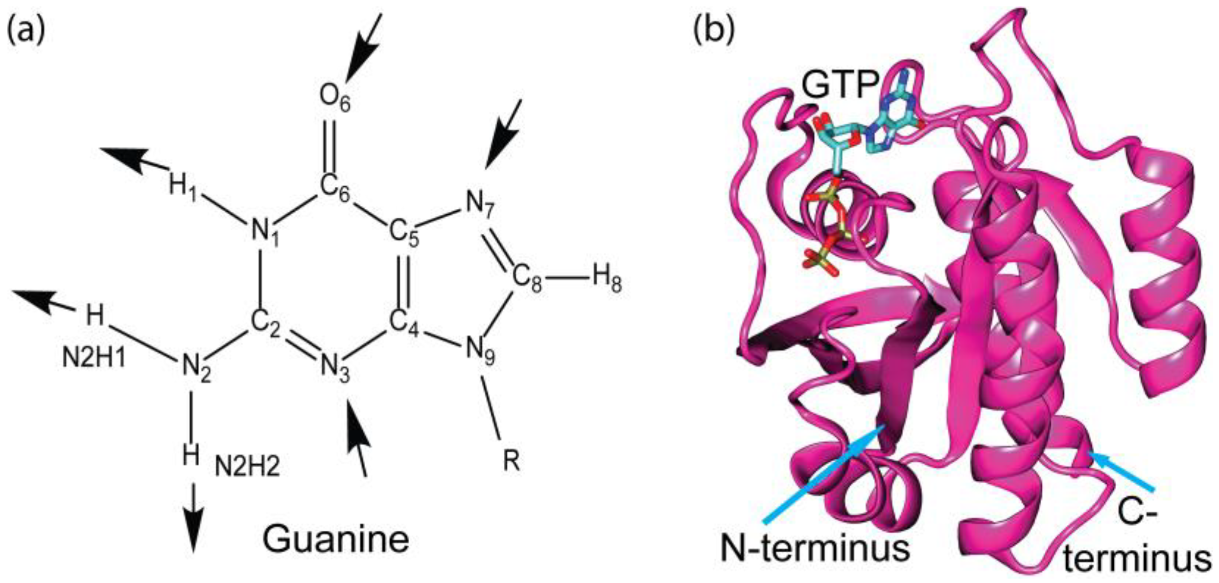
2.1. Hydrogen Bonding
2.2. Cation–π Interaction
2.3. π–π Stacking Interaction
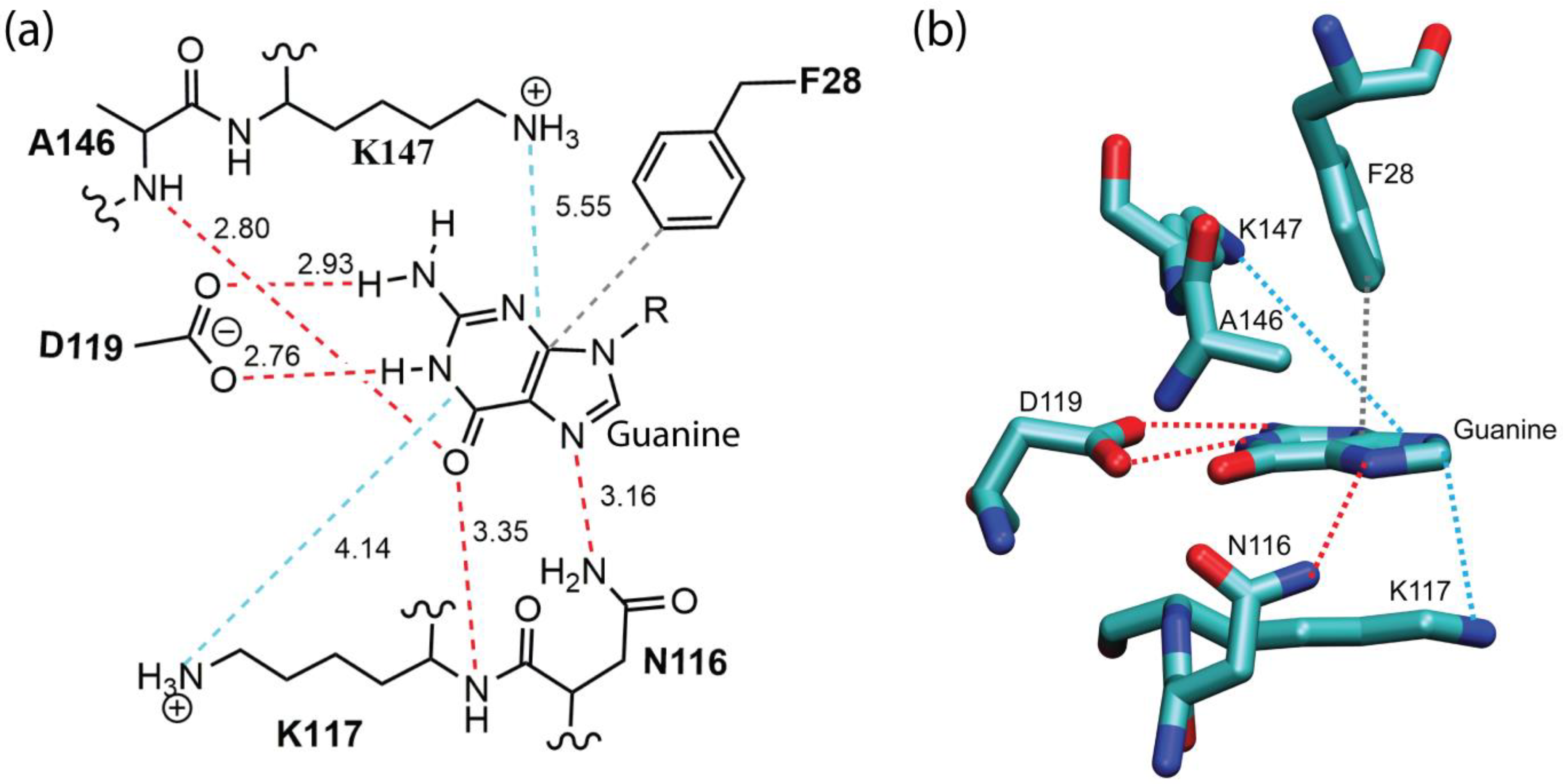
2.4. Energetic Contribution by Various Modes of Non-Bonded Interactions to the Binding of Guanine in a Representative Complex
2.5. Biological Significance
3. Theory and Methods
3.1. Data Mining
3.2. Analysis of Interaction Modes
3.3. Quantification of Intermolecular Interaction Energy
4. Conclusions
- Hydrogen bonds, particularly those involving N2 and O6 atoms of the guanine base, confer specificity to guanine recognition by distinguishing it from adenine.
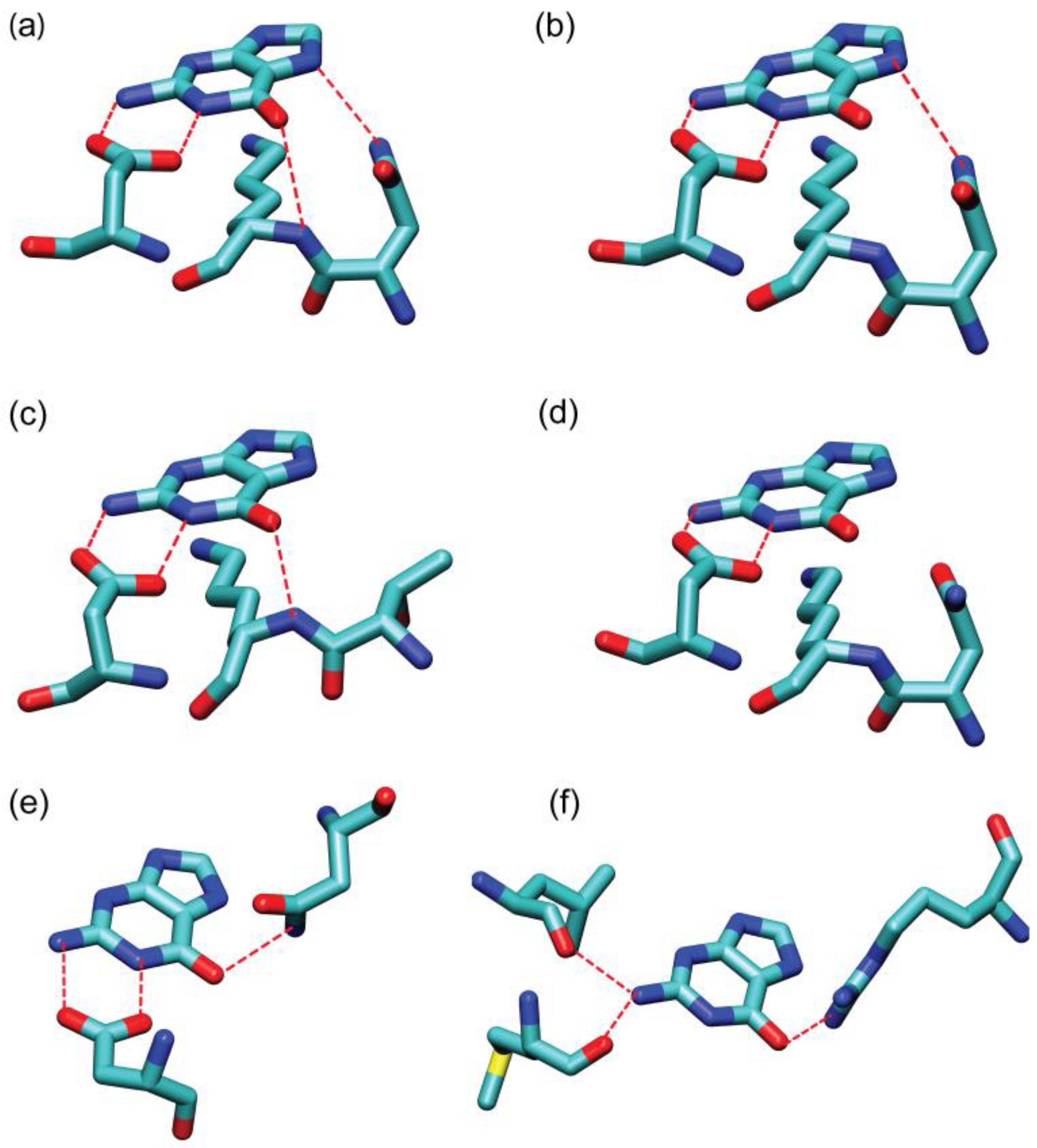
- Quantum chemical analysis revealed the critical role of cation–π interactions between the guanine ring and its surrounding basic residues (Lys and Arg) in stabilizing guanine binding within GTP-binding proteins. Intermolecular interaction energies for representative cation–π interactions range from −1.51 to −10.61 kcal/mol. The high-energy strength of cation–π interactions can be attributed to the multi-mode intermolecular interactions associated with the Lys and Arg residues. For example, the Lys residue of the NKXD motif can be involved in both the cation–π interactions between the positively charged ε-amino groups of lysine and the guanine ring, as well as in the CH–π interactions between the side chain alkyl groups of lysine and the guanine ring.
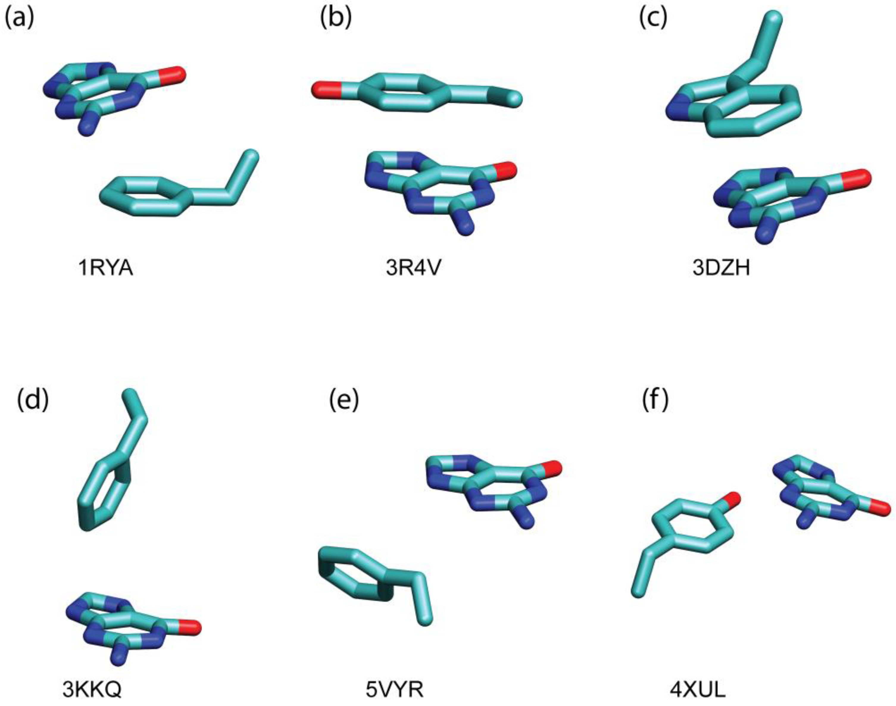
- π–π stacking interactions between the guanine ring and its surrounding aromatic residues (Phe, Tyr, and Trp) act as an auxiliary stabilizing factor. In complex featuring the NKXD motif, those aromatic residues are typically situated on the opposite side of the guanine ring relative to the Lys residue of the NKXD motif (see, for example, the table of content figure). Intermolecular interaction energies for representative π–π stacking interactions range from −0.34 to −6.57 kcal/mol.
Supplementary Materials
Author Contributions
Funding
Institutional Review Board Statement
Informed Consent Statement
Data Availability Statement
Acknowledgments
Conflicts of Interest
References
- Pfeuffer, T.; Helmreich, E.J.M. Structural and Functional-Relationships of Guanosine Triphosphate Binding-Proteins. Curr. Top. Cell. Regul. 1988, 29, 129–216. [Google Scholar] [PubMed]
- Spiegel, A.M. Signal Transduction by Guanine-Nucleotide Binding-Proteins. Mol. Cell. Endocrinol. 1987, 49, 1–16. [Google Scholar] [CrossRef] [PubMed]
- Bockaert, J.; Homburger, V.; Rouot, B. Gtp Binding-Proteins—A Key Role in Cellular Communication. Biochimie 1987, 69, 329–338. [Google Scholar] [CrossRef] [PubMed]
- Bourne, H.R.; Sanders, D.A.; McCormick, F. The Gtpase Superfamily—A Conserved Switch for Diverse Cell Functions. Nature 1990, 348, 125–132. [Google Scholar] [CrossRef] [PubMed]
- Li, G.; Zhang, X.C. GTP Hydrolysis Mechanism of Ras-like GTPases. J. Mol. Biol. 2004, 340, 921–932. [Google Scholar] [CrossRef]
- Guidolin, D.; Agnati, L.F.; Marcoli, M.; Borroto-Escuela, D.O.; Fuxe, K. G-protein-coupled receptor type A heteromers as an emerging therapeutic target. Expert Opin. Ther. Targets 2015, 19, 265–283. [Google Scholar] [CrossRef]
- Bourne, H.R.; Sanders, D.A.; McCormick, F. The Gtpase Superfamily—Conserved Structure And Molecular Mechanism. Nature 1991, 349, 117–127. [Google Scholar] [CrossRef]
- Sprang, S.R. G protein mechanisms: Insights from structural analysis. Annu. Rev. Biochem. 1997, 66, 639–678. [Google Scholar] [CrossRef]
- Kjeldgaard, M.; Nyborg, J.; Clark, B.F. The GTP binding motif: Variations on a theme. FASEB J. Off. Publ. Fed. Am. Soc. Exp. Biol. 1996, 10, 1347–1368. [Google Scholar] [CrossRef]
- Babor, M.; Sobolev, V.; Edelman, M. Conserved positions for ribose recognition: Importance of water bridging interactions among ATP, ADP and FAD-protein complexes. J. Mol. Biol. 2002, 323, 523–532. [Google Scholar] [CrossRef]
- Dreusicke, D.; Schulz, G.E. The glycine-rich loop of adenylate kinase forms a giant anion hole. FEBS Lett. 1986, 208, 301–304. [Google Scholar] [CrossRef] [PubMed]
- Sprang, S.R. Invited review: Activation of G proteins by GTP and the mechanism of Gα-catalyzed GTP hydrolysis. Biopolymers 2016, 105, 449–462. [Google Scholar] [CrossRef] [PubMed]
- Saraste, M.; Sibbald, P.R.; Wittinghofer, A. The P-loop--a common motif in ATP- and GTP-binding proteins. Trends Biochem. Sci. 1990, 15, 430–434. [Google Scholar] [CrossRef] [PubMed]
- Hirsch, A.K.; Fischer, F.R.; Diederich, F. Phosphate recognition in structural biology. Angew. Chem. (Int. Ed. Engl.) 2007, 46, 338–352. [Google Scholar] [CrossRef]
- Babine, R.E.; Bender, S.L. Molecular recognition of protein-ligand complexes: Applications to drug design. Chem. Rev. 1997, 97, 1359–1472. [Google Scholar] [CrossRef]
- McGaughey, G.B.; Gagné, M.; Rappé, A.K. π-Stacking interactions. Alive and well in proteins. J. Biol. Chem. 1998, 273, 15458–15463. [Google Scholar] [CrossRef]
- Bohm, H.J.; Klebe, G. What can we learn from molecular recognition in protein-ligand complexes for the design of new drugs? Angew. Chem.-Int. Ed. 1996, 35, 2589–2614. [Google Scholar] [CrossRef]
- Neel, A.J.; Hilton, M.J.; Sigman, M.S.; Toste, F.D. Exploiting non-covalent π interactions for catalyst design. Nature 2017, 543, 637–646. [Google Scholar] [CrossRef]
- Persch, E.; Dumele, O.; Diederich, F. Molecular Recognition in Chemical and Biological Systems. Angew. Chem. Int. Ed. 2015, 54, 3290–3327. [Google Scholar] [CrossRef]
- Zhu, Y.; Alqahtani, S.; Hu, X.C. Aromatic Rings as Molecular Determinants for the Molecular Recognition of Protein Kinase Inhibitors. Molecules 2021, 26, 1776. [Google Scholar] [CrossRef]
- Carter-Fenk, K.; Liu, M.L.; Pujal, L.; Loipersberger, M.; Tsanai, M.; Vernon, R.M.; Forman-Kay, J.D.; Head-Gordon, M.; Heidar-Zadeh, F.; Head-Gordon, T. The Energetic Origins of Pi-Pi Contacts in Proteins. J. Am. Chem. Soc. 2023, 145, 24836–24851. [Google Scholar] [CrossRef] [PubMed]
- Zhu, Y.; Hu, X.C. Molecular Recognition of FDA-Approved Small Molecule Protein Kinase Drugs in Protein Kinases. Molecules 2022, 27, 7124. [Google Scholar] [CrossRef] [PubMed]
- Gallivan, J.P.; Dougherty, D.A. Cation-π interactions in structural biology. Proc. Natl. Acad. Sci. USA 1999, 96, 9459–9464. [Google Scholar] [CrossRef] [PubMed]
- Mao, L.; Wang, Y.; Liu, Y.; Hu, X. Molecular determinants for ATP-binding in proteins: A data mining and quantum chemical analysis. J. Mol. Biol. 2004, 336, 787–807. [Google Scholar] [CrossRef]
- Dougherty, D.A. The cation-π interaction. Acc. Chem. Res. 2013, 46, 885–893. [Google Scholar] [CrossRef]
- Anderson, M.A.; Ogbay, B.; Arimoto, R.; Sha, W.; Kisselev, O.G.; Cistola, D.P.; Marshall, G.R. Relative Strength of Cation-π vs. Salt-Bridge Interactions: The Gtα(340−350) Peptide/Rhodopsin System. J. Am. Chem. Soc. 2006, 128, 7531–7541. [Google Scholar] [CrossRef]
- Kumar, K.; Woo, S.M.; Siu, T.; Cortopassi, W.A.; Duarte, F.; Paton, R.S. Cation–π interactions in protein–ligand binding: Theory and data-mining reveal different roles for lysine and arginine. Chem. Sci. 2018, 9, 2655–2665. [Google Scholar] [CrossRef]
- Sivasakthi, V.; Anitha, P.; Kumar, K.M.; Bag, S.; Senthilvel, P.; Lavanya, P.; Swetha, R.; Anbarasu, A.; Ramaiah, S. Aromatic-aromatic interactions: Analysis of π-π interactions in interleukins and TNF proteins. Bioinformation 2013, 9, 432–439. [Google Scholar] [CrossRef]
- Deng, J.H.; Luo, J.; Mao, Y.L.; Lai, S.; Gong, Y.N.; Zhong, D.C.; Lu, T.B. π-π stacking interactions: Non-negligible forces for stabilizing porous supramolecular frameworks. Sci. Adv. 2020, 6, eaax9976. [Google Scholar] [CrossRef]
- Zhu, Y.; Alqahtani, S.; Hu, X.C. An Assessment of Dispersion-Corrected DFT Methods for Modeling Nonbonded Interactions in Protein Kinase Inhibitor Complexes. Molecules 2024, 29, 304. [Google Scholar] [CrossRef]
- Grimme, S.; Neese, F. Double-hybrid density functional theory for excited electronic states of molecules. J. Chem. Phys. 2007, 127, 154116. [Google Scholar] [CrossRef]
- Grimme, S.; Ehrlich, S.; Goerigk, L. Effect of the damping function in dispersion corrected density functional theory. J. Comput. Chem. 2011, 32, 1456–1465. [Google Scholar] [CrossRef] [PubMed]
- Dunning, T.H. Gaussian-Basis Sets for Use in Correlated Molecular Calculations. 1. The Atoms Boron Through Neon and Hydrogen. J. Chem. Phys. 1989, 90, 1007–1023. [Google Scholar] [CrossRef]
- Mistry, J.; Chuguransky, S.; Williams, L.; Qureshi, M.; Salazar, G.A.; Sonnhammer, E.L.L.; Tosatto, S.C.E.; Paladin, L.; Raj, S.; Richardson, L.J.; et al. Pfam: The protein families database in 2021. Nucleic Acids Res. 2021, 49, D412–D419. [Google Scholar] [CrossRef] [PubMed]
- Leipe, D.D.; Wolf, Y.I.; Koonin, E.V.; Aravind, L. Classification and evolution of P-loop GTPases and related ATPases. J. Mol. Biol. 2002, 317, 41–72. [Google Scholar] [CrossRef] [PubMed]
- Tsuzuki, S.; Honda, K.; Uchimaru, T.; Mikami, M.; Tanabe, K. Origin of attraction and directionality of the π/π interaction: Model chemistry calculations of benzene dimer interaction. J. Am. Chem. Soc. 2002, 124, 104–112. [Google Scholar] [CrossRef] [PubMed]
- Scheidig, A.J.; Burmester, C.; Goody, R.S. The pre-hydrolysis state of p21ras in complex with GTP: New insights into the role of water molecules in the GTP hydrolysis reaction of ras-like proteins. Structure 1999, 7, 1311–1324. [Google Scholar] [CrossRef]
- Xiao, Y.; Woods, R.J. Protein-Ligand CH-π Interactions: Structural Informatics, Energy Function Development, and Docking Implementation. J. Chem. Theory Comput. 2023, 19, 5503–5515. [Google Scholar] [CrossRef]
- Mao, L.S.; Wang, Y.L.; Liu, Y.M.; Hu, X.C. Multiple intermolecular interaction modes of positively charged residues with adenine in ATP-binding proteins. J. Am. Chem. Soc. 2003, 125, 14216–14217. [Google Scholar] [CrossRef]
- Dever, T.E.; Glynias, M.J.; Merrick, W.C. GTP-Binding Domain-3 Consensus Sequence Elements with Distinct Spacing. Proc. Natl. Acad. Sci. USA 1987, 84, 1814–1818. [Google Scholar] [CrossRef]
- Takai, Y.; Sasaki, T.; Matozaki, T. Small GTP-binding proteins. Physiol. Rev. 2001, 81, 153–208. [Google Scholar] [CrossRef] [PubMed]
- Liu, S.; Cerione, R.A.; Clardy, J. Structural basis for the guanine nucleotide-binding activity of tissue transglutaminase and its regulation of transamidation activity. Proc. Natl. Acad. Sci. USA 2002, 99, 2743–2747. [Google Scholar] [CrossRef]
- Humphrey, W.; Dalke, A.; Schulten, K. VMD: Visual molecular dynamics. J. Mol. Graph. 1996, 14, 33–38. [Google Scholar] [CrossRef] [PubMed]
- Grimme, S. Semiempirical hybrid density functional with perturbative second-order correlation. J. Chem. Phys. 2006, 124, 034108. [Google Scholar] [CrossRef] [PubMed]
- Boys, S.F.; Bernardi, F. The calculation of small molecular interactions by the differences of separate total energies. Some procedures with reduced errors. Mol. Phys. 1970, 19, 553–566. [Google Scholar] [CrossRef]
- Li, J.; Zhu, T.; Hawkins, G.D.; Winget, P.; Liotard, D.A.; Cramer, C.J.; Truhlar, D.G. Extension of the platform of applicability of the SM5. 42R universal solvation model. Theor. Chem. Acc. 1999, 103, 9–63. [Google Scholar] [CrossRef]
- Schmidt, M.W.; Baldridge, K.K.; Boatz, J.A.; Elbert, S.T.; Gordon, M.S.; Jensen, J.H.; Koseki, S.; Matsunaga, N.; Nguyen, K.A.; Su, S. General atomic and molecular electronic structure system. J. Comput. Chem. 1993, 14, 1347–1363. [Google Scholar] [CrossRef]


| PDB ID | Protein | Resolution (Å) | Nucleotide a | Family b |
|---|---|---|---|---|
| 5DN8 | GTPase Der | 1.76 | GDP | 50S ribosome-binding GTPase |
| 5X4B | GTPase Der | 1.50 | GDP | 50S ribosome-binding GTPase |
| 5M04 | GTPase ObgE/CgtA | 1.85 | GDP | 50S ribosome-binding GTPase |
| 2DBY | GTP-binding protein | 1.76 | GDP | 50S ribosome-binding GTPase |
| 2DYK | GTP-binding protein | 1.96 | GDP | 50S ribosome-binding GTPase |
| 4DCU | GTP-binding protein ENGA | 2.00 | GDP | 50S ribosome-binding GTPase |
| 3EC1 | GTP-binding protein YqeH | 2.36 | GDP | 50S ribosome-binding GTPase |
| 1SVI | GTP-binding protein YSXC | 1.95 | GDP | 50S ribosome-binding GTPase |
| 1MKY | Probable GTP-binding protein EngA | 1.90 | GDP | 50S ribosome-binding GTPase |
| 3PQC | Probable GTP-binding protein EngB | 1.90 | GDP | 50S ribosome-binding GTPase |
| 5UCV | Probable GTP-binding protein EngB | 1.80 | GDP | 50S ribosome-binding GTPase |
| 3CNO | Putative uncharacterized protein | 2.30 | GDP | 50S ribosome-binding GTPase |
| 3QXX | Dethiobiotin synthetase | 1.36 | GDP | AAA domain |
| 2FQX | Membrane lipoprotein tmpC | 1.70 | GMP | ABC transporter substrate-binding protein PnrA-like |
| 4FWP | Propionate kinase | 2.50 | GDP | Acetokinase family |
| 2QVN | Adenosine deaminase | 2.19 | GMP | Adenosine deaminase |
| 1LON | Adenylosuccinate synthetase | 2.10 | GDP | Adenylosuccinate synthetase |
| 1P9B | Adenylosuccinate Synthetase | 2.00 | GDP | Adenylosuccinate synthetase |
| 1QF5 | Adenylosuccinate synthetase | 2.00 | GDP | Adenylosuccinate synthetase |
| 2V40 | Adenylosuccinate synthetase | 1.90 | GDP | Adenylosuccinate synthetase |
| 5I34 | Adenylosuccinate synthetase | 1.53 | GDP | Adenylosuccinate synthetase |
| 6C25 | Adenylosuccinate synthetase | 1.90 | GDP | Adenylosuccinate synthetase |
| 2X77 | ADP-ribosylation factor | 2.10 | GDP | ADP-ribosylation factor family |
| 1R8S | ADP-ribosylation factor 1 | 1.46 | GDP | ADP-ribosylation factor family |
| 3AQ4 | ADP-ribosylation factor 1 | 1.80 | GDP | ADP-ribosylation factor family |
| 3LRP | ADP-ribosylation factor 1 | 2.50 | GDP | ADP-ribosylation factor family |
| 4Y0V | ADP-ribosylation factor 1 | 1.80 | GDP | ADP-ribosylation factor family |
| 2B6H | ADP-ribosylation factor 5 | 1.76 | GDP | ADP-ribosylation factor family |
| 2A5D | ADP-ribosylation factor 6 | 1.80 | GTP | ADP-ribosylation factor family |
| 1UPT | ADP-ribosylation factor-like protein 1 | 1.70 | GTP | ADP-ribosylation factor family |
| 1ZD9 | ADP-ribosylation factor-like 10B | 1.70 | GDP | ADP-ribosylation factor family |
| 1YZG | ADP-ribosylation factor-like 8 | 2.00 | GDP | ADP-ribosylation factor family |
| 2H17 | ADP-ribosylation factor-like protein 5A | 1.70 | GDP | ADP-ribosylation factor family |
| 2H57 | ADP-ribosylation factor-like protein 6 | 2.00 | GTP | ADP-ribosylation factor family |
| 1FZQ | ADP-ribosylation factor-like protein3 | 1.70 | GDP | ADP-ribosylation factor family |
| 4V0K | ARF-LIKE SMALL GTPASE | 1.438 | GDP | ADP-ribosylation factor family |
| 3T1O | Gliding protein mglA | 1.90 | GDP | ADP-ribosylation factor family |
| 5UF8 | Potential ADP-ribosylation factor | 1.87 | GDP | ADP-ribosylation factor family |
| 3BH7 | Protein XRP2 | 1.90 | GDP | ADP-ribosylation factor family |
| 1F6B | SAR1 | 1.70 | GDP | ADP-ribosylation factor family |
| 1H65 | CHLOROPLAST OUTER ENVELOPE PROTEIN OEP34 | 2.00 | GDP | AIG1 family |
| 3V70 | GTPase IMAP family member 1 | 2.21 | GDP | AIG1 family |
| 2XTM | GTPASE IMAP FAMILY MEMBER 2 | 1.70 | GDP | AIG1 family |
| 3LXX | GTPase IMAP family member 4 | 2.15 | GDP | AIG1 family |
| 3DEF | T7I23.11 protein | 1.96 | GDP | AIG1 family |
| 4A7W | URIDYLATE KINASE | 1.80 | GTP | Amino acid kinase family |
| 3ZF8 | MANNAN POLYMERASE COMPLEXES SUBUNIT MNN9 | 1.98 | GDP | Anp1 |
| 2FP4 | Succinyl-CoA ligase [GDP-forming] alpha-chain | 2.08 | GTP | ATP-grasp domain |
| 3UFX | succinyl-CoA synthetase alpha subunit | 2.35 | GDP | ATP-grasp domain |
| 4Z87 | Inosine-5′-monophosphate dehydrogenase | 2.25 | GDP | CBS domain |
| 2Q0E | RNA uridylyl transferase | 2.10 | GTP | Cid1 family poly A polymerase |
| 4LPS | Hydrogenase/urease nickel incorporation protein HypB | 2.00 | GDP | CobW/HypB/UreG, nucleotide-binding domain |
| 4HI0 | Urease accessory protein UreF | 2.35 | GDP | CobW/HypB/UreG, nucleotide-binding domain |
| 5EY0 | GTP-sensing transcriptional pleiotropic repressor CodY | 1.60 | GTP | CodY GAF-like domain |
| 1YRB | ATP(GTP)binding protein | 1.75 | GDP | Conserved hypothetical ATP binding protein |
| 5HCI | GPN-loop GTPase 1 | 2.30 | GDP | Conserved hypothetical ATP binding protein |
| 2CXX | Probable GTP-binding protein engB | 1.70 | GDP | C-terminal region of MMR_HSR1 domain |
| 3LZZ | Putative uncharacterized protein | 2.50 | GDP | Cupin superfamily (DUF985) |
| 1IH7 | DNA primase | 2.21 | GMP | DNA polymerase family B |
| 4EDK | DNA primase | 2.00 | GTP | DNA primase catalytic core, N-terminal domain |
| 4B2P | DNA recombination and repair protein Rad51-like | 1.60 | GTP | DNA recombination/repair protein RadA |
| 5D3Q | Dynamin-1,Dynamin-1 | 1.70 | GDP | Dynamin family |
| 3W6P | Dynamin-1-like protein | 1.70 | GDP | Dynamin family |
| 3L43 | Dynamin-3 | 2.27 | GDP | Dynamin family |
| 4P4T | Interferon-induced GTP-binding protein Mx1 | 2.30 | GDP | Dynamin family |
| 1KK3 | eIF2gamma | 1.90 | GDP | Elongation factor Tu GTP-binding domain |
| 4TMX | eIF5B | 1.50 | GTP | Elongation factor Tu GTP-binding domain |
| 1IJE | Elongation factor 1-alpha | 2.40 | GDP | Elongation factor Tu GTP-binding domain |
| 1SKQ | Elongation factor 1-alpha | 1.80 | GDP | Elongation factor Tu GTP-binding domain |
| 3WXM | Elongation factor 1-alpha | 2.30 | GTP | Elongation factor Tu GTP-binding domain |
| 3WY9 | Elongation factor 1-alpha | 2.30 | GDP | Elongation factor Tu GTP-binding domain |
| 5H7K | Elongation factor 2 | 1.60 | GDP | Elongation factor Tu GTP-binding domain |
| 2BM0 | Elongation factor G | 2.40 | GDP | Elongation factor Tu GTP-binding domain |
| 2DY1 | Elongation factor G | 1.60 | GTP | Elongation factor Tu GTP-binding domain |
| 1D2E | Elongation factor Tu | 1.94 | GDP | Elongation factor Tu GTP-binding domain |
| 1HA3 | Elongation factor Tu | 2.00 | GDP | Elongation factor Tu GTP-binding domain |
| 4J0Q | Elongation factor Tu-A | 2.30 | GDP | Elongation factor Tu GTP-binding domain |
| 4NCN | Eukaryotic translation initiation factor 5B-like protein | 1.87 | GTP | Elongation factor Tu GTP-binding domain |
| 2YWH | GTP-binding protein LepA | 2.24 | GDP | Elongation factor Tu GTP-binding domain |
| 3TR5 | Peptide chain release factor 3 | 2.11 | GDP | Elongation factor Tu GTP-binding domain |
| 3VQT | Peptide chain release factor 3 | 1.80 | GDP | Elongation factor Tu GTP-binding domain |
| 5FG3 | Probable translation initiation factor IF-2 | 1.90 | GDP | Elongation factor Tu GTP-binding domain |
| 2HCJ | Protein chain elongation factor EF-Tu | 2.12 | GDP | Elongation factor Tu GTP-binding domain |
| 4ZKD | Superkiller protein 7 | 2.18 | GDP | Elongation factor Tu GTP-binding domain |
| 4B47 | Translation initiation factor IF-2 | 2.30 | GDP | Elongation factor Tu GTP-binding domain |
| 1G7S | Translation initiation factor IF2/EIF5B | 2.00 | GDP | Elongation factor Tu GTP-binding domain |
| 4RD1 | Translation initiation factor 2 subunit gamma | 1.50 | GDP | Elongation factor Tu GTP-binding domain |
| 2PHN | F420-0:gamma-glutamyl ligase | 1.35 | GDP | F420-0:Gamma-glutamyl ligase |
| 3I8X | Ferrous iron transport protein B | 2.25 | GDP | Ferrous iron transport protein B |
| 3W5J | Ferrous iron transport protein B | 1.93 | GDP | Ferrous iron transport protein B |
| 2WJH | Ferrous iron transport protein B HOMOLOG | 2.10 | GDP | Ferrous iron transport protein B |
| 4NON | Ferrous iron uptake transporter protein B | 2.50 | GDP | Ferrous iron transport protein B |
| 3A1S | Iron(II) transport protein B | 1.50 | GDP | Ferrous iron transport protein B |
| 4HDG | Polyprotein | 2.38 | GTP | Flavivirus RNA-directed RNA polymerase |
| 5VYR | Formyltransferase | 1.70 | GMP | Formyl transferase |
| 2PXA | Genome polyprotein | 2.30 | GTP | FtsJ-like methyltransferase |
| 4V0R | NS5 POLYMERASE | 2.40 | GTP | FtsJ-like methyltransferase |
| 3EVD | RNA-directed RNA polymerase NS5 | 1.50 | GTP | FtsJ-like methyltransferase |
| 5GOZ | RNA-directed RNA polymerase NS5 | 2.05 | GTP | FtsJ-like methyltransferase |
| 5U32 | tRNA ligase | 2.19 | GDP | Fungal tRNA ligase phosphodiesterase domain |
| 3ZY2 | GDP-fucose protein O-fucosyltransferase 1 | 1.54 | GDP | GDP-fucose protein O-fucosyltransferase |
| 5KXH | GDP-fucose protein O-fucosyltransferase 1 | 1.33 | GDP | GDP-fucose protein O-fucosyltransferase |
| 5FOE | GDP-fucose protein O-fucosyltransferase 2,Thrombospondin-1 | 1.98 | GDP | GDP-fucose protein O-fucosyltransferase |
| 2Z1M | GDP-D-mannose dehydratase | 2.00 | GDP | GDP-mannose 4,6 dehydratase |
| 1N7H | GDP-D-mannose-4,6-dehydratase | 1.80 | GDP | GDP-mannose 4,6 dehydratase |
| 5IN4 | GDP-mannose 4,6 dehydratase | 1.60 | GDP | GDP-mannose 4,6 dehydratase |
| 1RPN | GDP-mannose 4,6-dehydratase | 2.15 | GDP | GDP-mannose 4,6 dehydratase |
| 5UZH | NafoA.00085.b | 2.25 | GDP | GDP-mannose 4,6 dehydratase |
| 6DHM | Glutamate dehydrogenase 1, mitochondrial | 3.00 | GTP | Glutamate/Leucine/Phenylalanine/Valine dehydrogenase |
| 1S4O | Glycolipid 2-alpha-mannosyltransferase | 2.01 | GDP | Glycolipid 2-alpha-mannosyltransferase |
| 5A07 | Mannosyltransferase KTR4 | 1.90 | GDP | Glycolipid 2-alpha-mannosyltransferase |
| 5MLZ | Dolichol monophosphate mannose synthase | 2.00 | GDP | Glycosyl transferase family 2 |
| 2Y4K | MANNOSYLGLYCERATE SYNTHASE | 2.45 | GDP | Glycosyl transferase family 2 |
| 3OKC | Mannosyltransferase | 2.40 | GDP | Glycosyl transferases group 1 |
| 4N9W | Mannosyltransferase | 1.94 | GDP | Glycosyl transferases group 1 |
| 2NZX | Alpha1,3-fucosyltransferase | 1.90 | GDP | Glycosyltransferase family 10 (fucosyltransferase) C-term |
| 4F97 | VldE | 2.11 | GDP | Glycosyltransferase family 20 |
| 1ZCB | G alpha i/13 | 2.00 | GDP | G-protein alpha subunit |
| 2ODE | Guanine nucleotide-binding protein G(k) subunit alpha | 1.90 | GDP | G-protein alpha subunit |
| 1TAD | TRANSDUCIN-ALPHA | 1.70 | GDP | G-protein alpha subunit |
| 4DU6 | GTP cyclohydrolase 1 | 2.11 | GTP | GTP cyclohydrolase I |
| 1A8R | GTP CYCLOHYDROLASE I | 2.11 | GTP | GTP cyclohydrolase I |
| 2QV6 | GTP cyclohydrolase III | 2.00 | GTP | GTP cyclohydrolase III |
| 2QTH | GTP-binding protein | 2.00 | GDP | GTP-binding GTPase Middle Region |
| 1ZNY | Guanylate kinase | 2.30 | GDP | Guanylate kinase |
| 2AN9 | Guanylate kinase | 2.35 | GDP | Guanylate kinase |
| 6B9F | Atlastin-1 | 1.90 | GDP | Guanylate-binding protein, N-terminal domain |
| 5VGR | Atlastin-3 | 2.10 | GDP | Guanylate-binding protein, N-terminal domain |
| 1VJ7 | Bifunctional RELA/SPOT | 2.10 | GDP | HD domain |
| 4TNP | Deoxynucleoside triphosphate triphosphohydrolase SAMHD1 | 2.00 | GTP | HD domain |
| 2HEK | Hypothetical protein | 1.99 | GDP | HD domain |
| 2OGI | Hypothetical protein SAG1661 | 1.85 | GDP | HD domain |
| 4TZ0 | ATP-dependent RNA helicase MSS116, mitochondrial | 2.35 | GDP | Helicase conserved C-terminal domain |
| 4XBA | Aprataxin-like protein | 1.50 | GMP | HIT domain |
| 5AQK | HEAT SHOCK COGNATE 71 KDA PROTEIN | 2.09 | GDP | Hsp70 protein |
| 4Q46 | Polymerase basic protein 2 | 1.80 | GDP | Influenza RNA-dependent RNA polymerase subunit PB2 |
| 4LV5 | Rhoptry protein 5B | 1.70 | GDP | Interferon-inducible GTPase (IIGP) |
| 5GMF | Toll-like receptor 7 | 2.50 | GMP | Leucine-rich repeat |
| 4NXV | Mitochondrial dynamic protein MID51 | 2.30 | GDP | Mab-21 protein |
| 2ZU9 | Mannosyl-3-phosphoglycerate synthase | 2.00 | GDP | Mannosyl-3-phosphoglycerate synthase (osmo_MPGsynth) |
| 3NXS | LAO/AO transport system ATPase | 2.30 | GDP | Methylmalonyl Co-A mutase-associated GTPase MeaB |
| 3TK1 | Membrane ATPase/protein kinase | 2.40 | GDP | Methylmalonyl Co-A mutase-associated GTPase MeaB |
| 4LC1 | Methylmalonyl-CoA mutase accessory protein | 1.80 | GDP | Methylmalonyl Co-A mutase-associated GTPase MeaB |
| 3P32 | Probable GTPase Rv1496/MT1543 | 1.90 | GDP | Methylmalonyl Co-A mutase-associated GTPase MeaB |
| 3DMH | Probable ribosomal RNA small subunit methyltransferase | 1.55 | GMP | Methyltransferase small domain |
| 1FRW | MOLYBDOPTERIN-GUANINE DINUCLEOTIDE BIOSYNTHESIS PROTEIN | 1.75 | GTP | MobA-like NTP transferase domain |
| 2FB3 | Molybdenum cofactor biosynthesis protein A | 2.35 | GTP | Molybdenum Cofactor Synthesis C |
| 1SIW | Respiratory nitrate reductase 1 alpha chain | 2.20 | GDP | Molybdopterin oxidoreductase |
| 1CKM | MRNA CAPPING ENZYME | 2.50 | GTP | mRNA capping enzyme, catalytic domain |
| 4PZ6 | mRNA-capping enzyme subunit alpha | 2.41 | GMP | mRNA capping enzyme, catalytic domain |
| 1JWY | Myosin-2 heavy chain, Dynamin-A | 2.30 | GDP | Myosin head (motor domain) |
| 3SIW | Nodulation fucosyltransferase NodZ | 1.98 | GDP | Nodulation protein Z (NodZ) |
| 2E87 | Hypothetical protein PH1320 | 2.35 | GDP | NOG1 N-terminal helical domain |
| 2DXE | Nucleoside diphosphate kinase | 1.70 | GDP | Nucleoside diphosphate kinase |
| 3BBF | Nucleoside diphosphate kinase B | 1.70 | GDP | Nucleoside diphosphate kinase |
| 1RYA | GDP-mannose mannosyl hydrolase | 1.30 | GDP | NUDIX domain |
| 2A8S | U8 snoRNA-binding protein X29 | 2.45 | GTP | NUDIX domain |
| 4LC4 | Probable sugar kinase protein | 1.70 | GMP | pfkB family carbohydrate kinase |
| 2FAH | Phosphoenolpyruvate carboxykinase | 2.09 | GDP | Phosphoenolpyruvate carboxykinase C-terminal P-loop domain |
| 4R43 | Phosphoenolpyruvate carboxykinase [GTP] | 1.80 | GDP | Phosphoenolpyruvate carboxykinase C-terminal P-loop domain |
| 1JE1 | 5′-METHYLTHIOADENOSINE PHOSPHORYLASE | 1.80 | GMP | Phosphorylase superfamily |
| 1ODJ | PURINE NUCLEOSIDE PHOSPHORYLASE | 2.40 | GMP | Phosphorylase superfamily |
| 3IEX | Purine-nucleoside phosphorylase | 2.05 | GMP | Phosphorylase superfamily |
| 4DT9 | APH(2″)-Id | 2.10 | GMP | Phosphotransferase enzyme family |
| 4ORK | Bifunctional AAC/APH | 2.30 | GDP | Phosphotransferase enzyme family |
| 3TDW | Gentamicin resistance protein | 1.70 | GDP | Phosphotransferase enzyme family |
| 5IGI | Macrolide 2′-phosphotransferase | 1.20 | GMP | Phosphotransferase enzyme family |
| 5IH1 | Macrolide 2′-phosphotransferase II | 1.31 | GDP | Phosphotransferase enzyme family |
| 5UXC | Predicted aminoglycoside phosphotransferase | 1.72 | GDP | Phosphotransferase enzyme family |
| 3LDU | Putative methylase | 1.70 | GTP | Putative RNA methylase family UPF0020 |
| 5JCP | Arf-GAP with Rho-GAP domain | 2.10 | GDP | Ras family |
| 5OEC | GtgE | 2.30 | GDP | Ras family |
| 2G77 | GTPase-activating protein GYP1 | 2.26 | GDP | Ras family |
| 4DJT | GTP-binding nuclear protein GSP1 | 1.80 | GDP | Ras family |
| 3M1I | GTP-binding nuclear protein GSP1/CNR1 | 2.00 | GTP | Ras family |
| 3GJ0 | GTP-binding nuclear protein Ran | 1.48 | GDP | Ras family |
| 2GF0 | GTP-binding protein Di-Ras1 | 1.90 | GDP | Ras family |
| 2ERX | GTP-binding protein Di-Ras2 | 1.65 | GDP | Ras family |
| 2G3Y | GTP-binding protein GEM | 2.40 | GDP | Ras family |
| 2DPX | GTP-binding protein RAD | 1.80 | GDP | Ras family |
| 2NZJ | GTP-binding protein REM 1 | 2.50 | GDP | Ras family |
| 3CBQ | GTP-binding protein REM 2 | 1.82 | GDP | Ras family |
| 6BSX | GTP-binding protein Rheb | 1.65 | GDP | Ras family |
| 4KLZ | GTP-binding protein Rit1 | 2.30 | GDP | Ras family |
| 3RWO | GTP-binding protein YPT32/YPT11 | 1.70 | GDP | Ras family |
| 1KY3 | GTP-BINDING PROTEIN YPT7P | 1.35 | GDP | Ras family |
| 2ZEJ | Leucine-rich repeat kinase 2 | 2.00 | GDP | Ras family |
| 5UB8 | Likely Rab family GTP-binding protein | 2.35 | GDP | Ras family |
| 2WKQ | NPH1-1, RAS-RELATED C3 BOTULINUM TOXIN SUBSTRATE 1 | 1.60 | GTP | Ras family |
| 1QRA | P21RAS | 1.60 | GTP | Ras family |
| 5XC5 | Probable Rab-related GTPase | 1.40 | GTP | Ras family |
| 1EK0 | PROTEIN (GTP-BINDING PROTEIN YPT51) | 1.48 | GDP | Ras family |
| 2F9L | RAB11B, member RAS oncogene family | 1.55 | GDP | Ras family |
| 2IL1 | Rab12 | 2.10 | GDP | Ras family |
| 1Z0F | RAB14, member RAS oncogene family | 2.15 | GDP | Ras family |
| 3CLV | Rab5 protein, putative | 1.89 | GDP | Ras family |
| 1D5C | RAB6 GTPASE | 2.30 | GDP | Ras family |
| 3BWD | Rac-like GTP-binding protein ARAC6 | 1.53 | GDP | Ras family |
| 2J0V | RAC-LIKE GTP-BINDING PROTEIN ARAC7 | 1.78 | GDP | Ras family |
| 1KAO | RAP2A | 1.70 | GDP | Ras family |
| 2P5S | RAS and EF-hand domain containing | 2.15 | GDP | Ras family |
| 2Q3H | Ras homolog gene family, member U | 1.73 | GDP | Ras family |
| 5WDS | Ras protein | 1.85 | GDP | Ras family |
| 2ATV | RAS-like estrogen-regulated growth inhibitor | 1.90 | GDP | Ras family |
| 3C5C | RAS-like protein 12 | 1.85 | GDP | Ras family |
| 5O33 | Ras-related C3 botulinum toxin substrate 1 | 1.64 | GDP | Ras family |
| 5VCU | Ras-related c3 botulinum toxin substrate 1 isoform x2 | 1.85 | GDP | Ras family |
| 3KKQ | Ras-related protein M-Ras | 1.20 | GDP | Ras family |
| 1Z0I | Ras-related protein Rab-21 | 2.33 | GDP | Ras family |
| 1Z0J | Ras-related protein Rab-22A | 1.32 | GTP | Ras family |
| 1Z2A | Ras-related protein Rab-23 | 1.90 | GDP | Ras family |
| 2OIL | Ras-related protein Rab-25 | 2.30 | GDP | Ras family |
| 1Z0A | Ras-related protein Rab-2A | 2.12 | GDP | Ras family |
| 2A5J | Ras-related protein Rab-2B | 1.501 | GDP | Ras family |
| 3DZ8 | Ras-related protein Rab-3B | 1.90 | GDP | Ras family |
| 2GF9 | Ras-related protein Rab-3D | 1.53 | GDP | Ras family |
| 2HUP | RAS-related protein RAB-43 | 2.05 | GDP | Ras family |
| 2BMD | RAS-RELATED PROTEIN RAB4A | 1.80 | GDP | Ras family |
| 2O52 | Ras-related protein Rab-4B | 2.20 | GDP | Ras family |
| 1N6K | Ras-related protein Rab-5A | 1.55 | GDP | Ras family |
| 2E9S | Ras-related protein Rab-6B | 1.78 | GDP | Ras family |
| 1T91 | Ras-related protein Rab-7 | 1.90 | GTP | Ras family |
| 4LHV | Ras-related protein Rab-8A | 1.95 | GDP | Ras family |
| 1WMS | Ras-related protein Rab-9A | 1.25 | GDP | Ras family |
| 4QXA | Ras-related protein Rab-9A | 2.30 | GTP | Ras family |
| 1U8Z | Ras-related protein Ral-A | 1.50 | GDP | Ras family |
| 3X1W | Ras-related protein Rap-1b | 1.20 | GDP | Ras family |
| 2FN4 | Ras-related protein R-Ras | 1.65 | GDP | Ras family |
| 2ERY | Ras-related protein R-Ras2 | 1.70 | GDP | Ras family |
| 4MIT | Rho family GTPase | 2.35 | GTP | Ras family |
| 3REF | Rho-like small GTPase | 1.95 | GDP | Ras family |
| 2CLS | Rho-related GTP-binding protein RHO6 | 2.31 | GTP | Ras family |
| 2FV8 | Rho-related GTP-binding protein RhoB | 1.90 | GDP | Ras family |
| 2J1L | Rho-related GTP-binding protein RHOD | 2.50 | GDP | Ras family |
| 1M7B | Rnd3/RhoE small GTP-binding protein | 2.00 | GTP | Ras family |
| 2BCG | Secretory pathway GDP dissociation inhibitor | 1.48 | GDP | Ras family |
| 2EFH | Similarity to vacuolar protein sorting-associated protein VPS9 | 2.10 | GDP | Ras family |
| 3BFK | Small GTPase Rab11 | 1.80 | GDP | Ras family |
| 5C4M | Transforming protein RhoA | 1.30 | GDP | Ras family |
| 5C2K | Transforming protein RhoA, Rac GTPase-activating protein 1 | 1.42 | GDP | Ras family |
| 6EWZ | GTP pyrophosphokinase | 2.24 | GTP | Region found in RelA/SpoT proteins |
| 3O0Q | Ribonucleoside-diphosphate reductase | 1.80 | GDP | Ribonucleotide reductase, all-alpha domain |
| 2CVW | Ribonucleoside-diphosphate reductase large chain 1 | 2.40 | GDP | Ribonucleotide reductase, barrel domain |
| 5CA8 | Protein SEY1 | 2.30 | GDP | Root hair defective 3 GTP-binding protein (RHD3) |
| 2RCN | Probable GTPase EngC | 2.25 | GDP | RsgA GTPase |
| 2YV5 | YjeQ protein | 1.90 | GDP | RsgA GTPase |
| 4Z54 | Neuronal-specific septin-3 | 1.83 | GDP | Septin |
| 4KV9 | Septin | 1.93 | GDP | Septin |
| 5CYO | Septin-9 | 2.04 | GDP | Septin |
| 2FH5 | Signal recognition particle receptor alpha subunit | 2.45 | GTP | Signal recognition particle receptor beta subunit |
| 1NRJ | Signal recognition particle receptor alpha subunit homolog | 1.70 | GTP | Signal recognition particle receptor beta subunit |
| 2IYL | Cell division protein FTSY | 2.10 | GDP | SRP54-type protein, GTPase domain |
| 5L3V | Signal recognition particle 54 kDa protein | 2.30 | GDP | SRP54-type protein, GTPase domain |
| 2C03 | Signal recognition particle receptor | 1.24 | GDP | SRP54-type protein, GTPase domain |
| 3E70 | Signal recognition particle receptor | 1.97 | GDP | SRP54-type protein, GTPase domain |
| 5L3W | Signal recognition particle receptor FtsY | 2.40 | GDP | SRP54-type protein, GTPase domain |
| 2ZGY | Plasmid segregation protein parM | 1.90 | GDP | StbA protein |
| 4IEN | Putative acyl-CoA hydrolase | 2.00 | GDP | Thioesterase superfamily |
| 1OFU | Cell division protein FtsZ | 2.10 | GDP | Tubulin/FtsZ family, GTPase domain |
| 2R6R | Cell division protein ftsZ | 1.70 | GDP | Tubulin/FtsZ family, GTPase domain |
| 2RHL | Cell Division Protein ftsZ | 2.45 | GDP | Tubulin/FtsZ family, GTPase domain |
| 2VAP | Cell division protein FtsZ | 1.70 | GDP | Tubulin/FtsZ family, GTPase domain |
| 4B46 | Cell division protein ftsZ | 1.90 | GDP | Tubulin/FtsZ family, GTPase domain |
| 5XDT | Cell division protein FtsZ | 1.30 | GDP | Tubulin/FtsZ family, GTPase domain |
| 4EI7 | Plasmid replication protein RepX | 1.90 | GDP | Tubulin/FtsZ family, GTPase domain |
| 5IYZ | Tubulin alpha-1B chain | 1.80 | GTP | Tubulin/FtsZ family, GTPase domain |
| 2BTO | TUBULIN BTUBA | 2.50 | GTP | Tubulin/FtsZ family, GTPase domain |
| 3CB2 | Tubulin gamma-1 chain | 2.30 | GDP | Tubulin/FtsZ family, GTPase domain |
| 3ZID | TUBULIN/FTSZ, GTPASE | 2.00 | GDP | Tubulin/FtsZ family, GTPase domain |
| 4XCQ | TubZ | 2.39 | GDP | Tubulin/FtsZ family, GTPase domain |
| 1RA7 | Genome polyprotein | 2.35 | GTP | Viral RNA-dependent RNA polymerase |
| 1UVK | RNA-dependent RNA polymerase | 2.45 | GTP | Viral RNA-dependent RNA polymerase |
| 5XE0 | Genome polyprotein | 2.30 | GTP | Viral RNA-dependent RNA polymerase |
| 3N6M | RNA-dependent RNA polymerase | 2.50 | GTP | Viral RNA-dependent RNA polymerase |
| 4UCI | RNA-dependent RNA polymerase L | 2.21 | GTP | Virus-capping methyltransferase |
| 5KWK | Galactoside 2-alpha-L-fucosyltransferase | 1.90 | GDP | Xyloglucan fucosyltransferase |
| 2GJ8 | tRNA modification GTPase trmE | 1.70 | GDP | 50S ribosome-binding GTPase |
| 2IRX | DNA ligase-like protein Rv0938/MT0965 | 1.80 | GTP | DNA primase small subunit |
| 5KSP | Mitochondrial Rho GTPase 1 | 2.16 | GDP | Ras family |
| 5KU1 | Mitochondrial Rho GTPase 1 | 2.50 | GDP | Ras family |
| 5KUT | Mitochondrial Rho GTPase 2 | 1.69 | GDP | Ras family |
| 3ZBQ | PHIKZ039 | 1.70 | GDP | Tubulin/FtsZ family, GTPase domain |
| 3R4V | Putative uncharacterized protein | 1.67 | GDP | Tubulin/FtsZ family, GTPase domain |
| 3DZH | ADP-ribosyl cyclase 1 | 1.60 | GTP | - |
| 4XJ3 | Cyclic AMP-GMP synthase | 1.65 | GTP | - |
| 3T34 | Dynamin-related protein 1AA | 2.41 | GDP | - |
| 1MRE | IGG2B-KAPPA JEL103 FAB (LIGHT CHAIN) | 2.30 | GDP | - |
| 4XUL | mg662 | 2.26 | GTP | - |
| 5GOF | Mitofusin-1 | 1.60 | GTP | - |
| 5X6Z | mRNA capping enzyme P5 | 2.10 | GDP | - |
| 4GMU | Phosphoenolpyruvate carboxykinase | 1.20 | GTP | - |
| 3WNC | Protein translation elongation factor 1A | 1.90 | GDP | - |
| 2QU8 | Putative nucleolar GTP-binding protein 1 | 2.01 | GDP | - |
| 5CK4 | Putative signal recognition particle protein | 1.89 | GDP | - |
| 4KU4 | Ras-3 from Cryphonectria parasitica | 1.60 | GDP | - |
| 3SFV | Ras-related protein Rab-1A | 1.73 | GDP | - |
| 2RHD | Small GTP-binding protein rab1a | 2.06 | GDP | - |
| 1JLR | Uracil Phosphoribosyltransferase | 2.45 | GTP | - |
| S. N. | Motif | Hydrogen Bonding Pattern a | Percentage/No. of Complexes |
|---|---|---|---|
| 1. | NKXD | Ni-Ki+1-Xi+2-Di+3 | 19.1%: 57 out of 298 |
| 2. | NKXD | Ni-Ki+1-Xi+2-Di+3 | 19.1%: 57 out of 298 |
| 3. | NKXD | Ni-Ki+1-Xi+2-Di+3 | 6.7%: 20 out of 298 |
| 4. | NKXD | Ni-Ki+1-Xi+2-Di+3 | 11.7%: 35 out of 298 |
| 5. | Non-NKXD | Di/Ei plus | 20.5%: 61 out of 298 |
| 6. | Non-NKXD | Others | 22.8%: 68 out of 298 |
| S. N | Cation–π Interaction Pair a | PDB ID | Distance b (Å) | (Kcal/mol) c | (Kcal/mol) | d (Kcal/mol) |
|---|---|---|---|---|---|---|
| 1. | G…K131 | 1G7S | 3.44 | −10.74 | 0.13 | −10.61 |
| 2. | G…K124 | 2BMD | 4.25 | −6.07 | −4.23 | −10.30 |
| 3. | G…R130 | 1S4O | 3.42 | −8.51 | −1.30 | −9.81 |
| 4. | G…K205 | 3P32 | 4.26 | −6.49 | −2.89 | −9.38 |
| 5. | G…R52 | 1RYA | 2.77 | −33.65 | 24.57 | −9.08 |
| 6. | G…K126 | 1T91 | 3.87 | −4.05 | −4.76 | −8.81 |
| 7. | G…K55 | 2IRX | 2.97 | −36.38 | 28.30 | −8.09 |
| 8. | G…R142 | 5A07 | 3.57 | −6.38 | −1.56 | −7.94 |
| 9. | G…R217 | 6B9F | 2.87 | −28.58 | 23.07 | −5.51 |
| 10. | G…R181 | 4B2P | 3.73 | −9.12 | 4.59 | −4.53 |
| 11. | G…R90 | 2DYK | 4.29 | −2.78 | 0.72 | −2.06 |
| 12. | G…K45 | 2V40 | 4.92 | −11.4 | 9.89 | −1.51 |
| S. N | π–π Stacking Pair a | PDB ID | Angle (Degrees) | Distance b (Å) | (Kcal/mol) c | (Kcal/mol) | (Kcal/mol) d |
|---|---|---|---|---|---|---|---|
| 1. | G…F3 | 1RYA | 18.69 | 3.29 | −4.58 | −1.99 | −6.57 |
| 2. | G…Y161 | 3R4V | 10.94 | 3.53 | −5.36 | −1.03 | −6.39 |
| 3. | G…W189 | 3DZH | 2.95 | 3.40 | −7.13 | 1.08 | −6.05 |
| 4. | G…Y630 | 1UVK | 8.39 | 3.46 | −3.59 | −2.38 | −5.97 |
| 5. | G…F24 | 3EVD | 8.70 | 3.30 | −4.38 | −0.99 | −5.37 |
| 6. | G…Y94 | 5IGI | 20.39 | 3.51 | −9.69 | 6.14 | −3.55 |
| 7. | G…W359 | 4Q46 | 9.77 | 3.40 | −4.13 | 0.61 | −3.52 |
| 8. | G…F160 | 1JE1 | 53.83 | 3.54 | −3.06 | −0.18 | −3.24 |
| 9. | G…F28 | 3KKQ | 77.41 | 3.98 | −2.4 | 0.84 | −1.56 |
| 10. | G…F227 | 5VYR | 56.85 | 4.04 | −2.75 | 1.73 | −1.02 |
| 11. | G…F293 | 6B9F | 78.43 | 3.96 | −1.07 | 0.26 | −0.81 |
| 12. | G…F277 | 1RPN | 15.53 | 3.46 | −0.67 | 0.04 | −0.63 |
| 13. | G…Y344 | 4XUL | 12.69 | 5.28 | −3.29 | 2.92 | −0.37 |
| 14. | G…F190 | 4LPS | 46.47 | 3.48 | −1.68 | 1.34 | −0.34 |
| S. N. | Intermolecular Pair a | Interaction Mode b | (Kcal/mol) c | (Kcal/mol) | (Kcal/mol) d |
|---|---|---|---|---|---|
| 1. | G…N116 | HB | −2.47 | 1.98 | −0.49 |
| 2. | G…K117m | HB | −6.20 | 3.62 | −2.58 |
| 3. | G…D119 | HB | −28.59 | 24.51 | −4.08 |
| 4. | G…A146m | HB | −8.93 | 6.74 | −2.19 |
| 5. | G…K117 | Cation–π | −5.66 | −5.15 | −10.81 |
| 6. | G…K147 | Cation–π | −4.43 | −2.96 | −7.39 |
| 7. | G…F28 | π–π | −4.43 | 1.44 | −2.99 |
Disclaimer/Publisher’s Note: The statements, opinions and data contained in all publications are solely those of the individual author(s) and contributor(s) and not of MDPI and/or the editor(s). MDPI and/or the editor(s) disclaim responsibility for any injury to people or property resulting from any ideas, methods, instructions or products referred to in the content. |
© 2024 by the authors. Licensee MDPI, Basel, Switzerland. This article is an open access article distributed under the terms and conditions of the Creative Commons Attribution (CC BY) license (https://creativecommons.org/licenses/by/4.0/).
Share and Cite
Bhatta, P.; Hu, X. Molecular Determinants for Guanine Binding in GTP-Binding Proteins: A Data Mining and Quantum Chemical Study. Int. J. Mol. Sci. 2024, 25, 12449. https://doi.org/10.3390/ijms252212449
Bhatta P, Hu X. Molecular Determinants for Guanine Binding in GTP-Binding Proteins: A Data Mining and Quantum Chemical Study. International Journal of Molecular Sciences. 2024; 25(22):12449. https://doi.org/10.3390/ijms252212449
Chicago/Turabian StyleBhatta, Pawan, and Xiche Hu. 2024. "Molecular Determinants for Guanine Binding in GTP-Binding Proteins: A Data Mining and Quantum Chemical Study" International Journal of Molecular Sciences 25, no. 22: 12449. https://doi.org/10.3390/ijms252212449
APA StyleBhatta, P., & Hu, X. (2024). Molecular Determinants for Guanine Binding in GTP-Binding Proteins: A Data Mining and Quantum Chemical Study. International Journal of Molecular Sciences, 25(22), 12449. https://doi.org/10.3390/ijms252212449