Review

The Possible Roles of Glucosamine-6-Phosphate Deaminases in Ammonium Metabolism in Cancer

by
Roberto Lara-Lemus
1,2,*,
Manuel Castillejos-López
3 and
Arnoldo Aquino-Gálvez
2,4
1
Departamento de Biomedicina Molecular e Investigación Traslacional, Instituto Nacional de Enfermedades Respiratorias Ismael Cosío Villegas (INER), Mexico City 14080, Mexico
2
Departamento de Bioquímica, Facultad de Medicina, Universidad Nacional Autónoma de México (UNAM), Mexico City 04510, Mexico
3
Departamento de Epidemiología e Infectología Hospitalaria, Instituto Nacional de Enfermedades, Respiratorias Ismael Cosío Villegas (INER), Mexico City 14080, Mexico
4
Laboratorio de Biología Molecular, Departamento de Fibrosis Pulmonar, Instituto Nacional de Enfermedades Respiratorias Ismael Cosío Villegas (INER), Mexico City 14080, Mexico
*
Author to whom correspondence should be addressed.
Int. J. Mol. Sci. 2024, 25(22), 12054; https://doi.org/10.3390/ijms252212054
Submission received: 25 September 2024 / Revised: 21 October 2024 / Accepted: 22 October 2024 / Published: 9 November 2024
(This article belongs to the Section Biochemistry)

Abstract

:
Nearly 5% of the glucose-6-phosphate (Glc6P) in cells is diverted into the hexosamine biosynthetic pathway (HBP) to synthesize glucosamine-6-phosphate (GlcN6P) and uridine diphosphate N-acetyl-glucosamine-6-phosphate (UDP-GlcN6P). Fructose-6-phosphate (Fru6P) is a common intermediary between glycolysis and the HBP. Changes in HBP regulation cause abnormal protein N-glycosylation and O-linked-N-acetylglucosamine modification (O-GlcNAcylation), affecting protein function and modifying cellular responses to signals. The HBP enzymes glucosamine-6-phosphate deaminases 1 and 2 (GNPDA1 and 2) turn GlcN6P back into Fru6P and ammonium, and have been implicated in cancer and metabolic diseases. Despite the plentiful literature on this topic, the mechanisms involved are just beginning to be studied. In this review, we summarize, for the first time, the current knowledge regarding the possible roles of the isoenzymes of both GNPDAs in the pathogenesis and development of metabolic diseases and cancer from a molecular point of view, highlighting their importance not only in supplying carbon from glycolysis, but also in ammonia metabolism.

1. Introduction

Hexosamine metabolism has been studied for a long time in mammals, protozoans, bacteria, and fungi. However, in the past two decades, it has gained more relevance due to significant advances in our recognition of the role of the post-translational modification of O-linked β-N-acetylglucosamine (O-GlcNAc) in proteins. O-GlcNAc proteins have regulatory functions in several processes, such as metabolism, cell cycle control, histone methylation, and oncogenesis [1]. N-Acetyl-D-glucosamine (GlcNAc) is bound to the hydroxyl group of serine or threonine residues in target proteins from uridine diphosphate N-acetylglucosamine (UDP-GlcNAc). Intermediaries from other metabolic routes, such as Fru6P, glutamine (Gln), acetyl-Coenzyme A (AcCoA), and uridine-5′-triphosphate (UTP) [2], participate in the synthesis of this molecule (Figure 1). Thus, the HBP is connected not only to glycolysis, but also lipids, amino acids, and nucleotide metabolism [3]. In this sense, a better knowledge of the enzymes and regulatory mechanisms of the HBP is essential for improving our understanding of cancer and metabolic diseases. In this review, we focus on the allosteric enzyme glucosamine-6-phosphate isomerase deaminase (GNPDA, EC. 3.5.99.6), which catalyzes the reversible conversion of GlcN6P into Fru6P and ammonium (NH4+) [4], as follows:
Fru6P + NH4+ ↔ GlcN6P + H2O
Two different genes encoding the enzymes GNPDA1 and 2 have been identified in mammal genomes [5,6]. Recent studies have implied the involvement of these GNPDAs in some human diseases. As GNPDAs localize in metabolic intersections with glycolysis, in cancer cells, GNPDAs play a key role as suppliers of carbon for glycolysis [3]. In the same way, GNPDA2 can provide glycolytic precursors for tri-acyl glyceride synthesis, with this mechanism having been suggested as a possible explanation for the relationship between GNPDA2 and obesity. This review focuses on the survival of cancer cells in environments with high concentrations of NH4+, as the other product of GNPDA reactions.
Figure 1. Graphic description of the HBP showing the position of GNPDAs. Glucose, a key player in the metabolic pathways, is transported into the cell and then phosphorylated by hexokinase (1). It is then isomerized into Fru6P and follows the glycolytic pathway until pyruvate. GlcN6P, a significant metabolite, is synthesized from Fru6P and glutamine (Gln) by GFAT (Glutamine-fructose-6-phosphate transaminase or glucosamine-6-phosphate synthase) (2) or from NH4+ instead of Gln by the reverse reaction of GNPDA (3). GNPDA activity is a crucial part of the HBP, and the catabolic direction of this reaction is favored. GlcN6P is acetylated from Acetyl-CoA to form GlcNAc6P, the allosteric activator of GNPDA, by the enzyme Glucosamine-Phosphate N-Acetyltransferase 1 (4). Deacetylation can occur according to a reverse reaction or be driven by the enzyme N-acetylglucosamine-6-phosphate deacetylase (5). Phosphoacetylglucosamine mutase isomerizes GlcNAc6P into GlcNAc1P (6). Next, the UDP-N-acetyl hexosamine pyrophosphorylase produces UDP-GlcNAc from UTP and GlcNAc-1P (7). O-GlcNAc transferase (OGT) transfers the O-GlcNAc moiety to specific serine/threonine residues of diverse proteins (8). Finally, the enzyme Protein-O-GlcNAc hydrolase (OGA) removes GlcNAc from the modified protein (9). GlcNAc can be phosphorylated from ATP by a kinase to regenerate GlcNAc-1P. The fate of this metabolite depends on several conditions, such as the requirements of hyaluronan synthesis [1,7]. It can be recycled back into UDP-GlcNAc or to GlcN6P; then, GNPDA (3) can use the carbon in glycolysis and the ammonium in other biosynthetic pathways.
Ijms 25 12054 g001

2. Glucosamine-6-Phosphate Deaminases: A Short Story

In mammals, the activity of GNPDA was first described in 1956—in the kidneys by Leloir and Cardini [4] and in the brain by Faulkner and Quastel [8]. From then on, efforts were focused on the purification of this enzyme, aimed at determining its molecular, structural, and kinetic properties. Some publications reported partial purifications and kinetic studies with animal tissues taken from the human brain [9], pig and rat kidneys [10,11,12], and flies [13,14], as well as from human and rabbit erythrocytes [15,16]. At this point, no GNPDA genes had been cloned, and researchers faced considerable problems in obtaining pure and homogeneous enzymes from mammal sources. This led to misinterpretations of results, such as the band patterns obtained from SDS-denaturing acrylamide gel electrophoresis. The presence of more than a single band was described by Kikuchi et al., 1979, as demonstrating the instability of the purified enzyme [11] or that the enzyme was composed of two kinds of highly homologous sub-units (α and β) with slightly different molecular weights, as established by Cayli et al. [17]. These results contrasted with those obtained from bacteria, particularly Escherichia coli (E. coli), which have a single GNPDA gene (NagB), and the number of purification, crystallographic, and metabolic studies on E. coli grew rapidly within a few years [18,19,20,21,22,23]. By 1992, the first report was published showing the purification to homogeneity of the mammal GNPDA from a dog kidney cortex (dGNPDA); the purity of the protein was demonstrated by a single band both in non-denaturing and SDS-denaturing acrylamide gel electrophoresis, isoelectric focusing, and as a single peak in chromatofocusing chromatography [24].
Like bacterial deaminase, the dog kidney enzyme is also a homo-hexameric protein, but the mammal enzyme showed a slightly higher molecular weight of the monomer (30.5 vs. 29 kDa); subsequently, it was shown to be an isoform corresponding to GNPDA2. Soon after, the first mammal GNPDA gene was cloned. Notably, it came from a misinterpretation of some experimental results, which assigned the modulation of calcium oscillations in hamster eggs to GNPDA [25]. Afterward, some reports, including one from the authors of the hamster report, demonstrated that this enzyme lacked any oscillin (as it was named) activity. The molecular characterizations of the human, hamster, and mouse variants of the GNPDA gene were published in [25,26,27,28,29].
In summary, a single gene was cloned from different mammal sources, corresponding to the GNPDA1 enzyme. By then, molecular, kinetic, and allosteric analyses of an endogenous GNPDA (non-recombinant or modified enzyme) had been performed in GNPDA purified from a beef kidney (bGNPDA) [30]. This enzyme is also homo-hexameric, but with a higher molecular weight (32.5 kDa for the monomer) than that of dog deaminase. Notably, this enzyme displayed a very different kind of allosteric activation by GlcNAc6-P than that reported for the E. coli and dog kidney enzymes [17,20]. The amino-terminal sequence revealed that it corresponded to GNPDA1. It was difficult to explain how a single GNPDA gene could yield two different enzymes catalyzing the same reaction, but with different molecular and kinetic properties. This paradox was solved almost simultaneously by Zhang et al. and Arreola et al. when they reported the existence of a second GNPDA gene [5,6]. Considering this, the results reported by Cayli et al. [17] could be understood as two isoenzymes rather than one enzyme with two different sub-units. The research landscape of amino sugar metabolism, and particularly of the deaminase enzyme, suddenly changed now that there were two isoenzymes, GNPDA1 and GNPDA2, both highly homologous and homo-hexameric, catalyzing the same reaction but displaying different kinetic and molecular features. This allowed the possibility of there being distinctive functions for each isoenzyme in cell metabolism and physiology, with each being related differently to diverse human diseases.

3. GNPDAs Show Relevant Differences in Enzyme Kinetics

Significant differences in the kinetic behavior of the GNPDA isoenzymes are likely relevant for their physiologic and pathologic roles in mammal cells. A suitable starting point for this section is mentioning that GNPDA from E. coli (eGNPDA) is the most studied enzyme, both structurally and kinetically [18,19,20,21,22,23]. However, this enzyme has been studied in other bacteria and organisms [31,32,33,34,35]. The allosteric effector is GlcNAc6P, which activates eGNPDA by decreasing the Michaelis constant (kM) without modifying the maximum velocity (Vmax) as the activator concentration increases. Next, it is important to emphasize that when many of the publications cited in the previous section reported values for kM and Vmax, it is likely that those data were obtained from mammal tissues and cell lines containing both GNPDA isoenzymes; thus, the reported values are kM and Vmax (kM, Vmax app). On the other hand, kinetic assays were performed in the presence of the allosteric activator GlcNAc6P, which has an intense effect, increasing affinity for the substrate GlcN6P. These issues could explain why the reported kM values are quite similar [9,15,24,30,36]. As mentioned, mammal GNPDA1 was first characterized from a genic point of view, and kinetic studies specific to this purified isoenzyme are only available in two publications [30,36]. bGNPDA1 displays an uncommon kind of allosteric activation—in the absence of GlcNAc6P, the enzyme shows hyperbolic kinetics as the concentration of the substrate GlcN6P increases, but the catalytic constant (kcat) is tiny (kcat = 0.37 s−1). GlcNAc6P increases the Vmax of the enzyme and, at saturation, kcat reaches 10.1 s−1; that is, the allosteric transition produces a 27-fold increase in Vmax without affecting kM, which classifies bGNPDA1 as an allosteric enzyme of the Vmax or V-type [30]. In the reverse sense of the reaction, this enzyme also displays an intense V-effect, and it is practically inactive in the absence of the allosteric activator over a wide range of Fru6P and NH4+ concentrations [30]. The kM values reported in the presence of its allosteric activator were 5.9 and 3.7 mM for Fru6P and NH4+, respectively. The kM for NH4+ is lower than eGNPDA (31.4 mM), suggesting that this reverse reaction could play a biosynthetic role in bovine kidneys. A deeper kinetic study of GNPDA1 was performed in human recombinant enzyme (hrGNPDA1). In this case, GlcNAc6P affected both the kM and Vmax of the enzyme, behaving as a mixed allosteric enzyme (K and V) [36]; the authors named this behavior “antiergistic”, because GlcNAc6P has an inhibitory effect upon kM, but it activates the enzyme by increasing the Vmax. This is in contrast to “synergistic”, because, in this case, activation effects on both kM and Vmax can be expected [30]. In the absence of GlcNAc6P, both bGNPDA1 and hrGNPDA are almost inactive (showing a very low kcat), but small amounts of GlcNAc6P increase Vmax significantly, so the allosteric effector is described as an essential activator. In nature, allosteric V-enzymes are very scarce, and they are true mixed systems (K/V); so, it is possible that bGNPDA1 could also be an allosteric mixed system.
From a kinetic point of view, GNPDA2 exhibits similarities to eGNPDA. dGNPDA2 is a typical allosteric K-enzyme—it is activated by its allosteric ligand, which increases the affinity for its substrate [24]. In this case, GlcNAc6P is not an essential activator, because GNPDA2 is active in the absence of GlcNAc6P. dGNPDA2 has a kM value for GlcN6P of about one order of magnitude lower than the enzyme from E. coli (0.25 and 2.0 mM, respectively), but its kcat is lower (250 s−1 and 1800 s-l, respectively) [18,24]. As a result, the catalytic efficiency (kcat/kM) for both enzymes is quite similar. No dGNPDA2 kMs have been reported for Fru6P or NH4+, leaving the possibility that these values are close to the bacterial enzyme; however, this remains to be experimentally revealed.
The distinct responses of GNPDAs to GlcNAc6P could have significant metabolic implications. The activity of GNPDA1 is almost entirely dependent on the level of GlcNAc6P in the cell (essential allosteric activator) [30,36]. When GlcNAc6P levels increase, either due to the enhanced synthesis or accelerated degradation of UDP-GlcNAc, GNPDA1 works in the deaminating sense, using the carbon atoms of GlcN6P in glycolysis, yielding ATP or feeding tri-acyl glyceride metabolism (Figure 2). This increase in GlcNAc6P levels also leads to an increase in the concentration of NH4+, which can be passed into the bloodstream or sent to biosynthetic pathways, as discussed later. Due to the low kMs for Fru6P and NH4+, the GNPDA1 reaction could be easily reversible and contribute to GlcNAc6P synthesis [6,13,14,30,34]. On the other hand, GNPDA2 is more dependent on GlcN6P concentrations than GlcNAc6P, suggesting that this isoenzyme, along with GFAT, fuels the HBP.

4. GNPDAs and Disease

GNPDAs are associated with several diseases, including obesity and metabolic malfunctions, neurologic diseases, and cancer, primarily gastrointestinal cancer (Table 1). From Table 1, it is evident that each GNPDA is related to different kinds of pathologies. The GNPDA2 gene is associated with a greater body mass index and obesity, while colorectal cancer, hepatocellular, and other gastrointestinal malignancies show increased GNPDA1 protein levels; the latter is addressed in the next section.
Nearly all the reports linking GNPDA2 with obesity are based on genome-wide association studies (GWASs) conducted in different populations worldwide (Table 1). These studies identified an obesity-related single-nucleotide polymorphism (SNP), rs10938397, near to the GNPDA2 gene, which is associated with an increased body mass index and obesity; in fact, the relationship of GNPDA2 with type 2 diabetes and asthma is mediated through obesity. Due to the nature of these studies (primarily meta-analyses), they do not provide insights into the possible mechanisms by which GNPDA2, as an enzyme or protein, could be involved in the pathology of obesity; however, the few experimental publications in this field provide valuable data. GNPDA2 and four other genes (TMEM18, KCTD15, SH2B1, and NEGR1) are highly expressed in the hypothalamus [60]; it was recently shown in a mouse model that GNPDA2 in the hypothalamus does not control appetite, but regulates glucose homeostasis. The inhibition of GNPDA2 in the hypothalamus did not affect either food intake or body weight [61]. The only study on the metabolic role of GNPDA2 demonstrated that the over-expression of GNPDA2 enhances adipogenesis and lipid accumulation in adipose-derived mesenchymal stem cells [52]. The over-expression of this gene positively affects the mRNA level of peroxisome proliferator-activated receptor-γ (PPAR-γ) and signal transducer and activator of transcription 5 (STAT5), enhancing adipocyte differentiation [52] (Figure 3). Another notable finding from this study was the increased expression of genes related to non-alcoholic fatty liver disease (NAFLD) when the GNPDA2 gene was silenced.
In summary, GNPDA2 promotes lipogenesis and adipocyte proliferation, which is in accordance with the weight gain and obesity found in multiple GWAS and epidemiologic studies (Table 1); however, in the central nervous system, this enzyme does not increase food intake. It is important to recall that there is no direct evidence that the SNP rs10938397 alters endogenous GNPDA2 expression in adipose tissue; however, changes in glucose homeostasis could be associated with a higher risk of type 2 diabetes. Overall, these results indicate the involvement of the GNPDA2 isoenzyme in the metabolic changes underlying type 2 diabetes and some other related pathologies such as NAFLD.

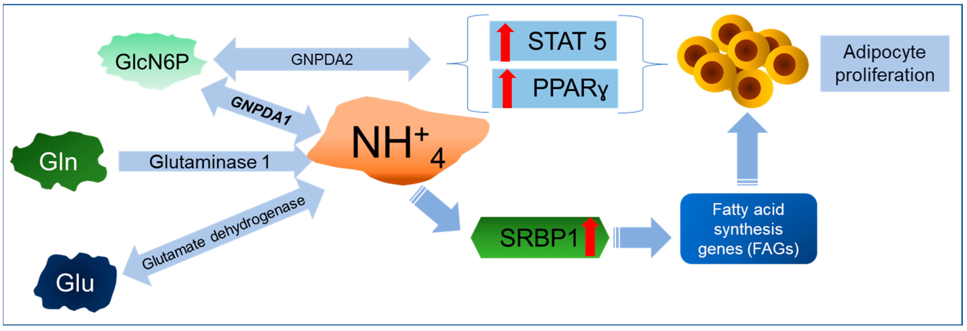
5. GNPDA1 and Cancer

Recently, evidence showing a clear relationship between GNPDA1 and some cancers was described (Table 1). However, the first indication that any GNPDA was related to cancer probably arose from a report by Sukeno et al. in 1971 [64]. In this report, they found an increased synthesis of glycogen and lactate in a rat model of Yoshida sarcoma cells. When incubated with 2 mM of glucosamine (GlcN), the cells showed an accumulation of GlcN6P and a rapid transformation into glycogen and lactate, with GNPDA being identified as the enzyme responsible. These cells had higher amounts of GNPDA than hepatic cells. From this, we can postulate that GNPDA1 is activated because higher concentrations of GlcN6P can be transformed in GlcNAc6P, producing more Fru6P and NH4+. Eight years later, Kikuchi et al. reported an increase of more than ten times in GNPDA enzymatic activity in rat hepatoma cells with respect to normal liver tissue. They also found a change in the fate of N-acetylglucosamine (GlcNAc)—while in normal hepatocytes, it is transformed mainly into sialic acid (biosynthetic route), hepatoma cells transform this amino sugar into glycolytic intermediates (catabolic pathway) [65]. Once again, in transformed cells, GNPDA1 activity can help in obtaining energy through glycolysis. Forty-one years later, it was confirmed that GNPDA1 mRNA expression was significantly increased in hepatocellular cancer (HCC) tissues and hepatoma cell lines, and it is a hallmark of a poor prognosis [66,67]. Experimental findings obtained from shRNA-knockdown GNPDA1 revealed that the expression of this isoenzyme promoted cell proliferation and raised the percentage of cells in the G0/G1 phase, favoring cell migration and invasiveness.
On the other hand, GNPDA1 knockdown in two different HCC cell lines increased the number of apoptotic cells, suggesting a possible role of GNPDA1 as an inhibitor of apoptosis [68]. Even though the mechanisms underlying these effects were not explored, the authors speculated that this increased GNPDA1 activity can provide an additional flux of Fru6P into the glycolytic pathway, favoring the energy supply in HCC cells. It is pertinent to emphasize that only the expression of GNPDA1, and not GNPDA2, was detected in the liver [5,26].
Furthermore, contradictory results were obtained when colorectal cancer SW480 cells were challenged with an anti-cancer activity compound derived from propolis [69]. As mentioned before, GNPDA1 displayed a pro-tumor effect in HCC, and we would expect that, by exposing cancer cells to an anti-tumor drug, the expression/activity of GNPDA1 would be down-regulated. Instead, this isoenzyme was up-regulated in SW480 cells treated with the anti-cancer drug. Notably, in addition to GNPDA1, the glycolytic enzyme glyceraldehyde-3-phosphate dehydrogenase (GAPDH) was also found to be up-regulated, reinforcing the proposed role of GNPDA1 as a “fuel supplier” for glycolysis. Nevertheless, for this type of cancer, this effect does not seem to be favorable. GAPDH has been implicated in some non-metabolic processes, for example, as an initiator of apoptosis [70]. This study did not suggest a reason for this; the fact is that colon and colorectal cancer SW480 cells express both GNPDAs, mostly GNPDA1, and its increased expression seems to be protective against tumor progression [69]. Therefore, more experimental studies are necessary to clarify this discrepancy. We can discern a possible explanation based on the metabolic roles of GFAT and GNPDA in UDP-GlcNAc6P and hyaluronan synthesis in human keratinocytes [2]. The following four enzymes catalyze the conversion of Fru6P into GlcN6P: GFAT1 and 2 and GNPDA1 and 2. When the biosynthesis of GlcNAc6P was blocked by silencing the GFAT1 gene, the expressions of both GNPDAs increased; then, the synthesis of GlcN6P was catalyzed, which was likely the reason for stable levels of UDP-GlcNAc being maintained.
On the other hand, the depletion of both GNPDA1 and GNPDA2 resulted in a significant increase in the cell surface hyaluronan coating. Hyaluronan, or hyaluronic acid (HA), plays an essential role in the epithelial–mesenchymal transition (EMT), cell migration, and cancer metastasis [71]. As was mentioned previously, the silencing of GFAT1 stimulated GNPDA1 and GDPDA2 expression, reducing cell migration [2]. Blocking HA synthesis in HCC Hepa 129 and Hep3B resulted in the inhibition of proliferation and increased apoptosis [72]. Whether a higher expression of GNPDA1 in hepatoma cells can increase HA synthesis, favoring pro-tumor effects, is still an open question. Finally, why the same protein behaves as a tumor suppressor or oncoprotein in different types of malignancies, as well as the mechanisms involved in this, are not entirely understood [73]. The existence of tissue-specific factors associated with proteins related to carcinogenesis and cancer development is a wide field to be explored.

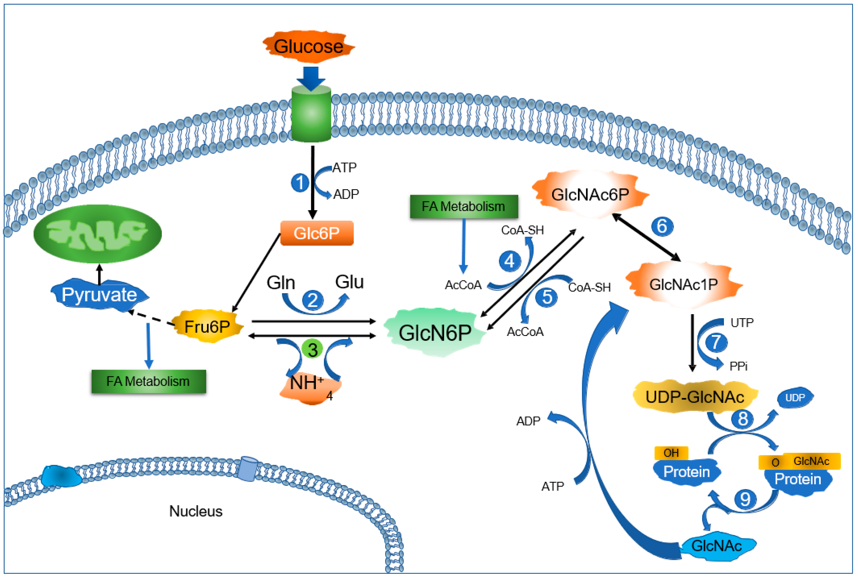
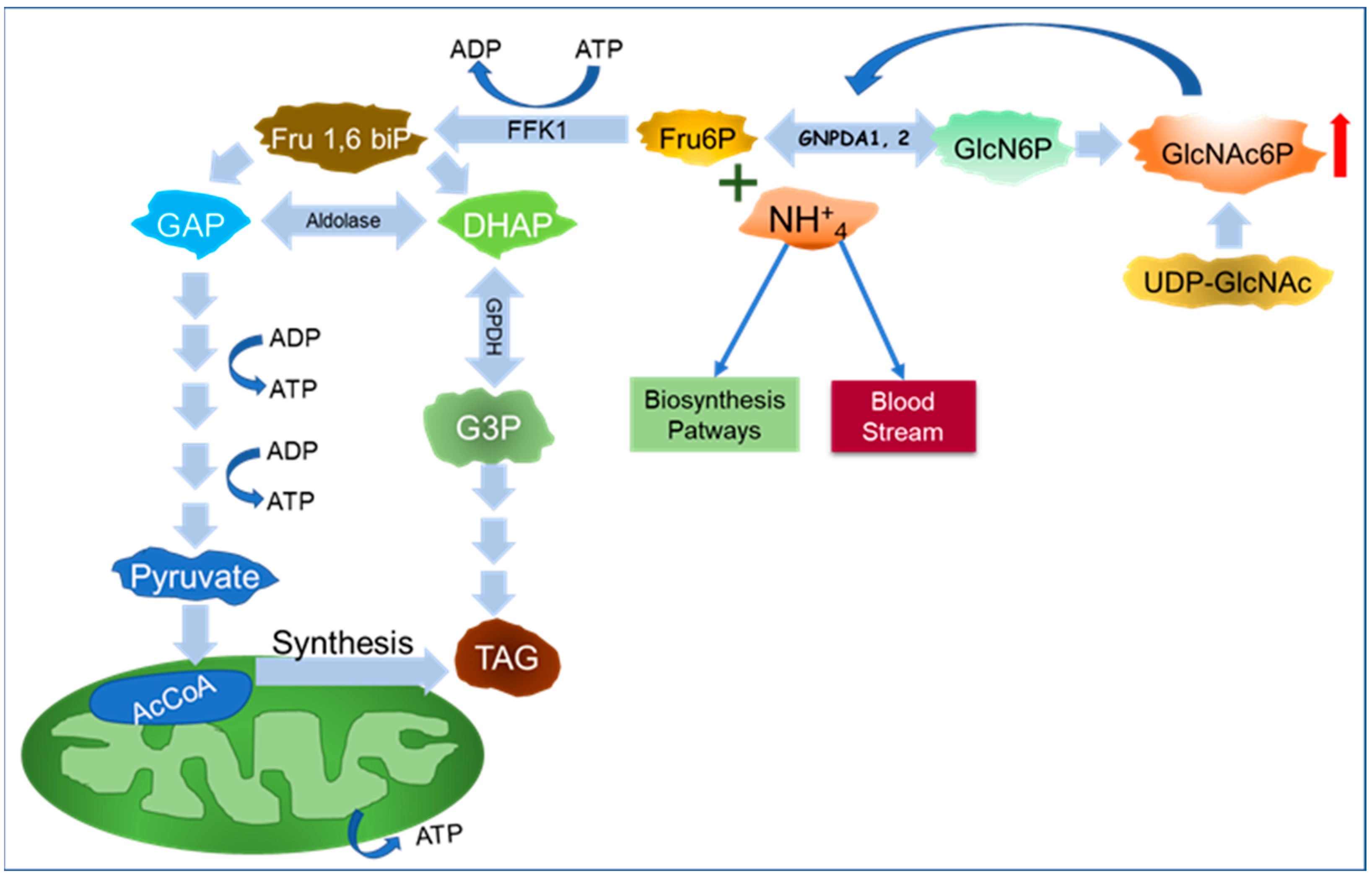
6. A New Perspective on GNPDAs, O-GlcNAcylation, and Ammonium

A good start for this section is the sentence “Glucosamine-6-phosphate isomerase catalyzes the conversion of glucosamine 6-phosphate to fructose 6-phosphate”, as referred to elsewhere. Of course, this is essential from a catabolic point of view because of the central role of GNPDAs in the degradation of GlcN6P, determining the catabolic fate of UDP-GlcNAc. However, most studies forget NH4+, the other product of the deaminating aspect of the GNPDA reaction. In order to discuss the relevance of NH4+, first, we take a brief look at GlcNAc6P metabolism and the O-GlcNAcylation of proteins. UDP-GlcNAc is synthesized through the HBP (Figure 1). GlcNAc is a common metabolite shared by the synthesis of the N- and O-glycan pathways and the O-GlcNAcylation of proteins. O-GlcNAcylation serves as a glucose-sensing mechanism. When cellular glucose levels are elevated, glucose is diverted into the HBP, increasing the levels of UDP-GlcNAc and the O-GlcNAcylation of proteins [74]. There is growing evidence supporting the role of the O-GlcNAcylation of proteins in different cellular processes and cancer [1,75,76,77,78]. The O-GlcNAcylation of cytoplasmic, nuclear, and mitochondrial proteins is the dynamic and reversible addition of a molecule of β-D-N-acetylglucosamine to specific serine and threonine residues. Once UDP-GlcNAc is synthesized, a GlcNAc moiety is added to a protein by the enzyme O-linked N-acetyl-glucosaminyltransferase (OGT), while O-linked N-acetyl β-D-glucosaminidase (OGA) controls its removal [1]. Released GlcNAc can be phosphorylated and recycled back into UDP-GlcNAc, creating a salvage loop. Both OGT and OGA are dysregulated in cancer. In general, OGT is up-regulated and OGA is down-regulated [79]. In most cancers, O-GlcNAcylation is enhanced, yielding more O-GlcNAcylation of regulatory proteins. Recently, it was suggested that OGA is down-regulated both transcriptionally and by epigenetic mechanisms [80]. In this sense, higher O-GlcNAc levels induce the transcription of several proliferative genes through different signaling routes [81]; e.g., in breast cancer cells, the Ras–Raf–MEK–ERK signaling pathway is affected, because O-GlcNAcylation avoids the degradation of MEK2, which extends its proliferative effects [82]. O-GlcNAcylation contributes to therapeutic resistance in cancer cells through different mechanisms, ranging from those affecting the amount or quality of anti-cancer drugs to cellular processes like autophagy, apoptosis, genome stability, the epithelial–mesenchymal transition, and the cell cycle [79].

Importance of GNPDA in Ammonium Metabolism

Traditionally, ammonia (NH3) and its protonated form (NH4+) were considered waste and toxic byproducts of amino acid and nucleotide catabolism, with NH4+ from GlnNAc6P catabolism also being identified recently. NH4+ toxicity is neutralized by integrating one molecule into glutamic acid (Glu), yielding Gln (the reaction is catalyzed by glutamine synthase; Figure 4), or even by the synthesis of urea in the liver. Then, urea is removed from the blood by the kidneys. Recently, the perception of NH4+ as a waste product of cellular metabolism has been changing, mainly with respect to cancer cells. Some mechanisms of ammonia metabolism supporting the growth and survival of cancer cells include (a) increased autophagy [83], (b) pyrimidine synthesis [84], (c) proline and aspartate synthesis [85], and (d) increased lipogenesis [86] (Figure 4). Indeed, it was found that NH4+ plays a key role as an activator of sterol regulatory element-binding transcription factor 1 (SREBP-1), promoting lipogenesis and tumor progression in different human cancer cells [86]. SREBPs are transcription factors that regulate the expressions of genes involved in cholesterol and lipid synthesis [87]. When cholesterol is high, SREBP-1 is located at the endoplasmic reticulum (ER) in a complex with SREBP-cleavage-activating protein (SCAP) and insulin-inducible gene protein (Insig) [88]. Experimental and computational data support that NH4+ released form Gln can bind SCAP through D428, releasing Insig and inducing the translocation of SREBP-1 from ER to the Golgi apparatus. In this organelle, SREBP1 is cleaved and the N-terminal domain is translocated into the nucleus, activating genes involved in lipid metabolism [86]. Increased lipogenesis is an essential hallmark of cancer progression and metastasis [89]. In this context, the enzymes involved in amino group metabolism have been intensely studied in neoplastic cells [90,91]. Due to the increased catabolism of Gln and decreased clearance caused by the poor vascularity inside solid tumors, cancer cells have higher levels of NH4+ compared with normal cells. The survival of cancer cells in high-NH4+ conditions depends on their ability to handle this excess by delivering NH4+ into anabolic pathways, providing building blocks such as nucleotides, amino acids, hexosamines, and indirectly, lipids, which are all necessary for an elevated rate of cell division (Figure 4).
As mentioned, under physiologic conditions, GlcN6P is synthetized from Fru6P and Gln by GFAT, and GNPDAs conduct the reversible deamination of GlcN6P. However, under some circumstances or metabolic conditions, GNPDAs can work in the anabolic direction, producing GlcN6P [13,14,64,65]. In this case, GNPDAs can incorporate NH4+ into the HBP. Exploring GNPDA’s role further, it is necessary to recall that the reaction catalyzed by GNPDAs is easily reversible (equilibrium constant of 0.2 M), while, on the other hand, the low kM for NH4+, as found for bGNPDA1 (3.7 mM), can push forward the biosynthetic function of GNPDA1 [18,30]. However, as mentioned previously, in ascites cancer cells, this enzyme functions mainly in the catabolism of GlcNAc, releasing NH4+ and incorporating the GlcN6P carbons into glycolysis and glycogen [64]. As represented in Figure 4, in cancer cells, the deaminating activity of GNPDA1 contributes to increasing the NH4+ cellular pool. At the same time, the enzyme supplies Fru6P to the glycolytic pathway, enhancing energy production [63] and fatty acid metabolism [91,92]. The link between GNPDA’s deaminating activity and the ammonium metabolism in cancer cells is still intriguing; a possible route for NH4+ coming from GlcN6P catabolism is its incorporation as the amino group for Glu or Gln by the reverse reaction of glutamate dehydrogenase [85,90,93,94] or by Gln-synthase [95,96]. In this way, the fate of NH4+ from GlcN6P could be in proteins, nucleic acids, or glutathione, which are necessary for cancer cell survival.
In conclusion, the role of GNPDA1 in cancer cells is currently unclear. The role of this enzyme in the synthesis or catabolism of GlcNAc clearly points toward an important function, in addition to GFAT1, in modulating the availability of GlcNAc for O-GlcNAcylation [7]. Dysregulation of the O-GlcNAcylation of proteins affects several cellular processes favoring cancer progression and other diseases, and both GNPDAs have kinetic properties that can support carcinogenic or anti-tumor mechanisms.
Despite the limitations of our current knowledge, we can still suggest some intriguing questions. Even when both deaminases catalyze the same reaction, why do GNPDA1 and 2 participate differentially in different diseases? Could these isoenzymes have other functions beyond their metabolic role? Moreover, is there any kind of epigenetic regulation operating on the expression of their genes? The first study aiming to explore the epigenetic regulation of the GNPDA2 gene was reported recently [97], and hopefully, more studies in this field will soon be produced. More experimental studies focused on these issues are necessary to clarify the roles of GNPDAs in metabolic and neoplastic diseases.

7. Future Directions

Future research in this field, mainly on GNPDA2, must go further than epidemiologic and genomic association studies and explore the mechanisms involved in the changes in sugar metabolism implied in the pathogenesis of obesity and related diseases such as diabetes mellitus type 2, and also in the development of complications derived from the glycation of blood and endothelial proteins, considering the central role of Fru6P in this process.
GNPDA2 is active in the hypothalamus, and its role in the mechanism of the regulation of glucose homeostasis is an open research field [61]. GNPDA1 is expressed in the brain to an even greater extent than GNPDA2; however, it is still unknown whether it plays any role in the metabolism of glucose in this organ in relation to obesity. In the same way, obesity is related to some types of cancer, such as breast cancer, and any possible involvement of GNPDA2 beyond GNPDA1 is yet to be identified
Another intriguing trait of GNPDA2 to be explored in the future is the existence of three isoforms. The human GNPDA2 variants show different numbers of amino acids, as follows: 276, 242, and 206 residues, corresponding to isoforms 1, 2, and 3, respectively. So far, there is no evidence regarding the different physiologic roles of these variants, or why GNPDA1 lacks isoforms.
The existence of natural hetero-hexameric forms of GNPDA (mixed oligomers of GNPDA1 and 2) has not been demonstrated. This indicates that evolutionary selection pressure has not led to monomer mixtures for either deaminase, and the reason for this is still unknown.

Author Contributions

R.L.-L. conceived the review concept, wrote the manuscript, and prepared the pictures. A.A.-G. and M.C.-L. critically reviewed the manuscript, contributed to the writing, and prepared the pictures. All authors have read and agreed to the published version of the manuscript.

Funding

Arnoldo Aquino-Gálvez was supported by CONAHCYT #194162 and Roberto Lara-Lemus, Arnoldo Aquino-Gálvez, and Manuel Castillejos-López by the Instituto Nacional de Enfermedades Respiratorias Ismael Cosio Villegas (INER).

Institutional Review Board Statement

Not applicable.

Informed Consent Statement

Not applicable.

Data Availability Statement

Not applicable.

Conflicts of Interest

The authors declare no conflicts of interest.

References

  1. Itano, N.; Iwamoto, S. Dysregulation of Hexosamine Biosynthetic Pathway Wiring Metabolic Signaling Circuits in Cancer. Biochim. Biophys. Acta Gen. Subj. 2023, 1867, 130250. [Google Scholar] [CrossRef] [PubMed]
  2. Vasconcelos-Dos-Santos, A.; de Queiroz, R.M.; da Costa Rodrigues, B.; Todeschini, A.R.; Dias, W.B. Hyperglycemia and Aberrant O-GlcNAcylation: Contributions to Tumor Progression. J. Bioenerg. Biomembr. 2018, 50, 175–187. [Google Scholar] [CrossRef] [PubMed]
  3. Akella, N.M.; Ciraku, L.; Reginato, M.J. Fueling the Fire: Emerging Role of the Hexosamine Biosynthetic Pathway in Cancer. BMC Biol. 2019, 17, 52. [Google Scholar] [CrossRef]
  4. Leloir, L.F.; Cardini, C.E. Enzymes Acting on Glucosamine Phosphates. Biochim. Biophys. Acta 1956, 20, 33–42. [Google Scholar] [CrossRef]
  5. Zhang, J.; Zhang, W.; Zou, D.; Chen, G.; Wan, T.; Li, N.; Cao, X. Cloning and Functional Characterization of GNPI2, a Novel Human Homolog of Glucosamine-6-Phosphate Isomerase/Oscillin. J. Cell. Biochem. 2003, 88, 932–940. [Google Scholar] [CrossRef]
  6. Arreola, R.; Valderrama, B.; Morante, M.L.; Horjales, E. Two Mammalian Glucosamine-6-Phosphate Deaminases: A Structural and Genetic Study. FEBS Lett. 2003, 551, 63–70. [Google Scholar] [CrossRef]
  7. Oikari, S.; Makkonen, K.; Deen, A.J.; Tyni, I.; Kärnä, R.; Tammi, R.H.; Tammi, M.I. Hexosamine Biosynthesis in Keratinocytes: Roles of GFAT and GNPDA Enzymes in the Maintenance of UDP-GlcNAc Content and Hyaluronan Synthesis. Glycobiology 2016, 26, 710–722. [Google Scholar] [CrossRef] [PubMed]
  8. Faulkner, P.; Quastel, J.H. Anaerobic Deamination of D-glucosamine by Bacterial and Brain Extracts. Nature 1956, 177, 1216–1218. [Google Scholar] [CrossRef]
  9. Pattabiraman, T.N.; Bachhawat, B.K. Purification of Glucosamine 6-phosphate Deaminase from Human Brain. Biochim. Biophys. Acta 1961, 54, 273–283. [Google Scholar] [CrossRef]
  10. Leloir, L.F.; Cardini, C.E. Glucosamine-6-Phosphate Deaminase from Pig Kidney. Methods Enzymol. 1962, 5, 418–422. [Google Scholar]
  11. Kikuchi, K.; Kikuchi, H.; Tsuiki, S. Stabilization and Purification of Glucosamine 6-Phosphate Isomerase from Rat Kidney. Sci. Rep. Res. Inst. Tohoku Univ. Ser. C Med. 1979, 26, 92–98. [Google Scholar]
  12. Comb, D.G.; Roseman, S. Glucosamine Metabolism. IV. Glucosamine-6-Phosphate Deaminase. J. Biol. Chem. 1958, 232, 807–827. [Google Scholar] [CrossRef] [PubMed]
  13. Benson, R.; Friedman, S. Allosteric Control of Glucosamine Phosphate Isomerase from the Adult Housefly and Its Role in the Synthesis of Glucosamine 6-Phosphate. J. Biol. Chem. 1970, 245, 2219–2228. [Google Scholar] [CrossRef] [PubMed]
  14. Enghofer, E.; Kress, H. Glucosamine Metabolism in Drosophila virilis Salivary Glands: Ontogenetic Changes of Enzyme Activities and Metabolite Synthesis. Dev. Biol. 1980, 78, 63–75. [Google Scholar] [CrossRef]
  15. Tesoriere, G.; Vento, R.; Magistro, D.; Dones, F. Glucosamine 6-Phosphate Deaminase from Rabbit Erythrocytes. Ital. J. Biochem. 1970, 19, 139–154. [Google Scholar]
  16. Weidanz, J.A.; Campbell, P.; De Lucas, L.J.; Jin, J.; Moore, D.; Rodén, L.; Yu, H.; Heilmann, E.; Vezza, A.C. Glucosamine 6-Phosphate Deaminase in Normal Human Erythrocytes. Br. J. Haematol. 1995, 91, 72–79. [Google Scholar] [CrossRef]
  17. Cayli, A.; Hirschmann, F.; Wirth, M.; Hauser, H.; Wagner, R. Cell Lines with Reduced UDP-N Acetylhexosamine Pool in the Presence of Ammonium. Biotechnol. Bioeng. 1999, 65, 192–200. [Google Scholar] [CrossRef]
  18. Calcagno, M.; Campos, P.J.; Mulliert, G.; Suastegui, J. Purification, Molecular and Kinetic Properties of Glucosamine-6-Phosphate Isomerase (Deaminase) from Escherichia coli. Biochim. Biophys. Acta 1984, 787, 165–173. [Google Scholar] [CrossRef]
  19. Oliva, G.; Fontes, M.R.; Garratt, M.C.; Altamirano, M.M.; Calcagno, M.L.; Horjales, E. Structure and Catalytic Mechanism of Glucosamine 6-Phosphate Deaminase from Escherichia coli at 2.1 A Resolution. Structure 1995, 3, 1323–1332. [Google Scholar] [CrossRef]
  20. Plumbridge, J.A.; Cochet, O.; Souza, J.M.; Altamirano, M.M.; Calcagno, M.L.; Badet, B. Coordinated Regulation of Amino Sugar-Synthesizing and -Degrading Enzymes in Escherichia coli K-12. J. Bacteriol. 1993, 175, 4951–4956. [Google Scholar] [CrossRef]
  21. Marcos-Viquez, J.; Rodríguez-Hernández, A.; Álvarez-Añorve, L.I.; Medina-García, A.; Plumbridge, J.; Calcagno, M.L.; Rodríguez-Romero, A.; Bustos-Jaimes, I. Substrate Binding in the Allosteric Site Mimics Homotropic Cooperativity in the SIS-fold Glucosamine-6-Phosphate Deaminases. Protein Sci. 2023, 32, e4651. [Google Scholar] [CrossRef] [PubMed]
  22. Álvarez-Añorve, L.I.; Gaugué, I.; Link, H.; Marcos-Viquez, J.; Díaz-Jiménez, D.M.; Zonszein, S.; Bustos-Jaimes, I.; Schmitz-Afonso, I.; Calcagno, M.L.; Plumbridge, J. Allosteric Activation of Escherichia coli Glucosamine-6-Phosphate Deaminase (NagB) In Vivo Justified by Intracellular Amino Sugar Metabolite Concentrations. J. Bacteriol. 2016, 198, 1610–1620. [Google Scholar] [CrossRef]
  23. Sosa-Peinado, A.; González-Andrade, M. Site-directed Fluorescence Labeling Reveals Differences on the R-Conformer of Glucosamine 6-Phosphate Deaminase of Escherichia coli Induced by Active or Allosteric Site Ligands at Steady State. Biochemistry 2005, 44, 15083–15092. [Google Scholar] [CrossRef] [PubMed]
  24. Lara-Lemus, R.; Libreros-Minotta, C.A.; Altamirano, M.M.; Calcagno, M.L. Purification and Characterization of Glucosamine-6-Phosphate Deaminase from Dog Kidney Cortex. Arch. Biochem. Biophys. 1992, 297, 213–220. [Google Scholar] [CrossRef]
  25. Parrington, J.; Swann, K.; Shevchenko, V.I.; Sesay, A.K.; Lai, F.A. Calcium Oscillations in Mammalian Eggs Triggered by a Soluble Sperm Protein. Nature 1996, 379, 364–368. [Google Scholar] [CrossRef] [PubMed]
  26. Wolosker, H.; Kline, D.; Bian, Y.; Blackshaw, S.; Cameron, A.M.; Fralich, T.J.; Schnaar, R.L.; Snyder, S.H. Molecularly Cloned Mammalian Glucosamine-6-Phosphate Deaminase Localizes to Transporting Epithelium and Lacks Oscillin Activity. FASEB J. 1998, 12, 91–99. [Google Scholar] [PubMed]
  27. Wolny, Y.M.; Fissore, R.A.; Wu, H.; Reis, M.M.; Colombero, L.T.; Ergün, B.; Rosenwaks, Z.; Palermo, G.D. Human Glucosamine-6-Phosphate Isomerase, a Homologue of Hamster oscillin, Does Not Appear To Be Involved in Ca2+ Release in Mammalian Oocytes. Mol. Rep. Dev. 1999, 52, 277–287. [Google Scholar] [CrossRef]
  28. Shevchenko, V.; Hogben, M.; Ekong, R.; Parrington, J.; Lai, F.A. The Human Glucosamine-6-Phosphate Deaminase Gene: cDNA Cloning and Expression, Genomic Organization and Chromosomal Localization. Gene 1998, 216, 31–38. [Google Scholar] [CrossRef]
  29. Amireault, P.; Dube, F. Cloning, Sequencing, and Expression Analysis of Mouse Glucosamine-6-Phosphate Deaminase (GNPDA/Oscillin). Mol. Rep. Dev. 2000, 56, 424–435. [Google Scholar] [CrossRef]
  30. Lara-Lemus, R.; Calcagno, M.L. Glucosamine-6-Phosphate Deaminase from Beef Kidney Is an Allosteric System of the V-type. Biochim. Biophys. Acta 1998, 1388, 1–9. [Google Scholar] [CrossRef]
  31. Davies, J.S.; Coombes, D.; Horne, C.R.; Pearce, F.G.; Friemann, R.; North, R.A.; Dobson, R.A.J. Functional and Solution Structure Studies of Amino Sugar Deacetylase and Deaminase Enzymes from Staphylococcus aureus. FEBS Lett. 2019, 593, 52–66. [Google Scholar] [CrossRef] [PubMed]
  32. Moye, Z.D.; Burne, R.A.; Zeng, L. Uptake and Metabolism of N-acetylglucosamine and Glucosamine by Streptococcus mutans. Appl. Environ. Microbiol. 2014, 80, 5053–5067. [Google Scholar] [CrossRef] [PubMed]
  33. Tanaka, T.; Takahashi, F.; Fukui, T.; Fujiwara, S.; Atomi, H.; Imanaka, T. Characterization of a Novel Glucosamine-6-Phosphate Deaminase from a Hyperthermophilic Archaeon. J. Bacteriol. 2005, 187, 7038–7044. [Google Scholar] [CrossRef]
  34. Solar, T.; Tursic, J.; Legisa, M. The Role of Glucosamine-6-Phosphate Deaminase at the Early Stages of Aspergillus niger Growth in a High-Citric-Acid-Yielding Medium. Appl. Microbiol. Biotechnol. 2008, 78, 613–619. [Google Scholar] [CrossRef]
  35. Aguilar-Díaz, H.; Díaz-Gallardo, M.; Laclette, J.P.; Carrero, J.C. In vitro Induction of Entamoeba histolytica Cyst-Like Structures from Trophozoites. PLoS Negl. Trop. Dis. 2010, 4, e607. [Google Scholar] [CrossRef] [PubMed]
  36. Álvarez-Añorve, L.I.; Alonzo, D.A.; Mora-Lugo, R.; Lara-González, S.; Bustos-Jaimes, I.; Plumbridge, J.; Calcagno, M.L. Allosteric Kinetics of the Isoform 1 of Human Glucosamine-6-Phosphate Deaminase. Biochim. Biophys. Acta 2011, 1814, 1846–1853. [Google Scholar] [CrossRef]
  37. Melén, E.; Himes, B.E.; Brehm, J.M.; Boutaoui, N.; Klanderman, E.J.; Sylvia, J.S.; Lasky-Su, J. Analyses of Shared Genetic Factors Between Asthma and Obesity in Children. J. Allergy Clin. Immunol. 2010, 126, e1–e8. [Google Scholar] [CrossRef]
  38. Colak, Y.; Afzal, S.; Lange, P.; Nordestgaard, B.G. Obese Individuals Experience Wheezing Without Asthma but not Asthma Without wheezing: A Mendelian Randomisation Study of 85 437 Adults from the Copenhagen General Population Study. Thorax 2016, 71, 247–254. [Google Scholar] [CrossRef]
  39. Ng, M.C.Y.; Tam, C.H.T.; So, W.Y.; Ho, J.S.; Chan, A.W.; Lee, H.M.; Wang, Y.; Lam, V.K.L.; Chan, J.C.N.; Ma, R.C.W. Implication of Genetic Variants Near NEGR1, SEC16B, TMEM18, ETV5/DGKG, GNPDA2, LIN7C/BDNF, MTCH2, BCDIN3D/FAIM2, SH2B1, FTO, MC4R, and KCTD15 with Obesity and Type 2 Diabetes in 7705 Chinese. J. Clin. Endocrinol. Metab. 2010, 95, 2418–2425. [Google Scholar] [CrossRef]
  40. Hebbar, P.; Abubaker, J.A.; Abu-Farha, M.; Tuomilehto, J.; Al-Mulla, F.; Thanaraj, T.A. A Perception on Genome-Wide Genetic Analysis of Metabolic Traits in Arab Populations. Front. Endocrinol. 2019, 10, 8. [Google Scholar] [CrossRef]
  41. Kong, X.; Zhang, X.; Zhao, Q.; He, J.; Chen, L.; Zhao, Z.; Li, Q.; Ge, J.; Chen, G.; Guo, X.; et al. Obesity-Related Genomic Loci Are Associated with Type 2 Diabetes in a Han Chinese Population. PLoS ONE 2014, 9, e104486. [Google Scholar] [CrossRef] [PubMed]
  42. Kang, J.; Guan, R.-C.; Zhao, Y.; Chen, Y. Obesity-Related Loci in TMEM18, CDKAL1 and FAIM2 Are Associated with Obesity and Type 2 Diabetes in Chinese Han patients. BMC Med. Genet. 2020, 21, 65. [Google Scholar] [CrossRef]
  43. Kong, X.; Xing, X.; Zhang, X.; Hong, J.; Yang, W. Sexual Dimorphism of a Genetic Risk Score for Obesity and Related Traits among Chinese Patients with Type 2 Diabetes. Obes. Facts 2019, 12, 328–343. [Google Scholar] [CrossRef] [PubMed]
  44. Hotta, K.; Nakamura, M.; Nakamura, T.; Matsuo, T.; Nakata, Y.; Kamohara, S.; Miyatake, N.; Kotani, K.; Komatsu, R.; Itoh, N.; et al. Association Between Obesity and Polymorphisms in SEC16B, TMEM18, GNPDA2, BDNF, FAIM2 and MC4R in a Japanese Population. J. Hum. Genet. 2009, 54, 727–731. [Google Scholar] [CrossRef]
  45. Robiou-du-Pont, S.; Bonnefond, A.; Yengo, L.; Vaillant, E.; Lobbens, S.; Durand, E.; Weill, J.; Lantieri, O.; Balkau, B.; Charpentier, G.; et al. Contribution of 24 Obesity-Associated Genetic Variants to Insulin Resistance, Pancreatic Beta-Cell Function and Type 2 Diabetes Risk in the French Population. Int. J. Obes. 2013, 37, 980–985. [Google Scholar] [CrossRef]
  46. Mejía-Benítez, A.; Klünder-Klünder, M.; Yengo, L.; Meyre, D.; Aradillas, C.; Cruz, E.; Pérez-Luque, E.; Malacara, J.M.; Garay, M.E.; Peralta-Romero, J.; et al. Analysis of the Contribution of FTO, NPC1, ENPP1, NEGR1, GNPDA2 and MC4R Genes to Obesity in Mexican Children. BMC Med. Genet. 2013, 14, 21. [Google Scholar] [CrossRef]
  47. Takeuchi, F.; Yamamoto, K.; Katsuya, T.; Nabika, T.; Sugiyama, T.; Fujioka, A.; Isono, M.; Ohnaka, K.; Fujisawa, T.; Nakashima, E.; et al. Association of Genetic Variants for Susceptibility to Obesity with Type 2 Diabetes in Japanese Individuals. Diabetologia 2011, 54, 1350–1359. [Google Scholar] [CrossRef] [PubMed]
  48. Wu, L.; Xi, B.; Zhang, M.; Shen, Y.; Zhao, X.; Cheng, H.; Hou, D.; Sun, D.; Ott, J.; Wang, X.; et al. Associations of Six Single Nucleotide Polymorphisms in Obesity-Related Genes with BMI and Risk of Obesity in Chinese Children. Diabetes 2010, 59, 3085–3089. [Google Scholar] [CrossRef]
  49. De, R.; Hu, T.; Moore, J.H.; Gilbert-Diamond, D. Characterizing Gene-Gene Interactions in a Statistical Epistasis Network of Twelve Candidate Genes for Obesity. BioData Min. 2015, 8, 45. [Google Scholar] [CrossRef]
  50. Costa-Urrutia, P.; Abud, C.; Franco-Trecu, V.; Colistro, V.; Rodríguez-Arellano, M.E.; Alvarez-Fariña, R.; Acuña-Alonso, V.; Bertoni, B.; Granados, J. Effect of 15 BMI-Associated Polymorphisms, Reported for Europeans, Across Ethnicities and Degrees of Amerindian Ancestry in Mexican Children. Int. J. Mol. Sci. 2020, 21, 374. [Google Scholar] [CrossRef]
  51. Hong, J.; Shi, J.; Qi, L.; Cui, B.; Gu, W.; Zhang, Y.; Li, L.; Xu, M.; Wang, L.; Zhai, Y.; et al. Genetic Susceptibility, Birth Weight and Obesity Risk in Young Chinese. Int. J. Obes. 2013, 37, 673–677. [Google Scholar] [CrossRef]
  52. Wu, L.; Ma, F.; Zhao, X.; Zhang, M.-X.; Wu, J.; Wu, J.; Jie, M. GNPDA2 Gene Affects Adipogenesis and Alters the Transcriptome Profile of Human Adipose-Derived Mesenchymal Stem Cells. Int. J. Endocrinol. 2019, 2019, 9145452. [Google Scholar] [CrossRef] [PubMed]
  53. Renström, F.; Payne, F.; Nordström, A.; Brito, E.C.; Rolandsson, O.; Hallmans, G.; Barroso, I.; Nordström, P.; Franks, P.W. Replication and Extension of Genome-Wide Association Study Results for Obesity in 4923 Adults from Northern Sweden. Hum. Mol. Genet. 2009, 18, 1489–1496. [Google Scholar] [CrossRef] [PubMed]
  54. Dorajoo, R.; Blakemore, A.I.F.; Sim, X.; Ong, R.T.-H.; Ng, D.P.K.; Seielstad, M.; Wong, T.-Y.; Saw, S.-M.; Froguel, P.; Liu, J.; et al. Replication of 13 Obesity Loci Among Singaporean Chinese, Malay and Asian-Indian Populations. Int. J. Obes. 2011, 36, 159–163. [Google Scholar] [CrossRef] [PubMed]
  55. Yılmaz, B.; Karadağ, M.G. The Current Review of Adolescent Obesity: The Role of Genetic Factors. J. Pediatr. Endocrinol. Metab. 2020, 34, 151–162. [Google Scholar] [CrossRef] [PubMed]
  56. Graff, M.; Gordon-Larsen, P.; Lim, U.; Fowke, J.H.; Love, S.-A.; Fesinmeyer, M.; Wilkens, L.R.; Vertilus, S.; Ritchie, M.D.; Prentice, R.L.; et al. The Influence of Obesity-Related Single Nucleotide Polymorphisms on BMI Across the Life Course: The PAGE study. Diabetes 2013, 62, 1763–1767. [Google Scholar] [CrossRef]
  57. Felix, J.F.; Bradfield, J.P.; Monnereau, C.; van der Valk, R.J.P.; Stergiakouli, E.; Chesi, A.; Gaillard, R.; Feenstra, B.; Thiering, E.; Kreiner-Møller, E.; et al. Genome-Wide Association Analysis Identifies Three New Susceptibility Loci for Childhood Body Mass Index. Hum. Mol. Genet. 2016, 25, 389–403. [Google Scholar] [CrossRef]
  58. Zhao, J.; Bradfield, J.P.; Zhang, H.; Sleiman, P.M.; Kim, C.E.; Glessner, J.T.; Deliard, S.; Thomas, K.A.; Frackelton, E.C.; Li, M.; et al. Role of BMI-Associated Loci Identified in GWAS Meta-Analyses in the Context of Common Childhood Obesity in European Americans. Obesity 2011, 19, 2436–2439. [Google Scholar] [CrossRef]
  59. Willer, C.J.; Speliotes, E.K.; Loos, R.J.F.; Li, S.; Lindgren, C.M.; Heid, I.M.; Berndt, S.I.; Elliott, A.L.; Jackson, A.U.; Lamina, C.; et al. Six New Loci Associated with Body Mass Index Highlight a Neuronal Influence on Body Weight Regulation. Nat. Genet. 2009, 41, 25–34. [Google Scholar] [CrossRef]
  60. Flores-Dorantes, M.T.; Díaz-López, Y.E.; Gutiérrez-Aguilar, R. Environment and Gene Association with Obesity and Their Impact on Neurodegenerative and Neurodevelopmental Diseases. Front. Neurosci. 2020, 14, 863. [Google Scholar] [CrossRef]
  61. Gutierrez-Aguilar, R.; Grayson, B.E.; Kim, D.-H.; Yalamanchili, S.; Calcagno, M.L.; Woods, S.C.; Seeley, R.J. CNS GNPDA2 Does Not Control Appetite, but Regulates Glucose Homeostasis. Frnt. Nutr. 2021, 8, 787470. [Google Scholar] [CrossRef] [PubMed]
  62. Lachén-Montes, M.; González-Morales, A.; Iloro, I.; Elortza, F.; Ferrer, I.; Gveric, D.; Fernández-Irigoyen, J.; Santamaría, E. Unveiling the Olfactory Proteostatic Disarrangement in Parkinson’s Disease by Proteome-Wide Profiling. Neurobiol. Aging 2019, 73, 123–134. [Google Scholar] [CrossRef]
  63. Lachén-Montes, M.; González-Morales, A.; Fernández-Irigoyen, J.; Santamaría, E. Deployment of Label-Free Quantitative Olfactory Proteomics to Detect Cerebrospinal Fluid Biomarker Candidates in Synucleinopathies. Methods Mol. Biol. 2019, 2044, 273–289. [Google Scholar] [CrossRef] [PubMed]
  64. Sukeno, T.; Kikuchi, H.; Saeki, H.; Tsuiki, S. Transformation of Glucosamine to Glycogen and Lactate by Ascites Tumor Cells. Biochim. Biophys. Acta 1971, 244, 19–29. [Google Scholar] [CrossRef]
  65. Kikuchi, K.; Tsuiki, S. Metabolism of Exogenous N-Acetylglucosamine in Extracts of Rat Kdney, Liver and Hepatoma. Biochim. Biophys. Acta 1979, 584, 246–253. [Google Scholar] [CrossRef] [PubMed]
  66. Li, D.; Cheng, X.; Zheng, W.; Chen, J. Glucosamine-6-Phosphate Isomerase 1 Promotes Tumor Progression and Indicates Poor Prognosis in Hepatocellular Carcinoma. Cancer Manag. Res. 2020, 12, 4923–4935. [Google Scholar] [CrossRef]
  67. Zhou, W.; Zhang, S.; Cai, Z.; Gao, F.; Deng, W.; Wen, Y.; Qiu, Z.-W.; Hou, Z.-K.; Chen, X.-L. A Glycolysis-Related Gene Pairs Signature Predicts Prognosis in Patients with Hepatocellular Carcinoma. Peer J. 2020, 8, e9944. [Google Scholar] [CrossRef] [PubMed]
  68. Xia, R.; Tang, H.; Shen, J.; Xu, S.; Liang, Y.; Zhang, Y.; Gong, X.; Min, Y.; Zhang, D.; Tao, C.; et al. Prognostic Value of a Novel Glycolysis-Related Gene Expression Signature for Gastrointestinal Cancer in the Asian Population. Cancer Cell Int. 2021, 21, 154. [Google Scholar] [CrossRef]
  69. He, Y.-J.; Li, W.-L.; Liu, B.-H.; Dong, H.; Mou, Z.-R.; Wu, Y.-Z. Identification of Differential Proteins in Colorectal Cancer Cells Treated with Caffeic Acid Phenethyl Ester. World J. Gastroenterol. 2014, 33, 11840–11849. [Google Scholar] [CrossRef]
  70. Tarze, A.; Deniaud, A.; Le Bras, M.; Maillier, E.; Molle, D.; Larochette, N.; Zamzami, N.; Jan, G.; Kroemer, G.; Brenner, C. GAPDH, a Novel Regulator of the pro-Apoptotic Mitochondrial Membrane Permeabilization. Oncogene 2007, 26, 2606–2620. [Google Scholar] [CrossRef]
  71. Carvalho-Cruz, P.; Alisson-Silva, F.; Todeschini, A.R.; Dias, W.B. Cellular Glycosylation Senses Metabolic Changes and Modulates Cell Plasticity During Epithelial to Mesenchymal Transition. Dev. Dyn. 2018, 247, 481–491. [Google Scholar] [CrossRef] [PubMed]
  72. Piccioni, F.; Malvicini, M.; Garcia, M.G.; Rodriguez, A.; Atorrasagasti, C.; Kippes, N.; Piedra Buena, I.T.; Rizzo, M.M.; Bayo, J.; Aquino, J.; et al. Antitumor effects of Hyaluronic Acid Inhibitor 4-methylumbelliferone in an Orthotopic HepatoCellular Carcinoma Model in Mice. Glycobiology 2012, 22, 400–410. [Google Scholar] [CrossRef] [PubMed]
  73. Lara-Lemus, R. On The Role of Myelin and Lymphocyte Protein (MAL) In Cancer: A Puzzle with Two Faces. J. Cancer 2019, 10, 2312–2318. [Google Scholar] [CrossRef]
  74. Yang, X.Y.; Qian, K.V. Protein O-GlcNAcylation: Emerging Mechanisms and Functions. Nat. Rev. Mol. Cell Biol. 2017, 18, 452–465. [Google Scholar] [CrossRef]
  75. Zhang, J.; Wang, Y. Emerging Roles of O-GlcNAcylation in Protein Trafficking and Secretion. J. Biol. Chem. 2024, 300, 105677. [Google Scholar] [CrossRef]
  76. Dupas, T.; Lauzier, B.; McGraw, S. O-GlcNAcylation: The Sweet Side of Epigenetics. Epigenet. Chromatin 2023, 16, 49. [Google Scholar] [CrossRef]
  77. He, X.-F.; Hu, X.; Wen, G.-J.; Wang, Z.; Lin, W.-J. O-GlcNAcylation in Cancer Development and Immunotherapy. Cancer Lett. 2023, 566, 216258. [Google Scholar] [CrossRef] [PubMed]
  78. Li, Y.; Qu, S.; Jin, H.; Jia, Q.; Li, M. Role of O-GlcNAcylation in Cancer Biology. Pathol. Res. Pract. 2024, 253, 155001. [Google Scholar] [CrossRef]
  79. Chen, L.; Hu, M.; Chen, L.; Peng, Y.; Zhang, C.; Wang, X.; Li, X.; Yao, Y.; Song, Q.; Li, J.; et al. Targeting O-GlcNAcylation in Cancer Therapeutic Resistance: The Sugar Saga Continues. Cancer Lett. 2024, 588, 216742. [Google Scholar] [CrossRef]
  80. Lin, C.-H.; Liao, C.-C.; Chen, M.-Y.; Chou, T.-Y. Feedback Regulation of O-GlcNAc Transferase through Translation Control to Maintain Intracellular O-GlcNAc Homeostasis. Int. J. Mol. Sci. 2021, 22, 3463. [Google Scholar] [CrossRef]
  81. Ciraku, L.; Esquea, E.M.; Reginato, M.J. O-GlcNAcylation Regulation of Cellular Signaling in Cancer. Cell. Signal. 2022, 90, 110201. [Google Scholar] [CrossRef] [PubMed]
  82. Xu, Y.; Sheng, X.; Zhao, T.; Zhang, L.; Ruan, Y.; Lu, H. O-GlcNAcylation of MEK2 Promotes the Proliferation and Migration of Breast Cancer Cells. Glycobiology 2021, 31, 571–581. [Google Scholar] [CrossRef] [PubMed]
  83. Eng, C.H.; Yu, K.; Lucas, J.; White, E.; Abraham, R.T. Ammonia Derived from Glutaminolysis is a Diffusible Regulator of Autophagy. Sci. Signal. 2010, 3, ra31. [Google Scholar] [CrossRef] [PubMed]
  84. Kim, J.; Hu, Z.; Cai, L.; Li, K.; Choi, E.; Faubert, B.; Bezwada, D.; Rodriguez-Canales, J.; Villalobos, P.; Lin, Y.F.; et al. CPS1 Maintains Pyrimidine Pools and DNA Synthesis in KRAS/LKB1-mutant Lung Cancer Cells. Nature 2017, 546, 168–172. [Google Scholar] [CrossRef] [PubMed]
  85. Spinelli, J.S.; Yoon, H.; Ringel, A.E.; Jeanfavre, S.; Clish, C.B.; Haigis, M.C. Metabolic Recycling of Ammonia via Glutamate Dehydrogenase Supports Breast Cancer Biomass. Science 2017, 358, 941–946. [Google Scholar] [CrossRef]
  86. Cheng, C.; Geng, F.; Li, Z.; Zhong, Y.; Wang, H.; Cheng, X.; Zhao, Y.; Mo, X.; Horbinski, C.; Duan, W.; et al. Ammonia Stimulates SCAP/Insig Dissociation and SREBP-1 Activation to Promote Lipogenesis and Tumour Growth. Nat. Metab. 2022, 4, 575–588. [Google Scholar] [CrossRef]
  87. Shimano, H.; Sato, R. SREBP-Regulated Lipid Metabolism: Convergent Physiology-Divergent Pathophysiology. Nat. Rev. Endocrinol. 2017, 13, 710–730. [Google Scholar] [CrossRef]
  88. Lee, H.M.; Nayak, A.; Kim, J. A new role for ammonia in tumorigenesis? Cell Metab. 2022, 34, 944–946. [Google Scholar] [CrossRef]
  89. Swinnen, J.V.; Brusselmans, K.; Verhoeven, G. Increased lipogenesis in cancer cells: New players, novel targets. Curr. Opin. Clin. Nutr. Metab. Care 2006, 9, 358–365. [Google Scholar] [CrossRef]
  90. Moreno-Sánchez, R.; Marín-Hernández, A.; Gallardo-Pérez, J.C.; Pacheco-Velázquez, S.C.; Robledo-Cadena, D.X.; Padilla-Flores, J.A.; Saavedra, E.; Rodríguez-Enríquez, S. Role of Glutamate Dehydrogenase in Cancer Cells. Front. Oncol. 2020, 10, 429. [Google Scholar] [CrossRef]
  91. McClain, D.A.; Hazel, M.; Parker, G.; Cooksey, R.C. Adipocytes with Increased Hexosamine Flux Exhibit Insulin Resistance, Increased Glucose Uptake, and Increased Synthesis and Storage of Lipid. Am. J. Physiol. Metab. 2005, 288, E973–E979. [Google Scholar] [CrossRef] [PubMed]
  92. Rumberger, J.M.; Wu, T.; Hering, M.A.; Marshall, S. Role of Hexosamine Biosynthesis in Glucose-Mediated Up-Regulation of Lipogenic Enzyme mRNA Levels. Effects of Glucose, Glutamine, and Glucosamine on Glycerophosphate Dehydrogenase, Fatty Acid Synthase, and Acetyl-coA Carboxylase mRNA Levels. J. Biol. Chem. 2003, 278, 28547–28552. [Google Scholar] [CrossRef] [PubMed]
  93. Chance, W.T.; Cao, L.; Nelson, J.L.; Foley-Nelson, T.; Fischer, J.E. Hyperammonemia in Anorectic Tumor-Bearing Rats. Life Sci. 1988, 43, 67–74. [Google Scholar] [CrossRef] [PubMed]
  94. Phang, J.M.; Liu, W.; Hancock, C.N.; Fischer, J.W. Proline Metabolism and Cancer: Emerging Links to Glutamine and Collagen. Curr. Opin. Clin. Nutr. Metab. Care 2015, 18, 71–77. [Google Scholar] [CrossRef] [PubMed]
  95. Kim, G.W.; Lee, D.H.; Jeon, Y.H.; Yoo, J.; Kim, S.Y.; Lee, S.W.; Cho, H.Y.; Kwon, S.H. Glutamine Synthetase as a Therapeutic Target for Cancer Treatment. Int. J. Mol. Sci. 2021, 22, 1701. [Google Scholar] [CrossRef]
  96. Dai, W.; Shen, J.; Yan, J.; Bott, A.J.; Maimouni, S.; Daguplo, H.Q.; Wang, Y.; Khayati, K.; Guo, J.Y.; Zhang, L.; et al. Glutamine Synthetase Limits β-Catenin–Mutated Liver Cancer Growth by Maintaining Nitrogen Homeostasis and Suppressing mTORC1. J. Clin. Investig. 2022, 132, e161408. [Google Scholar] [CrossRef]
  97. Chavira-Suárez, E.; Ramírez-Mendieta, A.J.; Martínez-Gutiérrez, S.; Zárate-Segura, P.; Beltrán-Montoya, J.; Espinosa-Maldonado, N.C.; de la Cerda-Ángeles, J.C.; Vadillo-Ortega, F. Influence of Pre-Pregnancy Body Mass Index (p-BMI) and Gestational Weight Gain (GWG) on DNA Methylation and Protein Expression of Obesogenic Genes in Umbilical Vein. PLoS ONE 2019, 14, e0226010. [Google Scholar] [CrossRef]
Figure 2. Graphic representation of GNPDAs’ role in producing energy and lipid synthesis. GNPDAs provide Fru6P for glycolytic pathways and ATP production. Fru6P is phosphorylated by Phosphofructokinase 1 (FFK1), yielding Fru1,6 biphosphate (Fru1, 6 bi P). Aldolase produces glyceraldehyde three phosphate (GAP) and dihydroxyacetone phosphate (DHAP). The glycolytic pathway generates ATP and pyruvate, which can enter the tricarboxylic cycle. Acetyl-coenzyme A (AcCoA) is the precursor of fatty acid synthase. The glycerol required for triacylglycerols is obtained from oxidation of DHAP into glycerol 3-phosphate (G3P). Increased GlcNAc6P activates GNPDAs.
Ijms 25 12054 g002
Figure 3. Graphic description of GNPDAs in adipocyte proliferation. GNPDA2, a key player in the metabolic process, significantly increases the levels of STAT5 and PPAR-γ, thereby stimulating the proliferation of adipose tissue cells. This pivotal role of GNPDA2 sheds light on the association of GNPDA2 with higher body mass index and obesity. The activation of both deaminases also contributes to the activation of fatty acid and cholesterol synthesis by increasing the amount of NH4+ and SRBP1 activation.
Ijms 25 12054 g003
Figure 4. Graphic illustration of ammonium metabolism in a tumor cell, emphasizing the role of GNPDA1.Tumor cells are known for surviving in reduced blood supply conditions and hypoxic environments. The concentration of NH4+ can reach significantly high levels of 0.14–5 mM, compared to the 0.027–0.05 mM found in normal tissues [90]. The GNPDA1 catabolic reaction releases NH4+, as well as glutaminase 1, and contributes to increasing the intracellular concentration of NH4+. On the contrary, the fixing NH4+ reactions include (a) carbamoyl phosphate synthase II (CPS-II) and (b) GlcN6P synthase (GFAT). CPS-II and GFAT use Gln to synthesize carbamoyl-phosphate (CP) and GlcN6P, respectively. CP is used in the pyrimidine nucleotides biosynthetic pathway, yielding uridine triphosphate (UTP). Finally, UTP is utilized in the synthesis of UDP-GlcNAc. In addition, amino acids, glutathione, DNA, and RNA purine nucleotides can also be synthesized from Gln. They are necessary for cell proliferation and survival. These mechanisms decrease the potential toxic effects of NH4+.
Ijms 25 12054 g004
Table 1. Summary of diseases associated with GNPDA isoenzymes.
EnzymeRelated DiseasesKind of StudyReferences
GNPDA2Obesity and
asthma
Epidemiologic a; no experimental data[37,38]
GNPDA2Obesity and
type 2 diabetes
Epidemiologic and
experimental b
[39,40,41,42,43,44,45,46,47,48,49,50,51,52,53,54,55,56,57,58,59]
GNPDA2Obesity and
neurologic diseases
Meta-analysis [59,60]
GNPDA2Metabolic control and neurologic diseasesExperimental c[61,62,63]
GNPDA1Gastrointestinal cancer,
hepatocellular cancer, and melanoma
Experimental d[64,65,66,67,68,69]
GNPDA2 is related to obesity, while GNPDA1 expression is altered in hepatocellular carcinoma and colon cancer. The lack of experimental studies clarifying the role of GNPDA2 in obesity is significant. a Epidemiologic studies include GWASs, genotyping SNP associations, observational, and genetic associations. b The only experimental study [52]. c Silencing and over-expression of GNPDA2, proteomic analyses, and transgenic mouse model. d In vitro metabolic studies with cell lines and tissue extracts, silencing, over-expression of genes, and pharmacologic treatments.
Disclaimer/Publisher’s Note: The statements, opinions and data contained in all publications are solely those of the individual author(s) and contributor(s) and not of MDPI and/or the editor(s). MDPI and/or the editor(s) disclaim responsibility for any injury to people or property resulting from any ideas, methods, instructions or products referred to in the content.

