Article

Electron-Transferring Flavoprotein and Its Dehydrogenase Required for Fungal Pathogenicity in Arthrobotrys oligospora

State Key Laboratory for Conservation and Utilization of Bio-Resources in Yunnan, Yunnan University, Kunming 650091, China
*
Author to whom correspondence should be addressed.
These authors contributed equally to this work.
Int. J. Mol. Sci. 2024, 25(20), 10934; https://doi.org/10.3390/ijms252010934
Submission received: 12 September 2024 / Revised: 4 October 2024 / Accepted: 9 October 2024 / Published: 11 October 2024

Abstract

Electron transfer flavoprotein (ETF) plays an important function in fatty acid beta oxidation and the amino acid metabolic pathway. It can provide pathogenicity to some opportunistic fungi via modulating cellular metabolite composition. Arthrobotrys oligospora is a typical invasion fungus to nematodes. Its ETF characterization is still unknown. Here, we showed that the mutations of A. oligospora ETF (Aoetfα and Aoetfβ) and its dehydrogenase (Aoetfdh) led to severe defects in mitochondrial integrity and blocked fatty acid metabolism. The pathogenicity-associated trap structures were completely suppressed when exposed to nematode-derived ascarosides and nutrition signals, including ammonia and urea. Compared to the wild-type strain, the nematode predatory activity was significantly reduced and delayed. But surprisingly, the rich nutrition could restore the massive trap and robust predatory activity in the mutant Aoetfβ beyond all induction cues. Moreover, the deletion of Aoetfβ has led to the accumulation of butyrate-like smell, which has a strong attraction to Caenorhabditis elegans nematodes. Ultimately, ETF and its dehydrogenase play a crucial role in nematode-trapping fungi, highlighting mitochondrial metabolite fluctuations that are connected to pathogenesis and further regulating the interactions between fungi and nematodes.

1. Introduction

Electron transfer flavoproteins (ETF) transfer electrons from mitochondrial dehydrogenases, such as acyl-CoA dehydrogenases, to the electron transport chain, which facilitates the production of ATP. This protein is conserved across a wide range of organisms and exists as a heterodimeric complex, consisting of ETFα and ETFβ subunits. ETF complexes are vital to fatty acid β-oxidation and amino acid metabolism. Both bacteria and eukaryotes contain ETF, that binds FAD and AMP as cofactors [1]. Flavoproteins rely on ETF to transfer electrons to the respiratory chain. Extensive research has been conducted on ETF in yeast, animals, and humans [2,3,4]. Mutations in the ETFα or ETFβ genes can lead to multiple acyl-CoA dehydrogenase deficiency (MADD) and other metabolic disorders [5]. Similarly, in plants, these mutations result in the accumulation of reactive oxygen species (ROS), which subsequently damage cellular components and disrupt plant growth and development [6]. It is therefore crucial to understand the function of ETF in order to gain a deeper understanding of cellular energy metabolism and diseases related to it.
Nematode-trapping fungi (NT fungi) are critical biological factors in regulating nematode populations within soil ecosystems [7,8,9]. These fungi capture, eliminate, and digest nematodes by forming various predatory structures, including adhesive hyphal networks, adhesive knobs, and constricting rings [10,11,12,13]. Signals, toxins, and effector proteins produced by fungi directly impact nematodes at each process of the interaction [14,15]. Approximately 380 species of nematode-trapping fungi have been identified, with most belonging to the genera Arthrobotrys, Cystopage, Dactylellina, Dactylella, Drechslerella, Hohenbuehelia, and so on [16].
After nematodes are captured by A. oligospora, they provide nutrients that facilitate the growth of more hyphae and spores [17]. Early studies indicate that nematode-derived nutrients are first converted into lipids by the fungus and then degraded via the β-oxidation pathway to support new hyphal growth [18]. During this process, fatty acid length determines whether degradation occurs in the mitochondria or peroxisomes: fatty acids with fewer than 20 carbons are oxidized in the mitochondria, while those with more than 20 carbons are initially oxidized in peroxisomes into shorter fatty acids, which are then fully oxidized in mitochondria [19]. The first dehydrogenation step in mitochondrial β-oxidation is catalyzed by acyl-CoA dehydrogenase, which transfers electrons to ETF. Then ETF transfers electrons to ubiquinone oxidoreductase through ETF dehydrogenase (ETFdh) in the respiratory chain, where electrons are transferred to coenzyme Q10 in the mitochondrial inner membrane for ATP synthesis [20,21,22]. Besides electron transport, ETF participates in aerobic metabolism and the production of superoxide and hydrogen peroxide. Mutations in ETF are linked to fatty acid oxidation disorders, such as multiple acyl-CoA dehydrogenase deficiency [23,24]. In summary, ETF plays a critical role in electron transfer and energy synthesis.
The research on ETF in filamentous fungi is limited. In this study, we identified the functions of etf and etfdh in A. oligospora through gene knockout. Our results demonstrated that both etf and etfdh are involved in the formation of traps in A. oligospora, particularly highlighting the strong attraction of Caenorhabditis elegans to the ΔAoetfβ mutant. In summary, our findings suggest that ETF and ETFdh contribute to predator–prey interactions between predatory fungi and nematodes, providing a theoretical basis for understanding these interactions.

2. Results

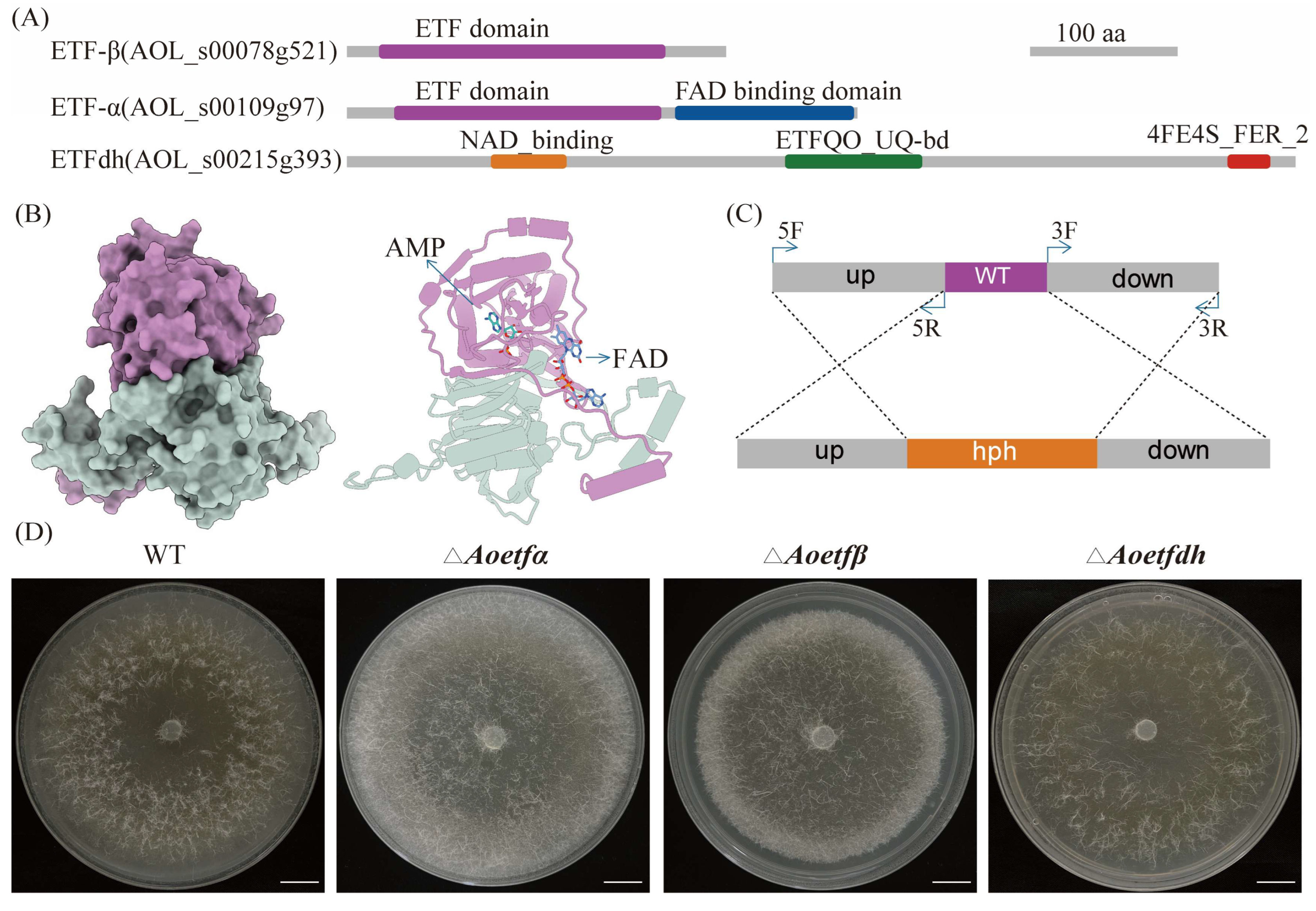
2.1. Analysis and Deletion of etf and etfdh in A. oligospora

Based on the genome annotation data of A. oligospora ATCC24927, we obtained probable electron transfer flavoprotein coding genes, Aoetfα (AOL_s00109g97), Aoetfβ (AOL_s00078g521), and ETF dehydrogenase coding gene Aoetfdh (AOL_s00215g393). A conserved domains analysis showed us that both subunits of ETF-α and ETF-β contain typical ETF domains (Figure 1A). Further modeling revealed that ETF-α associates with ETF-β to form a stable heterodimeric complex, designated AoETF. This complex binds both the FAD cofactor and an AMP molecule (Figure 1B). In contrast, ETFdh functions as a monomer and contains several cofactor binding sites for NAD, ETFQO_UQ, and an iron-sulfur cluster (4Fe-4S) (Figure 1A). So far in NT fungi, the roles of ETF heterodimer and ETFdh have not been characterized. Phylogenetic tree analysis revealed that ETF as well as ETFdh are highly conserved in NT fungi with other fungal species, suggesting that they may have similar functions in this group of fungi (Figures S1–S3). Using the homologous recombination strategy, we generated mutants for each of these three coding genes, designated as ΔAoetfα, ΔAoetfβ, and ΔAoetfdh (Figure 1C,D). To verify protoplast transformants using PCR, the sizes of the wild-type and mutant strains for gene Aoetfα are 1425 bp and 2563 bp, respectively. For gene Aoetfβ, the sizes of the wild-type and mutant strains are 1142 bp and 2353 bp, respectively. For gene Aoetfdh, the sizes of the wild-type and mutant strains are 1564 bp and 2460 bp, respectively (Figure S4A). On various nutrient-rich media, including PDA, CMY, TG, and TYGA, the vegetative growth of the three mutants was comparable to that of the wild-type strain (Figure S4B).

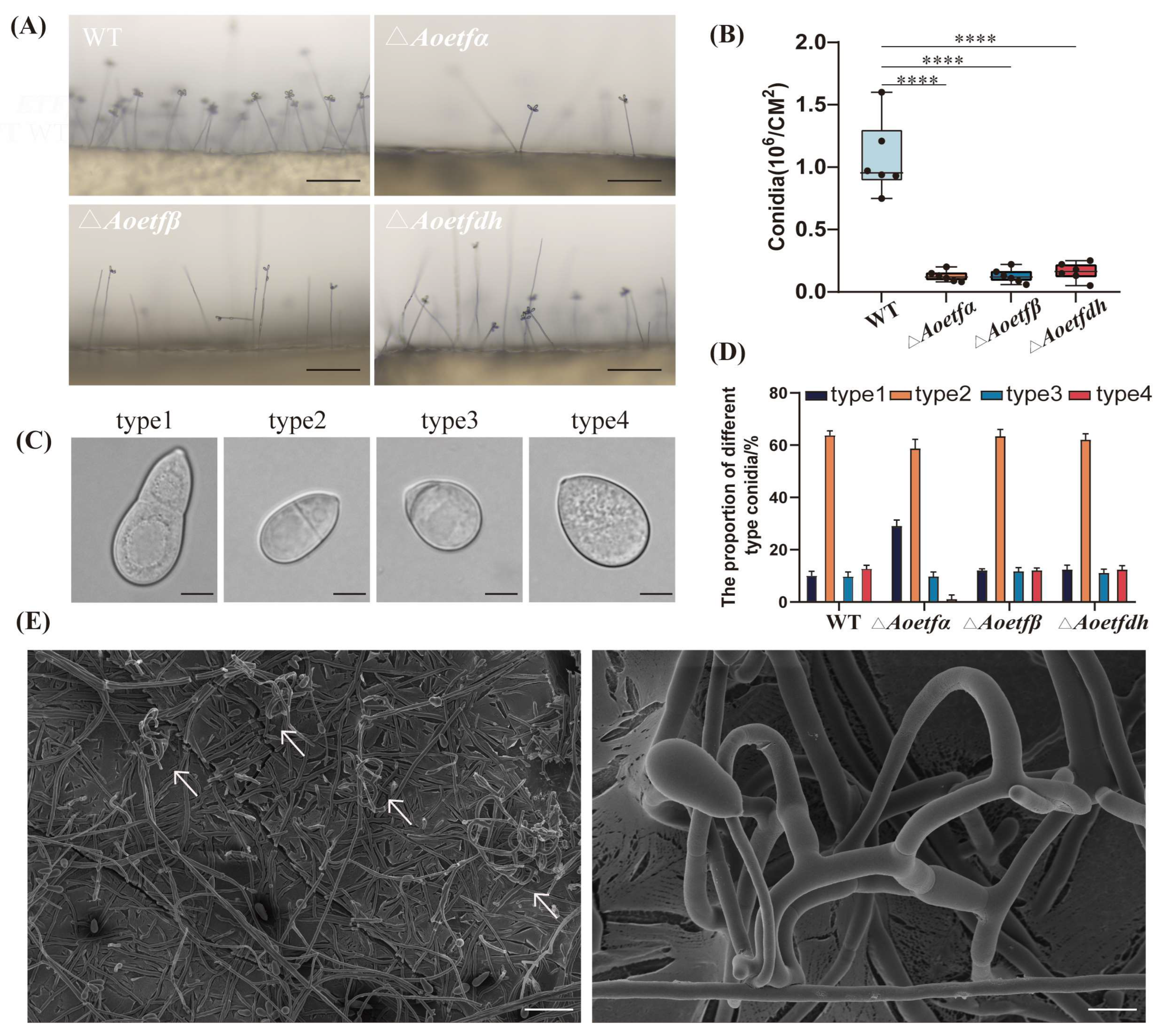
2.2. Aoetf and Aoetfdh Impair Conidiation in A. oligospora

We initially assessed whether Aoetf and Aoetfdh are involved in the sporulation of A. oligospora. Wild-type and mutant strains were cultivated on PDA medium for six days, after which conidial density and spore number were measured. The results showed a significant decrease in both conidial density and spore number in the ΔAoetfα, ΔAoetfβ, and ΔAoetfdh compared to the wild-type (Figure 2A,B). Regarding conidial morphology, both wild-type and mutant strains produced four main types of conidia, consistent with previous findings [25]. Among these, the second type, representing mature spores in the phialides, was predominant in both wild-type and mutant strains (64%, 59%, 63.67%, and 62.33%, respectively) (Figure 2C,D). However, the deletion of ΔAoetfα resulted in a notably low proportion of immature spores, constituting only 1.33% of the total. These observations suggest that the Aoetf and Aoetfdh have important involvement in conidiation in A. oligospora. Unexpectedly, the ΔAoetfβ strain was found to spontaneously produce nematode-capturing structures, adhesive three-dimensional networks, when grown on CMY medium (Figure 2E), which contrasts with the typical formation of predatory structures only under low-nutrient conditions.

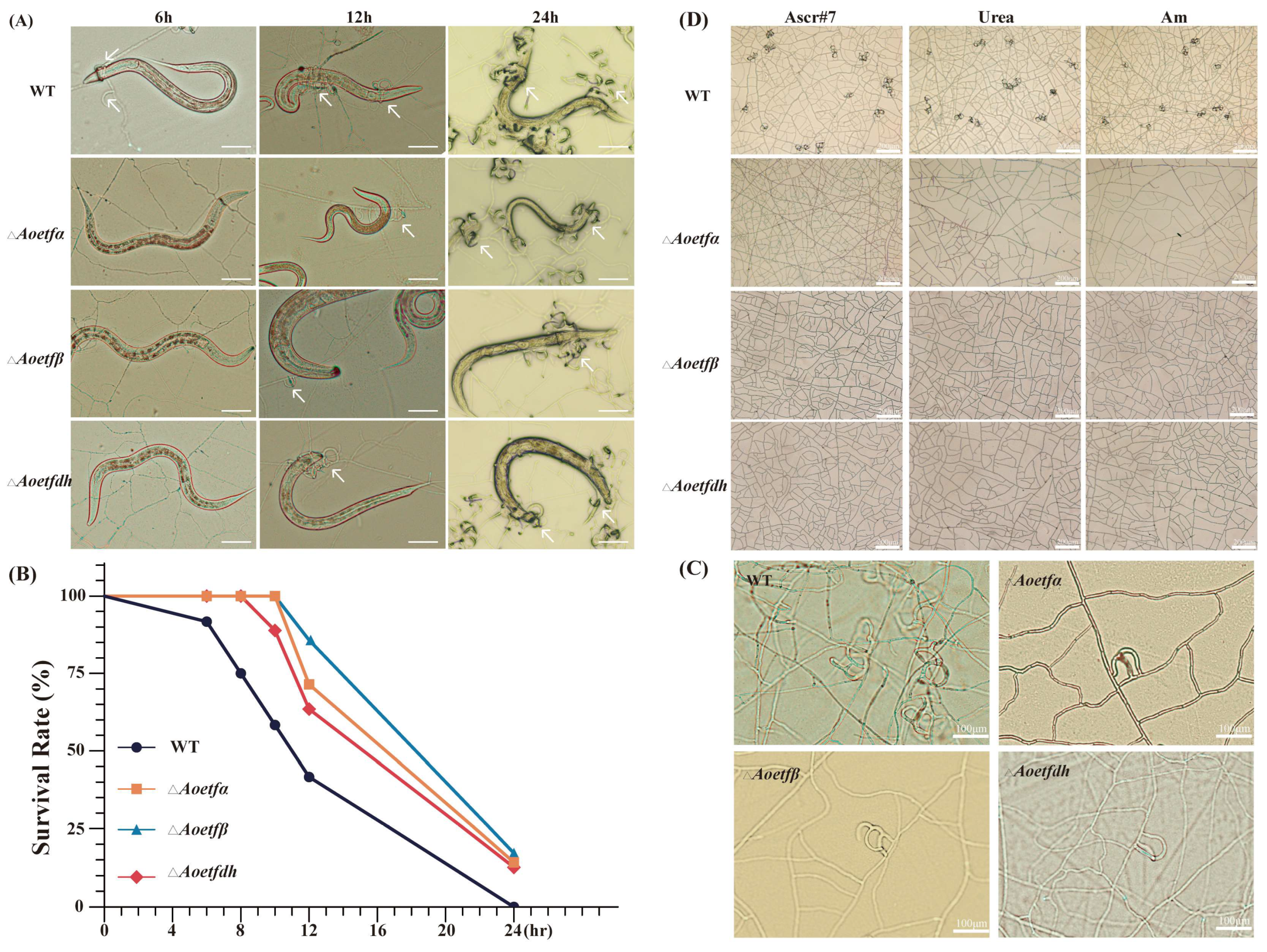
2.3. Aoetf and Aoetfdh Are Essential for the Formation of Traps in A. oligospora

To investigate whether Aoetf and Aoetfdh influence signal-induced trap formation in A. olilgospora, we compared trap production between wild-type and mutant strains in response to live C. elegans, nematode extracts, and trap-inducing signals such as ascaroside (ascr#7), urea, and ammonia (Am). Our findings revealed that the disruption of Aoetf or Aoetfdh genes delayed trap formation and reduced nematode-killing efficiency compared to the wild-type strain under same conditions. Specifically, when exposed to nematodes, the mutant strains produce traps later than the wild-type strains (Figure 3A). In the wild-type strain, only 41.67% of nematodes survived after 12 h, while the survival rates in the ΔAoetfα, ΔAoetfβ, and ΔAoetfdh strains were 71.43%, 85.71%, and 63.49%, respectively (Figure 3B). Furthermore, when exposed to whole nematode extracts, the mutants produced fewer traps than the wild-type strain (Figure 3C). Notably, when we tested trap induction in response to nematode-derived ascaroside, urea, and ammonia, the mutants completely failed to produce any traps (Figure 3D). These results suggest that mutations in Aoetf or Aoetfdh significantly impair trap formation in A. oligospora.

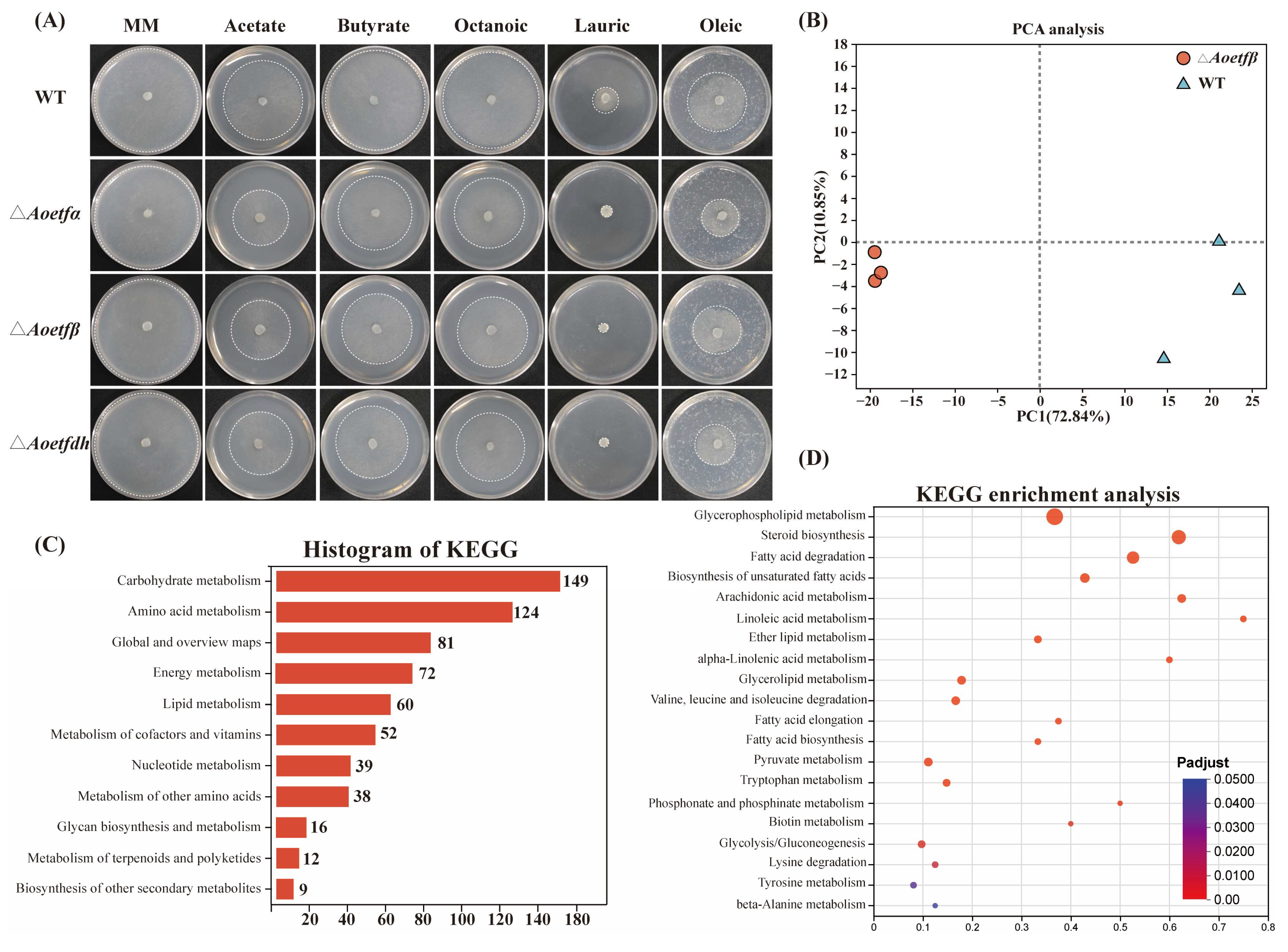
2.4. The Absence of Aoetf Affects Fatty Acid Metabolism in A. oligospora

In other organisms, ETF plays crucial roles in fatty acid metabolism, amino acid metabolism, and mitochondrial function. To determine whether Aoetf in A. oligospora has similar effects, we analyzed hyphal growth in wild-type and mutant strains on MM medium supplemented with sodium carbonate and four different fatty acids (sodium butyrate, caprylic acid, lauric acid, and oleic acid) as the sole carbon source. The results indicated that the growth of the mutant strains was significantly inhibited, with lauric acid completely blocking their growth. These findings suggest that Aoetf and Aoetfdh are essential for fatty acid metabolism in A. oligospora (Figure 4A). Furthermore, transcriptomic sequencing revealed that the Aoetf mutation led to significant changes of nearly 60 genes related to lipid metabolism in expression level; the metabolism pathways involving glycerophospholipid metabolism, steroid biosynthesis, fatty acid degradation, and biosynthesis of unsaturated fatty acids were significantly enriched (Figure 4B–D). Therefore, mutations in ETFs have a significant impact on intracellular fatty acid metabolism in fungi, which can inhibit the degradation of long-chain fats such as lauric acid by fungi.

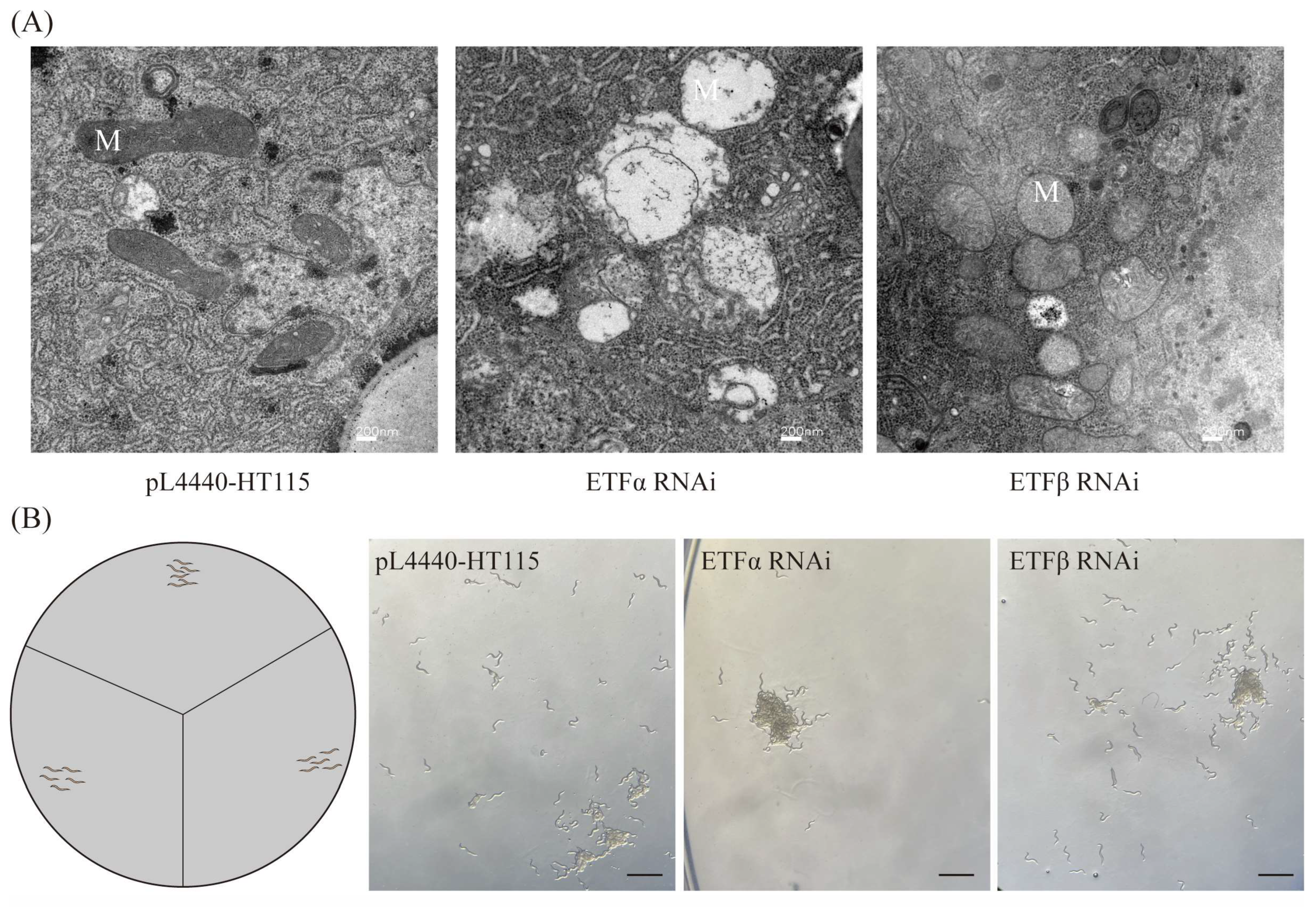
2.5. Aoetf Deletion Impairs Mitochondrial Function in A. oligospra

We guessed the defects in sporulation and pathogenicity are attributed to impaired mitochondrial function, so we conducted transmission electron microscopy (TEM) observation. The TEM results revealed significant mitochondrial damage in the mutant strains compared to the wild-type strain. Specifically, the mitochondria in the ΔAoetfα strain exhibited a complete loss of cristae, while those in the ΔAoetfβ strain displayed fragmented cristae (Figure 5A–C). Additionally, mitochondrial staining demonstrated that the wild-type strain had well-defined, prominent mitochondria, whereas the ΔAoetfα and ΔAoetfβ strains showed reduced staining intensity (Figure 5D–F), indicating a decrease in mitochondrial viability due to the mutations.

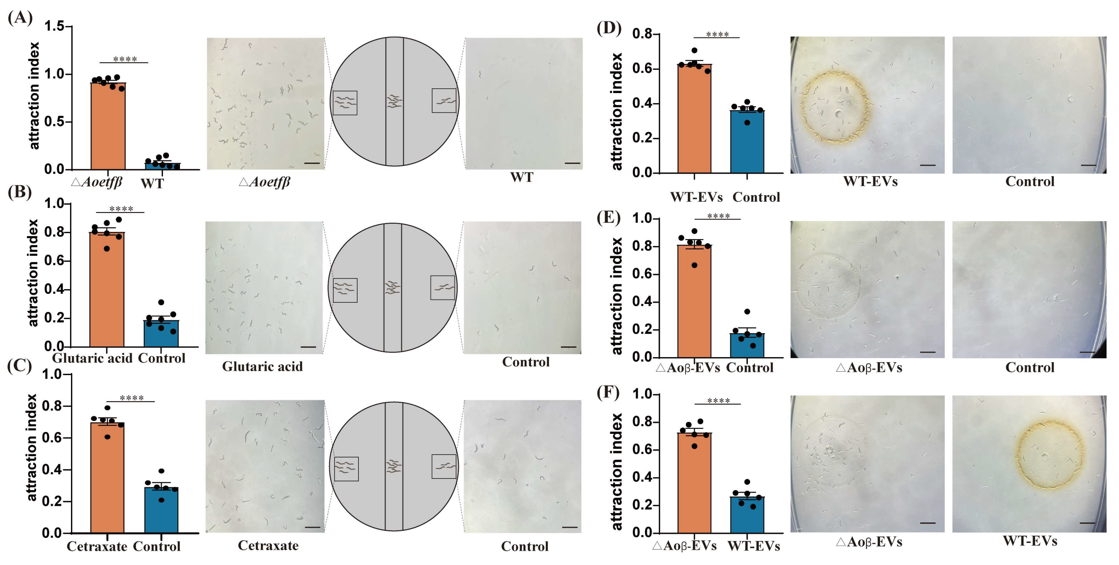
2.6. The Aoetf Mutant Strain Has the Ability to Attract C. elegans

ETF dysfunction has been implicated in a variety of metabolic disorders involving fatty acids and amino acids, particularly in the context of glutaric acidemia in humans [26]. During the growth of the mutant strains, a strong odor reminiscent of sweaty feet was detected. Considering the well-established attraction of NT fungi to nematodes, we conducted chemotaxis assays using the ΔAoetfβ strain and the wild-type strain in the presence of C. elegans. The results showed that ΔAoetfβ has a significant attraction to C. elegans, with a chemotaxis index of 0.92 (Figure 6A). Further, we evaluated the chemotactic response of C. elegans to glutaric acid and observed that this compound exhibited a higher attraction index of 0.8, significantly greater than that of the control (Figure 6B). In the metabolomics analysis, several significantly upregulated compounds were observed (Table 1, Figure S5A), among which the markedly upregulated compound cetraxate demonstrated an attraction to C. elegans with a value of 0.70 (Figure 6C). Given the role of fungal extracellular vesicles (exosomes) as mediators for shuttling chemical signals to communicate with host cells [27], we isolated exosomes from both the wild-type and ΔAoetfβ strains for further attraction assays. Nanoparticle Tracking Analysis (NTA) detection and TEM imaging revealed that the exosomes from the mutant strains were larger in size and present in higher concentrations compared to those from the wild-type strain (Figure S5B,C). Chemotaxis assays with exosomes demonstrated that exosomes from both the wild-type and ΔAoetfβ strains could attract C. elegans; however, the exosomes from the mutant strain exhibited a higher attraction index of 0.81, compared to 0.63 for the wild-type exosomes (Figure 6D,E). Furthermore, a comparison of the relative chemotactic strength revealed that the ΔAoetfβ strain had a significantly higher chemotaxis index of 0.73 compared to 0.26 for the wild-type strain (Figure 6F). Based on these observations, we conclude that the Aoetf mutation leads to the accumulation of metabolites with nematode-attracting activity, which may be secreted via fungal extracellular vesicles.

2.7. Nematode ETF Exhibited Similar Roles in Mitochondrial Function to A. oligospora

Nematode-NT fungi are consistently present in soil; we wondered about the impact of homologous genes of A. oligospora ETF on the mitochondrial function of nematodes. Based on the observation of the TEM, C. elegans exhibited mitochondrial swelling and cristae disruption when the ETF coding was interfered with (Figure 7A). Additionally, the behavior assay showed that obvious aggregation, nearly 100%, occurred following the interference of ETFα, while the aggregation ratio of ETFβ was 60% compared to N2 C. elegans (Figure 7B).

3. Discussion

ETF and ETFdh are highly conserved across bacteria, filamentous fungi, and animals. In our study, AoETF and AoETFdh also exhibited a high degree of conservation with other filamentous fungi (Figure 1 and Figure S1). We established multiple roles of Aoetf and Aoetfdh in the phenotypic regulation of conidiation and pathogenesis in A. oligospora (Figure 2 and Figure 3). In the Aoetf mutant strains, transcriptional profiles and metabolic omics revealed distinctive differences in fatty acid metabolism and amino acid metabolism, which were further confirmed by the growth defect against long chain fatty acids (Figure 4, Table 1). Similar results have been previously demonstrated in the rice blast fungus Magnaporthe oryzea and in the entomopathogenic fungus Beauveria bassiana [2,3].
In humans, ETF and ETFdh variants are typical causes of multiple acyl-CoA dehydrogenase deficiency (MADD) and glutaric aciduria type II [28]. In cases of ETF or ETFdh deficiency, the acyl-CoA dehydrogenases cannot transfer electrons from several dehydrogenases involved in fatty acid oxidation, as well as choline and amino acid metabolism [29]. This process would lead to the deficiencies of multiple acyl-CoA dehydrogenases and disruption of ATP production, causing a large accumulation of glutaric acid, lactic, ethylmalonic, butyric, isobutyric, 2-methyl-butyric, and isovaleric acids [30,31]. As we know, these compounds are volatile and emit unpleasant odors. Coincidentally, our A. oligospora mutant of Aoetf also produced a similar foul smell, which we suspected was glutaric acid. For fungi to capture nematodes, fatal attraction is an important process. In this study, we found that glutaric acid was strongly attracted to C. elegans, although we did not obtain exact compound information based on metabolomics (Figure 6). Moreover, the mutant and its exosomes showed stronger attraction to nematodes than the wild-type strain, suggesting that the disruption of Aoetf led to the secretion of certain functional metabolites (Figure 6). Therefore, the metabolic regulatory role of Aoetf and Aoetfdh in the fungus A. oligospora may be evolutionarily conserved with that in higher animals.
Few studies have described the integrity of mitochondrial morphology in ETF-ETFdh deletions [3,32]. In this study, we found that metabolic disorder-produced esters and acids might impair mitochondrial fitness in A. oligospora (Table 1). TEM images showed defective mitochondria with fragmented, cristae-free, or swollen mitochondria exist in the mutants of Aoetf (Figure 5). Since mitochondria are the key organelles for energy production, mutations or deletions in ETF and ETFdh in Magnaporthe oryzae lead to reduced ATP levels, affecting its energy metabolism, which may further impact its growth, development, and pathogenicity [3]. Based on these observations, mitochondrial dysfunction may be the reason why fungi fail to respond to signals to produce predatory organs. In most eukaryotic cells, mitochondria are responsible for the delicate metabolic activities and stress responses that maintain cellular stability [33,34]. Our research provides the possibility that inhibition of mitochondrial function could be a potential strategy for combating fungal infections in nematodes.
We have identified the multifaceted functions of Aoetf and Aoetfdh that include maintaining mitochondrial structure, fatty acid metabolism, and further controlling pathogenicity and sporulation. The lifestyle of NT fungi includes vegetative growth and parasitic growth, but the latter only occurs in nutrient-deprived conditions [35]. Unexpectedly, we identified a mutant strain of Aoetfβ that grew autonomously in the form of trapping hypha under rich nutrition, which can actively kill nematodes (Figure 2). In addition, Aoetf mutations acquired a strong ability to attract nematodes. From the perspective of the host nematode, Aoetf also plays a conservative role in maintaining mitochondrial integrity and has a significant regulatory effect on nematode aggregation behavior (Figure 7). This Aoetf mutation-induced change in animal behavior has been observed in other animal models, where ETF-resulted metabolic dysfunction is associated with aberrant neural phenotypes and paralysis [36]. The significance of these findings should further facilitate mutant strains’ potential in controlling nematodes in the field.
Faced with low or poor nutrition, the NT fungi can respond to context signals to form infection structures to capture and digest nematodes [35,37]. The involved signaling substances currently cover a wide range, including nematode or nematode-derived chemicals and nutrition signals like urea or ammonia [10,38,39]. The G protein signaling pathway plays an especially vital role for trap formation in NT fungi [39,40]. Unexpectedly, we discovered that changes in intracellular metabolic or energy levels induced by genetic manipulation might activate the regulation of trap formation in A. oligospora. Our future work will aim to link cellular metabolic fluctuations to signal transduction, extending the understanding of intracellular signal regulation to trap development and pathogenesis in the NT fungi.

4. Materials and Methods

4.1. Strains, Plasmids, and Culture Conditions

The wild-type (WT) strain of A. oligospora (ATCC 24927) and the mutant strains Aoetfα, Aoetfβ, and Aoetfdh were inoculated on PDA medium (200 g potato, 20 g glucose, and 18 g agar) and incubated at 28 °C. The plasmids pCSN44 and pCE-Zero (Vazyme Biotech Co., Ltd., Nanjing, China) were used for the amplification of the hygromycin fragment and the construction of knockout vectors, respectively. Escherichia coli strain (DH5α) (TransGen Biotech, Beijing, China) was used for plasmid cloning and storage, grown in LB medium at 37 °C. ssPDA medium (PDA supplemented with sucrose) was used for protoplast regeneration, while CMY medium (cornmeal-molasses-yeast), TG medium (tryptone-glucose), and TYGA medium (tryptone-yeast-extract-glucose-agar) were used to analyze the phenotypic characteristics of hyphae and spores [41]. C. elegans was cultured on NGM medium at 20 °C for bioassays.

4.2. Targeted Gene Deletion

The sequences of Aoetfα (AOL_s00109g97), Aoetfβ (AOL_s00078g521), and Aoetfdh (AOL_s00215g393) were obtained from the A. oligospora genome in the NCBI database. Primers for the upstream fragment, hygromycin fragment, and downstream fragment were designed using the NEB England online tool (https://nebuilderv1.neb.com). Paired primers were used to amplify the upstream and downstream fragments of Aoetfα, Aoetfβ, and Aoetfdh from A. oligospora by PCR, and the hygromycin fragment was amplified using the pCSN44 plasmid as a template. The pCE-Zero vector was digested with EcoRV, and the three fragments were ligated to the vector and transformed into E. coli competent cells. Primers for gene knockout are listed in Table S1. Subsequently, the fragments were transformed into A. oligospora protoplasts using the PEG/CaCl2-mediated method. Positive transformants were selected on TYGA medium containing 100 μg/mL hygromycin and verified by PCR.

4.3. Phenotypic Characterization of Strains

The wild-type and mutant strains were inoculated on PDA, CMY, TYGA, and TG media, and the colony diameters were measured every 24 h using a vernier caliper for a duration of 6 days. To determine conidial production, the wild-type and mutant strains were inoculated on CMY medium and cultured for 7 days. The spores were washed with 5 mL of double-distilled water, and 10 µL of the spore suspension was taken for counting using an optical microscope, with each experiment repeated three times [42]. Furthermore, different fatty acids were used as the sole carbon source to observe differences in fatty acid metabolism in the mutant strains. To observe conidia, the wild-type and mutant strains were inoculated on PDA medium and cultured for three days, then transferred to water agar plates for 24 h and observed under an optical microscope (Olympus, Tokyo, Japan).

4.4. Microscopic Observation

To observe the mitochondria, the hyphal cells were stained with 10 μg/mL mitochondrial staining (Mito Tracker™ Red. Invitrogen, Thermo Fisher Scientific, Waltham, MA, USA), and images were captured using a confocal laser scanning microscope (Zeiss, Oberkochen, Germany). Traps produced on nutrient-rich medium were scanned using a cryo-scanning electron microscope. Mycelia cultured on CMY were collected and fixed with glutaraldehyde electron microscopy fixation solution, then sent to the Kunming Institute of Zoology, Chinese Academy of Sciences, for transmission electron microscopy imaging. The interfered nematodes were fixed with glutaraldehyde and imaged using Transmission Electron Microscope (TEM) (Zeiss, Oberkochen, Germany).

4.5. Difference in Trap Formation

Spore suspensions containing 2000–3000 spores of the wild-type and mutant strains, cultured for 7 days, were spread onto WA plates and grown for 30 h. Different compounds (ascr#7, urea, and ammonia) were added to induce trap formation. The number of traps was counted, and photographs were taken 48 h later. Similarly, 1000 C. elegans nematodes were added to induce trap formation in the wild-type and mutant strains, and the traps were photographed and counted at 6, 12, and 24 h.

4.6. Chemotaxis Assay

The mutant and WT strains were inoculated on CMY solid and liquid media and cultured for 7 days. C. elegans nematodes were synchronized and allowed to mature to adulthood beforehand. Blocks of mutant and wild-type cultures, along with their fermentation broth, were placed on opposite sides of 6 cm NGM agar plates (without peptone), with 10 µL drops of nematode suspension (approximately 200 nematodes) added in the center. The direction of nematode movement was observed, and the number of nematodes moving toward the experimental group and the control group was counted, respectively. Perform a behavioral assay on the interfered nematodes, as described previously. Primers for gene interference are listed in Table S2 [43].

4.7. Exosome Assay

The mutant and WT strains were inoculated into TG medium and cultured for 7 days. Subsequently, the culture supernatant was collected and filtered sequentially through 0.45 µm and 0.22 µm filters. The filtered supernatant was subjected to centrifugation at 4 °C for 30 min at 100,000× g; the supernatant was discarded, and the pellet was collected. After resuspension in PBS, exosomes derived from mutant and wild-type strains were separately applied to opposite sides of 6 cm NGM agar plates (without peptone), with 10 µL drops containing approximately 200 adult-stage nematodes each added to the center. Nematode movement and direction were observed and quantified. Furthermore, the isolated exosomes were sent to Yanzai Biotechnology (Shanghai, China) Co., Ltd. for imaging and particle size measurement.

4.8. Transcriptome and Metabolome

The mutant and wild-type strains were cultured in CMY liquid medium for 7 days. Mycelia and fermentation broth were collected separately, flash-frozen in liquid nitrogen, and sent to Shanghai Majorbio Bio-Pharm Technology Co., Ltd. (Majorbio, Shanghai, China). Gene Ontology (GO) function and Kyoto Encyclopedia of Genes and Genomes (KEGG) pathway enrichment were analyzed using the Majorbio cloud platform (https://www.majorbio.com. accessed on 19 February 2025).

5. Conclusions

This study highlights the complications of ETF and its dehydrogenase ETFdh in the pathogenesis and sporulation of NT fungus A. oligospora. Further, the disruptions of Aoetf and Aoetfdh can alter metabolic and energy levels, possibly leading to mitochondrial dysfunction and modifying the interaction between NT fungi and nematodes. Moving forward, future research should focus on the regulatory mechanisms by which metabolic alterations influence signal transduction and phenotypic regulation in the NTF group.

Supplementary Materials

The supporting information can be downloaded at: https://www.mdpi.com/article/10.3390/ijms252010934/s1.

Author Contributions

Conceptualization, X.W.; writing—original draft preparation, Y.L. and Z.L.; writing—review and editing, J.L. and X.Z.; supervision, X.W. All authors have read and agreed to the published version of the manuscript.

Funding

This research was funded by the Science and Technology Major Project from Yunnan Province (202201BC070004), the National Natural Science Foundation of China (32370138 and 32260029), the Applied Basic Research Foundation of Yunnan Province (202201AT070089), the special fund from the Yunnan University “double first-class” construction, and the Scientific Research Fund Project of Yunnan Provincial Department of Education (2024Y019).

Institutional Review Board Statement

Not applicable.

Informed Consent Statement

Not applicable.

Data Availability Statement

The original contributions presented in the study are included in the article and Supplementary Material, further inquiries can be directed to the corresponding author, Xin Wang.

Acknowledgments

We thank Ying-qi Guo (Kunming Institute of Zoology, Chinese Academy of Sciences) for their help with taking and analyzing the TEM images, and we would like to thank Zhi-jia Gu (Kunming Institute of Botany, Chinese Academy of Scienses) for making sample and taking images.

Conflicts of Interest

The authors declare no conflicts of interest.

References

  1. Henriques, B.J.; Katrine Jentoft Olsen, R.; Gomes, C.M.; Bross, P. Electron transfer flavoprotein and its role in mitochondrial energy metabolism in health and disease. Gene 2021, 776, 145407. [Google Scholar] [CrossRef] [PubMed]
  2. Lin, D.; Wan, M.; Fan, Y. Electron-transferring flavoprotein and its dehydrogenase contributed to growth development and virulence in Beauveria bassiana. J. Invertebr. Pathol. 2024, 205, 108141. [Google Scholar] [CrossRef] [PubMed]
  3. Li, Y.; Zhu, J.; Hu, J.; Meng, X.; Zhang, Q.; Zhu, K.; Chen, X.; Chen, X.; Li, G.; Wang, Z.; et al. Functional characterization of electron-transferring flavoprotein and its dehydrogenase required for fungal development and plant infection by the rice blast fungus. Sci. Rep. 2016, 6, 24911. [Google Scholar] [CrossRef]
  4. Toplak, M.; Brunner, J.; Tabib, C.R.; Macheroux, P. Closing the gap: Yeast electron-transferring flavoprotein links the oxidation of d-lactate and d-α-hydroxyglutarate to energy production via the respiratory chain. FEBS J. 2019, 286, 3611–3628. [Google Scholar] [CrossRef] [PubMed]
  5. Wakitani, S.; Torisu, S.; Yoshino, T.; Hattanda, K.; Yamato, O.; Tasaki, R.; Fujita, H.; Nishino, K. Multiple Acyl-CoA Dehydrogenation Deficiency (Glutaric Aciduria Type II) with a Novel Mutation of Electron Transfer Flavoprotein-Dehydrogenase in a Cat. JIMD Rep. 2014, 13, 43–51. [Google Scholar]
  6. Hasanuzzaman, M.; Bhuyan, M.; Parvin, K.; Bhuiyan, T.F.; Anee, T.I.; Nahar, K.; Hossen, M.S.; Zulfiqar, F.; Alam, M.M.; Fujita, M. Regulation of ROS Metabolism in Plants under Environmental Stress: A Review of Recent Experimental Evidence. Int. J. Mol. Sci. 2020, 21, 8695. [Google Scholar] [CrossRef]
  7. Su, H.; Zhao, Y.; Zhou, J.; Feng, H.; Jiang, D.; Zhang, K.Q.; Yang, J. Trapping devices of nematode-trapping fungi: Formation, evolution, and genomic perspectives. Biol. Rev. Camb. Philos. Soc. 2017, 92, 357–368. [Google Scholar] [CrossRef]
  8. Soliman, M.S.; El-Deriny, M.M.; Ibrahim, D.S.S.; Zakaria, H.; Ahmed, Y. Suppression of root-knot nematode Meloidogyne incognita on tomato plants using the nematode trapping fungus Arthrobotrys oligospora Fresenius. J. Appl. Microbiol. 2021, 131, 2402–2415. [Google Scholar] [CrossRef]
  9. Pires, D.; Vicente, C.S.L.; Menéndez, E.; Faria, J.M.S.; Rusinque, L.; Camacho, M.J.; Inácio, M.L. The Fight against Plant-Parasitic Nematodes: Current Status of Bacterial and Fungal Biocontrol Agents. Pathogens 2022, 11, 1178. [Google Scholar] [CrossRef]
  10. Wang, X.; Li, G.H.; Zou, C.G.; Ji, X.L.; Liu, T.; Zhao, P.J.; Liang, L.M.; Xu, J.P.; An, Z.Q.; Zheng, X.; et al. Bacteria can mobilize nematode-trapping fungi to kill nematodes. Nat. Commun. 2014, 5, 5776. [Google Scholar] [CrossRef]
  11. Zhang, F.; Yang, Y.Q.; Zhou, F.P.; Xiao, W.; Boonmee, S.; Yang, X.Y. Morphological and Phylogenetic Characterization of Five Novel Nematode-Trapping Fungi (Orbiliomycetes) from Yunnan, China. J. Fungi 2023, 9, 735. [Google Scholar] [CrossRef] [PubMed]
  12. Zhang, D.; Zhu, X.; Sun, F.; Zhang, K.; Niu, S.; Huang, X. The roles of actin cytoskeleton and actin-associated protein Crn1p in trap formation of Arthrobotrys oligospora. Res. Microbiol. 2017, 168, 655–663. [Google Scholar] [CrossRef] [PubMed]
  13. Wang, B.L.; Chen, Y.H.; He, J.N.; Xue, H.X.; Yan, N.; Zeng, Z.J.; Bennett, J.W.; Zhang, K.Q.; Niu, X.M. Integrated Metabolomics and Morphogenesis Reveal Volatile Signaling of the Nematode-Trapping Fungus Arthrobotrys oligospora. Appl. Environ. Microbiol. 2018, 84, e02749. [Google Scholar] [CrossRef] [PubMed]
  14. Yu, X.; Hu, X.; Pop, M.; Wernet, N.; Kirschhöfer, F.; Brenner-Weiß, G.; Keller, J.; Bunzel, M.; Fischer, R. Fatal attraction of Caenorhabditis elegans to predatory fungi through 6-methyl-salicylic acid. Nat. Commun. 2021, 12, 5462. [Google Scholar] [CrossRef] [PubMed]
  15. Youssar, L.; Wernet, V.; Hensel, N.; Yu, X.; Hildebrand, H.G.; Schreckenberger, B.; Kriegler, M.; Hetzer, B.; Frankino, P.; Dillin, A.; et al. Intercellular communication is required for trap formation in the nematode-trapping fungus Duddingtonia flagrans. PLoS Genet. 2019, 15, e1008029. [Google Scholar] [CrossRef]
  16. Jiang, X.; Xiang, M.; Liu, X. Nematode-Trapping Fungi. Microbiol. Spectr. 2017, 5, 963–974. [Google Scholar] [CrossRef]
  17. Heintz, C.E.; Pramer, D. Ultrastructure of nematode-trapping fungi. J. Bacteriol. 1972, 110, 1163–1170. [Google Scholar] [CrossRef]
  18. Veenhuis, M.; Harder, W.; Nordbring-Hertz, B. Occurrence and metabolic significance of microbodies in trophic hyphae of the nematophagous fungus Arthrobotrys oligospora. Antonie Leeuwenhoek. 1989, 56, 241–249. [Google Scholar] [CrossRef]
  19. Talbot, N.J.; Foster, A.J. Genetics and genomics of the rice blast fungus Magnaporthe grisea: Developing an experimental model for understanding fungal diseases of cereals. Adv. Bot. Res. 2001, 34, 263–287. [Google Scholar]
  20. Watmough, N.J.; Frerman, F.E. The electron transfer flavoprotein: Ubiquinone oxidoreductases. Biochim. Biophys. Acta 2010, 1797, 1910–1916. [Google Scholar] [CrossRef]
  21. Wang, Y.; Palmfeldt, J.; Gregersen, N.; Makhov, A.M.; Conway, J.F.; Wang, M.; McCalley, S.P.; Basu, S.; Alharbi, H.; St Croix, C.; et al. Mitochondrial fatty acid oxidation and the electron transport chain comprise a multifunctional mitochondrial protein complex. J. Biol. Chem. 2019, 294, 12380–12391. [Google Scholar] [CrossRef] [PubMed]
  22. Chokchaiwong, S.; Kuo, Y.T.; Hsu, S.P.; Hsu, Y.C.; Lin, S.H.; Zhong, W.B.; Lin, Y.F.; Kao, S.H. ETF-QO Mutants Uncoupled Fatty Acid β-Oxidation and Mitochondrial Bioenergetics Leading to Lipid Pathology. Cells 2019, 8, 106. [Google Scholar] [CrossRef] [PubMed]
  23. Salerno, K.M.; Domenico, J.; Le, N.Q.; Stiles, C.D.; Solov’yov, I.A.; Martino, C.F. Long-Time Oxygen Localization in Electron Transfer Flavoprotein. J. Chem. Inf. Model 2022, 62, 4191–4199. [Google Scholar] [CrossRef] [PubMed]
  24. Herrero Martín, J.C.; Salegi Ansa, B.; Álvarez-Rivera, G.; Domínguez-Zorita, S.; Rodríguez-Pombo, P.; Pérez, B.; Calvo, E.; Paradela, A.; Miguez, D.G.; Cifuentes, A.; et al. An ETFDH-driven metabolon supports OXPHOS efficiency in skeletal muscle by regulating coenzyme Q homeostasis. Nat. Metab. 2024, 6, 209–225. [Google Scholar] [CrossRef]
  25. Liu, Q.; Bai, N.; Duan, S.; Shen, Y.; Zhu, L.; Yang, J. Characterizing the Role of AosfgA and AofluG in Mycelial and Conidial Development in Arthrobotrys oligospora and Their Role in Secondary Metabolism. Microorganisms 2024, 12, 615. [Google Scholar] [CrossRef]
  26. Cheng, Y.Y.; Tang, Y.; Liu, A.J.; Wei, L.; Lin, L.; Zhang, J.; Zhi, L. Clinical features and ETFDH mutations of children with late-onset glutaric aciduria type II: A report of two cases. Zhongguo Dang Dai Er Ke Za Zhi = Chin. J. Contemp. Pediatr. 2017, 19, 975–978. [Google Scholar]
  27. Stotz, H.U.; Brotherton, D.; Inal, J. Communication is key: Extracellular vesicles as mediators of infection and defence during host-microbe interactions in animals and plants. FEMS Microbiol. Rev. 2022, 46, fuab044. [Google Scholar] [CrossRef]
  28. Macchione, F.; Salviati, L.; Bordugo, A.; Vincenzi, M.; Camilot, M.; Teofoli, F.; Pancheri, E.; Zordan, R.; Bertolin, C.; Rossi, S.; et al. Multiple acyl-COA dehydrogenase deficiency in elderly carriers. J. Neurol. 2020, 267, 1414–1419. [Google Scholar] [CrossRef]
  29. Brandao, S.R.; Ferreira, R.; Rocha, H. Exploring the contribution of mitochondrial dynamics to multiple acyl-CoA dehydrogenase deficiency-related phenotype. Arch. Physiol. Biochem. 2021, 127, 210–216. [Google Scholar] [CrossRef]
  30. Angelini, C.; Burlina, A.; Blau, N.; Ferreira, C.R. Clinical and biochemical footprints of inherited metabolic disorders: X. Metabolic myopathies. Mol. Genet. Metab. 2022, 137, 213–222. [Google Scholar] [CrossRef]
  31. Szrok-Jurga, S.; Czumaj, A.; Turyn, J.; Hebanowska, A.; Swierczynski, J.; Sledzinski, T.; Stelmanska, E. The Physiological and Pathological Role of Acyl-CoA Oxidation. Int. J. Mol. Sci. 2023, 24, 14857. [Google Scholar] [CrossRef] [PubMed]
  32. Xiao, C.; Astiazaran-Symonds, E.; Basu, S.; Kisling, M.; Scaglia, F.; Chapman, K.A.; Wang, Y.; Vockley, J.; Ferreira, C.R. Mitochondrial energetic impairment in a patient with late-onset glutaric acidemia Type 2. Am. J. Med. Genet. A 2020, 182, 2426–2431. [Google Scholar] [CrossRef] [PubMed]
  33. Yuan, Q.; Zeng, Z.L.; Yang, S.; Li, A.; Zu, X.; Liu, J. Mitochondrial Stress in Metabolic Inflammation: Modest Benefits and Full Losses. Oxid. Med. Cell. Longev. 2022, 2022, 8803404. [Google Scholar] [CrossRef]
  34. Picard, M.; McEwen, B.S.; Epel, E.S.; Sandi, C. An energetic view of stress: Focus on mitochondria. Front. Neuroendocrinol. 2018, 49, 72–85. [Google Scholar] [CrossRef]
  35. Zhu, M.C.; Li, X.M.; Zhao, N.; Yang, L.; Zhang, K.Q.; Yang, J.K. Regulatory Mechanism of Trap Formation in the Nematode-Trapping Fungi. J. Fungi 2022, 8, 406. [Google Scholar] [CrossRef]
  36. Angelini, C.; Tavian, D.; Missaglia, S. Heterogeneous Phenotypes in Lipid Storage Myopathy Due to ETFDH Gene Mutations. JIMD Rep. 2018, 38, 33–40. [Google Scholar] [PubMed]
  37. Chen, S.A.; Lin, H.C.; Schroeder, F.C.; Hsueh, Y.P. Prey sensing and response in a nematode-trapping fungus is governed by the MAPK pheromone response pathway. Genetics 2021, 217, iyaa008. [Google Scholar] [CrossRef] [PubMed]
  38. Huang, J.; Zheng, X.; Tian, M.; Zhang, K. Ammonia and Nematode Ascaroside Are Synergistic in Trap Formation in Arthrobotrys oligospora. Pathogens 2023, 12, 1114. [Google Scholar] [CrossRef]
  39. Hu, X.; Hoffmann, D.S.; Wang, M.; Schuhmacher, L.; Stroe, M.C.; Schreckenberger, B.; Elstner, M.; Fischer, R. GprC of the nematode-trapping fungus Arthrobotrys flagrans activates mitochondria and reprograms fungal cells for nematode hunting. Nat. Microbiol. 2024, 9, 1752–1763. [Google Scholar] [CrossRef]
  40. Kuo, C.Y.; Tay, R.J.; Lin, H.C.; Juan, S.C.; Vidal-Diez de Ulzurrun, G.; Chang, Y.C.; Hoki, J.; Schroeder, F.C.; Hsueh, Y.P. The nematode-trapping fungus Arthrobotrys oligospora detects prey pheromones via G protein-coupled receptors. Nat. Microbiol. 2024, 9, 1738–1751. [Google Scholar] [CrossRef]
  41. Xie, M.; Yang, J.; Jiang, K.; Bai, N.; Zhu, M.; Zhu, Y.; Zhang, K.Q.; Yang, J. AoBck1 and AoMkk1 Are Necessary to Maintain Cell Wall Integrity, Vegetative Growth, Conidiation, Stress Resistance, and Pathogenicity in the Nematode-Trapping Fungus Arthrobotrys oligospora. Front. Microbiol. 2021, 12, 649582. [Google Scholar] [CrossRef] [PubMed]
  42. Zhou, L.; Li, M.; Cui, P.; Tian, M.; Xu, Y.; Zheng, X.; Zhang, K.; Li, G.; Wang, X. Arrestin-Coding Genes Regulate Endocytosis, Sporulation, Pathogenicity, and Stress Resistance in Arthrobotrys oligospora. Front. Cell Infect. Microbiol. 2022, 12, 754333. [Google Scholar] [CrossRef] [PubMed]
  43. Conte, D., Jr.; MacNeil, L.T.; Walhout, A.J.M.; Mello, C.C. RNA Interference in Caenorhabditis elegans. Curr. Protoc. Mol. Biol. 2015, 109, 26.23.21–26.23.30. [Google Scholar] [CrossRef] [PubMed]
Figure 1. Bioinformatics analysis of ETF, ETFdh and gene knockouts. (A) The domain structure of Arthrobotrys oligospora ETF-α, ETF-β and ETFdh as annotated at the Broad Institute of MIT. (B) Structural Model of ETFα-ETFβ complex (AoETF) bound to AMP and FAD, the purple structure represents ETF-β, while the light gray structure depicts ETF-α. (C) Homologous recombination knockout model diagram. (D) Growth of wild-type strain (WT) and the mutants of Aoetfα, Aoetfβ, and Aoetfdh on PDA medium for 7 days; scale bar: 1 cm.
Ijms 25 10934 g001
Figure 2. Mutations Aoetf and Aoetfdh affect spore production, morphology, and trap formation. (A) Comparison of conidia in wild-type, ΔAoetf, and ΔAoetfdh strains (Scale bar: 100 μm). (B) Comparison of conidia production in wild-type, ΔAoetf, and ΔAoetfdh strains. **** represents significant difference (p < 0.0001). (C,D) Analysis and statistics of conidia types in wild-type, ΔAoetf, and ΔAoetfdh strains (scale bar: 7 μm). (E) Scanning electron micrographs of the trapping structure in the ΔAoetfβ strain at different magnifications. The left image is at 150× magnification (scale bar: 100 μm; white arrow indicates the trap), and the right image is at 1000× magnification (scale bar: 10 μm).
Ijms 25 10934 g002
Figure 3. Trap formation in wild-type, ΔAoetf, and ΔAoetfdh strains. (A) Representative bright-field images of trap formation at different time points after nematode induction (scale bar: 200 μm; the arrows denote trap structures). (B) Survival rate of nematodes exposed to wild-type, ΔAoetfα, ΔAoetfβ, and ΔAoetfdh strains. (C). Representative bright-field images of traps induced by nematode extracts in wild-type, ΔAoetfα, ΔAoetfβ, and ΔAoetfdh strains (scale bar: 200 μm). (D) Representative bright-field images of traps induced by ascr#7, urea, and ammonia (Am) in wild-type, ΔAoetfα, ΔAoetfβ, and ΔAoetfdh strains (scale bar: 100 μm).
Ijms 25 10934 g003
Figure 4. ΔAoetf and ΔAoetfdh were involved in fatty acids metabolism. (A) Growth conditions of WT, ΔAoetfα, ΔAoetfβ, and ΔAoetfdh strains on MM medium with various fatty acids as the sole carbon source. (B) Principal component analysis (PCA) plot of Aoetfβ mutant and wild-type strains. (C) Histogram of KEGG-enriched metabolic pathways in the transcriptome, with 60 genes associated with lipid metabolism. (D) KEGG enrichment analysis of genes related to lipid metabolism.
Ijms 25 10934 g004
Figure 5. Aoetf deletion impairs mitochondria function. (AC) Ultrastructural analysis of wild-type, ΔAoetfα, and ΔAoetfβ strains using transmission electron microscopy (scale bars: 200 nm; M denotes mitochondria). (DF) Comparison of mitochondrial of wild-type and ΔAoetf strains were stained with MitoTracker® Red (scale bars: 3 μm).
Ijms 25 10934 g005
Figure 6. Chemotaxis observation of C. elegans. (A) Chemotaxis statistics of C. elegans towards fermentation broth of wild-type and ΔAoetfβ strains. (B) Chemotaxis statistics of C. elegans towards glutaric acid and control. (C) Chemotaxis statistics of C. elegans towards cetraxate and control (D) Chemotaxis statistics of C. elegans towards exosomes from wild-type strains. (E) Chemotaxis statistics of C. elegans towards exosomes from ΔAoetfβ strains. (F) Comparative chemotaxis statistics of C. elegans towards exosomes from wild-type and ΔAoetfβ strains. **** represents significant difference (p < 0.0001; scale bars: 2 mm).
Ijms 25 10934 g006
Figure 7. TEM observations and phenotypes of ETF Genes in C. elegans. (A) Transmission electron microscopy images of mitochondria after interfering with the ETF gene in C. elegans (scale bars: 200 nm). M denotes mitochondria. (B) Aggregation phenomenon observed in C. elegans after interfering with the ETFα gene (scale bars: 2 mm).
Ijms 25 10934 g007
Table 1. Metabolomics analysis of differentially upregulated compounds between wild-type and ΔAoetfβ strains.
Metabolism NameFCKEGG Pathway Description
Cetraxate47.8841-
S-Lactoylglutathione1.4921Pyruvate metabolism
Kynurenic acid1.2806Tryptophan metabolism
Gamma-glutamyl-L-putrescine1.3035Arginine and proline metabolism
2-Isopropylmalic acid1.1205Biosynthesis of secondary metabolites
Valine, leucine and isoleucine biosynthesis
2-Oxocarboxylic acid
Theophylline1.1008Biosynthesis of secondary metabolites
Caffeine metabolism
6-Lactoyltetrahydropterin1.1103Folate biosynthesis
Homogentisic acid1.1126Ubiquinone and other terpenoid-quinone biosynthesis
Tyrosine metabolism
Biosynthesis of cofactors
Imidazole lactic acid1.1166Histidine metabolism
D-xylonic acid1.0826Pentose and glucuronate interconversions
Ascorbate and aldarate metabolism
N-carbamoylputrescine1.0977Arginine and proline metabolism
5-methoxyindoleacetate1.0983Tryptophan metabolism
lactose1.0749ABC transporters
Galactose metabolism
Indole-3-acetaldehyde1.0808Tryptophan metabolism
Glucosamine1.0607Amino sugar and nucleotide sugar metabolism
Biosynthesis of nucleotide sugars
LL-2,6-diaminopimelic acid1.0692Biosynthesis of secondary metabolites
D-amino acid metabolism
Biosynthesis of amino acids
Lysine biosynthesis
Biotin1.0596Biotin metabolism;ABC transporters
Biosynthesis of cofactors
Kynurenine1.0558Tryptophan metabolism
Biosynthesis of cofactors
FC (ΔAoetfβ/WT): fold change in the expression of this metabolite between the two groups. WT: expression level of this metabolite in the control group WT. ΔAoetfβ: expression level of this metabolite in the experimental group ΔAoetfβ. Enriched KEGG metabolic pathways.
Disclaimer/Publisher’s Note: The statements, opinions and data contained in all publications are solely those of the individual author(s) and contributor(s) and not of MDPI and/or the editor(s). MDPI and/or the editor(s) disclaim responsibility for any injury to people or property resulting from any ideas, methods, instructions or products referred to in the content.

