Article

Rapid Detection of the Strawberry Foliar Nematode Aphelenchoides fragariae Using Recombinase Polymerase Amplification Assay with Lateral Flow Dipsticks

by
Sergei A. Subbotin
Plant Pest Diagnostic Centre, California Department of Food and Agriculture, 3294 Meadowview Road, Sacramento, CA 95832-1448, USA
Int. J. Mol. Sci. 2024, 25(2), 844; https://doi.org/10.3390/ijms25020844
Submission received: 27 November 2023 / Revised: 3 January 2024 / Accepted: 8 January 2024 / Published: 10 January 2024

Abstract

:
Rapid and reliable diagnostic methods for plant-parasitic nematodes are critical for facilitating the selection of effective control measures. A diagnostic recombinase polymerase amplification (RPA) assay for Aphelenchoides fragariae using a TwistAmp® Basic Kit (TwistDx, Cambridge, UK) and AmplifyRP® Acceler8® Discovery Kit (Agdia, Elkhart, IN, USA) combined with lateral flow dipsticks (LF) has been developed. In this study, a LF-RPA assay was designed that targets the ITS rRNA gene of A. fragariae. This assay enables the specific detection of A. fragariae from crude nematode extracts without a DNA extraction step, and from DNA extracts of plant tissues infected with this nematode species. The LF-RPA assay showed reliable detection within 18–25 min with a sensitivity of 0.03 nematode per reaction tube for crude nematode extracts or 0.3 nematode per reaction tube using plant DNA extracts from 0.1 g of fresh leaves. The LF-RPA assay was developed and validated with a wide range of nematode and plant samples. Aphelenchoides fragariae was identified from seed samples in California. The LF-RPA assay has great potential for nematode diagnostics in the laboratory with minimal available equipment.

Graphical Abstract

1. Introduction

The strawberry foliar nematode Aphelenchoides fragariae is an important pest of strawberry plants; it causes ‘strawberry crimp’ disease [1]. Aphelenchoides fragariae was described by Ritzema Bos [2] from specimens extracted from strawberry plants sent to him from England. The neotype of this species proposed and described by Allen [3] came from strawberries in Escalon, CA, USA. The nematode attacks above-ground parts of the plant and causes malformations, including twisting of the shoots and puckered undersized leaves with crinkled edges, reddened petioles and discolored areas that have hard, rough surfaces. The nematode damage symptoms are easily confused with symptoms of powdery mildew or infections with plant pathogenic bacteria. Aphelenchoides fragariae can severely impact the yield of strawberries, as heavily infected plants do not grow normally or produce fruit [4,5]. Over 250 plants in 47 families, including ornamental ferns and other ornamental plants, are also considered to be hosts of A. fragariae [3,4,5,6,7,8,9,10,11,12,13]. In California, A. fragariae has been recorded on 27 plant species [14]. This nematode is widely distributed in the USA, Canada, Japan and Europe and currently reported in 37 countries [7,15]. Interestingly, the strawberry foliar nematode may be endo- or ectoparasitic, but it can also be mycetophagous [16], attributes that enhance its survival in the absence of a host crop and which limit and complicate management options.
The two nematode species most associated with damage in California strawberries are the foliar nematode A. fragariae and the northern root-knot nematode Meloidogyne hapla. Strawberries are an important crop in the United States with about 90% of their national production occurring in California. Strawberries are California’s third highest grossing crop, valued at USD 3.02 billion in 2021. Strawberry production in California occurs primarily along 500 miles of coastal regions between San Diego in the south to Monterey Bay in the central coastal region [17]. Besides the survival of A. fragariae in the mycetophagus state, the western sword-fern Polystichum munitum, native to western North America from Alaska to Mexico, is often heavily infected by A. fragariae in California coastal forests. The high humidity of coastal areas provides very favorable conditions for a rapid increase in the foliar nematode population and its dispersal from infected natural areas to strawberry plantations.
The strawberry foliar nematode is the subject of a regulatory program in California. Strawberry nursery stock should be inspected and tested for nematodes through the California Strawberry Registration and Certification Program run by the California Department of Food and Agriculture and the Foundation Plant Services of the University of California, Davis [18]. The precise and rapid detection of regulated nematodes is the first and most essential step in the regulatory program.
The morphological identification of A. fragariae is complex and requires the microscopic examination of adult nematodes. This species can be distinguished from all other Aphelenchoides species by its slender body, lateral field with two incisures and tail terminus with a single mucro [15,19]. Distinguishing between foliar nematodes and fungal-feeding Aphelenchoides species based on morphological characteristics is quite problematic. Only molecular methods can provide reliable and rapid diagnostic tools for their differentiation.
Sequences of the nuclear ribosomal genes 18S rRNA, ITS rRNA and D2-D3 of 28S rRNA are clearly differentiate A. fragariae from other nematodes [20,21,22,23,24,25,26,27]. Ibrahim et al. [28] were the first to provide PCR-ITS-RFLP profiles for A. fragariae and other Aphelenchoides spp. McCuiston et al. [21] was the first to develop conventional PCR with species-specific primers designed on the ITS rRNA gene polymorphism for nematode detection in plant materials. Rybarczyk-Mydłowska et al. [22] designed a real-time PCR SYBR Green assay using species-specific primers, based on 18S rRNA gene polymorphism, for the quantitative detection of A. fragariae and other Aphelenchoides spp. Although these PCR assays are very reliable, new DNA amplification techniques may provide easier and more rapid nematode detection.
Recombinase polymerase amplification (RPA) is a relatively new isothermal in vitro nucleic acid amplification technique. It has been adopted as a novel molecular technology for simple, robust, rapid, reliable and low-resource diagnostics for different organisms [29]. RPA has several advantages over PCR-based methods for plant-parasitic nematode detection: (i) it does not require thermal cycling and can be used in areas with minimal laboratory infrastructure and run by personnel with minimal technical experience; (ii) it is more sensitive than PCR; (iii) sample processing does not require DNA extraction; (iv) amplicons may be detected, at the endpoint or in real-time, within 8 to 30 min. RPA assays have shown high sensitivity and specificity for detecting various agriculturally important plant-parasitic nematodes, including Meloidogyne hapla [30], but have not been developed for diagnostics in A. fragariae.
In this study, an LF-RPA assay was developed for the detection of A. fragariae from plant and nematode DNA samples and for crude nematode extracts. Species-specific primers and a probe were designed based on the polymorphism of the ITS ribosomal RNA gene sequences.

2. Results

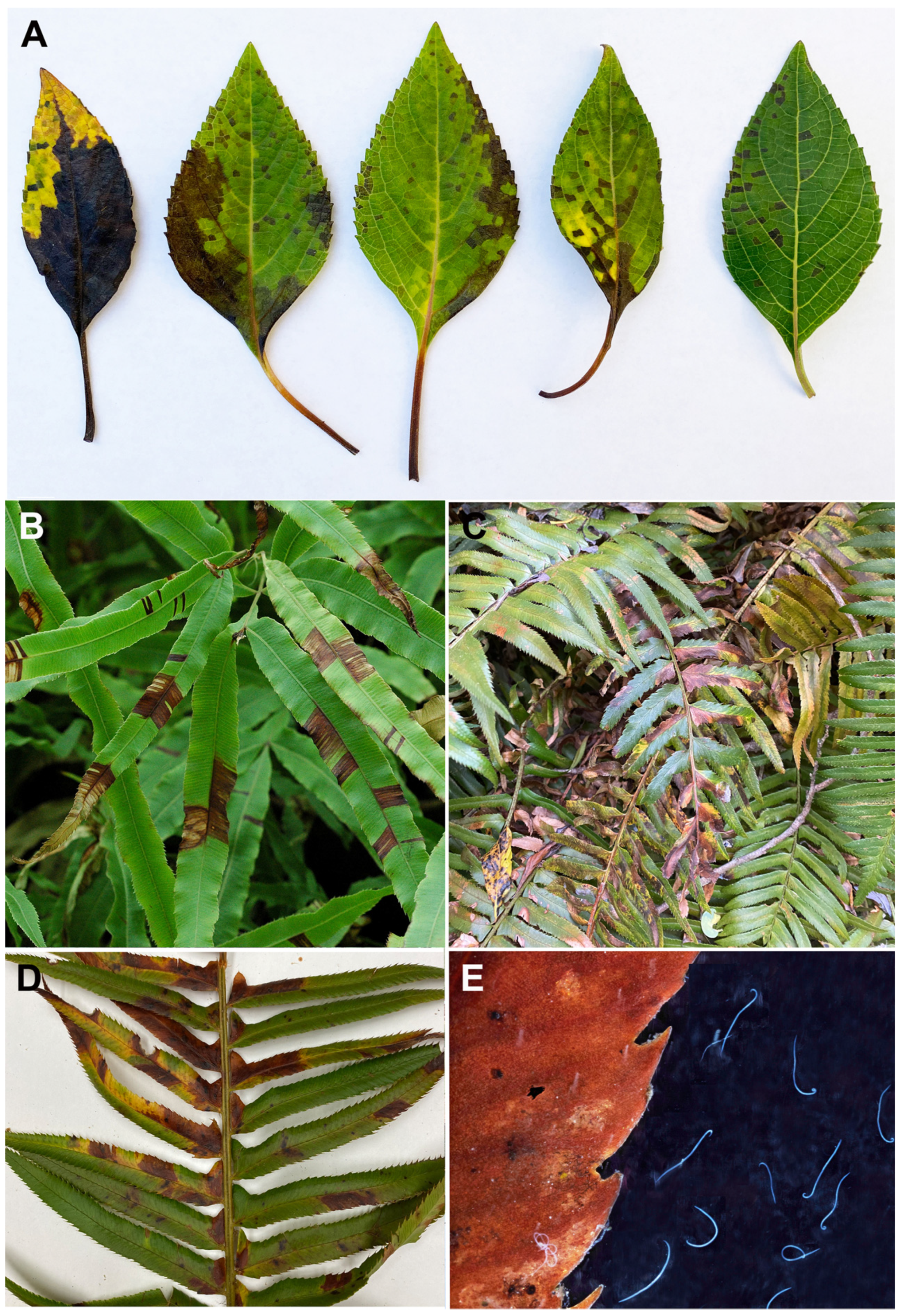
2.1. Nematode Samples

Twenty-seven isolates of A. fragariae were used to develop and validate the LF-RPA diagnostic assay. Nematodes of this species were collected from various plants in the USA (California, Connecticut, North Carolina, Washington), Russia, Germany and New Zealand (Figure 1; Table 1). Aphelenchoides fragariae was identified in rice seeds from California, and A. smolae was found in a strawberry sample from OR, USA.

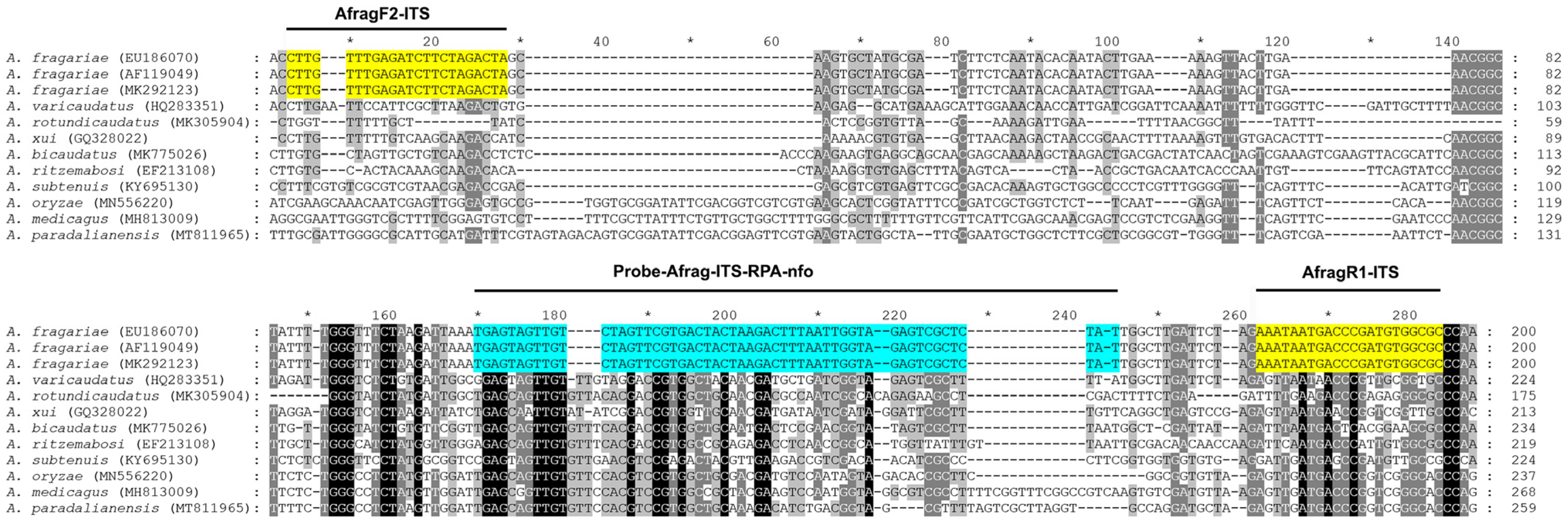
2.2. RPA Primers and Probe Design
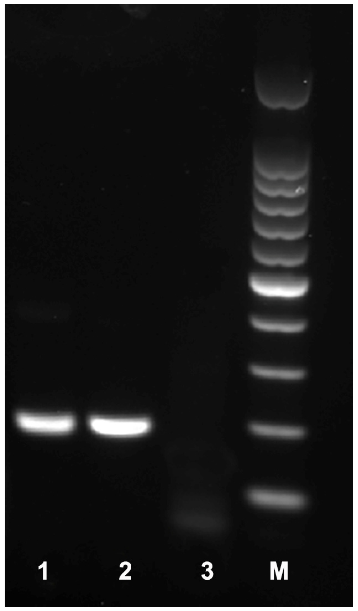
Several ITS rRNA gene sequences of A. fragariae and other Aphelenchoides species were downloaded from GenBank and aligned with ClustalX. Regions with high sequence dissimilarity between A. fragariae and other Aphelenchoides spp. were assessed, and four species-specific A. fragariae candidate primers and one probe were manually designed. The Blastn search of these species-specific candidate primer sequences and probe sequences showed high identity (100% similarity) only with the ITS rRNA fragments of A. fragariae deposited in GenBank.

2.3. RPA Detection

Four primer combinations were screened for the best performance under the same RPA conditions. The species-specific forward AfragF2-ITS and reverse AfragR1-ITS primers were found to be optimal with a clearly visible band. This primer set reliably and specifically amplified the target gene fragment on a gel, approximately 194 bp in length (Figure 2). The final sequences of the primers and probe used for the assays are listed in Table 2 and are indicated in the ITS rRNA gene alignment in Figure 3. Sometimes, non-specific weak bands of other sizes with this primer set were observed in experiments with other nematode samples or a negative control.

2.4. LF-RPA Assay

The workflows of LF-RPA detection assay for the strawberry foliar nematode Aphelenchoides fragariae are given in Figure 4.

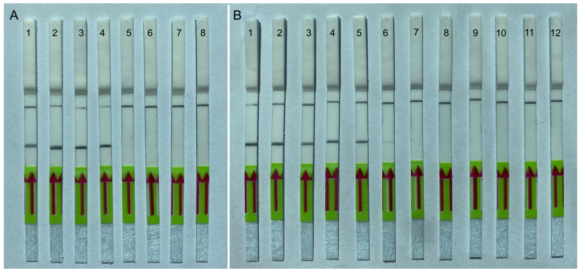
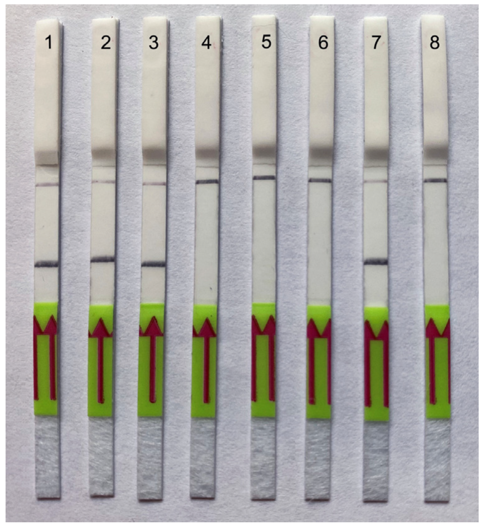
2.4.1. Specificity Testing

The RPA assay was tested for specificity using DNA extracted from eight populations of A. fragariae, six Aphelenchoides spp., six species belonging to other genera from the order Aphelenchida and an unidentified anguinid nematode parasitizing fern, P. munitum (Table 1). The RPA results showed high specificity to A. fragariae only, and no positive reactions were observed against any other nematodes. Positive test lines on the LF strips were observed for all A. fragariae samples, whereas the samples with other nematode species showed only a control line (Figure 5A).

2.4.2. Sensitivity Testing

The sensitivity assay evaluated the specimen number detection limit for a crude nematode extract and for DNA extracted from strawberry leaves and nematodes. Two-fold serial dilutions of crude nematode extract were prepared with a range between 0.5 and 0.001 nematode per reaction tube. The reliable detection level of A. fragariae was estimated to be 0.03 nematode per reaction tube, although weak test bands were also visible in some replicates at lower dilutions (Figure 5B). DNA extracted from 0.1 g of healthy strawberry leaves with 1, 5, 10, 15 and 20 nematodes was used in other sensitivity studies. RPA assays reliably detected this nematode species with plant DNA extracted from 0.1 g of healthy strawberry leaves containing 5 or more A. fragariae specimens (Figure 6A). Thus, reliable sensitivity was estimated at 0.3 nematode per reaction tube using plant DNA extracts. No positive results were obtained from the testing of crude plant extracts without target nematodes.

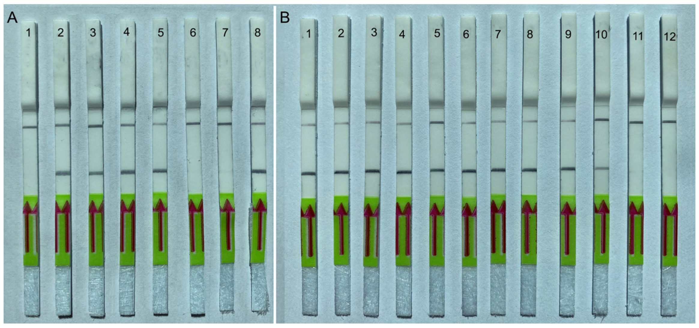
2.4.3. Testing of Plant Herbarium Materials

Aphelenchoides fragariae detection was confirmed using DNA extracts obtained from 20 dried leaf samples previously identified as infected by this nematode. Nematode-infected plants were collected and identified by Dr. D. Sturhan in Germany and New Zealand and preserved as herbarium specimens (Table 1). Positive test lines on the LF strips were observed for all DNA samples extracted from infected plant leaves only, whereas DNA samples without nematodes showed only a control line (Figure 6B).

2.4.4. Testing of Field Samples

Detection of the strawberry foliar nematode was confirmed in all strawberry samples with leaves and stolons in which fern leaves infected with A. fragariae were added. All extracts from the strawberry samples using Baermann funnels contained more than 30 specimens of non-target nematodes (cephalobids, rhabditids and tylenchids), and strawberry samples with target nematodes contained more than 5 moving A. fragariae specimens, which were visible under a binocular microscope. The samples were used to prepare crude nematode extracts. All samples containing A. fragariae showed strong positive test lines on the LF strips in RPA assays, whereas extracts from strawberry samples containing only non-target nematodes gave only a control line on the LF strips (Figure 7). No false negative or false positive reactions were observed in this experiment.

3. Discussion

The LF-RPA assay developed and tested in this study is a simple, fast and sensitive method for the detection of the strawberry foliar nematode, A. fragariae. The assay allows for the detection of this species from crude nematode extracts, nematode DNA and DNA extracts from infected plant tissues. The LF-RPA assay has some important advantages over our previously developed PCR method of A. fragariae detection [21], the first being that it uses crude nematode extract for the analysis instead of DNA extracts, which are required for PCR assays. The second advantage is that results are available for up to 25 min for RPA vs. more than 3.0 h for PCR assay including DNA extraction, PCR and electrophoresis.
The present A. fragariae diagnostic assay and our recently published Meloidogyne hapla diagnostics assay [30] can be used to detect these plant-parasitic nematodes in support of the strawberry certification program. These LF-RPA nematode diagnostic assays could also be performed without any special equipment.
The LF-RPA assay developed in this study is for detection of A. fragariae only. The testing did not reveal any false positive results with other nematode samples, including samples with an unidentified anguinid nematode parasitizing the fern, P. munitum. This putative new anguinid species found in Washington state, in rainforests, causes necrotic symptoms on leaves similar to those induced by the strawberry foliar nematode.
Diagnostic specificity of an assay facilitates the detection of a target organism even in the presence of non-target species that are potentially cross-reactive. The selection of appropriate species-specific primer and probe sequences that match the genomic region of the target species is critical for assay design. For example, recent extensive testing of primers and a probe proposed by Wang et al. [31] for an RPA diagnostic assay for Globodera rostochiensis with a wider range of Globodera and other cyst nematode species showed that the assay is not specific. The results of this testing showed that the proposed ITS rRNA gene putatively specific primers and the probe gave positive reactions not only with G. rostochiensis, but also with other Globodera and representatives of the genus Punctodera parasitising grasses [32]. It has been known that complementarity between primers and templates is often crucial for DNA amplification. It is likely that lower reaction temperatures during RPA might also lead to less specific DNA hybridization, compromise primer specificity, and may have a significant effect on the occurrence of false positive results despite mismatches [32].
In the present assay, the A. fragariae-specific primers have many mismatch differences with non-target Aphelenchoides, and testing did not reveal any false positive reactions with other non-target nematodes. However, the in silico and laboratory tests did not include two species, A. blastophthorus and A. saprophilus, that have close phylogenetic relationships with A. fragariae. Unfortunately, the ITS rRNA gene sequences of these two species are not currently available in GenBank, and their DNA was also not available for the present study. Further testing with other Aphelenchoides species is needed for confirmation of the high specificity of the described LF-RPA assay.
Several Aphelenchoides species are known to parasitize or to be associated with strawberry plants: A. besseyi, A. bicaudatus, A. blastophthorus, A. fragariae, A. pseudobesseyi, A. pseudogoodeyi, A. ritzemabosi and A. rutgersi [33,34,35,36,37,38,39]. In this study, A. smolae was reported to be a nematode species associated with strawberry for the first time. This species was recently isolated and described from medium soil and tissues of Lilium orientalis bulbs imported in China from the Netherlands [40].
In the results of our study, we also identified A. fragariae from fern, Polystichum munitum and rice seeds. This is the first molecular identification of this nematode in this fern species in California. Previously, A. fragariae on P. munitum were found in several western states [41] and California. To the best of our knowledge, it is also the first report of A. fragariae from rice. Presently, only A. oryzae and A. pseudogoodeyi belonging to the A. besseyi species complex are known to be parasites of rice [37,39]. In California, nematodes from the A. besseyi species complex were detected in a fungal culture of Sclerotium oryzae, which caused stem rot of rice in 1963. The fungus was collected from a rice field in Butte County that was used by a research facility that exchanged seeds with areas in the southeastern USA. In the last 20 years, there have been very occasional Aphelenchoides detections in Butte, Colusa, Sutter, Yolo and Yuba counties during phytosanitary inspections of rice for export [42]. The finding of A. fragariae in rice seeds will alert plant pathologists and nematologists to the necessity for further surveys of this pest in rice fields in California. The Aphelenchoides fragariae RPA-LFA assay, together with another novel A. besseyi species complex assay, that combines the RPA and CRISPR/Cas12a methods [43] could be used in rapid diagnostics of these Aphelenchoides pests in plant and soil samples.

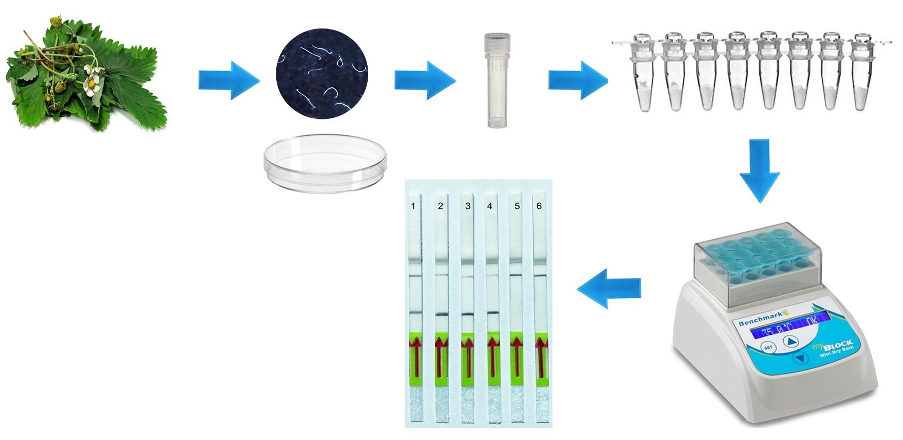
4. Materials and Methods

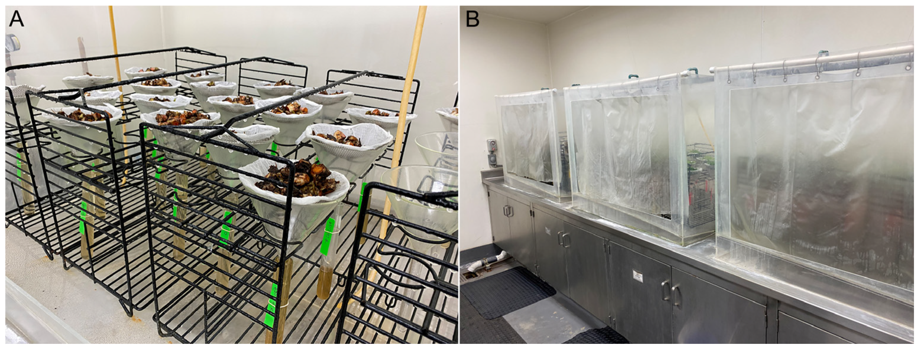
4.1. Nematode Samples

Aphenchoides fragariae isolates and other nematodes used in the present study were obtained from various sources (Table 1). Juvenile stages and adult stages of the strawberry foliar nematode were extracted from fresh leaf samples of different plants using a standard Baermann funnel method [44]. Extracted nematodes were morphologically and molecularly identified. A herbarium collection of A. fragariae-infected plant leaves collected in Germany, New Zealand and other locations during 1960–2000 were obtained from Dr. D. Sturhan (BBA, Münster, Germany), who identified the nematode morphologically. The herbarium materials were kept at room temperature until this study, and then, used for plant DNA extraction. Plant materials were also provided by Dr. J.L. McCuiston and identified as nematode-infected in our previous study [21]. DNA of several other nematodes (A. besseyi, A. oryzae, A. pseudobesseyi, A. ritzemabosi, A. smolae, Aphelenchoides sp., Aphelenchus sp., Bursaphelenchus fraudulentus, B. mucronatus, B. cocophilus and Laimaphelenchus hyrcanus) were also used in assay specificity experiments (Table 1). These species were also identified by molecular methods [37,38,39].
For assay validation with field samples, twelve healthy strawberry plants were provided to the CDFA Nematology lab by California growers. Approximately 20 g of plant tissues from each strawberry sample were placed in Baermann funnels in mist chambers. One gram of infected fern leaves was added to the funnels of half of these samples (Figure 8). The water gradually filled the collection tubes and overflowed slowly enough that nematodes remained in the bottom of the tube. All samples were incubated under the mist for 2 days, and then, each collection tube was carefully removed from its funnel without disturbing the contents. A large pipette was used to draw off the water carefully from each tube to avoid stirring up nematodes that had settled to the bottom, as described by Ayoub [44]. Extracts from all samples were visually inspected under a binocular microscope to reveal the presence of moving Aphelenchoides specimens and non-target nematodes. The samples were used to prepare crude nematode extracts.

4.2. Preparation of Nematode Crude Extract and DNA Extrication

Nematode crude extracts, nematode DNA and DNA from plant tissues were used for development and validation of the LF-RPA diagnostic assay.
For nematode crude extracts, live nematode specimens were placed into a drop of distilled water on a glass slide and cut by a stainless-steel dental needle under a stereo microscope. Cut nematodes were transferred in water suspension into a 0.2 mL PCR tube. Extracts from 20 adults or fourth juvenile stage nematode specimens in 40 μL of water were used to make a series of two-fold sequential dilutions to test the sensitivity of the assay.
Nematode DNA was extracted from several specimens. Nematodes were placed in 20 μL ddH2O on a glass slide and cut by a stainless-steel dental needle under a stereo microscope. Cut nematodes in water suspension were transferred into a 0.2 mL Eppendorf tube, and then, three μL of proteinase K (600 μg/mL) (Promega, Madison, WI, USA) and 2 μL of 10× PCR buffer (Taq PCR Core Kit, Qiagen, Germantown, MD, USA) were added to each tube. The tubes were incubated at 65 °C (1 h) and 95 °C (15 min) consecutively. After incubation, the tubes were centrifuged and kept at −20 °C until use.
DNA from infected and healthy control plant leaves was extracted using the Qiagen DNeasy Plant Mini Kit following the manufacturer’s protocol. Total genomic DNA was eluted in a final volume of 30 μL elution buffer and stored at −20 °C. Leaf tissues (0.1 g) were used for each extraction. In total, 1, 5, 10, 15 and 20 nematodes were also added into tubes with uninfected strawberry leaves (0.1 g), and then, used for DNA extraction as described above. These preparations were used to test the sensitivity of the assay for the detection of nematodes in plant tissues.
For validation of the assay with field samples, live nematodes in 300 mL of water obtained from each sample were added into individual tubes and homogenized with an ultimate laboratory homogenizer, Mini Bead Beater 1 (BioSpec Products, Bartlesville, OK, USA) and one glass bead (5 mm) for 30–60 s to obtain nematode extract.

4.3. Nematode Molecular Identification

The ITS rRNA and D2-D3 expansion segments of the 28S rRNA gene were amplified and sequenced from nematode isolates to confirm their species identity. PCR protocols were used as described by McCuiston et al. [21]. The following primer sets were used for PCR: (i) the forward D2A (5′-ACA AGT ACC GTG AGG GAA AGT TG-3′) and the reverse D3B (5′-TCG GAA GGA ACC AGC TAC TA-3′) primers for amplification of the D2–D3 expansion segments of the 28S rRNA gene [45] and (ii) the forward TW81 (5′-GTT TCC GTA GGT GAA CCT GC-3′) and the reverse AB28 (5′-ATA TGC TTA AGT TCA GCG GGT-3′) primers for amplification of the ITS1-5.8-ITS2 rRNA gene [46]. PCR products were purified using a QIAquick PCR Purification Kit (Qiagen, USA) and directly sequenced with the primers mentioned above or cloned using a pGEM-T Vector System II kit (Promega, Fitchburg, WI, USA), and then, the clones were sequenced. Sequencing was performed by Genewiz Inc. (Berkeley, CA, USA). Obtained sequences were compared with those deposited in GenBank using a Blastn search [47]. New sequences were deposited in the GenBank database under accession numbers OR685296-OR685297 (ITS rRNA gene) and OR691588-OR691594 (28S rRNA gene).

4.4. RPA Primer and Probe Design and Testing
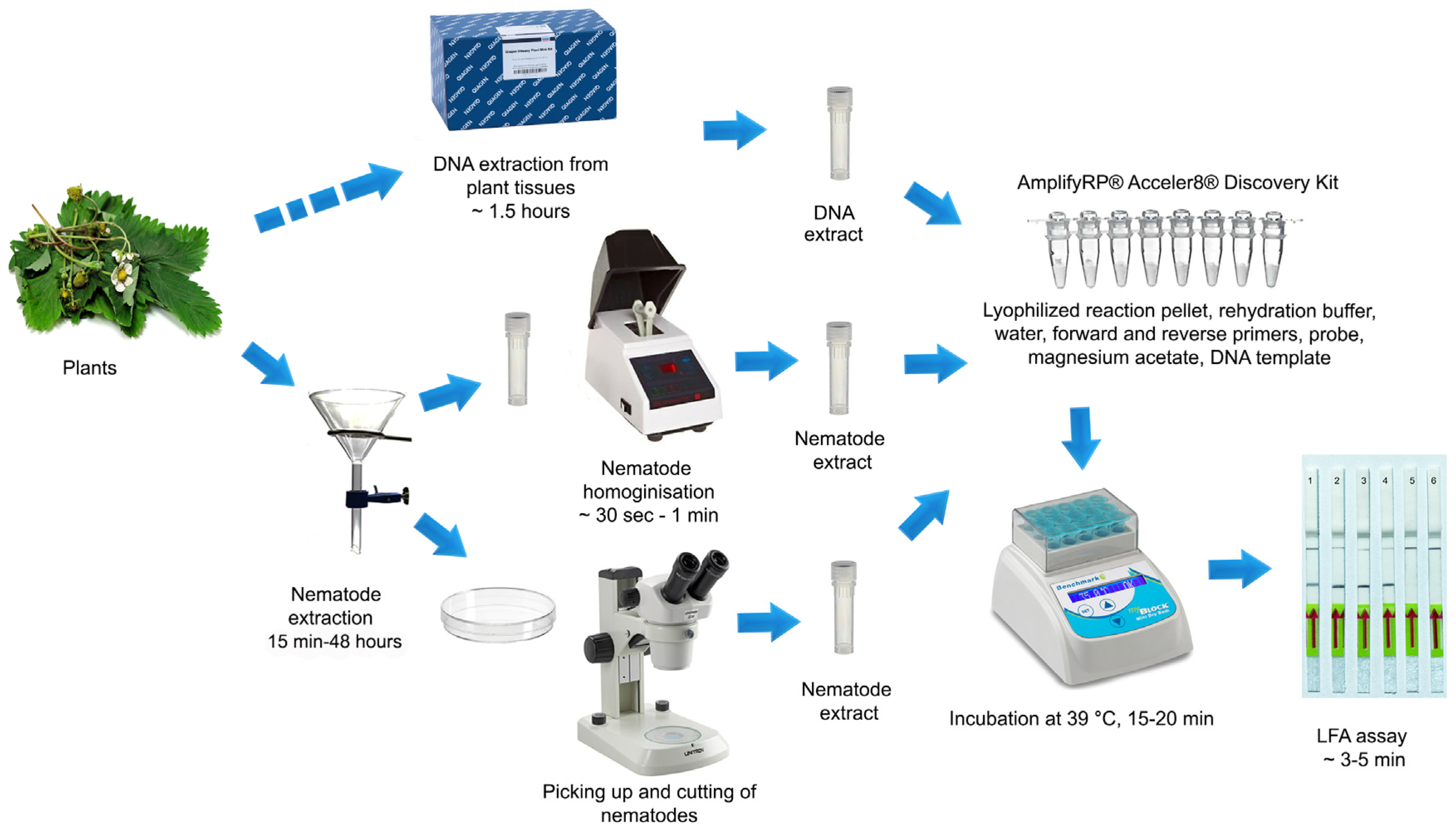
Two forward and two reverse RPA primers specific to A. fragariae were manually designed based on species sequence polymorphisms in the ITS rRNA gene. Primers were synthesized by Integrated DNA Technologies, Inc. (Redwood City, CA, USA). Primers were screened in different combinations using the TwistAmp® Basic kit (TwistDx, Cambridge, UK). Reactions were prepared according to the manufacturer’s instructions. The lyophilized reaction pellets were suspended in 29.5 μL of rehydration buffer, 2.4 μL each of forward and reverse primers (10 μM) (Table 2), 1 μL of DNA template or nematode extract and 12.2 μL of distilled water. For each sample, 2.5 μL of 280 mM magnesium acetate was added to the lid of the tube and the lids were closed carefully. The tubes were inverted 10–15 times and briefly centrifuged to initiate reactions simultaneously. Tubes were incubated at 39 °C (4 min) in a MyBlock Mini Dry Bath (Benchmark Scientific, Sayreville, NJ, USA), and then, they were inverted 10–15 times, briefly centrifuged and returned to the incubator block (39 °C) for 20 min. Sample tubes were then placed in a freezer to stop the reaction. Amplification products were purified with a QIAquick PCR Purification Kit (Qiagen, Germantown, MD, USA). Five microliters of purified product were run in a 1% TAE buffered agarose gel (100 V, 60 min) and visualized with a Gel Green stain. The primer set was selected based on amplification performance. The probe was designed based on species sequence polymorphisms in the ITS rRNA gene. RPA primers and probe were synthetized at Biosearch Technologies (Novato, CA, USA).

4.5. LF-RPA Assay

The LF-RPA assay was carried out using an AmplifyRP® Acceler8® Discovery Kit (Agdia, Elkhart, IN, USA). The reaction mixture for each RPA assay was prepared according to the manufacturer’s instructions: The lyophilized reaction pellet was suspended with a mixture containing 6 µL of the rehydration buffer, 2 µL of distilled water, 0.45 µL each of forward and reverse primers (10 µM), 0.15 µL of the probe (10 µM) and 0.5 µL of magnesium acetate. One microliter of the DNA template or extract was added to a reaction tube. The reaction tubes were incubated at 39 °C in a MyBlock Mini Dry Bath (Benchmark Scientific, Edison, NJ, USA) for 20 min. For visual analysis with Milenia® Genline Hybridetect-1 strips (Milenia Biotec GmbH, Giessen, Germany), 120 µL of HybriDetect assay buffer was added to a reaction tube, and then, a dipstick was placed in this mixture. Visual results were observed within 3–5 min, and then, photographed. The amplification product was indicated by the development of an intensively colored test line (lower) and/or a separate control line (upper) to confirm that the system worked properly. Three replicates of each variant were performed for sensitivity and specificity experiments.

Funding

This work was sponsored by the Specialty Crop Block Grant Program (USDA-CDFA Grant Number: 21-0001-045-MU).

Institutional Review Board Statement

Not applicable.

Informed Consent Statement

Not applicable.

Data Availability Statement

The data are contained within the article.

Acknowledgments

The author thanks V.N. Chizhov, I. Cid del Prado Vera, W. Crow, J.A. LaMondia, J.L. McCuiston, C. Oliveira, C. Overstreet and A. Ryss for providing the nematode materials; J. Burbridge and J. Ramirez Bonilla for providing technical assistance; P. Woods for performing DNA extraction from plant materials; and H. Ferris for critical reading of the manuscript draft.

Conflicts of Interest

The author declares no conflicts of interest.

References

  1. Christie, J.R. Recent observations on the strawberry dwarf nematode in Massachusetts. Plant Dis. Report. 1932, 16, 113–114. [Google Scholar]
  2. Ritzema Bos, J. Zwei neue Nematodenkrankheiten der Erdbeerpflanzen. Z. Für Pflanzenkrankh. Pflanzenschutz 1891, 1, 1–16. [Google Scholar]
  3. Allen, M.W. Taxonomic status of the bud and leaf nematodes related to Alphelenchoides fragariae (Ritzema Bos, 1891). Proc. Helminth. Soc. Wash. 1952, 19, 108–120. [Google Scholar]
  4. Raski, D.J.; Allen, M.W. Spring dwarf nematode. Calif. Agric. 1948, 4, 23–24. [Google Scholar]
  5. Maas, J.L. (Ed.) Compendium of Strawberry Diseases, 2nd ed.; APS Press: Saint Paul, MN, USA, 1998. [Google Scholar]
  6. Sturhan, D. Über neue Wirtspflanzen der Blattälchen Aphelenchoides fragariae und A. ritzemabosi, mit Bemerkungen zu den Wirtspflanzenkreisen beider Nematodenarten. Anz. Schädlingskunde 1962, 35, 65–67. [Google Scholar] [CrossRef]
  7. Siddiqi, M.R. Aphelenchoides fragariae. C.I.H. Description of Plant-Parasitic Nematodes; Set 5, No. 74; William Clowes & Sons Ltd.: London, UK, 1975; p. 4. [Google Scholar]
  8. Hunter, J.E.; Ko, W.H.; Kunimoto, R.K.; Higaki, T. A foliar disease of anthurium seedlings caused by Aphelenchoides fragariae. Phytopathology 1974, 64, 267–268. [Google Scholar] [CrossRef]
  9. Knight, K.W.L.; Hill, C.F.; Sturhan, D. Further records of Aphelenchoides fragariae and A. ritzemabosi (Nematoda: Aphelenchida) from New Zealand. Australas. Plant Pathol. 2002, 31, 93–94. [Google Scholar] [CrossRef]
  10. Khan, Z.; Son, S.; Moon, H.; Kim, S.; Shin, H.; Jeon, Y.; Kim, Y. Description of a foliar nematode, Aphelenchoides fragariae (Nematoda: Aphelenchida) with additional characteristics from Korea. J. Asia-Pac. Entomol. 2007, 10, 313–315. [Google Scholar] [CrossRef]
  11. Khan, Z.; Son, S.H.; Shin, H.D.; Kim, Y.H. First report of a foliar nematode Aphelenchoides fragariae (Aphelenchidae) on Stachys riederi var. japonica, a medicinal plant, in Korea. Plant Pathol. J. 2008, 24, 97–100. [Google Scholar] [CrossRef]
  12. Kohl, L.M. Foliar nematodes: A summary of biology and control with a compilation of host range. Plant Health Prog. 2011, 12, 1. [Google Scholar] [CrossRef]
  13. Zhen, F.; Agudelo, P.; Gerard, P. A protocol for assessing resistance to Aphelenchoides fragariae in hosta cultivars. Plant Dis. 2012, 96, 1438–1444. [Google Scholar] [CrossRef]
  14. Siddiqui, I.A.; Sher, S.A.; French, A.M. Distribution of Plant-Parasitic Nematodes in California; Division of Plant Industry, Department of Food and Agriculture: Sacramento, CA, USA, 1973; 324p. [Google Scholar]
  15. Handoo, Z.; Kantor, M.; Carta, L. Taxonomy and identification of principal foliar nematode species (Aphelenchoides and Litylenchus). Plants 2020, 9, 1490. [Google Scholar] [CrossRef]
  16. Hunt, D.J. Aphelenchida, Longidoridae and Trichodoridae: Their Systematics and Bionomics; CABI Publishing: Wallingford, UK, 1993; 352p. [Google Scholar]
  17. The California Strawberry Commission (CSC) & the California Minor Crops Council (CMCC). A Pest Management Strategic Plan for Strawberry Production in California. Available online: https://ipmdata.ipmcenters.org/documents/pmsps/CASTRAWBERRY.PDF (accessed on 27 December 2023).
  18. Strawberry Registration & Certification Program. Available online: https://www.cdfa.ca.gov/plant/pe/nsc/nursery/strawberry.html (accessed on 27 December 2023).
  19. Franklin, M.T. Aphelenchoides and related genera. In Plant Nematology; Southey, J.F., Ed.; Her Majesty’s Stationery Office: London, UK, 1978; pp. 172–187. [Google Scholar]
  20. Iwahori, H.; Tsuda, K.; Kanzaki, N.; Izui, K.; Futai, K. PCR-RFLP and sequencing analysis of DNA of Bursaphelenchus nematodes related to pine wilt disease. Fundam. Appl. Nematol. 1998, 21, 655–666. [Google Scholar]
  21. McCuiston, J.L.; Hudson, L.C.; Subbotin, S.A.; Davis, E.L.; Warfield, C.Y. Conventional and PCR detection of Aphelenchoides fragariae in diverse ornamental host plant species. J. Nematol. 2007, 39, 343–355. [Google Scholar]
  22. Rybarczyk-Mydłowska, K.; Mooyman, P.; van Megen, H.; van den Elsen, S.; Vervoort, M.; Veenhuizen, P.; van Doorn, J.; Dees, R.; Karssen, G.; Bakker, J.; et al. Small subunit ribosomal DNA-based phylogenetic analysis of foliar nematodes (Aphelenchoides spp.) and their quantitative detection in complex DNA backgrounds. Phytopathology 2012, 102, 1153–1160. [Google Scholar] [CrossRef]
  23. Chałańska, A.; Bogumił, A.; Winiszewska, G.; Kowalewska, K.; Malewski, T. Morphological and molecular characteristics of foliar nematode attacking silver birch (Betula pendula Roth) in Poland. Helminthologia 2017, 54, 250–256. [Google Scholar] [CrossRef]
  24. Holterman, M.; van der Wurff, A.; van den Elsen, S.; van Megen, H.; Bongers, T.; Holovachov, O.; Bakker, J.; Helder, J. Phylum-wide analysis of SSU rDNA reveals deep phylogenetic relationships among nematodes and accelerated evolution toward crown clades. Mol. Biol. Evol. 2006, 23, 1792–1800. [Google Scholar] [CrossRef]
  25. Holterman, M.; Karegar, A.; Mooijman, P.; van Megen, H.; van den Elsen, S.; Vervoort, M.T.; Quist, C.W.; Karssen, G.; Decraemer, W.; Opperman, C.H.; et al. Disparate gain and loss of parasitic abilities among nematode lineages. PLoS ONE 2017, 12, e0185445. [Google Scholar] [CrossRef]
  26. Sánchez-Monge, A.; Janssen, T.; Fang, Y.; Couvreur, M.; Karssen, G.; Bert, W. mtCOI successfully diagnoses the four main plant-parasitic Aphelenchoides species (Nematoda: Aphelenchoididae) and supports a multiple origin of plant-parasitism in this paraphyletic genus. Eur. J. Plant Pathol. 2017, 148, 853–866. [Google Scholar] [CrossRef]
  27. Djiwanti, S.R.; Miftakhurohmah. Molecular detection and identification of the foliar nematode Aphelenchoides fragariae on Andrographis paniculata in Indonesia. Australas. Plant Pathol. 2022, 51, 301–304. [Google Scholar] [CrossRef]
  28. Ibrahim, S.K.; Perry, R.N.; Burrows, P.R.; Hooper, D.J. Differentiation of species and populations of Aphelenchoides and of Ditylenchus angustus using a fragment of ribosomal DNA. J. Nematol. 1994, 26, 412–421. [Google Scholar] [CrossRef]
  29. Tan, M.; Liao, C.; Liang, L.; Yi, X.; Zhou, Z.; Wei, G. Recent advances in recombinase polymerase amplification: Principle, advantages, disadvantages and applications. Front. Cell. Infect. Microbiol. 2022, 12, 1019071. [Google Scholar] [CrossRef]
  30. Subbotin, S.A.; Burbridge, J. Sensitive, accurate and rapid detection of the northern root-knot nematode, Meloidogyne hapla, using Recombinase Polymerase Amplification assays. Plants 2021, 10, 336. [Google Scholar] [CrossRef]
  31. Wang, X.; Lei, R.; Peng, H.; Jiang, R.; Shao, H.D.; Ge, J.J.; Peng, D.L. Rapid diagnosis and visual detection of potato cyst nematode (Globodera rostochiensis) using recombinase polymerase amplification combination with lateral flow assay method (RPA-LFA). Agronomy 2022, 12, 2580. [Google Scholar] [CrossRef]
  32. Subbotin, S.A. On the reliability of recombinase polymerase amplification—Lateral flow assay using ITS rRNA gene primers and probe as a new detection method of the golden potato cyst nematode, Globodera rostochiensis. Russ. J. Nematol. 2023, 31, 115–120. [Google Scholar]
  33. Christie, J.R. A description of Aphelenchoides besseyi n. sp., the summer dwarf nematode of strawberries, with comments on the identity of Aphelenchoides subtenuis (Cobb, 1929) and Aphelenchoides hodsoni Goodey, 1935. Proceeding Helminthol. Soc. Wash. 1942, 9, 82–84. [Google Scholar]
  34. Franklin, M.T. Two species of Aphelenchoides associated with strawberry bud disease in Britain. Ann. Appl. Biol. 1950, 37, 1–10. [Google Scholar] [CrossRef]
  35. Haukeland, S.; Brekke, K. Yield loss in strawberries caused by Aphelenchoides blastophthorus. (Abstract). Nematology 2000, 2, 759. [Google Scholar]
  36. Consoli, E.; Ruthes, A.C.; Reinhard, E.; Dahlin, P. First morphological and molecular report of Aphelenchoides blastophthorus on strawberry plants in Switzerland. Plant Dis. 2019, 103, 2851–2856. [Google Scholar] [CrossRef]
  37. Oliveira, C.J.; Subbotin, S.A.; Álvarez-Ortega, S.; Desaeger, J.; Brito, J.A.; Xavier, K.; Freitas, L.G.; Vau, S.; Inserra, R.N. Morphological and molecular identification of two Florida populations of foliar nematodes (Aphelenchoides spp.) isolated from strawberry with the description of Aphelenchoides pseudogoodeyi sp. n. (Nematoda: Aphelenchoididae) and notes on their bionomics. Plant Dis. 2019, 103, 2825–2842. [Google Scholar] [CrossRef]
  38. Oliveira, C.J.; Subbotin, S.A.; Desaeger, J.A.; Dahlin, P.; Vau, S.; Inserra, R.N. Morphological and molecular analysis of two mycophagous nematodes, Aphelenchoides bicaudatus and A. rutgersi (Nematoda: Aphelenchoididae) from Florida strawberry. J. Nematol. 2024, unpublished. [Google Scholar]
  39. Subbotin, S.A.; Oliveira, C.J.; Alvarez-Ortega, S.; Desaeger, J.; Crow, W.; Overstreet, C.; Leany, R.; Vau, S.; Inserra, R.N. The taxonomic status of Aphelenchoides besseyi Christie, 1942 (Nematoda: Aphelenchoididae) populations from the Southeastern USA, and description of Aphelenchoides pseudobesseyi sp. n. Nematology 2021, 23, 381–413. [Google Scholar] [CrossRef]
  40. Cai, J.; Gu, J.; Wang, X.; Fang, Y.; Li, H. Aphelenchoides smolae n. sp. (Tylenchina: Aphelenchoididae) found in Lilium orientalis imported into China from The Netherlands. Nematology 2020, 22, 799–813. [Google Scholar] [CrossRef]
  41. Sandeno, J.L.; Jensen, H.J. A foliar nematode disease of western sword-fern, Polystichum munitum. Plant Dis. Report. 1962, 46, 699–701. [Google Scholar]
  42. Chitambar, J.J.; Westerdahl, B.B.; Subbotin, S.A. Plant-Parasitic Nematodes in California Agriculture. In Plant-Parasitic Nematodes in Sustainable Agriculture of North America: Vol.1—Canada, Mexico and Western USA; Subbotin, S.A., Chitambar, J.J., Eds.; Springer: Berlin/Heidelberg, Germany, 2018; pp. 131–192. [Google Scholar]
  43. Zhang, A.; Sun, B.; Zhang, J.; Cheng, C.; Zhou, J.; Niu, F.; Luo, Z.; Yu, L.; Yu, C.; Dai, Y.; et al. CRISPR/Cas12a coupled with Recombinase Polymerase Amplification for sensitive and specific detection of Aphelenchoides besseyi. Front. Bioeng. Biotechnol. 2022, 10, 912959. [Google Scholar] [CrossRef]
  44. Ayoub, S.M. Plant Nematology: An Agricultural Training Aid; Nema Aid Publications: Sacramento, CA, USA, 1980; 195p. [Google Scholar]
  45. De Ley, P.; Felix, M.A.; Frisse, L.M.; Nadler, S.A.; Sternberg, P.W.; Thomas, W.K. Molecular and morphological characterisation of two reproductively isolated species with mirror image anatomy (Nematoda: Cephalobidae). Nematology 1999, 2, 591–612. [Google Scholar] [CrossRef]
  46. Joyce, S.A.; Reid, A.; Driver, F.; Curran, J. Application of polymerase chain reaction (PCR) methods to identification of entomopathogenic nematodes. In COST 812 Biotechnology: Genetics of Entomopathogenic Nematode–Bacterium Complexes; Burnell, A.M., Ehlers, R.U., Masson, J.P., Eds.; Proceedings of Symposium & Workshop, St. Patrick’s College, Maynooth, Co.: Kildare, Ireland; Luxembourg, 1994; pp. 178–187. [Google Scholar]
  47. Sayers, E.W.; Bolton, E.E.; Brister, J.R.; Canese, K.; Chan, J.; Comeau, D.C.; Connor, R.; Funk, K.; Kelly, C.; Kim, S.; et al. Database resources of the national center for biotechnology information. Nucleic Acids Res. 2022, 50, D20–D26. [Google Scholar] [CrossRef]
Figure 1. Symptoms of Aphelenchoides fragariae infection. (A) leaves of Salvia sp.; (B) Pteris plants; (C) Polystichum munitum plants in Mendocino County, California; (D) leaves of P. munitum; (E) nematodes extracted from P. munitum leaves.
Ijms 25 00844 g001
Figure 2. RPA products on agarose gel amplified with AfragF2-ITS and AfragR1-ITS primers using TwistAmp® Basic kit. Lanes: 1: Aphelenchoides fragariae (sample CA38); 2: A. fragariae (CA53); 3: negative control (no DNA); M: 100 bp DNA marker (Promega, Madison, WI, USA).
Ijms 25 00844 g002
Figure 3. The fragment of the ITS rRNA gene sequence alignment for A. fragariae and several Aphelenchoides species with positions of RPA primers (yellow) and probe (blue) used in the present assay.
Ijms 25 00844 g003
Figure 4. Workflows of LF-RPA detection assay for the strawberry foliar nematode Aphelenchoides fragariae used in this study.
Ijms 25 00844 g004
Figure 5. Lateral flow recombinase polymerase amplification (LF-RPA) assay with examples of lateral flow strips. (A) Specificity assay with DNA samples of different nematodes. Strips: 1–4: Aphelenchoides fragariae (CA30, CA53, CD3764, CD3774); 5: Bursaphelenchus fraudulentus (CD2935); 6: Laimaphelenchus hyrcanus (CD3646); 7: A. pseudobesseyi (CD3702); 8: negative control (no DNA). (B) Sensitivity assay with crude nematode extract of A. fragariae (CD754). Strips: 1: 0.5 nematode per tube; 2: 0.25 nematode per tube; 3: 0.125 nematode per tube; 4: 0.06 nematode per tube; 5: 0.03 nematode per tube; 6: 0.015 nematode per tube; 7: 0.007 nematode per tube; 8: 0.004 per nematode tube; 9: 0.002 nematode per tube; 10: 0.001 nematode per tube; 11, 12: negative control (no DNA).
Ijms 25 00844 g005
Figure 6. Lateral flow recombinase polymerase amplification (LF-RPA) assay with examples of lateral flow strips. (A) Sensitivity assay with DNA extracted from samples containing strawberry leaves and different numbers of A. fragariae (CD3787) specimens. Strips: 1: 0.1 g strawberry leave + 1 nematode; 2: 0.1 g strawberry leaves + 5 nematodes; 3: 0.1 g strawberry leaves + 10 nematodes; 4: 0.1 g strawberry leaves + 15 nematodes; 5: 0.1 g strawberry leaves + 20 nematodes; 6: 0.1 g strawberry leaves; 7: negative control (no DNA); 8: positive control (sample CD3787). (B) Diagnostic assay with DNA extracted from infected plant leaves. Strips: 1: Polypodium californica (CD3794); 2: Lantana sp. (CD3795); 3: Pteris sp. (CD3796); 4: Anemone hupehensis (CD3797); 5: Heuchera sp. (CD3798); 6: Anemone x hybrida (CD3799); 7: Mimulus moschatus (CD3800); 8: Matteuccia orientalis (CD3801); 9, 10: Salvia sp. (CD3787); 11, 12: negative control (no DNA).
Ijms 25 00844 g006
Figure 7. Lateral flow recombinase polymerase amplification (LF-RPA) assay with examples of lateral flow strips. Testing of field samples. Strips: 1–3: nematodes extracted from strawberry plants and A. fragariae-infected fern leaves; 4–6: nematodes extracted from strawberry plants; 7: DNA of A. fragariae (CD4001); 8: negative control (no DNA).
Ijms 25 00844 g007
Figure 8. Baermann funnel technique in the mist system for nematode extraction. (A) Glass funnels and tubes with strawberry samples. (B) Mist extraction system in the Nematology lab, Plant Pest Diagnostic Centre of the California Department of Food and Agriculture.
Ijms 25 00844 g008
Table 1. Samples of Aphelenchoides fragariae and other nematodes tested in the present study.
SpeciesLocationsPlantsMaterials *Sample CodesSources
Aphelenchoides fragariaeUSA, California, Marin County, Point Reyes National SeashorePolystichum munitumND, PD, ExCD3774S.A. Subbotin
A. fragariaeUSA, California, Mendocino CountyP. munitumND, ExCD4001S.A. Subbotin
A. fragariaeUSA, Connecticut, WindsorSalvia sp.ND, ExCD3787J.A. LaMondia
A. fragariaeUSA, Washington, Clallam County, Storm King Ranger StationUnidentified plantND; ExCD3764S.A. Subbotin
A. fragariaeRussia, Moscow, Main Botanical Garden of the RASPteris sp.ND, PDCA30, CA38, CA53, CD3796V.N. Chizhov
A. fragariaeUSA, North CarolinaAnemone hupehensisND, PDCD386, CD3797J.L. McCuiston
A. fragariaeUSA, North CarolinaLantana camaraNDCD388J.L. McCuiston
A. fragariaeUSA, CaliforniaOryza sativaND, ExtCD754S.A. Subbotin
A. fragariaeGermanyPrimula denticulataPDCD3804D. Sturhan
A. fragariaeGermanyLagarosiphon cordofanusPDCD3806D. Sturhan
A. fragariaeNew ZealandPtisana salicinaPDCD3808D. Sturhan
A. fragariaeNew ZealandPolystichum sp.PDCD3809D. Sturhan
A. fragariaeUSA, North CarolinaPolypodium californicaPDCD3794J.L. McCuiston
A. fragariaeUSA, North CarolinaLantana sp.PDCD3795J.L. McCuiston
A. fragariaeUSA, North CarolinaAnemone × hybridaPDCD3799J.L. McCuiston
A. fragariaeUSA, North CarolinaHeuchera sp.PDCD3798J.L. McCuiston
A. fragariaeGermany, MünsterMimulus moschatusPDCD3800D. Sturhan
A. fragariaeGermanyMatteuccia orientalisPDCD3801D. Sturhan
A. fragariaeGermany, MünchenOsmunda regalisPDCD3810D. Sturhan
A. fragariaeNew ZealandTodea barbaraPDCD3811D. Sturhan
A. fragariaeGermany, MünsterPenstemon campanulatusPDCD3934D. Sturhan
A. fragariaeGermany, MünsterLithospermum arvensePDCD3944D. Sturhan
A. fragariaeNew ZealandBlechnum sp.PDCD3950D. Sturhan
A. fragariaeGermany, MünsterSolidago glomerataPDCD3946D. Sturhan
A. fragariaeGermany, MünsterTellima grandifloraPDCD3947D. Sturhan
A. fragariaeGermany, MünsterBergenia sp.PDCD3948D. Sturhan
A. fragariaeGermany, MünsterLigularia sp.PDCD3949D. Sturhan
A. besseyiUSA, FloridaFragaria × ananassaNDCD2415C. Oliveira
A. oryzaeUSA, Louisiana, Morehouse Parish, Mer RougeOryza sativaNDCD2471C. Overstreet
A. oryzaeRussia, KrasnodarO. sativaNDCD3790V.N. Chizhov
A. pseudobesseyiUSA, Florida, Sumter County, SumtervilleDryopteris erythrosoraNDCD2704W. Crow
A. pseudobesseyiUSA, North Carolina, Jackson County, CullowheeSoil sampleNDCD3097C. Oliveira
A. pseudobesseyiUSA, Florida, Alachua County, GainesvilleEchinacea sp.NDCD2491, CD3702W. Crow
A. ritzemabosiUSA, California, Mendocino CountyHelleborus sp.NDCD1366S.A. Subbotin
A. smolaeUSA, Oregon, BonanzaFragaria × ananassaNDCD3775S.A. Subbotin
Aphelenchoides sp.USA, California, San Diego CountyGrassesNDCD1300S.A. Subbotin
Aphelenchus sp.USA, California, Tehama CountyFragaria × ananassaNDCD3788bS.A. Subbotin
Cryptaphelenchus sp.Tomsk region, MalinovkaAbies sibiricaNDCD3657A. Ryss
Bursaphelenchus
cocophilus
Mexico, Guerrero stateCocos nuciferaNDCD3548, CD3572I. Cide del Prado Vera
B. fraudulentusRussia, Moscow, Main Botanical Garden of the RAS Quercus roburNDCD2935A. Ryss
B. mucronatusRussia, BuryatiaAbies sibiricaNDCD3642A. Ryss
Laimaphelenchus hyrcanusRussia, Saint Petersburg Q. roburNDCD3646A. Ryss
Unidentified
anguinid nematode
USA, Washington, Clallam County, Storm King Ranger
Station
P. munitumND, ExCD3762S.A. Subbotin
* ND—DNA extracted from nematodes; PD—DNA extracted from infected plants; Ex—crude nematode extract.
Table 2. RPA primers and probe for amplification of DNA of Aphelenchoides fragariae.
Primer or ProbeSequence (5′–3′)
AfragF2-ITS CTT GTT TGA GAT CTT CTA GAC TA
AfragR1-ITS GCG CCA CATC GGG TCA TTA TTT
AfragR1-ITS-biotin [Biotin] GCG CCA CATC GGG TCA TTA TTT
Probe-Afrag-ITS-RPA-nfo [FAM] TG AGT AGT TGT CTA GTT CGT GAC TAC TAA GAC TTT [THF] ATT GGT AGA GTC GCT CTA T [C3-spacer] *
* FAM—fluorophore, THF—tetrahydrofuran, C3—spacer block.
Disclaimer/Publisher’s Note: The statements, opinions and data contained in all publications are solely those of the individual author(s) and contributor(s) and not of MDPI and/or the editor(s). MDPI and/or the editor(s) disclaim responsibility for any injury to people or property resulting from any ideas, methods, instructions or products referred to in the content.

