
Molecular Mechanisms of Reelin in the Enteric Nervous System and the Microbiota–Gut–Brain Axis: Implications for Depression and Antidepressant Therapy
 , and
, and
Abstract
1. Introduction
2. Overview of Reelin
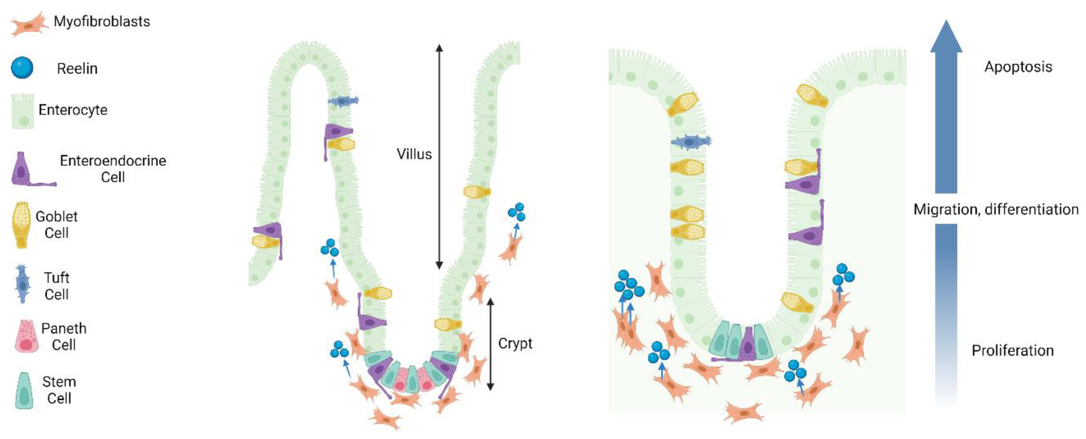
3. Reelin in the Enteric Nervous System and Microbiota–Gut–Brain Axis

| Molecule | Organ | Cell Type | Model | Reference |
|---|---|---|---|---|
| Reelin protein | Small intestine | Myofibroblasts, crypt cells | Mouse, rat, human | [18,19,59] |
| Colon | Ganglia, intramuscular nerve fibres | Mouse, rat, human | [16,18] | |
| VLDLR receptor | Small intestine | Myofibroblasts, epithelial cells, crypt cells, enterocytes | Rat, human | [18,19,59] |
| Colon | Myenteric ganglia | Mouse, rat, human | [18] | |
| APOER2 receptor | Small intestine | Myofibroblasts, epithelial cells, enterocytes | Rat, human | [16,18,59] |
| Colon | Myenteric ganglia | Mouse, rat, human | ||
| α3β1-integrin receptor | Small intestine | Myofibroblasts, enterocytes, epithelial cells, crypt cells | Mouse | [19,59,61] |
| Colon | Epithelial cells | Human (in vitro) | [61] | |
| EphB2 receptor | Small intestine | Epithelial cells, crypt cells | Mouse, human | [60,61,62] |
| Colon | Epithelial cells | Human | [60,61,62] |
4. Microbiota–Gut–Brain Axis Dysfunction in Depression
4.1. Alterations to the Gut Barrier and Blood–Brain Barrier Integrity in Depression
4.2. Alterations to the Microbiota in Depression
4.3. Effects of Antidepressants on the Microbiota–Gut–Brain Axis
5. Reelin in Depression and Putative Antidepressant Roles
5.1. Reelin in Depression
5.2. Putative Antidepressant Role of Reelin in the Enteric Nervous System
6. Conclusions
Author Contributions
Funding
Conflicts of Interest
Abbreviations
| AMPA | α-amino-3-hydroxy-5-methyl-4-isoxazolepropionic acid |
| ApoER2 | Apolipoprotein E receptor 2 |
| BBB | Blood–brain barrier |
| CNS | Central nervous system |
| CORT | Corticosterone |
| CUMS | Chronic unpredictable model of stress |
| Dab1 | Disabled-1 |
| DNA | Deoxyribonucleic acid |
| DSS | Dextran sulfate sodium |
| ECM | Extracellular matrix |
| ENS | Enteric nervous system |
| EphB2 | Ephrin type-B receptor 2 |
| FST | Forced swim test |
| GABA | γ-aminobutyric acid |
| HRM | Heterozygous reeler mouse |
| HPA | Hypothalamus–pituitary–adrenal |
| IBS | Irritable bowel syndrome |
| MGB | Microbiota–gut–brain |
| mTORC1 | Mechanistic target of rapamycin |
| NMDA | N-methyl-D-aspartate |
| SCFA | Short-chain fatty acid |
| SERT | Serotonin transporter |
| SGZ | Subgranular zone |
| SNRI | Serotonin and norepinephrine reuptake inhibitor |
| SSRI | Selective serotonin reuptake inhibitor |
| TNBS | 2,4,6-trinitrobenzene sulfonic acid |
| VLDLR | Very-low-density-lipoprotein receptor |
| VNS | Vagus nerve stimulation |
References
- Bromet, E.; Andrade, L.H.; Hwang, I.; Sampson, N.A.; Alonso, J.; de Girolamo, G.; de Graaf, R.; Demyttenaere, K.; Hu, C.; Iwata, N.; et al. Cross-National Epidemiology of DSM-IV Major Depressive Episode. BMC Med. 2011, 9, 90. [Google Scholar] [CrossRef]
- Treviño, L.A.; Ruble, M.W.; Treviño, K.; Weinstein, L.M.; Gresky, D.P. Antidepressant Medication Prescribing Practices for Treatment of Major Depressive Disorder. Psychiatr. Serv. 2017, 68, 199–202. [Google Scholar] [CrossRef] [PubMed]
- Rush, A.J.; Trivedi, M.H.; Wisniewski, S.R.; Nierenberg, A.A.; Stewart, J.W.; Warden, D.; Niederehe, G.; Thase, M.E.; Lavori, P.W.; Lebowitz, B.D.; et al. Acute and Longer-Term Outcomes in Depressed Outpatients Requiring One or Several Treatment Steps: A STAR*D Report. Am. J. Psychiatry 2006, 163, 1905–1917. [Google Scholar] [CrossRef]
- Kim, J.-W.; Suzuki, K.; Kavalali, E.T.; Monteggia, L.M. Ketamine: Mechanisms and Relevance to Treatment of Depression. Annu. Rev. Med. 2023. [Google Scholar] [CrossRef] [PubMed]
- Söderquist, F.; Syk, M.; Just, D.; Kurbalija Novicic, Z.; Rasmusson, A.J.; Hellström, P.M.; Ramklint, M.; Cunningham, J.L. A Cross-Sectional Study of Gastrointestinal Symptoms, Depressive Symptoms and Trait Anxiety in Young Adults. BMC Psychiatry 2020, 20, 535. [Google Scholar] [CrossRef] [PubMed]
- Lurie, I.; Yang, Y.-X.; Haynes, K.; Mamtani, R.; Boursi, B. Antibiotic Exposure and the Risk for Depression, Anxiety, or Psychosis: A Nested Case-Control Study. J. Clin. Psychiatry 2015, 76, 1522–1528. [Google Scholar] [CrossRef] [PubMed]
- Maes, M.; Kubera, M.; Leunis, J.-C. The Gut-Brain Barrier in Major Depression: Intestinal Mucosal Dysfunction with an Increased Translocation of LPS from Gram Negative Enterobacteria (Leaky Gut) Plays a Role in the Inflammatory Pathophysiology of Depression. Neuro Endocrinol. Lett. 2008, 29, 117–124. [Google Scholar]
- Allen, J.; Romay-Tallon, R.; Mitchell, M.A.; Brymer, K.J.; Johnston, J.; Sánchez-Lafuente, C.L.; Pinna, G.; Kalynchuk, L.E.; Caruncho, H.J. Reelin Has Antidepressant-like Effects after Repeated or Singular Peripheral Injections. Neuropharmacology 2022, 211, 109043. [Google Scholar] [CrossRef]
- Caruncho, H.J.; Brymer, K.; Romay-Tallón, R.; Mitchell, M.A.; Rivera-Baltanás, T.; Botterill, J.; Olivares, J.M.; Kalynchuk, L.E. Reelin-Related Disturbances in Depression: Implications for Translational Studies. Front. Cell Neurosci. 2016, 10, 48. [Google Scholar] [CrossRef]
- Jin, K.; Zhang, S.; Jiang, C.; Liu, R.; Chen, B.; Zhao, H.; Zhang, Q.; Shen, Z.; Xu, P.; Hu, X.; et al. The Role of Reelin in the Pathological Mechanism of Depression from Clinical to Rodents. Psychiatry Res. 2022, 317, 114838. [Google Scholar] [CrossRef]
- Johnston, J.N.; Thacker, J.S.; Desjardins, C.; Kulyk, B.D.; Romay-Tallon, R.; Kalynchuk, L.E.; Caruncho, H.J. Ketamine Rescues Hippocampal Reelin Expression and Synaptic Markers in the Repeated-Corticosterone Chronic Stress Paradigm. Front. Pharmacol. 2020, 11, 559627. [Google Scholar] [CrossRef]
- Johnston, J.N.; Allen, J.; Shkolnikov, I.; Sanchez-Lafuente, C.L.; Reive, B.S.; Scheil, K.; Liang, S.; Christie, B.R.; Kalynchuk, L.E.; Caruncho, H.J. Reelin Rescues Behavioral, Electrophysiological, and Molecular Metrics of a Chronic Stress Phenotype in a Similar Manner to Ketamine. eNeuro 2023, 10, ENEURO.0106-23.2023. [Google Scholar] [CrossRef] [PubMed]
- Kim, J.-W.; Herz, J.; Kavalali, E.T.; Monteggia, L.M. A Key Requirement for Synaptic Reelin Signaling in Ketamine-Mediated Behavioral and Synaptic Action. Proc. Natl. Acad. Sci. USA 2021, 118, e2103079118. [Google Scholar] [CrossRef] [PubMed]
- Jossin, Y. Reelin Functions, Mechanisms of Action and Signaling Pathways During Brain Development and Maturation. Biomolecules 2020, 10, 964. [Google Scholar] [CrossRef] [PubMed]
- Levenson, J.M.; Qiu, S.; Weeber, E.J. The Role of Reelin in Adult Synaptic Function and the Genetic and Epigenetic Regulation of the Reelin Gene. Biochim. Biophys. Acta 2008, 1779, 422–431. [Google Scholar] [CrossRef]
- Carvajal, A.E.; Serrano-Morales, J.M.; Vázquez-Carretero, M.D.; García-Miranda, P.; Calonge, M.L.; Peral, M.J.; Ilundain, A.A. Reelin Protects from Colon Pathology by Maintaining the Intestinal Barrier Integrity and Repressing Tumorigenic Genes. Biochim. et Biophys. Acta (BBA)-Mol. Basis Dis. 2017, 1863, 2126–2134. [Google Scholar] [CrossRef] [PubMed]
- Smalheiser, N.R.; Costa, E.; Guidotti, A.; Impagnatiello, F.; Auta, J.; Lacor, P.; Kriho, V.; Pappas, G.D. Expression of Reelin in Adult Mammalian Blood, Liver, Pituitary Pars Intermedia, and Adrenal Chromaffin Cells. Proc. Natl. Acad. Sci. USA 2000, 97, 1281–1286. [Google Scholar] [CrossRef]
- Böttner, M.; Ghorbani, P.; Harde, J.; Barrenschee, M.; Hellwig, I.; Vogel, I.; Ebsen, M.; Förster, E.; Wedel, T. Expression and Regulation of Reelin and Its Receptors in the Enteric Nervous System. Mol. Cell Neurosci. 2014, 61, 23–33. [Google Scholar] [CrossRef]
- Niec, R.E.; Chu, T.; Schernthanner, M.; Gur-Cohen, S.; Hidalgo, L.; Pasolli, H.A.; Luckett, K.A.; Wang, Z.; Bhalla, S.R.; Cambuli, F.; et al. Lymphatics Act as a Signaling Hub to Regulate Intestinal Stem Cell Activity. Cell Stem. Cell 2022, 29, 1067–1082.e18. [Google Scholar] [CrossRef]
- Falconer, D.S. Two New Mutants, “trembler” and “Reeler”, with Neurological Actions in the House Mouse (Mus musculus L.). J. Genet. 1951, 50, 192–201. [Google Scholar] [CrossRef]
- D’Arcangelo, G.; Miao, G.G.; Chen, S.C.; Soares, H.D.; Morgan, J.I.; Curran, T. A Protein Related to Extracellular Matrix Proteins Deleted in the Mouse Mutant Reeler. Nature 1995, 374, 719–723. [Google Scholar] [CrossRef]
- Armstrong, N.C.; Anderson, R.C.; McDermott, K.W. Reelin: Diverse Roles in Central Nervous System Development, Health and Disease. Int. J. Biochem. Cell Biol. 2019, 112, 72–75. [Google Scholar] [CrossRef] [PubMed]
- Lacor, P.N.; Grayson, D.R.; Auta, J.; Sugaya, I.; Costa, E.; Guidotti, A. Reelin Secretion from Glutamatergic Neurons in Culture Is Independent from Neurotransmitter Regulation. Proc. Natl. Acad. Sci. USA 2000, 97, 3556–3561. [Google Scholar] [CrossRef] [PubMed]
- Castellano, E.; Molina-Arcas, M.; Krygowska, A.A.; East, P.; Warne, P.; Nicol, A.; Downward, J. RAS Signalling through PI3-Kinase Controls Cell Migration via Modulation of Reelin Expression. Nat. Commun. 2016, 7, 11245. [Google Scholar] [CrossRef] [PubMed]
- Terashima, T.; Takayama, C.; Ichikawa, R.; Inoue, Y. Dendritic Arbolization of Large Pyramidal Neurons in the Motor Cortex of Normal and Reeler Mutant Mouse. Okajimas Folia Anat. Jpn. 1992, 68, 351–363. [Google Scholar] [CrossRef]
- Kriegstein, A.R.; Götz, M. Radial Glia Diversity: A Matter of Cell Fate. Glia 2003, 43, 37–43. [Google Scholar] [CrossRef] [PubMed]
- Gilmore, E.C.; Herrup, K. Cortical Development: Receiving Reelin. Curr. Biol. 2000, 10, R162–R166. [Google Scholar] [CrossRef]
- Dulabon, L.; Olson, E.C.; Taglienti, M.G.; Eisenhuth, S.; McGrath, B.; Walsh, C.A.; Kreidberg, J.A.; Anton, E.S. Reelin Binds Alpha3beta1 Integrin and Inhibits Neuronal Migration. Neuron 2000, 27, 33–44. [Google Scholar] [CrossRef]
- Jossin, Y. Neuronal Migration and the Role of Reelin during Early Development of the Cerebral Cortex. Mol. Neurobiol. 2004, 30, 225–251. [Google Scholar] [CrossRef]
- Jossin, Y.; Goffinet, A.M. Reelin Signals through Phosphatidylinositol 3-Kinase and Akt to Control Cortical Development and through mTor to Regulate Dendritic Growth. Mol. Cell Biol. 2007, 27, 7113–7124. [Google Scholar] [CrossRef]
- Matsuki, T.; Pramatarova, A.; Howell, B.W. Reduction of Crk and CrkL Expression Blocks Reelin-Induced Dendritogenesis. J. Cell Sci. 2008, 121, 1869–1875. [Google Scholar] [CrossRef] [PubMed]
- Niu, S.; Renfro, A.; Quattrocchi, C.C.; Sheldon, M.; D’Arcangelo, G. Reelin Promotes Hippocampal Dendrite Development through the VLDLR/ApoER2-Dab1 Pathway. Neuron 2004, 41, 71–84. [Google Scholar] [CrossRef] [PubMed]
- Pesold, C.; Impagnatiello, F.; Pisu, M.G.; Uzunov, D.P.; Costa, E.; Guidotti, A.; Caruncho, H.J. Reelin Is Preferentially Expressed in Neurons Synthesizing γ-Aminobutyric Acid in Cortex and Hippocampus of Adult Rats. Proc. Natl. Acad. Sci. USA 1998, 95, 3221–3226. [Google Scholar] [CrossRef] [PubMed]
- Groc, L.; Choquet, D.; Stephenson, F.A.; Verrier, D.; Manzoni, O.J.; Chavis, P. NMDA Receptor Surface Trafficking and Synaptic Subunit Composition Are Developmentally Regulated by the Extracellular Matrix Protein Reelin. J. Neurosci. 2007, 27, 10165–10175. [Google Scholar] [CrossRef] [PubMed]
- Rogers, J.T.; Rusiana, I.; Trotter, J.; Zhao, L.; Donaldson, E.; Pak, D.T.S.; Babus, L.W.; Peters, M.; Banko, J.L.; Chavis, P.; et al. Reelin Supplementation Enhances Cognitive Ability, Synaptic Plasticity, and Dendritic Spine Density. Learn. Mem. 2011, 18, 558–564. [Google Scholar] [CrossRef] [PubMed]
- Teixeira, C.M.; Kron, M.M.; Masachs, N.; Zhang, H.; Lagace, D.C.; Martinez, A.; Reillo, I.; Duan, X.; Bosch, C.; Pujadas, L.; et al. Cell-Autonomous Inactivation of the Reelin Pathway Impairs Adult Neurogenesis in the Hippocampus. J. Neurosci. 2012, 32, 12051–12065. [Google Scholar] [CrossRef] [PubMed]
- Bosch, C.; Masachs, N.; Exposito-Alonso, D.; Martínez, A.; Teixeira, C.M.; Fernaud, I.; Pujadas, L.; Ulloa, F.; Comella, J.X.; DeFelipe, J.; et al. Reelin Regulates the Maturation of Dendritic Spines, Synaptogenesis and Glial Ensheathment of Newborn Granule Cells. Cereb Cortex 2016, 26, 4282–4298. [Google Scholar] [CrossRef]
- Pujadas, L.; Gruart, A.; Bosch, C.; Delgado, L.; Teixeira, C.M.; Rossi, D.; de Lecea, L.; Martínez, A.; Delgado-García, J.M.; Soriano, E. Reelin Regulates Postnatal Neurogenesis and Enhances Spine Hypertrophy and Long-Term Potentiation. J. Neurosci. 2010, 30, 4636–4649. [Google Scholar] [CrossRef]
- Bock, H.H.; Herz, J. Reelin Activates SRC Family Tyrosine Kinases in Neurons. Curr. Biol. 2003, 13, 18–26. [Google Scholar] [CrossRef]
- Kuo, G.; Arnaud, L.; Kronstad-O’Brien, P.; Cooper, J.A. Absence of Fyn and Src Causes a Reeler-like Phenotype. J. Neurosci. 2005, 25, 8578–8586. [Google Scholar] [CrossRef]
- Gotthardt, M.; Trommsdorff, M.; Nevitt, M.F.; Shelton, J.; Richardson, J.A.; Stockinger, W.; Nimpf, J.; Herz, J. Interactions of the Low Density Lipoprotein Receptor Gene Family with Cytosolic Adaptor and Scaffold Proteins Suggest Diverse Biological Functions in Cellular Communication and Signal Transduction. J. Biol. Chem. 2000, 275, 25616–25624. [Google Scholar] [CrossRef] [PubMed]
- Nichols, A.J.; Olson, E.C. Reelin Promotes Neuronal Orientation and Dendritogenesis during Preplate Splitting. Cereb Cortex 2010, 20, 2213–2223. [Google Scholar] [CrossRef] [PubMed]
- Olson, E.C.; Kim, S.; Walsh, C.A. Impaired Neuronal Positioning and Dendritogenesis in the Neocortex after Cell-Autonomous Dab1 Suppression. J. Neurosci. 2006, 26, 1767–1775. [Google Scholar] [CrossRef]
- Villar-Cerviño, V.; Molano-Mazón, M.; Catchpole, T.; Valdeolmillos, M.; Henkemeyer, M.; Martínez, L.M.; Borrell, V.; Marín, O. Contact Repulsion Controls the Dispersion and Final Distribution of Cajal-Retzius Cells. Neuron 2013, 77, 457–471. [Google Scholar] [CrossRef] [PubMed]
- Kullander, K.; Klein, R. Mechanisms and Functions of Eph and Ephrin Signalling. Nat. Rev. Mol. Cell Biol. 2002, 3, 475–486. [Google Scholar] [CrossRef]
- Park, I.; Lee, H.-S. EphB/ephrinB Signaling in Cell Adhesion and Migration. Mol. Cells 2015, 38, 14–19. [Google Scholar] [CrossRef]
- Beffert, U.; Weeber, E.J.; Durudas, A.; Qiu, S.; Masiulis, I.; Sweatt, J.D.; Li, W.-P.; Adelmann, G.; Frotscher, M.; Hammer, R.E.; et al. Modulation of Synaptic Plasticity and Memory by Reelin Involves Differential Splicing of the Lipoprotein Receptor Apoer2. Neuron 2005, 47, 567–579. [Google Scholar] [CrossRef]
- Bouché, E.; Romero-Ortega, M.I.; Henkemeyer, M.; Catchpole, T.; Leemhuis, J.; Frotscher, M.; May, P.; Herz, J.; Bock, H.H. Reelin Induces EphB Activation. Cell Res. 2013, 23, 473–490. [Google Scholar] [CrossRef]
- Dalva, M.B.; Takasu, M.A.; Lin, M.Z.; Shamah, S.M.; Hu, L.; Gale, N.W.; Greenberg, M.E. EphB Receptors Interact with NMDA Receptors and Regulate Excitatory Synapse Formation. Cell 2000, 103, 945–956. [Google Scholar] [CrossRef]
- Klein, R. Bidirectional Modulation of Synaptic Functions by Eph/Ephrin Signaling. Nat. Neurosci. 2009, 12, 15–20. [Google Scholar] [CrossRef]
- Ballif, B.A.; Arnaud, L.; Arthur, W.T.; Guris, D.; Imamoto, A.; Cooper, J.A. Activation of a Dab1/CrkL/C3G/Rap1 Pathway in Reelin-Stimulated Neurons. Curr. Biol. 2004, 14, 606–610. [Google Scholar] [CrossRef]
- Botella-López, A.; Burgaya, F.; Gavín, R.; García-Ayllón, M.S.; Gómez-Tortosa, E.; Peña-Casanova, J.; Ureña, J.M.; Del Río, J.A.; Blesa, R.; Soriano, E.; et al. Reelin Expression and Glycosylation Patterns Are Altered in Alzheimer’s Disease. Proc. Natl. Acad. Sci. USA 2006, 103, 5573–5578. [Google Scholar] [CrossRef] [PubMed]
- Ikeda, Y.; Terashima, T. Expression of Reelin, the Gene Responsible for the Reeler Mutation, in Embryonic Development and Adulthood in the Mouse. Dev. Dyn. 1997, 210, 157–172. [Google Scholar] [CrossRef]
- Liu, J.; Tan, Y.; Cheng, H.; Zhang, D.; Feng, W.; Peng, C. Functions of Gut Microbiota Metabolites, Current Status and Future Perspectives. Aging Dis. 2022, 13, 1106–1126. [Google Scholar] [CrossRef] [PubMed]
- Liaqat, H.; Parveen, A.; Kim, S.Y. Neuroprotective Natural Products’ Regulatory Effects on Depression via Gut-Brain Axis Targeting Tryptophan. Nutrients 2022, 14, 3270. [Google Scholar] [CrossRef] [PubMed]
- Zhu, X.; Sakamoto, S.; Ishii, C.; Smith, M.D.; Ito, K.; Obayashi, M.; Unger, L.; Hasegawa, Y.; Kurokawa, S.; Kishimoto, T.; et al. Dectin-1 Signaling on Colonic Γδ T Cells Promotes Psychosocial Stress Responses. Nat. Immunol. 2023, 24, 625–636. [Google Scholar] [CrossRef]
- Furuya, S.; Furuya, K. Subepithelial Fibroblasts in Intestinal Villi: Roles in Intercellular Communication. Int. Rev. Cytol. 2007, 264, 165–223. [Google Scholar] [CrossRef]
- Pastuła, A.; Marcinkiewicz, J. Cellular Interactions in the Intestinal Stem Cell Niche. Arch. Immunol. Ther. Exp. 2019, 67, 19–26. [Google Scholar] [CrossRef]
- García-Miranda, P.; Peral, M.J.; Ilundain, A.A. Rat Small Intestine Expresses the Reelin–Disabled-1 Signalling Pathway. Exp. Physiol. 2010, 95, 498–507. [Google Scholar] [CrossRef]
- Hafner, C.; Schmitz, G.; Meyer, S.; Bataille, F.; Hau, P.; Langmann, T.; Dietmaier, W.; Landthaler, M.; Vogt, T. Differential Gene Expression of Eph Receptors and Ephrins in Benign Human Tissues and Cancers. Clin. Chem. 2004, 50, 490–499. [Google Scholar] [CrossRef]
- Qureshi, F.G.; Leaphart, C.; Cetin, S.; Li, J.; Grishin, A.; Watkins, S.; Ford, H.R.; Hackam, D.J. Increased Expression and Function of Integrins in Enterocytes by Endotoxin Impairs Epithelial Restitution. Gastroenterology 2005, 128, 1012–1022. [Google Scholar] [CrossRef] [PubMed]
- Islam, S.; Loizides, A.M.; Fialkovich, J.J.; Grand, R.J.; Montgomery, R.K. Developmental Expression of Eph and Ephrin Family Genes in Mammalian Small Intestine. Dig. Dis. Sci. 2010, 55, 2478–2488. [Google Scholar] [CrossRef]
- Aumailley, M. The Laminin Family. Cell Adh. Migr. 2013, 7, 48–55. [Google Scholar] [CrossRef]
- Ramovs, V.; Te Molder, L.; Sonnenberg, A. The Opposing Roles of Laminin-Binding Integrins in Cancer. Matrix Biol. 2017, 57–58, 213–243. [Google Scholar] [CrossRef] [PubMed]
- García-Miranda, P.; Vázquez-Carretero, M.D.; Sesma, P.; Peral, M.J.; Ilundain, A.A. Reelin Is Involved in the Crypt-Villus Unit Homeostasis. Tissue Eng. Part A 2013, 19, 188–198. [Google Scholar] [CrossRef] [PubMed]
- García-Miranda, P.; Vázquez-Carretero, M.D.; Gutiérrez, G.; Peral, M.J.; Ilundáin, A.A. Lack of Reelin Modifies the Gene Expression in the Small Intestine of Mice. J. Physiol. Biochem. 2012, 68, 205–218. [Google Scholar] [CrossRef] [PubMed]
- Biton, M.; Haber, A.L.; Rogel, N.; Burgin, G.; Beyaz, S.; Schnell, A.; Ashenberg, O.; Su, C.; Smillie, C.; Shekhar, K.; et al. T Helper Cell Cytokines Modulate Intestinal Stem Cell Renewal and Differentiation. Cell 2018, 175, 1307–1320.e22. [Google Scholar] [CrossRef]
- Lindemans, C.A.; Calafiore, M.; Mertelsmann, A.M.; O’Connor, M.H.; Dudakov, J.A.; Jenq, R.R.; Velardi, E.; Young, L.F.; Smith, O.M.; Lawrence, G.; et al. Interleukin-22 Promotes Intestinal-Stem-Cell-Mediated Epithelial Regeneration. Nature 2015, 528, 560–564. [Google Scholar] [CrossRef]
- Malhi, G.S.; Mann, J.J. Depression. Lancet 2018, 392, 2299–2312. [Google Scholar] [CrossRef]
- Buch, A.M.; Liston, C. Dissecting Diagnostic Heterogeneity in Depression by Integrating Neuroimaging and Genetics. Neuropsychopharmacology 2021, 46, 156–175. [Google Scholar] [CrossRef]
- Muzzi, C.; Watanabe, N.; Twomey, E.; Meers, G.K.; Reichardt, H.M.; Bohnenberger, H.; Reichardt, S.D. The Glucocorticoid Receptor in Intestinal Epithelial Cells Alleviates Colitis and Associated Colorectal Cancer in Mice. Cell Mol. Gastroenterol Hepatol. 2020, 11, 1505–1518. [Google Scholar] [CrossRef] [PubMed]
- Vanuytsel, T.; van Wanrooy, S.; Vanheel, H.; Vanormelingen, C.; Verschueren, S.; Houben, E.; Salim Rasoel, S.; Tόth, J.; Holvoet, L.; Farré, R.; et al. Psychological Stress and Corticotropin-Releasing Hormone Increase Intestinal Permeability in Humans by a Mast Cell-Dependent Mechanism. Gut 2014, 63, 1293–1299. [Google Scholar] [CrossRef]
- Prechtl, J.C.; Powley, T.L. B-Afferents: A Fundamental Division of the Nervous System Mediating Homeostasis? Behav. Brain Sci. 1990, 13, 289–300. [Google Scholar] [CrossRef]
- Cryan, J.F.; O’Riordan, K.J.; Cowan, C.S.M.; Sandhu, K.V.; Bastiaanssen, T.F.S.; Boehme, M.; Codagnone, M.G.; Cussotto, S.; Fulling, C.; Golubeva, A.V.; et al. The Microbiota-Gut-Brain Axis. Physiol. Rev. 2019, 99, 1877–2013. [Google Scholar] [CrossRef] [PubMed]
- O’Reardon, J.P.; Cristancho, P.; Peshek, A.D. Vagus Nerve Stimulation (VNS) and Treatment of Depression: To the Brainstem and Beyond. Psychiatry 2006, 3, 54–63. [Google Scholar] [PubMed]
- Marangell, L.B.; Rush, A.J.; George, M.S.; Sackeim, H.A.; Johnson, C.R.; Husain, M.M.; Nahas, Z.; Lisanby, S.H. Vagus Nerve Stimulation (VNS) for Major Depressive Episodes: One Year Outcomes. Biol. Psychiatry 2002, 51, 280–287. [Google Scholar] [CrossRef] [PubMed]
- Lv, H.; Zhao, Y.-H.; Chen, J.-G.; Wang, D.-Y.; Chen, H. Vagus Nerve Stimulation for Depression: A Systematic Review. Front. Psychol. 2019, 10, 64. [Google Scholar] [CrossRef]
- Schemann, M. Control of Gastrointestinal Motility by the “Gut Brain”--the Enteric Nervous System. J. Pediatr. Gastroenterol. Nutr. 2005, 41 (Suppl. S1), S4–S6. [Google Scholar] [CrossRef]
- Bonaz, B.; Bazin, T.; Pellissier, S. The Vagus Nerve at the Interface of the Microbiota-Gut-Brain Axis. Front. Neurosci. 2018, 12, 49. [Google Scholar] [CrossRef]
- Sahar, T.; Shalev, A.Y.; Porges, S.W. Vagal Modulation of Responses to Mental Challenge in Posttraumatic Stress Disorder. Biol. Psychiatry 2001, 49, 637–643. [Google Scholar] [CrossRef]
- Breit, S.; Kupferberg, A.; Rogler, G.; Hasler, G. Vagus Nerve as Modulator of the Brain–Gut Axis in Psychiatric and Inflammatory Disorders. Front. Psychiatry 2018, 9, 44. Available online: https://www.frontiersin.org/articles/10.3389/fpsyt.2018.00044/full (accessed on 20 July 2023). [CrossRef] [PubMed]
- Han, Y.; Wang, B.; Gao, H.; He, C.; Hua, R.; Liang, C.; Zhang, S.; Wang, Y.; Xin, S.; Xu, J. Vagus Nerve and Underlying Impact on the Gut Microbiota-Brain Axis in Behavior and Neurodegenerative Diseases. J. Inflamm. Res. 2022, 15, 6213–6230. [Google Scholar] [CrossRef] [PubMed]
- Mariotti, A. The Effects of Chronic Stress on Health: New Insights into the Molecular Mechanisms of Brain–Body Communication. Future Sci. OA 2015, 1, FSO23. [Google Scholar] [CrossRef] [PubMed]
- Schneider, K.M.; Blank, N.; Alvarez, Y.; Thum, K.; Lundgren, P.; Litichevskiy, L.; Sleeman, M.; Bahnsen, K.; Kim, J.; Kardo, S.; et al. The Enteric Nervous System Relays Psychological Stress to Intestinal Inflammation. Cell 2023, 186, 2823–2838.e20. [Google Scholar] [CrossRef]
- Hayes, C.L.; Dong, J.; Galipeau, H.J.; Jury, J.; McCarville, J.; Huang, X.; Wang, X.-Y.; Naidoo, A.; Anbazhagan, A.N.; Libertucci, J.; et al. Commensal Microbiota Induces Colonic Barrier Structure and Functions That Contribute to Homeostasis. Sci. Rep. 2018, 8, 14184. [Google Scholar] [CrossRef] [PubMed]
- Braniste, V.; Al-Asmakh, M.; Kowal, C.; Anuar, F.; Abbaspour, A.; Tóth, M.; Korecka, A.; Bakocevic, N.; Ng, L.G.; Kundu, P.; et al. The Gut Microbiota Influences Blood-Brain Barrier Permeability in Mice. Sci. Transl. Med. 2014, 6, 263ra158. [Google Scholar] [CrossRef] [PubMed]
- Cho, H.S.; Park, J.M.; Lim, C.H.; Cho, Y.K.; Lee, I.S.; Kim, S.W.; Choi, M.-G.; Chung, I.-S.; Chung, Y.K. Anxiety, Depression and Quality of Life in Patients with Irritable Bowel Syndrome. Gut Liver 2011, 5, 29–36. [Google Scholar] [CrossRef]
- Park, A.J.; Collins, J.; Blennerhassett, P.A.; Ghia, J.E.; Verdu, E.F.; Bercik, P.; Collins, S.M. Altered Colonic Function and Microbiota Profile in a Mouse Model of Chronic Depression. Neurogastroenterol. Motil. 2013, 25, 733-e575. [Google Scholar] [CrossRef]
- Rubio, C.A.; Huang, C.B. Quantification of the Sulphomucin-Producing Cell Population of the Colonic Mucosa during Protracted Stress in Rats. In Vivo 1992, 6, 81–84. [Google Scholar]
- Wei, L.; Li, Y.; Tang, W.; Sun, Q.; Chen, L.; Wang, X.; Liu, Q.; Yu, S.; Yu, S.; Liu, C.; et al. Chronic Unpredictable Mild Stress in Rats Induces Colonic Inflammation. Front. Physiol. 2019, 10, 1228. [Google Scholar] [CrossRef]
- Silva, S.D.; Robbe-Masselot, C.; Ait-Belgnaoui, A.; Mancuso, A.; Mercade-Loubière, M.; Salvador-Cartier, C.; Gillet, M.; Ferrier, L.; Loubière, P.; Dague, E.; et al. Stress Disrupts Intestinal Mucus Barrier in Rats via Mucin O-Glycosylation Shift: Prevention by a Probiotic Treatment. Am. J. Physiol.-Gastrointest. Liver Physiol. 2014, 307, G420–G429. [Google Scholar] [CrossRef] [PubMed]
- Bistoletti, M.; Bosi, A.; Banfi, D.; Giaroni, C.; Baj, A. Chapter Two - The Microbiota-Gut-Brain Axis: Focus on the Fundamental Communication Pathways. In Progress in Molecular Biology and Translational Science; Kasselman, L.J., Ed.; The Microbiome; Academic Press: Cambridge, MA, USA, 2020; Volume 176, pp. 43–110. [Google Scholar]
- Ohlsson, L.; Gustafsson, A.; Lavant, E.; Suneson, K.; Brundin, L.; Westrin, Å.; Ljunggren, L.; Lindqvist, D. Leaky Gut Biomarkers in Depression and Suicidal Behavior. Acta Psychiatr. Scand 2019, 139, 185–193. [Google Scholar] [CrossRef] [PubMed]
- Ait-Belgnaoui, A.; Durand, H.; Cartier, C.; Chaumaz, G.; Eutamene, H.; Ferrier, L.; Houdeau, E.; Fioramonti, J.; Bueno, L.; Theodorou, V. Prevention of Gut Leakiness by a Probiotic Treatment Leads to Attenuated HPA Response to an Acute Psychological Stress in Rats. Psychoneuroendocrinology 2012, 37, 1885–1895. [Google Scholar] [CrossRef] [PubMed]
- Miyauchi, E.; O’Callaghan, J.; Buttó, L.F.; Hurley, G.; Melgar, S.; Tanabe, S.; Shanahan, F.; Nally, K.; O’Toole, P.W. Mechanism of Protection of Transepithelial Barrier Function by Lactobacillus Salivarius: Strain Dependence and Attenuation by Bacteriocin Production. Am. J. Physiol. -Gastrointest. Liver Physiol. 2012, 303, G1029–G1041. [Google Scholar] [CrossRef] [PubMed]
- Li, H.; Wang, P.; Huang, L.; Li, P.; Zhang, D. Effects of Regulating Gut Microbiota on the Serotonin Metabolism in the Chronic Unpredictable Mild Stress Rat Model. Neurogastroenterol. Motil. 2019, 31, e13677. [Google Scholar] [CrossRef] [PubMed]
- Yang, C.; Hu, T.; Xue, X.; Su, X.; Zhang, X.; Fan, Y.; Shen, X.; Dong, X. Multi-Omics Analysis of Fecal Microbiota Transplantation’s Impact on Functional Constipation and Comorbid Depression and Anxiety. BMC Microbiol. 2023, 23, 389. [Google Scholar] [CrossRef]
- Linden, D.R.; Foley, K.F.; McQuoid, C.; Simpson, J.; Sharkey, K.A.; Mawe, G.M. Serotonin Transporter Function and Expression Are Reduced in Mice with TNBS-Induced Colitis. Neurogastroenterol. Motil. 2005, 17, 565–574. [Google Scholar] [CrossRef]
- O’Hara, J.R.; Skinn, A.C.; MacNaughton, W.K.; Sherman, P.M.; Sharkey, K.A. Consequences of Citrobacter Rodentium Infection on Enteroendocrine Cells and the Enteric Nervous System in the Mouse Colon. Cell Microbiol. 2006, 8, 646–660. [Google Scholar] [CrossRef]
- Chojnacki, C.; Medrek-Socha, M.; Blonska, A.; Zajdel, R.; Chojnacki, J.; Poplawski, T. A Reduced Tryptophan Diet in Patients with Diarrhoea-Predominant Irritable Bowel Syndrome Improves Their Abdominal Symptoms and Their Quality of Life through Reduction of Serotonin Levels and Its Urinary Metabolites. Int. J. Mol. Sci. 2022, 23, 15314. [Google Scholar] [CrossRef]
- Gershon, M.D.; Tack, J. The Serotonin Signaling System: From Basic Understanding to Drug Development for Functional GI Disorders. Gastroenterology 2007, 132, 397–414. [Google Scholar] [CrossRef]
- Camilleri, M. Serotonin in the Gastrointestinal Tract. Curr. Opin. Endocrinol. Diabetes Obes. 2009, 16, 53–59. [Google Scholar] [CrossRef] [PubMed]
- Feng, B.; Lin, L.; Li, L.; Long, X.; Liu, C.; Zhao, Z.; Li, S.; Li, Y. Glucocorticoid Induced Group 2 Innate Lymphoid Cell Overactivation Exacerbates Experimental Colitis. Front. Immunol. 2022, 13, 863034. [Google Scholar] [CrossRef]
- Cochran, K.E.; Lamson, N.G.; Whitehead, K.A. Expanding the Utility of the Dextran Sulfate Sodium (DSS) Mouse Model to Induce a Clinically Relevant Loss of Intestinal Barrier Function. PeerJ 2020, 8, e8681. [Google Scholar] [CrossRef] [PubMed]
- Sudeep, H.V.; Venkatakrishna, K.; Raj, A.; Reethi, B.; Shyamprasad, K. ViphyllinTM, a Standardized Extract from Black Pepper Seeds, Mitigates Intestinal Inflammation, Oxidative Stress, and Anxiety-like Behavior in DSS-Induced Colitis Mice. J. Food Biochem. 2022, 46, e14306. [Google Scholar] [CrossRef]
- Vecchiarelli, H.A.; Morena, M.; Keenan, C.M.; Chiang, V.; Tan, K.; Qiao, M.; Leitl, K.; Santori, A.; Pittman, Q.J.; Sharkey, K.A.; et al. Comorbid Anxiety-like Behavior in a Rat Model of Colitis Is Mediated by an Upregulation of Corticolimbic Fatty Acid Amide Hydrolase. Neuropsychopharmacol 2021, 46, 992–1003. [Google Scholar] [CrossRef] [PubMed]
- Zamani, M.; Alizadeh-Tabari, S.; Zamani, V. Systematic Review with Meta-Analysis: The Prevalence of Anxiety and Depression in Patients with Irritable Bowel Syndrome. Aliment. Pharmacol. Ther. 2019, 50, 132–143. [Google Scholar] [CrossRef]
- Pariante, C.M.; Lightman, S.L. The HPA Axis in Major Depression: Classical Theories and New Developments. Trends Neurosci. 2008, 31, 464–468. [Google Scholar] [CrossRef]
- Menard, C.; Pfau, M.L.; Hodes, G.E.; Kana, V.; Wang, V.X.; Bouchard, S.; Takahashi, A.; Flanigan, M.E.; Aleyasin, H.; LeClair, K.B.; et al. Social Stress Induces Neurovascular Pathology Promoting Depression. Nat. Neurosci. 2017, 20, 1752–1760. [Google Scholar] [CrossRef]
- Politi, P.; Brondino, N.; Emanuele, E. Increased Proapoptotic Serum Activity in Patients with Chronic Mood Disorders. Arch. Med. Res. 2008, 39, 242–245. [Google Scholar] [CrossRef]
- Zhang, Y.; Lu, W.; Wang, Z.; Zhang, R.; Xie, Y.; Guo, S.; Jiao, L.; Hong, Y.; Di, Z.; Wang, G.; et al. Reduced Neuronal cAMP in the Nucleus Accumbens Damages Blood-Brain Barrier Integrity and Promotes Stress Vulnerability. Biol. Psychiatry 2020, 87, 526–537. [Google Scholar] [CrossRef]
- Cheng, Y.; Desse, S.; Martinez, A.; Worthen, R.J.; Jope, R.S.; Beurel, E. TNFα Disrupts Blood Brain Barrier Integrity to Maintain Prolonged Depressive-like Behavior in Mice. Brain Behav. Immun. 2018, 69, 556–567. [Google Scholar] [CrossRef] [PubMed]
- Medina-Rodriguez, E.M.; Beurel, E. Blood Brain Barrier and Inflammation in Depression. Neurobiol. Dis. 2022, 175, 105926. [Google Scholar] [CrossRef] [PubMed]
- Najjar, S.; Pearlman, D.M.; Devinsky, O.; Najjar, A.; Zagzag, D. Neurovascular Unit Dysfunction with Blood-Brain Barrier Hyperpermeability Contributes to Major Depressive Disorder: A Review of Clinical and Experimental Evidence. J. Neuroinflammation 2013, 10, 906. [Google Scholar] [CrossRef] [PubMed]
- Kadry, H.; Noorani, B.; Cucullo, L. A Blood–Brain Barrier Overview on Structure, Function, Impairment, and Biomarkers of Integrity. Fluids Barriers CNS 2020, 17, 69. [Google Scholar] [CrossRef] [PubMed]
- Sun, Z.-W.; Wang, X.; Zhao, Y.; Sun, Z.-X.; Wu, Y.-H.; Hu, H.; Zhang, L.; Wang, S.-D.; Li, F.; Wei, A.-J.; et al. Blood-Brain Barrier Dysfunction Mediated by the EZH2-Claudin-5 Axis Drives Stress-Induced TNF-α Infiltration and Depression-like Behaviors. Brain Behav. Immun. 2024, 115, 143–156. [Google Scholar] [CrossRef] [PubMed]
- Chen, Z.; Li, J.; Gui, S.; Zhou, C.; Chen, J.; Yang, C.; Hu, Z.; Wang, H.; Zhong, X.; Zeng, L.; et al. Comparative Metaproteomics Analysis Shows Altered Fecal Microbiota Signatures in Patients with Major Depressive Disorder. Neuroreport 2018, 29, 417–425. [Google Scholar] [CrossRef] [PubMed]
- Cheung, S.G.; Goldenthal, A.R.; Uhlemann, A.-C.; Mann, J.J.; Miller, J.M.; Sublette, M.E. Systematic Review of Gut Microbiota and Major Depression. Front. Psychiatry 2019, 10, 34. [Google Scholar] [CrossRef]
- Barandouzi, Z.A.; Starkweather, A.R.; Henderson, W.A.; Gyamfi, A.; Cong, X.S. Altered Composition of Gut Microbiota in Depression: A Systematic Review. Front. Psychiatry 2020, 11, 541. [Google Scholar] [CrossRef]
- Knudsen, J.K.; Bundgaard-Nielsen, C.; Hjerrild, S.; Nielsen, R.E.; Leutscher, P.; Sørensen, S. Gut Microbiota Variations in Patients Diagnosed with Major Depressive Disorder-A Systematic Review. Brain Behav. 2021, 11, e02177. [Google Scholar] [CrossRef] [PubMed]
- Aizawa, E.; Tsuji, H.; Asahara, T.; Takahashi, T.; Teraishi, T.; Yoshida, S.; Ota, M.; Koga, N.; Hattori, K.; Kunugi, H. Possible Association of Bifidobacterium and Lactobacillus in the Gut Microbiota of Patients with Major Depressive Disorder. J. Affect Disord. 2016, 202, 254–257. [Google Scholar] [CrossRef]
- Dong, Z.; Shen, X.; Hao, Y.; Li, J.; Xu, H.; Yin, L.; Kuang, W. Gut Microbiome: A Potential Indicator for Predicting Treatment Outcomes in Major Depressive Disorder. Front. Neurosci. 2022, 16, 813075. [Google Scholar] [CrossRef] [PubMed]
- Akkasheh, G.; Kashani-Poor, Z.; Tajabadi-Ebrahimi, M.; Jafari, P.; Akbari, H.; Taghizadeh, M.; Memarzadeh, M.R.; Asemi, Z.; Esmaillzadeh, A. Clinical and Metabolic Response to Probiotic Administration in Patients with Major Depressive Disorder: A Randomized, Double-Blind, Placebo-Controlled Trial. Nutrition 2016, 32, 315–320. [Google Scholar] [CrossRef] [PubMed]
- Clarke, G.; Grenham, S.; Scully, P.; Fitzgerald, P.; Moloney, R.D.; Shanahan, F.; Dinan, T.G.; Cryan, J.F. The Microbiome-Gut-Brain Axis during Early Life Regulates the Hippocampal Serotonergic System in a Sex-Dependent Manner. Mol. Psychiatry 2013, 18, 666–673. [Google Scholar] [CrossRef] [PubMed]
- Yano, J.M.; Yu, K.; Donaldson, G.P.; Shastri, G.G.; Ann, P.; Ma, L.; Nagler, C.R.; Ismagilov, R.F.; Mazmanian, S.K.; Hsiao, E.Y. Indigenous Bacteria from the Gut Microbiota Regulate Host Serotonin Biosynthesis. Cell 2015, 161, 264–276. [Google Scholar] [CrossRef] [PubMed]
- Borrelli, L.; Aceto, S.; Agnisola, C.; De Paolo, S.; Dipineto, L.; Stilling, R.M.; Dinan, T.G.; Cryan, J.F.; Menna, L.F.; Fioretti, A. Probiotic Modulation of the Microbiota-Gut-Brain Axis and Behaviour in Zebrafish. Sci. Rep. 2016, 6, 30046. [Google Scholar] [CrossRef] [PubMed]
- Lukić, I.; Ivković, S.; Mitić, M.; Adžić, M. Tryptophan Metabolites in Depression: Modulation by Gut Microbiota. Front. Behav. Neurosci. 2022, 16, 987697. Available online: https://www.frontiersin.org/articles/10.3389/fnbeh.2022.987697 (accessed on 8 July 2023). [CrossRef] [PubMed]
- Peirce, J.M.; Alviña, K. The Role of Inflammation and the Gut Microbiome in Depression and Anxiety. J. Neurosci. Res. 2019, 97, 1223–1241. [Google Scholar] [CrossRef]
- Tahara, Y.; Yamazaki, M.; Sukigara, H.; Motohashi, H.; Sasaki, H.; Miyakawa, H.; Haraguchi, A.; Ikeda, Y.; Fukuda, S.; Shibata, S. Gut Microbiota-Derived Short Chain Fatty Acids Induce Circadian Clock Entrainment in Mouse Peripheral Tissue. Sci. Rep. 2018, 8, 1395. [Google Scholar] [CrossRef]
- Tan, J.; McKenzie, C.; Potamitis, M.; Thorburn, A.N.; Mackay, C.R.; Macia, L. The Role of Short-Chain Fatty Acids in Health and Disease. Adv. Immunol. 2014, 121, 91–119. [Google Scholar] [CrossRef]
- Tolhurst, G.; Heffron, H.; Lam, Y.S.; Parker, H.E.; Habib, A.M.; Diakogiannaki, E.; Cameron, J.; Grosse, J.; Reimann, F.; Gribble, F.M. Short-Chain Fatty Acids Stimulate Glucagon-like Peptide-1 Secretion via the G-Protein-Coupled Receptor FFAR2. Diabetes 2012, 61, 364–371. [Google Scholar] [CrossRef]
- Chen, J.-J.; Zhou, C.-J.; Liu, Z.; Fu, Y.-Y.; Zheng, P.; Yang, D.-Y.; Li, Q.; Mu, J.; Wei, Y.-D.; Zhou, J.-J.; et al. Divergent Urinary Metabolic Phenotypes between Major Depressive Disorder and Bipolar Disorder Identified by a Combined GC-MS and NMR Spectroscopic Metabonomic Approach. J. Proteome Res. 2015, 14, 3382–3389. [Google Scholar] [CrossRef] [PubMed]
- Krautkramer, K.A.; Fan, J.; Bäckhed, F. Gut Microbial Metabolites as Multi-Kingdom Intermediates. Nat. Rev. Microbiol. 2021, 19, 77–94. [Google Scholar] [CrossRef] [PubMed]
- Portincasa, P.; Bonfrate, L.; Vacca, M.; De Angelis, M.; Farella, I.; Lanza, E.; Khalil, M.; Wang, D.Q.-H.; Sperandio, M.; Di Ciaula, A. Gut Microbiota and Short Chain Fatty Acids: Implications in Glucose Homeostasis. Int. J. Mol. Sci. 2022, 23, 1105. [Google Scholar] [CrossRef] [PubMed]
- Skonieczna-Żydecka, K.; Grochans, E.; Maciejewska, D.; Szkup, M.; Schneider-Matyka, D.; Jurczak, A.; Łoniewski, I.; Kaczmarczyk, M.; Marlicz, W.; Czerwińska-Rogowska, M.; et al. Faecal Short Chain Fatty Acids Profile Is Changed in Polish Depressive Women. Nutrients 2018, 10, 1939. [Google Scholar] [CrossRef]
- Müller, B.; Rasmusson, A.J.; Just, D.; Jayarathna, S.; Moazzami, A.; Novicic, Z.K.; Cunningham, J.L. Fecal Short-Chain Fatty Acid Ratios as Related to Gastrointestinal and Depressive Symptoms in Young Adults. Psychosom. Med. 2021, 83, 693–699. [Google Scholar] [CrossRef] [PubMed]
- van de Wouw, M.; Boehme, M.; Lyte, J.M.; Wiley, N.; Strain, C.; O’Sullivan, O.; Clarke, G.; Stanton, C.; Dinan, T.G.; Cryan, J.F. Short-Chain Fatty Acids: Microbial Metabolites That Alleviate Stress-Induced Brain-Gut Axis Alterations. J. Physiol. 2018, 596, 4923–4944. [Google Scholar] [CrossRef]
- Duncan, S.H.; Louis, P.; Flint, H.J. Cultivable Bacterial Diversity from the Human Colon. Lett. Appl. Microbiol. 2007, 44, 343–350. [Google Scholar] [CrossRef]
- Jiang, H.; Ling, Z.; Zhang, Y.; Mao, H.; Ma, Z.; Yin, Y.; Wang, W.; Tang, W.; Tan, Z.; Shi, J.; et al. Altered Fecal Microbiota Composition in Patients with Major Depressive Disorder. Brain Behav. Immun. 2015, 48, 186–194. [Google Scholar] [CrossRef]
- Hou, K.; Wu, Z.-X.; Chen, X.-Y.; Wang, J.-Q.; Zhang, D.; Xiao, C.; Zhu, D.; Koya, J.B.; Wei, L.; Li, J.; et al. Microbiota in Health and Diseases. Sig. Transduct. Target Ther. 2022, 7, 135. [Google Scholar] [CrossRef]
- Caspani, G.; Kennedy, S.; Foster, J.A.; Swann, J. Gut Microbial Metabolites in Depression: Understanding the Biochemical Mechanisms. Microb. Cell 2019, 6, 454–481. [Google Scholar] [CrossRef]
- Bailey, M.T.; Dowd, S.E.; Galley, J.D.; Hufnagle, A.R.; Allen, R.G.; Lyte, M. Exposure to a Social Stressor Alters the Structure of the Intestinal Microbiota: Implications for Stressor-Induced Immunomodulation. Brain Behav. Immun. 2011, 25, 397–407. [Google Scholar] [CrossRef] [PubMed]
- Galley, J.D.; Yu, Z.; Kumar, P.; Dowd, S.E.; Lyte, M.; Bailey, M.T. The Structures of the Colonic Mucosa-Associated and Luminal Microbial Communities Are Distinct and Differentially Affected by a Prolonged Murine Stressor. Gut Microbes 2014, 5, 748–760. [Google Scholar] [CrossRef]
- Galley, J.D.; Nelson, M.C.; Yu, Z.; Dowd, S.E.; Walter, J.; Kumar, P.S.; Lyte, M.; Bailey, M.T. Exposure to a Social Stressor Disrupts the Community Structure of the Colonic Mucosa-Associated Microbiota. BMC Microbiol. 2014, 14, 189. [Google Scholar] [CrossRef] [PubMed]
- Duan, J.; Huang, Y.; Tan, X.; Chai, T.; Wu, J.; Zhang, H.; Li, Y.; Hu, X.; Zheng, P.; Ji, P.; et al. Characterization of Gut Microbiome in Mice Model of Depression with Divergent Response to Escitalopram Treatment. Transl. Psychiatry 2021, 11, 303. [Google Scholar] [CrossRef] [PubMed]
- Partrick, K.A.; Chassaing, B.; Beach, L.Q.; McCann, K.E.; Gewirtz, A.T.; Huhman, K.L. Acute and Repeated Exposure to Social Stress Reduces Gut Microbiota Diversity in Syrian Hamsters. Behav. Brain Res. 2018, 345, 39–48. [Google Scholar] [CrossRef] [PubMed]
- Kelly, J.R.; Borre, Y.; O’ Brien, C.; Patterson, E.; El Aidy, S.; Deane, J.; Kennedy, P.J.; Beers, S.; Scott, K.; Moloney, G.; et al. Transferring the Blues: Depression-Associated Gut Microbiota Induces Neurobehavioural Changes in the Rat. J. Psychiatr. Res. 2016, 82, 109–118. [Google Scholar] [CrossRef]
- Knowles, S.R.; Nelson, E.A.; Palombo, E.A. Investigating the Role of Perceived Stress on Bacterial Flora Activity and Salivary Cortisol Secretion: A Possible Mechanism Underlying Susceptibility to Illness. Biol. Psychol. 2008, 77, 132–137. [Google Scholar] [CrossRef]
- Karine de Sousa, A.; Rocha, J.E.; Gonçalves de Souza, T.; Sampaio de Freitas, T.; Ribeiro-Filho, J.; Melo Coutinho, H.D. New Roles of Fluoxetine in Pharmacology: Antibacterial Effect and Modulation of Antibiotic Activity. Microb. Pathog. 2018, 123, 368–371. [Google Scholar] [CrossRef]
- Lukić, I.; Getselter, D.; Ziv, O.; Oron, O.; Reuveni, E.; Koren, O.; Elliott, E. Antidepressants Affect Gut Microbiota and Ruminococcus Flavefaciens Is Able to Abolish Their Effects on Depressive-like Behavior. Transl. Psychiatry 2019, 9, 133. [Google Scholar] [CrossRef]
- Kelly, K.; Posternak, M.; Jonathan, E.A. Toward Achieving Optimal Response: Understanding and Managing Antidepressant Side Effects. Dialogues Clin. Neurosci. 2008, 10, 409–418. [Google Scholar] [CrossRef]
- Huang, J.; Cai, Y.; Su, Y.; Zhang, M.; Shi, Y.; Zhu, N.; Jin, F.; Peng, D.; Fang, Y. Gastrointestinal Symptoms During Depressive Episodes in 3256 Patients with Major Depressive Disorders: Findings from the NSSD. J. Affect. Disord. 2021, 286, 27–32. [Google Scholar] [CrossRef] [PubMed]
- Mawe, G.M.; Hoffman, J.M. Serotonin Signalling in the Gut--Functions, Dysfunctions and Therapeutic Targets. Nat. Rev. Gastroenterol. Hepatol. 2013, 10, 473–486. [Google Scholar] [CrossRef] [PubMed]
- Chen, J.J.; Li, Z.; Pan, H.; Murphy, D.L.; Tamir, H.; Koepsell, H.; Gershon, M.D. Maintenance of Serotonin in the Intestinal Mucosa and Ganglia of Mice That Lack the High-Affinity Serotonin Transporter: Abnormal Intestinal Motility and the Expression of Cation Transporters. J. Neurosci. 2001, 21, 6348–6361. [Google Scholar] [CrossRef] [PubMed]
- Coates, M.D.; Johnson, A.C.; Greenwood-Van Meerveld, B.; Mawe, G.M. Effects of Serotonin Transporter Inhibition on Gastrointestinal Motility and Colonic Sensitivity in the Mouse. Neurogastroenterol. Motil. 2006, 18, 464–471. [Google Scholar] [CrossRef] [PubMed]
- Lyte, M.; Daniels, K.M.; Schmitz-Esser, S. Fluoxetine-Induced Alteration of Murine Gut Microbial Community Structure: Evidence for a Microbial Endocrinology-Based Mechanism of Action Responsible for Fluoxetine-Induced Side Effects. PeerJ 2019, 7, e6199. [Google Scholar] [CrossRef] [PubMed]
- Van Hul, M.; Cani, P.D. The Gut Microbiota in Obesity and Weight Management: Microbes as Friends or Foe? Nat. Rev. Endocrinol. 2023, 19, 258–271. [Google Scholar] [CrossRef]
- Fava, M. Weight Gain and Antidepressants. J. Clin. Psychiatry 2000, 61 (Suppl. S11), 37–41. [Google Scholar]
- Ford, A.C.; Lacy, B.E.; Harris, L.A.; Quigley, E.M.M.; Moayyedi, P. Effect of Antidepressants and Psychological Therapies in Irritable Bowel Syndrome: An Updated Systematic Review and Meta-Analysis. Am. J. Gastroenterol. 2019, 114, 21–39. [Google Scholar] [CrossRef]
- Sun, L.; Zhang, H.; Cao, Y.; Wang, C.; Zhao, C.; Wang, H.; Cui, G.; Wang, M.; Pan, Y.; Shi, Y.; et al. Fluoxetine Ameliorates Dysbiosis in a Depression Model Induced by Chronic Unpredicted Mild Stress in Mice. Int. J. Med. Sci. 2019, 16, 1260–1270. [Google Scholar] [CrossRef]
- Perez-Caballero, L.; Torres-Sanchez, S.; Romero-López-Alberca, C.; González-Saiz, F.; Mico, J.A.; Berrocoso, E. Monoaminergic System and Depression. Cell Tissue Res. 2019, 377, 107–113. [Google Scholar] [CrossRef]
- Malhi, G.S.; Bell, E.; Morris, G.; Hamilton, A. The Delay in Response to Antidepressant Therapy: A Window of Opportunity? Aust. N. Z. J. Psychiatry 2020, 54, 127–129. [Google Scholar] [CrossRef] [PubMed]
- Fatemi, S.H.; Earle, J.A.; McMenomy, T. Reduction in Reelin Immunoreactivity in Hippocampus of Subjects with Schizophrenia, Bipolar Disorder and Major Depression. Mol. Psychiatry 2000, 5, 654–663. [Google Scholar] [CrossRef] [PubMed]
- Fatemi, S.H.; Kroll, J.L.; Stary, J.M. Altered Levels of Reelin and Its Isoforms in Schizophrenia and Mood Disorders. Neuroreport 2001, 12, 3209–3215. [Google Scholar] [CrossRef] [PubMed]
- Guidotti, A.; Grayson, D.R.; Caruncho, H.J. Epigenetic RELN Dysfunction in Schizophrenia and Related Neuropsychiatric Disorders. Front. Cell Neurosci. 2016, 10, 89. [Google Scholar] [CrossRef] [PubMed]
- Impagnatiello, F.; Guidotti, A.R.; Pesold, C.; Dwivedi, Y.; Caruncho, H.; Pisu, M.G.; Uzunov, D.P.; Smalheiser, N.R.; Davis, J.M.; Pandey, G.N.; et al. A Decrease of Reelin Expression as a Putative Vulnerability Factor in Schizophrenia. Proc. Natl. Acad. Sci. USA 1998, 95, 15718–15723. [Google Scholar] [CrossRef] [PubMed]
- Fatemi, S.H.; Reutiman, T.J.; Folsom, T.D. Chronic Psychotropic Drug Treatment Causes Differential Expression of Reelin Signaling System in Frontal Cortex of Rats. Schizophr. Res. 2009, 111, 138–152. [Google Scholar] [CrossRef] [PubMed]
- Trifu, S.C.; Trifu, A.C.; Aluaş, E.; Tătaru, M.A.; Costea, R.V. Brain Changes in Depression. Rom. J. Morphol. Embryol. 2020, 61, 361–370. [Google Scholar] [CrossRef]
- Wiswede, D.; Taubner, S.; Buchheim, A.; Münte, T.F.; Stasch, M.; Cierpka, M.; Kächele, H.; Roth, G.; Erhard, P.; Kessler, H. Tracking Functional Brain Changes in Patients with Depression under Psychodynamic Psychotherapy Using Individualized Stimuli. PLoS ONE 2014, 9, e109037. [Google Scholar] [CrossRef]
- Palmer, S.M.; Crewther, S.G.; Carey, L.M. START Project Team A Meta-Analysis of Changes in Brain Activity in Clinical Depression. Front. Hum. Neurosci. 2014, 8, 1045. [Google Scholar] [CrossRef]
- Lussier, A.L.; Caruncho, H.J.; Kalynchuk, L.E. Repeated Exposure to Corticosterone, but Not Restraint, Decreases the Number of Reelin-Positive Cells in the Adult Rat Hippocampus. Neurosci. Lett. 2009, 460, 170–174. [Google Scholar] [CrossRef]
- Lussier, A.L.; Romay-Tallón, R.; Kalynchuk, L.E.; Caruncho, H.J. Reelin as a Putative Vulnerability Factor for Depression: Examining the Depressogenic Effects of Repeated Corticosterone in Heterozygous Reeler Mice. Neuropharmacology 2011, 60, 1064–1074. [Google Scholar] [CrossRef] [PubMed]
- Teixeira, C.M.; Martín, E.D.; Sahún, I.; Masachs, N.; Pujadas, L.; Corvelo, A.; Bosch, C.; Rossi, D.; Martinez, A.; Maldonado, R.; et al. Overexpression of Reelin Prevents the Manifestation of Behavioral Phenotypes Related to Schizophrenia and Bipolar Disorder. Neuropsychopharmacol 2011, 36, 2395–2405. [Google Scholar] [CrossRef]
- Fenton, E.Y.; Fournier, N.M.; Lussier, A.L.; Romay-Tallon, R.; Caruncho, H.J.; Kalynchuk, L.E. Imipramine Protects against the Deleterious Effects of Chronic Corticosterone on Depression-like Behavior, Hippocampal Reelin Expression, and Neuronal Maturation. Prog. Neuropsychopharmacol Biol. Psychiatry 2015, 60, 52–59. [Google Scholar] [CrossRef] [PubMed]
- Hess, E.M.; Riggs, L.M.; Michaelides, M.; Gould, T.D. Mechanisms of Ketamine and Its Metabolites as Antidepressants. Biochem. Pharmacol. 2022, 197, 114892. [Google Scholar] [CrossRef] [PubMed]
- Green, S.M.; Roback, M.G.; Kennedy, R.M.; Krauss, B. Clinical Practice Guideline for Emergency Department Ketamine Dissociative Sedation: 2011 Update. Ann. Emerg. Med. 2011, 57, 449–461. [Google Scholar] [CrossRef] [PubMed]
- Sassano-Higgins, S.; Baron, D.; Juarez, G.; Esmaili, N.; Gold, M. A review of ketamine abuse and diversion. Depress. Anxiety 2016, 33, 718–727. [Google Scholar] [CrossRef]
- Zanos, P.; Moaddel, R.; Morris, P.J.; Riggs, L.M.; Highland, J.N.; Georgiou, P.; Pereira, E.F.R.; Albuquerque, E.X.; Thomas, C.J.; Zarate, C.A.; et al. Ketamine and Ketamine Metabolite Pharmacology: Insights into Therapeutic Mechanisms. Pharmacol. Rev. 2018, 70, 621–660. [Google Scholar] [CrossRef]
- Kokane, S.S.; Armant, R.J.; Bolaños-Guzmán, C.A.; Perrotti, L.I. Overlap in the Neural Circuitry and Molecular Mechanisms Underlying Ketamine Abuse and Its Use as an Antidepressant. Behav. Brain Res. 2020, 384, 112548. [Google Scholar] [CrossRef]
- Appleton, J. The Gut-Brain Axis: Influence of Microbiota on Mood and Mental Health. Integr. Med. 2018, 17, 28–32. [Google Scholar]
- Magnani, F.; Tate, C.G.; Wynne, S.; Williams, C.; Haase, J. Partitioning of the Serotonin Transporter into Lipid Microdomains Modulates Transport of Serotonin. J. Biol. Chem. 2004, 279, 38770–38778. [Google Scholar] [CrossRef]
- Rivera-Baltanas, T.; Olivares, J.M.; Calado-Otero, M.; Kalynchuk, L.E.; Martinez-Villamarin, J.R.; Caruncho, H.J. Serotonin Transporter Clustering in Blood Lymphocytes as a Putative Biomarker of Therapeutic Efficacy in Major Depressive Disorder. J. Affect. Disord. 2012, 137, 46–55. [Google Scholar] [CrossRef] [PubMed]
- Dong, E.; Caruncho, H.; Liu, W.S.; Smalheiser, N.R.; Grayson, D.R.; Costa, E.; Guidotti, A. A Reelin–Integrin Receptor Interaction Regulates Arc mRNA Translation in Synaptoneurosomes. Proc. Natl. Acad. Sci. USA 2003, 100, 5479–5484. [Google Scholar] [CrossRef] [PubMed]
- Singhal, M.; Turturice, B.A.; Manzella, C.R.; Ranjan, R.; Metwally, A.A.; Theorell, J.; Huang, Y.; Alrefai, W.A.; Dudeja, P.K.; Finn, P.W.; et al. Serotonin Transporter Deficiency Is Associated with Dysbiosis and Changes in Metabolic Function of the Mouse Intestinal Microbiome. Sci. Rep. 2019, 9, 2138. [Google Scholar] [CrossRef] [PubMed]
- Siopi, E.; Chevalier, G.; Katsimpardi, L.; Saha, S.; Bigot, M.; Moigneu, C.; Eberl, G.; Lledo, P.-M. Changes in Gut Microbiota by Chronic Stress Impair the Efficacy of Fluoxetine. Cell Rep. 2020, 30, 3682–3690.e6. [Google Scholar] [CrossRef]
- Kaushik, S.; Kaur, J. Effect of Chronic Cold Stress on Intestinal Epithelial Cell Proliferation and Inflammation in Rats. Stress 2005, 8, 191–197. [Google Scholar] [CrossRef] [PubMed]
- Greant, P.; Delvaux, G.; Willems, G. Influence of Stress on Epithelial Cell Proliferation in the Gut Mucosa of Rats. Digestion 2009, 40, 212–218. [Google Scholar] [CrossRef]
- Ampuero, E.; Jury, N.; Härtel, S.; Marzolo, M.-P.; van Zundert, B. Interfering of the Reelin/ApoER2/PSD95 Signaling Axis Reactivates Dendritogenesis of Mature Hippocampal Neurons. J. Cell Physiol. 2017, 232, 1187–1199. [Google Scholar] [CrossRef]
- Pierre, J.F.; Barlow-Anacker, A.J.; Erickson, C.S.; Heneghan, A.F.; Leverson, G.E.; Dowd, S.E.; Epstein, M.L.; Kudsk, K.A.; Gosain, A. Intestinal Dysbiosis and Bacterial Enteroinvasion in a Murine Model of Hirschsprung’s Disease. J. Pediatr. Surg. 2014, 49, 1242–1251. [Google Scholar] [CrossRef]
- Li, Y.; Poroyko, V.; Yan, Z.; Pan, L.; Feng, Y.; Zhao, P.; Xie, Z.; Hong, L. Characterization of Intestinal Microbiomes of Hirschsprung’s Disease Patients with or without Enterocolitis Using Illumina-MiSeq High-Throughput Sequencing. PLoS ONE 2016, 11, e0162079. [Google Scholar] [CrossRef]
- Hellwig, S.; Hack, I.; Kowalski, J.; Brunne, B.; Jarowyj, J.; Unger, A.; Bock, H.H.; Junghans, D.; Frotscher, M. Role for Reelin in Neurotransmitter Release. J. Neurosci. 2011, 31, 2352–2360. [Google Scholar] [CrossRef]
- Reive, B.S.; Johnston, J.N.; Sánchez-Lafuente, C.L.; Zhang, L.; Chang, A.; Zhang, J.; Allen, J.; Romay-Tallon, R.; Kalynchuk, L.E.; Caruncho, H.J. Intravenous Reelin Treatment Rescues Atrophy of Spleen White Pulp and Correlates to Rescue of Forced Swim Test Immobility and Neurochemical Alterations Induced by Chronic Stress. Chronic. Stress 2023, 7, 24705470231164920. [Google Scholar] [CrossRef] [PubMed]
- Scheil, K. Time-Dependent Parallel and Synergistic Antidepressant-like Effects of Reelin and Ketamine in an Animal Model of Chronic Stress. Master’s Thesis, University of Victoria, Victoria, BC, Canada, 2023. [Google Scholar]

Disclaimer/Publisher’s Note: The statements, opinions and data contained in all publications are solely those of the individual author(s) and contributor(s) and not of MDPI and/or the editor(s). MDPI and/or the editor(s) disclaim responsibility for any injury to people or property resulting from any ideas, methods, instructions or products referred to in the content. |
© 2024 by the authors. Licensee MDPI, Basel, Switzerland. This article is an open access article distributed under the terms and conditions of the Creative Commons Attribution (CC BY) license (https://creativecommons.org/licenses/by/4.0/).
Share and Cite
Halvorson, C.S.; Sánchez-Lafuente, C.L.; Johnston, J.N.; Kalynchuk, L.E.; Caruncho, H.J. Molecular Mechanisms of Reelin in the Enteric Nervous System and the Microbiota–Gut–Brain Axis: Implications for Depression and Antidepressant Therapy. Int. J. Mol. Sci. 2024, 25, 814. https://doi.org/10.3390/ijms25020814
Halvorson CS, Sánchez-Lafuente CL, Johnston JN, Kalynchuk LE, Caruncho HJ. Molecular Mechanisms of Reelin in the Enteric Nervous System and the Microbiota–Gut–Brain Axis: Implications for Depression and Antidepressant Therapy. International Journal of Molecular Sciences. 2024; 25(2):814. https://doi.org/10.3390/ijms25020814
Chicago/Turabian StyleHalvorson, Ciara S., Carla Liria Sánchez-Lafuente, Jenessa N. Johnston, Lisa E. Kalynchuk, and Hector J. Caruncho. 2024. "Molecular Mechanisms of Reelin in the Enteric Nervous System and the Microbiota–Gut–Brain Axis: Implications for Depression and Antidepressant Therapy" International Journal of Molecular Sciences 25, no. 2: 814. https://doi.org/10.3390/ijms25020814
APA StyleHalvorson, C. S., Sánchez-Lafuente, C. L., Johnston, J. N., Kalynchuk, L. E., & Caruncho, H. J. (2024). Molecular Mechanisms of Reelin in the Enteric Nervous System and the Microbiota–Gut–Brain Axis: Implications for Depression and Antidepressant Therapy. International Journal of Molecular Sciences, 25(2), 814. https://doi.org/10.3390/ijms25020814