Article

Chemical Constituents and Anticancer Activities of the Extracts from Phlomis × commixta Rech. f. (P. cretica × P. lanata)

by
Michalis K. Stefanakis
1,
Olga St. Tsiftsoglou
2,
Pavle Z. Mašković
3,
Diamanto Lazari
2,* and
Haralambos E. Katerinopoulos
1
1
Department of Chemistry, University of Crete, Voutes, 71003 Heraklion, Greece
2
Laboratory of Pharmacognosy, Faculty of Health Sciences, School of Pharmacy, Aristotle University of Thessaloniki, 54124 Thessaloniki, Greece
3
Department of Food Technology, Faculty of Agronomy, University of Kragujevac, Cara Dušana 34, 32000 Čačak, Serbia
*
Author to whom correspondence should be addressed.
Int. J. Mol. Sci. 2024, 25(2), 816; https://doi.org/10.3390/ijms25020816
Submission received: 17 December 2023 / Revised: 5 January 2024 / Accepted: 6 January 2024 / Published: 9 January 2024
(This article belongs to the Special Issue Molecular Studies of Natural Compounds and Plant Extracts)

Abstract

:
The present work is the first report on the ingredients of the P. × commixta hybrid, a plant of the genus Phlomis. So far, thirty substances have been isolated by various chromatographic techniques and identified by spectroscopic methods, such as UV/Vis, NMR, GC-MS and LC-MS. The compounds are classified as flavonoids: naringenin, eriodyctiol, eriodyctiol-7-O-β-D-glucoside, luteolin, luteolin-7-O-β-D-glucoside, apigenin, apigenin-7-O-β-D-glucoside, diosmetin-7-O-β-D-glucoside, quercetin, hesperetin and quercetin-3-O-β-D-glucoside; phenylpropanoids: martynoside, verbascoside, forsythoside B, echinacoside and allysonoside; chromene: 5,7-dihydroxychromone; phenolic acids: caffeic acid, p-hydroxybenzoic acid, chlorogenic acid, chlorogenic acid methyl ester, gallic acid, p-coumaric acid and vanillic acid; aliphatic hydrocarbon: docos-1-ene; steroids: brassicasterol and stigmasterol; a glucoside of allylic alcohol, 3-O-β-D-apiofuranosyl-(1→6)-O-β-D-glucopyranosyl-oct-1-ene-3-ol, was fully characterized as a natural product for the first time. Two tyrosol esters were also isolated: tyrosol lignocerate and tyrosol methyl ether palmitate, the latter one being isolated as a natural product for the first time. Moreover, the biological activities of the extracts from the different polarities of the roots, leaves and flowers were estimated for their cytotoxic potency. All root extracts tested showed a high cytotoxic activity against the Hep2c and RD cell lines.

1. Introduction

The genus Phlomis L. is one of the largest genera of the subfamily Lamioidae (Lamiaceae), which includes more than 100 species. Northwest Africa, Europe and Asia are the main places where this genus is found, but some species have been identified in southeastern Anatolia and northwestern Iraq [1]. Moench, in 1794, pointed out the morphological differences of the genus in two distinct genera, Phlomis and Phlomioides, and divided the genus into two groups distinguished by general differences, such as the chromosomal number (in the Phlomis group, it is generally 2n = 20, while in the Phlomioides group, it is 2n = 22) and the form (species of the Phlomis group are usually shrubs or subshrubs, while the Phlomioides group has mainly herbaceous plants) [2,3]. The flowers found in both groups are stalked and radially arranged by the axillary buds, while the lower edge of the flower is trilobate and the middle pod is wider than the other two. There are 12 records of the genus Phlomis in Europe [4], while in Greece, the genus is represented by 9 species. Three species of the genus are found in Crete: Phlomis cretica C. Preslin J. & C. Presl, Phlomis fruticosa L. and Phlomis lanata Willd. Phlomis is characterized as a difficult genus from a taxonomic point of view because it has a high frequency of hybridization and penetration between species [5]. The three species found on the island of Crete hybridize in pairs, producing three hybrids: P. × cytherea Rech. f. (P. cretica × P. fruticosa), P. × commixta Rech. f. (P. cretica × P. lanata) and P. × sieberi Vierh. (P. fruticosa × P. lanata). The hybrid P. × commixta has the intermediate morphological characteristics of P. cretica and P. lanata, as described above. The leaves are ovate–elliptic, with a sharply cut or rounded base. The hair on the leaves is dense or whitish. The bracts are linear or narrow lanceolate, with a length of 12–14 mm and a width of 1–3 mm. The width of the inflorescence vertebrae is 15–20 mm.
Over the last few years, there has been a rapid increase in the information available on the structures and pharmacological activities of new compounds isolated and identified from Phlomis species [6,7,8,9,10,11,12,13].
Phlomis species contain a diverse range of compounds, including iridoid glucosides [14,15,16], phenylpropanoid glycosides [17], flavonoid glycosides [18], lignans [19], neolignan glycosides [20], monoterpene glycosides [21], megastigmanes [22], diterpene glycosyl esters [23,24], nortriterpenes [25,26] and essential oils [5], which were reported to be responsible for various biological and pharmacological properties. Among them are the antinociceptive, anti-inflammatory [27,28,29], antioxidant [30,31,32,33,34], antimicrobial [35,36], wound-healing, antidiabetic [37,38] and antihemoroidal activity [39], as well as the antiplasmodial and anticancer activities [12,38,40,41,42,43].
To the best of our knowledge, no phytochemical investigation of the aerial parts and roots of P. × commixta has been previously carried out. The present work on the aerial and root system of the plant resulted in the isolation and identification of a series of components, including two new ones: 3-O-β-(3R)-D-apiofuranosyl-(1 6)-O-β-D-glucopyranosyl-(3S)-oct-1-en-3-ol (1) and the tyrosol methyl ether palmitate (2), as well as a third, tyrosol lignocerate (3), that is found for the first time in the genus Phlomis and the family Lamiaceae. The spectroscopic data of compound 3 are reported for the first time. The aim of this paper was to describe the isolation and structural elucidation of the components of the plant, with emphasis on the detailed structural identification of the new compounds, as well as the investigation of the anticancer potential of the plant’s extracts.
In the present study, the whole plant of the species Phlomis × commixta was collected, separated (leaves, flowers and roots), dried and extracted with solvents of increasing polarity, and the extracts were assessed for cytotoxicity in Hep2c and RD human cell lines, as well as in the L2OB cell line.

2. Results

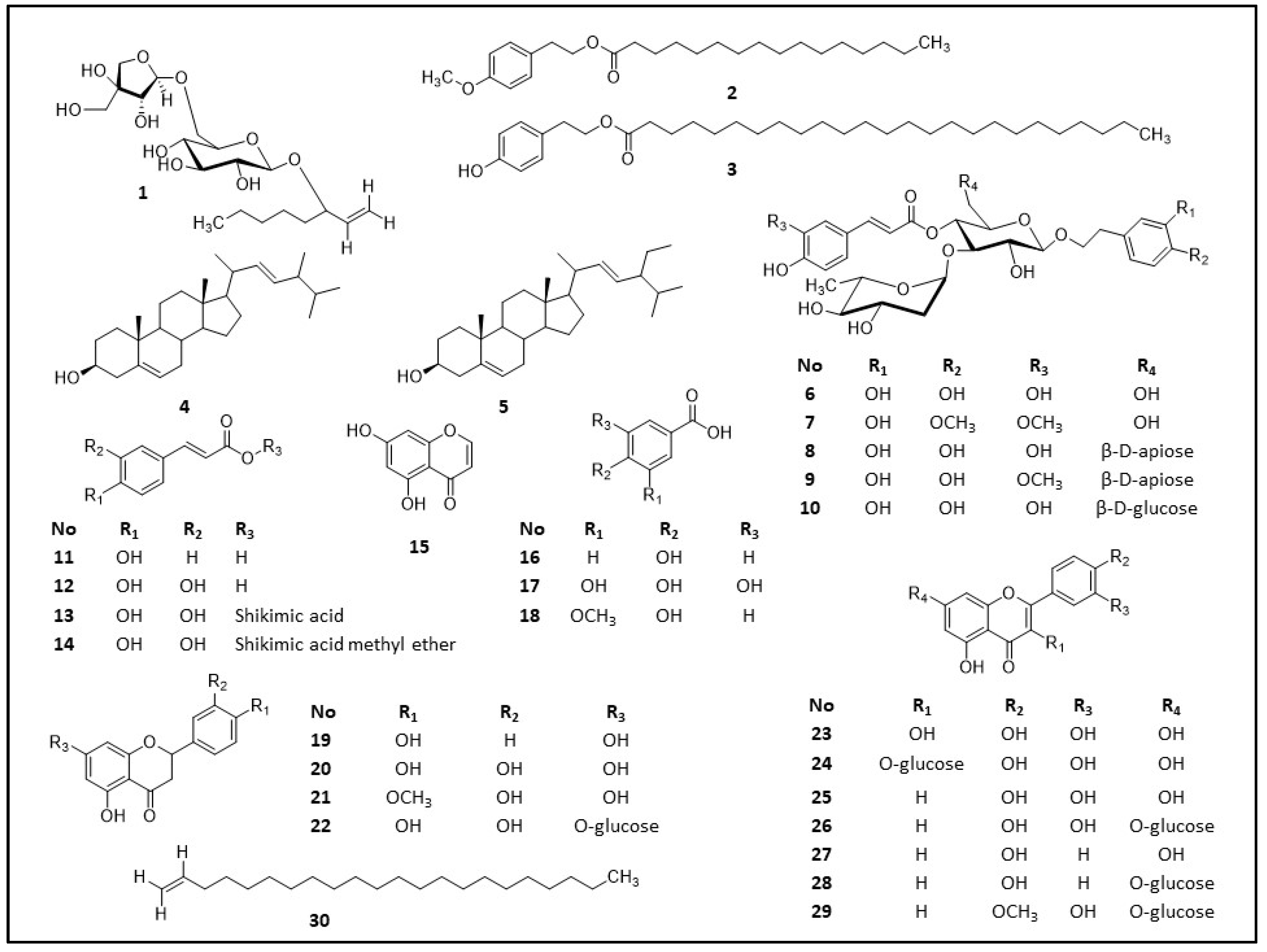
2.1. Ιsolated Compounds

Various chromatographic techniques, such as column chromatography, pTLC and semipreparative HPLC, of the extracts from the leaves, flowers and roots of Phlomis × commixta Rech. F. led to the isolation of twenty-seven known compounds. Seven compounds were isolated from the roots (2, 3, 4, 5, 6, 8, 9), ten from the leaves (1, 6, 8, 10, 18, 21, 23, 24, 26, 27) and sixteen from the flowers (6, 7, 11, 12, 13, 14, 15, 16, 17, 19, 20, 22, 25, 28, 29, 30). It must be mentioned that compound (6) is present in all the examined parts of the plant. Also, compound (8) has been isolated from both the roots and leaves, but not from the flowers. Both compound (6) and (8) are phenylpropanoids, which suggests that the plant can biosynthesize these kinds of secondary metabolites in each plant part. The compounds were identified as brassicasterol (4) (see Supplementary Data Table S2) [44,45], stigasterol (5) (see Supplementary Data Table S3) [46,47], verbascoside (6) (see Supplementary Data Table S4) [48], martynoside (7) (see Supplementary Data Table S5) [48], forsythoside B (8) (see Supplementary Data Table S6) [49], allysonoside (9) (see Supplementary Data Table S7) [50], echinacoside (10) (see Supplementary Data Table S8) [51], p-coumaric acid (11) (see Supplementary Data Note S1) [52], caffeic acid (12) (see Supplementary Data Note S2) [53], chlorogenic acid (13) (see Supplementary Data Table S9) [54], chlorogenic acid methyl ester (14) (see Supplementary Data Table S10) [55], 5,7-dihydroxychromone (15) (see Supplementary Data Note S3) [56], p-hydroxybenzoic acid (16) (see Supplementary Data Table S11) [57], gallic acid (17) (see Supplementary Data Note S4) [58], vanillic acid (18) (See Supplementary Data Note S5) [57], naringenin (19) (see Supplementary Data Table S12) [59], eriodyctiol (20) (see Supplementary Data Note S6) [60], hesperetin (21) (see Supplementary Data Table S13) [61,62], eriodyctiol-7-O-β-D-glucoside (22) (see Supplementary Data Table S14) [63], quercetin (23) (see Supplementary Data Table S15) [64], isoquercetin (24) (see Supplementary Data Table S16) [57], luteolin (25) (see Supplementary Data Table S17) [65], luteolin-7-O-β-D-glucoside (26) (see Supplementary Data Table S18) [66], apigenin (27) (see Supplementary Data Table S19) [67,68], apigenin-7-O-β-D-glucoside (28) (see Supplementary Data Table S20) [69], diosmetin-7-O-β-D-glucoside (29) (see Supplementary Data Table S21) [70] and docos-1-ene (30) (see Supplementary Data Table S22) [71]. Figure 1 shows the structures of the isolates, which were determined using 1D and 2D NMR spectra and by comparing them to those reported in the literature.

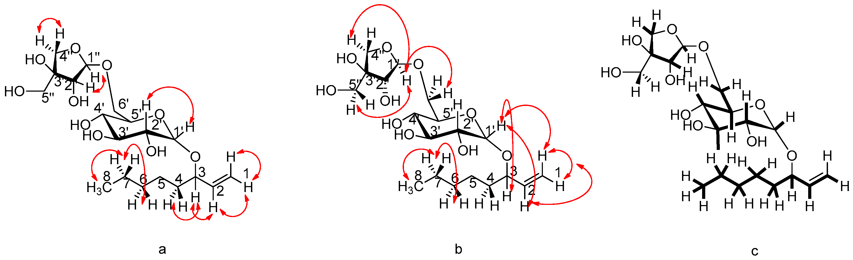
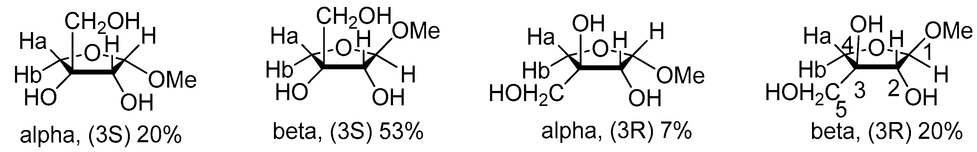
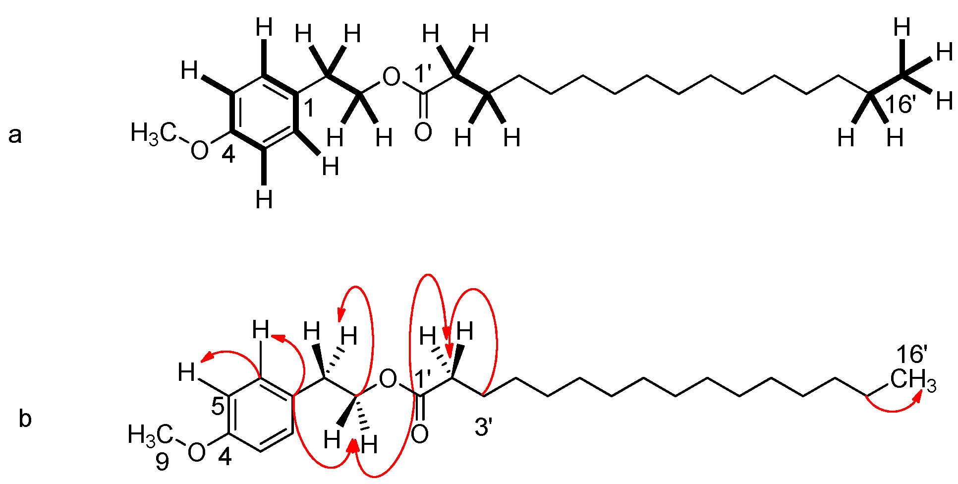
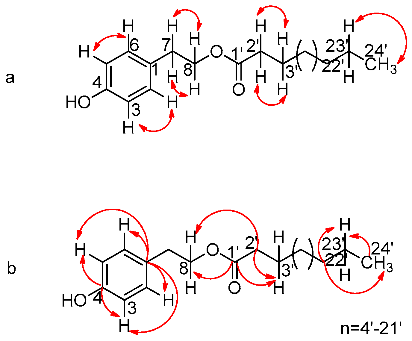
2.2. Elucidation of the Compounds

This research led to the isolation of two new natural products: 3-O-β-(3R)-D-apiofuranosyl-(1 6)-O-β-D-glucopyranosyl-(3S)-oct-1-en-3-ol (1) and the tyrosol methyl ether palmitate (2). Moreover, this is the first study that fully describes the NMR of tyrosol lignocerate (3). The combination of NMR spectroscopic data with those of the high-resolution mass spectrum (HRMS) data led to the molecular formula C19H34O10, with a molecular ion at m/z = 445.2044 [M + HNa]+ (theor. C19H34NaO10; exact mass: 445.2050). At the same time, the mass spectrum (ESI-MS, negative ionization) showed, as the base peak, the fragment m/z = 421.4 [MH]. Characteristic is the cleavage of the glycosidic O-bond, yielding a fragment of m/z = 131.0 of apiose, as well as a fragment of m/z = 289.3 [M-Apio-H]. The next set of data were from the homonuclear 1D NMR and heteronuclear 2D NMR experiments (Figure 2). The most characteristic system in the proton spectrum of the compound is that of the geminal (vinyl) protons of the terminal double bond, which appear at δ 5.21 and 5.11 ppm (see Supplementary Data Figures S1 and S2).
The HSQC spectrum confirmed the observation by correlation of these two protons to the secondary olefinic carbon at 116.3 ppm. The peak at 5.21 ppm appears as a ddd system, with a trans-vicinal coupling of J1 = 17.4 Hz, an allylic coupling of J2 = 1.7 Hz and a geminal coupling of J3 = 1.1 Hz. The peak at 5.11 ppm also appears as a ddd system, with a cis-vicinal coupling of J1 = 10.4 Hz, an allylic coupling of J2 = 1.7 Hz and a geminal coupling of J3 = 0.9 Hz. The C-2 olefinic carbon appears at 140.9 ppm, with the corresponding proton at 5.86 ppm. This proton also has the ddd system, with a trans-vicinal coupling of J1 = 17.4 Hz, a cis-vicinal coupling of J2 = 10.4 Hz and a value of J3 = 7.1 Hz, typical for the allylic proton. The high chemical shift δ of both the H-3 allylic proton and the C-3 tertiary allylic carbon (4.08 and 83.1 ppm, respectively) is indicative of the fact that the carbon is attached to a heteroatom; it is, in fact, attached to the epimeric oxygen of the glycoside, as will be shown below. The allylic proton is in a “key” position to identify the molecule, as it interacts through the HMBC spectrum with both the C-1 and C-2 olefinic carbons and the neighboring C-4 and C-5 aliphatic secondary carbons at δ 35.8 and 25.7 ppm, respectively (see Supplementary Data Table S1).
The glucoside bond between β-glucose and C-3 is doubly confirmed: (a) by the HMBC interaction between the anomeric proton H-1′ (δ 4.29 ppm) of glucose and C-3, and b) by the HMBC interaction of the H-3 (δ 4.08 ppm) of alcohol with the C-1 of glucose (δ 103.3).
The sequence of the carbons and the corresponding protons in the glucose molecule is clear, and can be confirmed by the data of the COSY, DEPT, HMBC, HSQC, NOESY and TOCSY spectra in, as well as by the coupling constant values of, the protons (J) in the proton spectrum, many of which are distinct (see Supplementary Data Figures S3–S9). A part of the data is in Table 1.
The peculiarity in the structure of apiose allows for the determination of the sequence of carbons and protons in the molecule. The anomeric proton H-1″ appears at δ 4.99 ppm and is correlated (HSQC) to the anomeric carbon that resonates at 110.8 ppm.
The glucoside bond with glucose C-6′ is confirmed by (a) the NOESY interaction (see Supplementary Data Table S1) of the H-1″ of apiose with glucose H-6′ (3.56/3.92 ppm), (b) the interaction of H-1″ with C-6′ (68.4 ppm) in the HMBC spectrum and (c) the interaction of H-6′ glucose with the epimeric C-1″ carbon at δ 110.8 ppm in the same spectrum.
The apiose H-1″ epimeric proton gives a signal at 4.99 ppm, which splits into a doublet, with J = 2.5 Hz, due to the presence of the neighboring H-2″ proton, which appears at 3.88 ppm as a doublet, with J = 2.5 Hz. The carbon C-3″ at 80.6 ppm is easily recognized by 13C NMR, as it is the only quaternary carbon in the molecule, and therefore does not appear in the DEPT-135. The C4″ (75.0 ppm) is also easily recognizable. This carbon is secondary (DEPT-135) and has two protons at 3.75 and 3.95 ppm, which are diastereotopic, since C-3″ is a stereogenic center. These protons split only with each other, with a typical coupling constant of J = 9.6 Hz. The pack corresponding to C-5″, with a resonance at 65.7 ppm, carries two protons at 3.57 ppm that appear as a broad singlet in spite of the fact that the neighboring quaternary carbon C3″ is a stereogenic center. Mass spectrometry (ESI-MS) gave negative-ionization fragments, which can be interpreted by the fragmentation process analyzed in Figures S10 and S11 in Supplementary Material. The specific rotation value of the molecule was determined as [α]D20 = −420 (c = 2.0, CH3OH).
A literature review on this structure showed an initial report with spectroscopic data referring to the structure of 3-O-β-D-apiofuranosyl-(1 6)-O-β-D-glucopyranosyl-(3S)-oct-1-en-3-οl from the plant Clerodendranthus spicatus (Labiateae) [72].
Spectral data in this work were very similar to ours, with small differences in the 13C-NMR values for the apiose molecule. Based on their literature reference [73], our observations and the data on apiose methyl glycosides [74], we believe that the isolated compound is a 3-S apiose glycoside (Figure 3).
The correct apiose structure can be clarified based on the NOESY spectrum, showing the interactions of the protons in space. Indeed, H-1″ interacts strongly with H-5″ and H-4b″ protons, which are “facing down” in a “beta (3R)”-like structure.
This hypothesis was supported by the NOESY data, which further indicated the interaction of the H-5″ proton with the H-4b″ proton, as well as the absence of an interaction between the H-4a″ and H-1″ protons, which are “on opposite sides of the molecule” in a beta (3R) structure. It is, therefore, very likely that the correct structure of the molecule isolated from our group is 3-O-β-(3R)-D-apiofuranosyl-(1 6)-O-β-D-glucopyranosyl-(3S)-oct-1-en-3-ol. The small difference in the specific rotation of the compound, a 20 D = −420, in relation to that of the literature, α 20 D = −340, is probably expected, given the small difference in the structure of the two molecules.
Metabolite 2: a tyrosol methyl ether palmitate (4-Methoxyphenethyl palmitate). The combination of the NMR and high-resolution mass spectrum (HRMS) spectroscopic data led to the molecular formula C25H42O3, with a molecular ion of m/z = 413.2666 [M + HNa]+ (theor. C25H42NaO3; exact mass: 413.3026) (see Supplementary Data Figures S12–S18). At the same time, the mass spectrum (ESI-MS, positive ionization) confirmed the molecular ion of m/z = 413.6 [M + HNa]+.
In the low-field range, two pairs of orthoconjugated aromatic protons, with peaks at δ 7.08 (J = 8.6 Hz, H-2/H-6) and 6.76 (J = 8.6 Hz, H-3/H-5), typical of an AΒ system, indicate the presence of a 1,4-substituted aromatic ring. There is also a triplet at 4.23 ppm (J = 7.0 Hz) for the ether moiety carbon (C-8) that split due to the coupling with the neighboring C-7 benzyl hydrogens, which resonate at 2.86 ppm, with a coupling constant of J = 7.0 Hz. Characteristic peaks in the spectrum are the singlet corresponding to the tyrosol methyl ether (C-9) at δ 3.48 and the triplet at δ 2.27, assigned to the methyne protons of the C-2′, split by the neighboring protons (H-3′) of the side chain, with a coupling constant of J = 7.3 Hz. A broad singlet at δ 1.25 ppm integrates to 22 protons, corresponding to an 11-carbon part of the aliphatic chain, and the triplet at δ 0.87 ppm is assigned to the terminal methyl group of this chain, split by the neighboring (C-15′) protons, with a coupling constant of J = 7.2 Hz.
In the carbon spectrum, the peak at 173.9 ppm confirmed the presence of the carbonyl carbon C-1′, and a very weak peak at 154.2 ppm was assigned to the quaternary C-4. By combining the NMR, HMBC (Figure 4) and HSQC spectroscopy techniques, it is understood that the peak at 115.2 ppm corresponds to C-3/C-5, since they interact only with the protons of the AB system, and the peak at 130.0 ppm corresponds to C-2/C-6, while the carbon at 50.9 ppm (C-9) corresponds to the methyl of tyrosol ether. The complete spectroscopic dataset for the compound is presented in Table 2 below.
Metabolite 2 is reported for the first time as component of the hybrid or the genus Phlomis. The literature review on this structure showed a report by the Gargouri research team [75], reporting synthetic approaches to lipophilic ester derivatives of tyrosol. The NMR spectroscopic data of the synthetic compound were similar to those reported by our group. Therefore, to the best of our knowledge, metabolite 2 has been isolated as a plant component for the first time. The same group investigated the germicidal activity of tyrosol and synthetic esters against Gram (+) and Gram (−) bacteria. The minimum inhibitory concentration (MIC) against the microorganisms concerned, as well as their antileishmaniasis activity against Leishmania major and L. infantum, were examined.
Metabolite 3: tyrosol lignocerate (2-(4′-hydroxyphenyl)-ethyl lignoceric ester), was identified using 1D and 2D NMR spectroscopy in combination with mass spectral data (ESI-MS), which showed a strong molecular ion of m/z = 487.4 [M-H] (see Supplementary Data Figures S19–S25). The signal sequence in the 1H-NMR spectrum showed several similarities to metabolite 2, which indicated that it was an analogue of a tyrosol ester. The 1H-NMR spectrum showed two pairs of orthoconjugated aromatic protons, with low-field peaks at δ 7.07 (J = 8.5 Hz, H-2) and 6.76 (J = 8.5 Hz, H-3), typical of an AB system, indicating the presence of a 1,4-substituted aromatic ring. There is also a triplet at 4.23 ppm (J = 7.1 Hz) for the protons on the ether-type carbon (C-8), which are split due to the coupling with the adjacent benzylic hydrogens C-7 that resonate at 2.86 ppm, with a coupling constant of J = 7.1 Hz. Characteristic is also the triplet at δ 2.28, corresponding to its methylene proton C-2′, split by neighboring hydrogens (H-3′) of the side chain, with a coupling constant of J = 7.5 Hz. A broad singlet at δ 1.25 ppm integrates for 36 protons, corresponding to the presence of an aliphatic chain segment comprising 18 methylene moieties, and finally the triplet at δ 0.88 ppm corresponds to the terminal methyl of the aliphatic chain, split by the protons of the neighboring carbon (C-23′), with a coupling constant of J = 7.2 Hz.
In the 13C NMR spectrum, the peak at 173.9 ppm confirmed the presence of the carbonyl carbon C-1′, whereas the peak at 154.2 ppm corresponds to the quaternary C-4. The combination of the NMR, HMBC (Figure 5) and HSQC techniques revealed that the peak at 115.2 ppm corresponds to C-3/C-5, since they interact only with the protons of the AB system. From the DEPT-135 range, it is clear that the secondary carbons of the aliphatic chain resonate in the range of 20–35 ppm, and the peak at 14.1 ppm corresponds to the terminal carbon of the chain. The complete spectroscopic data of the compound are presented in Table 3 below.
Metabolite 3 was first isolated from the bark of the Buddleja cordata subsp. cordata of the family Loganiaceae [76]. It is isolated for the first time regarding both the hybrid and the genus Phlomis in general. This is the second time that this particular metabolite is mentioned in the literature. Compound 3, according to the literature, showed moderate antibacterial activity against Mycobacterium tuberculosis (MIC = 64 μg/mL).

2.3. Cytotoxicity

The cytotoxic activity of the extracts from the roots, leaves and flowers of the hybrid P. × commixta was assessed using three different cell lines, i.e., Hep2c (human cervix carcinoma), RD (human rhabdomyosarcoma) and L2OB (murine fibroblast). There is a great interest regarding the biological activities of Phlomis spp. according to the literature. The results herein showed that all the samples from the roots expressed higher cytotoxic activity in the case of the Hep2c and RD cell lines, while the opposite was evidenced in the case of the L2OB cell line. The basic criterion for the cytotoxic activity of a plant extract is an IC50 < 30 µg/mL (Table 4).

3. Discussion

3.1. Chemotaxonomic Significance of the Secondary Metabolites

The genus Phlomis is well known for its richness in flavonoids and phenylpropanoid glycosides. A number of studies reveal that apigenin, luteolin, naringenin and their derivatives are very abundant in Phlomis’ polar extracts [12,25]. However, while there is only one report of the presence of diosmetin in Phlomis plants (P. fruticosa) [77], there is no one mentioning diosmetin-7-O-β-D-glucoside. It is noteworthy that quercetin is reported as a component only in three species of the genus Phlomis (P. bracteosa [78] and P. elliptica [79], P. sterwartii Hf. [38]). Zhang and Wang [80] initiated a study reporting the detection of phenolic acids in two Chinese Phlomis species. Phenolic acids isolated from Phlomis × commixta (caffeic acid, chlorogenic acid, p-coumaric acid, vanillic acid and gallic acid) have also been detected in acetone and methanol extracts of P. umbrosa and P. megalantha. There are very few references regarding the isolation and detection of caffeic acid (P. stewartii [7] and P. lycunitis [30], P. armeniaca [81]), p-hydroxy benzoic acid (P. stewartii [7] and P. bracteosa [78]) and chlorogenic acid (P. olivieri [82], P. syriaca [83]) in the genus Phlomis. The presence of chlorogenic acid and the methyl ester of an isomer (3-O-caffeoylquinic acid methyl ester) in Phlomis plants has been reported [25]; however, this is the first report on the presence of chlorogenic acid methyl ester in this genus.
Also, this is the first report of docos-1-ene (30) and 5,7-dihydroxy chromone (15) in the genus Phlomis. Stigmasterol is one of the most abundant sterols in the plant kingdom, and has also been isolated from the plants of the genus Phlomis (P. cashmeriana [8], P. bracteosa [84]). Brassicasterol, though, which is also a well-known sterol, is reported for the first time as component in the genus Phlomis.

3.2. Anticancer Activity of the Extracts

Cancer is a disease that has a significant global impact on people. To treat and prevent this fatal condition, there is a continuous need for novel therapeutics. Natural substances are gaining attention from scientists because they are considered to have fewer hazardous side-effects than existing treatments, like chemotherapy. In order to create novel medications, secondary metabolites from the plant kingdom have been isolated and assessed for their anticancer properties.
There are many studies regarding the cytotoxic effects of the extracts from the aerial parts of various Phlomis species (Table 5). It must be mentioned that the current study provides the first report on the cytotoxic effects of the extracts from the roots of a Phlomis × commixta Rech. f. Strangely, our literature search did not reveal any reports concerning the evaluation of anticancer activity on the extracts derived from the roots of species belonging to the genus Phlomis. However, there are some studies on the isolation, characterization and antitumor activity of nortriterpenoids from the roots of P. umbrosa var lutibracteata [85], the antitumor activity of phlomiol extracted from the roots of P. younghusbandii [86,87] and phenylethanoid glycosides from the roots of P. umbrosa [88], as well as the chemical constituents in the roots of P. medicinalis and P. younghusbandii [23,89].
According to the bibliography, there is a great interest for the antitumor activity exhibited by the extracts of the species belonging to the genus Phlomis. Table 5 describes studies on the antitumor activity of the extracts from Phlomis spp. using in vitro techniques in various human cancer cell lines. In order to compile all this bibliographic data, we used free chemistry databases, such as PubChem, Scopus, Reaxys and Google Scholar. It is interesting that, in all these reports, the plant material was from the aerial or part of the aerial parts (such as the flowers or/and leaves).
According to our results, the plant part that exhibited the highest cytotoxicity was the roots. In terms of the basic criterion for the cytotoxic activity of a plant extract according to American National Cancer Institute (activity < 30 µg/mL), all polar solvent extracts from the roots showed to be active for the human cancer cell lines in doses between 19.61 and 29.77 µg/mL.
Another important observation is that, with the exception of the less-polar extracts from the leaves and the flowers of the plant, the rest of them show a moderate to strong antitumor effect, with IC50 values between 21.84 and 48.29 µg/mL. Moreover, the flowers’ extracts showed lower IC50 values than the leaves. Apart from that, the results show that none of the extracts derived after the liquid–liquid extraction of the methanolic extracts from the leaves had a stronger anticancer activity compared to the initial methanolic extracts. This result indicates the possible synergistic activity of the compounds extracted from the leaves.

4. Materials and Methods

4.1. Plant Material

The plant material of Phlomis × commixta (Lamiaceae) was collected on 04/2012 from the village Tylisos of Heraklion, Crete, in Greece (coordinates: 35°18′42″ N, 25°01′07″ E). The plant was botanically identified by Prof. Dr. S. Pirintsos, Department of Biology, University of Crete, Greece, and a voucher specimen was deposited in the Department Herbarium. The plant samples were naturally dried (in the shade and in a well-ventilated environment), grinded by a laboratory mill (particle size approx. 1 mm) and stored in the darkness at room temperature.

4.2. Extraction and Isolation

The dried flower material (115 g) was extracted repeatedly in a Soxhlet apparatus (petroleum ether; dichloromethane; methanol). This process was thrice repeated (3 × 300 mL for 24 h each time), and after filtration and evaporation under reduced pressure, the petroleum extract (1.5 g), the dichloromethane extract (790 mg) and the methanol extract (27.0 g) residues were collected. The dried leaf herbal material (350 g) was extracted repeatedly at room temperature with petroleum ether, dichloromethane and methanol. This process was thrice repeated (3 × 300 mL for 24 h each time), and filtration and evaporation under reduced pressure afforded the petroleum extract (4.9 g), the dichloromethane extract (4.1 g) and the methanol extract (45.6 g) residues. The root systems of the plants were also extracted at room temperature with ethanol for six days under periodic shaking. The extract gave a solid residue weighing 7.2 g.

4.2.1. Component Isolation from the Flowers

The flower methanol extract was evaporated under reduced pressure to yield a crude residue (29.6 g), which was then suspended in 1 L of boiling water and partitioned with Et2O, EtOAc and n-BuOH, respectively. The Et2O residue (3.1 g) was subjected to column chromatography (C.C.) on a Sephadex LH-20 (4.00 × 40.00 cm) and eluted with a mixture of CH2Cl2:MeOH (1:1) to obtain fractions A–Q. Fraction D (184.5 mg) was submitted to Sephadex LH-20 separation using a mixture of CH2Cl2:MeOH (1:2) as an eluent to obtain eight fractions (DA–DS). One of these fractions, DO (4.3 mg), was identified as naringenin. Fraction E (111.2 mg) was submitted to Sephadex LH-20 separation using a mixture of CH2Cl2:MeOH (1:2) as an eluent to obtain eight fractions (EA–ES). One of these fractions, EM (10.1 mg), was identified again as naringenin. Fraction F (27.1 mg) was submitted to Sephadex LH-20 separation using MeOH as an eluent to obtain eight fractions (FA–FO). From these, fraction FB (5.8 mg) was identified as p-hydroxybenzoic acid and fraction FC (1.2 mg) as 5,7-dihydroxychromone. Fraction G (11.6 mg) was separated on the Sephadex LH-20 using MeOH as an eluent to obtain eight fractions (GA–GO). From these, fractions GH (2.5 mg) and GI (1.1 mg) were identified as naringenin. Fraction H (17.1 mg) was separated on the Sephadex LH-20 using MeOH as an eluent to obtain eight fractions (HA–HC). From these, fraction HC (0.7 mg) was identified as caffeic acid, while fractions HL (1.8 mg) and HM (1.0 mg) were identified as naringenin, fraction HO (1.7 mg) was identified as a mixture of two compounds (eriodyctiol and luteolin) and the HP (1.7 mg) fraction was identified as luteolin. Fraction I (7.8 mg) was submitted to Sephadex LH-20 separation using a mixture of CH2Cl2:MeOH (1:2) as an eluent to obtain eight fractions (IA–IH). From these, fraction IE (3.2 mg) was identified again as luteolin. Fraction K (24.0 mg) was submitted to Sephadex LH-20 separation using a mixture of CH2Cl2:MeOH (1:2) as an eluent to obtain eight fractions (KA–KQ). From these, fraction KL (7.5 mg) was identified as a mixture of two compounds (eriodyctiol and luteolin) and fraction KM (1.0 mg) was identified as luteolin.
The EtOAc residue (2.5 g) was subjected to column chromatography (C.C.) on the Sephadex LH-20 (4.00 × 40.00 cm) and eluted with mixture of CH2Cl2:MeOH (1:1) to obtain fractions A–O. Fraction C (382.4 mg) was submitted to C.C. on silica gel using CH2Cl2–MeOH mixtures of increasing polarity as eluents to obtain twenty-three fractions (CA–CW). One of these fractions, CR (13.5 mg), was identified as martynoside. Fractions D (610.2 mg) and E (1.1 g) were subjected to further chromatographic separations, as described below. Fraction D (610.2 mg) was submitted to C.C. on silica gel using CH2Cl2–MeOH–H2O mixtures of increasing polarity as eluents to obtain twenty-five fractions (DA–DZ). The hexane extract of fraction DA contained pure docos-1-ene (3.5 mg). Fraction DO (27.9 mg) was further fractionated by semipreparative HPLC (MeOH:H2O, 3:2, 1.00 mL/min) to obtain diosmetin-7-O-β-D-glucoside (Rt = 78.56 min, 1.1 mg). Fraction DU was identified as verbascoside (eluted with CH2Cl2–MeOH–H2O, 75:25:2.5, 265.6 mg). Fraction E (1.1 g) was submitted to C.C. on silica gel using CH2Cl2–MeOH–H2O mixtures of increasing polarity as eluents to obtain twenty-four fractions (EA–EY). Fraction EK (85.8) mg was submitted to Sephadex LH-20 (Merck KGaA, Darmstadt, Germany) separation using MeOH (100%) as an eluent to obtain twelve fractions (EKA–EKM). Fraction EKE (46.0 mg) was further purified on cellulose pTLC, which led to the isolation of chlorogenic acid methyl ester (14.3 mg, Rf = 0.87 on 30% acetic acid). Fraction EO (30.7 mg) was further purified on cellulose pTLC to yield apigenin-7-O-β-D-glucoside (1.0 mg, Rf = 0.28 on 30% acetic acid) and eriodyctiol-7-O-β-D-glucoside (16.6 mg, Rf = 0.69 on 30% acetic acid). Fraction ER was identified as verbascoside (eluted with CH2Cl2–MeOH–H2O, 75:25:2.5, 556.6 mg). Fraction EW was identified as chlorogenic acid (eluted with CH2Cl2–MeOH–H2O, 60:40:4.0, 50.7 mg).
The butanol residue (3.5 g) was subjected to column chromatography (C.C.) on the Sephadex LH-20 (4.00 × 44.00 cm) and eluted with MeOH (100%) to obtain the fractions A–L. Fraction H (341.0 mg) was submitted to Sephadex LH-20 fractionation using MeOH (100%) as an eluent to obtain eight fractions (HA–HO). Fraction HD (10.6 mg) was further fractionated by semipreparative HPLC (MeOH:H2O, 1:1, 1.0 mL/min), which allowed for the isolation of diosmetin-7-O-β-D-glucoside (Rt = 32.72 min, 4.3 mg). Fraction HE (41.6 mg) was further fractionated by semipreparative HPLC (MeOH:H2O, 1:1, 1.0 mL/min), which allowed for the isolation of chlorogenic acid (Rt = 10.94 min, 2.5 mg) and diosmetin-7-O-β-D-glucoside (Rt = 29.74 min, 9.8 mg). Fraction HF (63.2 mg) was further fractionated by semipreparative HPLC (MeOH:H2O, 1:1, 1.0 mL/min), which allowed for the isolation of chlorogenic acid (Rt = 9.51 min, 3.0 mg), gallic acid (Rt = 14.34 min, 1.0 mg) and p-coumaric acid (Rt = 23.8 min, 2.8 mg) compounds.

4.2.2. Component Isolation from the Leaves

The methanol extract was evaporated under reduced pressure to yield a crude residue (45.6 g), which was then suspended in 1 L of boiling water and partitioned with Et2O, EtOAc and n-BuOH, respectively. The EtOAc residue (3.4 g) was subjected to column chromatography (C.C.) on the Sephadex LH-20 (4.00 × 60.00 cm) and eluted with MeOH (100%) to obtain fractions A–L. Fraction E (88.0 mg) was further fractionated by semipreparative HPLC (MeOH:H2O, 1:1, 1.00 mL/min), which allowed for the isolation of echinacoside (Rt = 15.49 min, 11.9 mg) and isoquercetin (Rt = 40.34 min, 13.7 mg). Fraction H (98.0 mg) was submitted to Sephadex LH-20 fractionation using MeOH (100%) as an eluent to obtain nine fractions (HA–HP). Fraction HI (4.0 mg) was identified as apigenin. Fraction I (138.0 mg) was submitted to Sephadex LH-20 fractionation using MeOH (100%) as an eluent to obtain eleven fractions (IA–IL), which allowed for the isolation of quercetin from fraction ID (3.0 mg). Fraction IH (15.0 mg) was further fractionated by semipreparative HPLC (MeOH:H2O, 1:1, 1.00 mL/min), which allowed for the isolation of hesperetin (Rt = 46.47 min, 1.2 mg) and vanillic acid (Rt = 66.37 min, 1.0 mg).
The butanol residue (17.8 g) was subjected to VLC over silica gel (10 × 7 cm) using P.E., MeOH and H2O as eluent mixtures of increasing polarity to yield a total of twenty fractions (A–U). Fraction G (1.72 g) was submitted to Sephadex LH-20 fractionation using MeOH (100%) as an eluent to obtain twenty-one fractions (GA–GV). Fraction GH (104.5 mg) was further fractionated by semipreparative HPLC (MeOH:H2O, 1:1, 1.00 mL/min), which allowed for the isolation of 3-O-β-D-apiofuranosyl-(1 6)-O-β-D-glucopyranosyl-oct-1-en-3-ol) (Rt = 46.38 min, 4.7 mg). A portion of 167.0 mg from fraction GL (973.0 mg) was further purified on silica pTLC with EtOAc:AcOH:H2O/120:30:60, yielding forsythoside B (34.6 mg, Rf = 0.31) and verbascoside (38.6 mg, Rf = 0.55). Fraction GS (25.3 mg) was further purified on cellulose pTLC, which led to the isolation of luteolin-7-O-β-D-glucoside (6.2 mg) (Rf = 0.34 on EtOAc:AcOH:H2O/120:30:60).

4.2.3. Component Isolation from the Roots

The petroleum ether extract was evaporated under reduced pressure to yield a crude residue (1.3 g), which was then submitted to C.C. on silica gel using CH2Cl2–EtOAc–MeOH mixtures of increasing polarity as eluents to obtain twenty-one fractions (A–V). Fractions I (16.5 mg), K (92.7 mg) and M (110.7 mg) were subjected to further chromatographic separations, as described below. Fraction I was further purified on silica pTLC, which led to the isolation of the tyrosol methyl ether palmitate (3.0 mg) (Rf = 0.56, on DM:EtOAc, 8:2). Fraction K was further purified on semipreparative HPLC (Hex:EtOAc, 4:1, 1.00 mL/min), which allowed for the isolation of tyrosol lignocerate (Rt = 12.20 min, 6.0 mg) and brassicasterol (Rt = 25.97 min, 12.3 mg). Fraction M was further fractionated by semipreparative HPLC (Hex:EtOAc, 4:1, 1.00 mL/min), which allowed for the isolation of stigasterol (Rt = 23.65 min, 1.0 mg). The EtOAc residue (1.8 g) was subjected to column chromatography (C.C.) on the Sephadex LH-20 (4.00 × 40.00 cm) and eluted with a mixture of MeOH (100%) to obtain fractions A–U. Fraction M (78.6 mg) was further fractionated by semipreparative HPLC (MeOH:H2O, 1:1, 1.0 mL/min), which allowed for the isolation of forsythoside B (Rt = 15.02 min, 10.5 mg) and verbascoside (Rt = 17.47 min, 4.6 mg).
The butanol residue (1.9 g) was subjected to column chromatography (C.C.) on the Sephadex LH-20 (4.00 × 60.0 cm) and eluted with MeOH to obtain fractions A–N. Fractions E (587.4 mg), F (555.5 mg) and G (78.5 mg) were subjected to further chromatographic separations, as described below. A portion of 40.0 mg from fraction E was further fractionated by semipreparative HPLC (MeOH:H2O, 1:1, 1.00 mL/min), which allowed for the isolation of forsythoside B (Rt = 14.75 min, 9.5 mg) and a mixture of two compounds (allysonoside and forsythoside B) (Rt = 16.05 min, 1.6 mg). Fraction F (86.8 mg) was further fractionated by semipreparative HPLC (MeOH:H2O, 1:1, 1.00 mL/min), which allowed for the isolation of forsythoside B (Rt = 14.68 min, 40.6 mg), verbascoside (Rt = 16.93 min, 2.2 mg) and a mixture of allysonoside and forsythoside B (Rt = 17.27 min, 1.8 mg). A portion of 36.0 mg from fraction G (78.0 mg) was further fractionated by semipreparative HPLC (MeOH:H2O, 1:1, 1.00 mL/min), which allowed for the isolation of forsythoside B (Rt = 14.87 min, 8.0 mg) and verbascoside (Rt = 17.24 min, 1.1 mg).

4.3. Cytotoxic Activity of the Studied Extracts

A standard cytotoxic activity test was performed [113] using the MTT (3-[4,5-dimethylthiazol-2-yl]-2,5-diphenyl tetrazolium bromide) assay [114,115]. The following cell lines were used (cell lines were donated from the collection of cell lines of the Institute of Virology, Vaccines and Serums “Torlak”, Belgrade, Serbia): RD (substrate: MEM Eagle/10% FCS) (cell line derived from human rhabdomyosarcoma), Hep2c (medium: MEM Eagle/5% FCS) (cell line derived from human cervix carcinoma–HeLa derivative) and L2OB (medium: MEM Eagle/10% FCS) (cell line derived from murine fibroblast), against which the activity of the extracts was measured. The results were expressed as IC50 values (µg/mL), a threshold which was defined as the concentration of an agent inhibiting cell survival by 50% in comparison to a vehicle-treated control [116]. The experiment was conducted in accordance with the method described by Mašković et al., 2015 [116].

5. Conclusions

The work presented in this publication is the first report on the isolation and the identification of the components of the Phlomis × commixta hybrid, an endemic plant of Crete, Greece. The component analysis included extraction from the flowers, the leaves and the roots of the plant, the subsequent component isolation using various chromatographic techniques and identification by spectroscopic methods, such as UV/Vis, NMR, GC-MS and LC-MS.
A total of thirty components were isolated, including flavonoids, phenylethanoic glycosides, phenolic acids, tyrosol esters, steroids, a chromone and an aliphatic hydrocarbon.
This report includes a detailed structural elucidation of allylic alcohol glycoside 3-O-β-D-apiofuranosyl-(1 6)-O-β-D-glucopyranosyl-oct-1-ene-3-ol, which was fully characterized as a natural product for the first time, and two tyrosol esters: tyrosol lignocerate and the tyrosol methyl ether palmitate, the former one mentioned only once in the literature as a component in a different species, and the latter one isolated as a natural product for the first time.
All the root extracts tested showed a high cytotoxic activity against the Hep2c and RD cell lines, with the leaf and flower extracts showing a moderate, though distinct, activity.

Supplementary Materials

The following supporting information can be downloaded at: https://www.mdpi.com/article/10.3390/ijms25020816/s1.

Author Contributions

Conceptualization, M.K.S., H.E.K. and D.L.; methodology, M.K.S. and O.S.T.; validation, M.K.S., H.E.K., O.S.T. and D.L.; formal analysis, M.K.S., O.S.T., P.Z.M. and D.L.; investigation, M.K.S., O.S.T., P.Z.M., D.L. and H.E.K.; resources, D.L. and H.E.K.; data curation, O.S.T., M.K.S., H.E.K., P.Z.M. and D.L.; writing—original draft preparation, M.K.S., O.S.T., D.L. and H.E.K.; writing—review and editing, M.K.S., O.S.T., H.E.K. and D.L.; visualization, M.K.S., O.S.T., H.E.K. and D.L.; supervision, D.L., H.E.K. and M.K.S.; project administration, M.K.S., D.L. and H.E.K. All authors have read and agreed to the published version of the manuscript.

Funding

This research received no external funding.

Institutional Review Board Statement

Not applicable.

Informed Consent Statement

Not applicable.

Data Availability Statement

Data are contained within the article and Supplementary Materials.

Conflicts of Interest

The authors declare no conflicts of interest.

References

  1. Mathiensen, C.; Scheen, A.C.; Lindqvist, C. Phylogeny and biogeography of the lamioid genus Phlomis (Lamiaceae). Kew Bull. 2011, 66, 83–99. [Google Scholar] [CrossRef]
  2. Moench, C. Methodus Plantas Horti Botanici et Agri Marburgensis: A Staminum Situ Describendi; Officina Nova Libraria Academiae: Marburg, Germany, 1794; p. 619. [Google Scholar]
  3. Bariotakis, M. Thocus Issues in the Investigation of Hybridization Patterns. Ph.D. Thesis, University of Crete, Heraklion, Greece, 2016. Available online: https://elocus.lib.uoc.gr/dlib/2/0/5/metadata-dlib-1477377544-405386-26657.tkl (accessed on 3 November 2016).
  4. Basta, A.; Tzakou, O.; Couladis, M. The essential oil composition of Phlomis cretica C. Presl. Flavour Frag. J. 2006, 21, 795–797. [Google Scholar] [CrossRef]
  5. Georgescu, L.; Stefanakis, K.M.; Kokkini, S.; Katerinopoulos, E.H.; Pirintsos, A.S. Chemical and genetic characterization of Phlomis species and wild hybrids in Crete. Phytochemistry 2016, 122, 91–102. [Google Scholar] [CrossRef] [PubMed]
  6. Kyriakopoulou, I.; Magiatis, P.; Skaltsounis, A.L.; Aligiannis, N.; Harvala, C. Samioside, a new phenylethanoid glycoside with free-radical scavenging and antimicrobial activities from Phlomis samia. J. Nat. Prod. 2001, 64, 1095–1097. [Google Scholar] [CrossRef]
  7. Jabeen, B.; Riaz, N.; Saleem, M.; Naveed, M.; Ashraf, M.; Alam, U.; Rafiq, H.M.; Tareen, R.B.; Jabbar, A. Isolation of natural compounds from Phlomis stewartii showing α-glucosidase inhibitory activity. Phytochemistry 2013, 96, 443–448. [Google Scholar] [CrossRef] [PubMed]
  8. Hussain, J.; Ullah, R.; Khan, A.; Khan, F.U.; Muhammad, Z.; Shah, M.R. Phlomeoic acid: A new diterpene from Phlomis bracteosa. Nat. Prod. Commun. 2011, 6, 171–173. [Google Scholar] [CrossRef]
  9. Hussain, J.; Bukhari, N.; Hussain, H.; Haider, S.; Hassan, Z. Phlomisamide and Phlomisteriod: A New Ceramide and a New Stigmasterol Derivative from Phlomis cashmeriana. Helv. Chim. Acta 2010, 93, 1428–1431. [Google Scholar] [CrossRef]
  10. Sobeh, M.; Mamadalieva, N.Z.; Mohamed, T.; Krstin, S.; Youssef, F.S.; Ashour, M.L.; Azimova, S.S.; Wink, M. Chemical profiling of Phlomis thapsoides (Lamiaceae) and in vitro testing of its biological activities. Med. Chem. Res. 2016, 25, 2304–2315. [Google Scholar] [CrossRef]
  11. Olennikov, D.N.; Chirikova, N.K. Phlotuberosides I and II, new iridoid glycosides from Phlomoides tuberosa. Chem. Nat. Compd. 2017, 53, 269–272. [Google Scholar] [CrossRef]
  12. Sajjadi, S.E.; Delazari, Z.; Aghaei, M.; Ghannadian, M. Flavone constituents of Phlomis bruguieri Desf. with cytotoxic activity against MCF-7 breast cancer cells. Res. Pharm. Sci. 2018, 13, 422–429. [Google Scholar] [CrossRef]
  13. Caliş, I.; Bedir, E.; Kirmizibekmez, H.; Ersöz, T.; Dönmez, A.A.; Khan, I.A. Secondary metabolites from Phlomis oppositiflora. Nat. Prod. Res. 2005, 19, 493–501. [Google Scholar] [CrossRef] [PubMed]
  14. Kasai, R.; Katagiri, M.; Ohtani, K.; Yamasaki, K.; Chongren, Y.; Tanaka, O. Iridoid glycosides from Phlomis younghusbandii roots. Phytochemistry 1994, 36, 967–970. [Google Scholar] [CrossRef]
  15. Li, M.X.; Huang, J.; Zhang, C.; Jia, Z.; Fan, P.; Wei, L.; Zhang, Q.; Zhang, R. Iridoid glycosides and phenylethanoid glycosides from Phlomis younghusbandii roots. Chem. Nat. Compd. 2011, 47, 848–849. [Google Scholar] [CrossRef]
  16. Li, X.H.; Li, X.H.; Yao, Q.; Lu, L.H.; Li, Y.B.; Wu, D.S.; Fu, D.H.; Mei, S.X.; Cui, T.; Wang, J.K.; et al. Phlolosides A-F, iridoids from Phlomis likiangensis with a carbonate ester substituent. Tetrahedron Lett. 2017, 58, 3112–3118. [Google Scholar] [CrossRef]
  17. Le, D.; Nguyen, D.; Bingtian, Z.; Min, B.; Song, S.; Woo, M. Quantitation and radical scavenging activity evaluation of iridoids and phenylethanoids from the roots of Phlomis umbrosa (Turcz.) using DPPH free radical and DPPH-HPLC methods, and their cytotoxicity. Nat. Prod. Sci. 2019, 25, 122–129. [Google Scholar] [CrossRef]
  18. Bader, A.; Tuccinardi, T.; Granchi, C.; Martinelli, A.; Macchia, M.; Minutolo, F.; De Tommasi, N.; Braca, A. Phenylpropanoids and flavonoids from Phlomis kurdica as inhibitors of human lactate dehydrogenase. Phytochemistry 2015, 116, 262–268. [Google Scholar] [CrossRef] [PubMed]
  19. Kirmizibekmez, H.; Piacente, S.; Pizza, C.; Dönmez, A.A.; Çaliş, I. Iridoid and phenylethanoid glycosides from Phlomis nissolii and P. capitata. Z. Naturforsch. B J. Chem. Sci. 2004, 59, 609–613. [Google Scholar] [CrossRef]
  20. Ersöz, T.; Saracoğlu, I.; Taşdemir, D.; Kirmizibekmez, H.; Dönmez, A.; Ireland, C.M.; Çalış, I. Neolignan glucosides from Phlomis chimerae Boiss. Z. Naturforsch. C J. Biosci. 2002, 57, 221–225. [Google Scholar] [CrossRef]
  21. Saracoğlu, I.; Varel, M.; Çaliş, I.; Dönmez, A.A. Neolignan, flavonoid, phenylethanoid and iridoid glycosides from Phlomis integrifolia. Turk. J. Chem. 2003, 27, 739–747. [Google Scholar]
  22. Khitri, W.; Smati, D.; Mitaine-Offer, A.C.; Paululat, T.; Lacaille-Dubois, M.A. Chemical constituents from Phlomis bovei Noë and their chemotaxonomic significance. Biochem. Syst. Ecol. 2020, 91, 104054. [Google Scholar] [CrossRef]
  23. Katagiri, M.; Ohtani, K.; Kasai, R.; Yamasaki, K.; Yang, C.R.; Tanaka, O. Diterpenoid glycosyl esters from Phlomis younghusbandii and P. medicinalis roots. Phytochemistry 1994, 35, 439–442. [Google Scholar] [CrossRef] [PubMed]
  24. Yalçin, F.N.; Ersöz, T.; Bedir, E.; Dönmez Ali, A.; Stavri, M.; Zloh, M.; Gibbons, S.; Çaliş, I. Amanicadol, a pimarane-type diterpene from Phlomis amanica Vierch. Z. Naturforsch. B 2006, 61, 1433–1436. [Google Scholar] [CrossRef]
  25. Amor, I.L.B.; Boubaker, J.; Sgaier, M.B.; Skandrani, I.; Bhouri, W.; Neffati, A.; Kilani, S.; Bouhlel, I.; Ghedira, K.; Chekir-Ghedira, L. Phytochemistry and biological activities of Phlomis species. J. Ethnopharmacol. 2009, 125, 183–202. [Google Scholar] [CrossRef] [PubMed]
  26. Li, M.X.; Shang, X.F.; Jia, Z.P.; Zhang, R.X. Phytochemical and biological studies of plants from the genus Phlomis. Chem. Biodivers. 2010, 7, 283–301. [Google Scholar] [CrossRef] [PubMed]
  27. Hajarolasvadi, N.; Zamani, M.J.; Sarkhail, P.; Khorasani, R.; Mohajer, M.; Amin, G.; Shafiee, A.; Sharifzadeh, M.; Abdollahi, M. Comparison of antinociceptive effects of total, water, ethyl acetate, ether and n-butanol extracts of Phlomis anisodonta Boiss. and indomethacin in Mice. Intl. J. Pharmacol. 2006, 2, 209–212. [Google Scholar] [CrossRef]
  28. Wang, Q.S.; Yang, L.; Cui, W.Y.; Chen, L.; Jiang, Y.H. Anti-inflammatory and anti-nociceptive activities of methanol extract from aerial part of Phlomis younghusbandii Mukerjee. PLoS ONE 2014, 9, e89149. [Google Scholar] [CrossRef] [PubMed]
  29. Shang, X.; Wang, J.; Li, M.; Miao, X.; Pan, H.; Yang, Y.; Wang, Y. Antinociceptive and anti-inflammatory activities of Phlomis umbrosa Turcz extract. Fitoterapia 2011, 82, 716–721. [Google Scholar] [CrossRef]
  30. Lopez, V.; Jager, A.; Akerreta, S.; Cavero, R.; Calvo, M. Antioxidant activity and phenylpropanoids of Phlomis lychnitis L.: A traditional herbal tea. Plant Foods Hum. Nutr. 2010, 65, 179–185. [Google Scholar] [CrossRef]
  31. Keser, S.; Turkoglu, S.; Celik, S.; Turkoglu, I. Determination of antioxidant capacities of Phlomis pungens Wild. var. hispida Hub.-Mor. Asian J. Chem. 2012, 24, 2780–2784. [Google Scholar]
  32. Li, X.; Xie, Y.; Li, K.; Wu, A.; Xie, H.; Guo, Q.; Xue, P.; Maleshibek, Y.; Zhao, W.; Guo, J.; et al. Antioxidation and cytoprotection of acteoside and its derivatives: Comparison and mechanistic chemistry. Molecules 2018, 23, 498. [Google Scholar] [CrossRef]
  33. Eruygur, N.; Kirci, D.; Ayaz, F.; Doğu, S.; Bağci, Y. Biological activities of three Phlomis species. J. Res. Pharm. 2022, 26, 255–262. [Google Scholar] [CrossRef]
  34. Derafa, I.; Amira, S.; Benchikh, F.; Mamache, W.; Kaoudoune, C. Phenolic content and antioxidant activity of hydromethanolic and aqueous extracts of aerial parts of Phlomis crinita. Turk. JAF Sci. Tech. 2022, 10, 2061–2066. [Google Scholar] [CrossRef]
  35. Amor, I.L.B.; Neffati, A.; Sgaier, M.B.; Bhouri, W.; Boubaker, J.; Skandrani, I.; Bouhlel, I.; Kilani, S.; Ben Ammar, R.; Chraief, I.; et al. Antimicrobial activity of essential oils isolated from Phlomis crinita Cav. ssp. mauritanica Munby. J. Am. Oil Chem. Soc. 2008, 85, 845–849. [Google Scholar] [CrossRef]
  36. Alpay, M.; Dulger, G.; Sahin, I.E.; Dulger, B. Evaluating antimicrobial and antioxidant capacity of endemic Phlomis russeliana from Turkey and its antiproliferative effect on Human Caco-2 Cell Lines. An. Acad. Bras. Cienc. 2019, 91, e20180404. [Google Scholar] [CrossRef] [PubMed]
  37. Sarkhail, P.; Rahmanipour, S.; Fadyevatan, S.; Mohammadirad, A.; Dehghan, G.; Amin, G.; Shafiee, A.; Abdollahi, M. Antidiabetic effect of Phlomis anisodonta: Effects on hepatic cells lipid peroxidation and antioxidant enzymes in experimental diabetes. Pharmacol. Res. 2007, 56, 261–266. [Google Scholar] [CrossRef] [PubMed]
  38. Rasheed, M.U.; Naqvi, S.A.R.; Rasool, N.; Shah, S.A.A.; Zakaria, Z.A. Anti-Diabetic and Cytotoxic Evaluation of Phlomis stewartii Plant Phytochemicals on Cigarette Smoke Inhalation and Alloxan-Induced Diabetes in Wistar Rats. Metabolites 2022, 12, 1133. [Google Scholar] [CrossRef]
  39. Dönmez, C.; Koca Çalışkan, U. Ethnopharmacological survey on Phlomis grandiflora: In vivo antihemorrhoidal model. Curr. Pers. MAPs 2019, 2, 40–46. [Google Scholar] [CrossRef]
  40. Kirmizibekmez, H.; Çalis, I.; Perozzo, R.; Dönmez, A.A.; Linden, A.; Rüedi, P.; Tasdemir, D. Inhibiting activities of the secondary metabolites of Phlomis brunneogaleata against parasitic protozoa and plasmodial enoyl-ACP reductase, a crucial enzyme in fatty acid biosynthesis. Planta Med. 2004, 70, 711–717. [Google Scholar] [CrossRef]
  41. Kunter, I.; Kosar, M. Composition Analysis and Anti-Cancer Properties of Two Endemic Phlomis Species (Phlomis cypria Post and Phlomis brevibracteata Turrill) from Cyprus. Proceedings 2019, 40, 29. [Google Scholar] [CrossRef]
  42. Torky, Z.A.; Moussa, A.Y.; Abdelghffar, E.A.; Abdel-Hameed, U.K.; Eldahshan, O.A. Chemical profiling, antiviral and antiproliferative activities of the essential oil of: Phlomis aurea Decne grown in Egypt. Food Funct. 2021, 12, 4630–4643. [Google Scholar] [CrossRef]
  43. Kunter, I.; Tarabishi, M.T.; Zabib, N.; Erçetin, T.; İlktaç, M.; Göger, F.; Koşar, M. New Data for Endemic Phlomis cypria Post from North Cyprus: Biological Activities and LC MS/MS Analysis. Ind. J. Pharm. Educ. Res. 2023, 57, 511–518. [Google Scholar] [CrossRef]
  44. Sun, Y.; Lin, Y.; Cao, X.; Xiang, L.; Qi, J. Sterols from Mytilidae Show Anti-Aging and Neuroprotective Effects via Anti-Oxidative Activity. Int. J. Mol. Sci. 2014, 15, 21660–21673. [Google Scholar] [CrossRef] [PubMed]
  45. Lee, J.W.; Lee, D.Y.; Cho, J.G.; Baek, N.I.; Lee, Y.H. Isolation and identification of sterol compounds from the red kohlrabi sprouts (Brassica oleracea var. gongylodes). J. Appl. Biol. Chem. 2010, 53, 207–211. [Google Scholar] [CrossRef]
  46. Yahya, M.A.A.; Yaacob, W.A.; Nazlina, I. Isolation of chemical constituents from rhizomes of Etlingera sphaerocephala var. grandiflora. Malaysian J. Anal. Sci. 2011, 15, 22–26. [Google Scholar]
  47. Khatun, M.; Billah, M.; Quader, M.A. Sterols andsterol glucoside from Phyllanthus species. Dhaka Univ. J. Sci. 2012, 60, 5–10. [Google Scholar] [CrossRef]
  48. Venditti, A.; Bianco, A.; Nicoletti, M.; Quassinti, L.; Bramucci, M.; Lupidi, G.; Vitali, L.A.; Papa, F.; Vittori, S.; Petrelli, D.; et al. Characterization of secondary metabolites, biological activity and glandular trichomes of Stachys tymphaea Hausskn. from the Monti Sibillini National Park (Central Apennines, Italy). Chem. Biodivers. 2014, 11, 245–261. [Google Scholar] [CrossRef] [PubMed]
  49. Saracoğlu, I.; Harput, Ü.Ş.; Çaliş, I.; Ogihara, Y. Phenolic constituents from Phlomis lycia. Turk. J. Chem. 2002, 26, 133–142. [Google Scholar]
  50. Kirmizibekmez, H.; Montoro, P.; Piacente, S.; Dönmez, A.; Çaliş, I. Identification by HPLC-PAD-MS and quantification by HPLC-PAD of phenylethanoid glycosides of five Phlomis species. Phytochem. Anal. 2005, 16, 1–6. [Google Scholar] [CrossRef]
  51. Porter, E.A.; Kite, G.C.; Veitch, N.C.; Geoghegan, I.A.; Larsson, S.; Simmonds, M.S.J. Phenylethanoid glycosides in tepals of Magnolia salicifolia and their occurrence in flowers of Magnoliaceae. Phytochemistry 2015, 117, 185–193. [Google Scholar] [CrossRef]
  52. Ralph, J.; Hatfield, R.D.; Quideau, S.; Helm, R.F.; Grabber, J.H.; Jung, H.J.G. Pathway of p-coumaric acid incorporation into maize lignin as revealed by NMR. J. Am. Chem. Soc. 1994, 116, 9448–9456. [Google Scholar] [CrossRef]
  53. Frezza, C.; Venditti, A.; Rossi, G.; Serafini, I.; Pitorri, M.; Ciccóla, A.; Foddai, S.; Bianco, A.; Serafini, M. Phytochemical study on the leaves of Wollemia nobilis. Biochem. Syst. Ecol. 2017, 74, 63–66. [Google Scholar] [CrossRef]
  54. Suárez-Quiroz, M.L.; Alonso Campos, A.; Valerio Alfaro, G.; González-Ríos, O.; Villeneuve, P.; Figueroa-Espinoza, M.C. Isolation of green coffee chlorogenic acids using activated carbon. J. Food Comp. Anal. 2014, 33, 55–58. [Google Scholar] [CrossRef]
  55. Tsiftsoglou, O.S.; Lagogiannis, G.; Psaroudaki, A.; Vantsioti, A.; Mitić, M.N.; Mrmošanin, J.M.; Lazari, D. Phytochemical Analysis of the Aerial Parts of Campanula pelviformis Lam. (Campanulaceae): Documenting the Dietary Value of a Local Endemic Plant of Crete (Greece) Traditionally Used as Wild Edible Green. Sustainability 2023, 15, 7404. [Google Scholar] [CrossRef]
  56. Spencer, G.F. A Convenient Synthesis of 5,7-Dihydroxychromone. Org. Prep. Proc. Int. 1991, 23, 390–392. [Google Scholar] [CrossRef]
  57. Tsiftsoglou, O.S.; Lazari, D.M.; Stefanakis, M.K.; Kokkalou, E.L. Flavonoids and phenolic acids from the aerial parts of Alyssum alyssoides L. (Brassicaceae). Biochem. Syst. Ecol. 2019, 83, 51–53. [Google Scholar] [CrossRef]
  58. Kamatham, S.; Kumar, N.; Gudipalli, P. Isolation and characterization of gallic acid and methyl gallate from the seed coats of Givotia rottleriformis Griff. and their anti-proliferative effect on human epidermoid carcinoma A431 cells. Toxicol. Rep. 2015, 3, 520–529. [Google Scholar] [CrossRef] [PubMed]
  59. Kontogiorgis, C.; Ntella, M.; Mpompou, L.; Karallaki, F.; Papadopoulos, A.; Hadjipavlou-Litina, D.; Lazari, D. Study of the antioxidant activity of Thymus sibthorpii Bentham (Lamiaceae). J. Enzyme Inhib. Med. Chem. 2016, 31, 154–159. [Google Scholar] [CrossRef]
  60. Huang, Y.H.; Zeng, W.M.; Li, G.Y.; Liu, G.Q.; Zhao, D.D.; Wang, J.; Zhang, Y.L. Characterization of a new sesquiterpene and antifungal activities of chemical constituents from Dryopteris fragrans (L.) Schott. Molecules 2014, 19, 507–513. [Google Scholar] [CrossRef]
  61. Maltese, F.; Erkelens, C.; Kooy, F.V.D.; Choi, Y.H.; Verpoorte, R. Identification of natural epimeric flavanone glycosides by NMR spectroscopy. Food Chem. 2009, 116, 575–579. [Google Scholar] [CrossRef]
  62. Lee, N.K.; Choi, S.H.; Park, S.H.; Park, E.K.; Kim, D.H. Antiallergic activity of hesperidin is activated by intestinal microflora. Pharmacology 2004, 71, 174–180. [Google Scholar] [CrossRef]
  63. Vieira, M.N.; Winterhalter, P.; Jerz, G. Flavonoids from the flowers of Impatiens glandulifera Royle isolated by high performance countercurrent chromatography. Phytochem. Anal. 2016, 27, 116–125. [Google Scholar] [CrossRef] [PubMed]
  64. Güvenalp, Z.; Demirezer, Ö.L. Flavonol Glycosides from Asperula arvensis L. Turk. J. Chem. 2005, 29, 163–169. [Google Scholar]
  65. Patora, J.; Klimek, B. Flavonoids from lemon balm (Melissa officinalis L., Lamiaceae). Acta Pol. Pharm. 2002, 59, 139–143. [Google Scholar] [PubMed]
  66. Tsiftsoglou, O.S.; Krigas, N.; Gounaris, C.; Papitsa, C.; Nanouli, M.; Vartholomatos, E.; Markopoulos, G.S.; Isyhou, R.; Alexiou, G.; Lazari, D. Isolation of Secondary Metabolites from Achillea grandifolia Friv. (Asteraceae) and Main Compounds’ Effects on a Glioblastoma Cellular Model. Pharmaceutics 2023, 15, 1383. [Google Scholar] [CrossRef] [PubMed]
  67. Zhang, J.; Yang, J.; Duan, J.; Liang, Z.; Zhang, L.; Huo, Y.; Zhang, Y. Quantitative and qualitative analysis of flavonoids in leaves of Adinandra nitida by high performance liquid chromatography with UV and electrospray ionization tandem mass spectrometry detection. Anal. Chim. Acta 2005, 532, 97–104. [Google Scholar] [CrossRef]
  68. Da Silva, L.A.L.; Faqueti, L.G.; Reginatto, F.H.; Conceicão dos Santos, A.D.; Barison, A.; Biavatti, M.W. Phytochemical analysis of Vernonanthura tweedieana and a validated UPLC-PDA method for the quantification of eriodictyol. Rev. Bras. Farmacogn. 2015, 25, 375–381. [Google Scholar] [CrossRef]
  69. Ersöz, T.; Harput, Ü.Ş.; Saracoğlu, I.; Çalis, I. Phenolic compounds from Scutellaria pontica. Turk. J. Chem. 2002, 26, 581–588. [Google Scholar]
  70. Kim, Y.C.; Higuchi, R.; Kitamura, Y.; Komori, T. Thermal degradation of glycosides, IV. Degradation of flavonoid glycosides. Liebigs Ann. Chem. 1991, 12, 1285–1289. [Google Scholar] [CrossRef]
  71. Ihenetu, G.U.; Igwe, O.U.; Nnaji, J.C.; Ejezie, M.U. Isolation of 1-Eicosene from the leaves of Baphia nitida. J. Chem. Soc. Nigeria. 2020, 45, 434–440. [Google Scholar]
  72. Zou, J.; Zhu, Y.D.; Zhao, W.M. Two new alkyl glycosides from Clerodendranthus spicatus. J. Asian Nat. Prod. Res. 2008, 10, 602–606. [Google Scholar] [CrossRef]
  73. Yang, H.; Sang, H.S.; Young, C.K. Two new hepatoprotective stilbene glycosides from Acer mono leaves. J. Nat. Prod. 2005, 68, 101–103. [Google Scholar] [CrossRef] [PubMed]
  74. Ho, C.W.; Lin, R.D.; Lee, T.H.; Lin, C.H.; Wen, C.L.; Tseng, Y.T.; Lee, M.H. Chemical and pharmacological investigation of micropropagated Hygrophila pogonocalyx produced from leaf explants. Botan. Stud. 2013, 54, 51. [Google Scholar] [CrossRef]
  75. Aissa, I.; Sghair, R.; Bouaziz, M.; Laouini, D.; Sayadi, S.; Gargouri, Y. Synthesis of lipophilic tyrosyl esters derivatives and assessment of their antimicrobial and antileishmania activities. Lipids Health Dis. 2012, 11, 13. [Google Scholar] [CrossRef] [PubMed]
  76. Acevedo, L.; Martınez, E.; Castaneda, P.; Franzblau, S.; Timmermann, B.N.; Linares, E.; Bye, R.; Mata, R. New phenylethanoids from Buddleja cordata subsp. cordata. Plant Med. 2000, 66, 257–261. [Google Scholar] [CrossRef]
  77. Marin, P.D.; Veitch, N.C.; Grayer, R.J.; Kite, G.C.; Soković, M.; Janaćković, P. Flavonoids from Phlomis fruticosa (Lamiaceae) growing in Montenegro. Biochem. Syst. Ecol. 2007, 35, 462–466. [Google Scholar] [CrossRef]
  78. Ullah, R.; Khader, J.A.; AbdEIslam, N.M.; Arabia, S. Chemical Constituents from Phlomis bracteosa. Life Sci. J. 2013, 10, 969–970. [Google Scholar]
  79. Aghakhani, F.; Kharazian, N.; Lori Gooini, Z. Flavonoid Constituents of Phlomis (Lamiaceae) species using liquid chromatography nass Sspectrometry. Phytochem. Anal. 2018, 29, 180–195. [Google Scholar] [CrossRef] [PubMed]
  80. Zhang, Y.; Wang, Z.Z. Phenolic composition and antioxidant activities of two Phlomis species: A correlation study. Comptes Rendus Biol. 2009, 332, 816–826. [Google Scholar] [CrossRef]
  81. Sarikurkcu, C.; Uren, M.C.; Tepe, B.; Cengiz, M.; Kocak, M.S. Phlomis armeniaca: Phenolic compounds, enzyme inhibitory and antioxidant activities. Ind Crops Prod. 2015, 78, 95–101. [Google Scholar] [CrossRef]
  82. Delnavazi, M.R.; Mohammadifar, F.; Rustaie, A.; Aghaahmadi, M.; Yassa, N. Phytochemical constituents, antioxidant activity and toxicity potential of Phlomis olivieri Benth. Res. J. Pharmacogn. 2016, 3, 9–15. [Google Scholar]
  83. Harput, U.S.; Çaliş, I.; Saracoğlu, I.; Dönmez, A.A.; Nagatsu, A. Secondary Metabolites from Phlomis syriaca and their Antioxidant Activities. Turk. J. Chem. 2006, 30, 383–390. [Google Scholar]
  84. Ullah, R.; Ullah, Z.; Hussain, I.; Ahmad, S. Phlomiside: A new steroid from Phlomis bracteosa. Arab. J. Chem. 2017, 10, S1303–S1305. [Google Scholar] [CrossRef]
  85. Deng, R.X.; Xu, Y.F.; Liu, P. Study on nortriterpenes from roots of Phlomis umbrosa var. latibracteata. Chin. Tradit. Herb. Drugs 2021, 2, 1555–1561. [Google Scholar] [CrossRef]
  86. Xie, W.L.; Zhu, J.; Zhao, Y.W.; Li, L. Antitumor or effect of phlomiol in vivo and in vitro. Chin. J. Chin. Mater. Med. 2010, 35, 1189–1192. [Google Scholar]
  87. Tang, C.; Zhao, C.C.; Yi, H.; Geng, Z.J.; Wu, X.Y.; Zhang, Y.; Liu, Y.; Fan, G. Traditional Tibetan Medicine in Cancer Therapy by Targeting Apoptosis Pathways. Front. Pharmacol. 2020, 11, 976. [Google Scholar] [CrossRef] [PubMed]
  88. Liu, P.; Deng, R.X.; Duan, H.Q.; Yin, W.P.; Zhao, T.Z. Phenylethanoid glycosides from the roots of Phlomis umbrosa. J. Asian Nat. Prod. Res. 2009, 11, 69–74. [Google Scholar] [CrossRef]
  89. Yu, Z.X.; Wang, G.L.; Bianba, C.; Lin, R.C. Studies on chemical constituents in root of Phlomis medicinalis I. Zhongguo Zhong Yao Za Zhi 2006, 31, 656–658. (In Chinese) [Google Scholar]
  90. Sarkhail, P.; Sahranavard, S.; Nikan, M.; Gafari, S.; Eslami-Tehrani, B. Evaluation of the cytotoxic activity of extracts from six species of Phlomis genus. J. Appl. Pharm. Sci. 2017, 7, 180–184. [Google Scholar] [CrossRef]
  91. Soltani-Nasab, F.; Asgarpanah, J.; Majdzadeh, M.; Ostad, S.N. Investigating the effect of Phlomis lanceolata Boiss and Hohen on cancer cell lines. Acta Med. Iran. 2014, 52, 333–336. [Google Scholar]
  92. Abu-Dahab, R.; Afifi, F. Antiproliferative activity of selected medicinal plants of Jordan against a breast adenocarcinoma cell line (MCF7). Sci. Pharm. 2007, 75, 121–146. [Google Scholar] [CrossRef]
  93. Thoppil, R.J.; Harlev, E.; Mandal, A.; Nevo, E.; Bishayee, A. Antitumor activities of extracts from selected desert plants against HepG2 human hepatocellular carcinoma cells. Pharm. Biol. 2013, 51, 668–674. [Google Scholar] [CrossRef]
  94. Aydin, B.; Gönder, L.Y.; Çerçi, N.A.; Ateş, Y.C.; Yalçınkaya, İ.S.; Canbolat, N.; Açık, L.; Karacan, N.; Akyüz Turumtay, E.; Turumtay, H. Biological activities and DNA interactions of aqueous extract of Phlomis linearis (Boiss. & Bal.). Int. J. Plant Bas. Pharm. 2023, 3, 73–85. [Google Scholar] [CrossRef]
  95. Göger, G.; Türkyolu, Ü.; Gürşen, E.N.; Yur, S.; Karaduman, A.B.; Göger, F.; Tekin, M.; Özek, G. Phytochemical characterisation of Phlomis linearis Boiss. & Bal and screening for anticholinesterase, antiamylase, antimicrobial, and cytotoxic properties. Turk. J. Chem. 2021, 45, 387–399. [Google Scholar] [CrossRef] [PubMed]
  96. Abdelhady, N.M.; Badr, K.A. Comparative study of phenolic content, antioxidant potentials and cytotoxic activity of the crude and green synthesized silver nanoparticles’extracts of two Phlomis species growing in Egypt. J. Pharmacogn. Phytochem. 2016, 5, 377–383. [Google Scholar]
  97. Hegazy, M.F.; Abdelfatah, S.; Hamed, A.R.; Mohamed, T.A.; Elshamy, A.A.; Saleh, I.A.; Reda, E.H.; Abdel-Azim, N.S.; Shams, K.A.; Sakr, M.; et al. Cytotoxicity of 40 Egyptian plant extracts targeting mechanisms of drug-resistant cancer cells. Phytomedicine 2019, 59, 152771. [Google Scholar] [CrossRef]
  98. Asadi-Samani, M.; Rafieian-Kopaei, M.; Lorigooini, Z.; Shirzad, H. A screening of anti-breast cancer effects and antioxidant activity of twenty medicinal plants gathered from Chaharmahal va Bakhtyari province, Iran. J. Pharm. Pharmacogn. Res. 2019, 7, 213–222. [Google Scholar]
  99. Heydari, F.; Ghafarzadegan, R.; Mofasseri, M.; Ghasemi, S.V.; Kashefi, M.; Hajiaghaee, R.; Tavakoli, S. Phytochemical analysis and biological activities of essential oil and extract of Phlomis rigida Labill. J. Med. Plants 2021, 20, 13–22. [Google Scholar] [CrossRef]
  100. Badisa, R.B.; Tzakou, O.; Couladis, M.; Pilarinou, E. Cytotoxic activities of some Greek Labiatae herbs. Phytother. Res. 2003, 17, 472–476. [Google Scholar] [CrossRef]
  101. Ghaffari, K.; Ahmadi, R.; Saberi, B.; Moulavi, P. Anti-proliferative effects of Ziziphus spina-christi and Phlomis russeliana leaf extracts on HEK293 and MCF-7 Cell Lines and Evaluation of Bax and Bcl-2 Genes Expression Level in MCF-7 Cells. Asian Pac. J. Cancer Prev. 2021, 22, 81–87. [Google Scholar] [CrossRef]
  102. Sarkhail, P.; Salimi, M.; Sarkheil, P.; Heidarnezhad, F.; Saeidnia, S. Evaluation of anti-melanogenic and cytotoxic activities of Phlomis caucasica on human melanoma SKMEL-3 cells. Int. J. Cancer Manag. 2017, 10, e4633. [Google Scholar] [CrossRef]
  103. Salimi, M.; Sarkhail, P.; Tahmasvand, R.; Baeeri, M. Determination of anti-melanogenic activity of Phlomis kurdica in human melanoma SKMEL-3 cells. Iran. J. Pharm. Sci. 2016, 12, 1–10. [Google Scholar] [CrossRef]
  104. Sarkhail, P.; Salimi, M.; Saeidnia, S.; Nikan, M.; Kurpaz-Mahmoodabadi, M. Influence of Four Phlomis Species on melanogenesis in human malignant melanoma (SKMEL-3) cells. Res. J. Pharmacogn. 2019, 6, 19–24. [Google Scholar] [CrossRef]
  105. Stojković, D.; Gašić, U.; Drakulić, D.; Zengin, G.; Stevanović, M.; Rajčević, N.; Soković, M. Chemical profiling, antimicrobial, anti-enzymatic, and cytotoxic properties of Phlomis fruticosa L. J. Pharm. Biomed. Anal. 2021, 195, 113884. [Google Scholar] [CrossRef] [PubMed]
  106. Stojkovic, D.; Drakulic, D.; Dias, M.I.; Zengin, G.; Barros, L.; Ivanov, M.; Gašic, U.; Rajcevic, N.; Stevanovic, M.; Ferreira, I.C.F.R.; et al. Phlomis fruticosa L. exerts in vitro antineurodegenerative and antioxidant activities and induces prooxidant effect in glioblastoma cell line. EXCLI J. 2022, 21, 387–399. [Google Scholar] [CrossRef] [PubMed]
  107. Calderón-Montaño, J.M.; Martínez-Sánchez, S.M.; Burgos-Morón, E.; Guillén-Mancina, E.; Jiménez-Alonso, J.J.; García, F.; Aparicio, A.; López-Lázaro, M. Screening for Selective Anticancer Activity of Plants from Grazalema Natural Park, Spain. Nat. Prod. Res. 2019, 33, 3454–3458. [Google Scholar] [CrossRef]
  108. Ihoual, S.; Karaali, W.; Abidli, N. Antioxidant, anti-proliferative, and induction of apoptosis by Phlomis samia methanolic extract from Algeria. Der Pharma Chem. 2017, 9, 99–107. [Google Scholar]
  109. Yarmolinsky, L.; Budovsky, A.; Ben-Shabat, S.; Khalfin, B.; Gorelick, J.; Bishitz, Y.; Miloslavski, R.; Yarmolinsky, L. Recent Updates on the Phytochemistry and Pharmacological Properties of Phlomis viscosa Poiret. Rejuvenation Res. 2019, 22, 282–288. [Google Scholar] [CrossRef] [PubMed]
  110. Emami, S.A.; Kheshami, S.; Ramazani, E.; Akaberi, M.; Iranshahy, M.; Kazemi, S.M.; Tayarani-Najaran, Z. Cytotoxic activity of thirteen endemic and rare plants from Chaharmahal and Bakhtiari Province in Iran. Iran J. Pharm. Res. 2019, 18, 1912–1920. [Google Scholar] [CrossRef]
  111. Tarhan, L.; Urek, R.O.; Oner, A.; Nakiboglu, M. Evaluation of phenolic profiles, antioxidant activities, and cytotoxic and apoptotic potentials of Phlomis angustissima and Phlomis fruticosa, medicinal plants from Turkey. Eur. J. Integr. Med. 2022, 55, 102188. [Google Scholar] [CrossRef]
  112. Mamadalieva, N.Z.; Vinciguerra, V.; Sobeh, M.; Ovidi, E.; Ashour, M.L.; Wink, M.; Tiezzi, A. GLC-MS profiling of non-polar extracts from Phlomis bucharica and P. salicifolia and their cytotoxicity. Bol. Latinoam. Caribe Plant Med. Aromat. 2015, 14, 442–448. [Google Scholar]
  113. Radojković, M.; Zekovic, Z.; Maskovic, P.; Vidovic, S.; Mandić, A.; Mišan, A.; Ðurović, S. Biological activities and chemical composition of Morus leaves extracts obtained by maceration and supercritical fluid extraction. J. Supercrit. Fluids 2016, 117, 50–58. [Google Scholar] [CrossRef]
  114. Mosmann, T. Rapid colorimetric assay for cellular growth and survival: Application to proliferation and cytotoxicity assays. J. Immunol. Methods 1983, 65, 55–63. [Google Scholar] [CrossRef] [PubMed]
  115. Baviskara, B.A.; Khadabadia, S.S.; Deore, S.L.; Shiradkar, M.R. Synthesis of clubbed Triazolyl Indeno [1,2-C] Isoquinolines as a novel anticancer agent. Der Pharm. Sin. 2012, 3, 24–30. [Google Scholar]
  116. Mašković, P.Z.; Diamanto, L.D.; Vujic, J.M.; Cvetanović, A.D.; Radojković, M.M.; Gadžurić, S.B.; Zengin, G. Onosma aucheriana: A source of biologically active Molecules for novel food Ingredients and pharmaceuticals. J. Funct. Foods 2015, 19, 479–486. [Google Scholar] [CrossRef]
Figure 1. Isolated compounds from the leaves, flowers and roots of Phlomis × commixta Rech. f.
Ijms 25 00816 g001
Figure 2. Main correlations of the COSY (a), NOESY (b) and TOCSY (c) (highlighted) of metabolite 1.
Ijms 25 00816 g002
Figure 3. The four different diastereomers of apiose methyl glycosides.
Ijms 25 00816 g003
Figure 4. Main correlations of the COSY (highlighted) (a) and HMBC (b) of metabolite 2.
Ijms 25 00816 g004
Figure 5. Key correlations of COSY (a) and HMBC (b) for metabolite 3.
Ijms 25 00816 g005
Table 1. NMR spectroscopic data of metabolite 1 (CD3OD, 500 MHz).
#13CHSQCDEPT 135δ 1HMultiplicity (J, Hz)
Allylic part
1116.3H-1aCH25.21ddd, J1 = 17.4, J2 = 1.7, J3 = 1.1 Hz, 1H
H-1b5.11ddd, J1 = 10.4, J2 = 1.7, J3 = 0.9 Hz, 1H
2140.9H-2CH5.86ddd, J1 = 17.4, J2 = 10.4, J3 = 7.1 Hz, 1H
383.1H-3CH4.08dd, J1 = 13.1, J2 = 7.1 Hz, 1H
435.8H-4aCH21.51m, 1H
H-4b1.68dd, J1 = 10.2, J2 = 5.9 Hz, 1H
525.7H-5aCH21.39m, 1H
H-5b1.32m, 1H
633.1H-6CH21.31m, 2H
723.7H-7a
H-7b
CH21.33m, 1H
1.37m, 1H
814.4H-8CH30.90t, J = 7.0 Hz, 3H
β-D-Glucose
1′103.3H-1′CH4.29d, J = 7.8 Hz, 1H
2′75.3H-2′CH3.17dd, J1 = 7.8, J2 = 4.2, 1H
3′76.8H-3′CH3.34m, 1H
4′71.7H-4′CH3.25dd, J1 = 8.7, J2 = 1.0 Hz, 1H
5′78.2H-5′CH3.33m, 1H
6′68.4H-6a′CH23.56dd, J1 = 11.2, J2 = 5.2 Hz, 1H
H-6b′3.92dd, J1 = 11.2, J2 = 1.8 Hz, 1H
β-D-Apiose
1″110.8H-1″CH4.99d, J = 2.5 Hz, 1H
2″78.0H-2″CH3.88d, J = 2.5 Hz, 1H
3″80.6H-3″C--
4″75.0H-4a″CH23.95d, J = 9.6 Hz, 1H
H-4b″3.75d, J = 9.6 Hz, 1H
5″65.7H-5″CH23.57br.s, 2H
Table 2. NMR spectroscopic data of metabolite 2 (CDCl3, 500 ΜHz).
#13CHSQCδ 1HMultiplicity (J, Hz)COSYHMBC
1130.1 *----H-2/H-6, H-7, H-8
2/6130.0H-2/H-67.08d, J = 8.6 Hz, 2HH-3/H-5H-4, H-3/H-5
3/5115.2H-3/H-56.76d, J = 8.6 Hz, 2HH-2/H-6-
4154.2 *----H-2/H-6
734.3H-72.86t, J = 7.0 Hz, 2HH-8H-8
864.9H-84.23t, J = 7.0 Hz, 2HH-7H-7
950.9H-93.48s, 3H--
1′173.9----H-8, H-2′
2′34.2H-2′2.27t, J = 7.3 Hz, 2HH-3′-
3′24.9H-3′1.60–1.57m, 2HH-2′H-2′
4′/13′29.7H-4′/H-13′1.25br.s, 20H--
14′31.9H-14′1.25br.s, 2H-H-16′
15′22.6H-15′1.29–1.25m, 2HH-16′H-16′
16′14.1H-16′0.87t, J = 7.2 Hz, 3HH-15′-
* Reported correlation are from the HMBC and HSQC spectra.
Table 3. NMR spectroscopic data of metabolite 3 (CDCl3, 500 ΜHz).
#13CHSQCDEPT 135δ 1HMultiplicity (J, Hz)COSYHMBC
1129.9-C---H-2/H-6
H-3/H-5
2/6130.0H-2/H-6CH7.07d, J = 8.5 Hz, 2HH-3/H-5H-3/H-5
H-7, H-8
3/5115.2H-3/H-5CH6.76d, J = 8.5 Hz, 2HH-2/H-6H-2/H-6
4154.2-C --H-2/H-6
H-3/H-5
734.3H-7CH22.86t, J = 7.1 Hz, 2HH-8H-2/H-6
864.9H-8CH24.23t, J = 7.1 Hz, 2HH-7H-7
1′173.9-C---H-8, H-2′, H-3′
2′34.2H-2′CH22.28t, J = 7.5 Hz, 2HH-3′H-8, H-3′
3′24.9H-3′CH21.59dt, J1 = 7.5, J2 = 7.0 Hz, 2HH-2′H-2′
4′/21′29.6H-4′/H-21′(CH2)181.25br.s, 36H--
22′31.9H-22′CH21.25m, 2H-H-23′, H-24′
23′22.6H-23′CH21.29brq, J = 7.2 Hz, 2HH-24′H-24′
24′14.1H-24′CH30.88t, J = 7.2 Hz, 3HH-23′H-23′
Table 4. Cytotoxic activity of the extracts from P. × commixta in one murine (L2OB) and two human cell lines (Hep2c, RD) compared to the standard cytotoxic agent cis-diammine dichloroplatinum (cis-DDP).
ExtractIC50 Values (μg/mL)
SolventPlant PartsHep2c CellsRD CellsL2OB Cells
Petroleum etherroots19.61 ± 0.3923.18 ± 0.4330.84 ± 0.31
Ethyl acetateroots20.74 ± 0.3927.59 ± 0.2933.76 ± 0.48
Butanolroots25.89 ± 0.2128.57 ± 0.3943.65 ± 0.74
Waterroots26.32 ± 0.4129.77 ± 0.1846.74 ± 0.98
Petroleum etherleaves74.39 ± 0.1298.38 ± 0.22111.56 ± 0.37
Dichloromethaneleaves36.53 ± 0.6138.73 ± 0.7754.76 ± 0.39
Methanolleaves37.88 ± 0.9148.29 ± 0.4562.72 ± 0.91
Petroleum ether (a L.L.E of b ML)leaves52.92 ± 0.1949.29 ± 0.3385.38 ± 0.76
Ethyl acetate (L.L.E of ML)leaves52.78 ± 0.3262.90 ± 0.2888.75 ± 0.32
Butanol (L.L.E of ML)leaves79.61 ± 0.39100.91 ± 0.48121.42 ± 0.38
Water (L.L.E of ML)leaves91.44 ± 0.27104.19 ± 0.64129.39 ± 0.49
Petroleum etherflowers92.85 ± 0.24108.29 ± 0.88134.58 ± 0.29
Dichloromethaneflowers29.47 ± 0.2925.73 ± 0.9332.46 ± 0.29
Methanolflowers38.52 ± 0.8729.44 ± 0.3135.30 ± 0.91
Butanolflowers21.84 ± 0.7830.74 ± 0.2845.21 ± 0.37
Waterflowers38.63 ± 0.5931.53 ± 0.9848.93 ± 0.27
cis-DDP0.94 ± 0.551.4 ± 0.970.72 ± 0.64
American National Cancer Institute (NCI) < 30 μg/mL
a LLE: liquid–liquid extraction; b ML: methanol extract from leaves.
Table 5. Short synopsis of the cytotoxicity of the extracts from Phlomis spp. against human cancer cell lines.
Phlomis SpeciesPlant PartsCell LineBibliography
P. olivieri, P. caucasica, P. anisodontea, P. bruguieri, P. kurdica, P. persica MCF-7, HepG2, HT29, A549 and MDBK[90]
P. lanceolataTotal methanolic extract and partition fractions of flowering aerial partsHT29, Caco2, T47D and NIH3T3[91]
P. syriacaEthanolic extract from the
flowers
MCF-7[92]
P. cypria Post70% aqueous methanol extract of herbal partsSK-HEP-1 cancer cell line[43]
P. platystegia PostAqueous extracts of aerial partsHepG2[93]
P. linearis Boiss. & Bal.Aqueous extractMouse fibroblast L929 cell line, human H1299 cell line and human Caco-2 cell lines[94]
Hexane, diethyl ether, ethyl
acetate and methanol extracts of
the aerial parts
A549 and HT-29[95]
P. aurea Decne and P. floccosa D HEP-G2, HCT-116 and MCV-7[96]
P. aurea DecneAerial parts extracts methylene chloride: methanol (1:1) and methanol: H2O (7:3)CCRF–CEM leukemia cells[97]
P. persica Boiss.The aerial part of the plants macerated in ethanol (70%)MCF-7 and MDA-MB231[98]
P. rigida Labill.Methanol extract from leaves and flowersMCF-7, MDBK, HT-29 and A-549[99]
P. pungens Willd.Methanol extract of aerial parts powderCaco-2, HepG2 and MCF-7 cell lines[100]
P. russelianaEthanolic extract of aerial partCaco-2 cell lines[36]
Leaf extractHEK293 and MCF-7[101]
P. caucasicaAerial parts, 80% methanol extractHuman melanoma SKMEL-3 cells[102]
P. kurdica[103]
P. persica, P. brugieri, P. olivieri,
P. anisodontea
[104]
P. fruticosa L.Aerial parts, methanol extractHGF-1, MCF-7, SiHa and HepG2[105]
A172 glioblastoma cell line [106]
P. sterwartii Hf.Leaves, flowers and whole plantHepG2 cell lines[38]
P. × composita PauAerial partsA549 [107]
P. purpurea L. Flowering aerial parts
P. samiaAerial parts, methanol extractHepG2 and MDA[108]
P. viscosa PoiretCrude ethanolic extracts from leavesU-87 and MCF7[109]
P. thapsoidesAerial partsCaco-2 and HepG2[10]
P. aucheriAerial parts, methanol extractPC-3, MCF-7, HepG2, CHO and B16-F10[110]
P. angustissima and P. fruticosaMethanol/water, ethyl acetate and water extracts of leaves and flowersHeLa, MCF-7, ACC-201, OE-33 and HepG2[111]
P. bucharica and P. salicifolia Water, methanol, chloroform and hexane extracts from the whole plantHeLa and HL-60 cell lines[112]
Disclaimer/Publisher’s Note: The statements, opinions and data contained in all publications are solely those of the individual author(s) and contributor(s) and not of MDPI and/or the editor(s). MDPI and/or the editor(s) disclaim responsibility for any injury to people or property resulting from any ideas, methods, instructions or products referred to in the content.

