Article

Genome-Wide Identification and Expression Analysis of GATA Family Genes in Dimocarpus longan Lour

1
College of Computer and Information Sciences, Fujian Agriculture and Forestry University, Fuzhou 350002, China
2
College of Juncao Science and Ecology, Fujian Agriculture and Forestry University, Fuzhou 350002, China
3
College of Forestry, Fujian Agriculture and Forestry University, Fuzhou 350002, China
4
College of Horticulture, Fujian Agriculture and Forestry University, Fuzhou 350002, China
5
Institute of Horticultural Biotechnology, Fujian Agriculture and Forestry University, Fuzhou 350002, China
*
Authors to whom correspondence should be addressed.
Int. J. Mol. Sci. 2024, 25(2), 731; https://doi.org/10.3390/ijms25020731
Submission received: 25 November 2023 / Revised: 29 December 2023 / Accepted: 4 January 2024 / Published: 5 January 2024

Abstract

:
GATA transcription factors, which are DNA-binding proteins with type IV zinc finger binding domains, have a role in transcriptional regulation in biological organisms. They have an indispensable role in the growth and development of plants, as well as in improvements in their ability to face various environmental stresses. To date, GATAs have been identified in many gene families, but the GATA gene in longan (Dimocarpus longan Lour) has not been studied in previous explorations. Various aspects of genes in the longan GATA family, including their identification and classification, the distribution of their positions on chromosomes, their exon/intron structures, a synteny analysis, their expression at different temperatures, concentration of PEG, early developmental stages of somatic embryos and their expression levels in different tissues, and concentrations of exogenous hormones, were investigated in this study. This study showed that the 22 DlGATAs could be divided into four subfamilies. There were 10 pairs of homologous GATA genes in the synteny analysis of DlGATA and AtGATA. Four segmental replication motifs and one pair of tandem duplication events were present among the DlGATA family members. The cis-acting elements located in promoter regions were also found to be enriched with light-responsive elements, which contained related hormone-responsive elements. In somatic embryos, DlGATA4 is upregulated for expression at the globular embryo (GE) stage. We also found that DlGATA expression was strongly up-regulated in roots and stems. The study demonstrated the expression of DlGATA under hormone (ABA and IAA) treatments in embryogenic callus of longan. Under ABA treatment, DlGATA4 was up-regulated and the other DlGATA genes did not respond significantly. Moreover, as demonstrated with qRT-PCR, the expression of DlGATA genes showed strong up-regulated expression levels under 100 μ m o l · L 1 concentration IAA treatment. This experiment further studied these and simulated their possible connections with a drought response mechanism, while correlating them with their expression under PEG treatment. Overall, this experiment explored the GATA genes and dug into their evolution, structure, function, and expression profile, thus providing more information for a more in-depth study of the characteristics of the GATA family of genes.

1. Introduction

A plant’s life begins with the syncytium undergoing seed germination, transitions into the juvenile and mature stages, and then ends with the formation of a new syncytium. During the growth and development process, plants face many influences from the external natural environment, and the results indicate that transcription factors are an important part of a plant’s response to stress [1,2], Recently, many other transcription factors with diverse functions have also been identified, such as the basic leucine zipper [3], MYB (myeloblastosis) [4], NAC [5,6,7,8,9], bHLH (basic helix–loop–helix) [10], ERF (ethylene response factor) [11], CBF (CRT-binding factor) [12], and GATA (GATA-binding factor) [13].
The GATA gene family is a transcription factor in eukaryotes, including animals, plants, and fungi [14,15,16,17], and it plays a major part in biological processes, such as developmental differentiation, growth and proliferation, decomposition and apoptosis, and plant responses to environmental transformations. So far, GATA transcription factors have been found in many plants, such as rice [18], Arabidopsis thaliana [16], Pyrus bretschneideri [19], tobacco [20], Black Soybean [21], and tomato [22]. Within the context of plant hormone signaling pathways, the GATA gene family is important, particularly in hormone synthesis, signal transduction, and responsiveness. For instance, GATA transcription factors can modulate gibberellin biosynthesis and degradation, thereby influencing plant growth and development. Additionally, GATA genes are involved in the signal transduction and response of plant hormones, such as abscisic acid, ethylene, and ABA. In summary, GATA genes have diverse functions in plant hormone regulation. GATA transcription factors have a highly conserved class IV zinc finger structure and can recognize and specifically bind DNA sequences (T/A) (GATA(A/G)) [18,23]. Most GATA proteins contain CX2CX17–20CX2C zinc finger domains [24], followed by a basic region [19,25,26]. In addition, 30 and 28 gene members of the GATA family have been identified in Arabidopsis (Arabidopsis thaliana) [24] and rice (Oryza sativa subsp. japonica) [24], respectively. However, the complete genome of DlGATA has not yet been characterized in Dimocarpus longan.
Longan originates from Fujian, Guangdong, Southeast Asia, and it is also cultivated in Yunnan [27,28]. It has a long history of cultivation [29]. Therefore, it occupies an important position in the study of fruit trees. At the same time, a series of biotechnologies related to exogenous plant hormones also play an active role in overcoming harsh environments and genetic limitations, as well as improving crop quality and storage conditions [30,31]. Therefore, the study of plant hormones plays an indispensable role in understanding the effects of many plants, and this is particularly groundbreaking in the case of longan, which is the subject of the experiments described here. In this study, bioinformatics was used to analyze and identify members of the DlGATA family at the genome-wide level; this was mainly for the identification of DlGATA genes and their basic physicochemical properties, chromosomal localization, phylogenetic tree, gene structure, cis-regulatory elements, and expression patterns. In addition, the effects of treatments with the exogenous plant hormones IAA and ABA on the expression of GATA genes in early embryogenic callus of longan were also analyzed using real-time quantitative PCR. The study of the DlGATA family is conducive to gaining a deeper understanding of the GATA family and its structure and function, as well as the promotion of in-depth experiments.

2. Results

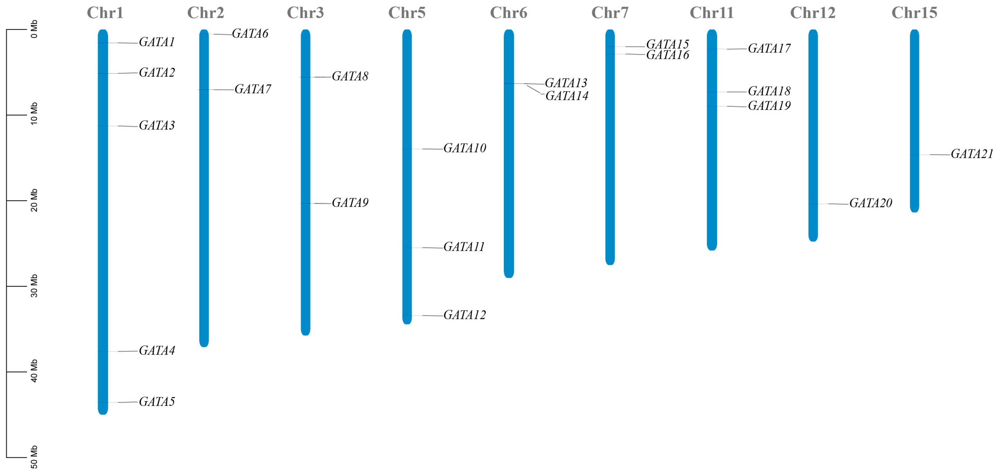
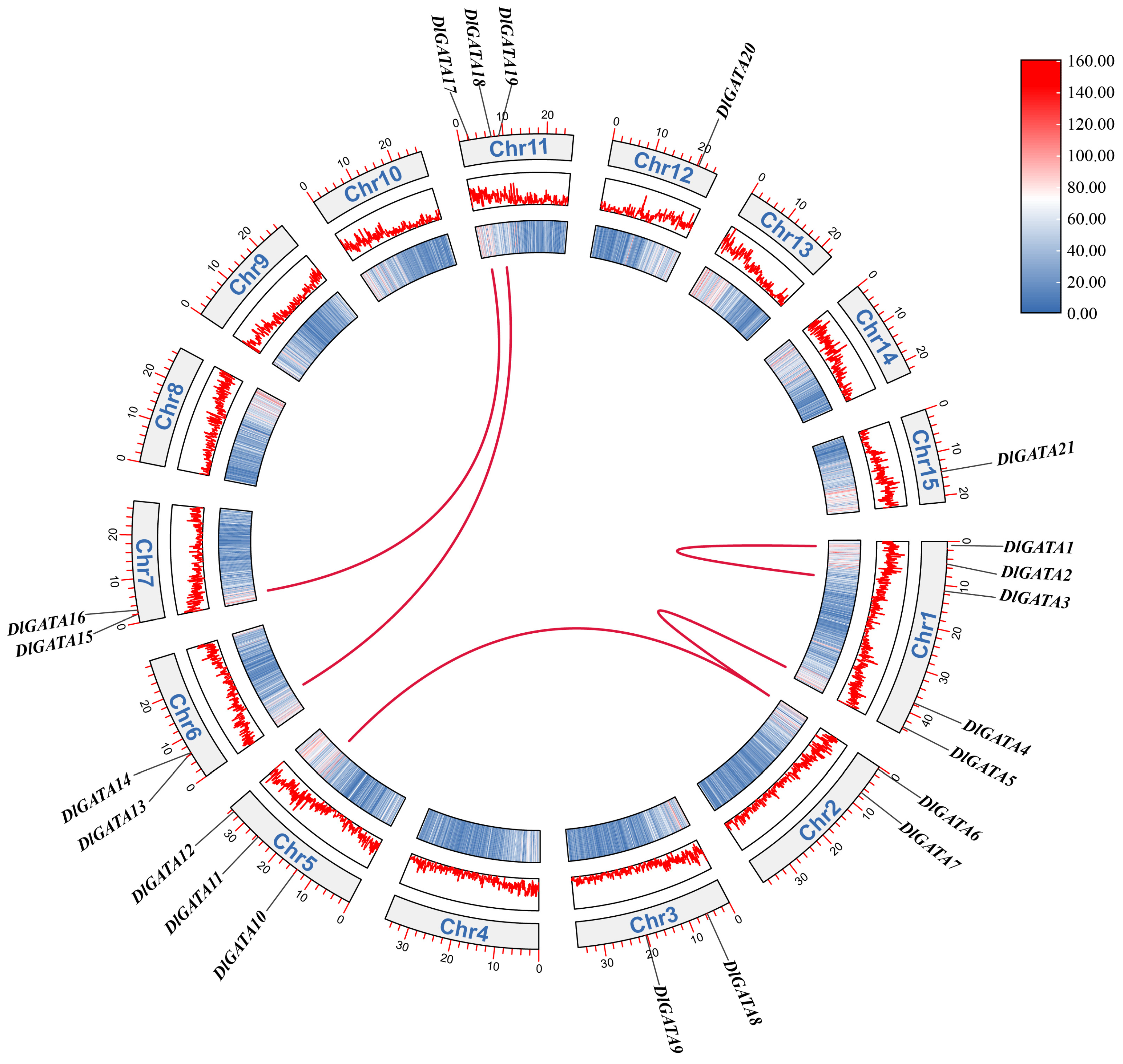
2.1. Characterization of the DlGATA Gene in Longan and Its Positional Distribution on Chromosomes

A total of 22 GATAs were identified among the longan genes. It was found that 21 GATAs were distributed on nine different chromosomes, and 1 was a GATA gene that was not mapped on the fixed chromosomes. They were renamed DlGATA1DlGATA22 according to their positions (Figure 1). Among these, chr1 was the longest and possessed the most DlGATAs (5 (23.8%)), and chr15 was the shortest. On chromosomes chr12 and chr15, only one GATA gene (4.8%) was found. To investigate the characteristics of DlGATA, its physicochemical properties were analyzed (Table 1). It was found that in DlGATA1DlGATA22, the protein-encoded amino acid numbers ranged from 112 to 542, the relative molecular weights ranged from 12,336.41 to 60,405.08, and the lengths of the proteins showed a close positive correlation with the relative molecular weights. Regarding the physicochemical properties of the theoretical isoelectric point, 10 DlGATAs were acidic, and the other 12 were alkaline. The highest isoelectric point for both DlGATA6 and DlGATA22 was 10.89. Except for the slightly lower stability index of DlGATA9, all GATA transcription factors had an instability index greater than 40, making them more prone to denaturation, aggregation, and sedimentation. Their aliphatic coefficients were close together, except for that of DlGATA16. All GRAVY values were less than 0, which indicated that all of the DlGATA proteins were hydrophilic. In terms of their subcellular localization, all DlGATAs were located in the nucleus.

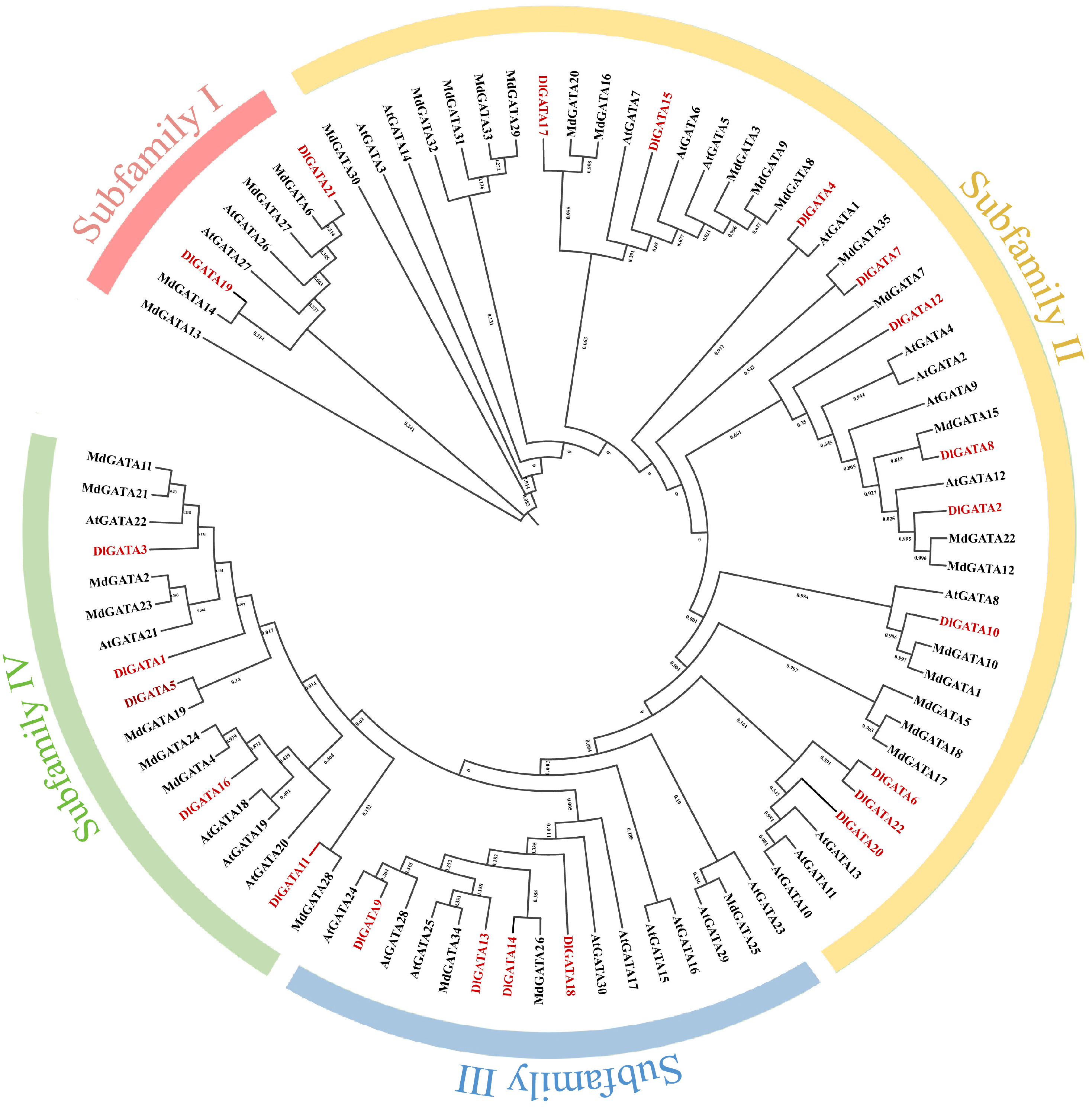
2.2. Phylogenetic Analysis between Longan, Apple, and Arabidopsis

The construction of phylogenetic trees is beneficial for the study of the biological relationships and genetic relationships of the GATA transcription factor gene family and other gene families. Using the MEGA X software, a biological evolutionary tree consisting of 35 MdGATA proteins, 22 DlGATA proteins, and 30 AtGATA proteins was obtained (Figure 2). According to the cluster analysis, we found that longan was the same as Arabidopsis and apple in that it could be categorized into four subfamilies; of these, subfamily II contained the most GATA genes, as it had 11 DlGATA proteins. It was followed by subfamily IV, which had five DlGATA proteins, namely, DlGATA1, DlGATA3, DlGATA5, DlGATA11, and DlGATA16. Subfamily Ⅰ was the smallest and only had two GATA proteins. Through the study of the DlGATA proteins, we aimed to gain a clearer understanding of the evolutionary process and possible connections with the evolution of other species.

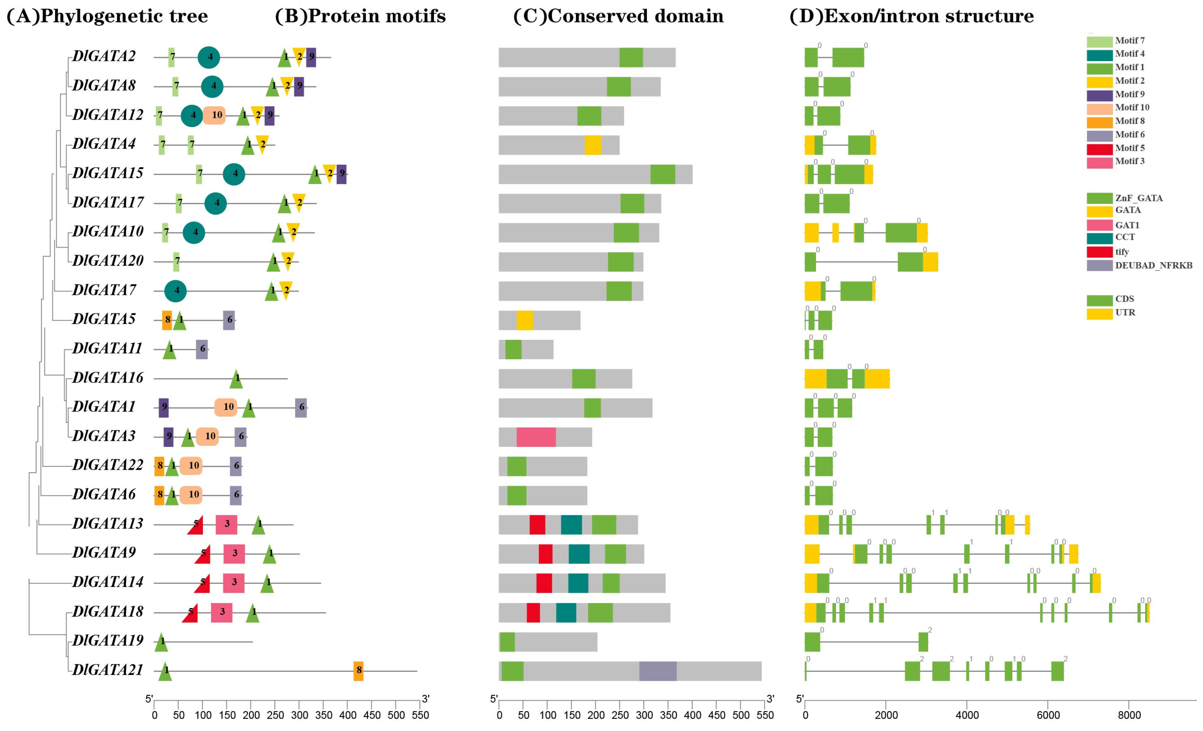
2.3. Analysis of the Gene Structure and Conserved Protein Sequence of DlGATA

In order to gain a clearer understanding of the gene structure of DlGATA and its protein sequence, we analyzed its exon–intron structure, its protein motifs, and its conserved domain structure. The results in Figure 3B show that some independent motifs appeared in only one family. For example, motif4 only existed in subfamily II, while motif5 and motif3 were only present in subfamily III. In subfamily III, motif5 was located in the upstream region of motif3 and motif1, and motif3 was upstream of motif1. Furthermore, motif1 was in all of the DlGATAs, suggesting that motif1 was a very important conserved domain within the DlGATA genes. In the exon/intron structure (Figure 3D), it was found that 2 to 3 exons were generally present in subfamily III, while 7 to 11 exons were present in subfamily III. The UTR region was not present in subfamily I. The present study was carried out to offer an essential basis to reveal the structural characteristics of longan.

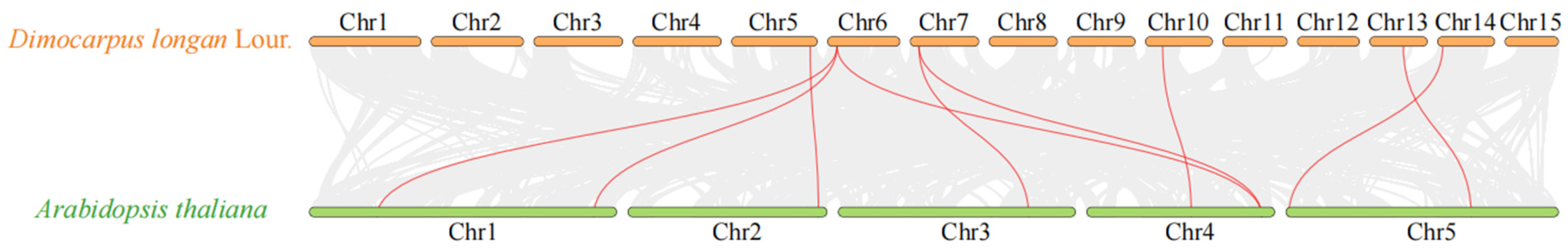
2.4. Synteny Analysis of Longan and Arabidopsis Genes and an Analysis of Replication Events within Gene Families

To understand the evolutionary mechanism of DlGATA and study its possible evolutionary links to other species, we analyzed the synteny of the DlGATA genome with the AtGATA genome (Figure 4). The results indicated that there were nine pairs with collinearity between DlGATA and AtGATA, with the largest number of collinearities being in chromosome 6 (three pairs). Two collinear pairs existed in chromosome 7, and one collinear gene pair between the DlGATA genome and AtGATA genome was found in chromosomes 5, 10, 13, and 14.
As can be seen in Figure 5, it was also found that there were five pairs of GATA genes in the longan chromosome, namely, DlGATA6/DlGATA11, DlGATA6/DlGATA5, DlGATA15/DlGATA17, DlGATA18/DlGATA13, and DlGATA1/DlGATA3. The first four pairs were segmental duplication events in DlGATA genes, and the last pair was a tandem duplication event.

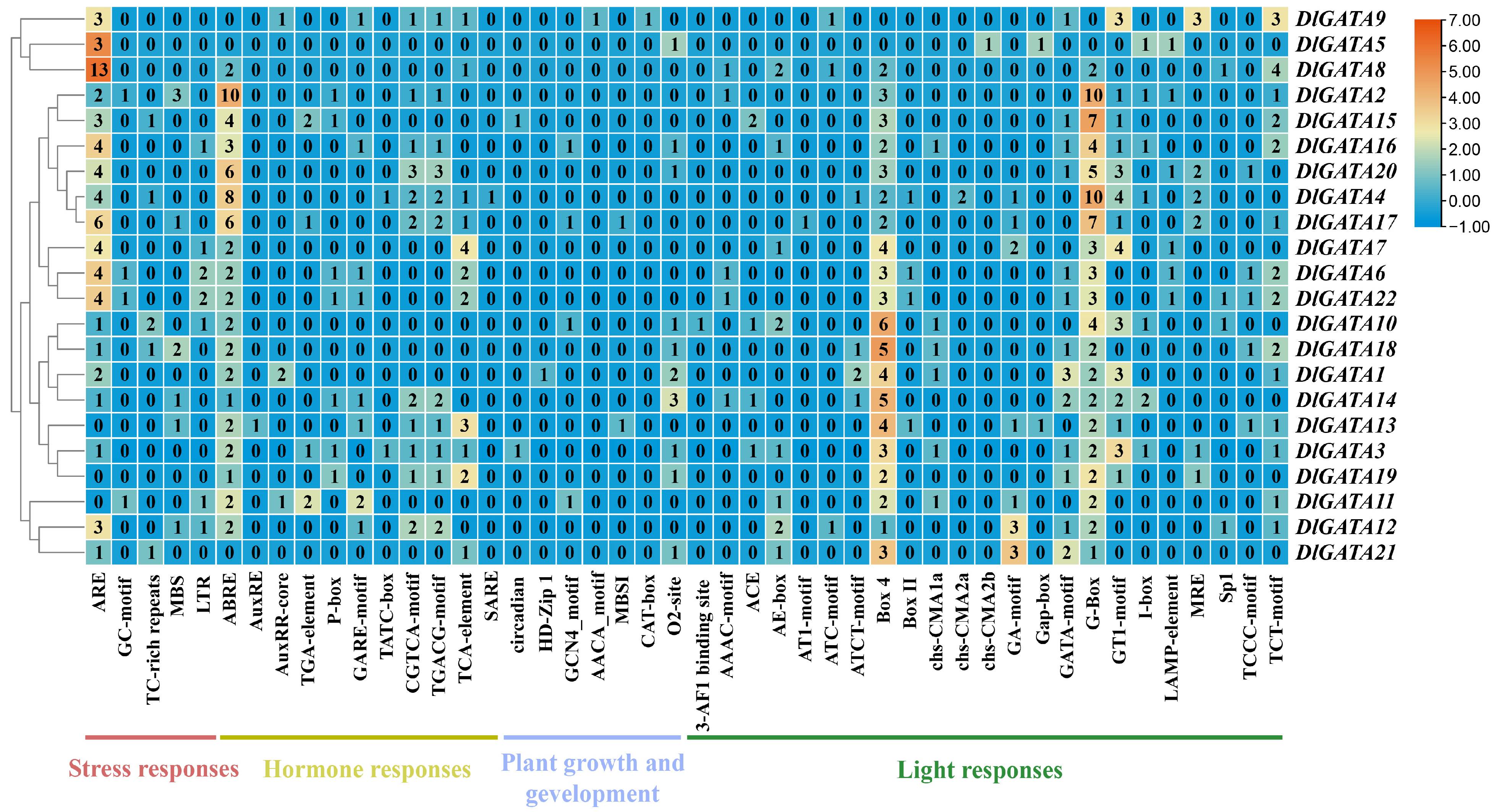
2.5. Analysis of the Cis-Acting Elements in the Promoter of the Longan GATA Genes

By analyzing the cis-acting elements, it was found that a total of 46 types of cis-acting elements existed in the promoter region of the GATA gene of longan (Figure 6), such as ABE, ABER, SARE, BOX4, BOX11, and so on. All of these cis-acting elements were divided into four types, namely, stress responses, hormone responses, plant growth and development, and light responses. Among the many cis-acting elements, Box 4 was the most widely distributed among the 22 DlGATAs; its number was 75 in total, and it was followed by ABE with 64. On the whole, the numbers of stress responses and light responses were significantly greater than those of hormone responses and plant growth and development elements. The results showed that the longan GATA family may be more sensitive to stress and light responses.

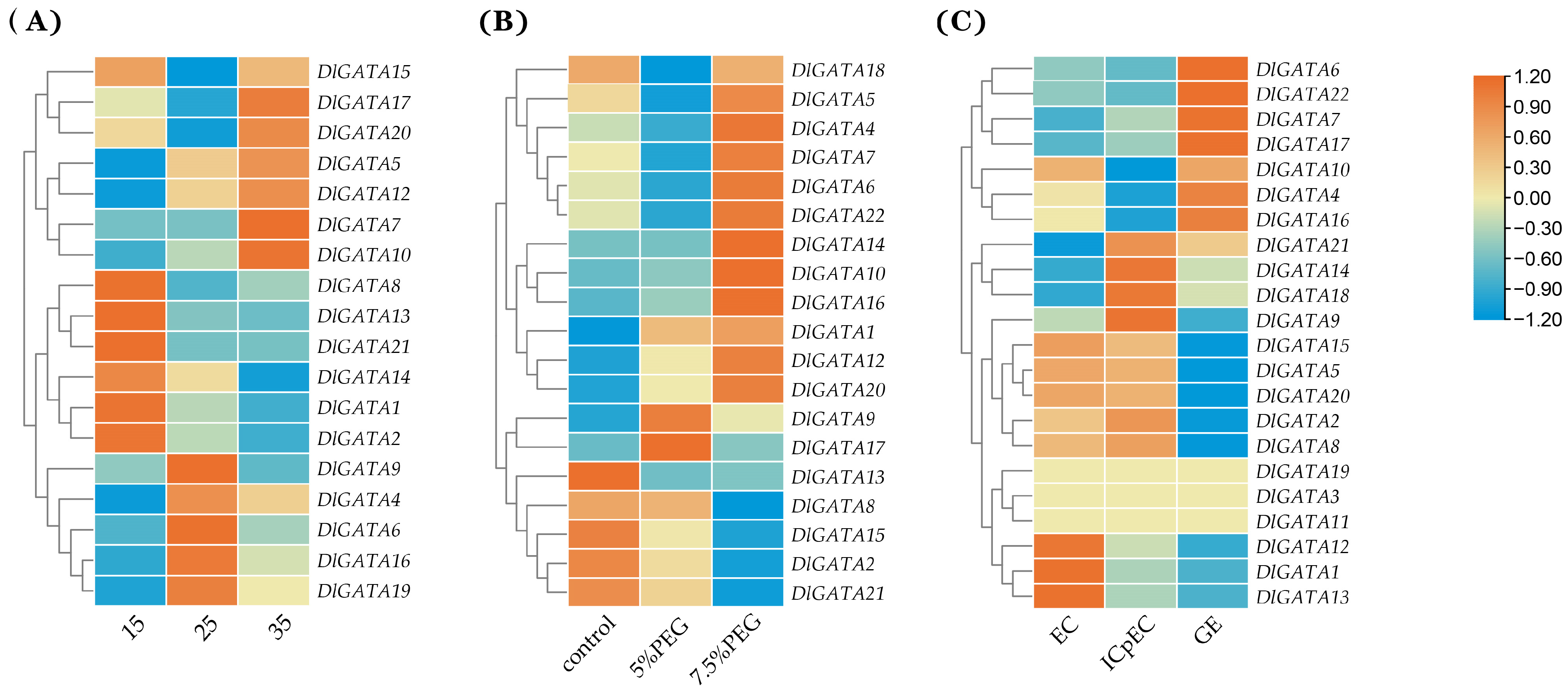
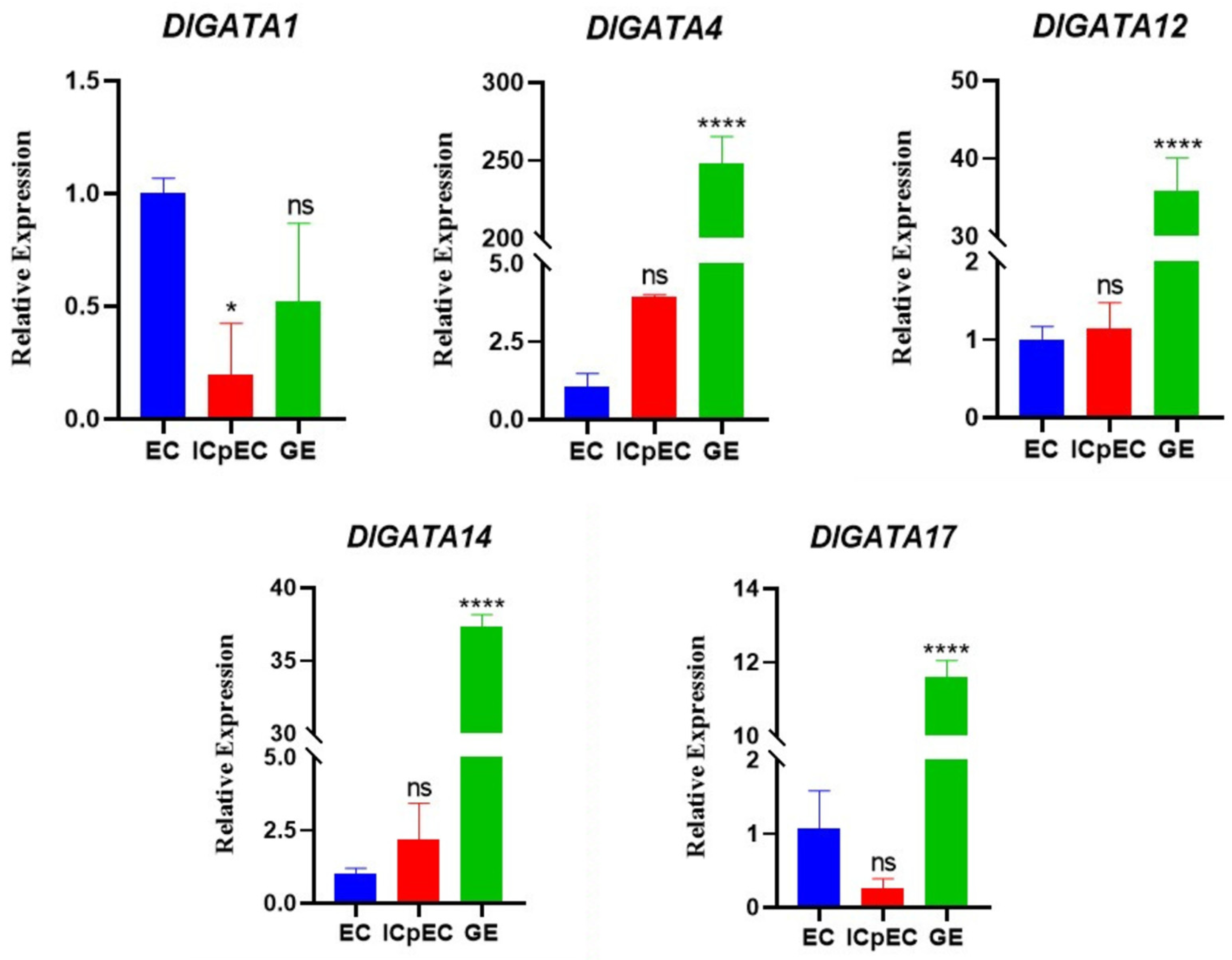
2.6. Expression Analysis of Longan at Different Temperatures, Concentration of PEG, and Early Developmental Stages of Somatic Embryos

By using data from the longan database, the gene expression of DlGATA at different temperatures, concentrations of PEG, and early developmental stages of somatic embryos was obtained, and a heat map was made (Figure 7A and Table S1). In the temperature expression section of DlGATA, the results showed that the GATA gene expression was roughly divided into three categories. In the first category, the most significant up-regulated expression at 15 °C occurred for DlGATA8, DlGATA13, DlGATA21, DlGATA14, DlGATA1, and DlGATA2. In the second category, the most significant up-regulated expression at 25 °C occurred for DlGATA9, DlGATA4, DlGATA6, DlGATA16, and DlGATA19. The third category included the last five GATA genes shown in the figure, which had a stronger up-regulated expression at 35 °C than at the other temperatures. Therefore, we hypothesized that the first group of genes contributed more to cold resistance in longan, while the third group of genes was important in the mechanism of heat tolerance in longan.
At different concentrations of PEG (5%, 7.5%) conditions in the DlGATA expression fraction, the following results were obtained (Figure 7B and Table S2): the GATA gene showed a strong up-regulated expression level under all PEG concentration conditions. Among them, DlGATA2, DlGATA8, DlGATA13, DlGATA15 and DlGATA21 had strong up-regulated expression levels in the absence of PEG. DlGATA9 and DlGATA17 showed high up-regulated levels of expression under 5% PEG. In addition to that, the DlGATAs showed a significantly up-regulated expression profile at 7.5% PEG.
The following types of expressions exist for DlGATA in the heatmap section during early developmental stages of somatic embryos (EC: embryogenic callus, ICpEC: incomplete pro-embryogenic callus, GE: globular embryo) (Figure 7C and Table S3) First, there was a continuous down-regulation in the expression from EC to ICpEC in the GE stage, and this included DlGATA12, DlGATA1, and DlGATA13. Second, expression was unchanged in these three stages, and this scenario included DlGATA19, DlGATA3, and DlGATA11. Third, there was an upward modulation during the EC and ICpEC phases and a downward modulation during the GE phase for DlGATA15, DlGATA5, DlGATA20, DlGATA2, and DlGATA8. Fourth, four genes—DlGATA21, DlGATA14, DlGATA18, and DlGATA9—were downregulated in EC and upwardly modulated in IcpEC and GE. Fifth, DlGATA10, DlGATA4, and DlGATA16 were upwardly modulated in EC and GE and downregulated in ICpEC. Lastly, DlGATA6, DlGATA22, DlGATA7, and DlGATA17 were downregulated in the EC and ICpEC stages and upwardly modulated in the GE stage.
To study the expression of DlGATA during the early developmental stages of longan somatic embryos in greater depth, five representative DlGATAs were selected for qRT-PCR (Figure 8). The following profiles were found: the four genes other than DlGATA1 had strong profiles in the GE phase, indicating that DlGATA may promote the differentiation of GE.

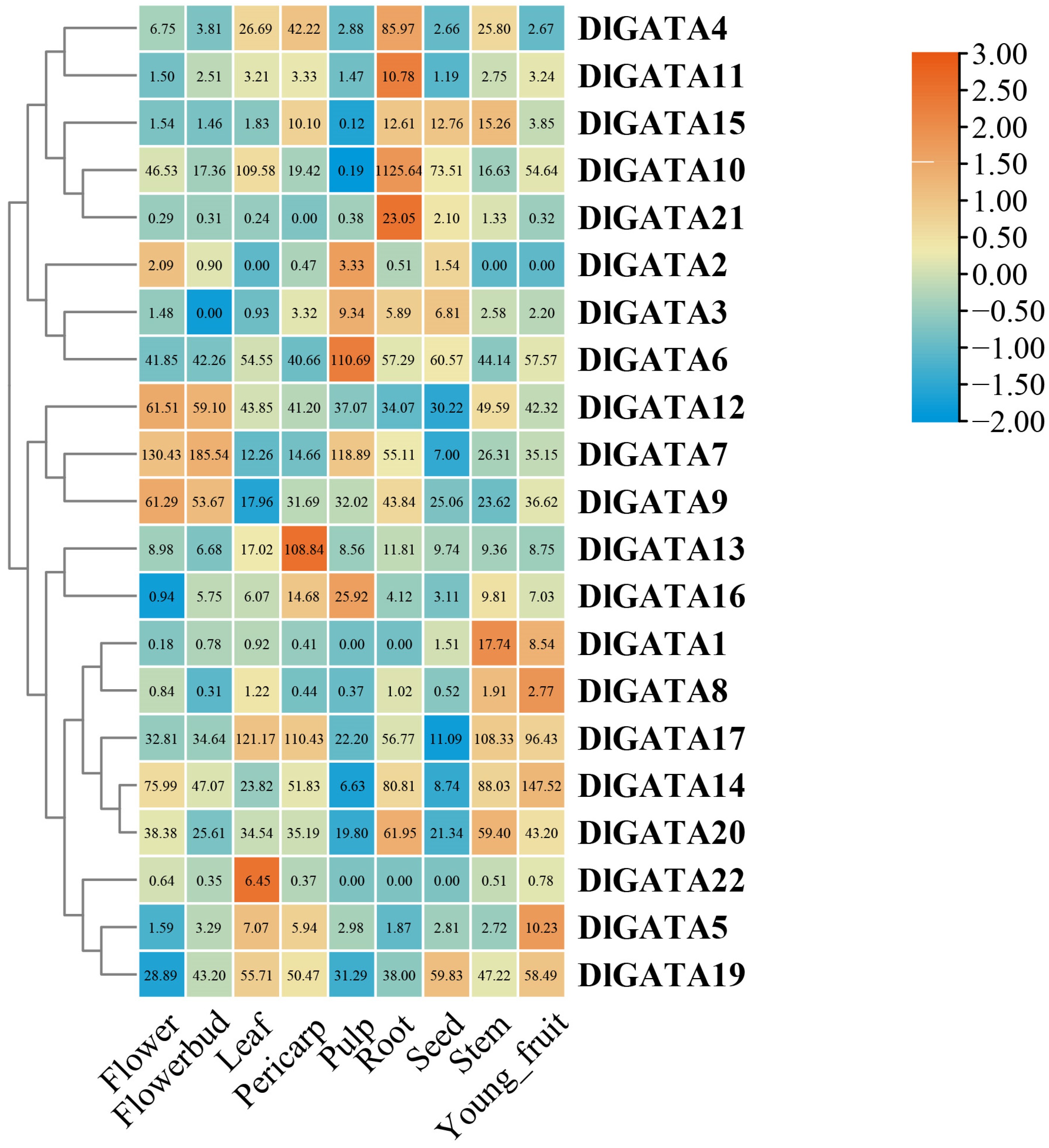
2.7. DlGATA Expression Profiles in Nine Different Tissues

The expression of DlGATAs was further explored using RNA-seq data from flowers, flowerbuds, leaves, pericarps, pulps, roots, seeds, stems, and young fruits. According to the expression conditions, the DlGATA expression levels were used to make a heatmap (Figure 9). On the whole, the different DlGATAs had different tissue expression levels, and high levels of expression were likely to be beneficial. In the current study, DlGATA12, DlGATA7, and DlGATA9 were highly expressed in the flowers and flowerbuds of longan, indicating that they may have a promotive role in the germination processes of flowers. DlGATA4, DlGATA11, DlGATA10, and DlGATA21 showed strong up-regulated expression in the root, which may have a driving effect on root extension. However, in the expression of DlGATA, not all expression levels were completely different, and a small number of genes had similar expressions. For example, DlGATA17, DlGATA14, and DlGATA20 had similar expressions.

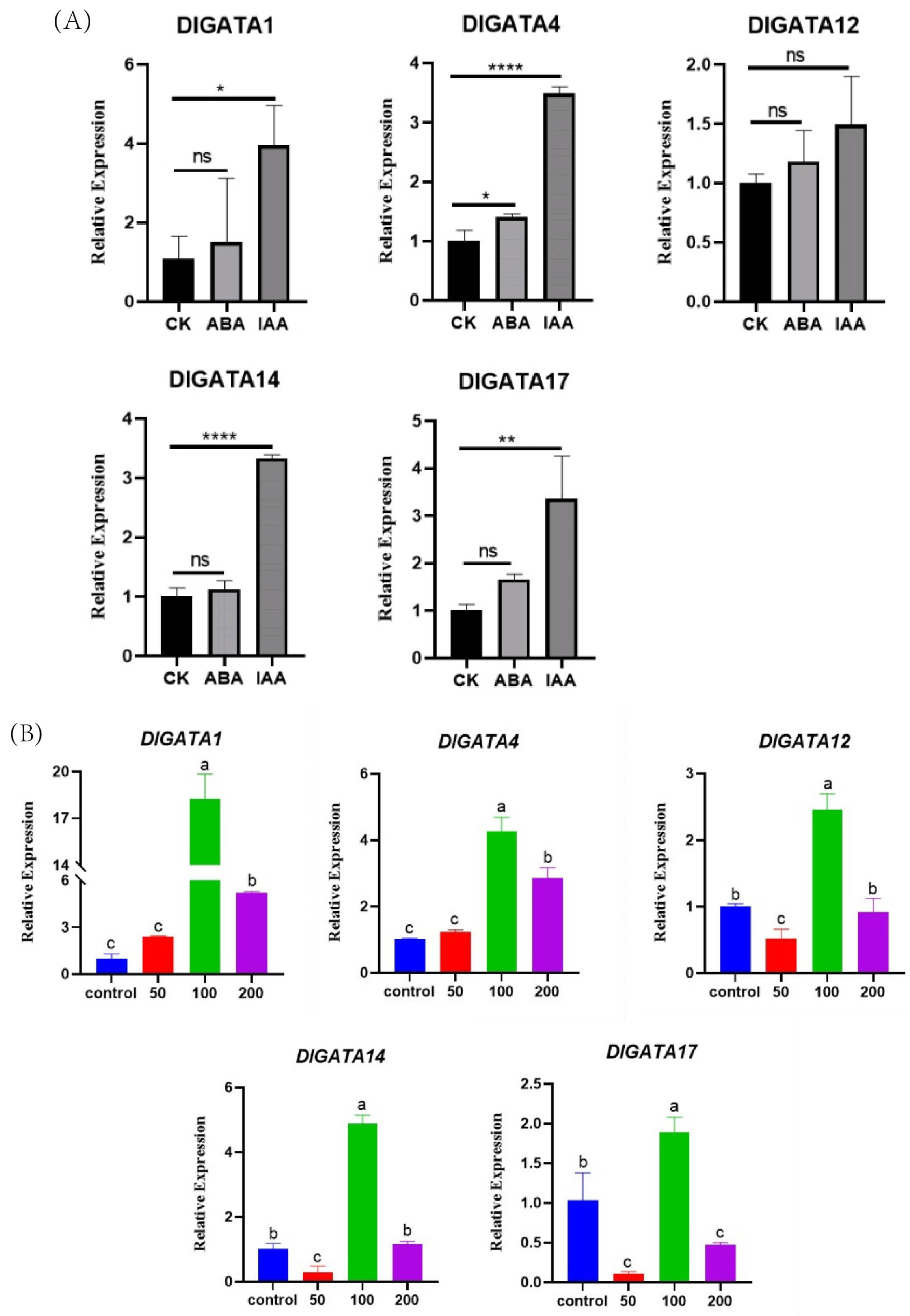
2.8. Expression of DlGATA Induced by Exogenous Hormone Treatments

According to the heatmap of DlGATA expression in the early somatic embryo (Figure 7), 5 representative GATA genes with different expression patterns were selected from the 22 GATA genes; these were DlGATA1, DlGATA4, DlGATA12, DlGATA14, and DlGATA17 (Figure 10). The data showed that, as a whole, there was a significant expression profile with IAA for all five genes, and there was a less significant one with ABA. DlGATA1 showed the most significant expression under IAA stress, as it was nearly five-times greater than that in the control group. In addition, the expression of DlGATA at different concentrations of IAA stress was analyzed in this experiment. The findings demonstrated that the five genes (DlGATA1, DlGATA4, DlGATA12, DlGATA14, and DlGATA17) were significantly up-regulated, expressed at a concentration of 100 μ m o l · L 1 . At the same time, some of the DlGATAs, such as DlGATA1 and DlGATA4, were greatly expressed at higher concentrations. However, at lower concentrations of the IAA treatment, the genes in the experimental group were mostly suppressed or maintained at expression levels similar to those in the untreated group. The results indicated that IAA at a concentration of 100 μ m o l · L 1 induced DlGATA expression, while concentrations too high or too low may inhibit this expression.

3. Discussion

In this study, the longan GATA gene family was divided using bioinformatic technology. A total of 22 DlGATA genes exist, and they can be divided into four families. Of these, subfamily II has the most members, while subfamily I has the fewest DlGATAs. In the assay of the physicochemical properties of DlGATA, it was found that DlGATA proteins and AcoDREB proteins [32] had similar isoelectric points, mostly centered on the 5 to 9 isoelectric points, and there was no obvious acidic versus basic nature overall. In the analysis of the structure of DlGATA and its protein conservation sequence (Figure 3), it was shown that the gene structures and protein conservation sequences in the same subfamily were roughly similar, and the structures of members of different subfamilies were different from the conserved domains of proteins [33]. This was in line with previous results. Furthermore, the number of introns has a key influence on the evolution and generation of plant gene families [34]. The intron number varied in the same GATA gene family in longan. The protein–protein role was closely linked to the motif composition of the transfer factors [35]. The motif combinations present in the GRAS family of Arabidopsis can function as transcriptional regulatory proteins to mediate many different interactions with the basic transcriptional machinery and accessory proteins [36]. In the motif study of longan GATA, only motif3 and motif5 were present in subfamily III, so it can be speculated that motif3 and motif5 are an important basis for its recognition, and this is most likely the reason for the specific functions carried out by subfamily III and its accessory proteins.
In the process of phytohormone signaling, related genes are substantially enriched [37,38]. Studies have revealed that phytohormones are instrumental in EC regeneration [39,40,41]. There are many singular genes related to embryonic growth and development in the EC of longan [42]. In the present study of the embryogenic callus of longan, it was found that growth hormones significantly induced the expression of DlGATA genes. This was in line with the findings of a previous study of longan development, which showed that there were large numbers of auxin and cytokinin signaling components in the transcriptomes of the four different stages of NEC (non-embryogenic callus), ICPEC, EC, and GC [37]. This suggests that DlGATA may synergize with IAA and participate in biotic or abiotic stresses associated with it. In addition, DlGATA1, which was significantly expressed under growth hormone treatment, showed strong expression levels in the stems and young fruits. In previous studies, it was shown that an elevated ratio of auxin to cytokinin favors the rooting of point–stem cuttings of rose shoots [43]. It was also reported that the concentration of auxin in Chinese fir stems affected the activity of cambium [44]. Studies have shown that increased concentrations of IAA reduce the sensitivity to exogenous material in ripe fruits of Citrus sinensis (L.) Osb [45]. It can be speculated that DlGATA may be involved in the stem elongation mechanism and fruit anti-shedding mechanism based on IAA signaling. However, the expression levels of DlGATA in various tissues under IAA treatment need to be further explored, as this will provide a rational foundation for further study of the growth and development of longan, as well as the function of DlGATA.
The effect of drought on longan was found to contribute to higher yields and higher economic returns. The GATA showed a close association with drought stress [14,46,47]. In a study of wheat GATA, we found that ABA with Ca2+ caused the up-regulation of TaGATAs, which activated related genes [48]. In studies of abiotic stress in grain crops such as rice and soybean, ABF, a response factor to ABA, has been shown to bind to ABER and promote the activation of related genes upon receipt of a signal [49]. In a study of the promoter region of DlGATAs, we found that ABER, a cis-acting element associated with the hormone ABA, was abundantly present in the promoter region of DlGATAs. Interestingly, we did not find a significant correlation with ABA in our analysis of DlGATA expression in longan, and this could be a result of the repressive effect of silencers in gene expression or other reasons, making it different from GATA genes in other species [50]. Furthermore, in the study of the IAA hormone, we found that DlGATA4, DlGATA10, DlGATA11, and DlGATA21 were significantly expressed in roots. Similarly, strong expression levels were also shown in the presence of IAA at a concentration of 100 μ m o l · L 1 . In previous studies, Arabidopsis PHB3 was found to regulate the degradation of IAA14/28 through the non-mediated growth hormone signaling pathway by releasing ARFs to bind to GATA23 and modulating the initiation of lateral root primordia [51]. During this experiment, we hypothesized that IAA may promote the transcription of DlGATAs through a series of unknown signaling mechanisms (Figure 11), which, in turn, modulate root traits. Changes in root traits can result in a better response to drought-stressed environments [52,53,54,55,56]. Therefore, we hypothesized that the drought response mechanism of longan GATA is less associated with ABA, while there is a strong link with IAA. In addition, to further investigate the role of DlGATA under drought conditions and the expression levels under different drought stresses, we conducted further studies on this. It was found that DlGATA1, DlGATA4, DlGATA12, and DlGATA14, which were mentioned in previous experiments (Figure 7), were also strongly up-regulated, expressed under 7.5% PEG at different concentrations of PEG. Meanwhile, DlGATA17 also showed strongly up-regulated expression at 5% PEG, further strengthening our speculation of DlGATA under drought conditions. Based on this, it is possible to predict the role and function of DlGATA in the drought resistance process.

4. Materials and Methods

4.1. Plant Materials

‘Honghezi’ longan used in this study was provided by the Institute of Horticultural Biotechnology of Fujian Agriculture and Forestry University, which grows mainly in Fuzhou, Fujian, and it has a high yield and cannot easily drop fruits; the climatic conditions required for growth are sunny, favorable temperatures, and high-humidity areas. The longan GATA gene sequences, CDS sequences, amino acid sequences, and gene annotation information were downloaded from the longan genome database, which was constructed in our laboratory [59].

4.2. Identification of GATA Genes in Longan

The reported Arabidopsis GATA gene family sequences were obtained from the TAIR [60] (https://www.arabidopsis.org/ (accessed on 9 July 2023)) website and homologated to the longan genome database using the TBtools V2.012 [61] software (Chen Chengjie from South China Agricultural University, China). The Interpro website [62] (https://www.ebi.ac.uk/interpro/ (accessed on 9 July 2023)) was used to view the conserved structural domains of the above sequences, and filtering based on whether or not the zinc finger structure was included (pfam00320) ultimately yielded 22 sequences of the longan GATA family. Referring to the longan genome annotation file and the nomenclature of the GATAs of other species, they were sequentially named DlGATA122. The number of amino acids, relative molecular weight, theoretical isoelectric point, instability index, aliphatic index, and grand average of hydropathicity of the DlGATA family members were analyzed using the Expasy online software [63] (https://www.expasy.org/ (accessed on 20 July 2023)) and the cello online software [64] (http://cello.life.nctu.edu.tw/ (accessed on 20 July 2023)) for the subcellular localization of the DlGATA family.

4.3. Phylogenetic Analysis, Gene Structure, and Conserved Motif Analysis

The MEGA X [65] software (Mega Limited, Auckland, New Zealand) was used to construct the phylogenetic tree of the GATA family for three species—longan, Malus domestica, and the model plant Arabidopsis thaliana. The GATA sequences of apple and Arabidopsis thaliana were obtained from the website of PlantTFDB [66] (http://planttfdb.gao-lab.org/ (accessed on 6 December 2022)), and the maximum likelihood was chosen to compute these sequences with 1000 bootstrap replicates. Finally, the above results were embellished using the iTOL online website [67]. The longan GATA sequences were imported into the MEME online website [68] (https://meme-suite.org/meme/ (accessed on 9 July 2023)), and the number of motifs was set to 10 to analyze the prediction of the above sequences. The structural domains contained in the amino acid sequence of GATA of longan were analyzed using the Batch CD-search function on the NCBI website (https://www.ncbi.nlm.nih.gov/ (accessed on 9 July 2023)). The longan GATA gene annotation files were imported into the Tbtools software [61]. Based on the Gene Structure View function in this software, the results of the above three types of analysis were visualized, and, finally, maps of the longan GATA motifs, introns, and conserved structures were acquired.

4.4. Chromosomal Location, Cis-Acting Element Analyses, and Analysis of Collinearity with Other Species

PlantCARE [69] (https://bioinformatics.psb.ugent.be/webtools/plantcare/html/ (accessed on 16 July 2023)) was utilized to analyze the cis-acting elements of the 2000 bp sequence upstream of GATA in longan. Finally, a two-dimensional heatmap was drawn, with the components involved in stress responses, hormone responses, plant growth and developmental regulation, and light responses as horizontal coordinates and the longan GATA genes as vertical coordinates. The genome-wide annotation information of Arabidopsis thaliana and rice was downloaded from the TAIR website and the Phytozomehttps website [70] (https://phytozome-next.jgi.doe.gov/ (accessed on 16 July 2023)), respectively, and analyses of the synteny of these species with longan were performed using the McscanX (https://github.com/wyp1125/MCScanX/ (accessed on 16 July 2023)) software (Tang Haibao from Fujian Agriculture and Forestry University, Fuzhou, China) [71]. Finally, Tbtools was used to draw and enhance the visualized images of the gene synteny among longan, Arabidopsis, and rice. The collinear relationships of the GATA gene within the longan species were analyzed using the MCscanX software, and the results were visualized using TBtools.

4.5. Hormonal Processing of Longan EC, RNA Extraction, and qRT-PCR Analysis

Longan embryogenic callus materials with a good growth state were selected, and equal amounts were added to a liquid medium with 50 μ m o l · L 1 , 100 μ m o l · L 1 , 200 μ m o l · L 1 IAA, and 100 μ m o l · L 1 ABA; longan EC was added to a liquid medium containing MS without any hormones as a blank control, and three replicates were set for the above treatments; they underwent 24 h of incubation at 25 °C in the dark, and they were then filter-dried and frozen at −80 °C in a refrigerator for later use. The DNAMAN6 software (Lynnon Biosoft, San Ramon, CA, USA) was used to design the qRT-PCR primers for the longan GATA gene, and the primer sequences are shown in Table S4. RNA was extracted from the above hormone-treated materials using the TransZol kit, as described in the instructions. The RNA obtained through extraction was reverse-transcribed into cDNA using the Revertaid Master Mix (Thermo Fisher Scientific, Shanghai, China) kit. By using ubiquitin [72] (UBQ) as an internal control, the expression level of GATA in longan after the IAA and ABA hormone treatments and the somatic embryo of longan (EC: embryogenic callus, IcpEC: Incomplete pro-embryogenic callus, GE: globular embryo) stage was examined in a Roche Light Cycler 96 (place of origin: Basel, Switzerland) for qRT-PCR. The 2−ΔΔCt method was used to calculate the assay data, then averaged over three repetitions and analyze the significant differences in the relative expression of DlGATA at the three stages of longan somatic embryo using the software Prism 8.0.2 as well as the one-way ANOVA method. The relative expression of DlGATA after IAA and ABA treatments was analyzed using SPSS 25 as well as Duncan’s one-way ANOVA method, and, finally, Prism 8.0.2 was used to visualize the above results. Three repetitions were used, and the average value was taken. The statistical method used in this experiment was standard deviation.

4.6. Analysis of the Specific Expression of DlGATA Family Genes

The FPKM of longan GATA family members at three stages of longan’s somatic embryogenesis (EC, IcpEC, GE) at high and low temperatures (35 °C, 15 °C), PEG (5%, 7%), 2,4-D treatments and at nine different tissue sites (seeds, roots, stems, leaves, flowers, flowerbuds, pulp, young fruits, and pericarp) was derived from the transcriptome database (SRA050205). The above data were log-transformed ( l o g 2 ) and plotted on a heatmap using Tbtools.

5. Conclusions

In the present experiment, based on a heatmap of expression in early embryonic calluses of longan, 5 representative genes extracted from 22 DlGATA genes were investigated. Based on the phylogeny and structure of GATA, its evolutionary relationship with Arabidopsis and apple was determined. In the analysis of Collinearity within the longan, there was one tandem gene pair and four fragment duplication gene pairs in the GATA gene family that provide information for studying gene amplification. Under the expression of DlGATA at different concentrations of IAA, they were all found to have a strong up-regulated expression profile under IAA conditions at 100 μ m o l · L 1 concentration. We also found that DlGATA was strongly up-regulated, expressed in roots and stems, which we correlated with drought stress. In addition, we investigated the expression of DlGATA under different concentrations of PEG. Further information about the relationship between DlGATA and drought was obtained. In addition, further studies of expression in the somatic embryo of longan can offer a rationale for exploring the functions and actions of GATA in the early period of development. All of these can help us to improve our understanding of DlGATA. However, there are still limitations to the in-depth study of the GATA gene family, so there are still questions that need to be answered, making this another fruitful area with room for progress.

Supplementary Materials

The following supporting information can be downloaded at: https://www.mdpi.com/article/10.3390/ijms25020731/s1.

Author Contributions

Conceptualization, Y.L., K.Z., J.L. and S.C.; methodology, Y.L.; software, S.L. and T.Z.; data curation, S.L. and T.Z.; formal analysis, K.Z., J.L. and X.H.; writing—original draft preparation, J.L.; writing—review and editing, Y.L., K.Z., J.L., X.H., S.L. and S.C.; investigation, K.Z.; supervision, Y.L. and S.C.; project administration, S.C.; visualization, J.L. and X.H. All authors have read and agreed to the published version of the manuscript.

Funding

This research was funded by the Fujian Provincial Natural Science Foundation (2020J01543); Fujian Province Plateau Discipline Construction Fund (102/71201801101); the Innovation Fund of Fujian Agriculture and Forestry University (KFb22022XA, CXZX2019033S); Fujian Agriculture and Forestry University Innovation and Entrepreneurship Training Program for College Students (FAFUXMPC20230718001-00081) co-funded.

Institutional Review Board Statement

Not applicable.

Informed Consent Statement

Not applicable.

Data Availability Statement

All analyzed data for this study are included in the contents of this article and Supplementary Materials.

Acknowledgments

Thanks to Fujian Natural Science Foundation of Fujian Province and other funds for their strong support for this experiment. We thank student Yin Ziyuan (College of Forestry, Fujian Agriculture and Forestry University) for his help in revising the manuscript.

Conflicts of Interest

The authors declare no conflicts of interest.

References

  1. Mitsuda, N.; Ohme-Takagi, M. Functional Analysis of Transcription Factors in Arabidopsis. Plant Cell Physiol. 2009, 50, 1232–1248. [Google Scholar] [CrossRef] [PubMed]
  2. Kim, K.-N.; Cheong, Y.H.; Grant, J.J.; Pandey, G.K.; Luan, S. CIPK3, a Calcium Sensor–Associated Protein Kinase That Regulates Abscisic Acid and Cold Signal Transduction in Arabidopsis. Plant Cell 2003, 15, 411–423. [Google Scholar] [CrossRef] [PubMed]
  3. Éva, C.; Moncsek, B.; Szőke-Pázsi, K.; Kunos, V.; Mészáros, K.; Makai, S.; Sági, L.; Juhász, A. bZIP transcription factors repress the expression of wheat (Triticum aestivum L.) high molecular weight glutenin subunit genes in vegetative tissues. Acta Physiol. 2023, 45, 29. [Google Scholar] [CrossRef]
  4. Abdullah-Zawawi, M.-R.; Ahmad-Nizammuddin, N.-F.; Govender, N.; Harun, S.; Mohd-Assaad, N.; Mohamed-Hussein, Z.-A. Comparative genome-wide analysis of WRKY, MADS-box and MYB transcription factor families in Arabidopsis and rice. Sci. Rep. 2021, 11, 19678. [Google Scholar] [CrossRef] [PubMed]
  5. Dudhate, A.; Shinde, H.; Yu, P.; Tsugama, D.; Gupta, S.K.; Liu, S.; Takano, T. Comprehensive analysis of NAC transcription factor family uncovers drought and salinity stress response in pearl millet (Pennisetum glaucum). BMC Genom. 2021, 22, 70. [Google Scholar] [CrossRef] [PubMed]
  6. Nakashima, K.; Takasaki, H.; Mizoi, J.; Shinozaki, K.; Yamaguchi-Shinozaki, K. NAC transcription factors in plant abiotic stress responses. Biochim. Biophys. Acta—Gene Regul. Mech. 2012, 1819, 97–103. [Google Scholar] [CrossRef]
  7. Olsen, A.N.; Ernst, H.A.; Leggio, L.L.; Skriver, K. NAC transcription factors: Structurally distinct, functionally diverse. Trends Plant Sci. 2005, 10, 79–87. [Google Scholar] [CrossRef]
  8. Nuruzzaman, M.; Sharoni, A.M.; Kikuchi, S. Roles of NAC transcription factors in the regulation of biotic and abiotic stress responses in plants. Front. Microbiol. 2013, 4, 248. [Google Scholar] [CrossRef]
  9. Puranik, S.; Sahu, P.P.; Srivastava, P.S.; Prasad, M. NAC proteins: Regulation and role in stress tolerance. Trends Plant Sci. 2012, 17, 369–381. [Google Scholar] [CrossRef]
  10. Khan, I.; Asaf, S.; Jan, R.; Bilal, S.; Lubna; Khan, A.L.; Kim, K.-M.; Al-Harrasi, A. Genome-wide annotation and expression analysis of WRKY and bHLH transcriptional factor families reveal their involvement under cadmium stress in tomato (Solanum lycopersicum L.). Front. Plant Sci. 2023, 14, 1100895. [Google Scholar] [CrossRef]
  11. Khoudi, H. SHINE clade of ERF transcription factors: A significant player in abiotic and biotic stress tolerance in plants. Plant Physiol. Biochem. 2023, 195, 77–88. [Google Scholar] [CrossRef]
  12. Medina, J.; Bargues, M.; Terol, J.; Pérez-Alonso, M.; Salinas, J. The Arabidopsis CBF Gene Family Is Composed of Three Genes Encoding AP2 Domain-Containing Proteins Whose Expression Is Regulated by Low Temperature but Not by Abscisic Acid or Dehydration. Plant Physiol. 1999, 119, 463–470. [Google Scholar] [CrossRef] [PubMed]
  13. Du, K.; Xia, Y.; Zhan, D.; Xu, T.; Lu, T.; Yang, J.; Kang, X. Genome-Wide Identification of the Eucalyptus urophylla GATA Gene Family and Its Diverse Roles in Chlorophyll Biosynthesis. Int. J. Mol. Sci. 2022, 23, 5251. [Google Scholar] [CrossRef] [PubMed]
  14. Gillis, W.Q.; St John, J.; Bowerman, B.; Schneider, S.Q. Whole genome duplications and expansion of the vertebrate GATA transcription factor gene family. Biology 2009, 9, 207. [Google Scholar] [CrossRef] [PubMed]
  15. Scazzocchio, C. The fungal GATA factors. Curr. Opin. Microbiol. 2000, 3, 126–131. [Google Scholar] [CrossRef] [PubMed]
  16. Reyes, J.C.; Muro-Pastor, M.I.; Florencio, F.J. The GATA Family of Transcription Factors in Arabidopsis and Rice. Plant Physiol. 2004, 134, 1718–1732. [Google Scholar] [CrossRef] [PubMed]
  17. Lindemose, S.; O’Shea, C.; Jensen, M.; Skriver, K. Structure, Function and Networks of Transcription Factors Involved in Abiotic Stress Responses. Int. J. Mol. Sci. 2013, 14, 5842–5878. [Google Scholar] [CrossRef] [PubMed]
  18. Gupta, P.; Nutan, K.K.; Singla-Pareek, S.L.; Pareek, A. Abiotic Stresses Cause Differential Regulation of Alternative Splice Forms of GATA Transcription Factor in Rice. Front. Plant Sci. 2017, 8, 1944. [Google Scholar] [CrossRef] [PubMed]
  19. Manzoor, M.A.; Sabir, I.A.; Shah, I.H.; Wang, H.; Yu, Z.; Rasool, F.; Mazhar, M.Z.; Younas, S.; Abdullah, M.; Cai, Y. Comprehensive Comparative Analysis of the GATA Transcription Factors in Four Rosaceae Species and Phytohormonal Response in Chinese Pear (Pyrus bretschneideri) Fruit. Int. J. Mol. Sci. 2021, 22, 12492. [Google Scholar] [CrossRef]
  20. Card, R.M. Functional Analysis of a Tobacco GATA Factor. 1999. Available online: https://www.researchgate.net/publication/263162054 (accessed on 24 November 2023).
  21. Nizamutdinova, I.T.; Kim, Y.M.; Il Chung, J.; Shin, S.C.; Jeong, Y.-K.; Seo, H.G.; Lee, J.H.; Chang, K.C.; Kim, H.J. Anthocyanins from Black Soybean Seed Coats Preferentially Inhibit TNF-α-Mediated Induction of VCAM-1 over ICAM-1 through the Regulation of GATAs and IRF-1. J. Agric. Food Chem. 2009, 57, 7324–7330. [Google Scholar] [CrossRef]
  22. Arens, P.; Odinot, P.; van Heusden, A.W.; Lindhout, P.; Vosman, B. GATA- and GACA-repeats are not evenly distributed throughout the tomato genome. Genome 1995, 38, 84–90. [Google Scholar] [CrossRef] [PubMed]
  23. Agnihotri, S.; Wolf, A.; Picard, D. GATA4 is a regulator of astrocyte cell proliferation and apoptosis in the human and murine central nervous system. Oncogene 2009, 28, 3033–3046. [Google Scholar] [CrossRef] [PubMed]
  24. Reyes, R.; Haendel, M.; Grant, D.; Melancon, E.; Eisen, J.S. Slow degeneration of zebrafish Rohon-Beard neurons during programmed cell death. Dev. Dyn. 2003, 229, 30–41. [Google Scholar] [CrossRef] [PubMed]
  25. Lowry, J.A.; Atchley, W.R. Molecular Evolution of the GATA Family of Transcription Factors: Conservation within the DNA-Binding Domain. J. Mol. Evol. 2000, 50, 103–115. [Google Scholar] [CrossRef] [PubMed]
  26. Krishna, S.S. Structural classification of zinc fingers: Survey and summary. Nucleic Acids Res. 2003, 31, 532–550. [Google Scholar] [CrossRef] [PubMed]
  27. Tindall, H.D. Sapindaceous fruits: Botany and horticulture. Hortic. Rev. Am. Soc. Hortic. Sci. 1994, 16, 143–196. [Google Scholar]
  28. Zhang, X.; Guo, S.; Ho, C.-T.; Bai, N. Phytochemical constituents and biological activities of longan (Dimocarpus longan Lour.) fruit: A review. Food Sci. Hum. Wellness 2020, 9, 95–102. [Google Scholar] [CrossRef]
  29. Li, L.R.; Zhuang, Y.M. Longan Cultivation; China Agricultural Pres: Beijing, China, 1983; Volume 1, p. 83. [Google Scholar]
  30. Nickell, L.G. Plant Growth Regulators in Agriculture and Horticulture; ACS Symposium Series; American Chemical Society: Washington, DC, USA, 1994; Volume 557, pp. 1–14. [Google Scholar]
  31. Bandara, P.M.; Tanino, K.K. Paclobutrazol enhances minituber production in Norland potatoes. J. Plant Growth Regul. 1995, 14, 151–155. [Google Scholar] [CrossRef]
  32. Chai, M.N.; Cheng, H.; Yan, M.K.; Priyadarshani, S.; Zhang, M.; He, Q.; Huang, Y.M.; Chen, F.Q.; Liu, L.P.; Huang, X.Y.; et al. Identification and expression analysis of the DREB transcription factor family in pineapple (Ananas comosus (L.) Merr.). PeerJ 2020, 8, e9006. [Google Scholar] [CrossRef]
  33. Zhang, Z.; Zou, X.; Huang, Z.; Fan, S.; Qun, G.; Liu, A.; Gong, J.; Li, J.; Gong, W.; Shi, Y.; et al. Genome-wide identification and analysis of the evolution and expression patterns of the GATA transcription factors in three species of Gossypium genus. Gene 2019, 680, 72–83. [Google Scholar] [CrossRef]
  34. Roy, S.W.; Penny, D. A Very High Fraction of Unique Intron Positions in the Intron-Rich Diatom Thalassiosira pseudonana Indicates Widespread Intron Gain. Mol. Biol. Evol. 2007, 24, 1447–1457. [Google Scholar] [CrossRef] [PubMed]
  35. Liu, L.; White, M.J.; MacRae, T.H. Transcription factors and their genes in higher plants. Functional domains, evolution and regulation. Eur. J. Biochem. 1999, 262, 247–257. [Google Scholar] [CrossRef] [PubMed]
  36. Pysh, L.D.; Wysocka-Diller, J.W.; Camilleri, C.; Bouchez, D.; Benfey, P.N. The GRAS gene family in Arabidopsis: Sequence characterization and basic expression analysis of the SCARECROW-LIKE genes. Plant J. 1999, 18, 111–119. [Google Scholar] [CrossRef] [PubMed]
  37. Chen, Y.; Xu, X.; Liu, Z.; Zhang, Z.; XuHan, X.; Lin, Y.; Lai, Z. Global scale transcriptome analysis reveals differentially expressed genes involve in early somatic embryogenesis in Dimocarpus longan Lour. BMC Genom. 2020, 21, 4. [Google Scholar] [CrossRef] [PubMed]
  38. Hellens, A.M.; Chabikwa, T.G.; Fichtner, F.; Brewer, P.B.; Beveridge, C.A. Identification of new potential downstream transcriptional targets of the strigolactone pathway including glucosinolate biosynthesis. Plant Direct 2023, 7, e486. [Google Scholar] [CrossRef] [PubMed]
  39. Anabel Lopez-Ruiz, B.; Thamara Juarez-Gonzalez, V.; Gomez-Felipe, A.; De Folter, S.; Dinkova, T.D. tasiR-ARFs Production and Target Regulation during In Vitro Maize Plant Regeneration. Plants 2020, 9, 849. [Google Scholar] [CrossRef] [PubMed]
  40. Ruan, C.-J.; Zheng, X.; da Silva, J.A.T.; Qin, P. Callus induction and plant regeneration from embryonic axes of Kosteletzkya virginica. Sci. Hortic. 2009, 120, 150–155. [Google Scholar] [CrossRef]
  41. Marimuthu, K.; Subbaraya, U.; Suthanthiram, B.; Marimuthu, S.S. Molecular analysis of somatic embryogenesis through proteomic approach and optimization of protocol in recalcitrant Musa spp. Physiol. Plant. 2019, 167, 282–301. [Google Scholar] [CrossRef]
  42. Lai, Z.; Lin, Y. Analysis of the global transcriptome of longan (Dimocarpus longan Lour.) embryogenic callus using Illumina paired-end sequencing. BMC Genom. 2013, 14, 561. [Google Scholar] [CrossRef]
  43. Otiende, M.A.; Fricke, K.; Nyabundi, J.O.; Ngamau, K.; Hajirezaei, M.R.; Druege, U. Involvement of the auxin–cytokinin homeostasis in adventitious root formation of rose cuttings as affected by their nodal position in the stock plant. Planta 2021, 254, 65. [Google Scholar] [CrossRef]
  44. Yang, L.; Zhu, S. The Interconnected Relationship between Auxin Concentration Gradient Changes in Chinese Fir Radial Stems and Dynamic Cambial Activity. Forests 2022, 13, 1698. [Google Scholar] [CrossRef]
  45. Yuan, R.; Kender, W.J.; Burns, J.K. Young Fruit and Auxin Transport Inhibitors Affect the Response of Mature ‘Valencia’ Oranges to Abscission Materials via Changing Endogenous Plant Hormones. J. Am. Soc. Hortic. 2003, 128, 302–308. [Google Scholar] [CrossRef]
  46. Hwarari, D.; Radani, Y.; Guan, Y.; Chen, J.; Liming, Y. Systematic Characterization of GATA Transcription Factors in Liriodendron chinense and Functional Validation in Abiotic Stresses. Plants 2023, 12, 2349. [Google Scholar] [CrossRef] [PubMed]
  47. Hussain, Q.; Asim, M.; Zhang, R.; Khan, R.; Farooq, S.; Wu, J. Transcription Factors Interact with ABA through Gene Expression and Signaling Pathways to Mitigate Drought and Salinity Stress. Biomolecules 2021, 11, 1159. [Google Scholar] [CrossRef] [PubMed]
  48. Du, X.; Lu, Y.; Sun, H.; Duan, W.; Hu, Y.; Yan, Y. Genome-Wide Analysis of Wheat GATA Transcription Factor Genes Reveals Their Molecular Evolutionary Characteristics and Involvement in Salt and Drought Tolerance. Int. J. Mol. Sci. 2022, 24, 27. [Google Scholar] [CrossRef]
  49. Nakashima, K.; Yamaguchi-Shinozaki, K.; Shinozaki, K. The transcriptional regulatory network in the drought response and its crosstalk in abiotic stress responses including drought, cold, and heat. Front. Plant Sci. 2014, 5, 170. [Google Scholar] [CrossRef] [PubMed]
  50. Cunningham, T.S.; Andhare, R.; Cooper, T.G. Nitrogen catabolite repression of DAL80 expression depends on the relative levels of Gat1p and Ure2p production in Saccharomyces cerevisiae. J. Biol. Chem. 2000, 275, 14408–14414. [Google Scholar] [CrossRef]
  51. Li, S.; Li, Q.; Tian, X.; Mu, L.; Ji, M.; Wang, X.; Li, N.; Liu, F.; Shu, J.; Crawford, N.M.; et al. PHB3 regulates lateral root primordia formation via NO-mediated degradation of AUXIN/INDOLE-3-ACETIC ACID proteins. J. Exp. Bot. 2022, 73, 4034–4045. [Google Scholar] [CrossRef]
  52. Shoaib, M.; Banerjee, B.P.; Hayden, M.; Kant, S. Roots’ Drought Adaptive Traits in Crop Improvement. Plants 2022, 11, 2256. [Google Scholar] [CrossRef]
  53. Steinemann, S.; Zeng, Z.H.; McKay, A.; Heuer, S.; Langridge, P.; Huang, C.Y. Dynamic root responses to drought and rewatering in two wheat (Triticum aestivum) genotypes. Plant Soil 2015, 391, 139–152. [Google Scholar] [CrossRef]
  54. Chen, Y.L.; Ghanem, M.E.; Siddique, K.H.M. Characterising root trait variability in chickpea (Cicer arietinum L.) germplasm. J. Exp. Bot. 2017, 68, 1987–1999. [Google Scholar]
  55. Manschadi, A.M.; Hammer, G.L.; Christopher, J.T.; deVoil, P. Genotypic variation in seedling root architectural traits and implications for drought adaptation in wheat (Triticum aestivum L.). Plant Soil 2008, 303, 115–129. [Google Scholar] [CrossRef]
  56. Andivia, E.; Zuccarini, P.; Grau, B.; de Herralde, F.; Villar-Salvador, P.; Savé, R. Rooting big and deep rapidly: The ecological roots of pine species distribution in southern Europe. Trees-Struct. Funct. 2019, 33, 293–303. [Google Scholar] [CrossRef]
  57. Bright, J.; Desikan, R.; Hancock, J.T.; Weir, I.S.; Neill, S.J. ABA-induced NO generation and stomatal closure in Arabidopsis are dependent on H2O2 synthesis. Plant J. 2005, 45, 113–122. [Google Scholar] [CrossRef] [PubMed]
  58. Spinelli, S.; Guida, L.; Vigliarolo, T.; Passalacqua, M.; Begani, G.; Magnone, M.; Sturla, L.; Benzi, A.; Ameri, P.; Lazzarini, E.; et al. The ABA-LANCL1/2 Hormone-Receptors System Protects H9c2 Cardiomyocytes from Hypoxia-Induced Mitochondrial Injury via an AMPK- and NO-Mediated Mechanism. Cells 2022, 11, 2888. [Google Scholar] [CrossRef] [PubMed]
  59. Lin, Y.; Min, J.; Lai, R.; Wu, Z.; Chen, Y.; Yu, L.; Cheng, C.; Jin, Y.; Tian, Q.; Liu, Q.; et al. Genome-wide sequencing of longan (Dimocarpus longan Lour.) provides insights into molecular basis of its polyphenol-rich characteristics. GigaScience 2017, 6, gix023. [Google Scholar] [CrossRef] [PubMed]
  60. Lamesch, P.; Berardini, T.Z.; Li, D.; Swarbreck, D.; Wilks, C.; Sasidharan, R.; Muller, R.; Dreher, K.; Alexander, D.L.; Garcia-Hernandez, M.; et al. The Arabidopsis Information Resource (TAIR): Improved gene annotation and new tools. Nucleic Acids Res. 2012, 40, D1202–D1210. [Google Scholar] [CrossRef]
  61. Chen, C.; Xia, R.; Chen, H.; He, Y. TBtools, a Toolkit for Biologists integrating various HTS-data handling tools with a user-friendly interface. bioRxiv 2018, 289660. [Google Scholar] [CrossRef]
  62. Paysan-Lafosse, T.; Blum, M.; Chuguransky, S.; Grego, T.; Pinto, B.L.; Salazar, G.A.; Bileschi, M.L.; Bork, P.; Bridge, A.; Colwell, L.; et al. InterPro in 2022. Nucleic Acids Res. 2023, 51, D418–D427. [Google Scholar] [CrossRef]
  63. Gasteiger, E. ExPASy: The proteomics server for in-depth protein knowledge and analysis. Nucleic Acids Res. 2003, 31, 3784–3788. [Google Scholar] [CrossRef]
  64. Chen, Y.; Yu, P.; Luo, J.; Jiang, Y. Secreted protein prediction system combining CJ-SPHMM, TMHMM, and PSORT. Mamm. Genome 2003, 14, 859–865. [Google Scholar] [CrossRef] [PubMed]
  65. Kumar, S.; Stecher, G.; Li, M.; Knyaz, C.; Tamura, K. MEGA X: Molecular Evolutionary Genetics Analysis across Computing Platforms. Mol. Biol. Evol. 2018, 35, 1547–1549. [Google Scholar] [CrossRef] [PubMed]
  66. Jin, J.; Tian, F.; Yang, D.C.; Meng, Y.Q.; Kong, L.; Luo, J.; Gao, G. PlantTFDB 4.0: Toward a central hub for transcription factors and regulatory interactions in plants. Nucleic Acids Res. 2017, 45, D1040–D1045. [Google Scholar] [CrossRef] [PubMed]
  67. Letunic, I.; Bork, P. Interactive Tree Of Life (iTOL) v5: An online tool for phylogenetic tree display and annotation. Nucleic Acids Res 2021, 49, W293–W296. [Google Scholar] [CrossRef] [PubMed]
  68. Bailey, T.L.; Boden, M.; Buske, F.A.; Frith, M.; Grant, C.E.; Clementi, L.; Ren, J.; Li, W.W.; Noble, W.S. MEME SUITE: Tools for motif discovery and searching. Nucleic Acids Res. 2009, 37, W202–W208. [Google Scholar] [CrossRef] [PubMed]
  69. Lescot, M.; Déhais, P.; Thijs, G.; Marchal, K.; Moreau, Y.; Van de Peer, Y.; Rouzé, P.; Rombauts, S. PlantCARE, a database of plant cis-acting regulatory elements and a portal to tools for in silico analysis of promoter sequences. Nucleic Acids Res. 2002, 30, 325–327. [Google Scholar] [CrossRef] [PubMed]
  70. Goodstein, D.M.; Shu, S.; Howson, R.; Neupane, R.; Hayes, R.D.; Fazo, J.; Mitros, T.; Dirks, W.; Hellsten, U.; Putnam, N.; et al. Phytozome: A comparative platform for green plant genomics. Nucleic Acids Res. 2012, 40, D1178–D1186. [Google Scholar] [CrossRef] [PubMed]
  71. Wang, Y.; Tang, H.; Debarry, J.D.; Tan, X.; Li, J.; Wang, X.; Lee, T.H.; Jin, H.; Marler, B.; Guo, H.; et al. MCScanX: A toolkit for detection and evolutionary analysis of gene synteny and collinearity. Nucleic Acids Res. 2012, 40, e49. [Google Scholar] [CrossRef]
  72. Lin, Y.L.; Lai, Z.X. Reference gene selection for qPCR analysis during somatic embryogenesis in longan tree. Plant Sci. 2010, 178, 359–365. [Google Scholar] [CrossRef]
Figure 1. Distribution of the DlGATA gene locations on the chromosomes of longan. The measurement bar on the left shows the relative position and the length of the chromosome in Mb.
Ijms 25 00731 g001
Figure 2. Phylogenetic tree of longan, Arabidopsis and apple. Different colors were used to represent subfamilies I, II, III and IV, consisting of 35 MdGATAs, 22 DlGATAs and 30 AtGATAs.
Ijms 25 00731 g002
Figure 3. Schematic representation of the phylogenetic relationships of GATA and a gene distribution diagram. (A) The phylogenetic tree of DlGATA was constructed using the MEGA X software, and the results were consistent with those obtained using the maximum similarity method with 1000 repetitions. (B) The positional distribution of protein motifs. (C) The conserved domain of the DlGATA proteins. (D) The exon/intron structure of DlGATA; standard green modules represent exons, grey lines indicate introns, and standard yellow modules represent UTR regions.
Ijms 25 00731 g003
Figure 4. Synteny analysis between longan and Arabidopsis genes. Red lines are collinear with GATA in two plants and gray indicates collinear gene pairs in longan and Arabidopsis genes.
Ijms 25 00731 g004
Figure 5. Collinearity analysis of the family of GATAs in the longan chromosomes. Red lines indicate homologous gene pairs in which segmental duplications were present. The gray curved frames represent the different chromosomes of longan.
Ijms 25 00731 g005
Figure 6. Schematic distribution of cis-acting element positions in each DlGATA. Plot of the positional distribution of the specific numbers of 46 cis-acting elements identified in the 22 DlGATAs.
Ijms 25 00731 g006
Figure 7. Heat map of DlGATA expression in longan at different temperatures, PEG concentrations and early developmental stages of somatic embryos. (A) Expression of DlGATA under temperature treatments (15 °C, 25 °C, 35 °C). (B) Expression of DlGATA under PEG treatment (0%, 5%, 7.5%). (C) Expression of DlGATA in three stages of early somatic embryo development.
Ijms 25 00731 g007
Figure 8. Expression levels of related genes in different early developmental stages of somatic embryos. Analyze the significant differences in the relative expression of DlGATA at the three stages of longan somatic embryo using the software Prism 8.0.2 as well as the one-way ANOVA method. Three experiments were repeated and their average values were taken. The statistical method used in this experiment was standard deviation. (* p < 0.1, **** p < 0.0001).
Ijms 25 00731 g008
Figure 9. Heatmap of the expression of the longan GATA gene in nine different tissues. Red represents higher relative expression levels, and blue indicates lower relative expression levels.
Ijms 25 00731 g009
Figure 10. The GATA gene expression in early somatic embryo longan was analyzed using qRT-PCR under stress due to different concentrations of the exogenous phytohormones IAA and ABA. (A) Expression of related genes under IAA and ABA stress at a concentration of 100 μ m o l · L 1 . (* p < 0.1, ** p < 0.01, **** p < 0.0001). (B) Expression of related genes treated with IAA stress at concentrations of 50 μ m o l · L 1 , 100 μ m o l · L 1 and 200 μ m o l · L 1 . The letters at the top of this bar graph: the same letter means there is no significant difference, different means there is a significant difference.
Ijms 25 00731 g010
Figure 11. Diagram of signaling of GATA genes in longan under drought conditions. Under drought conditions, the outside plant produces high stress, allowing Ca2+ to enter cells through ion channels. Both Ca2+ and drought stress induce an increase in ABA hormones. ABF acts as a response-binding factor for ABA and binds to the cis-acting element ABER located on DlGATA upon the receipt of a signal, but its expression is inhibited for other mechanisms, such as its internal silencers. Therefore, the ABA signal and its secondary messengers H2O2 and NO [57,58] may not directly cooperate with GATA. In the IAA response mechanism, drought stimulation leads to changes in the concentration of IAA, and signaling occurs in the IAA response factors—ARFs—which induce related genes and contribute to inter-root growth [51].
Ijms 25 00731 g011
Table 1. Physicochemical characterization of GATA family genes in longan.
GroupGene
Accession
Gene IdSize/aa 1MW 2/DaTheoretical
pI 3
Instability
Index
Aliphatic
Index
GRAVY 4Subcellular
Localization
IDlo023940DlGATA1920322,746.489.4158.3164.38−0.855Nuclear
Dlo031787DlGATA2154260,405.086.4553.8467.91−0.652Nuclear
IIDlo000544DlGATA236540,147.315.7550.2356.41−0.715Nuclear
Dlo002694DlGATA424927,832.378.8148.3259.08−0.642Nuclear
Dlo003563DlGATA618219,817.0510.8962.7467.58−0.513Nuclear
Dlo004225DlGATA729832,967.268.0756.5567.01−0.606Nuclear
Dol006515DlGATA833436,529.196.2645.7656.11−0.685Nuclear
Dlo011228DlGATA1033136,198.766.3360.0867.73−0.518Nuclear
Dlo012990DlGATA1228528,487.458.2547.2356.78−0.728Nuclear
Dlo015565DlGATA1540043,819.906.0958.0656.27−0.672Nuclear
Dlo023262DlGATA1733537,210.044.7470.6258.78−0.671Nuclear
Dlo026193DlGATA2029833,666.039.1562.3764.4−0.687Nuclear
Dlo033619DlGATA2218219,817.0510.8962.7467.58−0.513Nuclear
IIIDlo007382DlGATA930032,575.976.5639.4661.70−0.801Nuclear
Dlo013801DlGATA1328731,263.426.1540.0957.39−0.729Nuclear
Dlo013802DlGATA1434437,626.454.6248.3666.02−0.697Nuclear
Dlo023794DlGATA1835439,276.644.9949.8760.34−0.780Nuclear
IVDlo000173DlGATA131735,755.209.5961.8452.11−0.949Nuclear
Dlo001151DlGATA319221,530.589.8446.6755.36−0.776Nuclear
Dlo003321DlGATA516818,519.809.5858.867.38−0.943Nuclear
Dlo012045DlGATA1111212,336.419.9355.2361.07−0.563Nuclear
Dlo015654DlGATA1627530,480.318.3266.2734.11−0.946Nuclear
1 aa: amino acid number; 2 MW: molecular weight; 3 pI: isoelectric point; 4 GRAVY: grand average of hydropathicity.
Disclaimer/Publisher’s Note: The statements, opinions and data contained in all publications are solely those of the individual author(s) and contributor(s) and not of MDPI and/or the editor(s). MDPI and/or the editor(s) disclaim responsibility for any injury to people or property resulting from any ideas, methods, instructions or products referred to in the content.

