
Airways Type-2 Related Disorders: Multiorgan, Systemic or Syndemic Disease?

 , , , ,
 , ,
, ,  and
and 
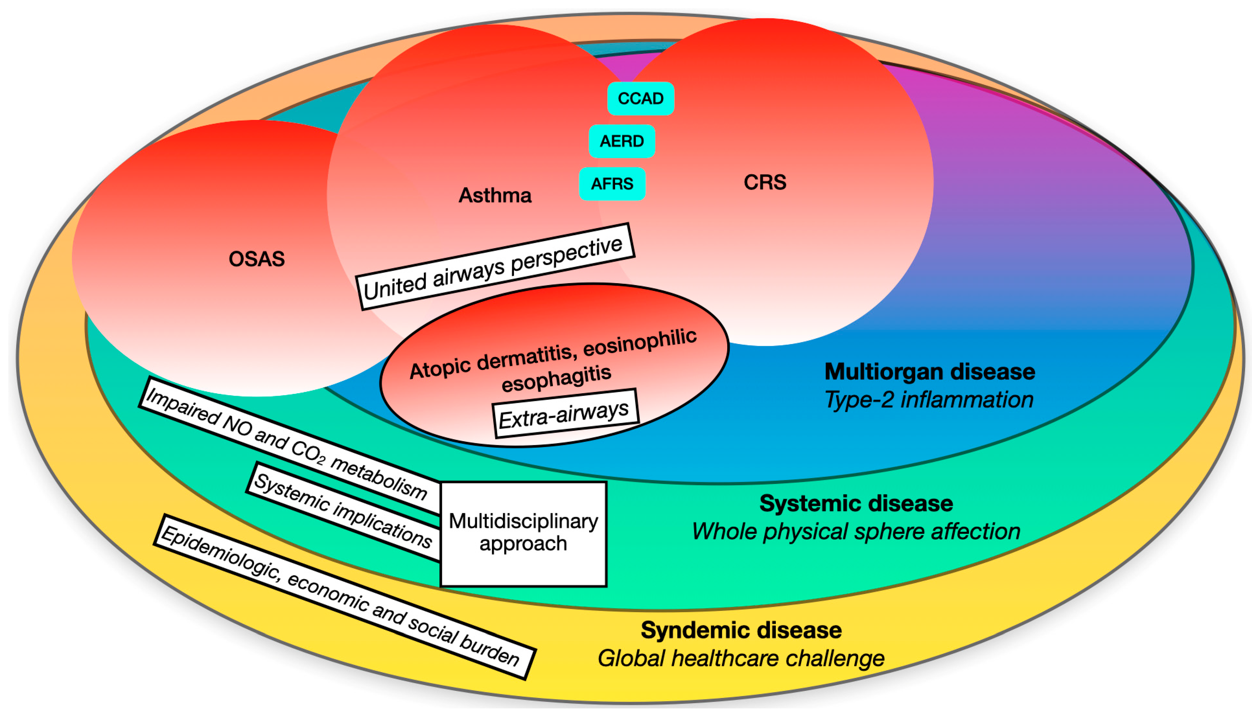
Abstract
1. Introduction
2. A Multiorgan Condition
2.1. Type-2 Inflammation: Pathophysiology and Nasal Implications
2.2. The Lower Airways: Bronchial Asthma
2.3. Other than the Airways: Atopic Dermatitis
3. A Systemic Condition
3.1. Further Implications: The Link with Sleep-Related Respiratory Disorders
3.2. Nitric Oxide: A Systemic Mediator
4. A Syndemic Condition
4.1. Burden
4.1.1. Epidemiological
4.1.2. Economic
4.1.3. Social
5. Conclusions
Author Contributions
Funding
Institutional Review Board Statement
Informed Consent Statement
Data Availability Statement
Conflicts of Interest
References
- Fokkens, W.J.; Lund, V.J.; Hopkins, C.; Hellings, P.W.; Kern, R.; Reitsma, S.; Toppila-Salmi, S.; Bernal-Sprekelsen, M.; Mullol, J.; Alobid, I.; et al. European position paper on rhinosinusitis and nasal polyps 2020. Rhinology 2020, 58 (Suppl. S29), 1–464. [Google Scholar] [CrossRef] [PubMed]
- Pelaia, C.; Pelaia, G.; Maglio, A.; Tinello, C.; Gallelli, L.; Lombardo, N.; Terracciano, R.; Vatrella, A. Pathobiology of Type 2 Inflammation in Asthma and Nasal Polyposis. J. Clin. Med. 2023, 12, 3371. [Google Scholar] [CrossRef] [PubMed]
- Dennis, S.K.; Lam, K.; Luong, A. A Review of Classification Schemes for Chronic Rhinosinusitis with Nasal Polyposis Endotypes. Laryngoscope Investig. Otolaryngol. 2016, 1, 130–134. [Google Scholar] [CrossRef] [PubMed]
- Kaur, R.; Chupp, G. Phenotypes and endotypes of adult asthma: Moving toward precision medicine. J. Allergy Clin. Immunol. 2019, 144, 1–12. [Google Scholar] [CrossRef] [PubMed]
- World Health Organization (WHO). Constitution of the World Health Organization. Basic Documents; World Health Organization: Geneva, Switzerland, 1946. [Google Scholar]
- The World Health Report. 2008–Primary Health Care (Now More Than Ever). Available online: https://www.who.int/director-general/speeches/detail/primary-health-care---now-more-than-ever (accessed on 16 April 2020).
- Alemayehu, D.; Cappelleri, J.C. Conceptual and Analytical Considerations toward the Use of Patient-Reported Outcomes in Personalized Medicine. Am. Health Drug Benefits 2012, 5, 310–317. [Google Scholar] [PubMed]
- Jebb, A.T.; Ng, V.; Tay, L. A Review of Key Likert Scale Development Advances: 1995–2019. Front. Psychol. 2021, 12, 637547. [Google Scholar] [CrossRef] [PubMed]
- Klimek, L.; Bergmann, K.C.; Biedermann, T.; Bousquet, J.; Hellings, P.; Jung, K.; Merk, H.; Olze, H.; Schlenter, W.; Stock, P.; et al. Visual analogue scales (VAS): Measuring instruments for the documentation of symptoms and therapy monitoring in cases of allergic rhinitis in everyday health care: Position Paper of the German Society of Allergology (AeDA) and the German Society of Allergy and Clinical Immunology (DGAKI), ENT Section, in collaboration with the working group on Clinical Immunology, Allergology and Environmental Medicine of the German Society of Otorhinolaryngology, Head and Neck Surgery (DGHNOKHC). Allergo J. Int. 2017, 26, 16–24. [Google Scholar] [CrossRef]
- Mercieca-Bebber, R.; King, M.T.; Calvert, M.J.; Stockler, M.R.; Friedlander, M. The importance of patient-reported out-comes in clinical trials and strategies for future optimization. Patient Relat. Outcome Meas. 2018, 9, 353–367. [Google Scholar] [CrossRef]
- Bachert, C.; Bhattacharyya, N.; Desrosiers, M.; Khan, A.H. Burden of Disease in Chronic Rhinosinusitis with Nasal Polyps. J. Asthma Allergy 2021, 14, 127–134. [Google Scholar] [CrossRef]
- Blanco-Aparicio, M.; Domínguez-Ortega, J.; Cisneros, C.; Colás, C.; Casas, F.; Del Cuvillo, A.; Alobid, I.; Quirce, S.; Mullol, J. Consensus on the management of united airways disease with type 2 inflammation: A multidisciplinary Delphi study. Allergy Asthma Clin. Immunol. 2023, 19, 34. [Google Scholar] [CrossRef]
- Bousquet, J.; Van Cauwenberge, P.; Khaltaev, N.; Aria Workshop Group; World Health Organization. Allergic rhinitis and its impact on asthma. J. Allergy Clin. Immunol. 2001, 108 (Suppl. S5), S147–S334. [Google Scholar] [CrossRef]
- Mullol, J.; Maldonado, M.; Castillo, J.A.; Miguel-Blanco, C.; Dávila, I.; Domínguez-Ortega, J.; Blanco-Aparicio, M. Management of United Airway Disease Focused on Patients with Asthma and Chronic Rhinosinusitis With Nasal Polyps: A Systematic Review. J. Allergy Clin. Immunol. Pr. 2022, 10, 2438–2447.e9. [Google Scholar] [CrossRef] [PubMed]
- Cao, P.P.; Wang, Z.C.; Schleimer, R.P.; Liu, Z. Pathophysiologic mechanisms of chronic rhinosinusitis and their roles in emerging disease endotypes. Ann. Allergy Asthma Immunol. 2019, 122, 33–40. [Google Scholar] [CrossRef]
- Dunican, E.M.; Fahy, J.V. The Role of Type 2 Inflammation in the Pathogenesis of Asthma Exacerbations. Ann. Am. Thorac. Soc. 2015, 12 (Suppl. S2), S144–S149. [Google Scholar] [CrossRef]
- Kato, A.; Schleimer, R.P.; Bleier, B.S. Mechanisms and pathogenesis of chronic rhinosinusitis. J. Allergy Clin. Immunol. 2022, 149, 1491–1503. [Google Scholar] [CrossRef] [PubMed]
- Chen, W.; Shu, Q.; Fan, J. Neural Regulation of Interactions Between Group 2 Innate Lymphoid Cells and Pulmonary Immune Cells. Front. Immunol. 2020, 11, 576929. [Google Scholar] [CrossRef] [PubMed]
- Vacca, F.; Le Gros, G. Tissue-specific immunity in helminth infections. Mucosal Immunol. 2022, 15, 1212–1223. [Google Scholar] [CrossRef]
- Strachan, D.P. Hay fever, hygiene, and household size. BMJ 1989, 299, 1259–1260. [Google Scholar] [CrossRef]
- Rankin, L.C.; Artis, D. Beyond Host Defense: Emerging Functions of the Immune System in Regulating Complex Tissue Physiology. Cell 2018, 173, 554–567. [Google Scholar] [CrossRef]
- Garn, H.; Potaczek, D.P.; Pfefferle, P.I. The Hygiene Hypothesis and New Perspectives-Current Challenges Meeting an Old Postulate. Front. Immunol. 2021, 12, 637087. [Google Scholar] [CrossRef]
- Pfefferle, P.I.; Keber, C.U.; Cohen, R.M.; Garn, H. The Hygiene Hypothesis—Learning from but Not Living in the Past. Front. Immunol. 2021, 12, 635935. [Google Scholar] [CrossRef] [PubMed]
- Scudellari, M. News Feature: Cleaning up the hygiene hypothesis. Proc. Natl. Acad. Sci. USA 2017, 114, 1433–1436. [Google Scholar] [CrossRef] [PubMed]
- von Mutius, E. The “Hygiene Hypothesi” and the Lessons Learnt from Farm Studies. Front. Immunol. 2021, 12, 635522. [Google Scholar] [CrossRef]
- Adeli, M.; El-Shareif, T.; Hendaus, M.A. Asthma exacerbation related to viral infections: An up to date summary. J. Fam. Med. Prim. Care 2019, 8, 2753–2759. [Google Scholar] [CrossRef] [PubMed]
- Edwards, M.R.; Strong, K.; Cameron, A.; Walton, R.P.; Jackson, D.J.; Johnston, S.L. Viral infections in allergy and immunology: How allergic inflammation influences viral infections and illness. J. Allergy Clin. Immunol. 2017, 140, 909–920. [Google Scholar] [CrossRef] [PubMed]
- Conteville, L.C.; Oliveira-Ferreira, J.; Vicente, A.C.P. Gut microbiome biomarkers and functional diversity within anmazoniann semi-nomadic hunter-gatherer group. Front. Microbiol. 2019, 10, 1743. [Google Scholar] [CrossRef] [PubMed]
- McLoughlin, R.; Berthon, B.S.; Rogers, G.B.; Baines, K.J.; Leong, L.E.X.; Gibson, P.G.; Williams, E.J.; Wood, L.G. Soluble fibre supplementation with and without a probiotic in adults with asthma: A 7-day randomised, double blind, three way cross-over trial. EBioMedicine 2019, 46, 473–485. [Google Scholar] [CrossRef]
- Enaud, R.; Prevel, R.; Ciarlo, E.; Beaufils, F.; Wieërs, G.; Guery, B.; Delhaes, L. The Gut-Lung Axis in Health and Respiratory Diseases: A Place for Inter-Organ and Inter-Kingdom Crosstalks. Front. Cell Infect. Microbiol. 2020, 10, 9. [Google Scholar] [CrossRef]
- Laue, H.E.; Coker, M.O.; Madan, J.C. The Developing Microbiome From Birth to 3 Years: The Gut-Brain Axis and Neurodevelopmental Outcomes. Front. Pediatr. 2022, 10, 815885. [Google Scholar] [CrossRef]
- Miettinen, M.E.; Honkanen, J.; Niinistö, S.; Vaarala, O.; Virtanen, S.M.; Knip, M.; DIABIMMUNE Study Group. Breastfeeding and circulating immunological markers during the first 3 years of life: The DIABIMMUNE study. Diabetologia 2022, 65, 329–335. [Google Scholar] [CrossRef]
- Li, J.; Jiao, J.; Gao, Y.; Zhang, Y.; Zhang, L. Association between methylation in nasal epithelial TSLP gene and chronic rhinosinusitis with nasal polyps. Allergy Asthma Clin. Immunol. 2019, 15, 71. [Google Scholar] [CrossRef] [PubMed]
- Nagarkar, D.R.; Poposki, J.A.; Tan, B.K.; Comeau, M.R.; Peters, A.T.; Hulse, K.E.; Suh, L.A.; Norton, J.; Harris, K.E.; Grammer, L.C.; et al. Thymic stromal lymphopoietin activity is increased in nasal polyps of patients with chronic rhinosinusitis. J. Allergy Clin. Immunol. 2013, 132, 593–600.e12. [Google Scholar] [CrossRef] [PubMed]
- Akdis, C.A.; Arkwright, P.D.; Brüggen, M.C.; Busse, W.; Gadina, M.; Guttman-Yassky, E.; Kabashima, K.; Mitamura, Y.; Vian, L.; Wu, J.; et al. Type 2 immunity in the skin and lungs. Allergy 2020, 75, 1582–1605. [Google Scholar] [CrossRef] [PubMed]
- Boita, M.; Bucca, C.; Riva, G.; Heffler, E.; Rolla, G. Release of Type 2 Cytokines by Epithelial Cells of Nasal Polyps. J. Immunol. Res. 2016, 2016, 2643297. [Google Scholar] [CrossRef]
- Heffler, E.; Malvezzi, L.; Boita, M.; Brussino, L.; De Virgilio, A.; Ferrando, M.; Puggioni, F.; Racca, F.; Stomeo, N.; Spriano, G.; et al. Immunological mechanisms underlying chronic rhinosinusitis with nasal polyps. Expert Rev. Clin. Immunol. 2018, 14, 731–737. [Google Scholar] [CrossRef] [PubMed]
- Konkel, J.E.; Zhang, D.; Zanvit, P.; Chia, C.; Zangarle-Murray, T.; Jin, W.; Wang, S.; Chen, W. Transforming Growth Factor-β Signaling in Regulatory T Cells Controls T Helper-17 Cells and Tissue-Specific Immune Responses. Immunity 2017, 46, 660–674. [Google Scholar] [CrossRef] [PubMed]
- Ojiaku, C.A.; Yoo, E.J.; Panettieri, R.A., Jr. Transforming Growth Factor β1 Function in Airway Remodeling and Hyperresponsiveness. The Missing Link? Am. J. Respir. Cell Mol. Biol. 2017, 56, 432–442. [Google Scholar] [CrossRef]
- Watelet, J.B.; Claeys, C.; Perez-Novo, C.; Gevaert, P.; Van Cauwenberge, P.; Bachert, C. Transforming growth factor beta1 in nasal remodeling: Differences between chronic rhinosinusitis and nasal polyposis. Am. J. Rhinol. 2004, 18, 267–272. [Google Scholar] [CrossRef]
- Shieh, J.M.; Tsai, Y.J.; Chi, J.C.; Wu, W.B. TGFβ mediates collagen production in human CRSsNP nasal mucosa-derived fibroblasts through Smad2/3-dependent pathway and CTGF induction and secretion. J. Cell Physiol. 2019, 234, 10489–10499. [Google Scholar] [CrossRef]
- Panek, M.; Stawiski, K.; Kaszkowiak, M.; Kuna, P. Cytokine TGFβ Gene Polymorphism in Asthma: TGF-Related SNP Analysis Enhances the Prediction of Disease Diagnosis (A Case-Control Study with Multivariable Data-Mining Model Development). Front. Immunol. 2022, 13, 746360. [Google Scholar] [CrossRef]
- Giombi, F.; Carrón-Herrero, A.; Pirola, F.; Paoletti, G.; Nappi, E.; Russo, E.; De Virgilio, A.; Mercante, G.; Canonica, G.W.; Spriano, G.; et al. Prevalence of familial link in patients affected by chronic rhinosinusitis with nasal polyposis. Int. Forum Allergy Rhinol. 2022, 12, 1562–1565. [Google Scholar] [CrossRef] [PubMed]
- Liang, Y.; Xie, R.; Xiong, X.; Hu, Z.; Mao, X.; Wang, X.; Zhang, J.; Sun, P.; Yue, Z.; Wang, W.; et al. Alterations of nasal microbiome in eosinophilic chronic rhinosinusitis. J. Allergy Clin. Immunol. 2023, 151, 1286–1295.e2. [Google Scholar] [CrossRef] [PubMed]
- Gangwar, R.S.; Bevan, G.H.; Palanivel, R.; Das, L.; Rajagopalan, S. Oxidative stress pathways of air pollution mediated toxicity: Recent insights. Redox Biol. 2020, 34, 101545. [Google Scholar] [CrossRef] [PubMed]
- Mumby, S.; Chung, K.F.; Adcock, I.M. Transcriptional effects of ozone and impact on airway inflammation. Front. Immunol. 2019, 10, 1610. [Google Scholar] [CrossRef] [PubMed]
- Leland, E.M.; Vohra, V.; Seal, S.M.; Zhang, Z.; Ramanathan, M., Jr. Environmental air pollution and chronic rhinosinusitis: A systematic review. Laryngoscope Investig. Otolaryngol. 2022, 7, 349–360. [Google Scholar] [CrossRef] [PubMed]
- Tiotiu, A.I.; Novakova, P.; Nedeva, D.; Chong-Neto, H.J.; Novakova, S.; Steiropoulos, P.; Kowal, K. Impact of Air Pollution on Asthma Outcomes. Int. J. Environ. Res. Public Health 2020, 17, 6212. [Google Scholar] [CrossRef]
- Maspero, J.; Adir, Y.; Al-Ahmad, M.; Celis-Preciado, C.A.; Colodenco, F.D.; Giavina-Bianchi, P.; Lababidi, H.; Ledanois, O.; Mahoub, B.; Perng, D.W.; et al. Type 2 inflammation in asthma and other airway diseases. ERJ Open Res. 2022, 8, 00576–02021. [Google Scholar] [CrossRef]
- Crisafulli, E.; Sartori, G.; Patruno, V.; Fantin, A. Bronchodilator reversibility in patients with asthma and persistent airflow limitation. Lancet Respir. Med. 2022, 10, e94–e95. [Google Scholar] [CrossRef]
- Kuruvilla, M.E.; Lee, F.E.; Lee, G.B. Understanding Asthma Phenotypes, Endotypes, and Mechanisms of Disease. Clin. Rev. Allergy Immunol. 2019, 56, 219–233. [Google Scholar] [CrossRef]
- Fahy, J.V. Type 2 inflammation in asthma–present in most, absent in many. Nat. Rev. Immunol. 2015, 15, 57–65. [Google Scholar] [CrossRef]
- Chung, K.F.; Wenzel, S.E.; Brozek, J.L.; Bush, A.; Castro, M.; Sterk, P.J.; Adcock, I.M.; Bateman, E.D.; Bel, E.H.; Bleecker, E.R.; et al. International ERS/ATS guidelines on definition, evaluation and treatment of severe asthma. Eur. Respir. J. 2014, 43, 343–373. [Google Scholar] [CrossRef] [PubMed]
- Frøssing, L.; Klein, D.K.; Hvidtfeldt, M.; Obling, N.; Telg, G.; Erjefält, J.S.; Bodtger, U.; Porsbjerg, C. Distribution of type 2 biomarkers and association with severity, clinical characteristics and comorbidities in the BREATHE real-life asthma population. ERJ Open Res. 2023, 9, 00483–02022. [Google Scholar] [CrossRef] [PubMed]
- Lambrecht, B.N.; Hammad, H.; Fahy, J.V. The Cytokines of Asthma. Immunity 2019, 50, 975–991. [Google Scholar] [CrossRef] [PubMed]
- Marone, G.; Granata, F.; Pucino, V.; Pecoraro, A.; Heffler, E.; Loffredo, S.; Scadding, G.W.; Varricchi, G. The Intriguing Role of Interleukin 13 in the Pathophysiology of Asthma. Front. Pharmacol. 2019, 10, 1387. [Google Scholar] [CrossRef] [PubMed]
- Pelaia, C.; Heffler, E.; Crimi, C.; Maglio, A.; Vatrella, A.; Pelaia, G.; Canonica, G.W. Interleukins 4 and 13 in Asthma: Key Pathophysiologic Cytokines and Druggable Molecular Targets. Front. Pharmacol. 2022, 13, 851940. [Google Scholar] [CrossRef] [PubMed]
- McBrien, C.N.; Menzies-Gow, A. The Biology of Eosinophils and Their Role in Asthma. Front. Med. 2017, 4, 93. [Google Scholar] [CrossRef] [PubMed]
- Schatz, M.; Zeiger, R.S.; Yang, S.J.; Chen, W.; Crawford, W.; Sajjan, S.; Allen-Ramey, F. Change in asthma control over time: Predictors and outcomes. J. Allergy Clin. Immunol. Pr. 2014, 2, 59–64. [Google Scholar] [CrossRef] [PubMed]
- Wenzel, S.E.; Balzar, S.; Ampleford, E.; Hawkins, G.A.; Busse, W.W.; Calhoun, W.J.; Castro, M.; Chung, K.F.; Erzurum, S.; Gaston, B.; et al. IL4R alpha mutations are associated with asthma exacerbations and mast cell/IgE expression. Am. J. Respir. Crit. Care Med. 2007, 175, 570–576. [Google Scholar] [CrossRef]
- Canonica, G.W.; Malvezzi, L.; Blasi, F.; Paggiaro, P.; Mantero, M.; Senna, G.; Heffler, E. Severe Asthma Network Italy (SANI). Chronic rhinosinusitis with nasal polyps impact in severe asthma patients: Evidences from the Severe Asthma Network Italy (SANI) registry. Respir. Med. 2022, 166, 105947. [Google Scholar] [CrossRef]
- Castro, M.; Corren, J.; Pavord, I.D.; Maspero, J.; Wenzel, S.; Rabe, K.F.; Busse, W.W.; Ford, L.; Sher, L.; FitzGerald, J.M.; et al. Dupilumab Efficacy and Safety in Moderate-to-Severe Uncontrolled Asthma. N. Engl. J. Med. 2018, 378, 2486–2496. [Google Scholar] [CrossRef]
- Doroudchi, A.; Pathria, M.; Modena, B.D. Asthma biologics: Comparing trial designs, patient cohorts and study results. Ann. Allergy Asthma Immunol. 2020, 124, 44–56. [Google Scholar] [CrossRef] [PubMed]
- Hanania, N.A.; Alpan, O.; Hamilos, D.L.; Condemi, J.J.; Reyes-Rivera, I.; Zhu, J.; Rosen, K.E.; Eisner, M.D.; Wong, D.A.; Busse, W. Omalizumab in severe allergic asthma inadequately controlled with standard therapy: A randomized trial. Ann. Intern. Med. 2011, 154, 573–582. [Google Scholar] [CrossRef] [PubMed]
- Juniper, E.F.; Buist, A.S.; Cox, F.M.; Ferrie, P.J.; King, D.R. Validation of a standardized version of the Asthma Quality of Life Questionnaire. Chest 1999, 115, 1265–1270. [Google Scholar] [CrossRef] [PubMed]
- Ortega, H.G.; Liu, M.C.; Pavord, I.D.; Brusselle, G.G.; FitzGerald, J.M.; Chetta, A.; Humbert, M.; Katz, L.E.; Keene, O.N.; Yancey, S.W.; et al. Mepolizumab treatment in patients with severe eosinophilic asthma. N. Engl. J. Med. 2014, 371, 1198–1207. [Google Scholar] [CrossRef] [PubMed]
- Wollenberg, A.; Werfel, T.; Ring, J.; Ott, H.; Gieler, U.; Weidinger, S. Atopic Dermatitis in Children and Adults—Diagnosis and Treatment. Dtsch. Arztebl. Int. 2023, 120, 224–234. [Google Scholar] [CrossRef] [PubMed]
- Plant, A.; Ardern-Jones, M.R. Advances in atopic dermatitis. Clin. Med. 2021, 21, 177–181. [Google Scholar] [CrossRef] [PubMed]
- Kim, J.; Kim, B.E.; Leung, D.Y.M. Pathophysiology of atopic dermatitis: Clinical implications. Allergy Asthma Proc. 2019, 40, 84–92. [Google Scholar] [CrossRef] [PubMed]
- Sugita, K.; Altunbulakli, C.; Morita, H.; Sugita, A.; Kubo, T.; Kimura, R.; Goto, H.; Yamamoto, O.; Rückert, B.; Akdis, M.; et al. Human type 2 innate lymphoid cells disrupt skin keratinocyte tight junction barrier by IL-13. Allergy 2019, 74, 2534–2537. [Google Scholar] [CrossRef]
- Suarez-Farinas, M.; Tintle, S.J.; Shemer, A.; Chiricozzi, A.; Nograles, K.; Cardinale, I.; Duan, S.; Bowcock, A.M.; Krueger, J.G.; Guttman-Yassky, E. Nonlesional atopic dermatitis skin is characterized by broad terminal differentiation defects and variable immune abnormalities. J. Allergy Clin. Immunol. 2011, 127, 954–964.e4. [Google Scholar] [CrossRef]
- Napolitano, M.; di Vico, F.; Ruggiero, A.; Fabbrocini, G.; Patruno, C. The hidden sentinel of the skin: An overview on the role of interleukin-13 in atopic dermatitis. Front. Med. 2023, 10, 1165098. [Google Scholar] [CrossRef]
- García-Reyes, M.M.; Zumaya-Pérez, L.C.; Pastelin-Palacios, R.; Moreno-Eutimio, M.A. Serum thymic stromal lym-phopoietin (TSLP) levels in atopic dermatitis patients: A systematic review and meta-analysis. Clin. Exp. Med. 2023, 23, 4129–4139. [Google Scholar] [CrossRef] [PubMed]
- Silverberg, J.I.; Guttman-Yassky, E.; Thaçi, D.; Irvine, A.D.; Stein Gold, L.; Blauvelt, A.; Simpson, E.L.; Chu, C.Y.; Liu, Z.; Gontijo Lima, R.; et al. Two Phase 3 Trials of Lebrikizumab for Moderate-to-Severe Atopic Dermatitis. N. Engl. J. Med. 2023, 388, 1080–1091. [Google Scholar] [CrossRef]
- Kuchibhotla, V.N.S.; Heijink, I.H. Join or Leave the Club: Jagged1 and Notch2 Dictate the Fate of Airway Epithelial Cells. Am. J. Respir. Cell Mol. Biol. 2020, 63, 4–6. [Google Scholar] [CrossRef] [PubMed]
- Carrer, M.; Crosby, J.R.; Sun, G.; Zhao, C.; Damle, S.S.; Kuntz, S.G.; Monia, B.P.; Hart, C.E.; Grossman, T.R. Antisense Oligonucleotides Targeting Jagged 1 Reduce House Dust Mite-induced Goblet Cell Metaplasia in the Adult Murine Lung. Am. J. Respir. Cell Mol. Biol. 2020, 63, 46–56. [Google Scholar] [CrossRef] [PubMed]
- Guo, C.J.; Mack, M.R.; Oetjen, L.K.; Trier, A.M.; Council, M.L.; Pavel, A.B.; Guttman-Yassky, E.; Kim, B.S.; Liu, Q. Kallikrein 7 Promotes Atopic Dermatitis-Associated Itch Independently of Skin Inflammation. J. Investig. Dermatol. 2020, 140, 1244–1252.e4. [Google Scholar] [CrossRef]
- Giunta, G.; Pirola, F.; Giombi, F.; Muci, G.; Pace, G.M.; Heffler, E.; Paoletti, G.; Puggioni, F.; Cerasuolo, M.; Ferreli, F.; et al. Care for Patients with Type-2 Chronic Rhinosinusitis. J. Pers. Med. 2023, 13, 618. [Google Scholar] [CrossRef] [PubMed]
- Swarbrick, M. A wellness approach. Psychiatr. Rehabil. J. 2006, 29, 311–314. [Google Scholar] [CrossRef]
- Gottlieb, D.J.; Punjabi, N.M. Diagnosis and Management of Obstructive Sleep Apnea: A Review. JAMA 2020, 323, 1389–1400. [Google Scholar] [CrossRef]
- Su, V.Y.-F.; Chou, K.T.; Tseng, C.H.; Kuo, C.Y.; Su, K.C.; Perng, D.W.; Chen, Y.M.; Chang, S.C. Mouth opening/breathing is common in sleep apnea and linked to more nocturnal water loss. Biomed. J. 2023, 46, 100536. [Google Scholar] [CrossRef]
- Värendh, M.; Andersson, M.; Bjørnsdottir, E.; Hrubos-Strøm, H.; Johannisson, A.; Arnardottir, E.S.; Gislason, T.; Juliusson, S. Nocturnal nasal obstruction is frequent and reduces sleep quality in patients with obstructive sleep apnea. J. Sleep Res. 2018, 27, e12631. [Google Scholar] [CrossRef]
- Friedman, M.; Maley, A.; Kelley, K.; Leesman, C.; Patel, A.; Pulver, T.; Joseph, N.; Catli, T. Impact of nasal obstruction on obstructive sleep apnea. Otolaryngol. Neck Surg. 2011, 144, 1000–1004. [Google Scholar] [CrossRef] [PubMed]
- Morris, L.G.; Burschtin, O.; Lebowitz, R.A.; Jacobs, J.B.; Lee, K.C. Nasal obstruction and sleep-disordered breathing: A study using acoustic rhinometry. Am. J. Rhinol. 2005, 19, 33–39. [Google Scholar] [CrossRef] [PubMed]
- Krouse, H.J.; Davis, J.E.; Krouse, J.H. Immune mediators in allergic rhinitis and sleep. Otolaryngol. Neck Surg. 2002, 126, 607–613. [Google Scholar] [CrossRef] [PubMed]
- Minoguchi, K.; Tazaki, T.; Yokoe, T.; Minoguchi, H.; Watanabe, Y.; Yamamoto, M.; Adachi, M. Elevated production of tumor necrosis factor-alpha by monocytes in patients with obstructive sleep apnea syndrome. Chest 2004, 126, 1473–1479. [Google Scholar] [CrossRef] [PubMed]
- Petrof, B.J.; Hendricks, J.C.; Pack, A.I. Does UA muscle injury trigger a vicious cycle in obstructive sleep apnea? A hypothesis. Sleep 1996, 19, 465–471. [Google Scholar] [CrossRef]
- Shah, F.; Stål, P. Myopathy of the UA in snoring and obstructive sleep apnea. Laryngoscope Investig. Otolaryngol. 2022, 7, 636–645. [Google Scholar] [CrossRef]
- Campos, A.I.; García-Marín, L.M.; Byrne, E.M.; Martin, N.G.; Cuéllar-Partida, G.; Rentería, M.E. Insights into the aetiology of snoring from observational and genetic investigations in the UK Biobank. Nat. Commun. 2020, 11, 817. [Google Scholar] [CrossRef]
- Lechner, M.; Breeze, C.E.; Ohayon, M.M.; Kotecha, B. Snoring and breathing pauses during sleep: Interview survey of a United Kingdom population sample reveals a significant increase in the rates of sleep apnoea and obesity over the last 20 years–Data from the UK sleep survey. Sleep Med. 2019, 54, 250–256. [Google Scholar] [CrossRef]
- Heinzer, R.; Vat, S.; Marques-Vidal, P.; Marti-Soler, H.; Andries, D.; Tobback, N.; Mooser, V.; Preisig, M.; Malhotra, A.; Waeber, G.; et al. Prevalence of sleep-disordered breathing in the general population: The HypnoLaus study. Lancet Respir. Med. 2015, 3, 310–318. [Google Scholar] [CrossRef]
- Sunderram, J.; Weintraub, M.; Black, K.; Alimokhtari, S.; Twumasi, A.; Sanders, H.; Udasin, I.; Harrison, D.; Chitkara, N.; de la Hoz, R.E.; et al. Chronic Rhinosinusitis Is an Independent Risk Factor for OSA in World Trade Center Responders. Chest 2019, 155, 375–383. [Google Scholar] [CrossRef]
- Serrano-Pariente, J.; Plaza, V.; Soriano, J.B.; Mayos, M.; López-Viña, A.; Picado, C.; Vigil, L.; CPASMA Trial Group. Asthma outcomes improve with continuous positive airway pressure for obstructive sleep apnea. Allergy 2017, 72, 802–812. [Google Scholar] [CrossRef] [PubMed]
- Mashaqi, S.; Badr, M.S. The Impact of Obstructive Sleep Apnea and Positive Airway Pressure Therapy on Metabolic Peptides Regulating Appetite, Food Intake, Energy Homeostasis, and Systemic Inflammation: A Literature Review. J. Clin. Sleep Med. 2019, 15, 1037–1050. [Google Scholar] [CrossRef] [PubMed]
- Abbasi, A.; Gupta, S.S.; Sabharwal, N.; Meghrajani, V.; Sharma, S.; Kamholz, S.; Kupfer, Y. A comprehensive review of obstructive sleep apnea. Sleep Sci. 2021, 14, 142–154. [Google Scholar] [PubMed]
- Sircu, V.; Colesnic, S.I.; Covantsev, S.; Corlateanu, O.; Sukhotko, A.; Popovici, C.; Corlateanu, A. The Burden of Comorbidities in Obstructive Sleep Apnea and the Pathophysiologic Mechanisms and Effects of CPAP. Clocks Sleep 2023, 5, 333–349. [Google Scholar] [CrossRef] [PubMed]
- Al Wadee, Z.; Ooi, S.L.; Pak, S.C. Serum Magnesium Levels in Patients with Obstructive Sleep Apnoea: A Systematic Review and Meta-Analysis. Biomedicines 2022, 10, 2273. [Google Scholar] [CrossRef]
- Andrabi, S.M.; Sharma, N.S.; Karan, A.; Shahriar, S.M.S.; Cordon, B.; Ma, B.; Xie, J. Nitric Oxide: Physiological Functions, Delivery, and Biomedical Applications. Adv. Sci. 2023, 10, e2303259. [Google Scholar] [CrossRef] [PubMed]
- Heffler, E.; Carpagnano, G.E.; Favero, E.; Guida, G.; Maniscalco, M.; Motta, A.; Paoletti, G.; Rolla, G.; Baraldi, E.; Pezzella, V.; et al. Fractional Exhaled Nitric Oxide (FENO) in the management of asthma: A position paper of the Italian Respiratory Society (SIP/IRS) and Italian Society of Allergy, Asthma and Clinical Immunology (SIAAIC). Multidiscip. Respir. Med. 2020, 15, 36. [Google Scholar] [CrossRef]
- Dweik, R.A.; Boggs, P.B.; Erzurum, S.C.; Irvin, C.G.; Leigh, M.W.; Lundberg, J.O.; Olin, A.C.; Plummer, A.L.; Taylor, D.R.; American Thoracic Society Committee on Interpretation of Exhaled Nitric Oxide Levels (FENO) for Clinical Applications. An official ATS clinical practice guideline: Interpretation of exhaled nitric oxide levels (FENO) for clinical applications. Am. J. Respir. Crit. Care Med. 2011, 184, 602–615. [Google Scholar] [CrossRef]
- Kambara, R.; Minami, T.; Akazawa, H.; Tsuji, F.; Sasaki, T.; Inohara, H.; Horii, A. Lower Airway Inflammation in Eosinophilic Chronic Rhinosinusitis as Determined by Exhaled Nitric Oxide. Int. Arch. Allergy Immunol. 2017, 173, 225–232. [Google Scholar] [CrossRef]
- Baraldi, E.; Azzolin, N.M.; Carrà, S.; Dario, C.; Marchesini, L.; Zacchello, F. Effect of topical steroids on nasal nitric oxide production in children with perennial allergic rhinitis: A pilot study. Respir. Med. 1998, 92, 558–561. [Google Scholar] [CrossRef]
- Ambrosino, P.; Molino, A.; Spedicato, G.A.; Parrella, P.; Formisano, R.; Motta, A.; Di Minno, M.N.D.; Maniscalco, M. Nasal Nitric Oxide in Chronic Rhinosinusitis with or without Nasal Polyps: A Systematic Review with Meta-Analysis. J. Clin. Med. 2020, 9, 200. [Google Scholar] [CrossRef] [PubMed]
- Fu, C.H.; Huang, C.C.; Chen, Y.W.; Chang, P.H.; Lee, T.J. Nasal nitric oxide in relation to quality-of-life improvements after endoscopic sinus surgery. Am. J. Rhinol. Allergy 2015, 29, e187–e191. [Google Scholar] [CrossRef] [PubMed]
- Paoletti, G.; Casini, M.; Malvezzi, L.; Pirola, F.; Russo, E.; Nappi, E.; Muci, G.Q.; Montagna, C.; Messina, M.R.; Ferri, S.; et al. Very rapid improvement of extended nitric oxide parameters, associated with clinical and functional betterment, in patients with chronic rhinosinusitis with nasal polyps (CRSwNP) treated with Dupilumab. J. Investig. Allergol. Clin. Immunol. 2022, 33, 457–463. [Google Scholar] [CrossRef] [PubMed]
- Singh, J.; Lee, Y.; Kellum, J.A. A new perspective on NO pathway in sepsis and ADMA lowering as a potential therapeutic approach. Crit. Care. 2022, 26, 246. [Google Scholar] [CrossRef] [PubMed]
- Al-Digheari, A.; Mahboub, B.; Tarraf, H.; Yucel, T.; Annesi-Maesano, I.; Doble, A.; Lahlou, A.; Tariq, L.; Aziz, F.; El Hasnaoui, A. The clinical burden of allergic rhinitis in five Middle Eastern countries: Results of the SNAPSHOT program. Allergy Asthma Clin. Immunol. 2018, 14, 63. [Google Scholar] [CrossRef] [PubMed]
- Savouré, M.; Bousquet, J.; Jaakkola, J.J.K.; Jaakkola, M.S.; Jacquemin, B.; Nadif, R. Worldwide prevalence of rhinitis in adults: A review of definitions and temporal evolution. Clin. Transl. Allergy 2022, 12, e12130. [Google Scholar] [CrossRef] [PubMed]
- Licari, A.; Magri, P.; De Silvestri, A.; Giannetti, A.; Indolfi, C.; Mori, F.; Marseglia, G.L.; Peroni, D. Epidemiology of Allergic Rhinitis in Children: A Systematic Review and Meta-Analysis. J. Allergy Clin. Immunol. Pr. 2023, 11, 2547–2556. [Google Scholar] [CrossRef]
- Hastan, D.; Fokkens, W.J.; Bachert, C.; Newson, R.B.; Bislimovska, J.; Bockelbrink, A.; Bousquet, P.J.; Brozek, G.; Bruno, A.; Dahlén, S.E.; et al. Chronic rhinosinusitis in Europe–an underestimated disease. A GA2LEN study. Allergy 2011, 66, 1216–1223. [Google Scholar] [CrossRef]
- Shi, J.B.; Fu, Q.L.; Zhang, H.; Cheng, L.; Wang, Y.J.; Zhu, D.D.; Lv, W.; Liu, S.X.; Li, P.Z.; Ou, C.Q.; et al. Epidemiology of chronic rhinosinusitis: Results from a cross-sectional survey in seven Chinese cities. Allergy 2015, 70, 533–539. [Google Scholar] [CrossRef]
- Stankiewicz, J.A.; Chow, J.M. A diagnostic dilemma for chronic rhinosinusitis: Definition accuracy and validity. Am. J. Rhinol. 2002, 16, 199–202. [Google Scholar] [CrossRef]
- Dietz de Loos, D.; Lourijsen, E.S.; Wildeman, M.A.M.; Freling, N.J.M.; Wolvers, M.D.J.; Reitsma, S.; Fokkens, W.J. Prevalence of chronic rhinosinusitis in the general population based on sinus radiology and symptomatology. J. Allergy Clin. Immunol. 2019, 143, 1207–1214. [Google Scholar] [CrossRef]
- The International Study of Asthma and Allergies in Childhood (ISAAC) Steering Committee. Worldwide variation in prevalence of symptoms of asthma allergic rhinoconjunctivitis atopic eczema: ISAAC. Lancet 1998, 351, 1225–1232. [Google Scholar] [CrossRef]
- Mortimer, K.; Lesosky, M.; García-Marcos, L.; Asher, M.I.; Pearce, N.; Ellwood, E.; Bissell, K.; El Sony, A.; Ellwood, P.; Marks, G.B.; et al. Global Asthma Network Phase I Study Group. The burden of asthma, hay fever and eczema in adults in 17 countries: GAN Phase I study. Eur. Respir. J. 2022, 60, 2102865. [Google Scholar] [CrossRef] [PubMed]
- García-Marcos, L.; Asher, M.I.; Pearce, N.; Ellwood, E.; Bissell, K.; Chiang, C.Y.; El Sony, A.; Ellwood, P.; Marks, G.B.; Mortimer, K.; et al. Global Asthma Network Phase I Study Group. The burden of asthma, hay fever and eczema in children in 25 countries: GAN Phase I study. Eur. Respir. J. 2022, 60, 2102866. [Google Scholar] [CrossRef]
- Promsopa, C.; Kansara, S.; Citardi, M.J.; Fakhri, S.; Porter, P.; Luong, A. Prevalence of confirmed asthma varies in chronic rhinosinusitis subtypes. Int. Forum Allergy Rhinol. 2016, 6, 373–377. [Google Scholar] [CrossRef]
- Philpott, C.M.; Erskine, S.; Hopkins, C.; Kumar, N.; Anari, S.; Kara, N.; Sunkaraneni, S.; Ray, J.; Clark, A.; Wilson, A.; et al. Prevalence of asthma, aspirin sensitivity and allergy in chronic rhi-nosinusitis: Data from the UK National Chronic Rhinosinusitis Epidemiology Study. Respir. Res. 2018, 19, 129. [Google Scholar] [CrossRef] [PubMed]
- Son, D.S.; Cho, M.S.; Kim, D.K. Chronic Rhinosinusitis and the Increased Incidence of Atopic Dermatitis. Am. J. Rhinol. Allergy 2022, 36, 574–582. [Google Scholar] [CrossRef]
- Khan, A.H.; Gouia, I.; Kamat, S.; Johnson, R.; Small, M.; Siddall, J. Prevalence and Severity Distribution of Type 2 Inflammation-Related Comorbidities Among Patients with Asthma, Chronic Rhinosinusitis with Nasal Polyps, and Atopic Dermatitis. Lung 2023, 201, 57–63. [Google Scholar] [CrossRef]
- Rudmik, L. Economics of Chronic Rhinosinusitis. Curr. Allergy Asthma Rep. 2017, 17, 20. [Google Scholar] [CrossRef]
- Wahid, N.W.; Smith, R.; Clark, A.; Salam, M.; Philpott, C.M. The socioeconomic cost of chronic rhinosinusitis study. Rhinology 2020, 58, 112–125. [Google Scholar] [CrossRef]
- Borsoi, L.; Armeni, P.; Donin, G.; Costa, F.; Ferini-Strambi, L. The invisible costs of obstructive sleep apnea (OSA): Systematic review and cost-of-illness analysis. PLoS ONE 2022, 17, e0268677. [Google Scholar] [CrossRef]
- Velasquez, N.; Gardiner, L.; Cheng, T.Z.; Moore, J.A.; Boudreau, R.M.; Presto, A.A.; Lee, S.E. Relationship between socioeconomic status, exposure to airborne pollutants, and chronic rhinosinusitis disease severity. Int. Forum Allergy Rhinol. 2022, 12, 172–180. [Google Scholar] [CrossRef] [PubMed]
- Cairns, C.; Kang, K. National Hospital Ambulatory Medical Care Survey: 2019 Emergency Department Summary Tables. 2022. Available online: https://www.cdc.gov/nchs/data/nhamcs/web_tables/2019-nhamcs-ed-web-tables-508.pdf (accessed on 16 April 2020).
- Jansson, S.A.; Backman, H.; Andersson, M.; Telg, G.; Lindberg, A.; Stridsman, C.; Lundbäck, B.; Rönmark, E. Severe asthma is related to high societal costs and decreased health related quality of life. Respir. Med. 2020, 162, 105860. [Google Scholar] [CrossRef] [PubMed]
- Augustin, M.; Misery, L.; von Kobyletzki, L.; Armario-Hita, J.C.; Mealing, S.; Redding, M. Unveiling the true costs and societal impacts of moderate-to-severe atopic dermatitis in Europe. J. Eur. Acad. Dermatol. Venereol. 2022, 36 (Suppl. S7), 3–16. [Google Scholar] [CrossRef]
- Manjelievskaia, J.; Boytsov, N.; Brouillette, M.A.; Onyekwere, U.; Pierce, E.; Goldblum, O.; Bonafede, M. The direct and indirect costs of adult atopic dermatitis. J. Manag. Care Spec. Pharm. 2021, 27, 1416–1425. [Google Scholar] [CrossRef] [PubMed]
- Schmidt, C. Leroy Hood looks forward to P4 medicine: Predictive, personalized, preventive, and participatory. J. Natl. Cancer Inst. 2014, 106, dju416. [Google Scholar] [CrossRef] [PubMed][Green Version]
- Hopkins, C.; Gillett, S.; Slack, R.; Lund, V.J.; Browne, J.P. Psychometric validity of the 22-item Sinonasal Outcome Test. Clin. Otolaryngol. 2009, 34, 447–454. [Google Scholar] [CrossRef] [PubMed]
- Giombi, F.; Pace, G.M.; Nappi, E.; Giunta, G.; Muci, G.; Pirola, F.; Ferreli, F.; Heffler, E.; Paoletti, G.; Giannitto, C.; et al. Radiological Versus Clinical 1-Year Outcomes of Dupilumab in Refractory CRSwNP: A Real-Life Study. Laryngoscope, 2023; ahead of print. [Google Scholar] [CrossRef]
- DeConde, A.S.; Soler, Z.M. Chronic rhinosinusitis: Epidemiology and burden of disease. Am. J. Rhinol. Allergy 2016, 30, 134–139. [Google Scholar] [CrossRef]
- Worth, A.; Hammersley, V.; Knibb, R.; Flokstra-de-Blok, B.; DunnGalvin, A.; Walker, S.; Dubois, A.E.; Sheikh, A. Patient-reported outcome measures for asthma: A systematic review. NPJ Prim. Care Respir. Med. 2014, 24, 14020. [Google Scholar] [CrossRef]
- Uchmanowicz, B.; Panaszek, B.; Uchmanowicz, I.; Rosińczuk, J. Clinical factors affecting quality of life of patients with asthma. Patient Prefer. Adherence 2016, 10, 579–589. [Google Scholar] [CrossRef]
- Luyster, F.S.; Strollo, P.J., Jr.; Holguin, F.; Castro, M.; Dunican, E.M.; Fahy, J.; Gaston, B.; Israel, E.; Jarjour, N.N.; Mauger, D.T.; et al. Association Between Insomnia and Asthma Burden in the Severe Asthma Research Program (SARP) III. Chest 2016, 150, 1242–1250. [Google Scholar] [CrossRef] [PubMed]
- Leander, M.; Lampa, E.; Rask-Andersen, A.; Franklin, K.; Gislason, T.; Oudin, A.; Svanes, C.; Torén, K.; Janson, C. Impact of anxiety and depression on respiratory symptoms. Respir. Med. 2014, 108, 1594–1600. [Google Scholar] [CrossRef] [PubMed]
- Blome, C.; Radtke, M.A.; Eissing, L.; Augustin, M. Quality of Life in Patients with Atopic Dermatitis: Disease Burden, Measurement, and Treatment Benefit. Am. J. Clin. Dermatol. 2016, 17, 163–169. [Google Scholar] [CrossRef] [PubMed]
- Birdi, G.; Cooke, R.; Knibb, R.C. Impact of atopic dermatitis on quality of life in adults: A systematic review and meta-analysis. Int. J. Dermatol. 2020, 59, e75–e91. [Google Scholar] [CrossRef]

Disclaimer/Publisher’s Note: The statements, opinions and data contained in all publications are solely those of the individual author(s) and contributor(s) and not of MDPI and/or the editor(s). MDPI and/or the editor(s) disclaim responsibility for any injury to people or property resulting from any ideas, methods, instructions or products referred to in the content. |
© 2024 by the authors. Licensee MDPI, Basel, Switzerland. This article is an open access article distributed under the terms and conditions of the Creative Commons Attribution (CC BY) license (https://creativecommons.org/licenses/by/4.0/).
Share and Cite
Giombi, F.; Pace, G.M.; Pirola, F.; Cerasuolo, M.; Ferreli, F.; Mercante, G.; Spriano, G.; Canonica, G.W.; Heffler, E.; Ferri, S.; et al. Airways Type-2 Related Disorders: Multiorgan, Systemic or Syndemic Disease? Int. J. Mol. Sci. 2024, 25, 730. https://doi.org/10.3390/ijms25020730
Giombi F, Pace GM, Pirola F, Cerasuolo M, Ferreli F, Mercante G, Spriano G, Canonica GW, Heffler E, Ferri S, et al. Airways Type-2 Related Disorders: Multiorgan, Systemic or Syndemic Disease? International Journal of Molecular Sciences. 2024; 25(2):730. https://doi.org/10.3390/ijms25020730
Chicago/Turabian StyleGiombi, Francesco, Gian Marco Pace, Francesca Pirola, Michele Cerasuolo, Fabio Ferreli, Giuseppe Mercante, Giuseppe Spriano, Giorgio Walter Canonica, Enrico Heffler, Sebastian Ferri, and et al. 2024. "Airways Type-2 Related Disorders: Multiorgan, Systemic or Syndemic Disease?" International Journal of Molecular Sciences 25, no. 2: 730. https://doi.org/10.3390/ijms25020730
APA StyleGiombi, F., Pace, G. M., Pirola, F., Cerasuolo, M., Ferreli, F., Mercante, G., Spriano, G., Canonica, G. W., Heffler, E., Ferri, S., Puggioni, F., Paoletti, G., & Malvezzi, L. (2024). Airways Type-2 Related Disorders: Multiorgan, Systemic or Syndemic Disease? International Journal of Molecular Sciences, 25(2), 730. https://doi.org/10.3390/ijms25020730