Review

Caenorhabditis elegans as a Screening Model for Probiotics with Properties against Metabolic Syndrome

by
Ignacio Goyache
1,2,
Deyan Yavorov-Dayliev
1,2,3,
Fermín I. Milagro
1,2,4,5,* and
Paula Aranaz
1,2,4
1
Faculty of Pharmacy and Nutrition, Department of Nutrition, Food Sciences and Physiology, University of Navarra, 31008 Pamplona, Spain
2
Center for Nutrition Research, University of Navarra, 31008 Pamplona, Spain
3
Genbioma Aplicaciones SL, Polígono Industrial Noain-Esquiroz, Calle S, Nave 4, 31191 Esquíroz, Spain
4
Navarra Institute for Health Research (IdiSNA), 31008 Pamplona, Spain
5
Spanish Biomedical Research Centre in Physiopathology of Obesity and Nutrition (CIBERObn), 28029 Madrid, Spain
*
Author to whom correspondence should be addressed.
Int. J. Mol. Sci. 2024, 25(2), 1321; https://doi.org/10.3390/ijms25021321
Submission received: 22 December 2023 / Revised: 17 January 2024 / Accepted: 18 January 2024 / Published: 22 January 2024
(This article belongs to the Special Issue C. elegans as a Disease Model: Molecular Perspectives)

Abstract

:
There is a growing need to develop new approaches to prevent and treat diseases related to metabolic syndromes, including obesity or type 2 diabetes, that focus on the different factors involved in the pathogenesis of these diseases. Due to the role of gut microbiota in the regulation of glucose and insulin homeostasis, probiotics with beneficial properties have emerged as an alternative therapeutic tool to ameliorate metabolic diseases-related disturbances, including fat excess or inflammation. In the last few years, different strains of bacteria, mainly lactic acid bacteria (LAB) and species from the genus Bifidobacterium, have emerged as potential probiotics due to their anti-obesogenic and/or anti-diabetic properties. However, in vivo studies are needed to demonstrate the mechanisms involved in these probiotic features. In this context, Caenorhabditis elegans has emerged as a very powerful simple in vivo model to study the physiological and molecular effects of probiotics with potential applications regarding the different pathologies of metabolic syndrome. This review aims to summarize the main studies describing anti-obesogenic, anti-diabetic, or anti-inflammatory properties of probiotics using C. elegans as an in vivo research model, as well as providing a description of the molecular mechanisms involved in these activities.

1. Introduction

During the last few years, different research studies have evidenced the important role that the gut microbiota play in the metabolic health of the host [1,2]. In this context, different research groups have identified specific modifications in the gut bacterial composition that could be associated with a differential risk of developing metabolic syndrome diseases, including obesity or diabetes, and which seem to play a fundamental role in the appearance of metabolic complications associated with these diseases, such as the increase in oxidative stress and chronic low-grade inflammation [3,4,5,6,7,8,9]. For this reason, it is not surprising that the modulation of the intestinal microbiota towards a healthier bacterial composition is currently considered a major factor to bear in mind for the development of therapeutic strategies in the treatment or prevention of metabolic diseases [10,11,12].
One of the best-known strategies for modulating gut microbiota is the use of probiotics. Probiotics are defined by the Food and Agriculture Organization (FAO) and the World Health Organization (WHO) as “live microorganisms which when administered in adequate amounts confer a health benefit on the host” [13]. Thus, different bacterial strains, mainly lactic acid bacteria (LAB) and strains from the genus Bifidobacterium, have emerged as potential treatments due to their health-promoting properties, including lipid-reducing activities, or the maintenance of glucose homeostasis, among others [14,15,16,17]. However, the mechanisms of action of some of these probiotic strains are scarcely understood. For this reason, in vivo studies are necessary to understand the molecular mechanisms involved in the metabolic effects observed with the incorporation of these probiotics into the diet. In this sense, the use of simple in vivo models such as Caenorhabditis elegans (C. elegans) represents a quick and effective advantage to describe these beneficial activities and understand the molecular mechanisms involved [18].
C. elegans has been widely employed as an animal model in different diseases and physiological processes, including obesity, diabetes, aging, and neurodegenerative disorders [19,20,21]. This microscopic nematode can be cultured and manipulated at low cost through conventional in vitro methods, which confers a great advantage as an in vivo model, in addition to its transparency, large number of progenies, short life span, and completely sequenced genomes [20,21]. Interestingly, the high conservation in humans of the genes involved in lipid and carbohydrate regulation makes C. elegans an excellent model for exploring the energy homeostasis and the regulation of the cellular lipid storage [22]. Thus, despite its simplicity, this nematode is currently considered a powerful experimental model for the study of physiological and molecular effect of health-promoting compounds, including probiotics, with potential applications in the different pathologies of metabolic syndrome [23,24].
This review aims to summarize the main studies describing anti-obesogenic, anti-diabetic or anti-inflammatory properties of probiotics using C. elegans as an in vivo research model, as well as a description of the molecular mechanisms involved in these activities. The literature search of this review was performed using PubMed, starting with the keywords Caenorhabditis elegans and probiotic, which retrieved 164 results. Then, we delimited the search using the following terms: Caenorhabditis elegans and probiotic and metabolic syndrome (five results); Caenorhabditis elegans and probiotics and obesity (ten results); Caenorhabditis elegans and probiotic and diabetes (four results); Caenorhabditis elegans and probiotics and insulin (fifteen results); Caenorhabditis elegans and probiotics and insulin resistance (seven results); Caenorhabditis elegans and probiotics and glucose tolerance (one result); Caenorhabditis elegans and probiotics and NAFLD (no results); Caenorhabditis elegans and probiotics and cardiovascular (four results); and Caenorhabditis elegans and probiotics and inflammation (thirteen results). The overlapping results were eliminated, yielding a final selection of 35 articles, which were carefully analyzed for the review preparation. The results were divided and discussed according to their potential anti-obesity, anti-diabetic, or anti-inflammatory properties in C. elegans.

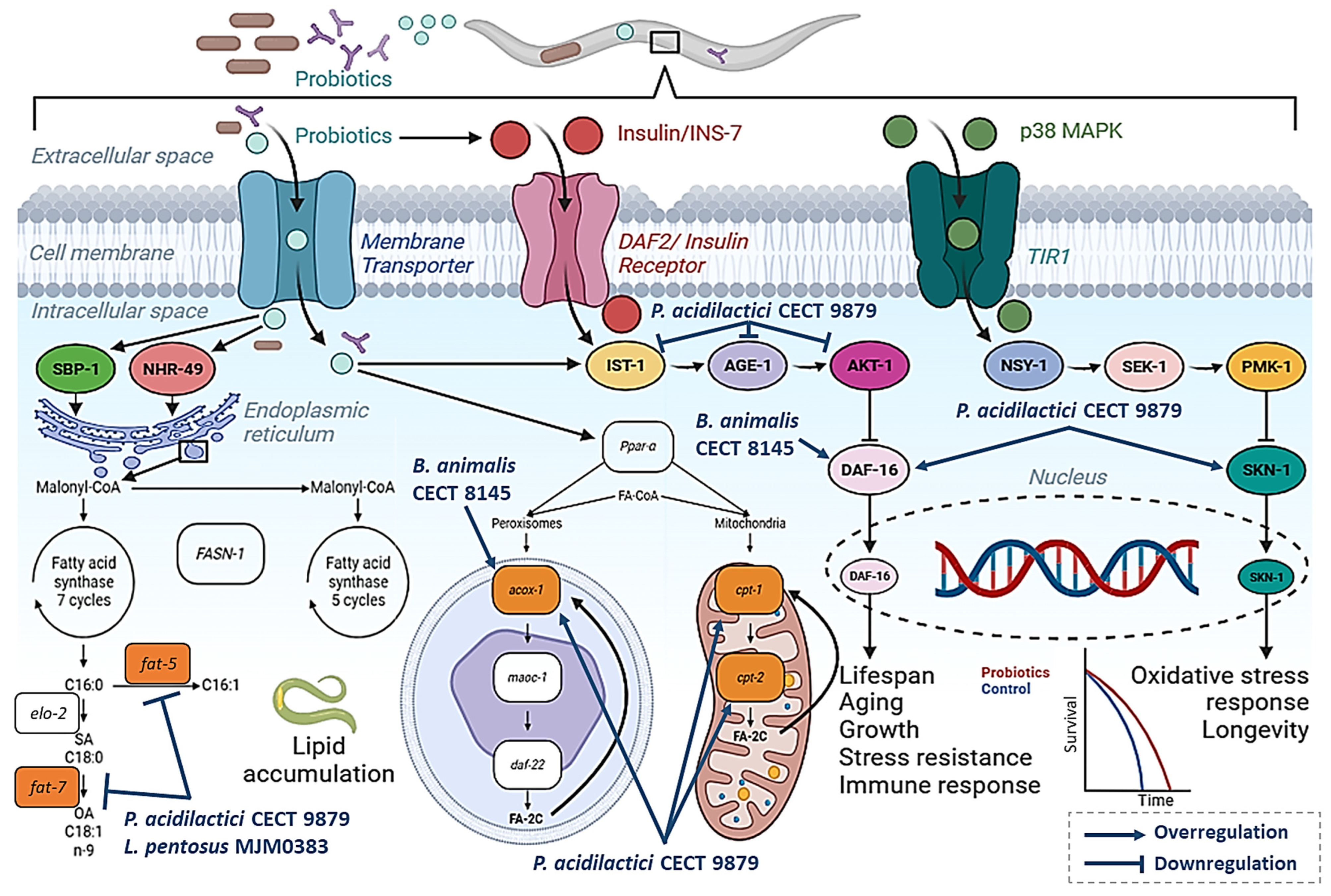
2. Probiotics with Lipid-Reducing Activity in C. elegans

In the last decade, the use of simple in vivo models such as C. elegans has been demonstrated to represent a powerful method to investigate microorganism–host interactions, as well as to evaluate the antioxidant, anti-aging, and life-extending properties of different probiotics strains [25]. Different Bifidobacteria and Lactobacilli strains have been demonstrated to extend nematode lifespan, mainly through modulations in the p-38 mitogen-activated protein kinases (p38-MAPK) signaling pathway [26]. For example, a recent study demonstrated that supplementation with Bacillus subtilis DG101, isolated from the traditional Japanese fermented food Nattō, extended worm lifespan by 45% and improved chemotaxis, in comparison with E. coli OP50-fed worms [27]. Similarly, supplementation with Lacticaseibacillus casei 62 and Lacticaseibacillus casei 63 increased nematode lifespan by improving the mitochondrial function, suggesting these bacterial strains as potential probiotics in sarcopenia [28].
However, the information about the potential anti-lipogenic, anti-diabetic, or anti-inflammatory properties of certain probiotic strains have been scarcely described in C. elegans [29]. Here, we summarize all the research describing potential anti-obesity or anti-diabetic properties of potential probiotic bacterial strains in C. elegans. The fat-reducing activities, cholesterol modulation, and the ability to counteract the effect of high-glucose exposure were some of the properties described for these probiotics.
C. elegans stores fat in lipid droplets accumulated in intestinal and skin-like hypodermal cells, mainly in the form of triglycerides [22]. Thus, probiotics might modulate lipid accumulation in C. elegans by affecting different signaling pathways, such as suppressing glucose uptake or triglyceride synthesis or activating fatty acid β-oxidation [30]. The main bacterial strains with fat-reducing activities are summarized in Table 1.

2.1. Bifidobacterium Strains with Anti-Obesity Properties in C. elegans

One of the first studies using C. elegans for the study of probiotic properties was published by Martorell et al. [23] (Table 1). In their work, this group performed a screening of the potential lipid-reducing activities of a Biopolis collection of 38 Lactobacillus (23) and Bifidobacterium (15) strains, previously isolated from the feces of healthy breast-fed babies. They found that the supplementation of the nematode growth medium (NGM) agar with a specific strain of Bifidobacterium animalis subsp. lactis CECT 8145 induced a reduction of approximately 33% in the lipid content of the worm, in comparison with nematodes fed E. coli OP50 as a standard diet. This reduction in fat accumulation was also maintained in soy-fermented milk treated with Bifidobacterium animalis subsp. lactis CECT 8145 [31]. Moreover, treatment with this Bifidobacterium strain also induced an improvement of the nematode oxidative stress-response, and an increase in lifespan by 64%. Gene expression analyses demonstrated the involvement of genes involved in energy metabolism, including the beta-oxidation genes acox-1 and daf-22, the energy modulator daf-16, and the unsaturated fatty acid synthesis gene fat-7, which was overexpressed in CECT 8145-treated worms (Figure 1).
Interestingly, the use of an inactivated form of Bifidobacterium animalis subsp. lactis CECT 8145 (BPL1), obtained via heat treatment (70 °C for 18 h), induced a similar reduction in the nematode fat content to that of the active form, supporting the idea that the strain efficacy still remained stable in non-viable cells, and that cell wall components might be responsible for the anti-obesogenic properties of this strain [23]. In this context, a subsequent study by this group demonstrated that an infant milk formula supplemented with heat-treated Bifidobacterium animalis subsp. lactis CECT 8145 (HT-BPL1, as a postbiotic) significantly reduced the fat content in C. elegans, confirming the anti-obesogenic properties of this strain in the nematode [31]. Further investigations demonstrated that lipoteichoic acid (LTA), a postbiotic isolated from the BPL1, was responsible for its lipid-reducing activities, both in NGM plates and glucose-loaded conditions [32]. In their work, Balaguer et al. demonstrated that the anti-lipogenic activities of both BPL1 and LTA were independent of SKN-1/p-38 MAPK pathway but were mediated by the modulation of the insulin-like signaling pathway (IGF-1), due to the lack of fat-reducing activity in daf-2 and daf-16 mutants [32].

2.2. Pediococcus Acidilactici Strains with Anti-Obesity Properties in C. elegans

One of the bacterial species that has emerged in recent years as a potential probiotic with anti-obesogenic and anti-diabetic properties is Pediococcus acidilactici [34,37,38,39,40]. Thus, different research groups have evaluated the potential beneficial activities of Pediococcus acidilactici strains on the lipid and carbohydrate metabolism using C. elegans (Table 1) [33,34,35]. In their study, Daliri et al. isolated lactic acid bacteria (LAB) strains from Korean fermented soybean paste, which included Pediococcus acidilactici SDL1402 and P. acidilactici SDL1406, together with Weisella cibaria SCCB2306 and Lactobacillus rhamnosus JDFM6 strains [35]. These four strains were able to reduce cholesterol levels in C. elegans, and increase the lifespan of the worms in comparison to E. coli OP50-fed worms [35].
In a similar study performed by Barathikannan et al. [33], a novel strain of Pediococcus acidilactici MNL5 was isolated through the screening of thirty-two LABs from fermented Indian herbal medicine with health-promoting activities in C. elegans. P. acidilactici MNL5 was able to counteract the lifespan-reduction induced by glucose supplementation in the medium, together with a reduction in the fat accumulation, in comparison with E. coli OP50-fed worms [33]. Gene expression analysis suggested that P. acidilactici MNL5 inhibited de novo lipogenesis by down-regulating the fatty acid desaturase-coding genes fat-4, fat-5, and fat-6, inducing a reduction in lipid accumulation (Figure 1) [33].
Similarly, our group demonstrated the anti-obesogenic and anti-diabetic properties of the strain Pediococcus acidilactici CECT 9879 (p1Ac®) in C. elegans [24,34] and rodents [34,40]. This strain was able to counteract the effect of glucose on C. elegans fat accumulation, lifespan, oxidative stress, and aging [24]. Thus, supplementation with P. acidilactici CECT 9879 at a dose of 5 × 10⁶ CFU/mL (in combination with the nematode standard diet E. coli OP50) was able to significantly reduce the nematode fat content in comparison with nematodes grown with OP50 only. Moreover, the probiotic was able to counteract the effect of high-glucose conditions by ameliorating aging (reducing lipofuscin pigment), enhancing the stress–oxidative response and prolonging lifespan without affecting worm development [24].
Gene expression analysis and mutant assays demonstrated that P. acidilactici CECT 9879 exerted health-promoting activities by affecting the IIS signaling pathway, by increasing the expression of daf-16, but also affecting the SKN-1/Nrf-2 signaling pathway. Moreover, the anti-obesity and anti-diabetic properties of P. acidilactici CECT 9879 included the inhibition of fatty acid biosynthesis (via the downregulation of fasn-1, fat-5, fat-7, and mdt-15 genes), and inducing FA degradation (by increasing the expression β-oxidation genes acox-1, daf-22, maoc-1, and cpt-2) (Figure 1) [24]. The properties of this probiotic strain and the molecular mechanism of action were confirmed and maintained in a subsequent study where P. acidilactici CECT 9879 was combined with the prebiotic ingredients chromium picolinate (0.5 μg/mL) and oat-beta glucans (50 μg/mL) [34]. Taken together, the research performed in C. elegans demonstrates the potential anti-obesity and anti-diabetic properties of P. acidilactici strains and supports the need for additional studies in higher models to determine its possible application in humans.

2.3. Other Lactic Acid Bacteria with Anti-Obesity Properties in C. elegans

Lactic acid bacteria (LAB) are important compositors of gut microbiota due to their beneficial properties by inhibiting the growth of pathogenic bacteria, enhancing intestinal function, modulating metabolic functions, and regulating the immune system. Thus, like Pediococcus, other LAB have been described to exert fat-reducing activities in C. elegans. In their study, Marquez et al. [30] described the anti-obesity properties of a probiotic mixture Lactobacillus delbrueckii subsp. indicus CRL1447, to which a mix of probiotics consisting of Limosilactobacillus fermentum CRL1446, Lactiplantibacillus paraplantarum CRL1449, and CRL1472 strains was then added in different formulations (Table 1). This combination was able to reduce worm TG content, when combined with E. coli OP50 at a ratio 25:75, in comparison with 100% of E. coli OP50 [30].
An interesting study performed by Gu et al. [36] described the efficacy of a probiotic strain of Lactobacillus pentosus MJM60383 in an obese model of C. elegans (Table 1). In this work, they established a new obesity model by feeding this nematode with a culture of Enterobacter cloacae, a proposed pathogenic bacterium that induces obesity in germ-free mice, in combination with high-glucose (100 mM) conditions (HGD-E) [36]. The proposed C. elegans obese model was characterized by an increase in the lipogenic and a decrease in the β-oxidation genes. With this model, they demonstrated that supplementation with L. pentosus MJM60383 strain was able to significantly reduce C. elegans fat accumulation, counteracting the effect of the obesity-related pathogenic bacteria E. cloacae [36].

3. Probiotics Counteracting the Effect of High Glucose Exposure in C. elegans

In C. elegans, the insulin-like signaling pathway (IGF-1) regulates aging, immunity, and lipid metabolism [41]. This signaling pathway is conserved across nematodes and mammals, including humans, and has been a key area of research in obesity. This signaling pathway is mainly controlled by DAF-2, homologous to the human insulin receptor, and DAF-16, which represents the Forkhead family of transcription factors (FOXO in humans) that play a central role in mediating the molecular mechanisms of this pathway [41,42]. The IIS pathway, with the daf-2/daf-16 axis as main actors, is involved in glucose transport and insulin sensitivity, being considered a target signaling pathway for the study of insulin resistance and type-2 diabetes [32]. Thus, C. elegans has been demonstrated to be a reliable model for evaluating the effect of high glucose exposure, which mimics overfeeding and diabetic conditions and is known to shorten lifespan, increase reactive oxygen species (ROS), and reduce stress response by modulating the insulin/IGF-1 signaling (IIS) pathway [32]. Importantly, since glucose is a known precursor of TGs synthesis, exposure to hyperglycemic conditions (glucose from 10 to 100 mM) has been demonstrated to increase the fat content and enlarge the body size of the worm [43].
Different probiotics have been demonstrated to be able to counteract the exposure to high doses of glucose. As previously mentioned, L. pentosus MJM60383 was able to ameliorate the obese phenotype induced by E. cloacae supplemented with high dose of glucose. Gene expression analyses demonstrated the downregulation of lipid-accumulation-related genes fat-6 and fat-7, which encode for a stearoyl-CoA desaturase (SCD) known to modulate the relative abundance of saturated and monounsaturated fatty acids in C. elegans [44]. Moreover, the fat-reducing activity of this probiotic strain was also mediated by the upregulation of the β-oxidation genes nhr-49 and acs-2 [36]. The gene nhr-49 encodes a transcription factor involved in the control of pathways regulating fat consumption and maintenance of the normal balance of fatty acid saturation by modulating the expression of genes involved in fatty acid beta-oxidation. Thus, the deletion of nhr-49 increases the fat accumulation in C. elegans. Acs-2, one of the downstream genes of nhr-49, is an acyl-coA synthetase catalyzing the conversion of fatty acid to acyl-CoA, resulting in a reduction in fatty acids [35]. A similar mechanism involving C. elegans nhr-49/acs-2 β-oxidation genes had also been reported to explain the fat-reducing activity of pasteurized Akkermansia muciniphila (p-AKK), the most-well described postbiotic for in vivo energy metabolism regulation [45].
Interestingly, P. acidilactici CECT 9897 was able to reduce the C. elegans fat content in NGM and glucose–NMG medium [24]. In this work, our group demonstrated that the anti-obesity properties of this strain were mediated by the IIS signaling pathway but also by inducing the peroxisomal and mitochondrial β-oxidation of fatty acids. In fact, the exposure of C. elegans to P. acidilactici CECT 9897 in high-glucose conditions demonstrated that this probiotic was able to revert the nuclear translocation of DAF-16 induced by the glucose and return to the cytosol. Indeed, the anti-obesity and anti-diabetic properties of P. acidilactici were accompanied by a reduction in the nematode oxidative stress, aging, and lifespan extension, demonstrating its ability to regulate the IIS signaling pathway [24].

4. Probiotics with Anti-Inflammatory Properties in C. elegans

As previously mentioned, metabolic diseases are characterized by an increase in cellular oxidative stress, aging, and chronic, low-grade inflammation [46]. Inflammation can be defined as an immune response during an infection or injury which aims to maintain an organism’s homeostasis [47]. In obesity, the increasing inflammation is considered a key risk factor for developing other metabolic diseases, including atherosclerosis or type-2 diabetes [46]. In this regard, C. elegans has also been used to investigate the potential anti-inflammatory effects of different probiotics, mainly attributed to their antioxidant, anti-aging, and life-extending properties or to their ability to enhance the functionality of the nematode immune response by conferring resistance to the infection of bacterial pathogens. At molecular level, the inflammatory response to harmful stimuli in C. elegans is mainly mediated by the IIS pathway but also by the p38 mitogen-activated protein kinase (p38 MAPK) [48].
The p38/MAPK pathway is a well-conserved inflammatory response in humans and C. elegans. It has been shown to be required in the resistance mechanism of C. elegans against several infections such as Cutibacterium acnes, Proteus spp. or Pseudomonas aeruginosa among others [48,49,50,51]. In this regard, metformin, the first-line oral drug for type-2 diabetes, has been shown to enhance C. elegans tolerance to pathogen infection by acting through pmk-1, the main actor of the nematode p38/MAPK signaling pathway [52].
Focusing on our main concern, probiotics, and their beneficial effects on C. elegans, there may be a link with their modulation of metabolic pathways and the requirement for the mentioned intestinal activity. Research conducted by Hu et al., where they described the fat-reducing properties of the P. acidilactici strain, also included a mechanistical exploration on the life-extending and enhanced oxidative stress response effects on C. elegans [53]. Taking advantage of mutant strains, they were able to characterize the molecular mechanism, showing that it is dependent on daf-16 but also on JNK/MAPK signaling. Treated mutants lacking the expression of jnk-1 had equal lifespan to the control worms.
Similarly to these findings, Kwon et al. described the probiotic effects of Propionibacterium freudenreichii KCTC 1063 on C. elegans [54]. After conducting similar lifespan assays with mutants, they showed that the life-extension effect was not observed in pmk-1, sek-1, mek-1, dbl-1, daf-12, or daf-2 mutants, suggesting crucial roles for these genes. Interestingly, the upregulation of the expression levels of daf-12 measured via qPCR also corroborated the role of this nuclear factor which induces the production of antimicrobial peptides and is related to the p38/MAPK pathway.
Similarly, Park et al. tested the effects of Lactobacillus fermentum strain JDFM216 on C. elegans [55]. After assessing its life-extending effect on the nematode’s life, they also showed how it can confer resistance against S. aureus and E. coli O157:H7 while colonizing the gut. The mechanism appeared to be dependent on pmk-1 phosphorylation, which was heavily upregulated in treated worms. Furthermore, no beneficial effects were observed in treated worms lacking the expression of pmk-1.
To sum up, the ability of other probiotics to upregulate the expression of pmk-1, like the Lactobacillus fermentum strain JDFM216 and Bacillus amyloliquefaciens SCGB1, or the expression of skn-1 (C. elegans ortholog of Nrf2), like the Lactococcus cremoris sp. nov. strain FC or Lactobacillus gasseri SBT2055, and extend the nematodes lifespan or enhance the host defenses [55,56,57,58], serves to highlight the beneficial effects of this pathway exerted by some probiotic strains.
Besides the activation of the IIS or MAPKS pathways, other mechanisms are able to promote inflammatory states, as summarized in Table 2. The prolonged inflammation of the gut has been associated with what is called “leaky gut”, a disruption of the intestinal barrier, and this is highly connected with the development and/or progression of several metabolic and autoimmune systemic diseases [59]. Therefore, the functional enhancement of this barrier by probiotics is one of the additional mechanisms against inflammation. The use of C. elegans to study the intestinal barrier and its functionality has allowed researchers like Shokouh Ahmadi and colleagues to unveil functional treatment-enhancements for five Lactobacillus strains (L. paracasei D3-5, L. rhamnosus D4-4, L. plantarum D6-2, L. rhamnosus D7-5, and L. plantarum D13-4) and five Enterococcus strains (E. raffinosus D24-1, E. INBio D24-2, E. avium D25-1, E. avium D25-2, and E. avium D26-1) isolated from healthy infant guts. After conducting several in vitro and in vivo assays in mice, the authors related the probiotics´ bile salt hydrolase activity to an increased taurine abundance in the gut that stimulated tight junctions though the upregulation of Zo1 and suppressed gut leakiness. With this data, the researchers conducted in vivo assays on wild type N2 C. elegans supplemented with taurine, which reduced intestinal permeability and in turn increased life span, reduced adiposity, and enhanced physical function [60].
The reduction in gut leakiness was assessed thought the “smurf assay” which, briefly, is carried out by feeding C. elegans with a dye and observing it though a fluorescent microscope if it passes though the intestinal epithelial barrier. This assay, together with similar ones based on the same principles [61], has also permitted Le et al. to characterize the response of C. elegans to different bacteria, including known pathogens and others with probiotic properties like Enterococcus faecalis, which did not affect the gut permeability when compared to pathogens.
To sum up, the inflammatory response in C. elegans has been mechanistically described over the past few years, giving researchers in the field of probiotics a powerful tool to further characterize the effects of beneficial bacteria and their mechanisms of action of immune and inflammation responses.

5. Study Limitations

The studies collected in Table 1 report very interesting and valuable information about the fat-reducing and glucose-counteracting effects of specific bacterial strains, including Bifidobacterium, Lactobacillus, and Pediococcus genera, among others. Moreover, as described in Table 2, different probiotics have been demonstrated to extend C. elegans lifespan counteracting inflammation. Nevertheless, some of the results are difficult to compare between studies due to the different methodologies used or the lack of information about the doses of probiotics used in some of the studies, which makes it difficult to compare the probiotics tested.
It is worth noting that many studies fail to evaluate possible modifications in the growth and/or development of the worms, including offspring, after exposure to the probiotic. These factors are particularly relevant when comparing the fat-reducing activity of a specific probiotic strain in comparison with a nematode’s standard diet of E. coli OP50. Bacterial dietary fatty acids are transformed into triglycerides, which are the main forms of fat deposited in the epithelial and intestine of C. elegans [33]. Thus, differences in the fatty acid composition of these bacteria might significantly affect the lipid content of the nematode. The E. coli OP50 strain is widely used as a standard diet for the proper growth and development of nematodes, with a specific development time [62].
However, the evaluated probiotic strains might have different fatty acid compositions, which could represent a lower energy source and might affect the nutritional status of the worm and, thus, its development [63]. In this case, the reported lipid-reducing effect would not be attributed to anti-lipogenic or lipolytic activity but rather to lower energy consumption. C. elegans has been widely used as a model to investigate the effects of calorie restriction, which promotes the extension of nematode lifespan, accompanied by an improvement in stress response and a reduction in the C. elegans fat accumulation mediated by the IIS pathway [64]. For this reason, the replacement of the standard food source by new bacterial strains should be investigated with caution, especially when attributing specific metabolic properties to the probiotics. For example, Zanni et al. [16] demonstrated that C. elegans supplementation with a foodborne LAB consortium, including Lactobacillus delbrueckii, Lactobacillus fermentum, and Leuconostoc lactis, induced a significant increase in the fat accumulation of the worm, in comparison with E. coli OP50-fed worms, through the downregulation of daf-16. In this work, the substitution of the nematode standard diet with this foodborne microbiota induced a reduction in worm survival accompanied by a reduction in the progeny number in comparison with nematodes grown with E. coli OP50. In this context, a recent study demonstrated that supplementation with gut microbiota (MCB) from murine feces represented lower energy density (glucose and triglycerides) in comparison with E. coli OP50. Consequently, MCB-fed worms exhibited smaller body length and size, lower fertility, lower fat content, and an extended lifespan in comparison to those fed with the standard diet [65]. Moreover, Gu et al. demonstrated the differential fatty acid composition of C. elegans exposed to E. cloacae in comparison with E. coli OP50 [36].
In contrast, Pediococcus acidilactici was shown to be able to reduce the nematode lipid content when administered in combination with the E. coli OP50, in comparison with the E. coli alone, without affecting worm development. A similar finding was identified by Balaguer and colleagues, who demonstrated that LTA from BPL1, in combination with OP50, was able to reduce C. elegans fat accumulation in comparison with OP50 alone [32].

6. Conclusions

The supplementation of the diet with beneficial bacteria has been proposed as an emerging therapy to combat or prevent the development of metabolic syndrome-related diseases, including obesity and type 2 diabetes. In vivo models are needed to characterize the strain-specific beneficial effects of these probiotics and to determine the molecular mechanisms involved in these effects. In this work, it has been shown that the supplementation of the nematode C. elegans with different probiotics, including species of the Bifidobacterium, Lactobacillus, or Pediococcus genera, can reduce lipid content compared to the standard diet through the modulation of the synthesis of triglycerides and the peroxisomal and mitochondrial β-oxidation of fatty acids. Furthermore, some of the described bacterial species are capable of counteracting the effects of exposure to high doses of glucose by reducing not only fat accumulation but also by reducing oxidative stress and aging and increasing the life expectancy of the nematodes. In most cases, the anti-obesity and anti-diabetic properties of these probiotics are mediated by the modulation of the insulin-like signaling pathway (IGF-1). Moreover, some specific strains have been shown to control the inflammatory response in C. elegans through the modulation of the p38/MAPK signaling pathway. Our review demonstrates that C. elegans represents a reliable model to evaluate the potential anti-obesity, anti-diabetic, or anti-inflammatory properties of specific bacterial strains proposed as probiotics for the prevention of metabolic syndrome.

Author Contributions

Conceptualization, P.A. and F.I.M.; methodology, P.A.; investigation, I.G.; resources, I.G.; data curation, I.G and P.A.; writing—original draft preparation, I.G, D.Y.-D. and P.A.; writing—review and editing, D.Y.-D. and F.I.M.; visualization, D.Y.-D.; supervision, F.I.M.; project administration, F.I.M.; funding acquisition, P.A. All authors have read and agreed to the published version of the manuscript.

Funding

This research was funded by Gobierno de Navarra (grant numbers 0011-1383-2022-000000 [PARABIOTICS], 0011-1383-2020-000010 [PREDISMET]) and Eatex Food Innovation Hub [PANACEA], and the MICINN (Gobierno de España; POSTBIOTICS project, PID2022-141766OB-I00).

Institutional Review Board Statement

Not applicable.

Informed Consent Statement

Not applicable.

Data Availability Statement

Data sharing is not applicable.

Acknowledgments

The authors thank LABORATORIOS CINFA, S.A., and VISCOFAN, S.A, for the financial support of the Center for Nutrition Research. The authors also want to thank Genbioma Aplicaciones, S.L., for their support of this work.

Conflicts of Interest

Author Deyan Yavorov-Dayliev was employed by the company Genbioma Aplicaciones, S.L. The remaining authors declare that the research was conducted in the absence of any commercial or financial relationships that could be construed as a potential conflict of interest.

References

  1. Wolters, M.; Ahrens, J.; Romaní-Pérez, M.; Watkins, C.; Sanz, Y.; Benítez-Páez, A.; Stanton, C.; Günther, K. Dietary Fat, the Gut Microbiota, and Metabolic Health—A Systematic Review Conducted within the MyNewGut Project. Clin. Nutr. 2019, 38, 2504–2520. [Google Scholar] [CrossRef] [PubMed]
  2. Lv, L.X.; Fang, D.Q.; Shi, D.; Chen, D.Y.; Yan, R.; Zhu, Y.X.; Chen, Y.F.; Shao, L.; Guo, F.F.; Wu, W.R.; et al. Alterations and Correlations of the Gut Microbiome, Metabolism and Immunity in Patients with Primary Biliary Cirrhosis. Environ. Microbiol. 2016, 18, 2272–2286. [Google Scholar] [CrossRef] [PubMed]
  3. Brial, F.; Le Lay, A.; Dumas, M.E.; Gauguier, D. Implication of Gut Microbiota Metabolites in Cardiovascular and Metabolic Diseases. Cell. Mol. Life Sci. 2018, 75, 3977–3990. [Google Scholar] [CrossRef] [PubMed]
  4. Tavassol, Z.H.; Ejtahed, H.S.; Atlasi, R.; Saghafian, F.; Khalagi, K.; Hasani-Ranjbar, S.; Siadat, S.D.; Nabipour, I.; Ostovar, A.; Larijani, B. Alteration in Gut Microbiota Composition of Older Adults Is Associated with Obesity and Its Indices: A Systematic Review. J. Nutr. Health Aging 2023, 27, 817–823. [Google Scholar] [CrossRef] [PubMed]
  5. Leung, C.; Rivera, L.; Furness, J.B.; Angus, P.W. The Role of the Gut Microbiota in NAFLD. Nat. Rev. Gastroenterol. Hepatol. 2016, 13, 412–425. [Google Scholar] [CrossRef] [PubMed]
  6. Xu, Z.; Jiang, W.; Huang, W.; Lin, Y.; Chan, F.K.L.; Ng, S.C. Gut Microbiota in Patients with Obesity and Metabolic Disorders—A Systematic Review. Genes Nutr. 2022, 17, 2. [Google Scholar] [CrossRef]
  7. Xiao, Y.; Niu, Y.; Mao, M.; Lin, H.; Wang, B.; Wu, E.; Zhao, H.; Li, S. Correlation Analysis between Type 2 Diabetes and Core Gut Microbiota. Nan Fang Yi Ke Da Xue Xue Bao 2021, 41, 358–369. [Google Scholar] [CrossRef]
  8. Iatcu, C.O.; Steen, A.; Covasa, M. Gut Microbiota and Complications of Type-2 Diabetes. Nutrients 2022, 14, 166. [Google Scholar] [CrossRef]
  9. Xu, Y.; Wang, N.; Tan, H.Y.; Li, S.; Zhang, C.; Feng, Y. Function of Akkermansia Muciniphila in Obesity: Interactions With Lipid Metabolism, Immune Response and Gut Systems. Front. Microbiol. 2020, 11, 219. [Google Scholar] [CrossRef]
  10. Antony, M.A.; Chowdhury, A.; Edem, D.; Raj, R.; Nain, P.; Joglekar, M.; Verma, V.; Kant, R. Gut Microbiome Supplementation as Therapy for Metabolic Syndrome. World J. Diabetes 2023, 14, 1502–1513. [Google Scholar] [CrossRef]
  11. Crudele, L.; Gadaleta, R.M.; Cariello, M.; Moschetta, A. Gut Microbiota in the Pathogenesis and Therapeutic Approaches of Diabetes. EBioMedicine 2023, 97, 104821. [Google Scholar] [CrossRef] [PubMed]
  12. Turroni, S.; Liu, F.; Wang, X.; Zhao, X.; Zhong, X.; Liu, X.; Gao, X. Therapeutic and Improving Function of Lactobacilli in the Prevention and Treatment of Cardiovascular-Related Diseases: A Novel Perspective from Gut Microbiota. Front. Nutr. 2021, 1, 693412. [Google Scholar] [CrossRef]
  13. Hill, C.; Guarner, F.; Reid, G.; Gibson, G.R.; Merenstein, D.J.; Pot, B.; Morelli, L.; Canani, R.B.; Flint, H.J.; Salminen, S.; et al. Expert Consensus Document. The International Scientific Association for Probiotics and Prebiotics Consensus Statement on the Scope and Appropriate Use of the Term Probiotic. Nat. Rev. Gastroenterol. Hepatol. 2014, 11, 506–514. [Google Scholar] [CrossRef] [PubMed]
  14. Tonucci, L.B.; Olbrich dos Santos, K.M.; Licursi de Oliveira, L.; Rocha Ribeiro, S.M.; Duarte Martino, H.S. Clinical Application of Probiotics in Type 2 Diabetes Mellitus: A Randomized, Double-Blind, Placebo-Controlled Study. Clin. Nutr. 2017, 36, 85–92. [Google Scholar] [CrossRef] [PubMed]
  15. Toshimitsu, T. Development of a Lactic Acid Bacteria Strain That Suppresses Chronic Inflammation and Improves Glucose and Lipid Metabolism. Biosci. Microbiota Food Health 2023, 42, 3–7. [Google Scholar] [CrossRef]
  16. Zanni, E.; Laudenzi, C.; Schifano, E.; Palleschi, C.; Perozzi, G.; Uccelletti, D.; Devirgiliis, C. Impact of a Complex Food Microbiota on Energy Metabolism in the Model Organism Caenorhabditis elegans. BioMed Res. Int. 2015, 2015, 621709. [Google Scholar] [CrossRef]
  17. Pedret, A.; Valls, R.M.; Calderón-Pérez, L.; Llauradó, E.; Companys, J.; Pla-Pagà, L.; Moragas, A.; Martín-Luján, F.; Ortega, Y.; Giralt, M.; et al. Effects of Daily Consumption of the Probiotic Bifidobacterium animalis subsp. lactis CECT 8145 on Anthropometric Adiposity Biomarkers in Abdominally Obese Subjects: A Randomized Controlled Trial. Int. J. Obes. 2019, 43, 1863–1868. [Google Scholar] [CrossRef]
  18. Chakravarty, B. The Evolving Role of the Caenorhabditis elegans Model as a Tool to Advance Studies in Nutrition and Health. Nutr. Res. 2022, 106, 47–59. [Google Scholar] [CrossRef]
  19. Shen, P.; Yue, Y.; Zheng, J.; Park, Y. Caenorhabditis elegans: A Convenient In Vivo Model for Assessing the Impact of Food Bioactive Compounds on Obesity, Aging, and Alzheimer’s Disease. Annu. Rev. Food Sci. Technol. 2018, 9, 1–22. [Google Scholar] [CrossRef]
  20. Shen, P.; Yue, Y.; Park, Y. A Living Model for Obesity and Aging Research: Caenorhabditis elegans. Crit. Rev. Food Sci. Nutr. 2018, 58, 741–754. [Google Scholar] [CrossRef]
  21. Lemieux, G.A.; Ashrafi, K. Insights and Challenges in Using C. elegans for Investigation of Fat Metabolism. Crit. Rev. Biochem. Mol. Biol. 2015, 50, 69–84. [Google Scholar] [CrossRef] [PubMed]
  22. Lemieux, G.A.; Liu, J.; Mayer, N.; Bainton, R.J.; Ashrafi, K.; Werb, Z. A Whole-Organism Screen Identifies New Regulators of Fat Storage. Nat. Chem. Biol. 2011, 7, 206–213. [Google Scholar] [CrossRef] [PubMed]
  23. Martorell, P.; Llopis, S.; González, N.; Chenoll, E.; López-Carreras, N.; Aleixandre, A.; Chen, Y.; Karoly, E.D.; Ramón, D.; Genovés, S. Probiotic Strain Bifidobacterium animalis subsp. lactis CECT 8145 Reduces Fat Content and Modulates Lipid Metabolism and Antioxidant Response in Caenorhabditis elegans. J. Agric. Food Chem. 2016, 64, 3462–3472. [Google Scholar] [CrossRef] [PubMed]
  24. Yavorov-Dayliev, D.; Milagro, F.I.; Ayo, J.; Oneca, M.; Aranaz, P. Pediococcus acidilactici CECT9879 (PA1c) Counteracts the Effect of a High-Glucose Exposure in C. elegans by Affecting the Insulin Signaling Pathway (IIS). Int. J. Mol. Sci. 2022, 23, 2689. [Google Scholar] [CrossRef] [PubMed]
  25. Poupet, C.; Chassard, C.; Nivoliez, A.; Bornes, S. Caenorhabditis elegans, a Host to Investigate the Probiotic Properties of Beneficial Microorganisms. Front. Nutr. 2020, 7, 135. [Google Scholar] [CrossRef] [PubMed]
  26. Komura, T.; Ikeda, T.; Yasui, C.; Saeki, S.; Nishikawa, Y. Mechanism Underlying Prolongevity Induced by Bifidobacteria in Caenorhabditis elegans. Biogerontology 2013, 14, 73–87. [Google Scholar] [CrossRef] [PubMed]
  27. Leñini, C.; Rodriguez Ayala, F.; Goñi, A.J.; Rateni, L.; Nakamura, A.; Grau, R.R. Probiotic Properties of Bacillus Subtilis DG101 Isolated from the Traditional Japanese Fermented Food Nattō. Front. Microbiol. 2023, 14, 3480. [Google Scholar] [CrossRef] [PubMed]
  28. Giron, M.; Thomas, M.; Jarzaguet, M.; Mayeur, C.; Ferrere, G.; Noordine, M.L.; Bornes, S.; Dardevet, D.; Chassard, C.; Savary-Auzeloux, I. Lacticaseibacillus Casei CNCM I-5663 Supplementation Maintained Muscle Mass in a Model of Frail Rodents. Front. Nutr. 2022, 9, 8798. [Google Scholar] [CrossRef]
  29. Wiciński, M.; Gębalski, J.; Gołębiewski, J.; Malinowski, B. Probiotics for the Treatment of Overweight and Obesity in Humans—A Review of Clinical Trials. Microorganisms 2020, 8, 1148. [Google Scholar] [CrossRef]
  30. Marquez, A.; Andrada, E.; Russo, M.; Bolondi, M.L.; Fabersani, E.; Medina, R.; Gauffin-Cano, P. Characterization of Autochthonous Lactobacilli from Goat Dairy Products with Probiotic Potential for Metabolic Diseases. Heliyon 2022, 8, E10462. [Google Scholar] [CrossRef]
  31. Silva, Á.; Gonzalez, N.; Terrén, A.; García, A.; Martinez-Blanch, J.F.; Illescas, V.; Morales, J.; Maroto, M.; Genovés, S.; Ramón, D.; et al. An Infant Milk Formula Supplemented with Heat-Treated Probiotic Bifidobacterium animalis subsp. lactis CECT 8145, Reduces Fat Deposition in C. elegans and Augments Acetate and Lactate in a Fermented Infant Slurry. Foods 2020, 9, 652. [Google Scholar] [CrossRef] [PubMed]
  32. Balaguer, F.; Enrique, M.; Llopis, S.; Barrena, M.; Navarro, V.; Álvarez, B.; Chenoll, E.; Ramón, D.; Tortajada, M.; Martorell, P. Lipoteichoic Acid from Bifidobacterium animalis subsp. lactis BPL1: A Novel Postbiotic That Reduces Fat Deposition via IGF-1 Pathway. Microb. Biotechnol. 2022, 15, 805–816. [Google Scholar] [CrossRef] [PubMed]
  33. Barathikannan, K.; Chelliah, R.; Elahi, F.; Tyagi, A.; Selvakumar, V.; Agastian, P.; Arasu, M.V.; Oh, D.H. Anti-Obesity Efficacy of Pediococcus acidilactici MNL5 in Canorhabditis elegans Gut Model. Int. J. Mol. Sci. 2022, 23, 1276. [Google Scholar] [CrossRef] [PubMed]
  34. Yavorov-Dayliev, D.; Milagro, F.I.; Ayo, J.; Oneca, M.; Goyache, I.; López-Yoldi, M.; Aranaz, P. Glucose-Lowering Effects of a Synbiotic Combination Containing Pediococcus acidilactici in C. elegans and Mice. Diabetologia 2023, 66, 2117–2138. [Google Scholar] [CrossRef] [PubMed]
  35. Daliri, E.B.M.; Kim, Y.; Do, Y.; Chelliah, R.; Oh, D.H. In Vitro and In Vivo Cholesterol Reducing Ability and Safety of Probiotic Candidates Isolated from Korean Fermented Soya Beans. Probiotics Antimicrob. Proteins 2022, 14, 87–98. [Google Scholar] [CrossRef] [PubMed]
  36. Gu, M.; Werlinger, P.; Cho, J.H.; Jang, N.; Choi, S.S.; Suh, J.W.; Cheng, J. Lactobacillus Pentosus MJM60383 Inhibits Lipid Accumulation in Caenorhabditis elegans Induced by Enterobacter Cloacae and Glucose. Int. J. Mol. Sci. 2022, 24, 280. [Google Scholar] [CrossRef] [PubMed]
  37. Ueda, T.; Tategaki, A.; Hamada, K.; Kishida, H.; Hosoe, K.; Morikawa, H.; Nakagawa, K. Effects of Pediococcus acidilactici R037 on Serum Triglyceride Levels in Mice and Rats after Oral Administration. J. Nutr. Sci. Vitaminol. 2018, 64, 41–47. [Google Scholar] [CrossRef]
  38. Mizoguchi, T.; Kasahara, K.; Yamashita, T.; Sasaki, N.; Yodoi, K.; Matsumoto, T.; Emoto, T.; Hayashi, T.; Kitano, N.; Yoshida, N.; et al. Oral Administration of the Lactic Acid Bacterium Pediococcus acidilactici Attenuates Atherosclerosis in Mice by Inducing Tolerogenic Dendritic Cells. Heart Vessel. 2017, 32, 768–776. [Google Scholar] [CrossRef]
  39. Cabello-Olmo, M.; Oneca, M.; Pajares, M.J.; Jiménez, M.; Ayo, J.; Encío, I.J.; Barajas, M.; Araña, M. Antidiabetic Effects of Pediococcus acidilactici PA1c on HFD-Induced Mice. Nutrients 2022, 14, 692. [Google Scholar] [CrossRef]
  40. Yavorov-Dayliev, D.; Milagro, F.; López-Yoldi, M.; Clemente, I.; Riezu-Boj, J.I.; Ayo, J.; Oneca, M.; Aranaz, P. Pediococcus acidilactici (PA1c®) Alleviates Obesity-Related Dyslipidemia and Inflammation in Wistar Rats by Activating Beta-Oxidation and Modulating the Gut Microbiota. Food Funct. 2023, 14, 10855–10867. [Google Scholar] [CrossRef]
  41. Tissenbaum, H.A. DAF-16/FOXO: Many Paths To a Single Fork(Head) in The Road. Antioxid. Redox Signal. 2010, 14, 623–634. [Google Scholar] [CrossRef]
  42. Tissenbaum, H.A. DAF-16: FOXO in the Context of C. elegans. In Current Topics in Developmental Biology; Elsevier: Amsterdam, The Netherlands, 2018. [Google Scholar]
  43. Peng, H.; Wei, Z.; Luo, H.; Yang, Y.; Wu, Z.; Gan, L.; Yang, X. Inhibition of Fat Accumulation by Hesperidin in Caenorhabditis elegans. J. Agric. Food Chem. 2016, 64, 5207–5214. [Google Scholar] [CrossRef] [PubMed]
  44. Brock, T.J.; Browse, J.; Watts, J.L. Genetic Regulation of Unsaturated Fatty Acid Composition in C. elegans. PLoS Genet. 2006, 2, e108. [Google Scholar] [CrossRef]
  45. Wu, Z.; Xiao, Y.; Zhou, F.; Chen, J.; Chen, X.; Hou, A.; Wang, Y.; Li, Z. Pasteurized Akkermansia Muciniphila Reduces Fat Accumulation via Nhr-49-Mediated Nuclear Hormone Signaling Pathway in Caenorhabditis elegans. Molecules 2022, 27, 6159. [Google Scholar] [CrossRef] [PubMed]
  46. Ellulu, M.S.; Patimah, I.; Khaza’ai, H.; Rahmat, A.; Abed, Y. Obesity & Inflammation: The Linking Mechanism & the Complications. Arch. Med. Sci. 2017, 13, 851–863. [Google Scholar] [CrossRef] [PubMed]
  47. Medzhitov, R. Inflammation 2010: New Adventures of an Old Flame. Cell 2010, 140, 771–776. [Google Scholar] [CrossRef] [PubMed]
  48. Roselli, M.; Schifano, E.; Guantario, B.; Zinno, P.; Uccelletti, D.; Devirgiliis, C. Caenorhabditis elegans and Probiotics Interactions from a Prolongevity Perspective. Int. J. Mol. Sci. 2019, 20, 5020. [Google Scholar] [CrossRef]
  49. Huang, X.; Pan, W.; Kim, W.; White, A.; Li, S.; Li, H.; Lee, K.; Fuchs, B.B.; Zeng, K.; Mylonakis, E. Caenorhabditis elegans Mounts a P38 MAPK Pathway-Mediated Defence to Cutibacterium Acnes Infection. Cell. Microbiol. 2020, 22, e13234. [Google Scholar] [CrossRef]
  50. JebaMercy, G.; Vigneshwari, L.; Balamurugan, K. A MAP Kinase Pathway in Caenorhabditis elegans Is Required for Defense against Infection by Opportunistic Proteus Species. Microbes Infect. 2013, 15, 550–568. [Google Scholar] [CrossRef]
  51. Xu, A.; Shi, G.; Liu, F.; Ge, B. Caenorhabditis elegans Mom-4 Is Required for the Activation of the P38 MAPK Signaling Pathway in the Response to Pseudomonas Aeruginosa Infection. Protein Cell 2013, 4, 53. [Google Scholar] [CrossRef]
  52. Xiao, Y.; Liu, F.; Li, S.; Jiang, N.; Yu, C.; Zhu, X.; Qin, Y.; Hui, J.; Meng, L.; Song, C.; et al. Metformin Promotes Innate Immunity through a Conserved PMK-1/P38 MAPK Pathway. Virulence 2020, 11, 39–48. [Google Scholar] [CrossRef] [PubMed]
  53. Hu, R.; Zhang, Y.; Qian, W.; Leng, Y.; Long, Y.; Liu, X.; Li, J.; Wan, X.; Wei, X. Pediococcus acidilactici Promotes the Longevity of C. elegans by Regulating the Insulin/IGF-1 and JNK/MAPK Signaling, Fat Accumulation and Chloride Ion. Front. Nutr. 2022, 9, 821685. [Google Scholar] [CrossRef] [PubMed]
  54. Kwon, G.; Lee, J.; Lim, Y.H. Dairy Propionibacterium Extends the Mean Lifespan of Caenorhabditis elegans via Activation of the Innate Immune System. Sci. Rep. 2016, 6, 31713. [Google Scholar] [CrossRef] [PubMed]
  55. Park, M.R.; Ryu, S.; Maburutse, B.E.; Oh, N.S.; Kim, S.H.; Oh, S.; Jeong, S.Y.; Jeong, D.Y.; Oh, S.; Kim, Y. Probiotic Lactobacillus Fermentum Strain JDFM216 Stimulates the Longevity and Immune Response of Caenorhabditis elegans through a Nuclear Hormone Receptor. Sci. Rep. 2018, 8, 7441. [Google Scholar] [CrossRef] [PubMed]
  56. Kang, M.; Choi, H.J.; Yun, B.; Lee, J.; Yoo, J.; Yang, H.J.; Jeong, D.Y.; Kim, Y.; Oh, S. Bacillus Amyloliquefaciens SCGB1 Alleviates Dextran Sulfate Sodium-Induced Colitis in Mice Through Immune Regulation. J. Med. Food 2021, 24, 709–719. [Google Scholar] [CrossRef] [PubMed]
  57. Komura, T.; Takemoto, A.; Kosaka, H.; Suzuki, T.; Nishikawa, Y. Prolonged Lifespan, Improved Perception, and Enhanced Host Defense of Caenorhabditis elegans by Lactococcus cremoris subsp. cremoris. Microbiol. Spectr. 2022, 10, e00454-21. [Google Scholar] [CrossRef]
  58. Nakagawa, H.; Shiozaki, T.; Kobatake, E.; Hosoya, T.; Moriya, T.; Sakai, F.; Taru, H.; Miyazaki, T. Effects and Mechanisms of Prolongevity Induced by Lactobacillus gasseri SBT2055 in Caenorhabditis elegans. Aging Cell 2016, 15, 227–236. [Google Scholar] [CrossRef]
  59. Di Vincenzo, F.; Del Gaudio, A.; Petito, V.; Lopetuso, L.R.; Scaldaferri, F. Gut Microbiota, Intestinal Permeability, and Systemic Inflammation: A Narrative Review. Intern. Emerg. Med. 2023, 1–19. [Google Scholar] [CrossRef]
  60. Ahmadi, S.; Wang, S.; Nagpal, R.; Wang, B.; Jain, S.; Razazan, A.; Mishra, S.P.; Zhu, X.; Wang, Z.; Kavanagh, K.; et al. A Human-Origin Probiotic Cocktail Ameliorates Aging-Related Leaky Gut and Inflammation via Modulating the Microbiota/Taurine/Tight Junction Axis. JCI Insight 2020, 5, e132055. [Google Scholar] [CrossRef]
  61. Le, T.A.N.; Selvaraj, B.; Lee, J.W.; Kang, K. Measuring the Effects of Bacteria and Chemicals on the Intestinal Permeability of Caenorhabditis elegans. J. Vis. Exp. 2019, 2019, e60419. [Google Scholar] [CrossRef]
  62. Brenner, S. The Genetics of Caenorhabditis elegans. Genetics 1974, 77, 71–94. [Google Scholar] [CrossRef]
  63. Houthoofd, K.; Johnson, T.E.; Vanfleteren, J.R. Dietary Restriction in the Nematode Caenorhabditis elegans. J. Gerontol. A Biol. Sci. Med. Sci. 2005, 60, 1125–1131. [Google Scholar] [CrossRef] [PubMed]
  64. Huayta, J.; Crapster, J.P.; San-Miguel, A. Endogenous DAF-16 Spatiotemporal Activity Quantitatively Predicts Lifespan Extension Induced by Dietary Restriction. Commun. Biol. 2023, 6, 203. [Google Scholar] [CrossRef] [PubMed]
  65. Alonzo-De la Rosa, C.M.; Miard, S.; Taubert, S.; Picard, F. Methods to Extract and Study the Biological Effects of Murine Gut Microbiota Using Caenorhabditis elegans as a Screening Host. PLoS ONE 2023, 18, e0281887. [Google Scholar] [CrossRef] [PubMed]
Figure 1. Schematic representation of different signaling pathways affected by the probiotic strains with anti-obesity or anti-diabetic properties in C. elegans. The figure includes a representation of the IIS signaling pathway, fatty acid synthesis, fatty acid β-oxidation, and oxidative stress responses.
Ijms 25 01321 g001
Table 1. Summary of the studies using C. elegans for the screening of probiotic strains with anti-obesity or anti-diabetic properties.
Probiotic StrainFood Sources and Culture ConditionsMain FindingsMechanisms (Signaling Pathways Involved)Reference
Bifidobacterium animalis subsp. lactis CECT 8145
Active form of Bifidobacterium animalis subsp. lactis CECT 8145 (probiotic)E. coli OP50 strain or B. animalis subsp. lactis CECT 8145;
No dose specification
20 °C;
Three days until young adults.
↓ Fat content (Nile red and TG quantification)
↑ Resistance to acute oxidative stress
↑ worm survival
Downregulation of positive regulators of growth rate and the xenobiotic metabolism.
Up-regulation of metabolic pathways for energy production.
↑ Lipid glycosylation
acox-1
[23]
Heat-treated Bifidobacterium animalis subsp. lactis CECT 8145NGM surface previously seeded with E. coli OP50;
Worms were incubated for 3 days at 20 °C.
↓ Fat content (Nile red and TG quantification)
↑ SCFAs production: acetate, lactic acids
NF-κB[31]
Lipoteichoic acid from Bifidobacterium animalis subsp. lactis BPL1 and LTA metaboliteEscherichia coli OP50 strain
NGM and glucose-NGM medium;
B. animalis and HI-B. animalis (108 cells/plate) were added to the NGM surface.
Lipoteichoic acid (LTA) as bioactive compound (50 to 0.1 µg mL−1).
↓ Fat accumulation by probiotic and LTA, also in NGM+ glucoseNo effect on fat reduction on daf-2 or daf-16 mutants.
Not dependent on skn-1 shown in mutants
[32]
Pediococcus acidilactici
Pediococcus acidilactici MNL5E. coli OP50 or P. acidilactici MNL5.↑ nematode lifespan and median survival
↓ Fat content vs. NGM OP50 + glucose (Nile red and oil red)
fat-4 fat-5 and fat-6
Glucose upregulated de novo fatty acid synthesis
[33]
Pediococcus acidilactici CECT 9879 (p1Ac)Worms were grown from L1 to L4 at 20 °C;
Probiotic dose: 5 × 10⁶ CFU/mL;
NGM and high-glucose NGM (10 mM) previously seeded with E. coli OP50 as normal nematode diet.
↓ Fat content (Nile red and oil red)
Normal worm development
↓ oxidative stress (ROS)
↓ aging (lipofuscin)
↑ nematode lifespan and median survival
IIS signaling pathway: pA1c inhibits the high-glucose-induced nuclear translocation of daf-16
fasn-1, fat-5, fat-7, and mdt-15 gene expression
acox-1, daf-22, maoc-1, and cpt-2 gene expression
skn-1 and nhr-49 gene expression
[24]
Pediococcus acidilactici CECT 9879 (p1Ac) combined with prebioticsWorms were grown from L1 to L4 at 20 °C;
Probiotic dose: 5 × 10⁶ CFU/mL; 0.5 µg/mL of PC; 50 µg/mL of BGC
NGM and high-glucose NGM (10 mM) previously seeded with E. coli OP50 as normal nematode diet.
↓ Fat content (Nile red and oil red)
Normal worm development
↓ oxidative stress (ROS)
↓ aging (lipofuscin)
↑ nematode lifespan and median survival
pA1c inhibits the high-glucose-induced nuclear translocation of daf-16
↓ expression of fatty acid biosynthesis genes: fat-5
↑ expression of β-oxidation genes: acox-1 and cpt-2
[34]
Other Lactic Acid Bacteria (LAB)
LAB strains from Korean Fermented Soya Beans:
Pediococcus acidilatici SDL1402
P. acidilactici SDL1406
Weisella cibaria SCCB2306
Lactobacillus rhamnosus JDFM6
E. coli OP50 or 50 µL of LAB;
(8 Log CFU/mL).
↓ Cholesterol accumulation irrespective to the order of treatment
↑ worm survival
[35]
Lactobacillus delbrueckii subsp. indicus CRL1447 combined with mixes of Limosilactobacillus fermentum CRL1446, Lactiplantibacillus paraplantarum CRL1449, and CRL1472 strainsE. coli OP50 (control group) or a combination of E. coli OP50 and each lactobacilli strain in a ratio of 25:75;
20 °C;
L1 to L4/adult.
↓ TG content [30]
Lactobacillus pentosus MJM60383E. coli OP50 or E. cloacae
20 °C;
Synchronized L1 worms were fed with OP50, or E. cloacae;
NGM plate supplemented with 100 mM glucose.
↓ Fat content (Nile red and oil content)
↓ ratio of C18:1∆9/C18:0
acs-2 and nhr-49 genes, enhancing fatty acid β-oxidation
fat6 and fat7 and tub1
[36]
↑: increased; ↓: reduced.
Table 2. Summary of the studies using C. elegans for the screening of probiotic strains with anti-inflammatory properties.
Probiotic StrainFood Source and Culture ConditionsMain FindingsMechanisms (Signaling Pathways Involved)Reference
Probiotic cocktail containing five Lactobacillus and five Enterococcus strains isolated from healthy infantsE. coli OP50 with or without taurine;
Supplementation of synchronized worms from L1 stage;
(proof of concept of the probiotic bile hydrolase activity).
↓ leaky gut (smurf assay)
↑ motility
↑ worm survival
Not described in C. elegans. [60]
Lactobacillus gasseri SBT2055E. coli OP50 or Lactobacillus gasseri SBT2055 (live or UV killed);
20 °C;
L1 to L4/adult.
↑ worm survival
↓ aging (lipofuscin)
↑ Oxidative stress response (Paraquat asay)
↑ Mitochondrial function measured by MitoTracker® CMXRos and cyanine dye JC-1
Skn-1, nsy-1, sek-1, and pmk-1 dependant mechanism for life-extension via p38 MAPK pathway signaling.
Independent effects from daf-2 or daf-16.
Upregulation of oxidative stress related genes: skn-1, gst-4, sod-1, trx-1 (thioredoxin), clk-1 (mitochondrial polypeptide), hsp16.2 (heat-shock protein), hsp-70, and gcs-1 (an ortholog of γ-glutamyl-cysteine synthetase).
[58]
Propionibacterium freudenreichii KCTC 1063E. coli OP50 or Propionibacterium freudenreichii KCTC 1063;
25 °C;
Assays performed on L4 adults.
↑ worm survival
↓ aging (lipofuscin)
resistance to Salmonella typhimurium
Skn-1 mutants failed to benefit from extended life.
Upregulation of p38/MAPKK pathway genes daf-2, pmk-1, sek-1, mek-1, dbl-1, daf-7, sma-3, and daf-12.
Upregulation of antimicrobial peptide-related genes lys-7 and lys-8.
[54]
Lactobacillus fermentum Strain JDFM216E. coli OP50 or Lactobacillus fermentum JDFM216;
25 °C;
L1 to L4/adult.
↑ worm survival
↑ Resistance to food-borne pathogens, including Staphylococcus aureus and E. coli O157:H7
Upregulation of the NHR and PMK-1 pathway.[55]
Bacillus amyloliquefaciens SCGB1Exposure to E. coli O157:H7 or Bacillus amyloliquefaciens SCGB1.↑ worm survival upon exposure to pathogen E. coli O157:H7.Upregulation of pmk-1.[56]
Lactococcus cremoris subsp. cremorisE. coli OP50 or Lactococcus cremoris subsp. Cremoris;
25 °C;
Young adult worms.
↑ Resistance to Salmonella enterica subsp. enterica serovar Enteritidis or Staphylococcus aureus
↓ aging (lipofuscin)
No beneficial effects on skn-1 lacking mutants.
Upregulation of heme oxygenase-1 ho-1, effector of the SKN-1/Nrf2 pathway.
[57]
↑: increased; ↓: reduced.
Disclaimer/Publisher’s Note: The statements, opinions and data contained in all publications are solely those of the individual author(s) and contributor(s) and not of MDPI and/or the editor(s). MDPI and/or the editor(s) disclaim responsibility for any injury to people or property resulting from any ideas, methods, instructions or products referred to in the content.

