Review

The Pivotal Role of Macrophages in the Pathogenesis of Pancreatic Diseases

1
Department of Biochemistry, College of Medicine, The Catholic University of Korea, Seoul 06591, Republic of Korea
2
Department of Biomedicine & Health Sciences, College of Medicine, The Catholic University of Korea, Seoul 06591, Republic of Korea
3
Institute for Aging and Metabolic Diseases, College of Medicine, The Catholic University of Korea, Seoul 06591, Republic of Korea
*
Author to whom correspondence should be addressed.
Int. J. Mol. Sci. 2024, 25(11), 5765; https://doi.org/10.3390/ijms25115765
Submission received: 25 April 2024 / Revised: 20 May 2024 / Accepted: 23 May 2024 / Published: 25 May 2024
(This article belongs to the Special Issue Pancreatic Disease: From Molecular Basis to Novel Therapies)

Abstract

:
The pancreas is an organ with both exocrine and endocrine functions, comprising a highly organized and complex tissue microenvironment composed of diverse cellular and non-cellular components. The impairment of microenvironmental homeostasis, mediated by the dysregulation of cell-to-cell crosstalk, can lead to pancreatic diseases such as pancreatitis, diabetes, and pancreatic cancer. Macrophages, key immune effector cells, can dynamically modulate their polarization status between pro-inflammatory (M1) and anti-inflammatory (M2) modes, critically influencing the homeostasis of the pancreatic microenvironment and thus playing a pivotal role in the pathogenesis of the pancreatic disease. This review aims to summarize current findings and provide detailed mechanistic insights into how alterations mediated by macrophage polarization contribute to the pathogenesis of pancreatic disorders. By analyzing current research comprehensively, this article endeavors to deepen our mechanistic understanding of regulatory molecules that affect macrophage polarity and the intricate crosstalk that regulates pancreatic function within the microenvironment, thereby facilitating the development of innovative therapeutic strategies that target perturbations in the pancreatic microenvironment.

1. Introduction

The pancreas is a complex organ that plays a pivotal role in maintaining metabolic homeostasis through its dual physiological functions [1,2]. It operates as an exocrine gland by producing and secreting digestive enzymes, such as amylase, lipase, and trypsin, via the acinar cells, facilitating the breakdown of carbohydrates, fats, and proteins in the digestive tract. Concurrently, it functions as an endocrine gland through the islet cells of Langerhans, which are responsible for the synthesis and release of vital hormones, including insulin, glucagon, and somatostatin, integral to the regulation of blood glucose levels [2,3].
In addition to its primary functions, the pancreatic microenvironment is characterized by a heterogeneous composition of cellular elements, including endothelial cells that constitute the vascular architecture, ductal cells that delineate the pancreatic ducts, neurons responsible for conveying neural signals, and an array of immune cells that regulate immune responses. This elaborate cellular network is supported by an intricate crosstalk of intercellular communication pathways, essential for the coordinated regulation of both exocrine and endocrine activities within the pancreas [4,5,6]. The structural and functional integrity of this environment is critical for the ability of the pancreas to respond to physiological challenges, necessitating precise and harmonious intercellular interactions to maintain the homeostasis of pancreatic function. Abnormal regulations in this crosstalk can lead to a wide spectrum of pancreatic pathologies such as pancreatitis, diabetes, and cancer, highlighting the importance of a comprehensive understanding of the intercellular crosstalk in maintaining pancreatic homeostasis within these microenvironments in health and disease states [7,8,9].
Growing evidence suggests that aberrations in cell-to-cell communications within the pancreatic microenvironment play a critical role in the pathophysiological responses of the pancreas to diverse stimuli [10]. In particular, this review will particularly emphasize the role of immune cells, notably macrophages, in modulating the cellular crosstalk in the pancreatic microenvironment, which, in turn leads, to a disruption of the homeostasis within the pancreatic milieu [11,12,13]. This dysregulation is increasingly recognized as a significant factor in the pathogenesis of several pancreatic diseases, such as pancreatitis, diabetes, and cancer [10,14,15]. This review aims to summarize current findings and offer insights into the mechanistic view of macrophage-mediated alteration and its regulators in the pathogenesis of pancreatic diseases. Through a comprehensive analysis of current research, this review seeks to advance our understanding of the complex interplay between cellular crosstalk and pancreatic function, paving the way for novel therapeutic strategies targeting the microenvironmental disruptions in pancreatic diseases.

2. Methodology

This review systematically collected references on macrophages and pancreatic disease from databases, including PubMed and Google Scholar, up to April 2024, using the search terms ‘macrophage’ combined with ‘pancreatitis’, ‘diabetes’, and ‘pancreatic cancer’. After an initial review of the current evidence, we sorted and grouped the articles and reviewed each in its appropriate context. Opinions, letters, and comments were excluded from the analysis.

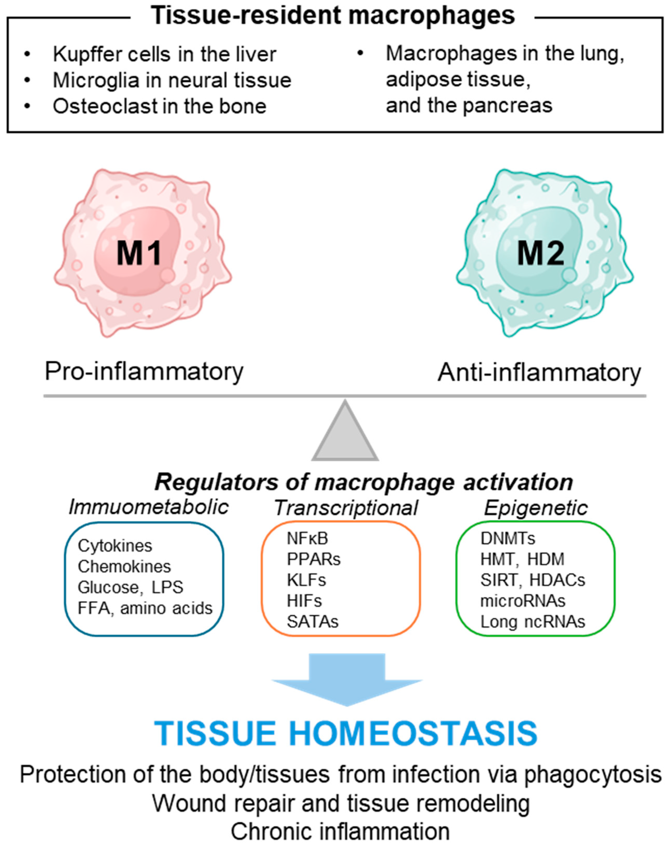
3. Macrophage and Its Polarity

Macrophages, pivotal components of the innate immune system, originate as monocytes within the hematopoietic niche of the bone marrow. These progenitors undergo a well-orchestrated differentiation process driven by a complex interplay of growth factors and cytokines, transitioning into monocytes that circulate transiently within the bloodstream. Upon receiving appropriate chemotactic signals, these monocytes extravasate into peripheral tissues, where they further differentiate into macrophages, adopting phenotypes that are intricately tailored to their specific microenvironmental context [16,17,18]. Macrophages play an important role in the host defense mechanisms, primarily through phagocytosis, wherein they engulf and degrade pathogens, apoptotic cells, and cellular debris. Beyond their phagocytic activity, macrophages are key arbiters of inflammation and tissue repair, secreting an array of cytokines, chemokines, and growth factors that modulate the immune response and orchestrate wound healing and tissue remodeling processes [17,19].
Macrophages exhibit remarkable phenotypic plasticity, enabling them to respond dynamically to a spectrum of immune-metabolic stimuli [20]. This functional versatility is underscored by their capacity to adopt distinct activation states, broadly categorized into the classical (M1) activation state, which is associated with pro-inflammatory, antimicrobial responses, and the alternative (M2) activation state, which is linked to anti-inflammatory, pro-repair activities [17,21]. M2 macrophages have subsets, including M2a, M2b, M2c, and M2d, based on their activation stimuli and unique gene expression profiles [17,22]. The M2a subtype is produced by interleukin-4 (IL-4)/IL-13 stimulation and mediates tissue repair and wound healing. The M2b subtype activated by LPS and IL-1β regulates immune response, while the M2c subtype stimulated by IL-10 exhibits strong anti-inflammatory activity. The M2d subtype resembles features of tumor-associated macrophages (reviewed in [23]).
The nomenclature of macrophages reflects their tissue-specific roles and origins, with Kupffer cells in the liver, microglia in the central nervous system, and osteoclasts in the bone representing well-characterized examples of tissue-resident macrophages [20,24]. Notably, macrophages also populate other tissues, such as the lungs, adipose tissue, and the pancreas, where they contribute to maintaining tissue homeostasis and resolving inflammation [25].
Recent advancements in research have elucidated the multifaceted roles of macrophages and the diverse regulatory elements that influence their function, as illustrated in Figure 1. This body of work highlights the significant impact of macrophages across a wide spectrum of pathophysiological states, encompassing metabolic dysfunctions, fibrotic conditions, oncogenesis, and persistent inflammatory disorders [11,13,19]. The dual nature of macrophages, serving both as guardians in host defense mechanisms and as contributors to the etiology of various diseases, underscores their potential as pivotal targets within therapeutic landscapes [17]. This may provide promising prospects for innovative strategies to fine-tune macrophage activity within disease milieus. This review aims to synthesize contemporary insights into the role and regulatory mechanisms of macrophages in the context of pancreatic pathologies, including pancreatitis, diabetes, and pancreatic cancer.

4. Macrophage Activation in Pancreatitis and Its Regulators

Pancreatitis is characterized by the inflammation of the pancreas mediated by the destruction of acinar cells [26,27]. Various factors, including gallstones, alcohol consumption, smoking, hyperglycemia, and genetic mutations, can trigger this pathological state. The disease manifests in two primary forms, distinguished by the duration of inflammation: acute pancreatitis (AP) and chronic pancreatitis (CP) [26,28]. In the acute phase, pancreatic injury leads to the disintegration of acinar cells, which subsequently initiates the activation of immune cells such as macrophages. These activated immune cells enhance the inflammatory response by releasing inflammatory cytokines, such as tumor necrosis factor-alpha (TNF-α), IL-1 beta (IL-1β), IL-6, C-C motif chemokine ligand 2 (CCL2), and exacerbating the condition [29]. Should the inflammatory stimuli persist, atrophic acinar cells engage in the prolonged activation of various immune cells. This sustained immune response culminates in the secretion of various inflammatory cytokines, including IL-1, IL-6, transforming growth factor-beta (TGF-β), and other growth factors, which, in turn, stimulate the activation of pancreatic stellate cells (PSC) [28]. The activation of PSCs leads to excessive extracellular matrix (ECM) production, which is a pivotal event in the progression of chronic pancreatitis, a condition characterized by its potential to induce fibrosis and significantly increase the risk of cancer development within the pancreatic tissue [8,27].
This intricate interplay between cellular destruction, immune activation, and cytokine release underscores the complex pathophysiological mechanisms underlying pancreatitis. Recent studies using several animal models for pancreatitis have shed light on the pivotal role of macrophages in the pathogenesis of pancreatitis. In this section, we aim to explore the characteristics of macrophages identified in the context of pancreatitis, the factors modulating the activation states of macrophages, and their possible roles in the progression of pancreatitis. By delving into the phenotype, activation states, and regulatory mechanisms of macrophages within the pancreatitis milieu, we seek to unravel the nuanced contributions of macrophages to the progression and resolution of the disease.

4.1. Factors Regulating Macrophage Polarization in AP

AP is a clinical condition characterized by a sudden inflammation of the pancreas, leading to systemic inflammatory responses and multiorgan dysfunction in severe cases [26,29]. This temporary pancreatic injury disrupts the organ’s ability to function normally, affecting the secretion of digestive enzymes and causing significant metabolic disturbances. Researchers often employ various models to understand the underlying mechanisms and potential therapeutic targets for AP [30]. These include models induced by cerulein (also known as caerulein and ceruletide), which simulate the hyperstimulation of pancreatic acinar cells; sodium taurocholate, which mimics the effects of bile acid reflux into the pancreatic duct; and lipopolysaccharide (LPS), which is used to induce an inflammatory response like that observed in bacterial infections. Each model helps to elucidate different aspects of the disease, from enzyme secretion and acinar cell injury to inflammatory pathways and systemic effects.
Recent studies have particularly emphasized the role of macrophages in AP [27]. Their versatile capacity, exhibiting either pro-inflammatory (M1) or anti-inflammatory (M2) phenotypes, is crucial in initiating and resolving inflammation in AP. The balance between these polarized states of macrophages can significantly influence the progression and severity of pancreatitis, making them key targets for potential interventions [27]. Understanding how various factors and signaling pathways influence macrophage polarization in the context of AP is essential for developing more effective treatments that can modulate the immune response and potentially mitigate the impact of the disease. One of the key regulators involved in this process is the mixed-lineage kinase domain-like protein (MLKL) [31]. It has been found that in cerulein-stimulated acinar cells, a model for AP, there is an upregulation in the expression and phosphorylation of MLKL. Knockout studies have shown that the absence of MLKL leads to decreased levels of CXCL10 in acinar cells and a reduction in M1 macrophage polarization, which subsequently lowers the severity of AP following cerulein and LPS stimulation. MicroRNAs (miRNAs), especially miR-29a, also play a significant role in modulating the pathology of pancreatitis [32]. Reports by Dey and colleagues indicate that miR-29a is consistently downregulated in murine and human pancreatitis samples. Specifically, the pancreas-specific deletion of the miR-29a/b1 gene results in a detrimental outcome in response to cerulein administration by enhancing the pancreatic inflammation, immune cell infiltration, and activation of pancreatic stellate cells. Moreover, long non-coding RNA (lncRNA) such as lncRNA-MM2P has been shown to have reduced expression in sodium taurocholate-induced models of AP [33]. The overexpression of lncRNA-MM2P can decrease M1 macrophage polarization, thereby alleviating pancreatic inflammation.
In addition to these findings, numerous factors that regulate macrophage polarization in various AP models have been reported. Pancreatic phospholipase A2 (PLA2) acts as a pathogenic trigger of AP [34]. Modulating PLA2 activity can reduce M1 macrophage polarization while potentially enhancing M2 polarization, making PLA2 targeting a consistent focus in the development of AP therapies [35]. Zhang and colleagues have developed nanoparticles that include both PLA2 attractants and inhibitors [36]. Applied in a cerulein-induced pancreatitis model, these nanoparticles demonstrated that the inhibition of PLA2 can reduce macrophage polarization, thereby ameliorating the effects of pancreatitis. Dopamine, which possesses anti-inflammatory properties, and its D2 receptor can also influence macrophage activation in pancreatitis [37]. The deletion of the D2 receptor specifically in myeloid cells has been shown to induce M1 polarization of macrophages, which exacerbates AP [38]. This suggests that dopamine D2 receptor pathways could be therapeutic targets to modulate immune responses in pancreatitis. Other agents like growth and differentiation factor 11 (GDF11, also known as BMP-11), tectoridin (Tec), and paeonol (Pae), which are known for their anti-inflammatory functions, can inhibit M1 polarization and, conversely, promote M2 polarization in various study models [39,40,41]. These findings suggest potential therapeutic benefits in targeting these molecules to balance macrophage responses in AP. Moreover, mesenchymal stem cells derived from the placental chorionic plate (CP-MSCs) have been shown to promote the secretion of TNF-stimulated gene 6 (TSG-6), which, in turn, enhances M2 polarization and plays a protective role in the pathology of AP [42].
Conversely, hyperglycemia can exacerbate AP symptoms by activating Notch signaling in macrophages, which promotes M1 polarization [43]. The application of a Notch inhibitor, such as DAPT, has been reported to alleviate symptoms of AP. Additionally, recent studies have shown that the release of extracellular vesicles (EVs) containing miR-183-5p increases in acinar cells during cerulein treatment, inducing the M1 polarization of macrophages and subsequently causing acinar cell damage [44]. This emerging evidence underscores the complex interplay of molecular signals that modulate macrophage polarization and their potential implications in AP therapy.
This growing body of evidence underscores the complexity of regulatory mechanisms governing macrophage polarization and its impact on the severity and progression of AP. Continued research into these pathways is crucial for developing targeted therapies that could modulate inflammatory responses in pancreatitis.

4.2. Factors Regulating Macrophage Polarization in CP

CP is characterized by irreversible damage to the pancreas, leading to the progressive replacement of both exocrine and endocrine tissues with fibrotic tissue [28,45]. It presents a significant clinical challenge due to its persistent and progressively worsening nature, often culminating in extensive pancreatic fibrosis. Currently, no effective therapies directly target these underlying fibrotic processes, highlighting the critical need for a more profound understanding of the molecular and cellular mechanisms driving this condition [27,45]. The pathogenesis of chronic pancreatitis is complex and remains only partially elucidated, involving a multitude of factors that sustain a chronic inflammatory state and contribute to fibrotic transformations. Importantly, several recent studies have begun to unravel the crucial role of macrophages in developing chronic pancreatitis, suggesting potential new avenues for therapeutic intervention.
Cytokines released from PSCs, notably IL-4 and IL-13, have been demonstrated to increase the population of alternatively activated (M2) macrophages, which, in turn, promote fibrosis [46]. The experimental model of CP in mice, induced by continuous pancreatic injury with cerulein, exhibits the enhancement in leukocyte infiltration, acinar cell loss, and pancreatic fibrosis. This model also shows increased the M2 polarization of macrophages within the pancreas, which is mediated by the release of IL-4 and IL-13 from PSCs. The blockade of IL-4/13 pathways ameliorates CP symptoms, suggesting that releasing these cytokines from pancreatic cells plays a critical regulatory role in the progression of chronic pancreatitis [46]. Beyond acinar cells, T-helper type 2 (Th2) cells and innate lymphoid cells (ILCs) also contribute to the release of IL-4/13 during the pathogenesis of CP [47]. Furthermore, the depletion of regulatory T (Treg) cells promotes a type 2 immune response, thereby enhancing M2 macrophage polarization and exacerbating the deleterious effects of CP insults.
In addition to cytokines, chemokines such as CCL5 play a pivotal role in CP by promoting the infiltration and maintenance of M2 macrophages, which support the fibrotic environment [48]. The influence of post-translational modifications in this process is illustrated by the action of MLN4924, a neddylation inhibitor, which exacerbates pancreatitis by boosting CCL5 secretion through hypoxia-inducible factor 1 alpha (HIF1α)-mediated gene expression. This highlights the critical interaction between cellular stress mechanisms and inflammatory signaling in CP. Additionally, several pharmacological agents, including pirfenidone [49], isoliquiritigenin [50], dasatinib [51], and berberine [52], have been implicated in contributing to the development of CP by enhancing PSC activation and macrophage polarization.
Factors regulating macrophage polarization in pancreatitis are listed in Table 1. Despite the identification of multiple factors that modulate PSC activation and macrophage polarization in the development of CP, the complexity of its pathogenesis presents significant challenges in achieving a full understanding. This underscores the importance of further studies into these regulatory mechanisms to develop targeted therapies that can effectively address the inflammatory and fibrotic processes underlying CP.

5. Macrophage Activation in Diabetes and Its Regulators

Diabetes is a systemic disease that manifests due to either a deficiency in insulin production or a failure in insulin action, leading to widespread pathological changes across multiple organ systems. This condition impacts a vast array of bodily functions, disrupting normal metabolic processes and increasing the risk of severe complications, such as cardiovascular disease, kidney damage, and neuropathy [53]. Recent research has highlighted the significant role of macrophages in maintaining the homeostasis of tissue microenvironments within these various organs affected by diabetes and insulin resistance [54,55,56]. These studies suggest that macrophages can influence a range of cellular processes, from inflammatory responses to tissue repair and fibrosis. The intricate interplay between macrophages and islet cells, such as pancreatic β cells and endothelial cells within the islets, as well as the broader interaction between macrophages and peripheral tissues in models of diabetes, are critical for the maintenance of tissue homeostasis, which can exacerbate pathological conditions if dysregulated. This section aims to discuss the characteristics of macrophage polarization in the context of diabetes and insulin resistance and the factors regulating this process.
Islet dysfunction can act as a primary driver of insulin resistance and the onset of diabetes, where the intricate interactions between β cells and other cellular components within the islets are crucial for maintaining islet microenvironment homeostasis [57,58,59]. This intercellular crosstalk in the islet is critical; its disturbances can lead to significant β cell dysfunction. For instance, endothelial cells and macrophages within the islets engage in dynamic communication with β cells, regulating their integrity and function through fine-tuned signaling networks [55,56]. Pancreatic β cells themselves are not passive in this environment; they actively secrete various factors, such as ATP, insulin, and pro-inflammatory molecules, such as colony-stimulating factor 1 (CSF1), islet amyloid polypeptide (IAPP), and IL-1β, which influence the growth, migration, and differentiation of neighboring cells, including macrophages [54,55,60,61,62]. Recent research has shown that pancreatic β cells can secrete factors like Fetuin-A, which increases macrophage infiltration within the islets and promotes intra-islet inflammation [63]. Additionally, advanced glycation end products (AGEs) in the islets have been observed to drive macrophage polarization toward an M1 phenotype, further exacerbating pancreatic β cell dysfunction [64]. Similarly, glucose-regulated protein 94 (GRP94) can promote M1 polarization, thereby enhancing systemic insulin resistance [65]. Interventions such as co-culturing with umbilical cord-derived mesenchymal stem cells (US-MSCs) and glucagon-like peptide-1 (GLP-1) treatment have shown protective effects on β cells against inflammatory responses by decreasing M1 polarization or enhancing M2 polarization of macrophage [66,67]. These studies suggest that macrophage polarization and the regulators that can modulate it within the islets significantly impact β cell dysfunction and could be therapeutic targets in the treatment and management of insulin resistance and diabetes. While research in this field continues to develop, these findings underscore the potential of targeting cellular interactions that contribute to intra-islet inflammation as a strategy to mitigate the progression of insulin resistance and diabetes.
Macrophages play a crucial role in maintaining tissue microenvironment homeostasis within the islets and in various peripheral tissues, including the kidneys and adipose tissues. Understanding the factors that regulate macrophage polarization in peripheral tissues is essential for intervening in insulin resistance, diabetes, and related pathological conditions. Recent studies have demonstrated specific instances where the modulation of macrophage polarization can have therapeutic effects in pathologic conditions related to diabetes. For example, hyperoside (HPS) has been shown to promote M2 polarization in the kidneys of db/db diabetic mice, improving fasting blood glucose levels and hyperlipidemia, thereby offering protection against diabetic nephropathy [68]. Cell therapy using UC-MSCs has also been applied to diabetic kidneys. Zhang and colleagues reported that miR-146a-5p derived from UC-MSCs can enhance M2 polarization and contribute to renal injury recovery [69]. Furthermore, insulin and IL-25 have been found to promote M2 polarization in the diabetic wound environment, enhancing wound healing [70,71].
In models of insulin resistance and diabetes, miRNAs have been reported to regulate macrophage polarization. For instance, miR-330-5p, which is induced in high-fat diet (HFD)-induced obesity, promotes M1 polarization exacerbating insulin resistance, while anti-miR-330-5p treatment may mitigate insulin resistance [72]. Conversely, the expression of miR-3061 is reduced in the cecal ligation and puncture (CLP)-induced sepsis models, leading to enhanced M1 polarization and worsening inflammation in diabetic intestines [73]. Furthermore, proline–serine–threonine phosphatase-interacting protein 2 (PSTPIP2) is downregulated in the adipose tissue of diabetic mice. The overexpression of PSTPIP2 decreases M1 polarization and simultaneously enhances M2 polarization, alleviating inflammation in obesity-associated adipose tissue and improving insulin resistance [74].
Macrophage polarization also regulates the diabetic osteoimmune microenvironment [75,76]. Bone morphogenic protein-4 (BMP-4), for instance, increases the M2 polarization of macrophages, thereby accelerating bone defect repair in diabetes mellitus [75]. Additionally, recent studies have reported that specific components from M2 macrophage-derived exosomes can improve diabetic fracture healing [76]. Furthermore, compounds like mogroside V (MV) and 4-octyl itaconate (OI), the cell-permeable itaconate derivate, exhibit anti-inflammatory effects [77,78]. Treatment with MV can mitigate the production of pro-inflammatory cytokines in bone marrow-derived macrophages (BMDM), reducing inflammation [77]. The administration of OI in the mouse models of streptozotocin (STZ)-induced type 1 diabetes (T1D) and spontaneous autoimmune diabetes reduce M1 polarization, alleviating β cell dysfunction and further improving glucose metabolism [78]. Additionally, high-intensity interval training (HIIT) in mice with type 2 diabetes mellitus (T2DM) has been shown to reduce M1 macrophage polarization in the liver, thereby decreasing liver inflammation and improving systemic inflammation associated with T2DM [79]. Factors regulating macrophage polarization in diabetic conditions are listed in Table 2.

6. Macrophage Polarization in PDAC and Its Regulators

Pancreatic ductal adenocarcinoma (PDAC) is the most common type of cancer originating in the pancreas and is characterized by a highly complex tumor microenvironment (TME) [80]. This complexity significantly influences the progression of the disease and the response to treatments. The TME of PDAC consists of various cellular and non-cellular components that interact in intricate ways, contributing to the aggressive nature of this cancer. Interactions among components in TME not only support tumor growth and spread but also create a formidable barrier to effective therapeutic interventions, making PDAC notoriously difficult to treat [81,82]. Recent studies have demonstrated that the TME of PDAC is markedly influenced by tissue-associated macrophage (TAM) polarization, particularly the dominance of M2 macrophages [83,84]. Macrophages polarized towards M2, which are pro-tumorigenic and anti-inflammatory, play a crucial role in promoting tumor growth and metastasis by suppressing anti-tumor immune responses and facilitating tissue remodeling and angiogenesis. The abundance of M2 macrophages in the PDAC microenvironment is associated with an immunosuppressive milieu characterized by activated cancer-associated fibroblasts (CAFs), reduced effectiveness of cytotoxic T cells, and enhanced activity of immunosuppressive cells like regulatory T cells (Treg) and myeloid-derived suppressor cells [13,83]. This polarization towards M2 macrophages contributes to chemotherapy resistance and poorer clinical outcomes. Strategies aimed at reprogramming or inhibiting M2 macrophages, thereby altering their recruitment, survival, or function, are being explored as promising approaches to disrupt the supportive tumor niche and enhance treatment efficacy in PDAC [84].
Several proteins, such as insulin-like growth factor binding protein 2 (IGFBP2), transforming growth factor-beta-induced protein (TGFBI), and pyruvate kinase M2 (PKM2), which are upregulated in the pancreatic tissues of PDAC patients, play significant roles in regulating macrophage polarization, thereby influencing PDAC progression [85,86,87]. IGFBP2 is a critical factor in determining TAM polarity in PDAC pathology, where it promotes polarization towards M2 macrophages. This shift increases the infiltration of Tregs and impairs anti-tumor T-cell immunity in mouse models [85]. TGFBI expression is elevated in PDAC, and the downregulation of TGFBI reduces M2 polarization, suppressing macrophage-stimulated tumor growth and enhancing anti-tumor immunity. [86]. Similarly, PKM2 correlates positively with the levels of M2 marker CD206 in PDAC specimens and promotes the M2 polarization of macrophages. The overexpression of PKM2 in PDAC cells enhances the M2 polarization of peripheral blood mononuclear cells (PBMCs) [87]. ALOX5, an enzyme encoded by the ALOX5 gene known as 5-lipoxygenase, plays a crucial role in M2 macrophage polarization and is a useful druggable target for cancer treatment [88]. A recent study by Hu and colleagues demonstrates that Zileuton, an FDA-approved ALOX5 inhibitor, mitigates the ALOX5-induced invasion and metastasis of PDAC cells [88]. Furthermore, the role of spleen tyrosine kinase (Syk) has been shown to promote PDAC growth and metastasis [89]. The inhibition of Syk using specific chemicals like R788, a Syk inhibitor, exhibits anticancer effects by affecting macrophage polarization [90].
Recent research has highlighted the significant role of extracellular vesicles in the pathology of PDAC [91,92]. Small extracellular vesicles derived from pancreatic cancer cells containing Ezrin (sEV-EZR) are known to potentially facilitate PDAC metastasis by enhancing M2 macrophage polarization [91]. Similarly, exosomes containing the oncogenic KRAS (KRASG12D), which are released through autophagy-dependent ferroptosis, have been observed to promote M2 polarization in TAMs [92]. Additional factors, including Doublecortin-like kinase 1 (DCLK1) isoform 2 [93], a member of the regenerating islet-derived family REG4 [94], NLR family pyrin domain containing 3 (NLRP3) [95], and Notch signaling pathways [96], also play roles in enhancing M2 polarization and have potential roles in cancer progression. Furthermore, non-coding RNAs, including miRNAs and lncRNAs, are critical in regulating the increase in M2 polarization in PDAC. For example, miR-548t-5p has a tumor-suppressive role in pancreatic cancer cells [97], and miR-506 enhances a shift from M2 to M1 polarization, thereby facilitating the anti-tumor immune response in pancreatic cancer [98]. Exosomes that contain lncRNAs LINC00460 or FGD5-AS1 contribute to the proliferation and metastasis of pancreatic cancer cells by enhancing M2 polarization in tumor-associated macrophages [99,100]. In addition, innovative strategies like the cytoplasmic delivery of polyinosinic-polycytidylic acid (pIC) and irreversible electroporation (IRE) are being explored to combat pancreatic cancer progression by promoting M1 polarization [101,102]. These approaches suggest potential new directions for therapeutic interventions aimed at modulating macrophage polarization to treat PDAC. Currently identified factors regulating macrophage polarization in PDAC pathology are listed in Table 3.

7. Discussion

Several studies have demonstrated the significant role of macrophages in the pathogenesis of pancreatic diseases, highlighting the biological and clinical importance of macrophage polarization in maintaining homeostasis within the pancreatic microenvironment [103,104,105,106]. Since macrophage polarization is a highly dynamic and finely tuned process, further research is needed to fully elucidate the detailed mechanisms underlying this phenomenon. Recent studies have highlighted the role of microbiota in regulating the tissue microenvironment of the pancreas [107,108,109]. Understanding the detailed mechanisms of microbiota-mediated cellular crosstalk in the pancreas is a significant challenge in overcoming pancreatic diseases. In addition to intrinsic factors, extrinsic conditions, such as endoscopic retrograde cholangiopancreatography (ERCP), may also contribute to the induction of pancreatitis [110]. These suggest that the complex and dynamic crosstalk among components of the pancreatic microenvironment is critical for maintaining tissue homeostasis, and its dysregulation is closely related to the pathological process of pancreatic diseases. However, the current research in this area is in the early stages and requires further development. Moreover, the complexity and heterogeneity of macrophage behavior across different stages of pancreatic diseases mean that findings may not be universally applicable. While this review focuses on macrophages, it does not extensively cover other important immune cells and factors in the pancreatic microenvironment. Therefore, to fully understand the mechanisms underlying pancreatic disease, it is essential to pursue a more integrated understanding of intercellular crosstalk within the complex pancreatic microenvironment, composed of diverse components such as islet cells, exocrine cells, acinar cells, immune cells, and microbiota.
Further research is crucial to unravel the complex signaling networks that regulate macrophage behavior. A detailed identification and characterization of the mediators and regulators of macrophage polarization could unveil new therapeutic targets [111,112,113]. Delving deeper into these molecular interactions will enable researchers to develop more effective strategies for preventing, managing, and treating pancreatic diseases. This may include creating novel pharmacological agents specifically aimed at targeting macrophage polarization pathways, thereby altering the disease microenvironment to improve therapeutic outcomes. Additionally, employing advanced technologies such as single-cell RNA sequencing and proteomics can provide exceptional insights into the cellular heterogeneity of the pancreas in both healthy and diseased states [114,115,116]. These technologies are crucial for precisely defining the roles of various macrophage subsets in the progression of pancreatic diseases and may identify previously unknown therapeutic targets.
Tissue-resident macrophages and peritoneal macrophages are related to the pathogenesis of pancreatic diseases such as pancreatitis, cancer, and fibrosis [117,118,119,120]. Tissue-resident macrophages are pivotal in early immune response, inflammation regulation, and tissue repair, maintaining a delicate balance between inflammation and healing. On the other hand, peritoneal macrophages are critical in the acute inflammatory phase, aiding in pathogen clearance and amplifying the immune response. Both types of macrophages are essential for managing inflammation and orchestrating the healing process in pancreatitis, highlighting their importance in disease progression and resolution. Specific markers are typically used to distinguish them: CD68, F4/80, and CD163 for tissue-resident macrophages, and GATA6 or CD11b for peritoneal macrophages. However, it is challenging to clearly define the functions of these two groups of macrophages in pancreatitis due to variations in marker combinations used across different studies and the occasional lack of differentiation between tissue-resident and peritoneal macrophages. Despite these challenges, understanding the roles of these macrophage populations is crucial for elucidating the pathophysiology of pancreatitis and other pancreatic diseases, highlighting the need for more extensive research in this area.
As research advances, it has the potential to yield groundbreaking insights that could transform the treatment landscape for pancreatic diseases. This process would ultimately enhance patient outcomes and pave the way for effective management and potential cures.

8. Conclusions

Macrophage polarization within the pancreatic tissue microenvironment plays a crucial role in the pathophysiology of pancreatitis, diabetes, and pancreatic cancer. These conditions reveal that the interactions between macrophages and other cellular components not only affect local tissue responses in the pancreas but also have significant systemic implications. The ability of macrophages to dynamically switch between pro-inflammatory and anti-inflammatory states is key to driving the pathways of inflammation and fibrosis that contribute to the progression of these pancreatic diseases. Understanding the factors that regulate macrophage polarization is essential for uncovering the molecular mechanisms that control systemic processes and cellular interactions within the pancreas. This advancing knowledge enhances our grasp of the complex orchestration of inflammation and immune responses in the pancreatic microenvironment, paving the way for novel therapeutic approaches.

Funding

This work was supported by the Basic Science Research Programs through the National Research Foundation of Korea (NRF) grant funded by the Korean government (MSIT) (2021R1A2C1004128).

Conflicts of Interest

The authors have no conflicts of interest to declare.

References

  1. Leung, P.S. Physiology of the pancreas. Adv. Exp. Med. Biol. 2010, 690, 13–27. [Google Scholar] [PubMed]
  2. Karpinska, M.; Czauderna, M. Pancreas-Its Functions, Disorders, and Physiological Impact on the Mammals’ Organism. Front. Physiol. 2022, 13, 807632. [Google Scholar] [CrossRef] [PubMed]
  3. Roder, P.V.; Wu, B.; Liu, Y.; Han, W. Pancreatic regulation of glucose homeostasis. Exp. Mol. Med. 2016, 48, e219. [Google Scholar] [CrossRef] [PubMed]
  4. Overton, D.L.; Mastracci, T.L. Exocrine-Endocrine Crosstalk: The Influence of Pancreatic Cellular Communications on Organ Growth, Function and Disease. Front. Endocrinol. 2022, 13, 904004. [Google Scholar] [CrossRef] [PubMed]
  5. Czako, L.; Hegyi, P.; Rakonczay, Z., Jr.; Wittmann, T.; Otsuki, M. Interactions between the endocrine and exocrine pancreas and their clinical relevance. Pancreatology 2009, 9, 351–359. [Google Scholar] [CrossRef] [PubMed]
  6. Rickels, M.R.; Norris, A.W.; Hull, R.L. A tale of two pancreases: Exocrine pathology and endocrine dysfunction. Diabetologia 2020, 63, 2030–2039. [Google Scholar] [CrossRef] [PubMed]
  7. Wei, J.; Wang, Z.; Han, T.; Chen, J.; Ou, Y.; Wei, L.; Zhu, X.; Wang, K.; Yan, Z.; Han, Y.P.; et al. Extracellular vesicle-mediated intercellular and interorgan crosstalk of pancreatic islet in health and diabetes. Front. Endocrinol. 2023, 14, 1170237. [Google Scholar] [CrossRef] [PubMed]
  8. Andersen, D.K.; Korc, M.; Petersen, G.M.; Eibl, G.; Li, D.; Rickels, M.R.; Chari, S.T.; Abbruzzese, J.L. Diabetes, Pancreatogenic Diabetes, and Pancreatic Cancer. Diabetes 2017, 66, 1103–1110. [Google Scholar] [CrossRef] [PubMed]
  9. Kahraman, S.; Dirice, E.; Basile, G.; Diegisser, D.; Alam, J.; Johansson, B.B.; Gupta, M.K.; Hu, J.; Huang, L.; Soh, C.L.; et al. Abnormal exocrine-endocrine cell cross-talk promotes beta-cell dysfunction and loss in MODY8. Nat. Metab. 2022, 4, 76–89. [Google Scholar] [CrossRef]
  10. Niedzwiedzka-Rystwej, P.; Wolacewicz, M.; Cywoniuk, P.; Klak, M.; Wszola, M. Crosstalk Between Immunity System Cells and Pancreas. Transformation of Stem Cells Used in the 3D Bioprinting Process as a Personalized Treatment Method for Type 1 Diabetes. Arch. Immunol. Ther. Exp. 2020, 68, 13. [Google Scholar] [CrossRef]
  11. Ying, W.; Fu, W.; Lee, Y.S.; Olefsky, J.M. The role of macrophages in obesity-associated islet inflammation and beta-cell abnormalities. Nat. Rev. Endocrinol. 2020, 16, 81–90. [Google Scholar] [CrossRef] [PubMed]
  12. Morris, D.L. Minireview: Emerging Concepts in Islet Macrophage Biology in Type 2 Diabetes. Mol. Endocrinol. 2015, 29, 946–962. [Google Scholar] [CrossRef] [PubMed]
  13. Storz, P. Roles of differently polarized macrophages in the initiation and progressionof pancreatic cancer. Front. Immunol. 2023, 14, 1237711. [Google Scholar] [CrossRef] [PubMed]
  14. Rehman, A.; Pacher, P.; Hasko, G. Role of Macrophages in the Endocrine System. Trends Endocrinol. Metab. 2021, 32, 238–256. [Google Scholar] [CrossRef] [PubMed]
  15. Lehuen, A.; Diana, J.; Zaccone, P.; Cooke, A. Immune cell crosstalk in type 1 diabetes. Nat. Rev. Immunol. 2010, 10, 501–513. [Google Scholar] [CrossRef] [PubMed]
  16. Ginhoux, F.; Jung, S. Monocytes and macrophages: Developmental pathways and tissue homeostasis. Nat. Rev. Immunol. 2014, 14, 392–404. [Google Scholar] [CrossRef] [PubMed]
  17. Chen, S.; Saeed, A.; Liu, Q.; Jiang, Q.; Xu, H.; Xiao, G.G.; Rao, L.; Duo, Y. Macrophages in immunoregulation and therapeutics. Signal Transduct. Target. Ther. 2023, 8, 207. [Google Scholar] [CrossRef] [PubMed]
  18. Murray, P.J. Macrophage Polarization. Annu. Rev. Physiol. 2017, 79, 541–566. [Google Scholar] [CrossRef]
  19. Liu, J.; Geng, X.; Hou, J.; Wu, G. New insights into M1/M2 macrophages: Key modulators in cancer progression. Cancer Cell Int. 2021, 21, 389. [Google Scholar] [CrossRef]
  20. Varol, C.; Mildner, A.; Jung, S. Macrophages: Development and tissue specialization. Annu. Rev. Immunol. 2015, 33, 643–675. [Google Scholar] [CrossRef]
  21. Italiani, P.; Boraschi, D. From Monocytes to M1/M2 Macrophages: Phenotypical vs. Functional Differentiation. Front. Immunol. 2014, 5, 514. [Google Scholar] [CrossRef]
  22. Roszer, T. Understanding the Mysterious M2 Macrophage through Activation Markers and Effector Mechanisms. Mediat. Inflamm. 2015, 2015, 816460. [Google Scholar] [CrossRef] [PubMed]
  23. Fuchs, A.L.; Costello, S.M.; Schiller, S.M.; Tripet, B.P.; Copie, V. Primary Human M2 Macrophage Subtypes Are Distinguishable by Aqueous Metabolite Profiles. Int. J. Mol. Sci. 2024, 25, 2407. [Google Scholar] [CrossRef] [PubMed]
  24. Meshkani, R.; Vakili, S. Tissue resident macrophages: Key players in the pathogenesis of type 2 diabetes and its complications. Clin. Chim. Acta 2016, 462, 77–89. [Google Scholar] [CrossRef]
  25. Gordon, S.; Pluddemann, A.; Martinez Estrada, F. Macrophage heterogeneity in tissues: Phenotypic diversity and functions. Immunol. Rev. 2014, 262, 36–55. [Google Scholar] [CrossRef] [PubMed]
  26. Mederos, M.A.; Reber, H.A.; Girgis, M.D. Acute Pancreatitis: A Review. JAMA 2021, 325, 382–390. [Google Scholar] [CrossRef] [PubMed]
  27. Hu, F.; Lou, N.; Jiao, J.; Guo, F.; Xiang, H.; Shang, D. Macrophages in pancreatitis: Mechanisms and therapeutic potential. Biomed. Pharmacother. 2020, 131, 110693. [Google Scholar] [CrossRef] [PubMed]
  28. Majumder, S.; Chari, S.T. Chronic pancreatitis. Lancet 2016, 387, 1957–1966. [Google Scholar] [CrossRef]
  29. Boxhoorn, L.; Voermans, R.P.; Bouwense, S.A.; Bruno, M.J.; Verdonk, R.C.; Boermeester, M.A.; van Santvoort, H.C.; Besselink, M.G. Acute pancreatitis. Lancet 2020, 396, 726–734. [Google Scholar] [CrossRef]
  30. Hyun, J.J.; Lee, H.S. Experimental models of pancreatitis. Clin. Endosc. 2014, 47, 212–216. [Google Scholar] [CrossRef]
  31. Peng, C.; Tu, G.; Wang, J.; Wang, Y.; Wu, P.; Yu, L.; Li, Z.; Yu, X. MLKL signaling regulates macrophage polarization in acute pancreatitis through CXCL10. Cell Death Dis. 2023, 14, 155. [Google Scholar] [CrossRef] [PubMed]
  32. Dey, S.; Udari, L.M.; RiveraHernandez, P.; Kwon, J.J.; Willis, B.; Easler, J.J.; Fogel, E.L.; Pandol, S.; Kota, J. Loss of miR-29a/b1 promotes inflammation and fibrosis in acute pancreatitis. JCI Insight 2021, 6, e149539. [Google Scholar] [CrossRef] [PubMed]
  33. Peng, K.; Biao, C.; Zhao, Y.Y.; Jun, L.C.; Wei, W.; YLNYZ, A.B.L.Z.; Song, L. Long non-coding RNA MM2P suppresses M1-polarized macrophages-mediated excessive inflammation to prevent sodium taurocholate-induced acute pancreatitis by blocking SHP2-mediated STAT3 dephosphorylation. Clin. Exp. Med. 2023, 23, 3589–3603. [Google Scholar] [CrossRef] [PubMed]
  34. Aufenanger, J.; Samman, M.; Quintel, M.; Fassbender, K.; Zimmer, W.; Bertsch, T. Pancreatic phospholipase A2 activity in acute pancreatitis: A prognostic marker for early identification of patients at risk. Clin. Chem. Lab. Med. 2002, 40, 293–297. [Google Scholar] [CrossRef] [PubMed]
  35. Ashley, J.W.; Hancock, W.D.; Nelson, A.J.; Bone, R.N.; Tse, H.M.; Wohltmann, M.; Turk, J.; Ramanadham, S. Polarization of Macrophages toward M2 Phenotype Is Favored by Reduction in iPLA2beta (Group VIA Phospholipase A2). J. Biol. Chem. 2016, 291, 23268–23281. [Google Scholar] [CrossRef] [PubMed]
  36. Zhang, Q.; Zhou, J.; Zhou, J.; Fang, R.H.; Gao, W.; Zhang, L. Lure-and-kill macrophage nanoparticles alleviate the severity of experimental acute pancreatitis. Nat. Commun. 2021, 12, 4136. [Google Scholar] [CrossRef]
  37. Han, X.; Li, B.; Ye, X.; Mulatibieke, T.; Wu, J.; Dai, J.; Wu, D.; Ni, J.; Zhang, R.; Xue, J.; et al. Dopamine D(2) receptor signalling controls inflammation in acute pancreatitis via a PP2A-dependent Akt/NF-kappaB signalling pathway. Br. J. Pharmacol. 2017, 174, 4751–4770. [Google Scholar] [CrossRef] [PubMed]
  38. Han, X.; Ni, J.; Wu, Z.; Wu, J.; Li, B.; Ye, X.; Dai, J.; Chen, C.; Xue, J.; Wan, R.; et al. Myeloid-specific dopamine D(2) receptor signalling controls inflammation in acute pancreatitis via inhibiting M1 macrophage. Br. J. Pharmacol. 2020, 177, 2991–3008. [Google Scholar] [CrossRef]
  39. Duan, F.; Wang, X.; Wang, H.; Wang, Y.; Zhang, Y.; Chen, J.; Zhu, X.; Chen, B. GDF11 ameliorates severe acute pancreatitis through modulating macrophage M1 and M2 polarization by targeting the TGFbetaR1/SMAD-2 pathway. Int. Immunopharmacol. 2022, 108, 108777. [Google Scholar] [CrossRef]
  40. Zhou, L.; Yu, J.; Wang, S.; Ma, Y.; Liu, X.; Zhang, X.; Luo, Y.; Wen, S.; Li, L.; Li, W.; et al. Tectoridin alleviates caerulein-induced severe acute pancreatitis by targeting ERK2 to promote macrophage M2 polarization. Arch. Biochem. Biophys. 2024, 752, 109873. [Google Scholar] [CrossRef]
  41. Yuan, C.; Xu, X.; Wang, N.; Zhu, Q.; Zhang, J.; Gong, W.; Ding, Y.; Xiao, W.; Chen, W.; Lu, G.; et al. Paeonol protects against acute pancreatitis by inhibiting M1 macrophage polarization via the NLRP3 inflammasomes pathway. Biochem. Biophys. Res. Commun. 2022, 600, 35–43. [Google Scholar] [CrossRef] [PubMed]
  42. Huang, Q.; Cheng, X.; Luo, C.; Yang, S.; Li, S.; Wang, B.; Yuan, X.; Yang, Y.; Wen, Y.; Liu, R.; et al. Placental chorionic plate-derived mesenchymal stem cells ameliorate severe acute pancreatitis by regulating macrophage polarization via secreting TSG-6. Stem Cell Res. Ther. 2021, 12, 337. [Google Scholar] [CrossRef] [PubMed]
  43. Hu, N.; Zhang, X.; Zhang, X.; Guan, Y.; He, R.; Xue, E.; Zhang, X.; Deng, W.; Yu, J.; Wang, W.; et al. Inhibition of Notch activity suppresses hyperglycemia-augmented polarization of macrophages to the M1 phenotype and alleviates acute pancreatitis. Clin. Sci. 2022, 136, 455–471. [Google Scholar] [CrossRef] [PubMed]
  44. Tang, D.S.; Cao, F.; Yan, C.S.; Cui, J.T.; Guo, X.Y.; Cheng, L.; Li, L.; Li, Y.L.; Ma, J.M.; Fang, K.; et al. Acinar Cell-Derived Extracellular Vesicle MiRNA-183-5p Aggravates Acute Pancreatitis by Promoting M1 Macrophage Polarization through Downregulation of FoxO1. Front. Immunol. 2022, 13, 869207. [Google Scholar] [CrossRef] [PubMed]
  45. Habtezion, A. Inflammation in acute and chronic pancreatitis. Curr. Opin. Gastroenterol. 2015, 31, 395–399. [Google Scholar] [CrossRef] [PubMed]
  46. Xue, J.; Sharma, V.; Hsieh, M.H.; Chawla, A.; Murali, R.; Pandol, S.J.; Habtezion, A. Alternatively activated macrophages promote pancreatic fibrosis in chronic pancreatitis. Nat. Commun. 2015, 6, 7158. [Google Scholar] [CrossRef] [PubMed]
  47. Glaubitz, J.; Wilden, A.; Golchert, J.; Homuth, G.; Volker, U.; Broker, B.M.; Thiele, T.; Lerch, M.M.; Mayerle, J.; Aghdassi, A.A.; et al. In mouse chronic pancreatitis CD25(+)FOXP3(+) regulatory T cells control pancreatic fibrosis by suppression of the type 2 immune response. Nat. Commun. 2022, 13, 4502. [Google Scholar] [CrossRef] [PubMed]
  48. Lin, Y.; Chen, Y.; Feng, W.; Hua, R.; Zhang, J.; Huo, Y.; Jiang, H.; Yin, B.; Yang, X. Neddylation pathway alleviates chronic pancreatitis by reducing HIF1alpha-CCL5-dependent macrophage infiltration. Cell Death Dis. 2021, 12, 273. [Google Scholar] [CrossRef] [PubMed]
  49. Guo, H.L.; Liang, X.S.; Zeng, X.P.; Liu, Y.; Li, Z.S.; Wang, L.J.; Hu, L.H. Pirfenidone alleviates chronic pancreatitis via suppressing the activation of pancreatic stellate cells and the M1 polarization of macrophages. Int. Immunopharmacol. 2024, 130, 111691. [Google Scholar] [CrossRef]
  50. Wang, L.J.; He, L.; Hao, L.; Guo, H.L.; Zeng, X.P.; Bi, Y.W.; Lu, G.T.; Li, Z.S.; Hu, L.H. Isoliquiritigenin ameliorates caerulein-induced chronic pancreatitis by inhibiting the activation of PSCs and pancreatic infiltration of macrophages. J. Cell. Mol. Med. 2020, 24, 9667–9681. [Google Scholar] [CrossRef]
  51. Zeng, X.P.; Wang, L.J.; Guo, H.L.; He, L.; Bi, Y.W.; Xu, Z.L.; Li, Z.S.; Hu, L.H. Dasatinib ameliorates chronic pancreatitis induced by caerulein via anti-fibrotic and anti-inflammatory mechanism. Pharmacol. Res. 2019, 147, 104357. [Google Scholar] [CrossRef] [PubMed]
  52. Bansod, S.; Doijad, N.; Godugu, C. Berberine attenuates severity of chronic pancreatitis and fibrosis via AMPK-mediated inhibition of TGF-beta1/Smad signaling and M2 polarization. Toxicol. Appl. Pharmacol. 2020, 403, 115162. [Google Scholar] [CrossRef] [PubMed]
  53. Deshpande, A.D.; Harris-Hayes, M.; Schootman, M. Epidemiology of diabetes and diabetes-related complications. Phys. Ther. 2008, 88, 1254–1264. [Google Scholar] [CrossRef] [PubMed]
  54. Cosentino, C.; Regazzi, R. Crosstalk between Macrophages and Pancreatic beta-Cells in Islet Development, Homeostasis and Disease. Int. J. Mol. Sci. 2021, 22, 1765. [Google Scholar] [CrossRef] [PubMed]
  55. Eguchi, K.; Nagai, R. Islet inflammation in type 2 diabetes and physiology. J. Clin. Investig. 2017, 127, 14–23. [Google Scholar] [CrossRef] [PubMed]
  56. Xiao, X.; Gittes, G.K. Concise Review: New Insights Into the Role of Macrophages in beta-Cell Proliferation. Stem Cells Transl. Med. 2015, 4, 655–658. [Google Scholar] [CrossRef] [PubMed]
  57. Cerf, M.E. Beta cell dysfunction and insulin resistance. Front. Endocrinol. 2013, 4, 37. [Google Scholar] [CrossRef] [PubMed]
  58. Donath, M.Y.; Dalmas, E.; Sauter, N.S.; Boni-Schnetzler, M. Inflammation in obesity and diabetes: Islet dysfunction and therapeutic opportunity. Cell Metab. 2013, 17, 860–872. [Google Scholar] [CrossRef] [PubMed]
  59. Dludla, P.V.; Mabhida, S.E.; Ziqubu, K.; Nkambule, B.B.; Mazibuko-Mbeje, S.E.; Hanser, S.; Basson, A.K.; Pheiffer, C.; Kengne, A.P. Pancreatic beta-cell dysfunction in type 2 diabetes: Implications of inflammation and oxidative stress. World J. Diabetes 2023, 14, 130–146. [Google Scholar] [CrossRef]
  60. Weitz, J.R.; Makhmutova, M.; Almaca, J.; Stertmann, J.; Aamodt, K.; Brissova, M.; Speier, S.; Rodriguez-Diaz, R.; Caicedo, A. Mouse pancreatic islet macrophages use locally released ATP to monitor beta cell activity. Diabetologia 2018, 61, 182–192. [Google Scholar] [CrossRef]
  61. Brissova, M.; Aamodt, K.; Brahmachary, P.; Prasad, N.; Hong, J.Y.; Dai, C.; Mellati, M.; Shostak, A.; Poffenberger, G.; Aramandla, R.; et al. Islet microenvironment, modulated by vascular endothelial growth factor-A signaling, promotes beta cell regeneration. Cell Metab. 2014, 19, 498–511. [Google Scholar] [CrossRef] [PubMed]
  62. Saunders, D.C.; Aamodt, K.I.; Richardson, T.M.; Hopkirk, A.J.; Aramandla, R.; Poffenberger, G.; Jenkins, R.; Flaherty, D.K.; Prasad, N.; Levy, S.E.; et al. Coordinated interactions between endothelial cells and macrophages in the islet microenvironment promote beta cell regeneration. NPJ Regen. Med. 2021, 6, 22. [Google Scholar] [CrossRef] [PubMed]
  63. Mukhuty, A.; Fouzder, C.; Kundu, R. Fetuin-A secretion from beta-cells leads to accumulation of macrophages in islets, aggravates inflammation and impairs insulin secretion. J. Cell Sci. 2021, 134, jcs258507. [Google Scholar] [CrossRef] [PubMed]
  64. He, S.; Hu, Q.; Xu, X.; Niu, Y.; Chen, Y.; Lu, Y.; Su, Q.; Qin, L. Advanced glycation end products enhance M1 macrophage polarization by activating the MAPK pathway. Biochem. Biophys. Res. Commun. 2020, 525, 334–340. [Google Scholar] [CrossRef] [PubMed]
  65. Song, L.; Kim, D.S.; Gou, W.; Wang, J.; Wang, P.; Wei, Z.; Liu, B.; Li, Z.; Gou, K.; Wang, H. GRP94 regulates M1 macrophage polarization and insulin resistance. Am. J. Physiol. Endocrinol. Metab. 2020, 318, E1004–E1013. [Google Scholar] [CrossRef]
  66. Yin, Y.; Hao, H.; Cheng, Y.; Zang, L.; Liu, J.; Gao, J.; Xue, J.; Xie, Z.; Zhang, Q.; Han, W.; et al. Human umbilical cord-derived mesenchymal stem cells direct macrophage polarization to alleviate pancreatic islets dysfunction in type 2 diabetic mice. Cell Death Dis. 2018, 9, 760. [Google Scholar] [CrossRef] [PubMed]
  67. Wan, S.; Sun, H. Glucagon-like peptide-1 modulates RAW264.7 macrophage polarization by interfering with the JNK/STAT3 signaling pathway. Exp. Ther. Med. 2019, 17, 3573–3579. [Google Scholar] [CrossRef] [PubMed]
  68. Liu, J.; Zhang, Y.; Sheng, H.; Liang, C.; Liu, H.; Moran Guerrero, J.A.; Lu, Z.; Mao, W.; Dai, Z.; Liu, X.; et al. Hyperoside Suppresses Renal Inflammation by Regulating Macrophage Polarization in Mice With Type 2 Diabetes Mellitus. Front. Immunol. 2021, 12, 733808. [Google Scholar] [CrossRef] [PubMed]
  69. Zhang, Y.; Le, X.; Zheng, S.; Zhang, K.; He, J.; Liu, M.; Tu, C.; Rao, W.; Du, H.; Ouyang, Y.; et al. MicroRNA-146a-5p-modified human umbilical cord mesenchymal stem cells enhance protection against diabetic nephropathy in rats through facilitating M2 macrophage polarization. Stem Cell Res. Ther. 2022, 13, 171. [Google Scholar] [CrossRef]
  70. Yu, T.; Gao, M.; Yang, P.; Liu, D.; Wang, D.; Song, F.; Zhang, X.; Liu, Y. Insulin promotes macrophage phenotype transition through PI3K/Akt and PPAR-gamma signaling during diabetic wound healing. J. Cell Physiol. 2019, 234, 4217–4231. [Google Scholar] [CrossRef]
  71. Li, S.; Ding, X.; Zhang, H.; Ding, Y.; Tan, Q. IL-25 improves diabetic wound healing through stimulating M2 macrophage polarization and fibroblast activation. Int. Immunopharmacol. 2022, 106, 108605. [Google Scholar] [CrossRef] [PubMed]
  72. Sun, J.; Huang, Q.; Li, S.; Meng, F.; Li, X.; Gong, X. miR-330-5p/Tim-3 axis regulates macrophage M2 polarization and insulin resistance in diabetes mice. Mol. Immunol. 2018, 95, 107–113. [Google Scholar] [CrossRef] [PubMed]
  73. Tan, F.; Cao, Y.; Zheng, L.; Wang, T.; Zhao, S.; Chen, J.; Pang, C.; Xia, W.; Xia, Z.; Li, N.; et al. Diabetes exacerbated sepsis-induced intestinal injury by promoting M1 macrophage polarization via miR-3061/Snail1 signaling. Front. Immunol. 2022, 13, 922614. [Google Scholar] [CrossRef] [PubMed]
  74. Xu, J.; Lv, H. PSTPIP2 alleviates obesity associated adipose tissue inflammation and insulin resistance in diabetes mice through promoting M2 macrophage polarization via activation of PPARgamma. J. Diabetes Complicat. 2023, 37, 108479. [Google Scholar] [CrossRef]
  75. Sun, X.; Ma, Z.; Zhao, X.; Jin, W.; Zhang, C.; Ma, J.; Qiang, L.; Wang, W.; Deng, Q.; Yang, H.; et al. Three-dimensional bioprinting of multicell-laden scaffolds containing bone morphogenic protein-4 for promoting M2 macrophage polarization and accelerating bone defect repair in diabetes mellitus. Bioact. Mater. 2021, 6, 757–769. [Google Scholar] [CrossRef] [PubMed]
  76. Wang, Y.; Lin, Q.; Zhang, H.; Wang, S.; Cui, J.; Hu, Y.; Liu, J.; Li, M.; Zhang, K.; Zhou, F.; et al. M2 macrophage-derived exosomes promote diabetic fracture healing by acting as an immunomodulator. Bioact. Mater. 2023, 28, 273–283. [Google Scholar] [CrossRef] [PubMed]
  77. Dong, X.; Ye, Z.; Li, C.; Li, K.; Zhong, X.; Li, H. Mogroside V Inhibits M1 Polarization and Inflammation of Diabetic Mouse Macrophages via p38 MAPK/NF-Kappab Signaling Pathway. Immunol. Investig. 2024, 1–18. [Google Scholar] [CrossRef]
  78. He, S.; Zhao, Y.; Wang, G.; Ke, Q.; Wu, N.; Lu, L.; Wu, J.; Sun, S.; Jin, W.; Zhang, W.; et al. 4-Octyl itaconate attenuates glycemic deterioration by regulating macrophage polarization in mouse models of type 1 diabetes. Mol. Med. 2023, 29, 31. [Google Scholar] [CrossRef]
  79. Wang, Y.; Guo, Y.; Xu, Y.; Wang, W.; Zhuang, S.; Wang, R.; Xiao, W. HIIT Ameliorates Inflammation and Lipid Metabolism by Regulating Macrophage Polarization and Mitochondrial Dynamics in the Liver of Type 2 Diabetes Mellitus Mice. Metabolites 2022, 13, 14. [Google Scholar] [CrossRef]
  80. Karamitopoulou, E. Tumour microenvironment of pancreatic cancer: Immune landscape is dictated by molecular and histopathological features. Br. J. Cancer 2019, 121, 5–14. [Google Scholar] [CrossRef]
  81. Truong, L.H.; Pauklin, S. Pancreatic Cancer Microenvironment and Cellular Composition: Current Understandings and Therapeutic Approaches. Cancers 2021, 13, 5028. [Google Scholar] [CrossRef]
  82. Liu, Q.; Liao, Q.; Zhao, Y. Chemotherapy and tumor microenvironment of pancreatic cancer. Cancer Cell Int. 2017, 17, 68. [Google Scholar] [CrossRef]
  83. Yang, S.; Liu, Q.; Liao, Q. Tumor-Associated Macrophages in Pancreatic Ductal Adenocarcinoma: Origin, Polarization, Function, and Reprogramming. Front. Cell Dev. Biol. 2020, 8, 607209. [Google Scholar] [CrossRef] [PubMed]
  84. Wang, S.; Wang, J.; Chen, Z.; Luo, J.; Guo, W.; Sun, L.; Lin, L. Targeting M2-like tumor-associated macrophages is a potential therapeutic approach to overcome antitumor drug resistance. NPJ Precis. Oncol. 2024, 8, 31. [Google Scholar] [CrossRef] [PubMed]
  85. Sun, L.; Zhang, X.; Song, Q.; Liu, L.; Forbes, E.; Tian, W.; Zhang, Z.; Kang, Y.; Wang, H.; Fleming, J.B.; et al. IGFBP2 promotes tumor progression by inducing alternative polarization of macrophages in pancreatic ductal adenocarcinoma through the STAT3 pathway. Cancer Lett. 2021, 500, 132–146. [Google Scholar] [CrossRef]
  86. Zhou, J.; Lyu, N.; Wang, Q.; Yang, M.; Kimchi, E.T.; Cheng, K.; Joshi, T.; Tukuli, A.R.; Staveley-O’Carroll, K.F.; Li, G. A novel role of TGFBI in macrophage polarization and macrophage-induced pancreatic cancer growth and therapeutic resistance. Cancer Lett. 2023, 578, 216457. [Google Scholar] [CrossRef]
  87. Hu, H.; Tu, W.; Chen, Y.; Zhu, M.; Jin, H.; Huang, T.; Zou, Z.; Xia, Q. The combination of PKM2 overexpression and M2 macrophages infiltration confers a poor prognosis for PDAC patients. J. Cancer 2020, 11, 2022–2031. [Google Scholar] [CrossRef]
  88. Hu, W.M.; Liu, S.Q.; Zhu, K.F.; Li, W.; Yang, Z.J.; Yang, Q.; Zhu, Z.C.; Chang, J. The ALOX5 inhibitor Zileuton regulates tumor-associated macrophage M2 polarization by JAK/STAT and inhibits pancreatic cancer invasion and metastasis. Int. Immunopharmacol. 2023, 121, 110505. [Google Scholar] [CrossRef] [PubMed]
  89. Layton, T.; Stalens, C.; Gunderson, F.; Goodison, S.; Silletti, S. Syk tyrosine kinase acts as a pancreatic adenocarcinoma tumor suppressor by regulating cellular growth and invasion. Am. J. Pathol. 2009, 175, 2625–2636. [Google Scholar] [CrossRef]
  90. Rohila, D.; Park, I.H.; Pham, T.V.; Weitz, J.; Hurtado de Mendoza, T.; Madheswaran, S.; Ishfaq, M.; Beaman, C.; Tapia, E.; Sun, S.; et al. Syk Inhibition Reprograms Tumor-Associated Macrophages and Overcomes Gemcitabine-Induced Immunosuppression in Pancreatic Ductal Adenocarcinoma. Cancer Res. 2023, 83, 2675–2689. [Google Scholar] [CrossRef]
  91. Chang, Y.T.; Peng, H.Y.; Hu, C.M.; Huang, S.C.; Tien, S.C.; Jeng, Y.M. Pancreatic cancer-derived small extracellular vesical Ezrin regulates macrophage polarization and promotes metastasis. Am. J. Cancer Res. 2020, 10, 12–37. [Google Scholar] [CrossRef] [PubMed]
  92. Dai, E.; Han, L.; Liu, J.; Xie, Y.; Kroemer, G.; Klionsky, D.J.; Zeh, H.J.; Kang, R.; Wang, J.; Tang, D. Autophagy-dependent ferroptosis drives tumor-associated macrophage polarization via release and uptake of oncogenic KRAS protein. Autophagy 2020, 16, 2069–2083. [Google Scholar] [CrossRef] [PubMed]
  93. Chandrakesan, P.; Panneerselvam, J.; May, R.; Weygant, N.; Qu, D.; Berry, W.R.; Pitts, K.; Stanger, B.Z.; Rao, C.V.; Bronze, M.S.; et al. DCLK1-Isoform2 Alternative Splice Variant Promotes Pancreatic Tumor Immunosuppressive M2-Macrophage Polarization. Mol. Cancer Ther. 2020, 19, 1539–1549. [Google Scholar] [CrossRef] [PubMed]
  94. Ma, X.; Wu, D.; Zhou, S.; Wan, F.; Liu, H.; Xu, X.; Xu, X.; Zhao, Y.; Tang, M. The pancreatic cancer secreted REG4 promotes macrophage polarization to M2 through EGFR/AKT/CREB pathway. Oncol. Rep. 2016, 35, 189–196. [Google Scholar] [CrossRef] [PubMed]
  95. Gu, H.; Deng, W.; Zhang, Y.; Chang, Y.; Shelat, V.G.; Tsuchida, K.; Lino-Silva, L.S.; Wang, Z. NLRP3 activation in tumor-associated macrophages enhances lung metastasis of pancreatic ductal adenocarcinoma. Transl. Lung Cancer Res. 2022, 11, 858–868. [Google Scholar] [CrossRef] [PubMed]
  96. Yan, W.; Menjivar, R.E.; Bonilla, M.E.; Steele, N.G.; Kemp, S.B.; Du, W.; Donahue, K.L.; Brown, K.L.; Carpenter, E.S.; Avritt, F.R.; et al. Notch Signaling Regulates Immunosuppressive Tumor-Associated Macrophage Function in Pancreatic Cancer. Cancer Immunol. Res. 2024, 12, 91–106. [Google Scholar] [CrossRef] [PubMed]
  97. Wang, Y.; Ge, W.L.; Wang, S.J.; Liu, Y.Y.; Zhang, Z.H.; Hua, Y.; Zhang, X.F.; Zhang, J.J. MiR-548t-5p regulates pancreatic ductal adenocarcinoma metastasis through an IL-33-dependent crosstalk between cancer cells and M2 macrophages. Cell Cycle 2024, 23, 169–187. [Google Scholar] [CrossRef] [PubMed]
  98. Yang, T.; Han, Y.; Chen, J.; Liang, X.; Sun, L. MiR-506 Promotes Antitumor Immune Response in Pancreatic Cancer by Reprogramming Tumor-Associated Macrophages toward an M1 Phenotype. Biomedicines 2023, 11, 2874. [Google Scholar] [CrossRef] [PubMed]
  99. Yao, J.; Gao, R.; Luo, M.; Li, D.; Guo, L.; Yu, Z.; Xiong, F.; Wei, C.; Wu, B.; Xu, Z.; et al. Exosomal LINC00460/miR-503-5p/ANLN positive feedback loop aggravates pancreatic cancer progression through regulating T cell-mediated cytotoxicity and PD-1 checkpoint. Cancer Cell Int. 2022, 22, 390. [Google Scholar] [CrossRef]
  100. He, Z.; Wang, J.; Zhu, C.; Xu, J.; Chen, P.; Jiang, X.; Chen, Y.; Jiang, J.; Sun, C. Exosome-derived FGD5-AS1 promotes tumor-associated macrophage M2 polarization-mediated pancreatic cancer cell proliferation and metastasis. Cancer Lett. 2022, 548, 215751. [Google Scholar] [CrossRef]
  101. Bhoopathi, P.; Kumar, A.; Pradhan, A.K.; Maji, S.; Mannangatti, P.; Windle, J.J.; Subler, M.A.; Zhang, D.; Vudatha, V.; Trevino, J.G.; et al. Cytoplasmic-delivery of polyinosine-polycytidylic acid inhibits pancreatic cancer progression increasing survival by activating Stat1-CCL2-mediated immunity. J. Immunother. Cancer 2023, 11, e007624. [Google Scholar] [CrossRef] [PubMed]
  102. He, C.; Sun, S.; Zhang, Y.; Xie, F.; Li, S. The role of irreversible electroporation in promoting M1 macrophage polarization via regulating the HMGB1-RAGE-MAPK axis in pancreatic cancer. Oncoimmunology 2021, 10, 1897295. [Google Scholar] [CrossRef] [PubMed]
  103. Gea-Sorli, S.; Closa, D. Role of macrophages in the progression of acute pancreatitis. World J. Gastrointest. Pharmacol. Ther. 2010, 1, 107–111. [Google Scholar] [CrossRef] [PubMed]
  104. Xiang, H.; Yu, H.; Zhou, Q.; Wu, Y.; Ren, J.; Zhao, Z.; Tao, X.; Dong, D. Macrophages: A rising star in immunotherapy for chronic pancreatitis. Pharmacol. Res. 2022, 185, 106508. [Google Scholar] [CrossRef] [PubMed]
  105. Poh, A.R.; Ernst, M. Tumor-Associated Macrophages in Pancreatic Ductal Adenocarcinoma: Therapeutic Opportunities and Clinical Challenges. Cancers 2021, 13, 2860. [Google Scholar] [CrossRef] [PubMed]
  106. Kraakman, M.J.; Murphy, A.J.; Jandeleit-Dahm, K.; Kammoun, H.L. Macrophage polarization in obesity and type 2 diabetes: Weighing down our understanding of macrophage function? Front. Immunol. 2014, 5, 470. [Google Scholar] [CrossRef]
  107. Boicean, A.; Ichim, C.; Todor, S.B.; Anderco, P.; Popa, M.L. The Importance of Microbiota and Fecal Microbiota Transplantation in Pancreatic Disorders. Diagnostics 2024, 14, 861. [Google Scholar] [CrossRef]
  108. Thomas, R.M.; Jobin, C. Microbiota in pancreatic health and disease: The next frontier in microbiome research. Nat. Rev. Gastroenterol. Hepatol. 2020, 17, 53–64. [Google Scholar] [CrossRef] [PubMed]
  109. Schepis, T.; De Lucia, S.S.; Nista, E.C.; Manilla, V.; Pignataro, G.; Ojetti, V.; Piccioni, A.; Gasbarrini, A.; Franceschi, F.; Candelli, M. Microbiota in Pancreatic Diseases: A Review of the Literature. J. Clin. Med. 2021, 10, 5920. [Google Scholar] [CrossRef]
  110. Boicean, A.; Birlutiu, V.; Ichim, C.; Todor, S.B.; Hasegan, A.; Bacila, C.; Solomon, A.; Cristian, A.; Dura, H. Predictors of Post-ERCP Pancreatitis (P.E.P.) in Choledochal Lithiasis Extraction. J. Pers. Med. 2023, 13, 1356. [Google Scholar] [CrossRef]
  111. Mantovani, A.; Allavena, P.; Marchesi, F.; Garlanda, C. Macrophages as tools and targets in cancer therapy. Nat. Rev. Drug Discov. 2022, 21, 799–820. [Google Scholar] [CrossRef] [PubMed]
  112. Kuntzel, T.; Bagnard, D. Manipulating Macrophage/Microglia Polarization to Treat Glioblastoma or Multiple Sclerosis. Pharmaceutics 2022, 14, 344. [Google Scholar] [CrossRef] [PubMed]
  113. He, W.; Kapate, N.; Shields, C.W., IV; Mitragotri, S. Drug delivery to macrophages: A review of targeting drugs and drug carriers to macrophages for inflammatory diseases. Adv. Drug Deliv. Rev. 2020, 165–166, 15–40. [Google Scholar] [CrossRef] [PubMed]
  114. Van de Sande, B.; Lee, J.S.; Mutasa-Gottgens, E.; Naughton, B.; Bacon, W.; Manning, J.; Wang, Y.; Pollard, J.; Mendez, M.; Hill, J.; et al. Applications of single-cell RNA sequencing in drug discovery and development. Nat. Rev. Drug Discov. 2023, 22, 496–520. [Google Scholar] [CrossRef] [PubMed]
  115. Loscalzo, J. Molecular interaction networks and drug development: Novel approach to drug target identification and drug repositioning. FASEB J. 2023, 37, e22660. [Google Scholar] [CrossRef] [PubMed]
  116. Ha, J.; Park, H.; Park, J.; Park, S.B. Recent advances in identifying protein targets in drug discovery. Cell Chem. Biol. 2021, 28, 394–423. [Google Scholar] [CrossRef] [PubMed]
  117. Ardavin, C.; Alvarez-Ladron, N.; Ferriz, M.; Gutierrez-Gonzalez, A.; Vega-Perez, A. Mouse Tissue-Resident Peritoneal Macrophages in Homeostasis, Repair, Infection, and Tumor Metastasis. Adv. Sci. 2023, 10, e2206617. [Google Scholar] [CrossRef] [PubMed]
  118. Louwe, P.A.; Badiola Gomez, L.; Webster, H.; Perona-Wright, G.; Bain, C.C.; Forbes, S.J.; Jenkins, S.J. Recruited macrophages that colonize the post-inflammatory peritoneal niche convert into functionally divergent resident cells. Nat. Commun. 2021, 12, 1770. [Google Scholar] [CrossRef] [PubMed]
  119. Zinselmeyer, B.H.; Vomund, A.N.; Saunders, B.T.; Johnson, M.W.; Carrero, J.A.; Unanue, E.R. The resident macrophages in murine pancreatic islets are constantly probing their local environment, capturing beta cell granules and blood particles. Diabetologia 2018, 61, 1374–1383. [Google Scholar] [CrossRef]
  120. Baer, J.M.; Zuo, C.; Kang, L.I.; de la Lastra, A.A.; Borcherding, N.C.; Knolhoff, B.L.; Bogner, S.J.; Zhu, Y.; Yang, L.; Laurent, J.; et al. Fibrosis induced by resident macrophages has divergent roles in pancreas inflammatory injury and PDAC. Nat. Immunol. 2023, 24, 1443–1457. [Google Scholar] [CrossRef]
Figure 1. Schematic diagram of macrophage polarization and its regulators. Macrophages respond to changes in the tissue microenvironment by altering their polarity towards M1 or M2, thus playing a role in regulating tissue homeostasis. They engage in phagocytosis to combat infections and contribute to tissue remodeling through wound repair. Additionally, macrophages can induce chronic inflammation, which disrupts the tissue microenvironment. Abnormal macrophage polarization can contribute to the development of pancreatitis, diabetes, and cancer. Various immunometabolic, transcriptional, and epigenetic factors influence macrophage polarization towards M1 or M2 phenotypes, and identifying new regulators is essential for understanding macrophage-mediated tissue homeostasis.
Ijms 25 05765 g001
Table 1. Factors regulating macrophage polarization in pancreatitis.
RegulatorsMφ PolarizationEffect on PancreatitisRef.
MLKL ↓ in acinar cellM1 polarization ↓AP promotion ↓[31]
miR-29a/b1M1 polarization ↑AP promotion ↑[32]
MM2PM1 polarization ↓AP promotion ↓[33]
PLA2 inhibitorM1 polarization ↓AP promotion ↓[36]
Dopamine & D2 receptorM1 polarization ↓AP promotion ↓[37,38]
GDF11M1 polarization ↓AP promotion ↓[39]
TectoridinM1 polarization ↓AP promotion ↓[40]
PaeonolM1 polarization ↓AP promotion ↓[41]
CP-MSCsM2 polarization ↑AP promotion ↓[42]
HyperglycemiaM1 polarization ↑AP promotion ↑[43]
miR-183-5p in acinar EVsM1 polarization ↑AP promotion ↑[44]
IL-4/13 in PSCs
IL-4/13 in T cells
M2 polarization ↑CP promotion ↑[46,47]
MLN4924M2 polarization ↑CP promotion ↑[48]
PirfenidoneM1 polarization ↓
PSC activation ↓
CP promotion ↓[49]
IsoliquiritigeninM1 polarization ↓
PSC activation ↓
CP promotion ↓[50]
DasatinibM1 polarization ↓
PSC activation ↓
CP promotion ↓[51]
BerberineM2 polarization ↓
PSC activation ↓
CP promotion ↓[52]
Table 2. Factors regulating macrophage polarization in diabetic conditions.
Tissue/CellRegulatorsMφ ConditionEffect on DiabetesRef.
Pancreatic isletSeveral factors
from β cell
(insulin, ATP, etc.)
Mφ crosstalk with β cellβ cell integrity
Islet inflammation
Reviewed in [54,55]
Pancreatic isletFetuin-A from β cellMφ accumulation in isletIntra-islet inflammation[63]
β cellAGEsM1 polarization ↑β cell dysfunction[64]
SystemicGRP94M1 polarization ↑Insulin resistance ↑[65]
β cellUS-MSCM2 polarization ↑β cell protection[66]
β cellGLP-1M2 polarization ↑β cell protection[67]
KidneyHPSM2 polarization ↑Diabetic nephropathy protection [68]
KidneymiR-146a-5p
from US-MSC
M2 polarization ↑Renal injury recovery[69]
SkinInsulin, IL-25M2 polarization ↑Wound healing[70,71]
SystemicAnti-miR-330-5pM2 polarization ↑Insulin resistance ↓[72]
Intestine in sepsismiR-3061M1 polarization ↑Intestinal injury ↑[73]
Adipose tissuePSTPIP2M2 polarization ↑Inflammation,
Insulin resistance ↓
[74]
BoneBMP-4M2 polarization ↑Bone defect repair ↑[75]
BoneM2-derived exosomeM2 polarization ↑Diabetic fracture healing ↑[76]
SystemicMogroside VM1 polarization ↓Inflammation ↓[77]
Systemic4-Octyl itaconateM1 polarization ↓Glucose metabolism ↑[78]
LiverHIITM1 polarization ↓Inflammation ↓[79]
Table 3. Factors regulating macrophage polarization in PDAC.
RegulatorsMφ PolarizationEffect on PDACRef.
IGFBP2M2 polarization ↑PDAC progression ↑[85]
TGFBIM2 polarization ↑PDAC progression ↑[86]
PKM2M2 polarization ↑PDAC progression ↑[87]
Zileuton (ALOX5 inhibitor)M2 polarization ↓PDAC progression ↓[88]
R788 (Syk inhibitor)M2 polarization ↓PDAC progression ↓[90]
sEV-EZRM2 polarization ↑PDAC metastasis ↑[91]
Oncogenic KRASM2 polarization ↑PDAC progression ↑[92]
DCLK1-iso2M2 polarization ↑PDAC progression ↑[93]
REG4M2 polarization ↑PDAC progression ↑[94]
NLRP3M2a/c/d polarization ↑PDAC progression ↑[95]
Notch signaling pathwayM2 polarization ↑PDAC progression ↑[96]
miR-548t-5pM2 polarization ↓PDAC progression ↓[97]
miR-506M2 polarization ↓PDAC progression ↓[98]
Exosomal-LINK00460M2 polarization ↑PDAC progression ↑[99]
Exosomal-FGD5-AS1M2 polarization ↑PDAC progression ↑[100]
Cytoplasmic delivery of pICM1 polarization ↑PDAC progression ↓[101]
Irreversible electroporation (IRE)M1 polarization ↑PDAC progression ↓[102]
Disclaimer/Publisher’s Note: The statements, opinions and data contained in all publications are solely those of the individual author(s) and contributor(s) and not of MDPI and/or the editor(s). MDPI and/or the editor(s) disclaim responsibility for any injury to people or property resulting from any ideas, methods, instructions or products referred to in the content.

