Article

Does Salmonella diarizonae 58:r:z53 Isolated from a Mallard Duck Pose a Threat to Human Health?

1
Laboratory of Molecular Biology, Vet-Lab Brudzew, 62-720 Brudzew, Poland
2
Department of Medical Microbiology, Faculty of Medicine, Medical University of Gdańsk, 80-204 Gdańsk, Poland
3
DNA Sequencing and Synthesis Facility, Institute of Biochemistry and Biophysics, Polish Academy of Sciences, 02-106 Warsaw, Poland
4
Department of Food Hygiene of Animal Origin, Faculty of Veterinary Medicine, University of Life Sciences in Lublin, 20-950 Lublin, Poland
5
Department of Food Hygiene and Public Health Protection, Institute of Veterinary Medicine, Warsaw University of Life Sciences, 02-776 Warsaw, Poland
*
Authors to whom correspondence should be addressed.
Int. J. Mol. Sci. 2024, 25(11), 5664; https://doi.org/10.3390/ijms25115664
Submission received: 26 March 2024 / Revised: 13 May 2024 / Accepted: 13 May 2024 / Published: 23 May 2024
(This article belongs to the Special Issue Salmonella Pathophysiology and Host-Bacteria Relationship)

Abstract

:
Salmonella diarizonae (IIIb) is frequently isolated from reptiles and less frequently from birds and mammals. However, its isolation from invasive human infections has not been widely reported. Migratory mallard ducks are excellent bioindicators of pathogen presence and pathogen antibiotic resistance (AMR). We present the first isolation from a mallard duck in central Europe of the antibiotic-resistant Salmonella enterica subsp. diarizonae with the unique antigenic pattern 58:r:z53 and report its whole-genome sequencing, serosequencing, and genotyping, which enabled the prediction of its pathogenicity and comparison with phenotypic AMR. The isolated strain was highly similar to S. diarizonae isolated from humans and food. Twenty-four AMR genes were detected, including those encoding aminoglycoside, fluoroquinolone, macrolide, carbapenem, tetracycline, cephalosporin, nitroimidazole, peptide antibiotic, and disinfecting agent/antiseptic resistance. Six Salmonella pathogenicity islands were found (SPI-1, SPI-2, SPI-3, SPI-5, SPI-9, and SPI-13). An iron transport system was detected in SPI-1 centisome C63PI. Plasmid profile analyses showed three to be present. Sequence mutations in the invA and invF genes were noted, which truncated and elongated the proteins, respectively. The strain also harbored genes encoding type-III secretion-system effector proteins and many virulence factors found in S. diarizonae associated with human infections. This study aims to elucidate the AMR and virulence genes in S. enterica subsp. diarizonae that may most seriously threaten human health.

1. Introduction

The Salmonella genus, containing 2659 serovars, is divided into two species: Salmonella enterica with 2637 and Salmonella bongori with the remaining 22. There are six subspecies of Salmonella enterica: enterica (I) containing 1586 serovars, salamae (II) with 522, arizonae (IIIa) with 102, diarizonae (IIIb) with 338, houtenae (IV) with 76, and indica (VI) with the final 13 [1]. The Salmonella enterica species is a widely known pathogen causing serious illness in humans and animals. Some serotypes of Salmonella enterica subsp. enterica are particularly well described and are known to cause a wide range of food- and water-borne illnesses [2,3,4,5,6,7]. Salmonella enterica subsp. diarizonae is the third most abundant subspecies of the genus. The diarizonae (IIIb) subspecies is commonly isolated from a variety of cold-blooded animals, including snakes, turtles, and lizards, as well as from birds, sheep, and even humans [8,9,10,11]. Although human infection caused by S. diarizonae seems to be rare, more and more cases of gastroenteritis and bacteremia are being described in the global literature [12,13,14].
Salmonella diarizonae 58:r:z53 was first isolated in Germany in 1985 from snake feces and in 2021 it was isolated in Poland as a multidrug-resistant bacterium from a mallard duck (Anas platyrhynchos). In this study, we present a genome analysis of this rare multidrug-resistant strain to demonstrate its potential human pathogenicity and virulence features.

2. Results

2.1. Source Animal and Antimicrobial Resistance of the Isolate

The mallard duck from the intestine of which the S. enterica subsp. diarizonae 58:r:z53 (S. IIIb 58:r:z53) strain was isolated had no pathological changes when given an anatomopathological examination and was in good physical condition. The isolated strain was found to be resistant to 14 of the 33 antibiotics tested [15]. It was sensitive to ampicillin, amoxicillin, trimethoprim-sulfamethoxazole, third-generation cephalosporin, and imipenem.

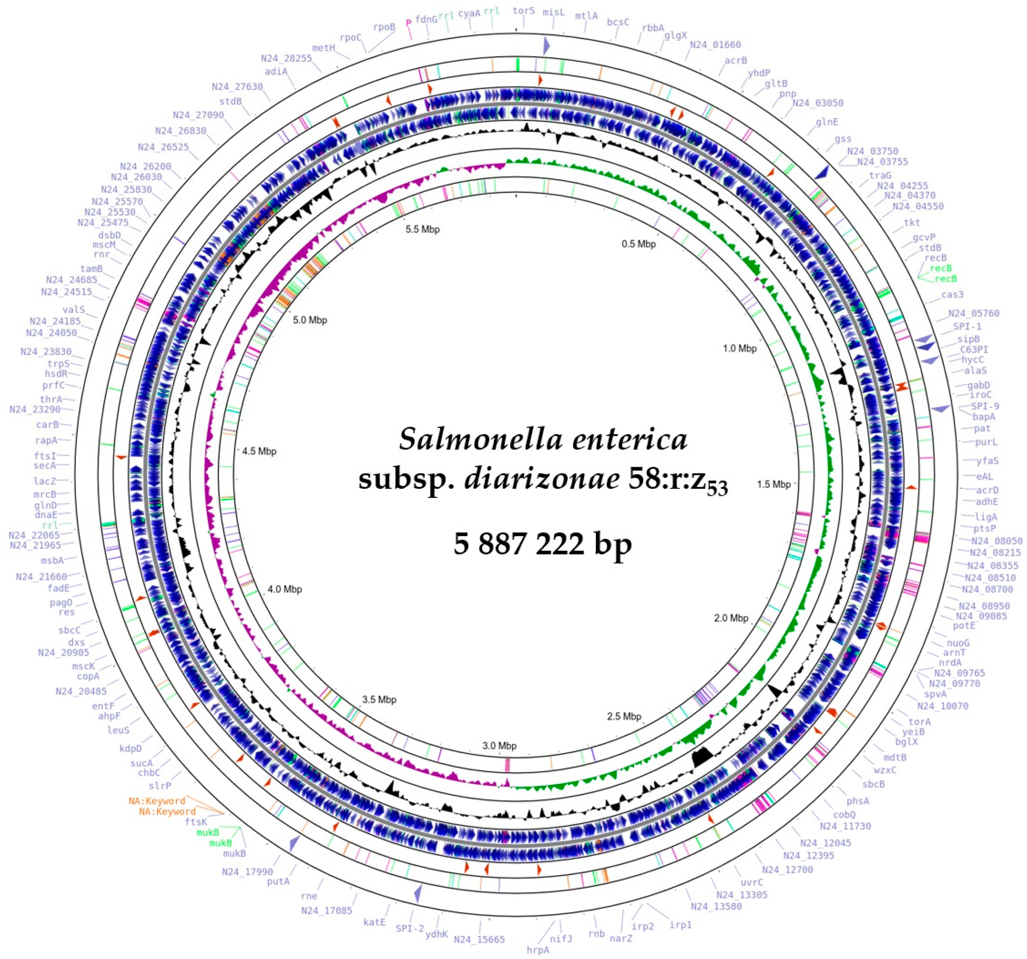
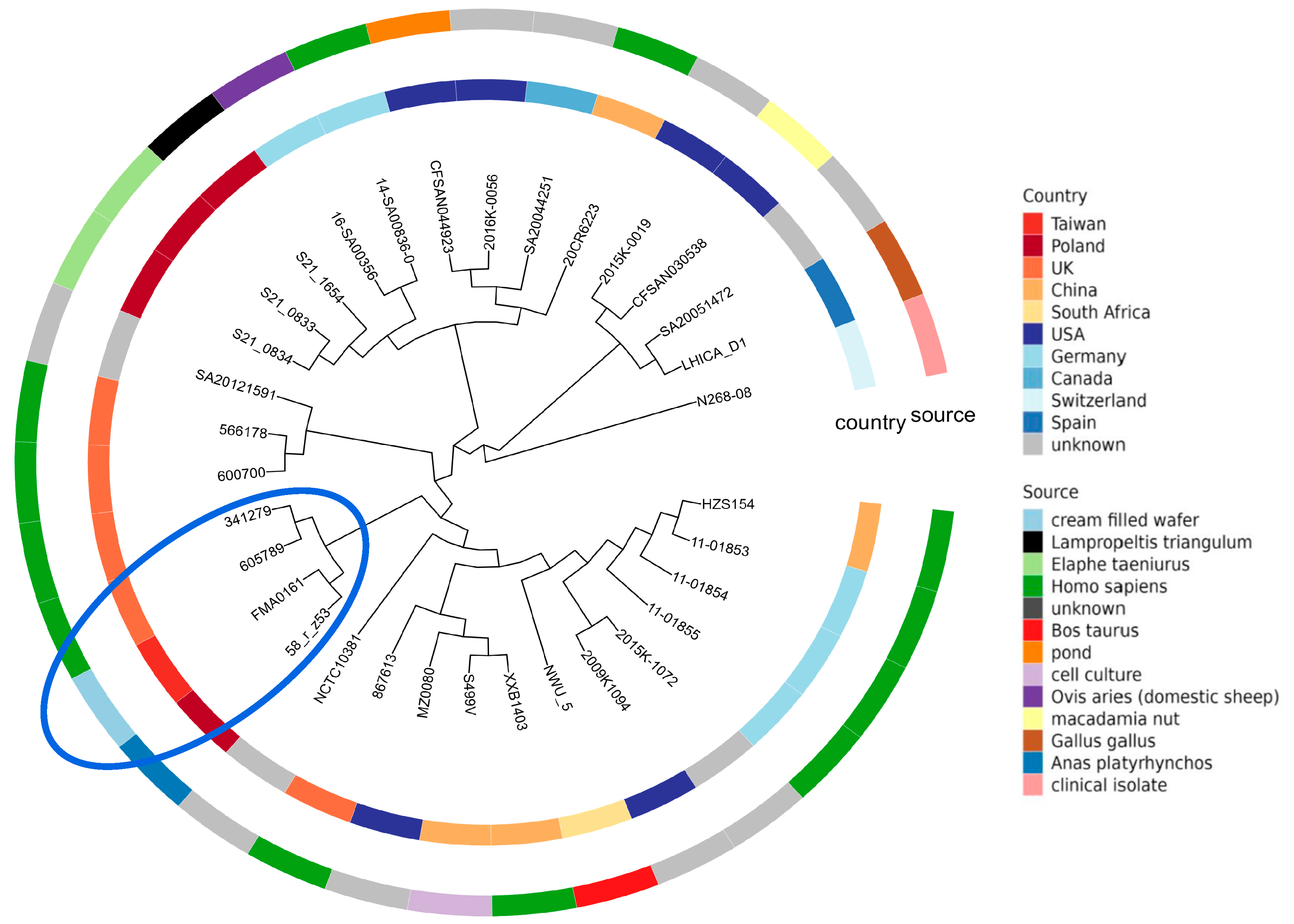
2.2. Phylogenetic Analysis of Salmonella enterica subsp. diarizonae IIIb 58:r:z53

The assembly of sequence reads revealed a single circular chromosome with a total length of 5,887,222 bp and an average guanine–cytosine content of 51.32%. This genome had 6308 protein-coding sequences (CDS), 102 transfer RNA (tRNA) genes, and 22 ribosomal RNA (rRNA) genes (Figure 1), Whole Genome map of S. enterica subsp. diarizonae 58:r:z53 (Figure 2).

2.3. Genotypic Serotyping

Multidirectional whole-genome sequencing (WGS) analysis predicted the O antigen to be the serotype 58 variant, the H1 antigen (fliC) to be r, and the H2 antigen (fljB) to be z53. The strain was tentatively classified as Salmonella enterica subsp. diarizonae (subspecies IIIb) serotype 58:r:z53 and named Salmonella IIIb 58:r:z53.

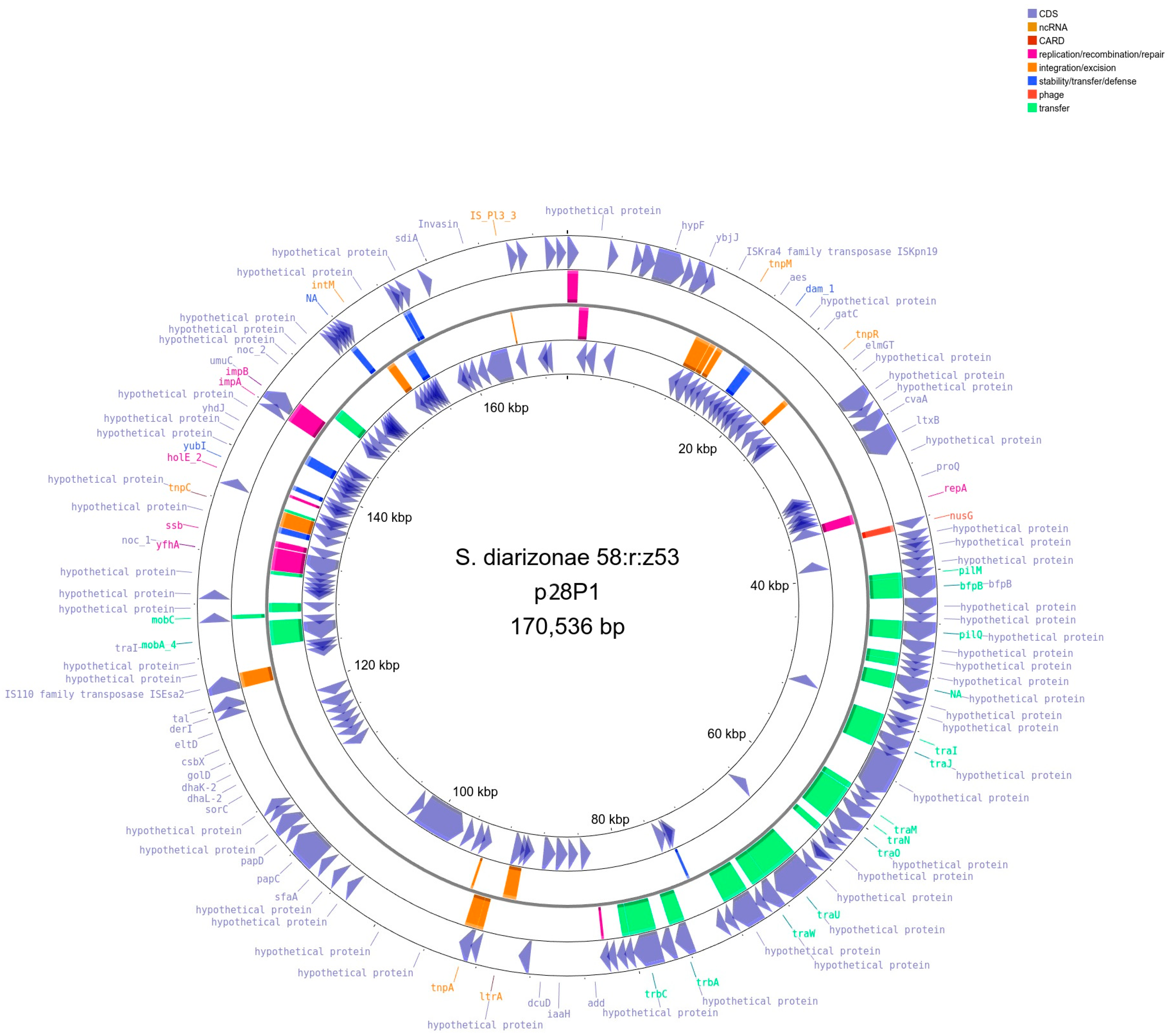
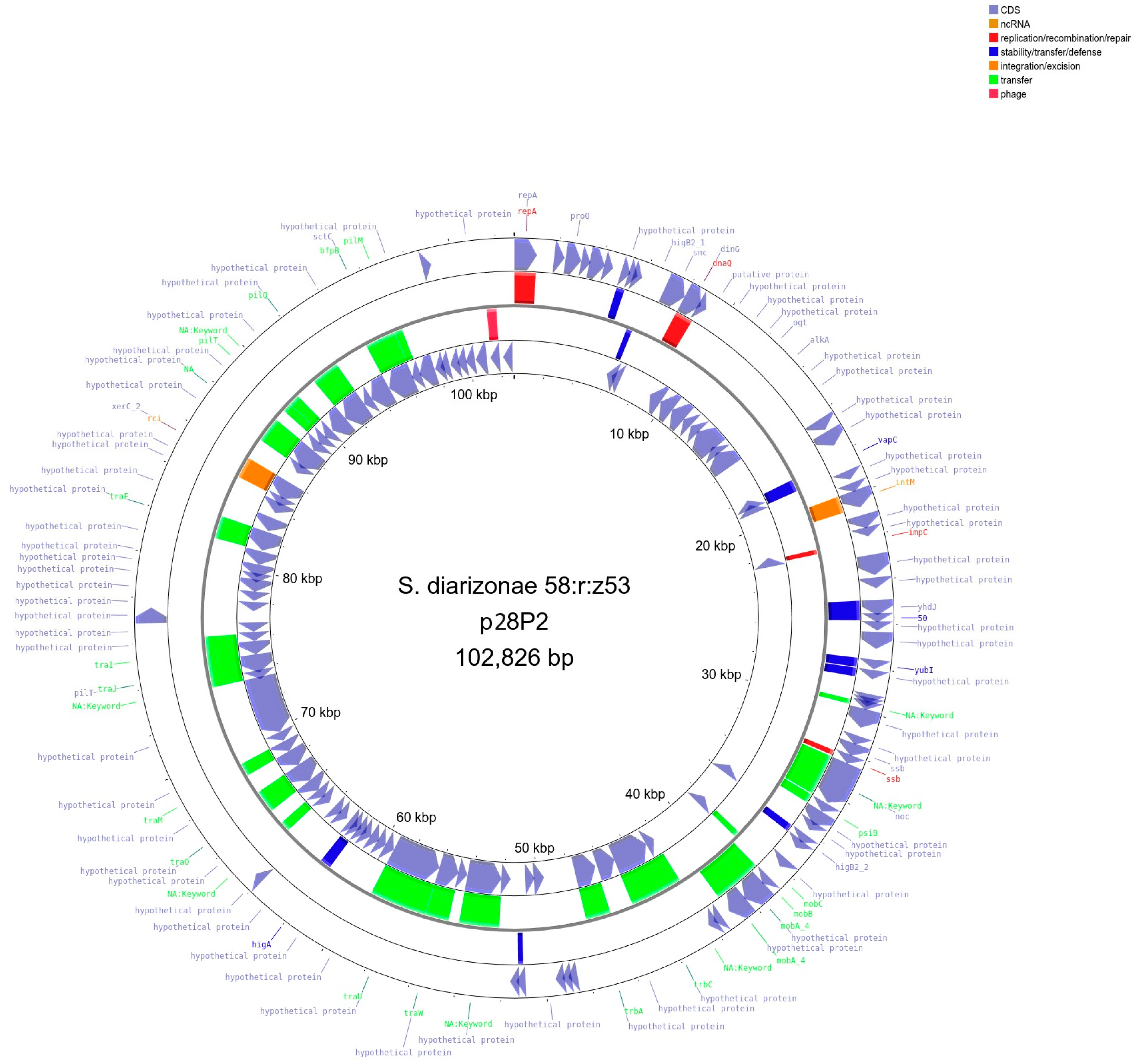
2.4. Detection and Initial Characterization of S. diarizonae 58:r:z53 Plasmids

Genome sequencing revealed that S. diarizonae 58:r:z53 contained three circular plasmids: plasmid 1 (p28P1, length 170,536 bp, Figure 3), plasmid 2 (p28P2, length 102,826 bp, Figure 4), and plasmid 3 (p28P3, 6462 bp, Figure 5). The plasmids did not carry antibiotic resistance genes but encoded several hypothetical proteins of unknown function, putative regulatory proteins, and genes associated with conjugation.

2.5. Salmonella diarizonae 58:r:z53 Pathogenicity Islands

Salmonella IIIb 58:r:z53 harbored six known pathogenicity islands (SPI-1, SPI-2, SPI-3, SPI-5, SPI-9, and SPI-13). An iron transport system was detected in SPI-1 centisome C63PI (Table 1).

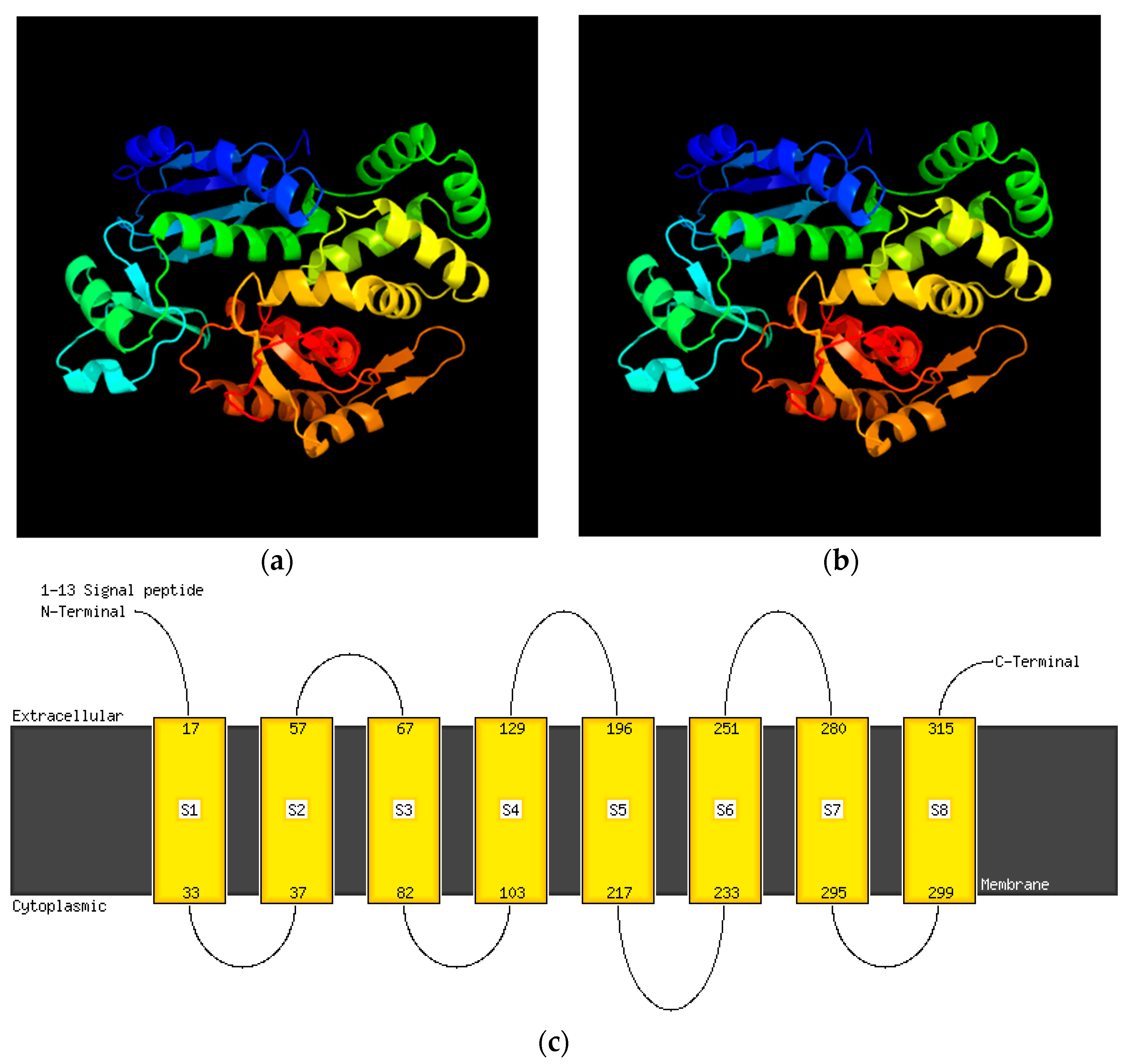
2.6. Virulence and Pathogenic Genes

The chromosome of S. diarizonae 58:r:z53 carried multiple virulence genes including type-III secretion systems, i.e., invA-J, spaO-S, sipA-D, sptP, prgH-J, orgA-C, avrA, cytolethal distending toxin B (cdtB), sopB, sopD2, sopE2, steC, sseF, sseG, sseJ, ssaC, ssaD, ssaG-V, sseB-D, and slrP. Besides the type-III secretion system genes, others for virulence in the chromosome were those for magnesium uptake (mgtA and mgtC), invasion proteins (orgA, orgB, and orgC), iron transporters (sitA, sitB, sitC, and sitD), and aerobactin siderophores (iucA, iucB, and iucC). Yersiniabactin genes involved in iron uptake (ybtA, ybtE, ybtP, ybtQ, ybtT, ybtU, ybtX, ybt S, and irp1,2) were also carried. The S. diarizonae 58:r:z53 genome was observed to contain operons of the chaperone/usher assembly class fimbrial genes: stc (yehB and yehB), bcf (bcfF), fim (fimA, fimC, fimD, fimF, fimH, fimI, fimW, fimY, and fimZ), stb (stbB, stbC, and stbD), and std (stdA, stdB, and stdC), all located on the chromosome. Genes coding thin aggregative fimbria (csgA, csgB, csgC, csgD, csgE, csgF, and csgG) and antimicrobial peptide resistance protein Mig-14 were also components of the genome. Plasmids p28P1 and p28P2 bore a significant portion of the SPI-7 genes predicted to be related to virulence (the pil-locus and tra-region). The relE and vapC genes were also identified in plasmid p28P2.

2.7. Comparative Analysis of Virulence Determinants in Salmonella enterica subsp. diarizonae Strains

Salmonella diarizonae 58:r:z53 InvA invasion protein was truncated (665 amino acids (aa)) and lacked the first 20 aa present in clinical isolates of Salmonella diarizonae (GenBank accession numbers CP059886.1, CP123007.1, CP054422.1, and CP011288.1) [18]. This truncation did not result in the loss of the InvA transmembrane structure (Figure 6, Supplementary Figure S1).
In contrast, the InvF protein was truncated (216 aa) in the clinical strains referred to in [18,19], one of which caused diarrhea and sepsis with a fatal outcome (GenBank accession number CP011288.1) [19] but these proteins had 100% identity with the InvF protein of the isolate (249 aa) (Figure 7 and Figure 8, secondary structure Figures S2 and S3) [20]. The fimbrial biogenesis chaperone StbB protein of S. diarizonae 58:r:z53 shared 100% of its sequence (253 aa) with that of an S. Typhi serovar (NCBI RefSeq number WP_053508616.1) and the fimbrial outer membrane usher StbC protein also shared its full sequence (230 aa) with that of Salmonella enterica subsp. diarizonae isolated from children (GenBank accession number JAHQRS010000001.1) [11,21].
Other invasion proteins (InfE and InvG), SPI-1 type-III secretion-system export apparatus proteins (SpaP, SpaQ, and SpaS), SPI-2 type-III secretion-system apparatus proteins (SsaS, SsaR, SsaP, and SsaK), the EscJ/YscJ/HrcJ family type-III secretion inner-membrane ring protein (SsaJ), the type-III secretion-system needle filament protein (SsaG), and the pathogenicity island 2 effector protein (SseG) showed 100% identity with their equivalents in clinical isolates of Salmonella diarizonae (GenBank accession numbers CP059886.1, CP123007.1, CP054422.1, and CP011288.1)

2.8. Antimicrobial Resistance of Salmonella IIIb 58:r:z53

A total of 20 different antimicrobial resistance genes were identified, all having been detected in the chromosome. Additionally, the tolC, marA, sdiA, and emrD genes were detected, which are associated with resistance to disinfecting agents and antiseptics including triclosan (Table 2, Supplementary Table S1).
The concordance between phenotypic resistance and the presence of known AMR genes was not consistent with the genotype for all antimicrobials. Most of the detected AMR genes were associated with aminoglycoside resistance and were in a Salmonella isolate, which was phenotypically gentamycin-, streptomycin-, and neomycin-resistant. Similarly, the present macrolide-resistance genes were associated with phenotypic resistance to erythromycin and tylosin in the IIIb 58:r:z53 strain. Fluoroquinolone-resistance genes were present in the S. IIIb 58:r:z53 strain, which was nevertheless sensitive to enrofloxacin and marbofloxacin but not to flumequine. Salmonella IIIb 58:r:z53 showed intermediate resistance to florphenicol and four genes (emrD, sdiA, acrA, and acrB) from the multidrug efflux pump family were identified. The carriage of tolC and marA, which confer carbapenem resistance, was detected in Salmonella IIIb 58:r:z53 but did not prevent the strain from being sensitive to imipenem.

2.9. Phylogenetic Analysis of Salmonella IIIb 58:r:z53

To infer the phylogenetic affinities of the Salmonella IIIb 58:r:z53 isolate, the maximum-likelihood phylogeny was estimated using kSNP4 (https://sourceforge.net/projects/ksnp/) and based on the core single-nucleotide polymorphisms computed from 31 genomes downloaded from the GenBank database (Supplementary Table S2). Additionally, whole-genome- and whole-proteome-based trees were inferred using the TYGS strain-typing server (Supplementary Figure S4).
Phylogenetic analysis using both tools showed that the strains named in the National Center for Biotechnology Information BioSample database as Salmonella enterica diarizonae serovar Salmonella arizonae 341279 and Salmonella enterica diarizonae serovar Salmonella diarizonae 605789 isolated from humans in the UK formed a single distinct clade with the tested strain. This single clade also included strain FMA0161 (Salmonella enterica subsp. diarizonae IIIb 58:r:-) isolated from cream-filled wafers in Taiwan in 2011, which showed the highest average nucleotide identity (99.65%) and conserved DNA threshold (84.15%) (Figure 9) (Supplementary Table S2).

3. Discussion

Salmonella enterica subsp. enterica is responsible for over 99% of cases of human salmonellosis and is therefore the subject of extensive research. However, there is still only very limited published research and genomic information about the non-enterica subspecies [12,13,23,24]. Our whole-genome analysis revealed that there were six known Salmonella pathogenicity islands in Salmonella IIIb 58:r:z53 (SPI-1, SPI-2, SPI-3, SPI-5, SPI-9, and SPI-13) and there was also centisome C63PI, an iron transport system. Genes from SPI-2, SPI-3, and SPI-13 are reported to be required for S. Typhimurium survival and replication in macrophages [24]. All of them were found in Salmonella IIIb 58:r:z53. In addition, SPI-1 and SPI-5, which are crucial for the intracellular lifestyle of the pathogen, were present in Salmonella IIIb 58:r:z53. The iron transport system C63PI in SPI-1, crucial for the entry of Salmonella into host cells, was also found in Salmonella IIIb 58:r:z53 [25].
Plasmids P1 and P2 contained many of the SPI-7 genes predicted to be related to virulence (the type IVb pilus pil and tra regions). Therefore, the presence of the pil locus in diarrheagenic bacteria may have contributed to their ability to infect humans.
The comparison of Salmonella IIIb 58:r:z53 with S. diarizonae isolated from newborn child infections revealed that 11 out of 12 type-III effector genes were present in all strains, namely steC, sseJ, sseG, sseF, sptP, sopE2, sopB, sipA, sipB, sipD, and avrA [26]. Moreover, Salmonella IIIb 58:r:z53 harbored the gene encoding the glycosyltransferase SseK2, which interferes with the proper immune response to infection through tumor necrosis factor-alpha-stimulated nuclear factor kappa B cell signaling. The host immune response is also hampered by the E3 ubiquitin ligase SlrP, known to have been produced by S. diarizonae and isolated from human and animal infections and by Salmonella IIIb 58:r:z53 because it inhibits the release of interleukin-1β. Fimbriae on the Salmonella cell surface mediate adhesion to host cells [27]. Salmonella IIIb 58:r:z53 bore the mig-14 gene coding antimicrobial resistance protein Mig-14, important in bacterial resistance to antimicrobial peptides and necessary for replication of the S. Typhimurium serovar in the liver and spleen [28].
The Salmonella IIIb 58:r:z53 P1 plasmid bore the samA gene, part of the samAB operon. The operon of which samA is part efficiently promotes UV mutagenesis when carried on a high-copy-number 60-MDa cryptic plasmid, as observed in research concerning the samAB operon in the Typhimurium serovar chromosome [29].
Aminoglycosides interfere with bacterial protein synthesis by binding to the bacterial 30S or ribosomal subunit. Aminoglycoside 6ʹ-N-acetyltransferase (AAC(6ʹ)) inactivates aminoglycoside antibiotics through acetylation of the 6-amino-acid group of the compound. One aminoglycoside found in S. enterica, AAC(6′)-Iy, is a cryptic chromosomally encoded aminoglycoside acetyltransferase that has been shown to confer extensive aminoglycoside resistance in strains expressing the structural gene.
Most of the Salmonella IIIb 58:r:z53 AMR mechanism was associated with its antibiotic efflux pump. Genes involved in the pump mechanism—marR encoding the MarR regulator of the AcrAB multidrug efflux pump and msbA encoding the multidrug resistance transporter—were found in Salmonella IIIb 58:r:z53 and S. diarizonae isolated from invasive newborn child infections [26]. Additionally, the Salmonella IIIb 58:r:z53 genome also contained tolC, marA, sdiA, and emrD associated with resistance to disinfecting agents and antiseptics including triclosan. A human isolate of S. enterica subsp. diarizonae serovar IIIb 48:i:z was found to contain the marA gene [26].
Three approaches to phylogenetic relationship reconstruction were taken in this study: core SNP identification and whole-genome- and whole-proteome-based strategies. They revealed that the S. IIIb 58:r:z53 strain was clustered with a Taiwanese strain (isolated from food) and S. enterica subsp. diarizonae from the UK (isolated from humans). This shows that the isolate presented in this study could unquestionably infect humans and the fact that it could be present in meat for human consumption indicates that it could be widespread and a real threat to public health.

4. Materials and Methods

4.1. Sampled Animal

The analyzed S. enterica spp. diarizonae (S. IIIb 58:r:z53) strain was isolated from the intestine of a mallard duck (Anas platyrhynchos) shot as prey in accordance with the hunting law in force in Poland [30,31]. The mallard duck was in good physical condition, with no pathological changes observed when given an anatomopathological examination.

4.2. Salmonella spp. Isolation and Identification

Salmonella spp. were isolated in accordance with PN-EN ISO 6579-1:2017-04 Microbiology of the food chain—horizontal method for the detection, enumeration, and serotyping of Salmonella—Part 1: Detection of Salmonella spp. [32]. The microbiological media used were described in a publication by Pławińska-Czarnak et al. [15]. Biochemical strain identification was performed using a VITEK® 2 GN (Gram-Negative) card and API20E test (BioMérieux, Craponne, France) according to the manufacturer’s instructions. For serological typing, the strains were originally grown on 2% nutrient agar slants and re-isolated on Salmonella-Shigella agar, Hektoen agar, Bismuth sulfite agar, and Xylose Lysine Deoxycholate agar (Merck, Darmstadt, Germany) before serotyping. The presumptive colonies of Salmonella strains were chosen and were cultured on Enrichment LAB-AGARTM (BioMaxima, Lublin, Poland) at 37 °C overnight. These cultures were used for serological identification. The antigenic formula of the Salmonella strains was determined with the use of standard agglutination methods and the serotype name was assigned according to the White–Kaufmann–LeMinor (WKL) scheme [1,33]. A small amount of bacterial mass from one colony was first checked by slide agglutination for a positive reaction with polyvalent HM serum and next, somatic O antigen was identified by slide agglutination in a drop of serum. Then, each strain was grown on swarm agar plates (BioMaxima, Lublin, Poland) at 37 °C overnight to test phases 1 and 2 of H antigens by slide agglutination. Polyvalent and monovalent anti-O and anti-H diagnostic sera for Salmonella antigens were purchased from SSI Diagnostica A/S (Hillerød, Denmark), Sifin Diagnostics GmbH (Berlin, Germany), and BIOMED (Kraków, Poland). Salmonella antigens were classified into serotypes by the WKL scheme. When a serovar had not been previously recorded in Poland, the strain representing this newly recognized serovar was sent to the WHO Collaborating Centre for Reference and Research on Salmonella (Institut Pasteur, Paris, France) for the identification to be verified and confirmed [15].

4.3. Antimicrobial Sensitivity Testing

The Salmonella strain was subcultured as described previously. From an 18–24 h culture, a suspension was prepared to 0.5 McFarland turbidity with a DensiCHEK Plus instrument (BioMérieux, Marcy-l’Étoile, France) and the inoculum was transferred to another VITEK® tube containing 3 mL of 0.45% saline. The card was automatically filled by a vacuum device and automatically sealed. It was manually inserted in the VITEK2 Compact reader–incubator module (BioMérieux, Craponne, France) and the card was automatically subjected to a kinetic fluorescence measurement every 15 min. This is a test methodology based on the minimum inhibitory concentration (MIC) technique reported by MacLowry and Marsh [34] and Gerlach [35]. To analyze MIC patterns of S. enterica subsp. diarizonae, a MERLIN MICRONAUT system (MERLIN Diagnostika GmbH, Bremen, Germany) was used. The MICs were interpreted according to the Clinical and Laboratory Standards Institute and Food and Drug Administration breakpoints [36]. The antimicrobial susceptibility was assessed by determining the MIC values using 96-well MICRONAUT Special Plates in a protocol described by Pławińska-Czarnak et al. in 2022 [37].

4.4. Whole-Genome Sequencing

Whole-genome sequencing of Salmonella spp. was performed at Genomed S.A. (Warsaw, Poland). Illumina sequencing was conducted in paired-end 300 bp mode on the MiSeq device (Illumina, San Diego, CA, USA) targeting 100× genome coverage. Sequencing was also performed using an SQK-RBK004 kit and an R9.4.1 flow cell on a MinION instrument (Oxford Nanopore Technologies, Oxford, UK). Short-read quality was assessed using FASTQC (https://www.bioinformatics.babraham.ac.uk/projects/fastqc) and sequencing data were quality trimmed using fastp [38]. Long reads were quality-filtered using NanoFilt [39] and the residual adaptor was removed using Porechop (https://github.com/rrwick/Porechop, accessed on 3 February 2024). The filtered dataset was finally quality-checked using NanoPlot [39]. Long-read assembly was performed using the Trycycler pipeline [40]. In brief, nanopore reads were initially assembled using four long-read assemblers—flye v. 2.9, unicycler v. 04.8, Raven v. 1.8.1, and miniasm v. 0.3-r179—next, the assemblies were reconciled and circularized, the final consensus was generated, and it was polished with Medaka (Oxford Nanopore Technologies). Circular replicons were further polished with short Illumina reads using the Polypolish (https://github.com/rrwick/Polypolish) [40] and POLCA [41] programs. The remaining ambiguities in the genome assembly were verified by PCR amplification of DNA fragments followed by Sanger sequencing with an ABI3730xl Genetic Analyzer (Life Technologies, Carlsbad, CA, USA) using BigDye Terminator Mix v. 3.1 chemistry (Life Technologies). All of the possible sequence errors and misassemblies were further manually corrected using Seqman software (DNAStar, Madison, WI, USA, https://www.dnastar.com/software/lasergene/seqman-ultra/?utm_source=google&utm_medium=cpc&utm_campaign=6773110442&utm_content=134971714457&utm_term=&utm_term=&utm_campaign=Branded+Keywords&utm_source=adwords&utm_medium=ppc&hsa_acc=7537872482&hsa_cam=6773) to obtain the complete nucleotide sequence of the bacterial genome.

4.5. Whole-Genome Analysis of Salmonella enterica spp. diarizonae 58:r:z53

General information about the assembly quality and gene content of Salmonella enterica subsp. diarizonae 58:r:z53 isolates and genomic components were obtained using the genomics tools of the Bacterial and Viral Bioinformatics Resource Center (BV-BRC, https://www.bv-brc.org, accessed on 29 February 2024). The serotype of strain S. IIIb 58:r:z53 was elucidated using SeqSero2 [42,43] and the genome sequence was annotated using Bakta software v. 1.8.2 and the Bakta database v. 5.0 (https://github.com/oschwengers/bakta) [44]. Salmonella pathogenic islands were detected using SPIFinder (https://cge.food.dtu.dk/services/SPIFinder/, accessed on 29 February 2024). Antibiotic resistance and virulence factor coding genes were detected using abricate (https://github.com/tseemann/abricate) by screening against the CARD (https://card.mcmaster.ca/) and VFDB (http://www.mgc.ac.cn/VFs/) databases, respectively. Genome maps of the S. IIIb 58:r:z53-strain chromosome and plasmids were visualized using the Proksee [17] and GenoVi packages [16].

4.6. Phylogenetic Analysis

The phylogenetic analysis of S. IIIb strain 58:r:z53 was performed including the genomic sequences of other S. enterica subsp. diarizonae members that are available in GenBank.
In total, 31 sequences were selected using the Referenceseeker tool [45] and based on an ANI value of 99% and conserved DNA threshold of 0.69. Those 31 comprised 10 incomplete genome sequences (drafts) and 21 complete ones. Salmonella bongori N268-08 was used as the outgroup. The sequences were uploaded to the TYGS typing server [46] and a whole-genome sequence-based tree was constructed.
Phylogenetic analysis was also performed taking a reference-free SNP-based approach using kSNP4 software [22]. A phylogenetic tree was constructed based on the maximum-likelihood method and visualized using the ggtree R package 3.12.0 (https://bioconductor.org/packages/release/bioc/html/ggtree.html, accessed on 29 February 2024).
The Phyre2 [47] web portal served as a resource for protein modeling, prediction, and analysis (http://www.sbg.bio.ic.ac.uk/phyre2, accessed on 19 March 2024).

Supplementary Materials

The supporting information can be downloaded at https://www.mdpi.com/article/10.3390/ijms25115664/s1.

Author Contributions

Conceptualization, J.P.-C. and K.W.; methodology, J.P.-C., K.W. and R.G.; Methodology—biochemical and serological typing and molecular typing, J.P.-C., K.W. and E.T.-P.; data curation, J.P.-C., K.W., E.T.-P., J.G., Z.S., J.B. and L.P.; software, J.P.-C., J.G., K.W. and T.N.; validation, J.P.-C., K.W. and K.A.; formal analysis, J.P.-C., K.W., E.T.-P., L.P., R.G,. and J.G.; investigation, K.W., Z.S. and Z.B.; resources, Z.B., J.B., J.W., T.N. and Z.B.; data curation, T.N., J.B., R.G. and J.G.; writing—original draft preparation, J.P.-C., K.W., J.B., J.W., T.N., E.T.-P., L.P., Z.B., R.G. and J.G.; writing—review and editing, J.P.-C., T.N., R.G. and K.A.; visualization, K.W., J.W., R.G. and J.G.; supervision, J.P.-C. and K.W.; project administration, J.P.-C.; funding acquisition, K.A. and Z.B. All authors have read and agreed to the published version of the manuscript.

Funding

This research received no external funding.

Institutional Review Board Statement

The study complied with Directive 2010/63/EU and the Act of the Polish Parliament dated 15 January 2015 on the protection of animals used for scientific purposes (Journal of Laws of the Republic of Poland 2015, item 266). The wild ducks were not killed for the purposes of the study. The hunt took place in accordance with Polish hunting law (Act of the Polish Parliament dated 13 October 1995, item 713, the Hunting law, Chapter 3, Art. 8 Hunt) during the 2021 hunting season.

Informed Consent Statement

Not applicable.

Data Availability Statement

The datasets presented in this study have been deposited in the BioProject database under accession number PRJNA1101854 and in the BioSample database under SAMN41003542, Genome accession numbers: CP152066-CP152069.

Acknowledgments

Special thanks to Przemysław Walerjan for the linguistic revision of the manuscript.

Conflicts of Interest

The authors declare no conflicts of interest. The funders had no role in the design of the study; in the collection, analyses, or interpretation of data; in the writing of the manuscript; or in the decision to publish the results.

References

  1. Issenhuth-Jeanjean, S.; Roggentin, P.; Mikoleit, M.; De Pinna, E.; Nair, S.; Fields, P.I.; Guibourdenche, M.; Weill, F.-X. Supplement 2008–2010 (no. 48) to the White–Kauffmann–Le Minor scheme. Res. Microbiol. 2014, 165, 526–530. [Google Scholar] [CrossRef]
  2. Havelaar, A.H.; Kirk, M.D.; Torgerson, P.R.; Gibb, H.J.; Hald, T.; Lake, R.J.; Praet, N.; Bellinger, D.C.; de Silva, N.R.; Gargouri, N.; et al. World Health Organization Global Estimates and Regional Comparisons of the Burden of Foodborne Disease in 2010. PLoS Med. 2015, 12, e1001923. [Google Scholar] [CrossRef] [PubMed]
  3. Gil, M.; Duma-kocan, P.; Rudy, M.; Stanisławczyk, R. Serowary Salmonella w żywności wg powiadomień systemu RASFF w latach 2000–2017. Med. Weter. 2019, 75, 394–397. [Google Scholar]
  4. Pires, S.M.; Vieira, A.R.; Hald, T.; Cole, D. Source attribution of human salmonellosis: An overview of methods and estimates. Foodborne Pathog. Dis. 2014, 11, 667–676. [Google Scholar] [CrossRef]
  5. Andino, A.; Hanning, I. Salmonella enterica: Survival, colonization, and virulence differences among serovars. Sci. World J. 2015, 2015, 520179. [Google Scholar] [CrossRef]
  6. EFSA. (European Food Safety Authority) Multi-country outbreak of multiple Salmonella enterica serotypes linked to imported sesame-based products. EFSA Support. Publ. 2021, 18, 6922E. [Google Scholar] [CrossRef]
  7. Toyting, J.; Nuanmuang, N.; Utrarachkij, F.; Supha, N.; Thongpanich, Y. Genomic analysis of Salmonella isolated from canal water in Bangkok, Thailand. Microbiol. Spectr. 2024, 12, e04216-23. [Google Scholar] [CrossRef]
  8. Schröter, M.; Roggentin, P.; Hofmann, J.; Speicher, A.; Laufs, R.; Mack, D. Pet Snakes as a Reservoir for Salmonella enterica subsp. diarizonae (Serogroup IIIb): A Prospective Study. Appl. Environ. Microbiol. 2004, 70, 613–615. [Google Scholar] [CrossRef]
  9. Dec, M.; Zając, M.; Puchalski, A.; Szczepaniak, K.; Urban-Chmiel, R. Pet Reptiles in Poland as a Potential Source of Transmission of Salmonella. Pathogens 2022, 11, 1125. [Google Scholar] [CrossRef]
  10. Molina-Lopez, R.A.; Valverdú, N.; Martin, M.; Mateu, E.; Obon, E.; Cerdà-Cuéllar, M.; Darwich, L. Wild raptors as carriers of antimicrobialresistant Salmonella and Campylobacter strains. Vet. Rec. 2011, 168, 565. [Google Scholar] [CrossRef]
  11. Zhou, M.; Shi, Q.; Zhang, X.; Mei, L.; Ye, Y.; Fang, C.; Shang, S. Salmonella enterica subsp. diarizonae Harboring ST233, ST1263, and ST1845 in Children. Front. Cell. Infect. Microbiol. 2021, 11, 727811. [Google Scholar] [CrossRef] [PubMed]
  12. Hervás, J.A.; Rosell, A.; Hervás, D.; Rubio, R.; Dueñas, J.; Mena, A. Reptile Pets–associated Salmonella enterica Subspecies diarizonae Gastroenteritis in a Neonate. Pediatr. Infect. Dis. J. 2012, 31, 1102–1103. [Google Scholar] [CrossRef] [PubMed]
  13. Fermaglich, L.J.; Routes, J.M.; Lye, P.S.; Kehl, S.C.; Havens, P.L. Salmonella cervical lymphadenitis in an immunocompetent child exposed to a snake at an educational exhibit: A case report. Infect. Dis. Clin. Pract. 2012, 20, 289–290. [Google Scholar] [CrossRef]
  14. Chong, Y.; Kwon, O.H.; Lee, S.Y.; Chung, K.S.; Shimada, T. Salmonella enterica subspecies diarizonae bacteremia in an infant with enteritis. A case report. Yonsei Med. J. 1991, 32, 275–278. [Google Scholar] [CrossRef] [PubMed]
  15. Pławińska-Czarnak, J.; Wódz, K.; Piechowicz, L.; Tokarska-Pietrzak, E.; Bełkot, Z.; Bogdan, J.; Wiśniewski, J.; Kwieciński, P.; Kwieciński, A.; Anusz, K. Wild Duck (Anas platyrhynchos) as a Source of Antibiotic-Resistant Salmonella enterica subsp. diarizonae O58—The First Report in Poland. Antibiotics 2022, 11, 530. [Google Scholar] [CrossRef] [PubMed]
  16. Cumsille, A.; Durán, R.E.; Rodríguez-Delherbe, A.; Saona-Urmeneta, V.; Cámara, B.; Seeger, M.; Araya, M.; Jara, N.; Buil-Aranda, C. GenoVi, an open-source automated circular genome visualizer for bacteria and archaea. PLoS Comput. Biol. 2023, 19, e1010998. [Google Scholar] [CrossRef]
  17. Grant, J.R.; Enns, E.; Marinier, E.; Mandal, A.; Herman, E.K.; Chen, C.Y.; Graham, M.; Van Domselaar, G.; Stothard, P. Proksee: In-depth characterization and visualization of bacterial genomes. Nucleic Acids Res. 2023, 51, W484–W492. [Google Scholar] [CrossRef]
  18. Uelze, L.; Borowiak, M.; Flieger, A.; Simon, S.; Tausch, S.H. Complete Genome Sequence of Salmonella enterica subsp. diarizonae Serovar 61:k:1,5,(7) Strain 14-SA00836-0, Isolated from Human Urine Laura. Microbiol. Resour. Announc. 2020, 9, 10–1128. [Google Scholar] [CrossRef] [PubMed]
  19. Gerlach, R.G.; Walter, S.; McClelland, M.; Schmidt, C.; Steglich, M.; Prager, R.; Bender, J.K.; Fuchs, S.; Schoerner, C.; Rabsch, W.; et al. Comparative whole genome analysis of three consecutive Salmonella diarizonae isolates. Int. J. Med. Microbiol. 2017, 307, 542–551. [Google Scholar] [CrossRef]
  20. Dufresne, K.; Saulnier-Bellemare, J.; Daigle, F. Functional analysis of the chaperone-usher fimbrial gene clusters of Salmonella enterica serovar Typhi. Front. Cell. Infect. Microbiol. 2018, 8, 26. [Google Scholar] [CrossRef]
  21. Seah, X.F.V.; Ngeow, J.H.A.; Thoon, K.C.; Tee, W.S.N.; Maiwald, M.; Chong, C.Y.; Tan, N.W.H. Infant With Invasive Nontyphoidal Salmonellosis and Mastitis. Glob. Pediatr. Health 2015, 2, 2333794X1559156. [Google Scholar] [CrossRef] [PubMed]
  22. Hall, B.G.; Nisbet, J. Building Phylogenetic Trees From Genome Sequences With kSNP4. Mol. Biol. Evol. 2023, 40, msad235. [Google Scholar] [CrossRef] [PubMed]
  23. Horvath, L.; Kraft, M.; Fostiropoulos, K.; Falkowski, A.; Tarr, P.E. Salmonella enterica subspecies diarizonae maxillary sinusitis in a snake handler: First report. Open Forum Infect. Dis. 2016, 3, ofw066. [Google Scholar] [CrossRef]
  24. Jiang, L.; Wang, P.; Song, X.; Zhang, H.; Ma, S.; Wang, J.; Li, W.; Lv, R.; Liu, X.; Ma, S.; et al. Salmonella Typhimurium reprograms macrophage metabolism via T3SS effector SopE2 to promote intracellular replication and virulence. Nat. Commun. 2021, 12, 879. [Google Scholar] [CrossRef] [PubMed]
  25. Schwan, C.L.; Lomonaco, S.; Bastos, L.M.; Cook, P.W.; Maher, J.; Trinetta, V.; Bhullar, M.; Phebus, R.K.; Gragg, S.; Kastner, J.; et al. Genotypic and Phenotypic Characterization of Antimicrobial Resistance Profiles in Non-typhoidal Salmonella enterica Strains Isolated From Cambodian Informal Markets. Front. Microbiol. 2021, 12, 711472. [Google Scholar] [CrossRef] [PubMed]
  26. Giner-Lamia, J.; Vinuesa, P.; Betancor, L.; Silva, C.; Bisio, J.; Soleto, L.; Chabalgoity, J.A.; Puente, J.L.; Soncini, F.C.; García-Vescovi, E.; et al. Genome analysis of Salmonella enterica subsp. diarizonae isolates from invasive human infections reveals enrichment of virulence-related functions in lineage ST1256. BMC Genom. 2019, 20, 99. [Google Scholar] [CrossRef] [PubMed]
  27. Jennings, E.; Thurston, T.L.M.; Holden, D.W. Salmonella SPI-2 Type III Secretion System Effectors: Molecular Mechanisms And Physiological Consequences. Cell Host Microbe 2017, 22, 217–231. [Google Scholar] [CrossRef] [PubMed]
  28. Brodsky, I.E.; Ghori, N.; Falkow, S.; Monack, D. Mig-14 is an inner membrane-associated protein that promotes Salmonella typhimurium resistance to CRAMP, survival within activated macrophages and persistent infection. Mol. Microbiol. 2005, 55, 954–972. [Google Scholar] [CrossRef] [PubMed]
  29. Gruz, P.; Matsui, K.; Sofuni, T.; Nohmi, T. Roles of the mutagenesis proteins SamA’B and MucA’B in chemically induced frameshift mutagenesis in Salmonella typhimurium hisD3052. Mutat. Res.-Fundam. Mol. Mech. Mutagen. 1998, 398, 33–42. [Google Scholar] [CrossRef]
  30. Gawin, P. Wild game in Poland. In Hunting Traditions, Law, Game; Gawin, P., Ed.; SBM Sp. z o.o.: Warsaw, Poland, 2015; pp. 129–131. ISBN 9788378458661. [Google Scholar]
  31. Regulation of the Minister of the Environment of March 11, 2005 on the List of Game Species Dz.U. 2005 nr 45 poz. 433. 2005. Available online: https://isap.sejm.gov.pl/isap.nsf/DocDetails.xsp?id=WDU20050450433 (accessed on 3 February 2024).
  32. PN-EN ISO 6579-1:2017-04; Microbiology of the Food Chain—Horizontal Method for the Detection, Enumeration, and Serotyping of Salmonella—Part 1: Detection of Salmonella spp. Polish Committee for Standardization: Warsaw, Poland, 2020.
  33. Grimont, P.A.D.; Weill, F.-X. Antigenic formulae of the Salmonella serovars. In WHO Collaborating Centre for Reference and Research on Salmonella, 9th ed.; WHO: Geneva, Switzerland, 2007; pp. 1–167. [Google Scholar]
  34. MacLowry, J.D.; Marsh, H.H. Semi-automatic microtechnique for serial dilution antibiotic sensitivity testing in the clinical laboratory. J. Lab. Clin. Med. 1968, 72, 685–687. [Google Scholar]
  35. Gerlach, E. Microdilution 1: A Comparative Study. In Current Techniques for Antibiotic Susceptibility Testing; Thomas, C.C., Ed.; Charles C. Thomas: Springfield, IL, USA, 1974; pp. 63–76. [Google Scholar]
  36. CLSI. Performance Standards for Antimicrobial Susceptibility Testing, 28th ed.; CLSI supplement M100; Clinical and Laboratory Standards Institute: Wayne, PA, USA, 2018; Volume 8, ISBN 156238838X. [Google Scholar]
  37. Pławińska-Czarnak, J.; Wódz, K.; Kizerwetter-Świda, M.; Bogdan, J.; Kwieciński, P.; Nowak, T.; Strzałkowska, Z.; Anusz, K. Multi-Drug Resistance to Salmonella spp. When Isolated from Raw Meat Products. Antibiotics 2022, 11, 876. [Google Scholar] [CrossRef] [PubMed]
  38. Chen, S.; Zhou, Y.; Chen, Y.; Gu, J. Fastp: An ultra-fast all-in-one FASTQ preprocessor. Bioinformatics 2018, 34, i884–i890. [Google Scholar] [CrossRef] [PubMed]
  39. De Coster, W.; D’Hert, S.; Schultz, D.T.; Cruts, M.; Van Broeckhoven, C. NanoPack: Visualizing and processing long-read sequencing data. Bioinformatics 2018, 34, 2666–2669. [Google Scholar] [CrossRef] [PubMed]
  40. Wick, R.R.; Holt, K.E. Polypolish: Short-read polishing of long-read bacterial genome assemblies. PLoS Comput. Biol. 2022, 18, e1009802. [Google Scholar] [CrossRef] [PubMed]
  41. Zimin, A.V.; Puiu, D.; Luo, M.C.; Zhu, T.; Koren, S.; Marçais, G.; Yorke, J.A.; Dvořák, J.; Salzberg, S.L. Hybrid assembly of the large and highly repetitive genome of Aegilops tauschii, a progenitor of bread wheat, with the MaSuRCA mega-reads algorithm. Genome Res. 2017, 27, 787–792. [Google Scholar] [CrossRef] [PubMed]
  42. Zhang, S.; Yin, Y.; Jones, M.B.; Zhang, Z.; Kaiser, B.L.D.; Dinsmore, B.A.; Fitzgerald, C.; Fields, P.I.; Deng, X. Salmonella serotype determination utilizing high-throughput genome sequencing data. J. Clin. Microbiol. 2015, 53, 1685–1692. [Google Scholar] [CrossRef] [PubMed]
  43. Zhang, S.; den Bakker, H.C.; Li, S.; Chen, J.; Dinsmore, B.A.; Lane, C.; Lauer, A.C.; Fields, P.I.; Deng, X. SeqSero2: Rapid and improved salmonella serotype determination using whole-genome sequencing data. Appl. Environ. Microbiol. 2019, 85, e01746-19. [Google Scholar] [CrossRef]
  44. Schwengers, O.; Jelonek, L.; Dieckmann, M.A.; Beyvers, S.; Blom, J.; Goesmann, A. Bakta: Rapid and standardized annotation of bacterial genomes via alignment-free sequence identification. Microb. Genom. 2021, 7, 000685. [Google Scholar] [CrossRef]
  45. Schwengers, O.; Hain, T.; Chakraborty, T.; Goesmann, A. ReferenceSeeker: Rapid determination of appropriate reference genomes. J. Open Source Softw. 2020, 5, 1994. [Google Scholar] [CrossRef]
  46. Meier-Kolthoff, J.P.; Göker, M. TYGS is an automated high-throughput platform for state-of-the-art genome-based taxonomy. Nat. Commun. 2019, 10, 2182. [Google Scholar] [CrossRef]
  47. Kelley, L.A.; Mezulis, S.; Yates, C.M.; Wass, M.N.; Sternberg, M.J. The Phyre2 web portal for protein modeling, prediction and analysis. Nat. Protoc. 2015, 10, 845–858. [Google Scholar] [CrossRef] [PubMed]
Figure 1. Illustration of the distribution of the genome annotations of Salmonella enterica subsp. diarizonae 58:r:z53 isolated from a mallard duck in Poland. The protein-coding sequence (CDS) elements of the figure are shown in gray for the position label (Mbp: megabase pairs); GC: guanine and cytosine; tRNA: transfer RNA; rRNA: ribosomal RNA. Visualized with GenoVi [16], accessed on 6 March 2024.
Ijms 25 05664 g001
Figure 2. Genome map of S. enterica subsp. diarizonae 58:r:z53. The innermost rings show genome positions (Mbp: megabase pairs) and GC content, shown in black. The outer rings represent the coding orientation, with the forward strand on the outside and the reverse strand on the inside. Created with Proksee [17].
Ijms 25 05664 g002
Figure 3. Illustration of the S. diarizonae 58:r:z53 plasmid 1 (p28P1, length 170,536 base pairs (bp)). The outermost and innermost rings represent the coding orientation, with the forward and reverse strands, respectively. The two central rings present mobile genetic element (MGE) annotation with the mobile orthologous groups database (Mobile OG DB). Regions involved in stability/transfer/defense are shown in blue, plasmid transfer in green, integration/excision in orange, and replication/recombination/repair in pink. Created with Proksee [17].
Ijms 25 05664 g003
Figure 4. Illustration of S. diarizonae 58:r:z53 plasmid 2 (p28P2, length 102,826 bp). The outermost and innermost rings represent the coding orientation, with the forward and reverse strands, respectively. The two central rings present MGE annotation with Mobile OG DB. Regions involved in stability/transfer/defense are shown in blue, plasmid transfer in green, integration/excision in orange, and replication/recombination/repair in pink. Created with Proksee [17].
Ijms 25 05664 g004
Figure 5. Illustration of S. diarizonae 58:r:z53 plasmid 3 (p28P3, 6462 bp). The outermost and innermost rings represent the coding orientation, with the forward and reverse strands, respectively. Regions involved in stability/transfer/defense are shown in blue, plasmid transfer in green, and replication/recombination/repair in pink. Created with Proksee [17].
Ijms 25 05664 g005
Figure 6. Three-dimensional view of (a) the full-length InvA protein (685 amino acids (aa)), (b) the truncated S. diarizonae 58:r:z53 InvA protein (665 aa), and (c) the truncated S. diarizonae 58:r:z53 InvA protein with no loss of transmembrane structure. Created with Phyre2, Protein Homology/analogY Recognition Engine V 2.0, Structural Bioinformatics Group, Imperial College, London, UK. (a,b) are rainbow-colored from the N to C terminus.
Ijms 25 05664 g006
Figure 7. Alignment of the InvF 28N protein isolated from S. diarizonae 58:r:z53 (249 aa) and the same protein from a clinical strain (GenBank accession number CP011288.1, 216 aa). Created with Color Align Properties in the Sequence Manipulation Suite (https://www.bioinformatics.org/sms2/color_align_prop.html, accessed on 26 April 2024).
Ijms 25 05664 g007
Figure 8. Three-dimensional view of (a) the truncated InvF protein (216 aa, 24.34 kDa) in S. diarizonae clinical isolates and (b) the full-length S. diarizonae 58:r:z53 28N InvF protein (249 aa, 28.30 kDa). Created by Phyre2. (a,b) are rainbow-colored from the N to C terminus.
Ijms 25 05664 g008
Figure 9. Phylogenetic analysis of the Salmonella enterica subsp. diarizonae IIIb 58:r:z53 strain as a maximum-likelihood tree of S. diarizonae isolates built using core genome single-nucleotide polymorphisms. The inner ring represents the country from which each isolate originated and the outer ring indicates the isolation source. Closely related strains are marked by the blue oval. Salmonella bongori N268-08 was used as the outgroup. All strains are presented in Table S2. Single-nucleotide polymorphisms were identified with kSNP4 [22].
Ijms 25 05664 g009
Table 1. Salmonella pathogenicity islands (SPIs) detected in the genome of Salmonella enterica subsp. diarizonae 58:r:z53 isolated from a mallard duck in Poland and the GenBank references of previous detections of those islands.
NameTypeStart/StopIdentity %Query/Template LengthGenesBacteriumAccession No.
C63PICDS1 219 752/1 223 75195.584000/4000sitA, sitB, sitC, sitDSalmonella-enterica-Typhimurium-SL1344AF128999
SPI-1CDS1 190 777/1 191 19196.14415/415invASalmonella-enterica-Gallinarum-SGB_8AY956825
SPI-1CDS1 189 639/1 189 89795.37259/257invASalmonella-enterica-Typhimurium-J4STEHOJN982040
SPI-1CDS1 171 725/1 174 41495.322692/3141fhlA/mutSSalmonella-enterica-Typhimurium-SL1344U16303
SPI-13CDS745 447/745 78498.22338/338gacDSalmonella-enterica-Gallinarum-SGD_3AY956832
SPI-13CDS746 092/746 49598.02404/404gtrASalmonella-enterica-Gallinarum-SGG_1AY956833
SPI-13CDS747 863/748 20398.53341/341gtrASalmonella-enterica-Gallinarum-SGA_10AY956834
SPI-2CDS3 159 479/3 164 10495.474634/4631ORF32, ORF48, pykFSalmonella-enterica-Typhimurium-LT2X99945
SPI-3CDS66 847/67 58496.75738/738mgtCSalmonella-enterica-Typhimurium-14028sAJ000509
SPI-5CDS3 455 305/3 456 55799.361253/1253sopBSalmonella-diarizonae-SARC7AF323077
SPI-9CDS1 323 107/1 335 75495.6512651/15696 Salmonella-enterica-Typhi-CT18NC_003198
ORF: open reading frame.
Table 2. Genes related to antibiotic resistance found in S. diarizonae 58:r:z53.
AMR MechanismGenesDrug Class
antibiotic inactivationAAC(6′)-Iyaminoglycoside
antibiotic effluxacrD, kdpEaminoglycoside
mdtB, mdtCaminocoumarin
baeR, cpxAaminocoumarin, aminoglycoside
msbAnitroimidazole
yojIpeptide antibiotic
emrA, emrB, emrRfluoroquinolone
crpfluoroquinolone, macrolide, penam
msrBstreptogramin, macrolide
h-nsmacrolide, penam, cephamycin, tetracycline, cephalosporin, fluoroquinolone
emrDphenicol, disinfecting agent, antiseptic
sdiA, E. coli acrAcephalosporin, glycylcycline, penam, tetracycline, fluoroquinolone, rifamycin, phenicol, triclosan
acrBtetracycline, rifamycin, glycylcycline, phenicol, penam, cephalosporin, fluoroquinolone, disinfecting agent, antiseptic
tolCpeptide antibiotic, aminoglycoside, tetracycline, glycylcycline, macrolide, fluoroquinolone, penam, carbapenem, penem, aminocoumarin, phenicol, cephalosporin, rifamycin, cephamycin, disinfecting agent, antiseptic
antibiotic target alterationbacApeptide antibiotic
glpT, uhpTfosfomycin
reduced permeability to antibiotics, antibiotic effluxmarAglycylcycline, cephalosporin, penam, cephamycin, monobactam, penem, tetracycline, fluoroquinolone, rifamycin, phenicol, carbapenem, triclosan
AMR: antimicrobial resistance.
Disclaimer/Publisher’s Note: The statements, opinions and data contained in all publications are solely those of the individual author(s) and contributor(s) and not of MDPI and/or the editor(s). MDPI and/or the editor(s) disclaim responsibility for any injury to people or property resulting from any ideas, methods, instructions or products referred to in the content.

