Review

Cross-Species Insights into Autosomal Dominant Polycystic Kidney Disease: Provide an Alternative View on Research Advancement

1
College of Animal Sciences, Zhejiang University, Hangzhou 310027, China
2
Department of Biotechnology, Hindustan Institute of Technology and Science, Tamil Nadu 603103, India
*
Author to whom correspondence should be addressed.
Int. J. Mol. Sci. 2024, 25(11), 5646; https://doi.org/10.3390/ijms25115646
Submission received: 1 April 2024 / Revised: 9 May 2024 / Accepted: 20 May 2024 / Published: 22 May 2024
(This article belongs to the Section Molecular Pathology, Diagnostics, and Therapeutics)

Abstract

:
Autosomal Dominant Polycystic Kidney Disease (ADPKD) is a prevalent hereditary disorder that affects the kidneys, characterized by the development of an excessive number of fluid-filled cysts of varying sizes in both kidneys. Along with the progression of ADPKD, these enlarged cysts displace normal kidney tissue, often accompanied by interstitial fibrosis and inflammation, and significantly impair renal function, leading to end-stage renal disease. Currently, the precise mechanisms underlying ADPKD remain elusive, and a definitive cure has yet to be discovered. This review delineates the epidemiology, pathological features, and clinical diagnostics of ADPKD or ADPKD-like disease across human populations, as well as companion animals and other domesticated species. A light has been shed on pivotal genes and biological pathways essential for preventing and managing ADPKD, which underscores the importance of cross-species research in addressing this complex condition. Treatment options are currently limited to Tolvaptan, dialysis, or surgical excision of large cysts. However, comparative studies of ADPKD across different species hold promise for unveiling novel insights and therapeutic strategies to combat this disease.

1. Introduction

In humans, numerous hereditary diseases, including Autosomal Dominant Polycystic Kidney Disease (ADPKD), can induce the development of renal cysts. In ADPKD, the primary pathogenic genes are typically PKD1 and PKD2, responsible for encoding Polycystin-1 (PC-1) and Polycystin-2 (PC-2), respectively. PC-1, with an approximate size of 460 kDa [1], presents as a membrane protein characterized by an extended N-terminal extracellular domain, 11 transmembrane domains, and a short intracellular C-terminal domain (Figure 1). The predominant localization of PC-1 spans in primary cilia of cells, tight junctions of cell membranes, desmosomes, and adhesion plaques [2]. In contrast, PC-2, smaller in size at around 110 kDa, harbors six transmembrane domains with intracellular N and C terminals (Figure 1). As a member of the transient receptor potential channel superfamily, PC-2 is situated in primary cilia, centrosomes, and the endoplasmic reticulum [2]. Functionally, PC-2 serves as a Ca2+ permeable non-selective cation channel.
ADPKD presents as a systemic disorder marked by the growth of numerous cysts within both kidneys. In addition to renal manifestations, ADPKD encompasses a range of extrarenal pathologies and symptoms, including the emergence of liver cysts, seminal vesicle cysts, pancreatic cysts, and hypertension, among other complications [3,4,5]. Cyst formation within the kidneys predominantly originates from the renal tubules and collecting ducts. The fluid composition within these cysts mirrors plasma, containing vital constituents such as Na+, K+, Cl, H+, creatinine, and urea. As the disease advances, cyst enlargement exerts continuous pressure on adjacent renal tissues, triggering cytokine activation and the recruitment of inflammatory cells, ultimately leading to interstitial inflammation and fibrosis [6]. Progressive decline in renal function culminates in renal insufficiency and eventual progression to end-stage renal disease, necessitating interventions such as dialysis or kidney transplantation. Furthermore, ADPKD represents a chronic condition with variable progression among individuals and species, posing diagnostic and management challenges. This variability underscores the considerable risk of misdiagnosis among susceptible patients. Symptoms resembling ADPKD have been documented in various animal species, including dogs, cats, pigs, cattle, and sheep [7,8,9,10,11,12,13,14,15]. Affected animals exhibit notable similarities in inheritance patterns, age of onset, clinical symptoms, and pathological features (Figure 2). The presence of genetically akin diseases across diverse species may be attributed to mutations in homologous genes [16].
The precise mechanisms governing the initiation and development of ADPKD remain incompletely elucidated. However, numerous models have been proposed to elucidate the pathogenesis of ADPKD and the mechanisms by which mutations in the PKD1 and PKD2 genes lead to the development of polycystic kidneys. These include the “two-hit” (carry a germline mutation with somatic mutations) and “third hit” (external factor stimulation) theories, the over-expression model (the expression of polycystic protein increased), the haploinsufficiency model (the expression level of the gene is reduced to 50% of the usual level), as well as the trans-heterozygosity (the expression of one allele related to cyst formation is reduced to 50% and a mutation occurs in another related gene within the somatic cell) and threshold models (expression above or below the steady-state threshold), which have been substantiated through experimental studies in mice [17,18,19,20,21,22]. Nevertheless, several signaling pathways are implicated in its pathogenesis, consisting of the HIF-1α, mTOR, TSC-1/2 complex, Wnt, JAK/STAT, AMPK, and PPAR-γ pathways [23,24,25,26,27,28,29]. In addition, the dysregulation of Ca2+ channels and cAMP signaling has been observed as contributing to the accelerated proliferation of cystic epithelial cells [30]. In vitro data and animal models of non-human origin have demonstrated a reduction in autophagy flux in ADPKD [31]. Furthermore, current studies indicate that metabolic disorientation emerges as a pivotal characteristic of ADPKD, with affected individuals predisposed to aberrant metabolic reactions or accumulation of metabolites, exacerbating disease progression [32].

2. Comprehensive Investigation of Multi-Species Epidemiology

ADPKD represents a widespread condition globally, standing as the most prevalent form of polycystic kidney diseases. Being autosomal dominant inheritance in nature, ADPKD exerts its impact on millions of people worldwide. Present estimation indicates that around 12 million individuals globally are inflicted with ADPKD [33], with approximately 1.5 million cases documented in China alone [34]. As lifestyles shift with aging and altered habits, the incidence of this malady is expected to surge further, while prevalence rates manifest variability across distinct regions and among diverse ethnic cohorts [35]. Thus, ADPKD imposes a pressing global health concern, affecting approximately 1 in 400 to 1 in 1000 individuals. This condition manifests across both adult and pediatric populations without evident gender bias. The presence of a familial history of ADPKD significantly escalates the risk of other family members to develop the disease. In Europe, ADPKD ranks as the fourth most prevalent cause of kidney disease necessitating dialysis or replacement therapy, with one in ten patients requiring renal replacement therapy due to ADPKD [35,36]. In China, data gleaned from Shanghai’s registration records reveal that ADPKD accounts for 4.7% of end-stage renal disease (ESRD) cases [34], emerging as a principal cause of end-stage renal failure, accounting for 10% of all cases [33].
In addition to humans, ADPKD or other related polycystic kidney diseases emerge as a frequently encountered hereditary kidney disorder in cats to date [37,38,39]. Currently, the PKD1 gene has been pinpointed as the primary cause of the ADPKD in cats, yet the presence of additional mutations potentially influencing feline polycystic kidney diseases remains an area of investigation [40,41,42]. Polycystic kidney disease is prevalent among adult Persian cats and breeds with Persian lineage, encompassing cherished breeds like the Garfield and other hybrid varieties. However, ADPKD is not confined solely to Persian-related felines; occurrences have also been reported in British shorthairs, American shorthairs, exotic longhairs, kinjiras, maines, German curlys, and various other cat breeds [43,44]. Globally, an estimated 40% to 50% of Persian cats, comprising approximately 6% of the worldwide feline population, are afflicted with ADPKD [45,46,47,48], affecting both sexes without marked gender preference [49].
While a number of reports have addressed polycystic kidney disease in dogs [50,51,52,53,54], a conclusive genetic link to ADPKD remains elusive. Inbreeding practices within specific dog breeds have increased the likelihood of harboring the mutated gene. Notably, breeds such as the West highland white terrier, bull terrier, cairn terrier, and various other terrier breeds exhibit heightened susceptibility, although the instances of polycystic kidneys have also been noted in other breeds, including shepherd dogs. In Australia, bull terrier polycystic kidney disease (BTPKD) demonstrates particularly high incidence rates, particularly among dogs showcased in pet exhibitions and breeding establishments. Data summarization indicates a comparable incidence between male and female dogs, with no discernible gender bias observed [55].
Polycystic kidney disease was also seen in the domesticated animals such as pigs, cattle, and sheep. Clinically diagnosed cases in these animals are rare, and it is typically identified post-mortem during slaughter, revealing kidneys riddled with numerous cysts. Studies estimate that approximately 26% of cattle and 47% of pigs exhibit renal cysts [56,57]. The restricted breeding practices in livestock, characterized by a small number of selected paternal and maternal lines, result in nearly half of the offspring inheriting kidney cysts. However, since most livestock are slaughtered at a young age, the potential risks associated with this disease are often overlooked. However, considering the impact of ADPKD on human fertility [58], it is reasonable to speculate that, in addition to kidney cyst formation, cysts may also arise in the seminal vesicles of livestock. This could potentially compromise the reproductive performance of breeding animals, leading to diminished sperm production and quality, as well as reduced fertilization and conception rates. Consequently, this may lead into lower pregnancy rates among females, an increased risk of miscarriage, fewer newborn offspring, and the birth of weaker, malformed young animals. Such outcomes markedly escalate the expenses associated with breeding while diminishing production efficiency. Therefore, it is imperative to conduct research into ADPKD in livestock to gain a deeper understanding of the disease and address these potential challenges.
Regardless of the species—such as human, dog, cat, pig, or other animals—the actual prevalence of ADPKD is often considerably higher than commonly perceived due to the absence of early diagnosis and effective reproductive management. If left unaddressed, this could result in a more widespread occurrence of ADPKD. Therefore, it is imperative to screen and test individuals with a family history of the disease and animals exhibiting related symptoms to detect ADPKD at the earliest opportunity. Moreover, genetic testing should be prioritized in animal breeding programs to mitigate the propagation of the polycystic kidney gene, thereby reducing the incidence of ADPKD.

3. Pathological Features and Cross-Species Manifestations of ADPKD

ADPKD is an inherited condition that affects various species. Despite variations in symptoms and severities across different species, there are common pathological characteristics. The hallmark of ADPKD is the formation of cysts of varying sizes in both kidneys, which can be found across the renal cortex and medulla. Additionally, a subset of patients may develop cysts in the liver, potentially progressing to severe Polycystic Liver Disease (PLD). The condition exhibits a higher prevalence in females, while male patients typically exhibit cysts in the kidneys and seminal vesicles. Moreover, cysts can also occur in the pancreas, spleen, thyroid, and other areas (Table 1) [57,58,59,60]. Experimental research has shed light on the role of Pkd1 gene variations in the development and severity of ADPKD in mice [61]. Mice possessing a typical Pkd1 gene remain disease-free, while the presence of even a single, less potent variant of the Pkd1 gene can trigger swift cystic changes within the kidneys. Furthermore, mice with two copies of such gene variants exhibit a gradual onset of renal cysts. These observations underscore the critical influence of PC1 protein dosage, which is encoded by the Pkd1 gene, on disease severity. The phenotype of ADPKD intensifies as the levels of PC1 protein decrease, thereby highlighting the profound effects of gene dosage on the disease’s manifestation. Collectively, these insights substantiate the pivotal role of gene dosage effects in the pathogenesis of ADPKD. As the disease progresses, these cysts gradually enlarge, exerting pressure on surrounding normal tissues and causing local tissue damage. Histological examination of the kidneys in ADPKD patients reveals significant interstitial inflammation, characterized by a high infiltration of inflammatory cells into the interstitium (Table 1). Immunohistochemical analysis of kidneys from advanced ADPKD patients demonstrates increased expression of fibrosis markers such as α-SMA, indicating interstitial fibrosis. Moreover, there is a decrease in the expression of E-cadherin, a marker of epithelial cells, alongside an increase in N-cadherin, a mesenchymal marker, suggesting a dedifferentiation of renal epithelial cells and consequent changes in cell function [62]. Furthermore, the disease is characterized by epithelial-mesenchymal transition (EMT) and excessive accumulation of extracellular matrix (ECM) (Table 1) [62].
In the advanced stages, renal tissue undergoes atrophy, healthy renal parenchyma is gradually replaced, and ultimately leads to chronic renal failure. At this stage, both kidneys may increase in volume, often assuming irregular shapes. While symptoms may be subtle in early stages, as the disease progresses to intermediate or late stages, kidneys gradually lose normal physiological function due to the relentless compression of healthy tissue by expanding cysts. Severe cases result in irreversible renal failure, accompanied by complications such as azotemia, hyperphosphatemia, acidosis, and anemia. Additionally, studies indicate that ADPKD patients and their families face an increased risk of heart disease, including aortic valve malformation and aortic root dilation [63], this is due to the fact that polycystin proteins also play an important role in the development and maintenance of the cardiovascular system, and their abnormalities may indirectly affect the cardiovascular system, leading to vascular dysregulation, which significantly impacting their quality of life and posing life-threatening risks. In addition, most patients in the early stages of ADPKD exhibit normal renal function; however, hypertension prevalence can escalate to as high as 50%. Hypertension will also heighten the risk of cardiovascular disease. Without the timely control of ADPKD, the condition will persist in deteriorating, resulting in a continuous rise in blood pressure. This imposes a significant burden on both the heart and kidneys, hastening the progression towards renal failure. ADPKD-like disease in dogs and cats closely resembles that in humans (Table 1 and Table 2). The majority of cysts were formed within the renal cortex and medulla, with the quantity and size varying based on individuals and distinct breeds [64]. Additionally, cysts may form in the liver, pancreas, uterus, and abdominal cavity. Some dogs and cats may exhibit focal proliferation of renal tubular epithelium and develop heart disease, particularly the Bulldogs, who appear to have an elevated risk for cardiac issues [65]. Moreover, in cats, chronic renal failure has been linked to hypertension [66,67,68,69]. Studies comparing blood pressure indices in normal cats versus those with mild and severe ADPKD symptoms have shown that cats with ADPKD have an increased average arterial pressure compared to the control group [70]. Histological examination, ultrastructural analysis, and lectin staining have indicated that the cysts most likely originate from the nephron segments and collecting ducts. BrdU-based proliferation studies have revealed a higher proliferation rate of cyst lining epithelial cells in some ADPKD-affected kittens compared to healthy kittens [64]. Additionally, research has delved into the histological and ultrastructural characteristics of renal lesions in BTPKD, identifying that cysts in BTPKD may arise from either nephrons or collecting ducts [71,72]. Initially, most young animals exhibit minimal symptoms, typically presenting with small cysts in limited numbers, while renal function remains relatively unaffected. However, in adult or elderly dogs and cats, cysts significantly increase in size and quantity, often accompanied by renal dysfunction. As time progresses, the condition of affected dogs and cats deteriorates gradually, culminating in end-stage renal disease. Nevertheless, some dogs and cats may remain asymptomatic for most of their lives, only experiencing renal failure in later stages (usually after the age of 7). Cats, in particular, tend to develop renal failure earlier than dogs and may succumb to other ailments before renal failure becomes a significant factor.
In livestock, such as pigs, cattle, and sheep, cyst formation typically occurs in the kidneys and liver, leading to the enlargement of these organs (Table 1). Some animals may also develop ascites, wherein affected piglets accumulate significant amounts of fluid in their abdomens, resulting in pronounced distension. Both kidneys of the livestock are usually affected, exhibiting a marked increase in volume compared to healthy organs. Upon sectioning, numerous cysts are visible, permeating throughout the medulla and cortex, creating a honeycomb-like structure [73]. Conversely, the affected liver presents a greenish-brown hue, displaying evident bile duct hyperplasia and containing numerous irregularly shaped cystic vesicles filled with fluid. Microscopic examination of the kidneys using Hematoxylin and Eosin (H&E) and Masson’s trichrome staining methods reveals that the cysts exert pressure on the surrounding cortex and medulla (Table 2). These cysts are lined by a layer of flattened epithelial cells, bordered by fibrous connective tissue that often extends and replaces the renal parenchyma. In these regions, renal tubules undergo atrophy and degeneration. While ADPKD in livestock may not attract the same societal attention as sudden events like infectious epidemics, its impact on the health and production efficiency of livestock is significant and cannot be overlooked. In humans, ADPKD exhibits considerable clinical diversity not only among different families but also among members of the same family. This diversity is evident in the disease’s manifestations, its severity, and the age at which it becomes apparent, resulting in a spectrum of disease experiences even among individuals sharing identical genetic predispositions. Although ADPKD is the most prevalent hereditary kidney condition in adults, the age of disease onset varies, with some individuals experiencing early onset forms, while others have a later onset.
The alignment of symptoms across diverse species hints at the probability of shared gene mutations underlying ADPKD. Unraveling the differences and connections about ADPKD across various species can enrich our comprehension of the molecular mechanisms and genetic traits associated with this condition. Through syntheses and analyses of these data, we can pinpoint commonalities that aid in identifying pivotal genes and pathways involved in the disease’s pathogenesis. This interdisciplinary research methodology promises invaluable insights and direction for the prevention and treatment of ADPKD, furnishing us with a more robust toolkit to address the challenges posed by this condition.

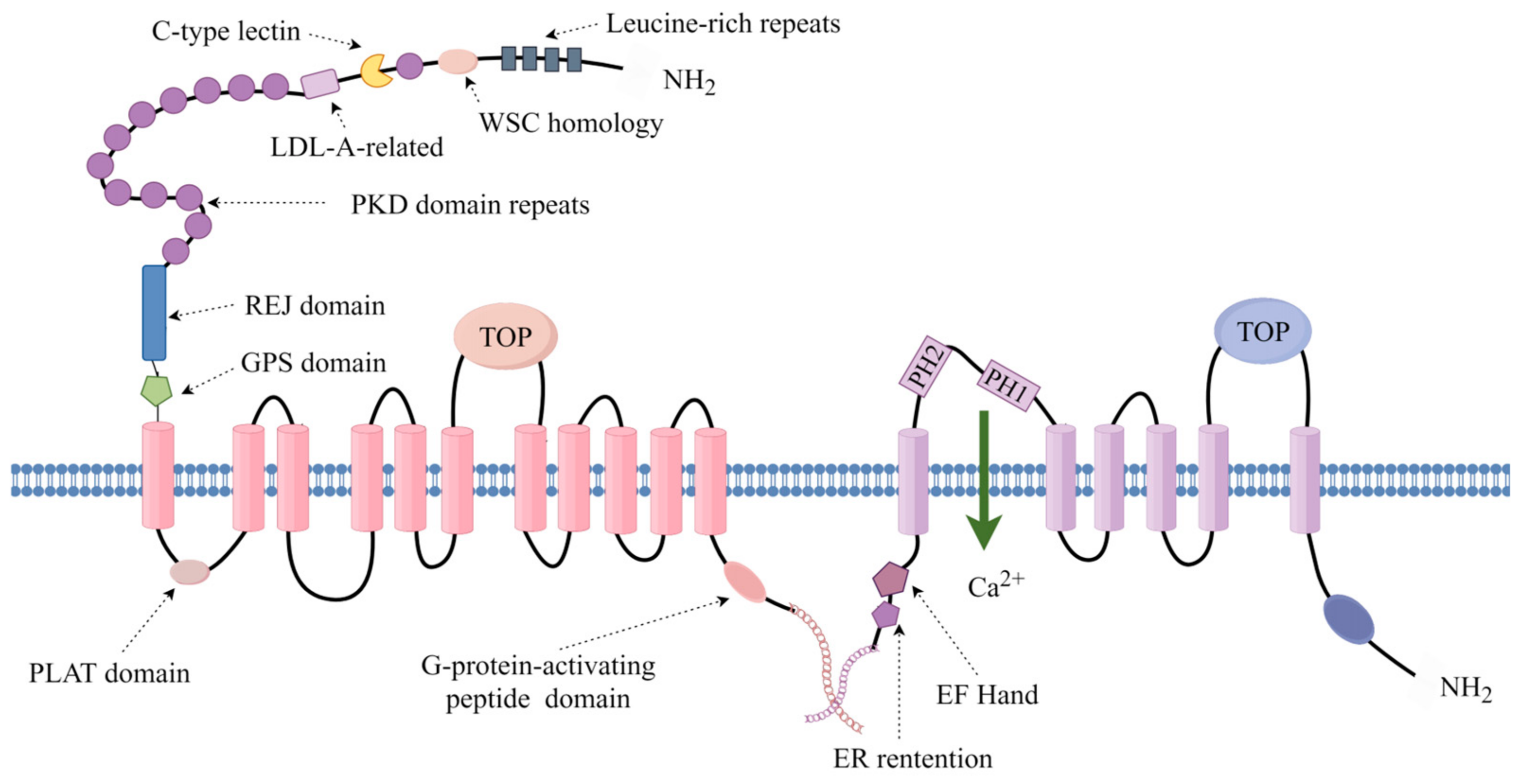
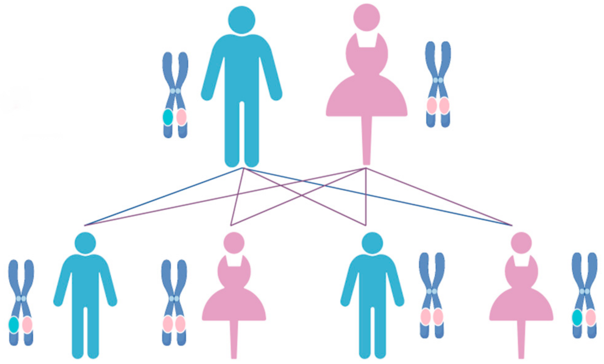
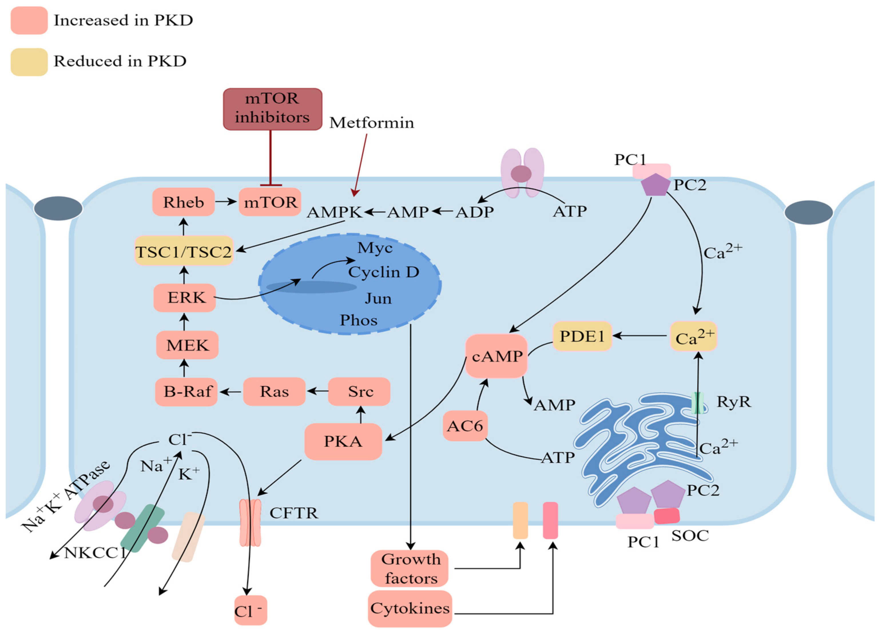
4. Molecular and Genetic Mechanisms of ADPKD across Species

In humans, ADPKD is usually caused by mutations in two genes, PKD1 and PKD2, and the presence of a mutated gene in an individual can precipitate symptom manifestation [74]. Most of the ADPKD cases arise from inherited mutated genes from parents, with only around 10% stemming from de novo mutations. PKD1 and PKD2 encode PC-1 and PC-2, respectively and it is recognized that several proteins can interact with PC-1 or PC-2, thereby regulating diverse signaling pathways [75]. Both PC-1 and PC-2 harbor N-linked glycosylation modification sites, and ALG8, ALG9 also undergo glycosylation within the endoplasmic reticulum. GANAB (neutral α-glucosidase AB) is an α-glucosidase primarily responsible for the catalysis of hydrolysis reactions involving incomplete glucose moieties on the endoplasmic reticulum. Furthermore, the genes encoding these proteins have been reported as one of the potential pathogenic loci for ADPKD. Studies have suggested that GANAB and PC-1 can confer PC-1 stability through interaction.
PC-1 and PC-2 form unique heterotetrametric complexes (1 PC-1 + 3 PC-2) that can interact at the primary cilia of renal epithelial cells [75,76] through their C-terminal interactions, participating in the regulation of cell differentiation, proliferation, and apoptosis [77]. Studies have demonstrated that primary cilia function as sensory antennae, adept at detecting chemical and mechanical stimuli, including blood flow sensation in renal tubules, and are integral in maintaining renal epithelial cell homeostasis. Disruption of cilia has been implicated in renal cysts initiation [78]. These complexes play a role in regulating cellular calcium ion channel function, in which process ALG8 and ALG9 play important roles. Upon stimulation by abnormal fluid flow within the renal tubules, this channel can disrupt calcium-related pathways, leading to an imbalance in Ca2+ signaling (Figure 3). This disruption alters intracellular Ca2+ levels, impacting cAMP synthesis and degradation [79]. Fluctuations in cAMP levels can stimulate cell proliferation derived from cysts [80]. Furthermore, signaling pathways involving MEK, ERK, CREB, STAT, STAT6, and mTOR are upregulated (Figure 3), contributing to the repair and proliferation of damaged renal tissue [81,82]. Remarkably, mTOR plays a pivotal role in regulating cell growth and division, as well as inhibiting catabolic processes. mTOR promotes cell growth by inhibiting protein catabolism, such as through autophagy regulation [83]. A study has identified a decline in lysosomal acidification, LAMP degradation, and CTSB/tissue protease B processing and activity in PKD1-deficient mIMCD3 cells. This reduction disrupts autophagosome-lysosome fusion, decreases ubiquitination from multivesicular bodies (MVB)/exosomes to lysosomes, and increases the secretion of unprocessed CTSB into the extracellular space [84]. Moreover, mTOR can activate the PI3K/Akt pathway. When Akt is activated, it phosphorylates the GSK3 subtype at its highly conserved N-terminal regulatory site, thereby inactivating the kinase and regulating cell apoptosis and glucose metabolism through GSK3. Akt also influences cell apoptosis, growth, proliferation, metabolism, protein synthesis, and transcription by phosphorylating and inhibiting FoxO1/3a transcription and TSC2, and can reduce intracellular calcium levels [85]. Under conditions of low ATP levels, hypoxia, or DNA damage, the stress response metabolic regulatory factor AMPK is activated to inhibit TSC2 [83].
The Wnt signaling pathway plays a crucial role in cyst formation, influencing cell proliferation, differentiation, and apoptosis [86]. Dysregulation of this pathway can significantly contribute to cyst growth. Abnormal activation of Wnt signaling leads to increased proliferation and fluid secretion by renal epithelial cells, facilitating the continuous expansion and dissemination of cysts. Equally, beyond signaling irregularities within cystic structures, abnormal expression of TGF-β in renal fibrosis areas suggests a potential contribution of renal fibrosis to eventual renal failure in ADPKD [87]. In addition, abnormal oxidative stress and inflammatory responses can damage cells and tissues. In ADPKD, oxidative stress may accelerate the development of renal cysts by affecting cellular nucleic acids and proteins [88].
A vast amount of data is available on the mechanisms underlying human ADPKD, particularly through the research conducted in mice, which has yielded valuable insights into the underlying mechanism. However, there is a lack of study specifically examining the molecular mechanisms of ADPKD in dogs, cats, pigs, and other animal species. Human PKD1 contains several polypyrimidine tracts, and many ADPKD patients exhibit deletion mutations in one or more polypyrimidine regions near the intron 21 and intron 22 [89]. In contrast, in other species, the polypyrimidine region may be absent or possess a different length and sequence composition. For example, mice and rats lack this region, and spontaneous ADPKD has not been reported in these rodent models. In the pigs, the polypyrimidine region may be involved in the regulation of gene expression, affecting the development, growth and diseases, although there is no exact research or evidence that the polypyrimidine region is directly related to its spontaneous PKD [90]. To date, no reported homozygous animals with PKD1 or PKD2 mutations exist, suggesting that such mutations may result in embryonic lethality. Thus, several heterozygous mutations in these animals have been detected. Specific mutation in the PKD1 gene of cats has been identified (Table 3), where the adenine at the 3284th position in exon 32 is replaced by cytosine (C.10063C>A), accounting for approximately 30% of diseased Persian cats. This mutation introduces premature termination codons into the mRNA, leading to the loss of 25% of the C-terminus during the formation of PC-1. A linkage analysis study on seven Persian cat families revealed a significant association between the ADPKD phenotype and the FCA476 marker, which is located within the 10 cR region of the cat PKD1 gene on the E3 chromosome [91]. This suggests that the PKD1 gene or other genes within this region may be implicated in ADPKD in cats. In dogs, ADPKD is prevalent in certain Bulldog lines, resembling the condition observed in humans. The gene, situated on the dog chromosome 6, is linked to ADPKD and shares homology with the region of the human chromosome containing the PKD1 gene. Studies have identified a mutation of PKD1 gene G>A on dog chromosome 29 associated with BTPKD (Table 3). This mutation serves as a diagnostic tool to assess the risk of ADPKD in young and breeding dogs [92]. Additionally, the symptoms and pathological characteristics of ADPKD in pigs, cattle, and sheep closely resemble those in humans. However, due to restricted breeding within these domestic animal populations, offspring are more susceptible to inheriting pathogenic genes, increasing the genetic risk of ADPKD within specific populations. Regrettably, genetic localization studies targeting the polycystic kidney phenotype in domestic species like pigs, cattle, and sheep are currently lacking, representing a significant void in the existing research landscape.
The genetic models and molecular mechanisms of ADPKD across various species exhibit significant similarities, though with subtle variations likely stemming from shared gene mutations among different species. Consequently, cross-species comparisons can enrich our understanding of ADPKD pathogenesis in human and animals, providing clearer guidance for diagnosis and treatment strategies.

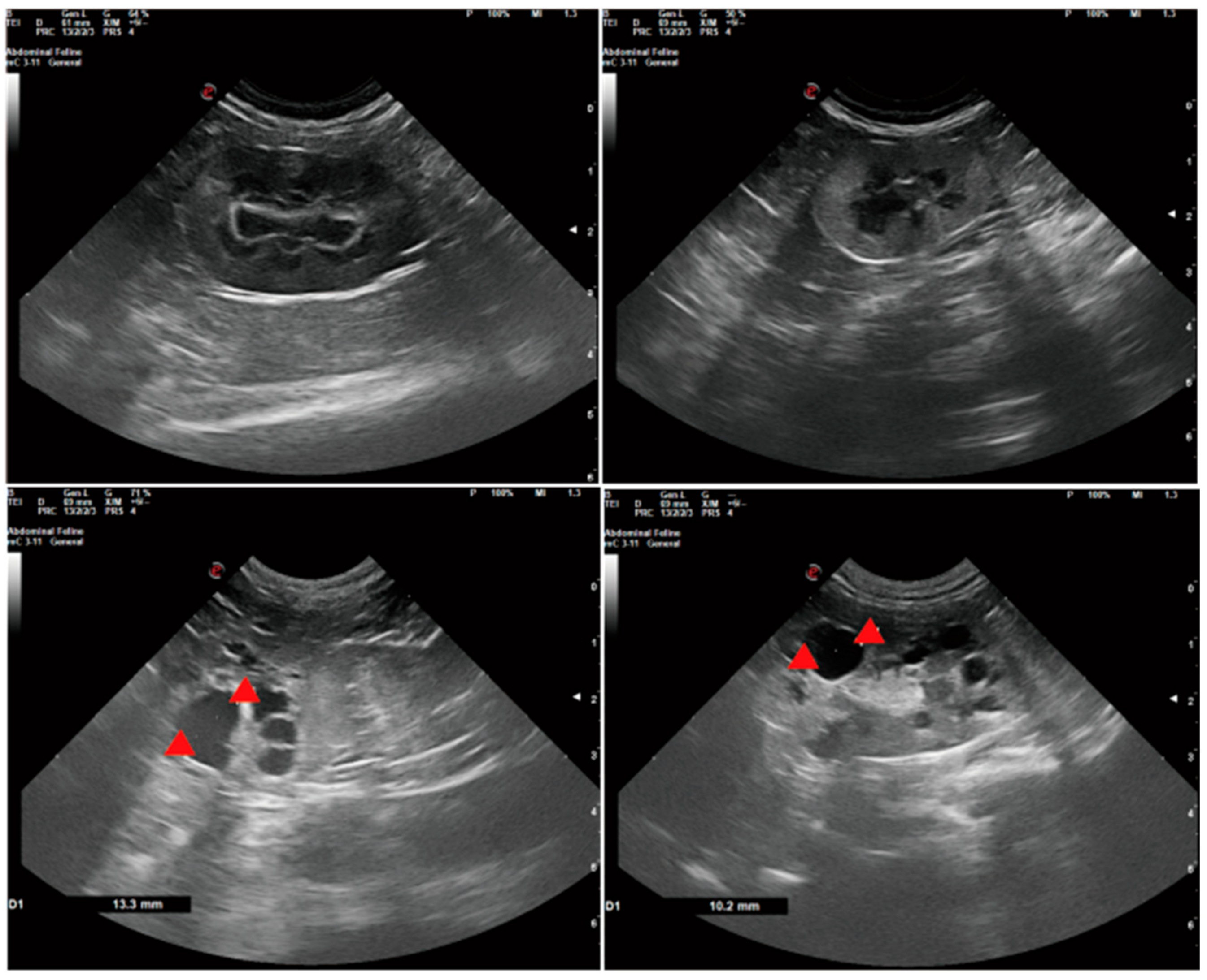
5. Diagnostic Approaches and Clinical Manifestations

While the exact pathogenesis of ADPKD remains elusive, continuous research and advancements in modern medical technology have facilitated its diagnosis through various methods. Clinical symptoms observed in dogs, cats, and other animals closely mirror those in humans, implying analogous diagnostic approaches.
In clinical settings, early stages of ADPKD often manifest without significant changes in kidney size, with patients experiencing subtle symptoms like headaches, insomnia, and blurred vision (Table 4, Figure 4 and Figure 5). Hypertension emerges in nearly all patients at this stage, progressively worsening as the disease advances, becoming a key clinical feature that accelerates disease progression. Furthermore, patients may present with proteinuria, hematuria, renal pain, kidney stones, urinary tract stones, urinary tract infections, intracranial aneurysms, heart valve abnormalities, and other complications. Notably, the incidence of intracranial aneurysms in ADPKD patients is approximately 12%, significantly contributing to early mortality [93].
In the advanced stages of ADPKD, the kidneys filled with numerous cysts, causing significant enlargement and an irregular shape, ultimately leading to declining renal function and potential renal failure in patients. In veterinary clinical practice, animals with clinical symptoms may exhibit loss of appetite, lethargy, dehydration, weight loss, increased thirst and urination, and reduced urine output or even anuria (Table 4, Figure 4 and Figure 5). Palpation of the kidneys may reveal enlargement and an irregular texture. However, these symptoms are nonspecific to ADPKD and can be observed in various other conditions. Some individuals, including dogs and cats, may remain subclinical without obvious symptoms. Blood indicators and biochemical parameters—such as urea, creatinine, and phosphorus—may remain within normal limits, especially at a young age.
Consequently, the diagnosis of ADPKD cannot rely solely on clinical manifestations or general physiological indicators like blood tests and urea levels. Additionally, it is imperative to consider other forms of cystic nephropathy that exhibit symptoms similar to those of ADPKD, including Autosomal Recessive Polycystic Kidney Disease (ARPKD), Nephonophthisis, and Autosomal Dominant Hereditary Renal Tubulointerstitial Nephropathy (ADTKD). ARPKD presents analogous symptoms to ADPKD such as renal failure, liver disease, and portal hypertension, albeit typically affecting children and young animals. Nephonophthisis and ADTKD have not been documented in veterinary clinics. In the human clinical context, Nephonophthisis is a hereditary renal condition characterized by tubulointerstitial damage, often manifesting as chronic nephropathy progressing to end-stage renal disease, with clinical features including progressive renal function deterioration or mild proteinuria. ADTKD is typified by renal interstitial fibrosis, presenting clinical signs such as progressive renal dysfunction, polyuria, polydipsia, anemia, and metabolic acidosis. Given the striking similarity in symptoms among these nephropathies, the diagnosis of polycystic kidney disease should not rely solely on clinical manifestations and general physiological indicators like blood and urea levels. Rather, monitoring and evaluating body state and renal function through these symptoms and indicators is crucial [94].
In human clinical practice, imaging studies are indispensable for diagnosing ADPKD, with kidney size assessment serving as the most reliable biomarker for predicting disease progression. Diagnostic criteria for individuals with a family history of ADPKD include the detection of at least three cysts in unilateral or bilateral kidneys in the 15–39 age group, at least two cysts in each kidney in the 40–59 age group, and at least four cysts in each kidney in the 60 and older age group, confirming an ADPKD diagnosis [95]. Another CT scan offering higher accuracy than ultrasound can be utilized. While ultrasound can detect cysts ranging from 0.5 to 1 cm, CT can identify cysts larger than 0.3 cm. In cases of unclear ultrasound findings or when molecular phenotyping is not feasible, magnetic resonance imaging (MRI) serves as an alternative to rule out other conditions. For individuals lacking a family history of ADPKD, a multifaceted diagnostic approach is necessary. During ultrasound examination, evaluation should extend beyond the kidneys to include organs like the liver, pancreas, and spleen. Provisional diagnosis of ADPKD can be made if more than 10 kidney cysts are detected alongside cysts in the liver, pancreas, or other organs. When the cyst count approaches the threshold and there is no significant increase in kidney size, ADPKD diagnosis can be confirmed through regular imaging to monitor cyst growth or via further genetic testing.
In veterinary clinical practice, ultrasound imaging serves as a practical tool for assessing the progression of ADPKD in dogs and cats. An ultrasound examination reveals numerous hypoechoic to anechoic cysts, characterized by thin or absent walls, circular or oval shapes, and clear demarcation from the surrounding renal parenchyma. Due to the lack of overt clinical signs in the early stages, early intervention is rare in dogs and cats. However, in advanced cases, ultrasound evaluation of total kidney volume and cyst volume is crucial for staging the severity of the disease. Ultrasound has proven to be an efficient method for detecting ADPKD of pets, with a sensitivity of 91% and a specificity of 100% at 36 weeks of age [96], cysts in the kidneys of cats can be detected as early as 7 weeks of age.
In addition to imaging examinations, genetic testing serves as a diagnostic tool for ADPKD. It is the preferred and reliable method for confirming the presence of mutations in genes associated with the disease and for early diagnosis. Genetic testing complements findings from CT scans, B-mode ultrasound, and MRI by providing genetic evidence. Through DNA analysis, mutations in genes linked to ADPKD, such as PKD1 and PKD2, can be identified. This testing can be promptly conducted at any age or stage of disease. Early detection is preferred as it can effectively prevent the familial transmission of ADPKD and designing improved breeding scheme for cats or dogs carrying mutant genes. However, genetic testing is not yet widely performed, posing challenges for prevention and early intervention in ADPKD. In addition, cats and dogs typically do not undergo genetic testing before breeding, contributing to the rising number of ADPKD cases in these animals.
ADPKD is a chronic renal condition that typically manifests overt symptoms only in its later stages, posing significant challenges for diagnosis. By the time ADPKD is detected, it often has advanced to intermediate or advanced stages, precluding the opportunity for early intervention and treatment. In human or veterinary clinics, there is a widespread lack of awareness regarding the importance of early screening and monitoring for ADPKD. Therefore, establishing an effective screening protocol is crucial for the prompt diagnosis, treatment, and management of this condition.

6. Advancements and Challenges in ADPKD Treatment Strategies

ADPKD is a chronic renal condition frequently culminating in renal failure. Currently, there is no definitive cure for the disease, with treatment primarily centered on symptom management and supportive care. Although this approach can alleviate symptoms, it does not address the underlying condition. Drugs play a pivotal role in ADPKD treatment, with Tolvaptan emerging as a prominent therapeutic option. Tolvaptan, a selective vasopressin V2 receptor antagonist, has demonstrated efficacy in randomized clinical trials by attenuating the decline in renal function among ADPKD patients at high risk for rapid progression. To date, Tolvaptan remains the sole medication approved for ADPKD treatment [97]. Tolvaptan functions as a selective antagonist of the V2 receptor, regulating water reabsorption by modulating aquaporin-2 (AQP2) channels, concurrently, the inhibition of the cAMP pathway led to a decrease in the proliferation of renal tubular epithelial cells. When antidiuretic hormone (ADH)/vasopressin binds to the V2 receptor, it activates the cAMP signaling pathway, prompting the translocation of AQP2-expressing cells to the apical plasma membrane, thus enhancing water reabsorption [98]. The blockade of V2 receptors effectively inhibits the signaling cascade of the cAMP pathway and the actions of ADH. Consequently, the expression and activity of AQP2 are suppressed, leading to a reduction in water reabsorption by the renal tubules. This decrease in water reabsorption is accompanied by an increase in the plasma concentration of Na+. These combined effects enhance the kidney’s capacity to manage water balance, promoting the excretion of excess water through urine and concurrently diminishing the production of fluid. However, Tolvaptan is associated with various adverse effects, including polyuria, thirst, dry mouth, nausea, vomiting, headache, and fatigue. It may not be appropriate for individuals with renal impairment or those intolerant to the medication. In addition, Tolvaptan can interact with other drugs, particularly those affecting liver metabolic enzymes [99]. Given these limitations, there is a necessity to explore strategies that either mitigate adverse reactions or enhance the efficacy of ADPKD treatment. Due to the limitations inherent in the treatment of Tolvaptan, it is imperative to explore alternative approaches to mitigate adverse reactions in patients and to develop more effective treatments for ADPKD. Extensive research has identified and validated that agents such as sirolimus [100] and rapamycin [101] can exert beneficial effects on the murine model of ADPKD. These drugs demonstrate the ability to slow cyst growth by inhibiting the mTOR signaling pathway. However, they have yet to successfully navigate clinical trials, and their efficacy for human ADPKD patients remains uncertain. Consequently, there is a compelling need to shift focus to inter-species research. Given the anatomical and functional similarities between porcine and human kidneys, the porcine model may offer a more suitable platform for investigating ADPKD pathogenesis and therapeutic strategies.
Other than medications, surgical intervention represents a conventional approach to managing ADPKD, involving several procedures like cyst decaplication and decompression; capsulotomy, puncture and sclerosis; kidney transplantation; and nephrectomy [102]. For a solitary large cyst, a surgeon can perform a small incision into the cyst, drain the fluid, and then close the wound. This method can alleviate cystic pressure and potentially enhance renal function. While surgical interventions can effectively alleviate the symptoms associated with polycystic kidney disease, they are not without significant risks. The procedure itself carries a high operational risk, with the potential for damage to normal tissues during surgery. Additionally, postoperative complications such as urinary tract obstructions are not uncommon. It is important to note that these treatments address the symptoms rather than the underlying cause of the disease, and there is a heightened risk of cyst recurrence. In cases of severe renal impairment caused by numerous cysts, the consideration of nephrectomy or kidney transplantation may arise. A recent study evaluated the intraoperative assistance of three-dimensional virtual models (3DVMs) in highly complex minimally invasive partial nephrectomy for renal tumors [103], highlighting their potential value in improving surgical accuracy and postoperative recovery for patients. This may also offer insights for the resection of renal cysts. Through kidney transplantation, the patient’s symptoms can indeed be alleviated. However, the potential complications and risks of rejection associated with the transplant cannot be ignored. Moreover, kidney transplantation only addresses the issue of kidney cysts; if the patient has cysts in other areas, the transplant will not resolve these issues. Nonetheless, these procedures are intricate, donor kidneys are scarce, and the associated costs are prohibitive, rendering widespread implementation challenging.
Renal replacement therapy is imperative for patients with ESRD. Given the enlarged abdomen and diminished effective peritoneal area in ADPKD patients, hemodialysis typically prevails over peritoneal dialysis as the preferred choice. However, a retrospective cohort study has indicated no substantial disparity in 5-year survival rates or peritonitis risk between patients undergoing peritoneal dialysis and those receiving hemodialysis [97]. Consequently, peritoneal dialysis stands as a viable treatment alternative for ADPKD patients with ESRD.
With the increasing knowledge of ADPKD, novel treatment strategies continue to emerge. Promising drugs like octreotide-LAR, somatostatin [104] and hydrochlorothiazide [105] are currently in development, offering potential additional options for ADPKD management. In particular, the hydrochlorothiazide has been shown to mitigate the polyureic effect associated with Tolvaptan in patients. Moreover, it significantly reduces multiple markers indicative of kidney damage and dysregulation in glucose metabolism within a murine model [105]. Gene therapy constitutes a burgeoning frontier in research, exemplified by innovative treatments like Casgevy. Casgevy, an ex vivo gene editing therapy, utilizes CRISPR/Cas9 technology to modify hematopoietic stem cells. This groundbreaking treatment is a collaborative effort between Vertex Pharmaceuticals, an American biotechnology firm, and CRISPR Therapeutics, a Swiss pharmaceutical company. The approval of Casgevy by the FDA marks a significant milestone in gene therapy [106,107,108,109,110]. Concurrently, stem cell therapy is emerging as a rapidly evolving field, particularly in renal disease research. Stem cell therapy approaches include direct stem cell differentiation into renal parenchymal cells, enhancement of renal secretion, mitigation of inflammatory responses, and restoration of renal blood circulation, all contributing to renal injury repair and kidney function improvement. As stem cell research technology progresses, further advancements are anticipated in understanding stem cell differentiation into kidney cells, thereby broadening the therapeutic scope of stem cell transplantation in treating kidney diseases [111,112,113,114,115].

7. Advancements in Animal Models for ADPKD Research

Animal disease models play a crucial role in the fields of medicine and life sciences, aiding in the comprehensive understanding of disease etiologies and pathophysiological mechanisms. They serve as a cornerstone for the formulation of preventive and therapeutic strategies. Additionally, these models are indispensable for evaluating the effectiveness and safety of novel pharmacological interventions and treatment approaches. The controlled laboratory setting enables meticulous assessment of drug safety, efficacy, and potential adverse reactions, thus yielding invaluable insights for therapeutic intervention. Furthermore, through the integration of genetics and disease prediction methodologies, animal models contribute significantly to the advancement of innovative gene therapies. Therefore, animal disease models represent an essential tool for unraveling the complexities of various diseases, serving as a crucial platform for medical progress.
At present, there is a dearth of established animal models for ADPKD in canines and felines, although relevant models exist in murine, rodent, and miniature porcine species. Currently, two primary categories of animal models for ADPKD prevail. The first comprises spontaneous genetic models exhibiting characteristic ADPKD phenotypes, while the second involves models generated through homologous gene mutations mirroring those found in humans. Published studies have elucidated several genes implicated in spontaneous genetic models, especially, Cys1, Nphp3, Anks6, and Pkhd1, among others, which are associated with renal cystogenesis in cpk mice, pcy mice, Han: SPRD cy rats, and PCK rats [116,117,118,119]. It has been observed that these gene products also correspond to the PC-1 and PC-2 proteins [116,117,118,119].
To advance our understanding of ADPKD pathogenesis, a multitude of genetically modified animal models have been developed. Initially, ADPKD mouse models predominantly employed traditional homologous recombination techniques to knock out the PKD1 or PKD2 genes. Presently, the Cre-loxP system enables precise gene modifications in specific organs by flanking the target region with loxP sites. The Ksp-Cre; PKD1flox/flox model involves breeding mice carrying a single loxP gene locus on each side of the target area with mice that express Ksp-Cre to create a kidney-specific knockout model of PKD1. Using this model, people have carried out a variety of studies, finding that cysts originate from cells with active Cre recombinase, accompanied by activated MAPK/ERK signaling. Nonetheless, inhibition of ERK1/2 activation fails to impede cyst growth [120]. The Ksp Cre; Pkd1flox/flox mouse model also served as a platform to evaluate mitochondrial abnormalities associated with ADPKD. The observed accumulation of mitochondrial abnormalities in this ADPKD model lends support to the role of disrupted Ca2+ influx and increased cAMP levels in ADPKD progression [121,122]. In addition, the human PKD1 mutant p.Arg3277Cys (RC) represents a sub-allelic variant, and introducing this mutation into mice (as Pkd1RC/RC) can attenuate the progression of ADPKD in these animals. In contrast to typical homozygous ADPKD models, the Pkd1RC/RC model can induce slowly progressed ADPKD and has been extensively utilized in preclinical investigations [123]. A study utilizing the Pkd1RC/RC mouse model to explore Tolvaptan treatment demonstrated a reduction in kidney weight-to-body weight (KW/BW) ratio in Tolvaptan-treated mice, along with decreased volume and number of cysts, accompanied by significant reductions in blood urea nitrogen and cAMP levels [124]. Another model, the doxycycline-induced Pax8rtTA; TetO-Cre; Pkd1flox/flox model, enabling spatial-temporal control of gene activity, presents a progressively slower and more human-like ADPKD phenotype. In this model, rtTA is exclusively expressed in kidney cells under the control of the Pax8 promoter. Upon activation, rtTA binds to the TetO sequence, thereby initiating the transcription of the Cre gene. The Cre recombinase, once produced, recognizes and recombines the loxP sites flanking the Pkd1 gene, leading to its specific knockout in kidney cells. The expression of TetO-Cre is regulated by tetracycline administration, by manipulating the timing and duration of tetracycline treatment, researchers can precisely control when the Pkd1 gene is knocked out, allowing for the study of its effects on kidney function at defined stages [125]. Researchers recently utilized the Pax8rtTA; TetO-Cre; Pkd1flox/flox model to explore the therapeutic potential of 11 beta-dichloro in ADPKD. Their study demonstrated that 11 beta-dichloro significantly improved renal structure and function in mice of this model, without any indication of significant toxicity [126].
In PKD, mutations classified as non-protein truncating and protein truncating significantly impact the average age of onset for renal failure [127]. This variability is attributed to individual differences, genetic backgrounds, lifestyle factors, and disease management strategies. Non-protein truncating mutations, such as missense mutations, can cause partial functional impairment of PC-1 or PC-2, resulting in partial protein activity. Consequently, these mutations are associated with a slower disease progression, with renal failure typically occurring later in middle or later adulthood. Conversely, protein truncating mutations, including nonsense mutations, alterations in splicing sites, or large deletions, lead to the premature termination of polycystin synthesis, resulting in non-functional protein fragments or complete absence of the protein. These truncating mutations are associated with more severe disease manifestations and accelerated progression, leading to an earlier onset of renal failure, potentially in early adulthood or middle age. Specifically, patients with PKD1 gene mutations, particularly those that are truncated, may experience ESRD between the ages of 30 and 50. In contrast, PKD2 gene mutations typically result in a slower disease progression, with ESRD typically occurring after the age of 50 or at a later stage.
Miniature pigs and monkeys are widely accepted large animal models in human disease modeling. Our group have established a PKD2 transgenic miniature pig model [127]. Despite the initial high expression of the PKD2 gene observed through PCR and Western blot analyses, early stages of ADPKD progression did not manifest evident pathological alterations, and there were no significant differences in urea nitrogen and creatinine levels between wild-type and transgenic pigs. Therefore, continued monitoring is essential to detect any potential pathological changes in the kidneys of transgenic pigs. Subsequently, our group [128] utilized the first-generation genome editing technology, zinc-finger nucleases (ZFNs), to establish a miniature pig model with a single-allele knockout of PKD1. By approximately 6 months of age, cyst formation commenced in the pig kidneys, exhibiting a more rapid growth compared to the previously established polycystic kidney model of transgenic miniature pigs with PKD2, as well as faster progression than ADPKD mice with a single-allele knockout of PKD1 (PKD1+/−). Masahito Watanabe and colleagues [129] recently developed a PKD1insG/+ pig model using the CRISPR-Cas9 gene editing technique. Investigations have revealed that newborn piglets in the PKD1insG/+ model demonstrate renal cysts and interstitial fibrosis, accompanied by an early asymptomatic phase, mirroring the characteristics observed in ADPKD patients with PKD1 heterozygous mutations. Tomoyuki Tsukiyama et al. [130] pioneered the creation of an ADPKD model with a PKD1 mutation in crab-eating macaques using CRISPR/Cas9. Heterozygous crab-eating macaques manifested a small number of cysts either at birth or around 2 months of age, predominantly located in the distal tubules, resembling the scenario observed in human heterozygous pediatric patients. Utilizing monkey models for studying the cyst formation process proves to be more practical than relying solely on mice, which necessitate conditional knockout strategies to mimic human phenotypes. Nevertheless, the significant time and financial investments associated with using monkeys as model organisms must be duly considered.
To date, the mouse model has been extensively employed across various disciplines, encompassing immunology, infectious diseases, cancer biology, and drug research, significantly augmenting our understanding of ADPKD. However, many mouse disease models inadequately represent the human internal environment, and some exhibit excessively rapid disease progression, diverging starkly from the pace observed in humans. This discrepancy is particularly pronounced in ADPKD, where mouse models often demonstrate a rapid disease onset and progression, leading to the development of drugs with limited efficacy in humans. Consequently, there is an urgent need for disease models that more accurately mirrors human physiological structure and disease progression. Among the available options, the pig disease model emerges as an alternative. Pigs exhibit a high degree of anatomical and physiological similarity to humans, including weight, organ formation, and disease occurrence, without any artificial manipulation. Additionally, mice possess a unilobular kidney and are substantially smaller than human kidneys, comprising only about one-fifth of their size, with fewer nephrons. In contrast, both pigs and humans possess multiple renal papillae with comparable morphology, size, and anatomical structure. A comparative assessment of kidneys across various species underscores that pigs and humans exhibit greater morphological, size, and physiological functional resemblances than other animal models (Table 5) [131,132,133,134,135,136,137].
Moreover, pigs exhibit a remarkable degree of genomic and chromosomal resemblance to humans. Their genome size, structure, number, and chromosomal arrangement closely parallel those of humans, accompanied by a plethora of shared gene sequences. With the advent of gene editing technologies, traditional homologous recombination methods and cutting-edge nuclease technologies—such as ZFNs, Transcription Activator-Like Effector Nucleases (TALENs), and the CRISPR-Cas9 system [109,110]—alike have significantly advanced. Concurrently, there has been rapid progress in generating transgenic models, gene knockout/knock-in models, and pigs bearing precise gene modifications [111]. Cloning techniques have become more mature compared to before, while the establishment of cell lines has become relatively facile, bolstering the development of gene-editing models in miniature pigs. As pig genome sequencing continues to refine, researchers will gain access to more potent genetic and proteomic tools tailored for pig-specific research endeavors. Furthermore, ethical concerns associated with pigs are comparatively fewer than those associated with mice, dogs, cats, and monkeys. Thus, utilizing pig disease models offers substantial advantages for investigating pathological mechanisms and therapeutic strategies applicable to both animal and human contexts.

8. Conclusions

Research on polycystic kidney disease has been ongoing for decades. Currently, significant advancements have been achieved in understanding the pathological changes, molecular mechanisms, and diagnosis of human ADPKD. The progress in molecular genetics and imaging techniques has remarkably enhanced the accuracy of diagnosis. However, it is often overlooked that polycystic kidney disease not only has a high incidence and heredity in humans, but also in other animals, such as dogs, cats, pigs, cattle, and sheep. The genetic patterns and molecular mechanisms of polycystic kidney disease among different species are highly similar. For instance, ADPKD in humans, dogs, and cats is all caused by a single gene mutation, and polycystic kidneys in domestic animals such as pigs are often confined to the same populations as humans. Human ADPKD shares many similarities in pathological and clinical features with animals such as dogs, cats, and pigs, including the occurrence of cysts and impaired renal function in the kidneys. However, due to species and individual differences, there is still a great deal of complexity and diversity. Therefore, when conducting research on polycystic kidney disease, it is important to note that the incidence, cycle, and degree of ADPKD vary among different species, with small pigs being the closest to humans. When studying genes related to polycystic kidney disease, these different manifestations can be combined to identify gene mutations and further explore the diverse pathogenesis of polycystic kidney disease. At present, there is no method or drug that can cure polycystic kidney disease. Only Tolvaptan is approved for the treatment of human ADPKD, and there are no suitable drugs available for use in veterinary clinical practice or animal husbandry. It is time for us to explore alternative research methods. Presently, there are numerous mouse models available, yet it is evident that pig models offer closer parallels to humans in terms of function, structure, and response. They can better replicate human disease characteristics. The gene-edited pig model, established through genetic editing in pigs, has emerged as one of the most favored large animal models. Perhaps shifting our research focus from mice to small pigs could yield significant breakthroughs in ADPKD research.

Funding

This research was funded by National Key R&D Program of China with grant number 2021YFA0805902.

Conflicts of Interest

The authors declare no conflicts of interest.

References

  1. Sutters, M. The pathogenesis of autosomal dominant polycystic kidney disease. Nephron Exp. Nephrol. 2006, 103, 149–155. [Google Scholar] [CrossRef] [PubMed]
  2. Horie, S.; Mochizuki, T.; Muto, S.; Hanaoka, K.; Fukushima, Y.; Narita, I.; Nutahara, K.; Tsuchiya, K.; Tsuruya, K.; Kamura, K.; et al. Evidence-based clinical practice guidelines for polycystic kidney disease. Clin. Exp. Nephrol. 2016, 20, 510. [Google Scholar] [CrossRef] [PubMed]
  3. Müller, R.U.; Haas, C.S.; Sayer, J.A. Practical approaches to the management of autosomal dominant polycystic kidney disease patients in the era of tolvaptan. Clin. Kidney J. 2018, 11, 62–69. [Google Scholar] [CrossRef] [PubMed]
  4. Chebib, F.T.; Torres, V.E. Autosomal Dominant Polycystic Kidney Disease: Core Curriculum 2016. Am. J. Kidney Dis. 2016, 67, 792–810. [Google Scholar] [CrossRef] [PubMed]
  5. Grantham, J.J. Clinical practice. Autosomal dominant polycystic kidney disease. N. Engl. J. Med. 2008, 359, 1477–1485. [Google Scholar] [CrossRef] [PubMed]
  6. Leonhard, W.N.; Zandbergen, M.; Veraar, K.; van den Berg, S.; van der Weerd, L.; Breuning, M.; de Heer, E.; Peters, D.J. Scattered Deletion of PKD1 in Kidneys Causes a Cystic Snowball Effect and Recapitulates Polycystic Kidney Disease. J. Am. Soc. Nephrol. 2015, 26, 1322–1333. [Google Scholar] [CrossRef] [PubMed]
  7. Gubbels, E.J.; Prins, P. Polycystic Kidney Disease (PKD). Tijdschr. Diergeneeskd. 2005, 130, 184–185. [Google Scholar] [PubMed]
  8. Lowrie, M.; Garosi, L. Classification of involuntary movements in dogs: Paroxysmal dyskinesias. Vet. J. 2017, 220, 65–71. [Google Scholar] [CrossRef]
  9. O’Leary, C.A.; Duffy, D.; Biros, I.; Corley, S. Linkage confirms canine pkd1 orthologue as a candidate for bull terrier polycystic kidney disease. Anim. Genet. 2009, 40, 543–546. [Google Scholar] [CrossRef]
  10. Schmidt, U. Angeborene zystische Veränderungen in Leber und Nieren bei einem Kalb [Congenital cystic changes of the liver and kidneys in a calf]. Dtsch. Tierarztl. Wochenschr. 1973, 80, 329. [Google Scholar]
  11. Patterson, D.S.; Done, J.T. Neurochemistry as a diagnostic aid in the congenital tremor syndrome of piglets. Br. Vet. J. 1977, 133, 111–117. [Google Scholar] [CrossRef] [PubMed]
  12. Chebib, F.T.; Torres, V.E. Recent Advances in the Management of Autosomal Dominant Polycystic Kidney Disease. Clin. J. Am. Soc. Nephrol. 2018, 13, 1765–1776. [Google Scholar] [CrossRef] [PubMed]
  13. Messchendorp, A.L.; Spithoven, E.M.; Casteleijn, N.F.; Dam, W.A.; van den Born, J.; Tonnis, W.F.; Gaillard, C.A.J.M.; Meijer, E. Association of plasma somatostatin with disease severity and progression in patients with autosomal dominant polycystic kidney disease. BMC Nephrol. 2018, 19, 368. [Google Scholar] [CrossRef] [PubMed]
  14. Al-Hamed, M.H.; Alsahan, N.; Rice, S.J.; Edwards, N.; Nooreddeen, E.; Alotaibi, M.; Kurdi, W.; Alnemer, M.; Altaleb, N.; Ali, W.; et al. Bialleleic PKD1 mutations underlie early-onset autosomal dominant polycystic kidney disease in Saudi Arabian families. Pediatr. Nephrol. 2019, 34, 1615–1623. [Google Scholar] [CrossRef] [PubMed]
  15. Wu, R.; Bai, B.; Li, F.; Bai, R.; Zhuo, Y.; Zhu, Z.; Jia, R.; Li, S.; Chen, Y.; Lan, X. Phenotypes and genetic etiology of spontaneous polycystic kidney and liver disease in cynomolgus monkey. Front. Vet. Sci. 2023, 10, 1106016. [Google Scholar] [CrossRef]
  16. Mellersh, C.S.; Hitte, C.; Richman, M.; Vignaux, F.; Priat, C.; Jouquand, S.; Werner, P.; André, C.; DeRose, S.; Patterson, D.F.; et al. An integrated linkage-radiation hybrid map of the canine genome. Mamm. Genome 2000, 11, 120–130. [Google Scholar] [CrossRef]
  17. Sinha, D.; Ivan, D.; Gibbs, E.; Chetluru, M.; Goss, J.; Chen, Q. Fission yeast polycystin Pkd2p promotes cell size expansion and antagonizes the Hippo-related SIN pathway. J. Cell Sci. 2022, 135, 259046. [Google Scholar] [CrossRef]
  18. Weimbs, T. Polycystic kidney disease and renal injury repair: Common pathways, fluid flow, and the function of polycystin-1. Am. J. Physiol. Ren. Physiol. 2007, 293, F1423–F1432. [Google Scholar] [CrossRef]
  19. Weimbs, T. Third-hit signaling in renal cyst formation. J. Am. Soc. Nephrol. 2011, 22, 793–795. [Google Scholar] [CrossRef]
  20. Raman, A.; Parnell, S.C.; Zhang, Y.; Reif, G.A.; Dai, Y.; Khanna, A.; Daniel, E.; White, C.; Vivian, J.L.; Wallace, D.P. Periostin overexpression in collecting ducts accelerates renal cyst growth and fibrosis in polycystic kidney disease. Am. J. Physiol. Ren. Physiol. 2018, 315, 1695–1707. [Google Scholar] [CrossRef]
  21. Raman, A.; Reif, G.A.; Dai, Y.; Khanna, A.; Li, X.; Astleford, L.; Parnell, S.C.; Calvet, J.P.; Wallace, D.P. Integrin-Linked Kinase Signaling Promotes Cyst Growth and Fibrosis in Polycystic Kidney Disease. J. Am. Soc. Nephrol. 2017, 28, 2708–2719. [Google Scholar] [CrossRef] [PubMed]
  22. Qian, Q.; Hunter, L.W.; Li, M.; Marin-Padilla, M.; Prakash, Y.S.; Somlo, S.; Harris, P.C.; Torres, V.E.; Sieck, G.C. Pkd2 haploinsufficiency alters intracellular calcium regulation in vascular smooth muscle cells. Hum. Mol. Genet. 2003, 12, 1875–1880. [Google Scholar] [CrossRef] [PubMed]
  23. Kraus, A.; Peters, D.J.M.; Klanke, B.; Weidemann, A.; Willam, C.; Schley, G.; Kunzelmann, K.; Eckardt, K.U.; Buchholz, B. HIF-1α promotes cyst progression in a mouse model of autosomal dominant polycystic kidney disease. Kidney Int. 2018, 94, 887–899. [Google Scholar] [CrossRef] [PubMed]
  24. Nowak, K.L.; Edelstein, C.L. Apoptosis and autophagy in polycystic kidney disease (PKD). Cell. Signal. 2020, 68, 109518. [Google Scholar] [CrossRef]
  25. Sekine, A.; Hidaka, S.; Moriyama, T.; Shikida, Y.; Shimazu, K.; Ishikawa, E.; Uchiyama, K.; Kataoka, H.; Kawano, H.; Kurashige, M.; et al. Cystic Kidney Diseases That Require a Differential Diagnosis from Autosomal Dominant Polycystic Kidney Disease (ADPKD). J. Clin. Med. 2022, 11, 6528. [Google Scholar] [CrossRef]
  26. Bergmann, C.; Guay-Woodford, L.M.; Harris, P.C.; Horie, S.; Peters, D.J.M.; Torres, V.E. Polycystic kidney disease. Nat. Rev. Dis. Primers 2018, 4, 50. [Google Scholar] [CrossRef] [PubMed]
  27. Song, C.J.; Zimmerman, K.A.; Henke, S.J.; Yoder, B.K. Inflammation and Fibrosis in Polycystic Kidney Disease. Results Probl. Cell Differ. 2017, 60, 323–344. [Google Scholar] [PubMed]
  28. Yuan, J.; Rozengurt, E. PKD, PKD2, and p38 MAPK mediate Hsp27 serine-82 phosphorylation induced by neurotensin in pancreatic cancer PANC-1 cells. J. Cell Biochem. 2008, 103, 648–662. [Google Scholar] [CrossRef]
  29. Antignac, C.; Calvet, J.P.; Germino, G.G.; Grantham, J.J.; Guay-Woodford, L.M.; Harris, P.C.; Hildebrandt, F.; Peters, D.J.; Somlo, S.; Torres, V.E.; et al. The Future of Polycystic Kidney Disease Research—As Seen By the 12 Kaplan Awardees. J. Am. Soc. Nephrol. 2015, 26, 2081–2095. [Google Scholar] [CrossRef]
  30. Lakhia, R.; Mishra, A.; Biggers, L.; Malladi, V.; Cobo-Stark, P.; Hajarnis, S.; Patel, V. Enhancer and super-enhancer landscape in polycystic kidney disease. Kidney Int. 2023, 103, 87–99. [Google Scholar] [CrossRef]
  31. Magistroni, R.; Boletta, A. Defective glycolysis and the use of 2-deoxy-D-glucose in polycystic kidney disease: From animal models to humans. J. Nephrol. 2017, 30, 511–519. [Google Scholar] [CrossRef]
  32. Nowak, K.L.; Hopp, K. Metabolic Reprogramming in Autosomal Dominant Polycystic Kidney Disease: Evidence and Therapeutic Potential. Clin. J. Am. Soc. Nephrol. 2020, 15, 577–584. [Google Scholar] [CrossRef] [PubMed]
  33. Gaur, P.; Gedroyc, W.; Hill, P. ADPKD-what the radiologist should know. Br. J. Radiol. 2019, 92, 20190078. [Google Scholar] [CrossRef] [PubMed]
  34. Xue, C.; Zhou, C.C.; Wu, M.; Mei, C.L. The Clinical Manifestation and Management of Autosomal Dominant Polycystic Kidney Disease in China. Kidney Dis. 2016, 2, 111–119. [Google Scholar] [CrossRef] [PubMed]
  35. Pippias, M.; Kramer, A.; Noordzij, M.; Afentakis, N.; Alonso de la Torre, R.; Ambühl, P.M.; Aparicio Madre, M.I.; Arribas Monzón, F.; Åsberg, A.; Bonthuis, M.; et al. The European Renal Association—European Dialysis and Transplant Association Registry Annual Report 2014: A summary. Clin. Kidney J. 2017, 10, 154–169. [Google Scholar] [CrossRef] [PubMed]
  36. Spithoven, E.M.; Kramer, A.; Meijer, E.; Laruelle, E.; Couchoud, C.; Collart, F.; Cases, A.; Arici, M.; Helve, J.; Waldum-Grevbo, B.; et al. Analysis of data from the ERA-EDTA Registry indicates that conventional treatments for chronic kidney disease do not reduce the need for renal replacement therapy in autosomal dominant polycystic kidney disease. Kidney Int. 2014, 86, 1244–1252. [Google Scholar] [CrossRef] [PubMed]
  37. Lulich, J.P.; Osborne, C.A.; Walter, P.A. O’Brien TD:Feline idiopathic polycystic kidney disease. Compend. Cont. Ed. Pract. Vet. 1988, 10, 1029–1041. [Google Scholar]
  38. Podell, M.; DiBartola, S.P.; Rosol, T.J. Polycystic kidney disease and renal lymphoma in a cat. J. Am. Vet. Med. Assoc. 1992, 201, 906–909. [Google Scholar] [CrossRef] [PubMed]
  39. Stebbins, K.E. Polycystic disease of the kidney and liver in an adult Persian cat. J. Comp. Pathol. 1989, 100, 327–330. [Google Scholar] [CrossRef]
  40. Kimberling, W.J.; Kumar, S.; Gabow, P.A.; Kenyon, J.B.; Connolly, C.; Somlo, S. Autosomal dominant polycystic kidney disease: Localization of the second gene to chromosome 4q13-q23. Genomics 1993, 18, 467–472. [Google Scholar] [CrossRef]
  41. Gendron, K.; Owczarek-Lipska, M.; Lang, J.; Leeb, T. Maine Coon renal screening: Ultrasonographical characterisation and preliminary genetic analysis for common genes in cats with renal cysts. J. Feline Med. Surg. 2013, 15, 1079–1085. [Google Scholar] [CrossRef] [PubMed]
  42. Helps, C.; Tasker, S.; Harley, R. Correlation of the feline PKD1 genetic mutation with cases of PKD diagnosed by pathological examination. Exp. Mol. Pathol. 2007, 83, 264–268. [Google Scholar] [CrossRef] [PubMed]
  43. Torres, J.A.; Kruger, S.L.; Broderick, C.; Amarlkhagva, T.; Agrawal, S.; Dodam, J.R.; Mrug, M.; Lyons, L.A.; Weimbs, T. Ketosis Ameliorates Renal Cyst Growth in Polycystic Kidney Disease. Cell Metab. 2019, 30, 1007–1023. [Google Scholar] [CrossRef] [PubMed]
  44. Volta, A.; Manfredi, S.; Gnudi, G.; Gelati, A.; Bertoni, G. Polycystic Kidney Disease in a Chartreux Cat. J. Feline Med. Surg. 2010, 12, 138–140. [Google Scholar] [CrossRef]
  45. Barrs, V.R.; Gunew, M.; Foster, S.F.; Beatty, J.A.; Malik, R. Prevalence of autosomal dominant polycystic kidney disease in Persian cats and related-breeds in Sydney and Brisbane. Aust. Vet. J. 2001, 79, 257–259. [Google Scholar] [CrossRef] [PubMed]
  46. Kobayashi, S.; Kawarasaki, M.; Aono, A.; Cho, J.; Hashimoto, T.; Sato, R. Renoprotective effects of docosahexaenoic acid in cats with early chronic kidney disease due to polycystic kidney disease: A pilot study. J. Feline Med. Surg. 2022, 24, 505–512. [Google Scholar] [CrossRef]
  47. Gerwing, M.; Michele, U.; Kramer, M. PKD (polycystic kidney disease)-Polyzystisches syndrome. Prakt. Tierarzt 1999, 80, 374–396. [Google Scholar]
  48. Lyons, L.A.; Biller, D.S.; Erdman, C.A.; Lipinski, M.J.; Young, A.E.; Roe, B.A.; Qin, B.; Grahn, R.A. Feline polycystic kidney disease mutation identified in PKD1. J. Am. Soc. Nephrol. 2004, 15, 2548–2555. [Google Scholar] [CrossRef] [PubMed]
  49. Domanjko-Petric, A.; Cernec, D.; Cotman, M. Polycystic kidney disease: A review and occurrence in Slovenia with comparison between ultrasound and genetic testing. J. Feline Med. Surg. 2008, 10, 115–119. [Google Scholar] [CrossRef]
  50. O’Leary, C.A.; Mackay, B.M.; Malik, R.; Edmondston, J.E.; Robinson, W.F.; Huxtable, C.R. Polycystic kidney disease in bull terriers: An autosomal dominant inherited disorder. Aust. Vet. J. 1999, 77, 361–366. [Google Scholar] [CrossRef]
  51. McAloose, D.; Casal, M.; Patterson, D.F.; Dambach, D.M. Polycystic kidney and liver disease in two related West Highland White Terrier litters. Vet. Pathol. 1998, 35, 77–81. [Google Scholar] [CrossRef] [PubMed]
  52. O’Leary, C.A.; Mackay, B.M.; Taplin, R.H.; Atwell, R.B. Echocardiographic parameters in 14 healthy English Bull Terriers. Aust. Vet. J. 2003, 81, 535–542. [Google Scholar] [CrossRef]
  53. McKenna, S.C.; Carpenter, J.L. Polycystic disease of the kidney and liver in the Cairn Terrier. Vet. Pathol. 1980, 17, 436–442. [Google Scholar] [CrossRef] [PubMed]
  54. Monaghan, M.L.; Hannan, J. Abattoir survey of bovine kidney disease. Vet. Rec. 1983, 113, 55–57. [Google Scholar] [CrossRef] [PubMed]
  55. Wells, G.A.; Hebert, C.N.; Robins, B.C. Renal cysts in pigs: Prevalence and pathology in slaughtered pigs from a single herd. Vet. Rec. 1980, 106, 532–535. [Google Scholar] [CrossRef]
  56. Knox, R.V. Artificial insemination in pigs today. Theriogenology 2016, 85, 83–93. [Google Scholar] [CrossRef]
  57. Gao, Z.; Ruden, D.M.; Lu, X. PKD2 cation channel is required for directional sperm movement and male fertility. Curr. Biol. 2003, 13, 2175–2178. [Google Scholar] [CrossRef]
  58. Lanktree, M.B.; Haghighi, A.; Guiard, E.; Iliuta, I.A.; Song, X.; Harris, P.C.; Paterson, A.D.; Pei, Y. Prevalence estimates of polycystic kidney and liver disease by population sequencing. J. Am. Soc. Nephrol. 2018, 29, 2593–2600. [Google Scholar] [CrossRef]
  59. Messa, P.; Alfieri, C.M.; Montanari, E.; Ferraresso, M.; Cerutti, R. ADPKD: Clinical issues before and after renal transplantation. J. Nephrol. 2016, 29, 755–763. [Google Scholar] [CrossRef]
  60. Porpiglia, F.; Checcucci, E.; Cillis, S.D.E.; Piramide, F.; Amparore, D.; Piana, A.; Volpi, G.; Granato, S.; Zamengo, D.; Stura, I.; et al. A prospective randomized controlled trial comparing target prostate biopsy alone approach vs. target plus standard in naïve patients with positive mpMRI. Minerva Urol. Nephrol. 2023, 75, 31–41. [Google Scholar] [CrossRef]
  61. Ambrosini, E.; Montanari, F.; Cristalli, C.P.; Capelli, I.; La Scola, C.; Pasini, A.; Graziano, C. Modifiers of Autosomal Dominant Polycystic Kidney Disease Severity: The Role of PKD1 Hypomorphic Alleles. Genes 2023, 14, 1230. [Google Scholar] [CrossRef] [PubMed]
  62. Roitbak, T.; Ward, C.J.; Harris, P.C.; Bacallao, R.; Ness, S.A.; Wandinger-Ness, A. A polycystin-1 multiprotein complex is disrupted in polycystic kidney disease cells. Mol. Biol. Cell 2004, 15, 1334–1346. [Google Scholar] [CrossRef] [PubMed]
  63. Boulter, C.; Mulroy, S.; Webb, S.; Fleming, S.; Brindle, K.; Sandford, R. Cardiovascular, skeletal, and renal defects in mice with a targeted disruption of the Pkd1 gene. Proc. Natl. Acad. Sci. USA 2001, 98, 12174–12179. [Google Scholar] [CrossRef] [PubMed]
  64. Eaton, K.A.; Biller, D.S.; DiBartola, S.P.; Radin, M.J.; Wellman, M.L. Autosomal dominant polycystic kidney disease in Persian and Persian-cross cats. Vet. Pathol. 1997, 34, 117–126. [Google Scholar] [CrossRef] [PubMed]
  65. O’Leary, C.A.; Mackay, B.M.; Taplin, R.H.; Atwell, R.B. Auscultation and echocardiographic findings in Bull Terriers with and without polycystic kidney disease. Aust. Vet. J. 2005, 83, 270–275. [Google Scholar] [CrossRef] [PubMed]
  66. Elliott, J.; Barber, P.J.; Syme, H.M.; Rawlings, J.M.; Markwell, P.J. Feline hypertension: Clinical findings and response to antihypertensive treatment in 30 cases. J. Small Anim. Pract. 2001, 42, 122–129. [Google Scholar] [CrossRef] [PubMed]
  67. Maggio, F.; DeFrancesco, T.C.; Atkins, C.E.; Pizzirani, S.; Gilger, B.C.; Davidson, M.G. Ocular lesions associated with systemic hypertension in cats: 69 cases (1985–1998). J. Am. Vet. Med. Assoc. 2000, 217, 695–702. [Google Scholar] [CrossRef]
  68. Littman, M.P. Spontaneous systemic hypertension in 24 cats. J. Vet. Intern. Med. 1994, 8, 79–86. [Google Scholar] [CrossRef]
  69. Kobayashi, D.L.; Peterson, M.E.; Graves, T.K.; Lesser, M.; Nichols, C.E. Hypertension in cats with chronic renal failure or hyperthyroidism. J. Vet. Intern. Med. 1990, 4, 58–62. [Google Scholar] [CrossRef]
  70. Beck, C.; Lavelle, R.B. Feline polycystic kidney disease in Persian and other cats: A prospective study using ultrasonography. Aust. Vet. J. 2001, 79, 181–184. [Google Scholar] [CrossRef]
  71. Drögemüller, M.; Klein, N.; Steffensen, R.L.; Keiner, M.; Jagannathan, V.; Leeb, T. PKD1 Nonsense Variant in a Lagotto Romagnolo Family with Polycystic Kidney Disease. Genes 2023, 14, 1210. [Google Scholar] [CrossRef] [PubMed]
  72. O’Leary, C.A.; Ghoddusi, M.; Huxtable, C.R. Renal pathology of polycystic kidney disease and concurrent hereditary nephritis in Bull Terriers. Aust. Vet. J. 2002, 80, 353–361. [Google Scholar] [CrossRef]
  73. Webster, W.R.; Summers, P.M. Congenital polycystic kidney and liver syndrome in piglets. Aust. Vet. J. 1978, 54, 451. [Google Scholar] [CrossRef] [PubMed]
  74. Cornec-Le, G.E.; Alam, A.; Perrone, R.D. Autosomal dominant polycystic kidney disease. Lancet 2019, 393, 919–935. [Google Scholar] [CrossRef] [PubMed]
  75. Fedeles, S.; Gallagher, A.R. Cell polarity and cystic kidney disease. Pediatr. Nephrol. 2013, 28, 1161–1172. [Google Scholar] [CrossRef] [PubMed]
  76. Su, Q.; Hu, F.; Ge, X.; Lei, J.; Yu, S.; Wang, T.; Zhou, Q.; Mei, C.; Shi, Y. Structure of the human PKD1-PKD2 complex. Science 2018, 361, 9819. [Google Scholar] [CrossRef]
  77. Mekahli, D.; Parys, J.B.; Bultynck, G.; Missiaen, L.; De Smedt, H. Polycystins and cellular Ca2+ signaling. Cell. Mol. Life Sci. 2013, 70, 2697–2712. [Google Scholar] [CrossRef] [PubMed]
  78. Ko, J.Y.; Park, J.H. Mouse models of polycystic kidney disease induced by defects of ciliary proteins. BMB Rep. 2013, 46, 73–79. [Google Scholar] [CrossRef]
  79. Sussman, C.R.; Wang, X.; Chebib, F.T.; Torres, V.E. Modulation of polycystic kidney disease by G-protein coupled receptors and cyclic AMP signaling. Cell. Signal. 2020, 72, 109649. [Google Scholar] [CrossRef]
  80. Hansen, J.N.; Kaiser, F.; Leyendecker, P.; Stüven, B.; Krause, J.H.; Derakhshandeh, F.; Irfan, J.; Sroka, T.J.; Preval, K.M.; Desai, P.B.; et al. A cAMP signalosome in primary cilia drives gene expression and kidney cyst formation. EMBO Rep. 2022, 23, 54315. [Google Scholar] [CrossRef]
  81. Ye, H.; Wang, X.; Constans, M.M.; Sussman, C.R.; Chebib, F.T.; Irazabal, M.V.; Young, W.F., Jr.; Harris, P.C.; Kirschner, L.S.; Torres, V.E. The regulatory 1α subunit of protein kinase A modulates renal cystogenesis. Am. J. Physiol. Ren. Physiol. 2017, 313, 677–686. [Google Scholar] [CrossRef] [PubMed]
  82. Formica, C.; Peters, D.J.M. Molecular pathways involved in injury-repair and ADPKD progression. Cell. Signal. 2020, 72, 109648. [Google Scholar] [CrossRef] [PubMed]
  83. Saxton, R.A.; Sabatini, D.M. mTOR Signaling in Growth, Metabolism, and Disease. Cell 2017, 69, 361–371. [Google Scholar] [CrossRef] [PubMed]
  84. Morleo, M.; Vieira, H.L.A.; Pennekamp, P.; Palma, A.; Bento-Lopes, L.; Omran, H.; Lopes, S.S.; Barral, D.C.; Franco, B. Crosstalk between cilia and autophagy: Implication for human diseases. Autophagy 2023, 19, 24–43. [Google Scholar] [CrossRef] [PubMed]
  85. Reiterová, J.; Tesař, V. Autosomal Dominant Polycystic Kidney Disease: From Pathophysiology of Cystogenesis to Advances in the Treatment. Int. J. Mol. Sci. 2022, 23, 3317. [Google Scholar] [CrossRef]
  86. Benzing, T.; Simons, M.; Walz, G. Wnt signaling in polycystic kidney disease. J. Am. Soc. Nephrol. 2007, 18, 1389–1398. [Google Scholar] [CrossRef]
  87. Waddell, S.H.; Yao, Y.; Olaizola, P.; Walker, A.; Jarman, E.J.; Gournopanos, K.; Gradinaru, A.; Christodoulou, E.; Gautier, P.; Boerrigter, M.M.; et al. A TGFβ-ECM-integrin signaling axis drives structural reconfiguration of the bile duct to promote polycystic liver disease. Sci. Transl. Med. 2023, 15, 5930. [Google Scholar] [CrossRef]
  88. Torres, V.E.; Harris, P.C.; Pirson, Y. Autosomal dominant polycystic kidney disease. Lancet 2007, 369, 1287–1301. [Google Scholar] [CrossRef] [PubMed]
  89. Rossetti, S.; Consugar, M.B.; Chapman, A.B.; Torres, V.E.; Guay-Woodford, L.M.; Grantham, J.J.; Bennett, W.M.; Meyers, C.M.; Walker, D.L.; Bae, K.; et al. Comprehensive molecular diagnostics in autosomal dominant polycystic kidney disease. J. Am. Soc. Nephrol. 2007, 18, 2143–2160. [Google Scholar] [CrossRef]
  90. Song, Y.; Wang, L.; Xu, M.; Lu, X.; Wang, Y.; Zhang, L. Molecular and functional characterization of porcine poly C binding protein 1 (PCBP1). BMC Vet. Res. 2024, 20, 25. [Google Scholar] [CrossRef]
  91. Young, A.E.; Biller, D.S.; Herrgesell, E.J.; Roberts, H.R.; Lyons, L.A. Feline polycystic kidney disease is linked to the PKD1 region. Mamm. Genome 2005, 16, 59–65. [Google Scholar] [CrossRef] [PubMed]
  92. Gharahkhani, P.; O’Leary, C.A.; Kyaw-Tanner, M.; Sturm, R.A.; Duffy, D.L. A non-synonymous mutation in the canine Pkd1 gene is associated with autosomal dominant polycystic kidney disease in Bull Terriers. PLoS ONE 2011, 6, 22455. [Google Scholar] [CrossRef]
  93. Duarte-Chavez, R.; Stoltzfus, J.; Yellapu, V.; Martins, N.; Nanda, S.; Longo, S.; Geme, B.; Schneider, Y. Colonic diverticular disease in autosomal dominant polycystic kidney disease: Is there really an association? A nationwide analysis. Int. J. Colorectal Dis. 2021, 36, 83–91. [Google Scholar] [CrossRef]
  94. Vucicevic, M.; Slijepcevic, D.; Davitkov, D.; Avdalovic, V.; Aleksic-Kovacevic, S.; Stevanovic, J.; Stanimirovic, Z. First report of Polycystic kidney disease occurrence in Persian cats in Serbia. Vet. Ital. 2016, 52, 51–56. [Google Scholar] [PubMed]
  95. Belibi, F.A.; Edelstein, C.L. Unified Ultrasonographic Diagnostic Criteria for Polycystic Kidney Disease. J. Am. Soc. Nephrol. 2009, 20, 6–8. [Google Scholar] [CrossRef]
  96. Biller, D.S.; DiBartola, S.P.; Eaton, K.A.; Pflueger, S.; Wellman, M.L.; Radin, M.J. Inheritance of polycystic kidney disease in Persian cats. J. Hered. 1996, 87, 1–5. [Google Scholar] [CrossRef] [PubMed]
  97. Gkekas, E.; Tang, T.Y.T.; Green, A.; Davidson, H.; Fraser, R.; Sayer, J.A.; Srivastava, S. Outcomes from the Northeast England cohort of autosomal dominant polycystic kidney disease (ADPKD) patients on tolvaptan. Front. Nephrol. 2022, 2, 984165. [Google Scholar] [CrossRef]
  98. Tamma, G.; Di Mise, A.; Ranieri, M.; Geller, A.; Tamma, R.; Zallone, A.; Valenti, G. The V2 receptor antagonist tolvaptan raises cytosolic calcium and prevents AQP2 trafficking and function: An in vitro and in vivo assessment. J. Cell Mol. Med. 2017, 21, 1767–1780. [Google Scholar] [CrossRef]
  99. Sans-Atxer, L.; Joly, D. Tolvaptan in the treatment of autosomal dominant polycystic kidney disease: Patient selection and special considerations. Int. J. Nephrol. Renovasc. Dis. 2018, 11, 41–51. [Google Scholar] [CrossRef]
  100. Holditch, S.J.; Brown, C.N.; Atwood, D.J.; Lombardi, A.M.; Nguyen, K.N.; Toll, H.W.; Hopp, K.; Edelstein, C.L. A study of sirolimus and mTOR kinase inhibitor in a hypomorphic Pkd1 mouse model of autosomal dominant polycystic kidney disease. Am. J. Physiol. Ren. Physiol. 2019, 317, 187–196. [Google Scholar] [CrossRef]
  101. Shillingford, J.M.; Piontek, K.B.; Germino, G.G.; Weimbs, T. Rapamycin ameliorates PKD resulting from conditional inactivation of Pkd1. J. Am. Soc. Nephrol. 2010, 21, 489–497. [Google Scholar] [CrossRef]
  102. Torres, V.E. Treatment strategies and clinical trial design in ADPKD. Adv. Chronic Kidney Dis. 2010, 17, 190–204. [Google Scholar] [CrossRef]
  103. Amparore, D.; Pecoraro, A.; Piramide, F.; Verri, P.; Checcucci, E.; De Cillis, S.; Piana, A.; Burgio, M.; Di Dio, M.; Manfredi, M.; et al. Three-dimensional imaging reconstruction of the kidney’s anatomy for a tailored minimally invasive partial nephrectomy: A pilot study. Asian J. Urol. 2022, 9, 263–271. [Google Scholar] [CrossRef] [PubMed]
  104. Capuano, I.; Buonanno, P.; Riccio, E.; Rizzo, M.; Pisani, A. Tolvaptan vs. somatostatin in the treatment of ADPKD: A review of the literature. Clin. Nephrol. 2022, 97, 131–140. [Google Scholar] [CrossRef] [PubMed]
  105. Kramers, B.J.; Koorevaar, I.W.; van Gastel, M.D.A.; van Goor, H.; Hallows, K.R.; Heerspink, H.L.; van Goor, H.; Hallows, K.R.; Heerspink, H.L.; Li, H.; et al. Effects of Hydrochlorothiazide and Metformin on Aquaresis and Nephroprotection by a Vasopressin V2 Receptor Antagonist in ADPKD: A Randomized Crossover Trial. Clin. J. Am. Soc. Nephrol. 2022, 17, 507–517. [Google Scholar] [CrossRef]
  106. Ledford, H. CRISPR gene therapy shows promise against blood diseases. Nature 2020, 588, 383. [Google Scholar] [CrossRef]
  107. Malech, H.L. Treatment by CRISPR-Cas9 Gene Editing—A Proof of Principle. N. Engl. J. Med. 2021, 384, 286–287. [Google Scholar] [CrossRef]
  108. Dodds, W.J. One Health: Animal Models of Heritable Human Bleeding Diseases. Animals 2022, 13, 87. [Google Scholar] [CrossRef] [PubMed]
  109. Parums, D.V. Editorial: First Regulatory Approvals for CRISPR-Cas9 Therapeutic Gene Editing for Sickle Cell Disease and Transfusion-Dependent β-Thalassemia. Med. Sci. Monit. 2024, 30, 944204. [Google Scholar] [CrossRef]
  110. Mehta, J. CRISPR-Cas9 Gene Editing for Sickle Cell Disease and β-Thalassemia. N. Engl. J. Med. 2021, 384, 91. [Google Scholar]
  111. Geng, Y.; Zhang, L.; Fu, B.; Zhang, J.; Hong, Q.; Hu, J.; Li, D.; Luo, C.; Cui, S.; Zhu, F.; et al. Mesenchymal stem cells ameliorate rhabdomyolysis-induced acute kidney injury via the activation of M2 macrophages. Stem Cell Res. Ther. 2014, 5, 80. [Google Scholar] [CrossRef] [PubMed]
  112. Rota, C.; Morigi, M.; Cerullo, D.; Introna, M.; Colpani, O.; Corna, D.; Capelli, C.; Rabelink, T.J.; Leuning, D.G.; Rottoli, D.; et al. Therapeutic potential of stromal cells of non-renal or renal origin in experimental chronic kidney disease. Stem Cell Res. Ther. 2018, 9, 220. [Google Scholar] [CrossRef]
  113. Shyh-Chang, N.; Daley, G.Q.; Cantley, L.C. Stem cell metabolism in tissue development and aging. Development 2013, 140, 2535–2547. [Google Scholar] [CrossRef] [PubMed]
  114. Chen, F.; Chen, N.; Xia, C.; Wang, H.; Shao, L.; Zhou, C.; Wang, J. Mesenchymal Stem Cell Therapy in Kidney Diseases: Potential and Challenges. Cell Transplant. 2023, 32, 9636897231164251. [Google Scholar] [CrossRef]
  115. Perico, N.; Remuzzi, G.; Griffin, M.D.; Cockwell, P.; Maxwell, A.P.; Casiraghi, F.; Rubis, N.; Peracchi, T.; Villa, A.; Todeschini, M.; et al. Safety and Preliminary Efficacy of Mesenchymal Stromal Cell (ORBCEL-M) Therapy in Diabetic Kidney Disease: A Randomized Clinical Trial (NEPHSTROM). J. Am. Soc. Nephrol. 2023, 34, 1733–1751. [Google Scholar] [CrossRef] [PubMed]
  116. Fry, J.L., Jr.; Koch, W.E.; Jennette, J.C.; McFarland, E.; Fried, F.A.; Mandell, J. A genetically determined murine model of infantile polycystic kidney disease. J. Urol. 1985, 134, 828–833. [Google Scholar] [CrossRef] [PubMed]
  117. Nagao, S.; Kugita, M.; Yoshihara, D.; Yamaguchi, T. Animal models for human polycystic kidney disease. Exp. Anim. 2012, 61, 477–488. [Google Scholar] [CrossRef] [PubMed]
  118. Riwanto, M.; Kapoor, S.; Rodriguez, D.; Edenhofer, I.; Segerer, S.; Wüthrich, R.P. Inhibition of Aerobic Glycolysis Attenuates Disease Progression in Polycystic Kidney Disease. PLoS ONE 2016, 11, 0146654. [Google Scholar] [CrossRef]
  119. Wang, X.; Wu, Y.; Ward, C.J.; Harris, P.C.; Torres, V.E. Vasopressin directly regulates cyst growth in polycystic kidney disease. J. Am. Soc. Nephrol. 2008, 19, 102–108. [Google Scholar] [CrossRef]
  120. Shibazaki, S.; Yu, Z.; Nishio, S.; Tian, X.; Thomson, R.B.; Mitobe, M.; Louvi, A.; Velazquez, H.; Ishibe, S.; Cantley, L.G.; et al. Cyst formation and activation of the extracellular regulated kinase pathway after kidney specific inactivation of Pkd1. Hum. Mol. Genet. 2008, 17, 1505–1516. [Google Scholar] [CrossRef]
  121. Ishimoto, Y.; Inagi, R.; Yoshihara, D.; Kugita, M.; Nagao, S.; Shimizu, A.; Takeda, N.; Wake, M.; Honda, K.; Zhou, J.; et al. Mitochondrial Abnormality Facilitates Cyst Formation in Autosomal Dominant Polycystic Kidney Disease. Mol. Cell Biol. 2017, 37, e00337-17. [Google Scholar] [CrossRef] [PubMed]
  122. Calvet, J.P. The Role of Calcium and Cyclic AMP in PKD. In Polycystic Kidney Disease; Li, X., Ed.; Codon Publications: Brisbane, AU, Australia, 2015. [Google Scholar]
  123. Hopp, K.; Ward, C.J.; Hommerding, C.J.; Nasr, S.H.; Tuan, H.F.; Gainullin, V.G.; Rossetti, S.; Torres, V.E.; Harris, P.C. Functional polycystin-1 dosage governs autosomal dominant polycystic kidney disease severity. J. Clin. Investig. 2012, 122, 4257–4273. [Google Scholar] [CrossRef] [PubMed]
  124. Arroyo, J.; Escobar-Zarate, D.; Wells, H.H.; Constans, M.M.; Thao, K.; Smith, J.M.; Sieben, C.J.; Martell, M.R.; Kline, T.L.; Irazabal, M.V.; et al. The genetic background significantly impacts the severity of kidney cystic disease in the Pkd1RC/RC mouse model of autosomal dominant polycystic kidney disease. Kidney Int. 2021, 99, 1392–1407. [Google Scholar] [CrossRef] [PubMed]
  125. Schena, G.; Carmosino, M.; Chiurlia, S.; Onuchic, L.; Mastropasqua, M.; Maiorano, E.; Schena, F.P.; Caplan, M.J. β3 adrenergic receptor as potential therapeutic target in ADPKD. Physiol. Rep. 2021, 9, 15058. [Google Scholar] [CrossRef] [PubMed]
  126. Fedeles, B.I.; Bhardwaj, R.; Ishikawa, Y.; Khumsubdee, S.; Krappitz, M.; Gubina, N.; Volpe, I.; Andrade, D.C.; Westergerling, P.; Staudner, T.; et al. A synthetic agent ameliorates polycystic kidney disease by promoting apoptosis of cystic cells through increased oxidative stress. Proc. Natl. Acad. Sci. USA 2024, 121, 2317344121. [Google Scholar] [CrossRef] [PubMed]
  127. Rossetti, S.; Kubly, V.J.; Consugar, M.B.; Hopp, K.; Roy, S.; Horsley, S.W.; Chauveau, D.; Hopp, K.; Roy, S.; Horsley, S.W.; et al. PKD1 alleles suggest a role for gene dosage in cyst initiation in polycystic kidney disease. Kidney Int. 2009, 75, 848–855. [Google Scholar] [CrossRef]
  128. He, J.; Ye, J.; Li, Q.; Feng, Y.; Bai, X.; Chen, X.; Wu, C.; Yu, Z.; Zhao, Y.; Hu, X.; et al. Construction of a transgenic pig model overexpressing polycystic kidney disease 2 (PKD2) gene. Transgenic Res. 2013, 22, 861–867. [Google Scholar] [CrossRef]
  129. He, J.; Li, Q.; Fang, S.; Guo, Y.; Liu, T.; Ye, J.; Yu, Z.; Zhang, R.; Zhao, Y.; Hu, X.; et al. PKD1 mono-allelic knockout is sufficient to trigger renal cystogenesis in a mini-pig model. Int. J. Biol. Sci. 2015, 11, 361–369. [Google Scholar] [CrossRef]
  130. Watanabe, M.; Umeyama, K.; Nakano, K.; Matsunari, H.; Fukuda, T.; Matsumoto, K.; Tajiri, S.; Yamanaka, S.; Hasegawa, K.; Okamoto, K.; et al. Generation of heterozygous PKD1 mutant pigs exhibiting early-onset renal cyst formation. Lab. Investig. 2022, 102, 560–569. [Google Scholar] [CrossRef]
  131. Tsukiyama, T.; Kobayashi, K.; Nakaya, M.; Iwatani, C.; Seita, Y.; Tsuchiya, H.; Matsushita, J.; Kitajima, K.; Kawamoto, I.; Nakagawa, T.; et al. Monkeys mutant for PKD1 recapitulate human autosomal dominant polycystic kidney disease. Nat. Commun. 2019, 10, 5517. [Google Scholar] [CrossRef]
  132. Kim, D.; Le, Q.V.; Wu, Y.; Park, J.; Oh, Y.K. Nanovesicle-Mediated Delivery Systems for CRISPR/Cas Genome Editing. Pharmaceutics 2020, 12, 1233. [Google Scholar] [CrossRef] [PubMed]
  133. Wang, H.Y.; Wu, S.; Capecchi, M.R.; Jaenisch, R. A brief review of genome editing technology for generating animal models. Front. Agric. Sci. Eng. 2020, 7, 123–128. [Google Scholar] [CrossRef]
  134. Nørregaard, R.; Mutsaers, H.A.M.; Frøkiær, J.; Kwon, T.H. Obstructive nephropathy and molecular pathophysiology of renal interstitial fibrosis. Physiol. Rev. 2023, 103, 2827–2872. [Google Scholar] [CrossRef] [PubMed]
  135. Gupta, N.; Matsumoto, T.; Hiratsuka, K.; Garcia Saiz, E.; Galichon, P.; Miyoshi, T.; Susa, K.; Tatsumoto, N.; Yamashita, M.; Morizane, R. Modeling injury and repair in kidney organoids reveals that homologous recombination governs tubular intrinsic repair. Sci. Transl. Med. 2022, 14, 4772. [Google Scholar] [CrossRef] [PubMed]
  136. Vallon, V. Micropuncturing the nephron. Pflug. Arch. 2009, 458, 189–201. [Google Scholar] [CrossRef]
  137. Iwase, H.; Klein, E.C.; Cooper, D.K. Physiologic Aspects of Pig Kidney Transplantation in Nonhuman Primates. Comp. Med. 2018, 68, 332–340. [Google Scholar] [CrossRef]
Figure 1. Schematics of PC-1 and PC-2 with labeled domains. The schemas of PC-1 and PC-2 illustrate their complex structures and key domains. Both proteins interact through coiled-coil domains in their tails, critical for calcium signaling, which is disrupted in polycystic kidney disease. The pictures in this paper were created through the FigDraw website, https://www.figdraw.com/static/index.html#/ (accessed on 11 January 2024).
Ijms 25 05646 g001
Figure 2. Hereditary mode of ADPKD. Autosomal dominant inheritance is a genetic pattern in which a trait or disorder is passed down through families. In this pattern, one copy of the altered gene in each cell is sufficient to increase the risk of developing a condition. Autosomal dominant conditions can occur in every generation of an affected family, and the risk of passing the condition from an affected parent to a child is 50%. Blue dots: normal genes; pink dots: pathogenic genes. The pictures in this paper were created through the FigDraw website, https://www.figdraw.com/static/index.html#/ (accessed on 13 January 2024).
Ijms 25 05646 g002
Figure 3. Molecular mechanism of ADPKD. In ADPKD, altered levels of Ca2+ and elevated cAMP contribute to disease progression. Dysregulated calcium signaling can lead to increased cell proliferation and changes in extracellular matrix metabolism, promoting kidney cyst growth. Concurrently, increased cAMP levels activate PKA, which further stimulates cell proliferation and extracellular matrix secretion. The interplay between dysregulated calcium and elevated cAMP creates a harmful loop that intensifies renal damage in ADPKD, offering potential targets for therapeutic intervention to normalize these pathways and stop disease advancement. The pictures in this paper were created through the FigDraw website, https://www.figdraw.com/static/index.html#/ (accessed on 21 May 2024).
Ijms 25 05646 g003
Figure 4. Ultrasonographic image of the felines’ kidneys. The first two images show the normal kidneys of felines. Red arrows indicate cysts in the kidney.
Ijms 25 05646 g004
Figure 5. Contrast enhanced computed tomography of the pigs’ kidneys and livers. Red arrows indicate cysts in the kidney; blue arrows indicate cysts in the liver.
Ijms 25 05646 g005
Table 1. Pathological Characteristics of ADPKD in Humans, Dogs, Cats, and Livestock.
SpeciesHumanDogCatLivestock
Cyst FormationKidneys, liverKidneys, liverKidneys, liverKidneys, liver
Additional Organ
Involvement
Liver, pancreas,
spleen, thyroid
Liver, pancreas,
uterus, abdominalcavity
Liver, pancreas,
uterus, abdominal
cavity
Ascites, bile ducthyperplasia
Histological FeaturesInterstitial inflammation, fibrosis, EMT, ECM accumulationeProliferation of renal
Tubular epithelium, heartdisease
Increased arterial
pressure, proliferation ofcyst lining epithelial cells
Atrophy, degeneration, fibrous connective tissue, cyst formation
Clinical ProgressionChronic renal failure,
hypertension,
cardiovascular
complications
Gradual deterioration,
end-stage renal disease
Early renal failure, variecsymptoms, end-stage
renal disease
Organ enlargement,
reduced production efficiency
Table 2. Molecular Characteristics of ADPKD in Humans, Dogs, Cats, and Livestock.
SpeciesHumanDogCatLivestock
Genetic FactorsShared gene mutations,
familial inheritance
Familial inheritance,
breed-specific mutations
Familial inheritance,
breed-specific mutations
Familial inheritance,
breed-specific mutations
Research ApproachesGenetic sequencing, geneexpresslon analysisComparative genomics, histological studiesGenetic testingGenetic mapping, histopathological analysis
Commonalities IdentifiedPathways involving fibrosis, ECM accumulationRenal tubular epithelial cell
proliferation
Arterial pressure increase, cystlining epithelial cell proliferationRenal and liver cyst formation, fibrous connective tissue
Table 3. Summary of Mutations Associated with ADPKD Across Species.
SpeciesHumanDogCatLivestock
GenePKD1
PKD2
PKD1
PKD1
Unknown
MutationVarious deletions, insertions, substitutions in PKD1 geneG>A mutation on chromosome 29C.10063C>A mutation in exon 32Unknown mutations
Phenotypic EffectPrecipitation of symptom manifestation, contribution to ADPKD development Associated with bull terrier polycystic kidney disease (BTPKD)Premature termination codon, loss of 25% of PC-1 C-terminusResemble human ADPKD symptoms and pathology
Table 4. Comparison of Diagnostic Approaches for ADPKD in Human and Veterinary Clinical Practice.
Diagnostic
Method
Human Clinical PracticeVeterinary Clinical Practice
Clinical
Symptoms
Headaches, insomnia, blurred vision, hypertension, proteinuria, hematuria, renalpain, kidney stones, urinary tract infections, intracranial aneurysms, heart valve
abnormalities
Loss of appetite, lethargy, dehydration, weight loss, increased thirst andurination, reduced urine output, anuria, palpable kidney enlargement and
irregular texture
Imaging studiesUltrasound, CT scanning, MRIUltrasound
BiomarkersBlood indicators: urea, creatinine,
phosphorus
Blood indicators: urea, creatinine,
phosphorus
Genetic TestingIdentifies mutations in PKD1 and PKD2 genesIdentifies mutations in PKD1 and PKD2 genes
Diagnostic CriteriaBased on cyst counts on ultrasound, family history Based on cyst counts on ultrasound, family history
Table 5. Renal Structure and Physiological Indexes of Different Species.
Structure and IndexHumanPigDogMouse
Kidney weight120–150 g150 g39 g0.176 g
Renal volume120–490 cm338–118 cm320–34 cm30.34 cm3
Renal papillaMultipapillaMultipapillaMonopapillaMonopapilla
Bilateral nephron2–3 million2.2 million0.8 million30,000
Glomerular filtration rate80–120 mL/min100 mL/min61.3 mL/min0.28 mL/min
Filtration fraction0.200.240.320.23
Relative medullary thickness3.014.35.6
Urine pH4.5–86.25–7.556–77.3–8.5
24-h urine volume1–2 L2–5 L0.25–1 L0.001–0.003 L
Disclaimer/Publisher’s Note: The statements, opinions and data contained in all publications are solely those of the individual author(s) and contributor(s) and not of MDPI and/or the editor(s). MDPI and/or the editor(s) disclaim responsibility for any injury to people or property resulting from any ideas, methods, instructions or products referred to in the content.

