Article

Serum Calretinin and Genetic Variability as a Prognostic and Predictive Factor in Malignant Mesothelioma

1
Military Medical Unit-Slovenian Army, 1000 Ljubljana, Slovenia
2
Faculty of Medicine, University of Ljubljana, 1000 Ljubljana, Slovenia
3
Clinical Institute of Occupational Medicine, University Medical Centre Ljubljana, 1000 Ljubljana, Slovenia
4
Institute of Oncology Ljubljana, 1000 Ljubljana, Slovenia
5
Pharmacogenetics Laboratory, Institute of Biochemistry and Molecular Genetics, Faculty of Medicine, University of Ljubljana, 1000 Ljubljana, Slovenia
*
Author to whom correspondence should be addressed.
Int. J. Mol. Sci. 2024, 25(1), 190; https://doi.org/10.3390/ijms25010190
Submission received: 25 November 2023 / Revised: 19 December 2023 / Accepted: 20 December 2023 / Published: 22 December 2023
(This article belongs to the Special Issue Molecular Mechanisms and Therapies of Malignant Mesothelioma 2.0)

Abstract

:
Calretinin is a promising diagnostic biomarker for malignant mesothelioma (MM), but less is known about its prognostic role. Our aim was to evaluate the association between serum calretinin concentration or genetic factors and the survival or outcome of cisplatin-based chemotherapy in MM. Our study included 265 MM patients. Serum calretinin concentration was determined using ELISA. Patients were genotyped for seven polymorphisms in CALB2, E2F2, MIR335, NRF1, and SEPTIN7 using competitive allele-specific PCR. Nonparametric tests, logistic regression, and survival analysis were used for statistical analysis. Higher serum calretinin concentration was associated with shorter progression-free (PFS) (HR = 1.18 (1.02–1.37), p = 0.023) and overall survival (OS) (HR = 1.20 (1.03–1.41), p = 0.023), but the association was not significant after adjusting for clinical factors (HR = 1.05 (0.85–1.31), p = 0.653 and HR = 1.06 (0.84–1.34), p = 0.613, respectively). SEPTIN7 rs3801339 and MIR335 rs3807348 were associated with survival even after adjustment (HR = 1.76 (1.17–2.64), p = 0.007 and HR = 0.65 (0.45–0.95), p = 0.028, respectively). Calretinin concentration was higher in patients who progressed after treatment with cisplatin-based chemotherapy (1.68 vs. 0.45 ng/mL, p = 0.001). Calretinin concentration above 0.89 ng/mL was associated with shorter PFS and OS from the start of chemotherapy (HR = 1.88 (1.28–2.77), p = 0.001 and HR = 1.91 (1.22–2.97), p = 0.004, respectively), even after adjusting for clinical factors (p < 0.05). MIR335 rs3807348 was associated with a better response to chemotherapy (OR = 2.69 (1.17–6.18), p = 0.020). We showed that serum calretinin is associated with survival and chemotherapy treatment outcomes in MM and could serve as a predictive biomarker.

1. Introduction

Malignant mesothelioma (MM) is a rare cancer and is frequently associated with asbestos exposure [1]. It most often appears on the pleura and the peritoneum, but it can also occur in other parts of the body, such as the pericardium and tunica vaginalis. MM is very aggressive and has a poor prognosis, with a 5-year overall survival (OS) below 10% [2]. Currently, MM treatment consists of chemotherapy, surgery, and radiation. The introduction of chemotherapy significantly improved OS, and several trials confirmed the benefit of systemic treatment with drug combinations, such as doublets of cisplatin, of pemetrexed or gemcitabine, but standard treatment approaches have not changed much since the introduction of pemetrexed/cisplatin chemotherapy in 2004 [1,3]. In recent years, immunotherapy also emerged as a potential treatment option, but it is mostly not used as a first-line treatment [1,4]. Due to the limitations of individual treatment regimens, several new treatment targets and approaches are currently being investigated in preclinical studies and clinical trials [1,4,5], but despite recent advances, long-term benefits for MM patients are often lacking [6].
Minimally invasive serum or plasma biomarkers are one of the factors that could improve the prognosis of patients with MM. Diagnostic biomarkers, such as commonly studied mesothelin and fibulin, could allow an earlier diagnosis of MM and, therefore, enable detection of the disease in earlier stages, leading to a better prognosis [7,8,9,10]. Predictive or prognostic biomarkers, such as mesothelin or high-mobility group box protein 1 (HMGB1), could help guide treatment selection and enable a more personalized treatment, leading to better treatment outcomes [7,8,9,10]. Mesothelin and fibulin are increased in MM but have limited sensitivity or specificity [7,8,9]. Therefore, studies have tried to establish new biomarkers or combinations of biomarkers that could help diagnose MM or predict its prognosis. Among protein biomarkers, the highest predictive value was reported for mesothelin, fibulin-3, osteopontin, and HMGB1 [10]. On the mRNA level, more than 50 overexpressed genes were reported in MM that could serve as diagnostic, predictive, or prognostic markers, or even therapeutic targets, including MSLN, encoding for mesothelin, EFEMP1, encoding for fibulin 3, BIRC5, encoding for survivin and CALB2, encoding for calretinin [11]. Among other biomarker types, most studies focused on microRNAs (miRNAs), non-coding RNA that have an important role in the post-transcriptional regulation of gene expression [10] and can be detected in various bodily fluids, as well as in extracellular vesicles [12]. MiRNAs might play an oncogenic or a tumor-suppressive role in multiple cancers and have already been proposed as both biomarkers and therapeutic targets in MM [1]. For example, an altered expression of miR-103-3p, miR-126-3p, and miR-625-3p was reported in MM, and miR-126-3p and miR-625-3p were also associated with MM prognosis [10,13]. However, even though several biomarkers were reported, none have resulted in a clinically validated test, partly due to the heterogeneity among studies regarding control groups, cutoff values, and biological fluids [10].
One of the clinically validated immunohistological MM biomarkers is calretinin, a calcium-binding protein that plays an important role in neurons and is also expressed on the surface of mesothelial cells during MM tumorigenesis [14]. Calretinin can activate the focal adhesion kinase (FAK) signaling pathway and promote invasion and epithelial-to-mesenchymal transition [15]. Calretinin is a useful tissue diagnostic biomarker that can distinguish MM from other asbestos-related diseases and is commonly used in immunohistochemical diagnosis of MM. It can also be found in serum, plasma, and pleural effusions of MM patients, as well as extracellular vesicles (EVs) derived from MM cell lines [16,17]. Calretinin plasma or serum concentration is higher in patients with MM than healthy people or patients with benign asbestos-related diseases [16,18,19,20] and it could, therefore, also contribute to an earlier non-invasive diagnosis of MM as a circulating biomarker [18].
Less is known about the association of calretinin with survival and treatment outcome, even though the expression of calretinin in tumor tissue was previously associated with the survival of MM patients. Higher tissue calretinin expression was associated with the epithelioid histological type and was an independent predictor of better survival [21,22,23]. Previous studies also suggest that calretinin might be associated with a response to cisplatin-based chemotherapy, which is commonly used in MM. In MM cell lines, calretinin downregulation was associated with resistance to cisplatin through the FAK and Wnt signaling pathways [15]. In ovarian cancer patients treated with cisplatin, higher serum calretinin at diagnosis or a relative serum calretinin increase during treatment were both independent predictors of platinum resistance, as well as survival [24]. However, the predictive and prognostic value of soluble calretinin has not been previously investigated in MM.
Different clinical and tumor characteristics can affect the concentration of soluble calretinin, such as sex and histological type [25,26]. Calretinin expression is also affected by different molecular factors: transcription factors, e.g., septin 7, E2F transcription factor 2 (E2F2), and nuclear respiratory factor 1 (NRF-1), as well as miRNAs, e.g., miR-335-5p [14,27,28], but the regulation of calretinin in different tissues is still not well understood [14]. Clinical characteristics, genetic variability, and molecular factors that can affect calretinin expression could potentially be used to improve the predictive value of soluble calretinin as a biomarker in MM.
The aim of our study was, therefore, to investigate the association between serum calretinin concentration or genetic factors associated with calretinin expression and the survival of MM patients or the outcomes of cisplatin-based chemotherapy in MM.

2. Results

2.1. Patients’ Characteristics and Genotype Frequencies

In total, 265 MM patients were included in this study, and among them, 239 were treated with cisplatin-based chemotherapy. The clinical characteristics of all MM patients and MM patients treated with cisplatin-based chemotherapy are presented in Table 1. In the whole group, 197 (74.3%) patients were male, 235 (88.7%) had pleural MM, and 204 (77.0%) had the epithelioid histological type. Median progression-free survival (PFS) from diagnosis was 10.0 (6.3–16.2) months and median OS from diagnosis was 19.1 (10.0–29.4) months, with a follow-up from diagnosis of 45.5 (22.8–78.7) months. Serum calretinin concentration was determined at diagnosis for 146 patients and was 0.52 (0.22–1.43) ng/mL.
All patients were genotyped for seven single nucleotide polymorphisms (SNPs) in CALB2, E2F2, MIR335, NRF1, and SEPTIN7 genes. The genotype frequencies of the studied SNPs are presented in Table S1. Variant allele frequencies ranged between 12 and 56%. All SNPs met the genotyping quality control criteria. All genotype frequencies were consistent with the expected distribution in the population under the Hardy–Weinberg equilibrium (HWE) (all p > 0.05).

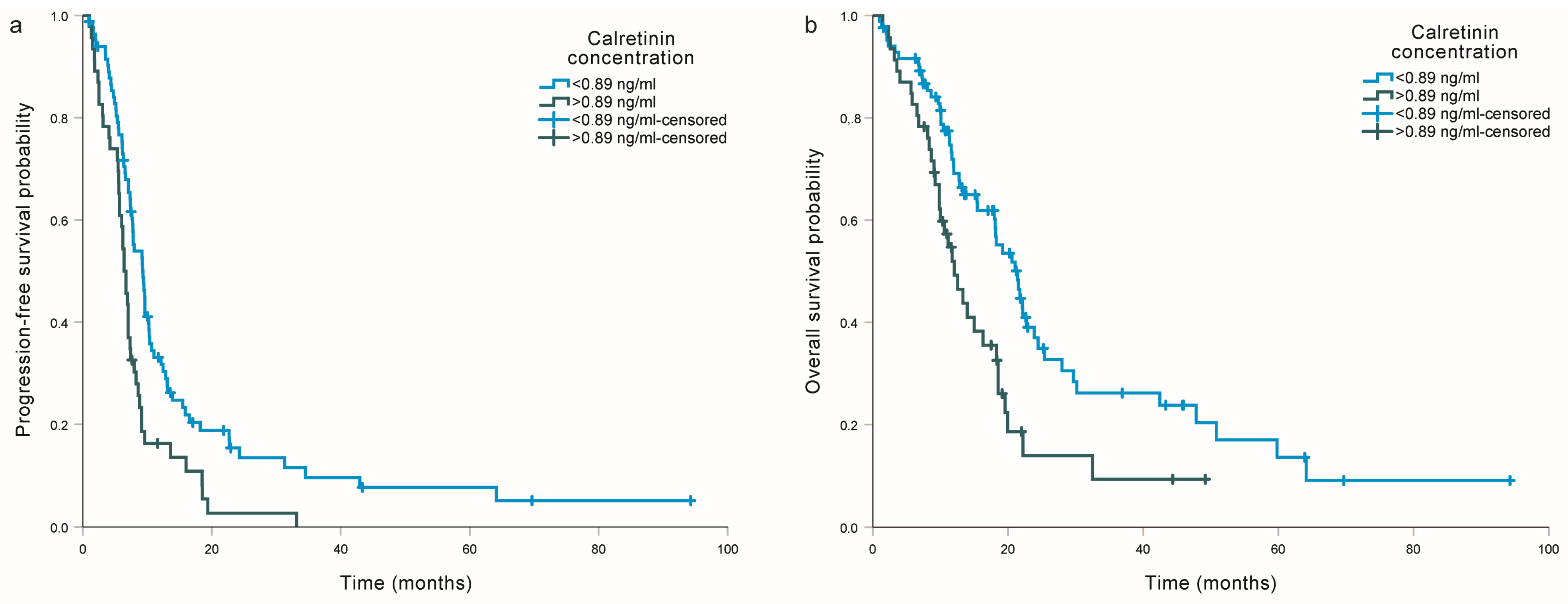
2.2. Association of Calretinin with Progression-Free Survival and Overall Survival from Diagnosis

Among clinical factors, asbestos exposure, smoking, weight loss, C-reactive protein, and histological type were significantly associated with PFS from diagnosis (all p < 0.05), while age, asbestos exposure, smoking, C-reactive protein, and histological type were significantly associated with OS from diagnosis (all p < 0.05).
Higher serum calretinin concentration was associated with shorter PFS (HR = 1.18, 95% CI = 1.02–1.37, p = 0.023) and shorter OS in MM patients (HR = 1.20, 95% CI = 1.03–1.41, p = 0.023). However, after adjusting for the significant clinical factors, the association was no longer statistically significant (HR = 1.05, 95% CI = 0.85–1.31, p = 0.653 and HR = 1.06, 95% CI = 0.84–1.34, p = 0.613, respectively). If patients were divided into two groups based on median calretinin concentration, PFS was slightly shorter if serum calretinin concentration was above the median (8.3 (5.7–14.2) compared to 10.0 (6.8–14.5) months), but the difference was not significant in univariable (HR = 1.25, 95% CI = 0.89–1.77, p = 0.205) or multivariable analysis (HR = 0.95, 95% CI = 0.63–1.43, p = 0.816). Similarly, OS was shorter if serum calretinin concentration was above the median (14.9 (9.9–23.9) compared to 20.7 (11.3–43.4) months), but the difference did not reach statistical significance in univariable (HR = 1.39, 95% CI = 0.94–2.05, p = 0.104) or multivariable analysis (HR = 0.97, 95% CI = 0.60–1.55, p = 0.884).
The association of investigated SNPs with the survival of MM patients from diagnosis is presented in Table 2. Among genetic factors, SEPTIN7 rs3801339 was associated with slightly shorter PFS in carriers of two polymorphic alleles (HR = 1.47, 95% CI = 1.02–2.13, p = 0.039), even after adjusting for clinical factors (HR = 1.76, 95% CI = 1.17–2.64, p = 0.007) (Figure S1). In multivariable analysis, the association was also significant in the dominant model (p = 0.046, Table 2). Heterozygotes for the MIR335 rs3807348 polymorphism had longer OS compared to the carriers of two reference alleles (HR = 0.69, 95% CI = 0.49–0.98, p = 0.038), even after adjusting for clinical factors (HR = 0.65, 95% CI = 0.45–0.95, p = 0.028).

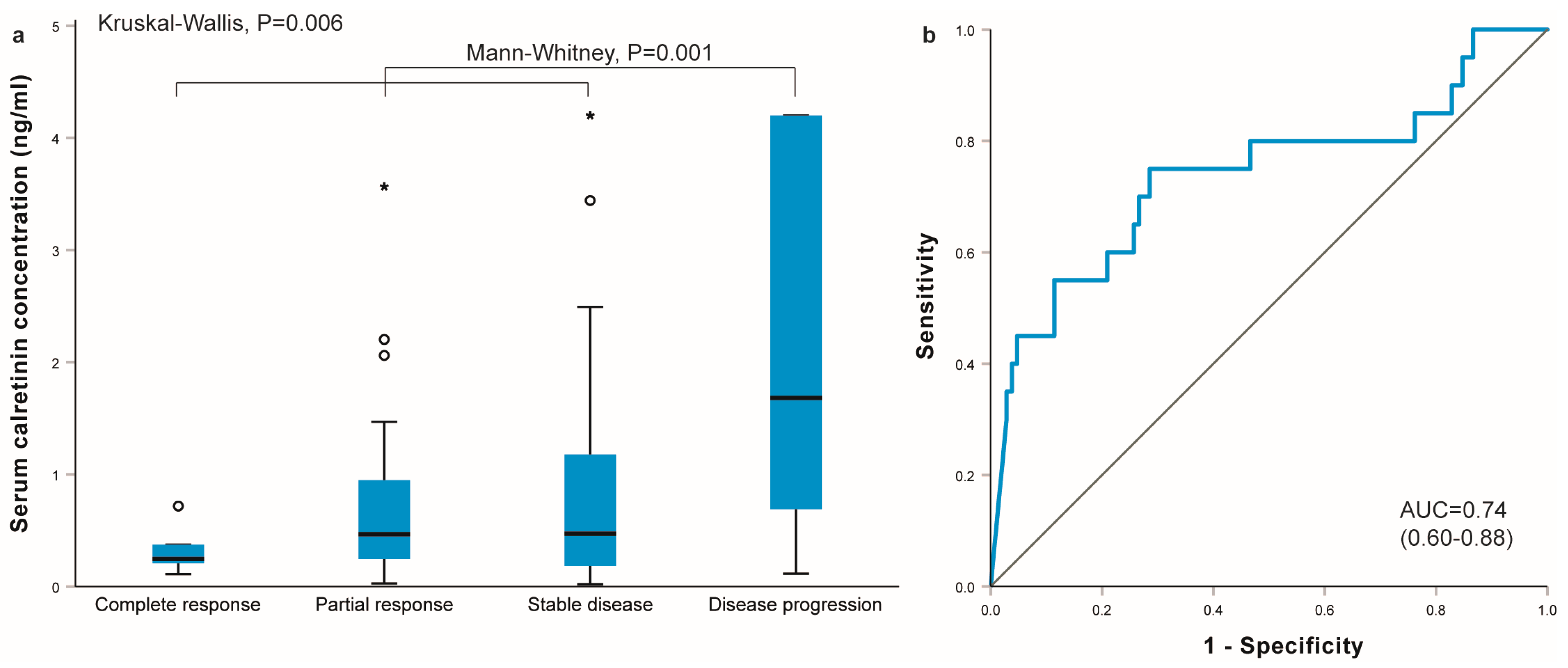
2.3. Association of Calretinin with the Outcome of Cisplatin-Based Chemotherapy

Among patients treated with cisplatin-based chemotherapy, 154 (64.4%) patients received a combination of gemcitabine and cisplatin, while 85 (35.6%) patients received a combination of pemetrexed and cisplatin. Regarding treatment outcome, eight (3.5%) patients had a complete response, 72 (31.2%) patients had a partial response, 112 (48.5%) had stable disease, and 39 (16.9%) experienced disease progression.
Serum calretinin concentration differed in MM patients with different treatment outcomes. The highest calretinin concentrations were observed in patients with disease progression after chemotherapy and the lowest were observed in patients who responded completely to chemotherapy (p = 0.006, Figure 1a). Calretinin concentration was statistically significantly higher in patients who progressed after the completion of chemotherapy compared to others (1.68 (0.58–4.20) ng/mL versus 0.45 (0.21–1.07) ng/mL, p = 0.001). Calretinin concentration at diagnosis was a good predictor of disease progression (area under the curve 0.74 (0.60–0.88); p = 0.001) (Figure 1b). At a cutoff of 0.89 ng/mL, the sensitivity for predicting disease progression was 0.750 and the specificity was 0.714.
The association of serum calretinin concentration with disease control rate is presented in Table 3 and Table S2. Patients with calretinin concentration above the median were more likely to experience disease progression (OR = 3.56, 95% CI = 1.21–10.52, p = 0.021), but the difference was no longer significant after adjusting for CRP and chemotherapy type (p = 0.168). The difference was even more pronounced if patients were divided based on the cutoff value determined in ROC curve analysis: patients with a calretinin concentration above 0.89 ng/mL were much more likely to experience disease progression (OR = 7.50, 95% CI = 2.50–22.47, p < 0.001), even after adjusting for clinical factors (p = 0.016, Table S2).
When evaluating the association with response rate, patients with higher serum calretinin concentration were less likely to achieve good treatment response (OR = 0.61, 95% CI = 0.40–0.93, p = 0.021), but the association was no longer significant after adjusting for smoking, CRP, and weight loss (OR = 0.77, 95% CI = 0.48–1.24, p = 0.278). Additionally, serum calretinin above the median or above 0.89 ng/mL was not significantly associated with the response rate in univariable (p = 0.315 and p = 0.632, respectively) or multivariable analysis (p = 0.948 and p = 0.695, respectively).
When analyzing the effect of selected polymorphisms on the treatment response rate (Table 4), carriers of at least one MIR335 rs3807348 polymorphic allele had a significantly better response (OR = 2.41, 95% CI = 1.19–4.87, p = 0.015), even after adjusting for clinical factors (OR = 2.69, 95% CI = 1.17–6.18, p = 0.020). Other investigated polymorphisms were not significantly associated with response rate (all p > 0.05).

2.4. Association of Calretinin with Progression-Free Survival and Overall Survival from the Beginning of Chemotherapy

Serum calretinin concentration was also associated with survival from the beginning of chemotherapy in MM patients treated with cisplatin-based chemotherapy (Table 3 and Table S2). Higher serum calretinin concentration was associated with worse PFS (p = 0.010) and worse OS (p = 0.013) in univariable analysis, but not after adjusting for clinical factors (p = 0.206 and p = 0.378, respectively). Patients with a calretinin concentration above 0.89 ng/mL had significantly shorter PFS (HR = 1.88, 95% CI = 1.28–2.77, p = 0.001; Figure 2a) and shorter OS (HR = 1.91, 95% CI = 1.22–2.97, p = 0.004, Figure 2b). This association remained statistically significant, even after adjusting for clinical factors (p = 0.006 and 0.049, respectively).
None of the studied polymorphisms were significantly associated with PFS or OS in this group of patients, either in univariable or multivariable analysis (Table S3).

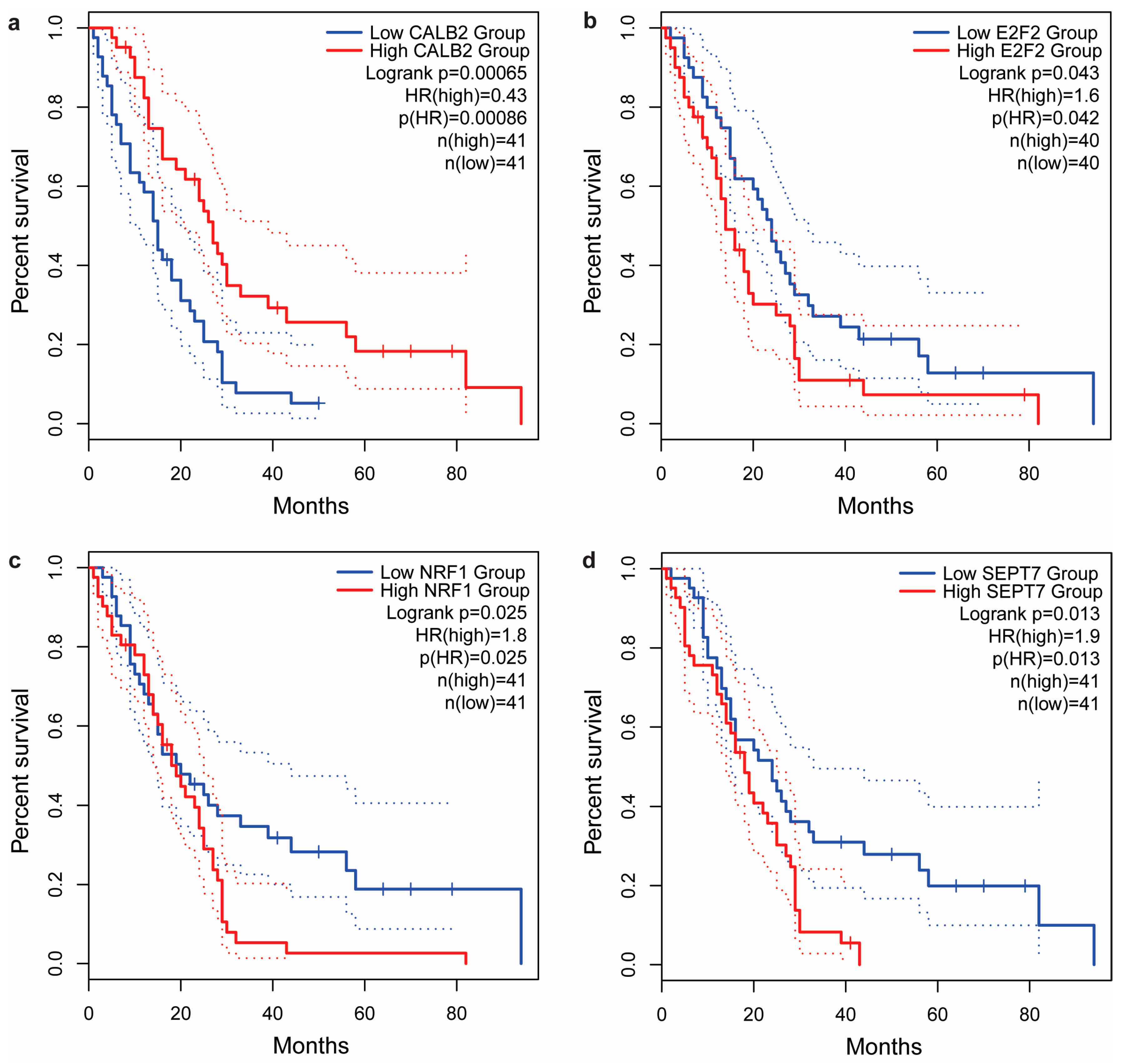
2.5. In Silico Analysis of Tumor Tissue mRNA Expression

We also evaluated the mRNA expression of investigated protein-coding genes in TCGA MESO samples using the online tool GEPIA2. There were several correlations between the expression of different genes in the MM tumor tissue. We observed significant negative correlations between CALB2 and E2F2 (ρ = −0.26, p = 0.017), as well as between CALB2 and SEPTIN7 (ρ = −0.29, p = 0.007) expression, while the correlation between CALB2 and NRF1 (ρ = −0.20, p = 0.058) did not reach statistical significance. On the other hand, positive correlations were observed between E2F2 and NRF1 (ρ = 0.32, p = 0.002), E2F2 and SEPTIN7 (ρ = 0.28, p = 0.009), and NRF1 and SEPTIN7 expression (ρ = 0.28, p < 0.001).
The association of gene expression with the survival of MM patients showed that disease-free survival (DFS) was only statistically significantly affected by SEPTIN7 (HR = 1.9, p = 0.03), while no significant differences were observed for CALB2 (HR = 0.80, p = 0.42), E2F2 (HR = 1.6, p = 0.099), and NRF1 (HR = 1.7, p = 0.065). On the other hand, all four genes were significantly associated with OS of MM patients: patients with high CALB2 expression had longer survival than patients with low CALB2 expression (HR = 0.43, p < 0.001, Figure 3a), while patients with higher E2F2 (HR = 1.6, p = 0.042, Figure 3b), NRF1 (HR = 1.8, p = 0.025, Figure 3c), or SEPTIN7 (HR = 1.9, p = 0.013, Figure 3d) expression had shorter OS.

3. Discussion

In the present study, we evaluated the prognostic and predictive role of serum calretinin concentration in patients with MM. We also assessed if genetic polymorphisms in the CALB2 gene or in genes coding for miRNAs and transcription factors that regulate calretinin expression can serve as independent prognostic factors in MM. In the whole patient group, higher serum calretinin concentration was associated with shorter PFS and shorter OS, but the association was no longer significant after adjusting for clinical factors, indicating that calretinin was not an independent prognostic factor in our patients with MM. On the other hand, calretinin was an important predictive biomarker in MM patients treated with cisplatin-based chemotherapy, even in multivariable analysis: serum calretinin concentration was significantly higher in patients who had disease progression during or after the completion of chemotherapy and was associated with worse PFS and OS. Among genetic factors, SEPTIN7 rs3801339 was associated with shorter PFS, and MIR335 rs3807348 was associated with longer OS and better response to cisplatin-based chemotherapy.
In our study, serum calretinin concentration was associated with shorter PFS and OS from diagnosis, regardless of the treatment regimen, but after adjusting for clinical factors, the association was no longer statistically significant, suggesting that serum calretinin is not an independent prognostic factor in MM. As MM is commonly treated with cisplatin-based chemotherapy, we also determined the association of calretinin with the outcome of cisplatin-based chemotherapy and survival from the start of chemotherapy. We demonstrated that serum calretinin concentration was higher in patients with a worse treatment outcome and shorter PFS and OS from the beginning of chemotherapy, even after adjusting for clinical factors, suggesting that it could serve as an independent predictive biomarker in MM patients treated with cisplatin-based chemotherapy. A calretinin concentration above 0.89 ng/mL was a good predictor of disease control rate and survival, but not response rate, suggesting that it is associated more with disease progression. This concentration is much higher than the concentration of 0.32 ng/mL, which could be used as a diagnostic biomarker to discriminate between MM patients and other asbestos-exposed subjects based on our previous results [18].
So far, none of the previous studies investigated the association of soluble calretinin with prognosis or treatment outcome in MM. Consistent with our results, higher serum calretinin concentration at diagnosis, as well as its dynamics during chemotherapy, were independent predictors of poor PFS and OS, as well as chemotherapy resistance, in ovarian cancer patients treated with cisplatin-based chemotherapy [24]. Additional validation studies are needed to better evaluate the association between calretinin and cisplatin and determine whether serum calretinin could serve as a predictive biomarker of disease progression in cancer patients treated with cisplatin in clinical practice.
Contrary to our results and results in ovarian cancer showing that higher serum calretinin concentration is associated with shorter survival [24], previous studies reported that the higher expression of calretinin in MM tumor tissue was independently associated with longer survival, even after adjusting for significant clinical parameters, such as histological subtype or age [21,22,23]. Therefore, we also evaluated mRNA expression in MM tumor tissue using in silico analysis of publicly available databases. Consistent with previous studies, higher calretinin mRNA expression was associated with longer OS in the TCGA MESO cohort. So far, the association between tissue calretinin expression and soluble calretinin in MM was only investigated in one study: patients with calretinin-positive tumors had higher plasma calretinin [29], and, therefore, a similar prognostic role in tissue and blood could be expected. However, the positive correlation of calretinin expression in tissue and plasma could be attributed to the fact that calretinin is more often detected in epithelioid and biphasic MM compared to the sarcomatoid subtype in both tissue and plasma or serum of MM patients [18,20,30,31]. As patients with sarcomatoid MM still have much shorter survival [32], this could explain the observed differences regarding the association of calretinin with survival. However, a more detailed comparison would be needed to determine the different prognostic roles of calretinin in tissue and blood samples.
In our bioinformatic analysis, we also evaluated tissue mRNA expression for other protein-coding genes investigated in our study. For E2F2, NRF1, and SEPTIN7, MM patients with lower mRNA expression had longer OS. Additionally, significant correlations were observed between the mRNA expression of CALB2 and genes involved in calretinin regulations in MM tumor tissue, as well as among the investigated regulating genes, further confirming that E2F2, NRF1, and SEPTIN7 could act as regulators of calretinin expression as previously proposed [14,27]. Further studies are needed to assess whether a combination of these genes might serve as a potential biomarker in MM.
In the genetic part of our study, we also investigated the association of several different SNPs in CALB2 and its regulating genes with survival and treatment response in MM. We found that carriers of two polymorphic SEPTIN7 rs3801339 alleles had shorter PFS from diagnosis, even after adjusting for clinical factors. Septin 7 is a GTP-binding protein that plays an important role in various cellular processes [14,33]. Its binding to the CALB2 promoter region resulted in decreased calretinin expression in MM cell lines [14]. We also observed a negative correlation between SEPTIN7 and CALB2 expression in TCGA MESO tissue samples. Septin 7 can also contribute to cancer development [33,34] and it was previously implicated in cell migration and invasion in breast cancer cell lines [35]. Similar to our results, another polymorphism in this gene, rs1143149, was previously associated with shorter survival in non-small cell lung cancer patients who were long-term former smokers [33]. On the other hand, SEPTIN7 rs3801339 was not significantly associated with MM susceptibility or with serum calretinin concentration in our previous study [36]. Additional studies are needed to better evaluate the association of septin 7 and its genetic variability with both calretinin and cancer prognosis.
In the present study, the polymorphic MIR335 rs3807348 allele was associated with longer OS from diagnosis, as well as a better response to cisplatin-based chemotherapy, even after adjusting for clinical factors. In our previous study, this allele was also associated with higher serum calretinin concentration [36]. MiR-335 can influence apoptosis, proliferation, migration, and invasion of cancer cells through various target genes involved in different signaling pathways, including CALB2, and is often dysregulated in different cancer types [37]. In most cancer types, miR-335 is downregulated and acts as a tumor suppressor, but some studies reported that it is upregulated and acts as an oncogene in specific cancers [37]. Some studies also reported that it could function as a prognostic biomarker; however, both lower and higher miR-335 expression was associated with worse prognosis in different cancer types [37,38]. In MM, miR-335 expression was only evaluated in a very small study, where increased expression was reported in the serum of MM patients [39]. Interestingly, miR-335 was also associated with response to chemotherapy, including cisplatin-based chemotherapy [37]. In ovarian cancer, increased miR-335 expression was associated with increased sensitivity to cisplatin through its target gene BCL2L2, an anti-apoptotic protein increased in several cancer types [40]. However, the mechanism of action of miR-335 is complex and associated with many cellular processes. Additionally, the role of MIR335 genetic variability is not well established. Further studies are, therefore, needed to improve the understanding of miR-335’s association with calretinin and its role in MM and cancer in general.
Other investigated SNPs in CALB2, E2F2, and NRF1 genes were not associated with survival or response to cisplatin-based chemotherapy, even though we have previously shown that they could contribute to the interindividual variability observed for serum calretinin concentration in MM patients or asbestos-exposed subjects [36], suggesting that the genetic variability in these genes does not play an important role as a prognostic or predictive biomarker in MM.
This is the first study to show that serum calretinin and genetic factors associated with calretinin expression might play a role in prognosis and treatment response in MM, but this study also has some limitations. In our study, serum calretinin concentration was only available for a subgroup of patients and we did not have tissue samples available for comparison of calretinin expression in tumor samples. To overcome this, we used in silico analysis in MM samples included in publicly available databases. As expression data for miR-335 are not available in the GEPIA2 database, this part of the analysis was only performed for protein-coding genes. On the other hand, we evaluated the role of genetic variability in a large MM cohort. Still, other genetic factors could also influence calretinin expression. All MM patients were also treated in the same institution according to the same protocols. In the future, further large studies in this field and validation of these results in an independent population are needed. Additionally, evaluation of the mRNA and protein expression of investigated genes and the miRNA expression of investigated miRNAs in different biological samples could provide new insights about the processes involved in MM and its prognosis.
So far, several circulating biomarkers have been identified in MM, especially diagnostic, but also prognostic. Still, none of them have entered routine clinical use yet, and further validation studies and studies focusing on new combinations of biomarkers are needed. The association of calretinin with response to cisplatin reported in our study is also important because many new treatment options are being investigated in MM, such as immunotherapy and targeted therapy. With the help of new biomarkers and genetic factors, we could identify patients who are expected to respond poorly to standard forms of chemotherapy and would benefit more from other treatment options.

4. Materials and Methods

4.1. Study Population

Our retrospective study included patients with histologically confirmed pleural or peritoneal MM treated at the Institute of Oncology Ljubljana between November 2001 and March 2019.
For all patients, data on demographic and clinical characteristics were obtained from medical records or an interview. Asbestos exposure was defined as known occupational or environmental exposure. The stage of MM was determined using the TNM staging system for pleural MM. The performance status of MM patients was determined using Eastern Cooperative Oncology Group (ECOG) scores.
All participants provided written informed consent. This study was approved by the National Medical Ethics Committee of the Republic of Slovenia (31/07/04, 39/04/06, and 41/02/09) and was carried out according to the Declaration of Helsinki.

4.2. Treatment Response and Survival Assessment

Tumor response was evaluated using modified Response Evaluation Criteria in Solid Tumors (RECISTs) [41]. The response rate was defined as the percentage of patients that achieved complete or partial response, while the disease control rate was defined as the percentage of patients that achieved complete response, partial response, or stable disease.
In survival analysis, PFS and OS were evaluated. PFS was defined as the time from diagnosis (for all patients) or from the beginning of chemotherapy (for patients with systemic treatment) to disease progression, and OS was defined as the time from diagnosis or from the beginning of chemotherapy to death from any cause.

4.3. Single-Nucleotide Polymorphism (SNP) Selection, DNA Extraction, and Genotyping

Using bioinformatic analysis, we previously identified common SNPs that could affect calretinin expression within the CALB2 gene and its regulating transcription factors and miRNAs [36]. We selected three SNPs in 5′ and 3′ UTR of the CALB2 gene (rs1862818, rs889704, rs8063760), three SNPs in genes coding for transcription factors (E2F2 rs2075995, NRF1 rs13241028, SEPTIN7 rs3801339) and one SNP in genes coding for miRNAs associated with calretinin expression (MIR335 rs3807348).
We used the Qiagen FlexiGene Kit (Qiagen, Hilden, Germany) to extract genomic DNA from peripheral venous blood samples. All SNPs were genotyped using competitive allele-specific PCR following the manufacturer’s instructions (KASP assays, LGC Biosearch Technologies, Hoddesdon, UK). For all SNPs, 15% of the samples were genotyped in duplicates. Genotyping quality control criteria were 100% duplicate call rate and 95% SNP-wise call rate.

4.4. Measurement of Serum Calretinin

Serum samples were collected at diagnosis, prepared within 6 h of blood collection, and stored at −20 °C until analysis. Serum calretinin concentrations were determined using a commercially available enzyme-linked immunosorbent calretinin ELISA assay (DLD Diagnostika GmbH, Hamburg, Germany) according to the manufacturer’s instructions, as previously described [18,20,42]. The manufacturer reported the intra-assay coefficient of variation as 8.1–6.6% and the inter-assay coefficient of variation as 10.4–10.0%.

4.5. Bioinformatic Analysis: Tissue Gene Expression

To analyze the expression of calretinin and associated genes in MM tissue samples, we used the Gene Expression Profiling Interactive Analysis (GEPIA) 2 tool, an interactive web server for analyzing the RNA sequencing expression data of tumors and normal samples from The Cancer Genome Atlas (TCGA). We used GEPIA2 to assess the correlations between the expression of different protein-coding genes in MM and evaluate the association of the investigated genes with survival in MM in the TCGA mesothelioma tumor samples (TCGA MESO study) [43]. Expression above the median was classified as high expression, while expression below the median was classified as low expression.

4.6. Statistical Analysis

Continuous and categorical variables were described using a median with interquartile range and frequencies, respectively. For all investigated SNPs, both additive and dominant models were used in the analysis. A chi-square test was used to evaluate deviation from HWE. Nonparametric Mann–Whitney or Kruskal–Wallis tests were used to compare the distribution of continuous variables. Spearman’s rho correlation coefficient (ρ) was used to evaluate correlations between continuous variables. Receiver operating characteristic (ROC) curve analysis was used to determine the area under the curve (AUC), sensitivity, and specificity. Cutoff values were determined as the values with the highest sum of sensitivity and specificity. Univariable and multivariable logistic regression was used to evaluate the association with response to chemotherapy to determine odds ratios (ORs) and 95% confidence intervals (CIs). In survival analysis, Kaplan–Meier analysis was used to calculate the median survival or follow-up time and analysis of GEPIA survival data, while univariable and multivariable Cox regression was used to calculate the hazard ratios (HRs) with 95% CIs. Patients without progression or death at the time of the analysis were censored at the date of the last follow-up. Clinical parameters used for adjustment in multivariable analysis were selected using stepwise forward conditional selection. To determine the median survival in patients with different serum calretinin concentrations, patients were divided into two groups according to median concentration or cutoff values determined using ROC curve analysis. All statistical tests were two-sided, and the level of significance was set at 0.05. The statistical analyses were carried out using IBM SPSS Statistics version 27.0 (IBM Corporation, Armonk, NY, USA).

5. Conclusions

In conclusion, our study showed that higher serum calretinin concentration was associated with shorter PFS and OS in MM, even though it was not an independent prognostic factor in our study. SEPTIN7 rs3801339 and MIR335 rs3807348 were associated with survival among genetic factors. Additionally, serum calretinin concentration could serve as a predictive biomarker in MM patients treated with cisplatin-based chemotherapy. In the future, it could help improve treatment stratification and potentially contribute to better treatment outcomes in MM.

Supplementary Materials

The following supporting information can be downloaded at https://www.mdpi.com/article/10.3390/ijms25010190/s1.

Author Contributions

Conceptualization, V.D. and K.G.; methodology, A.F., V.D. and K.G.; validation, C.Z. and K.G.; formal analysis, C.Z. and K.G.; investigation, C.Z., A.F., D.Š., V.K., V.D. and K.G.; resources, A.F., D.Š., V.K. and V.D.; writing—original draft preparation, C.Z. and K.G.; writing—review and editing, C.Z., A.F., D.Š., V.K., V.D. and K.G.; visualization, C.Z. and K.G.; supervision, V.D. and K.G.; project administration, A.F., V.K., V.D. and K.G.; funding acquisition, V.D. All authors have read and agreed to the published version of the manuscript.

Funding

This study and APC were funded by the Javna Agencija za Raziskovalno Dejavnost RS (Eng. Slovenian Research Agency) (ARRS) research grants P1-0170, L3-8203, and L3-2622.

Institutional Review Board Statement

The study was conducted according to the guidelines of the Declaration of Helsinki and was approved by the Republic of Slovenia National Medical Ethics Committee (31/07/04, 13 July 2004; 39/04/06, 14 April 2006; 41/02/09, 7 March 2009).

Informed Consent Statement

Informed consent was obtained from all subjects involved in the study.

Data Availability Statement

All the data are presented within the article and Supplementary Materials. Any additional information is available upon request from the corresponding author.

Conflicts of Interest

The authors declare no conflict of interest. The funders had no role in the design of the study; in the collection, analyses, or interpretation of data; in the writing of the manuscript; or in the decision to publish the results.

References

  1. Štrbac, D.; Dolžan, V. Novel and Future Treatment Options in Mesothelioma: A Systematic Review. Int. J. Mol. Sci. 2022, 23, 1975. [Google Scholar] [CrossRef]
  2. He, J.; Xu, S.; Pan, H.; Li, S.; He, J. Does size matter?—A population-based analysis of malignant pleural mesothelioma. Transl. Lung Cancer Res. 2020, 9, 1041–1052. [Google Scholar] [CrossRef]
  3. Kovac, V.; Zwitter, M.; Zagar, T. Improved survival after introduction of chemotherapy for malignant pleural mesothelioma in Slovenia: Population-based survey of 444 patients. Radiol. Oncol. 2012, 46, 136–144. [Google Scholar] [CrossRef]
  4. Rondon, L.; Fu, R.; Patel, M.R. Success of Checkpoint Blockade Paves the Way for Novel Immune Therapy in Malignant Pleural Mesothelioma. Cancers 2023, 15, 2940. [Google Scholar] [CrossRef] [PubMed]
  5. Deiana, C.; Fabbri, F.; Tavolari, S.; Palloni, A.; Brandi, G. Improvements in Systemic Therapies for Advanced Malignant Mesothelioma. Int. J. Mol. Sci. 2023, 24, 10415. [Google Scholar] [CrossRef] [PubMed]
  6. Chevallier, M.; Kim, F.; Friedlaender, A.; Addeo, A. Pleural Mesothelioma in the Era of Immunotherapy. Clin. Med. Insights Oncol. 2023, 17, 11795549231178173. [Google Scholar] [CrossRef] [PubMed]
  7. Cristaudo, A.; Bonotti, A.; Guglielmi, G.; Fallahi, P.; Foddis, R. Serum mesothelin and other biomarkers: What have we learned in the last decade? J. Thorac. Dis. 2018, 10, S353–S359. [Google Scholar] [CrossRef]
  8. Pass, H.I.; Levin, S.M.; Harbut, M.R.; Melamed, J.; Chiriboga, L.; Donington, J.; Huflejt, M.; Carbone, M.; Chia, D.; Goodglick, L.; et al. Fibulin-3 as a blood and effusion biomarker for pleural mesothelioma. N. Engl. J. Med. 2012, 367, 1417–1427. [Google Scholar] [CrossRef]
  9. Kovac, V.; Dodic-Fikfak, M.; Arneric, N.; Dolzan, V.; Franko, A. Fibulin-3 as a biomarker of response to treatment in malignant mesothelioma. Radiol. Oncol. 2015, 49, 279–285. [Google Scholar] [CrossRef]
  10. Schillebeeckx, E.; van Meerbeeck, J.P.; Lamote, K. Clinical utility of diagnostic biomarkers in malignant pleural mesothelioma: A systematic review and meta-analysis. Eur. Respir. Rev. 2021, 30, 210057. [Google Scholar] [CrossRef]
  11. Barone, E.; Gemignani, F.; Landi, S. Overexpressed genes in malignant pleural mesothelioma: Implications in clinical management. J. Thorac. Dis. 2018, 10, S369–S382. [Google Scholar] [CrossRef] [PubMed]
  12. Goricar, K.; Dolzan, V.; Lenassi, M. Extracellular Vesicles: A Novel Tool Facilitating Personalized Medicine and Pharmacogenomics in Oncology. Front. Pharmacol. 2021, 12, 671298. [Google Scholar] [CrossRef] [PubMed]
  13. Goričar, K.; Holcar, M.; Mavec, N.; Kovač, V.; Lenassi, M.; Dolžan, V. Extracellular Vesicle Enriched miR-625-3p Is Associated with Survival of Malignant Mesothelioma Patients. J. Pers. Med. 2021, 11, 1014. [Google Scholar] [CrossRef]
  14. Blum, W.; Pecze, L.; Rodriguez, J.W.; Steinauer, M.; Schwaller, B. Regulation of calretinin in malignant mesothelioma is mediated by septin 7 binding to the CALB2 promoter. BMC Cancer 2018, 18, 475. [Google Scholar] [CrossRef] [PubMed]
  15. Wörthmüller, J.; Salicio, V.; Oberson, A.; Blum, W.; Schwaller, B. Modulation of Calretinin Expression in Human Mesothelioma Cells Reveals the Implication of the FAK and Wnt Signaling Pathways in Conferring Chemoresistance towards Cisplatin. Int. J. Mol. Sci. 2019, 20, 5391. [Google Scholar] [CrossRef]
  16. Casjens, S.; Johnen, G.; Raiko, I.; Pesch, B.; Taeger, D.; Töpfer, C.; Schonefeld, S.; Moebus, S.; Jöckel, K.-H.; Brüning, T.; et al. Re-evaluation of potential predictors of calretinin and mesothelin in a population-based cohort study using assays for the routine application in clinical medicine. BMJ Open 2021, 11, e039079. [Google Scholar] [CrossRef] [PubMed]
  17. Greening, D.W.; Ji, H.; Chen, M.; Robinson, B.W.S.; Dick, I.M.; Creaney, J.; Simpson, R.J. Secreted primary human malignant mesothelioma exosome signature reflects oncogenic cargo. Sci. Rep. 2016, 6, 32643. [Google Scholar] [CrossRef]
  18. Zupanc, C.; Franko, A.; Štrbac, D.; Dodič Fikfak, M.; Kovač, V.; Dolžan, V.; Goričar, K. Serum Calretinin as a Biomarker in Malignant Mesothelioma. J. Clin. Med. 2021, 10, 4875. [Google Scholar] [CrossRef]
  19. Aguilar-Madrid, G.; Pesch, B.; Calderón-Aranda, E.S.; Burek, K.; Jiménez-Ramírez, C.; Juárez-Pérez, C.A.; Ochoa-Vázquez, M.D.; Torre-Bouscoulet, L.; Acosta-Saavedra, L.C.; Sada-Ovalle, I.; et al. Biomarkers for Predicting Malignant Pleural Mesothelioma in a Mexican Population. Int. J. Med. Sci. 2018, 15, 883–891. [Google Scholar] [CrossRef]
  20. Johnen, G.; Gawrych, K.; Raiko, I.; Casjens, S.; Pesch, B.; Weber, D.G.; Taeger, D.; Lehnert, M.; Kollmeier, J.; Bauer, T.; et al. Calretinin as a blood-based biomarker for mesothelioma. BMC Cancer 2017, 17, 386. [Google Scholar] [CrossRef]
  21. Linton, A.; Pavlakis, N.; O’Connell, R.; Soeberg, M.; Kao, S.; Clarke, S.; Vardy, J.; van Zandwijk, N. Factors associated with survival in a large series of patients with malignant pleural mesothelioma in New South Wales. Br. J. Cancer 2014, 111, 1860–1869. [Google Scholar] [CrossRef] [PubMed]
  22. Thapa, B.; Walkiewicz, M.; Murone, C.; Asadi, K.; Deb, S.; Barnett, S.; Knight, S.; Mitchell, P.; Liew, D.; Watkins, D.N.; et al. Calretinin but not caveolin-1 correlates with tumour histology and survival in malignant mesothelioma. Pathology 2016, 48, 660–665. [Google Scholar] [CrossRef] [PubMed]
  23. Kao, S.C.; Klebe, S.; Henderson, D.W.; Reid, G.; Chatfield, M.; Armstrong, N.J.; Yan, T.D.; Vardy, J.; Clarke, S.; van Zandwijk, N.; et al. Low calretinin expression and high neutrophil-to-lymphocyte ratio are poor prognostic factors in patients with malignant mesothelioma undergoing extrapleural pneumonectomy. J. Thorac. Oncol. 2011, 6, 1923–1929. [Google Scholar] [CrossRef] [PubMed]
  24. Link, T.; Passek, S.; Wimberger, P.; Frank, K.; Vassileva, Y.D.; Kramer, M.; Kuhlmann, J.D. Serum calretinin as an independent predictor for platinum resistance and prognosis in ovarian cancer. Int. J. Cancer 2020, 146, 2608–2618. [Google Scholar] [CrossRef] [PubMed]
  25. Casjens, S.; Weber, D.G.; Johnen, G.; Raiko, I.; Taeger, D.; Meinig, C.; Moebus, S.; Jöckel, K.H.; Brüning, T.; Pesch, B. Assessment of potential predictors of calretinin and mesothelin to improve the diagnostic performance to detect malignant mesothelioma: Results from a population-based cohort study. BMJ Open 2017, 7, e017104. [Google Scholar] [CrossRef] [PubMed]
  26. Li, D.; Wang, B.; Long, H.; Wen, F. Diagnostic accuracy of calretinin for malignant mesothelioma in serous effusions: A meta-analysis. Sci. Rep. 2015, 5, 9507. [Google Scholar] [CrossRef] [PubMed]
  27. Kresoja-Rakic, J.; Kapaklikaya, E.; Ziltener, G.; Dalcher, D.; Santoro, R.; Christensen, B.C.; Johnson, K.C.; Schwaller, B.; Weder, W.; Stahel, R.A.; et al. Identification of cis- and trans-acting elements regulating calretinin expression in mesothelioma cells. Oncotarget 2016, 7, 21272–21286. [Google Scholar] [CrossRef] [PubMed]
  28. Tavazoie, S.F.; Alarcon, C.; Oskarsson, T.; Padua, D.; Wang, Q.; Bos, P.D.; Gerald, W.L.; Massague, J. Endogenous human microRNAs that suppress breast cancer metastasis. Nature 2008, 451, 147–152. [Google Scholar] [CrossRef]
  29. Lehnert, M.; Weber, D.G.; Taeger, D.; Raiko, I.; Kollmeier, J.; Stephan-Falkenau, S.; Brüning, T.; Johnen, G.; Brik, A.; Burek, K.; et al. Determinants of plasma calretinin in patients with malignant pleural mesothelioma. BMC Res. Notes 2020, 13, 359. [Google Scholar] [CrossRef]
  30. Chapel, D.B.; Schulte, J.J.; Husain, A.N.; Krausz, T. Application of immunohistochemistry in diagnosis and management of malignant mesothelioma. Transl. Lung Cancer Res. 2020, 9, S3–S27. [Google Scholar] [CrossRef]
  31. Chirieac, L.R.; Pinkus, G.S.; Pinkus, J.L.; Godleski, J.; Sugarbaker, D.J.; Corson, J.M. The immunohistochemical characterization of sarcomatoid malignant mesothelioma of the pleura. Am. J. Cancer Res. 2011, 1, 14–24. [Google Scholar] [PubMed]
  32. Calthorpe, L.; Romero-Hernandez, F.; Miller, P.; Conroy, P.C.; Hirose, K.; Kim, A.; Kirkwood, K.; Nakakura, E.; Corvera, C.; Maker, A.V.; et al. Contemporary Trends in Malignant Peritoneal Mesothelioma: Incidence and Survival in the United States. Cancers 2022, 15, 229. [Google Scholar] [CrossRef] [PubMed]
  33. Shen, S.; Wei, Y.; Li, Y.; Duan, W.; Dong, X.; Lin, L.; You, D.; Tardon, A.; Chen, C.; Field, J.K.; et al. A multi-omics study links TNS3 and SEPT7 to long-term former smoking NSCLC survival. NPJ Precis. Oncol. 2021, 5, 39. [Google Scholar] [CrossRef] [PubMed]
  34. Wang, X.; Fei, F.; Qu, J.; Li, C.; Li, Y.; Zhang, S. The role of septin 7 in physiology and pathological disease: A systematic review of current status. J. Cell Mol. Med. 2018, 22, 3298–3307. [Google Scholar] [CrossRef] [PubMed]
  35. Zhang, N.; Liu, L.; Fan, N.; Zhang, Q.; Wang, W.; Zheng, M.; Ma, L.; Li, Y.; Shi, L. The requirement of SEPT2 and SEPT7 for migration and invasion in human breast cancer via MEK/ERK activation. Oncotarget 2016, 7, 61587–61600. [Google Scholar] [CrossRef]
  36. Zupanc, C.; Franko, A.; Štrbac, D.; Dodič Fikfak, M.; Kovač, V.; Dolžan, V.; Goričar, K. Genetic factors influence serum calretinin levels in asbestos-related diseases. Abstracts from the 55th European Society of Human Genetics (ESHG) Conference. Eur. J. Hum. Genet. 2023, 31 (Suppl. S1), 1–12. [Google Scholar]
  37. Ye, L.; Wang, F.; Wu, H.; Yang, H.; Yang, Y.; Ma, Y.; Xue, A.; Zhu, J.; Chen, M.; Wang, J.; et al. Functions and Targets of miR-335 in Cancer. Oncol. Targets Ther. 2021, 14, 3335–3349. [Google Scholar] [CrossRef] [PubMed]
  38. Huo, W.; Zhang, M.; Li, C.; Wang, X.; Zhang, X.; Yang, X.; Fei, H. Correlation of microRNA-335 expression level with clinical significance and prognosis in non-small cell lung cancer. Medicine 2020, 99, e21369. [Google Scholar] [CrossRef] [PubMed]
  39. Lamberti, M.; Capasso, R.; Lombardi, A.; Di Domenico, M.; Fiorelli, A.; Feola, A.; Perna, A.F.; Santini, M.; Caraglia, M.; Ingrosso, D. Two Different Serum MiRNA Signatures Correlate with the Clinical Outcome and Histological Subtype in Pleural Malignant Mesothelioma Patients. PLoS ONE 2015, 10, e0135331. [Google Scholar] [CrossRef]
  40. Liu, R.; Guo, H.; Lu, S. MiR-335-5p restores cisplatin sensitivity in ovarian cancer cells through targeting BCL2L2. Cancer Med. 2018, 7, 4598–4609. [Google Scholar] [CrossRef]
  41. Byrne, M.J.; Nowak, A.K. Modified RECIST criteria for assessment of response in malignant pleural mesothelioma. Ann. Oncol. 2004, 15, 257–260. [Google Scholar] [CrossRef]
  42. Raiko, I.; Sander, I.; Weber, D.G.; Raulf-Heimsoth, M.; Gillissen, A.; Kollmeier, J.; Scherpereel, A.; Bruning, T.; Johnen, G. Development of an enzyme-linked immunosorbent assay for the detection of human calretinin in plasma and serum of mesothelioma patients. BMC Cancer 2010, 10, 242. [Google Scholar] [CrossRef] [PubMed]
  43. Tang, Z.; Kang, B.; Li, C.; Chen, T.; Zhang, Z. GEPIA2: An enhanced web server for large-scale expression profiling and interactive analysis. Nucleic Acids Res. 2019, 47, W556–W560. [Google Scholar] [CrossRef] [PubMed]
Figure 1. (a) Serum calretinin concentration and response to chemotherapy in malignant mesothelioma patients; (b) ROC curve analysis for the prediction of disease progression based on serum calretinin concentration at diagnosis. AUC, area under the curve. Circles represent outliers and stars (*) represent extreme outliers.
Ijms 25 00190 g001
Figure 2. Association of serum calretinin concentration with (a) progression-free survival and (b) overall survival from the beginning of chemotherapy in malignant mesothelioma patients treated with cisplatin-based chemotherapy.
Ijms 25 00190 g002
Figure 3. Association of the tumor tissue gene expression of the investigated genes with overall survival based on The Cancer Genome Atlas mesothelioma (TCGA MESO) data evaluated using the GEPIA2 online tool: (a) CALB2, (b) E2F2, (c) NRF1, (d) SEPTIN7. Dotted lines represent 95% confidence intervals.
Ijms 25 00190 g003
Table 1. Clinical characteristics of malignant mesothelioma patients.
All Patients (N = 265)Patients Treated with Cisplatin (N = 239)
CharacteristicCategory/UnitN (%)N (%)
SexMale197 (74.3)179 (74.9)
Female68 (25.7)60 (25.1)
AgeYears, median (25–75%)66 (59–72)66 (59–72)
Stage117 (7.2)15 (7.1)
260 (25.5)53 (25.1)
385 (36.2)77 (36.5)
472 (30.6)65 (30.8)
Not determined1 (0.4)1 (0.5)
LocationPleura235 (88.7)211 (88.3)
Peritoneum30 (11.3)28 (11.7)
Histological typeEpithelioid204 (77.0)186 (77.8)
Biphasic23 (8.7)21 (8.8)
Sarcomatoid26 (9.8)23 (9.6)
Not determined12 (4.5)9 (3.8)
ECOG performance status015 (5.7)15 (6.3)
1137 (51.7)129 (54.0)
2106 (40.0)92 (38.5)
37 (2.6)3 (1.3)
Asbestos exposureNo66 (25.0) {1}60 (25.2) {1}
Yes198 (75.0)178 (74.8)
SmokingNo146 (55.7) {3}132 (55.7) {2}
Yes116 (44.3)105 (44.3)
CRPmg/L, median (25–75%)23 (9–68.8) {29}21 (8–61) {20}
LDHµkat/L, median (25–75%)2.72 (2.28–3.19) {28}2.69 (2.26–3.17) {20}
PainNo109 (42.6) {9}101 (43.5) {7}
Yes147 (57.4)131 (56.5)
Weight lossNo87 (34.7) {14}77 (34.1) {13}
Yes164 (65.3)149 (65.9)
Chemotherapy regimenNo16 (6.0)0 (0.0)
Gemcitabine/cisplatin154 (58.1)154 (64.4)
Pemetrexed/cisplatin85 (32.1)85 (35.6)
Other10 (3.8)0 (0.0)
Disease progressionNo27 (10.2)25 (10.5)
Yes238 (89.8)214 (89.5)
DeathNo71 (26.8)67 (28.0)
Yes194 (73.2)172 (72.2)
PFSMonths, median (25–75%)10.0 (6.3–16.2) **8.2 (5.3–13.8) ***
OSMonths, median (25–75%)19.1 (10.0–29.4) **9.5 (18.1–28.4) ***
Follow-upMonths, median (25–75%)45.5 (22.8–78.7) **20.2 (44.4–75.5) ***
Calretinin concentration *Ng/mL, median (25–75%)0.52 (0.22–1.43)0.51 (0.22–1.37)
The number of missing data is presented in {} brackets. * Data available for 146 patients in the whole cohort and 130 patients treated with cisplatin; ** survival and follow-up from diagnosis; *** survival and follow-up from the beginning of chemotherapy. CRP, C reactive protein, ECOG, Eastern Cooperative Oncology Group, LDH, lactate dehydrogenase, OS, overall survival, PFS, progression-free survival.
Table 2. Association of selected polymorphisms with progression-free survival (PFS) and overall survival (OS) from diagnosis of MM patients.
PFSOS
SNPGenotypeMedian (25–75%)HR (95% CI)pHR (95% CI)adj1Padj1Median (25–75%)HR (95% CI)pHR (95% CI)adj2Padj2
CALB2 rs1862818CC9.8 (5.9–14.5)Reference Reference 16.7 (9.4–28.3)Reference Reference
CT9.9 (6.6–16.9)0.88 (0.67–1.15)0.3330.82 (0.60–1.11)0.19921.2 (11.3–35.1)0.78 (0.58–1.05)0.0990.79 (0.57–1.09)0.156
TT11.5 (6.6–20.5)0.81 (0.52–1.27)0.3640.74 (0.45–1.21)0.23324.4 (12.5–27.2)0.84 (0.52–1.37)0.4800.85 (0.50–1.44)0.545
CT + TT10.0 (6.6–17.4)0.86 (0.67–1.11)0.2560.80 (0.60–1.07)0.13022.0 (11.5–33.7)0.79 (0.59–1.05)0.0990.80 (0.59–1.09)0.157
CALB2 rs889704CC10.0 (6.6–16.9)Reference Reference 18.5 (10.1–30.3)Reference Reference
CA10.0 (6.0–12.6)1.14 (0.83–1.56)0.4151.03 (0.72–1.46)0.89020.3 (9.5–28.6)1.05 (0.73–1.51)0.7771.14 (0.78–1.66)0.501
AA8.7 (4.8–8.7)1.76 (0.43–7.13)0.432.60 (0.63–10.68)0.18516.2 (6.1–16.2)2.17 (0.53–8.81)0.2793.59 (0.86–14.96)0.079
CA + AA9.7 (6.0–12.6)1.15 (0.85–1.57)0.3621.06 (0.75–1.49)0.76220.3 (9.4–28.6)1.08 (0.76–1.54)0.6651.18 (0.81–1.71)0.381
CALB2 rs8063760CC10.2 (6.6–17.1)Reference Reference 19.4 (10.0–29)Reference Reference
CT9.4 (6.2–14.3)1.07 (0.81–1.41)0.6221.26 (0.93–1.71)0.14319.3 (11.6–32.5)0.88 (0.65–1.21)0.4330.98 (0.70–1.38)0.920
TT7.9 (4.8–14.3)1.54 (0.90–2.63)0.1141.22 (0.67–2.24)0.51812.7 (7.1–24.7)1.59 (0.90–2.83)0.1121.22 (0.66–2.25)0.527
CT + TT9.4 (6.1–14.3)1.13 (0.87–1.46)0.3711.25 (0.94–1.67)0.12818.2 (10.0–31.1)0.96 (0.72–1.29)0.7831.02 (0.74–1.41)0.895
E2F2 rs2075995CC8.4 (5.5–15.0)Reference Reference 16.0 (8.3–25.7)Reference Reference
CA11.0 (6.6–16.2)0.82 (0.60–1.12)0.2051.00 (0.71–1.42)0.98920.7 (11.0–31.4)0.73 (0.52–1.02)0.0660.95 (0.65–1.39)0.805
AA9.9 (6.2–16.5)0.93 (0.65–1.33)0.6951.04 (0.70–1.55)0.84618.1 (10.8–28.6)0.80 (0.54–1.19)0.2681.03 (0.66–1.60)0.886
CA + AA10.2 (6.4–16.5)0.85 (0.64–1.14)0.2861.02 (0.73–1.41)0.92819.7 (10.8–31.2)0.75 (0.55–1.03)0.0750.98 (0.68–1.40)0.903
MIR335 rs3807348GG9.4 (5.6–15.0)Reference Reference 16.1 (7.6–25.6)Reference Reference
GA10.6 (7.3–17.9)0.83 (0.61–1.13)0.2390.98 (0.68–1.40)0.90022.0 (11.8–35.1)0.69 (0.49–0.98)0.0380.65 (0.45–0.95)0.028
AA8.2 (5.5–13.4)1.18 (0.83–1.68)0.3531.40 (0.92–2.11)0.11415.6 (8.3–26.2)1.08 (0.74–1.57)0.7091.02 (0.67–1.55)0.914
GA + AA10.0 (6.6–16.3)0.93 (0.69–1.24)0.6141.08 (0.77–1.52)0.65019.7 (11.0–31.2)0.81 (0.59–1.11)0.1840.76 (0.53–1.08)0.129
NRF1 rs13241028TT10.7 (6.8–15.0)Reference Reference 18.2 (11.8–27.8)Reference Reference
TC7.5 (5.6–16.3)1.13 (0.86–1.49)0.3651.23 (0.92–1.67)0.16619.3 (7.3–33.7)0.94 (0.69–1.26)0.6681.04 (0.75–1.45)0.794
CC9.9 (5.5–14.1)0.88 (0.47–1.62)0.6720.70 (0.28–1.77)0.44929.4 (9.6–*)0.58 (0.27–1.25)0.1640.72 (0.29–1.81)0.489
TC + CC8.0 (5.6–16.3)1.10 (0.84–1.43)0.4901.19 (0.88–1.59)0.25719.3 (8.0–35.1)0.89 (0.66–1.19)0.4161.01 (0.74–1.39)0.942
SEPTIN7 rs3801339TT11.6 (7.9–19.4)Reference Reference 22.0 (11.5–28.6)Reference Reference
TC8.8 (6.0–16.3)1.27 (0.90–1.80)0.1761.28 (0.88–1.88)0.20315.6 (10.0–29.0)1.08 (0.74–1.56)0.7021.21 (0.81–1.80)0.361
CC9.7 (6.1–14.7)1.47 (1.02–2.13)0.0391.76 (1.17–2.64)0.00720.6 (9.6–31.4)09.5 (0.64–1.42)0.7961.11 (0.72–1.71)0.630
TC + CC9.4 (6.0–14.9)1.35 (0.97–1.86)0.0741.44 (1.01–2.06)0.04617.5 (9.6–30.0)1.02 (0.72–1.45)0.9051.17 (0.80–1.69)0.421
Adj1: adjusted for asbestos exposure, smoking, weight loss, C-reactive protein, and histological type; Adj2: adjusted for age, asbestos exposure, smoking, C-reactive protein, and histological type. * not yet reached. Cl, confidence interval, HR, hazard ratio, OS, overall survival, PFS, progression-free survival, SNP, single-nucleotide polymorphism.
Table 3. Association of serum calretinin concentration with disease control rate, progression-free (PFS), and overall survival (OS) from the beginning of chemotherapy in MM patients treated with cisplatin-based chemotherapy.
Disease Control RatePFSOS
Calretinin ConcentrationCR + PR + SDPDOR (95% CI)pMedian (25–75%)HR (95% CI)pMedian (25–75%)HR (95% CI)p
Serum concentration (ng/mL)0.45 (0.21–1.07) *1.68 (0.58–4.20) *2.18 (1.51–3.14)<0.001/1.21 (1.05–1.40)0.010/1.23 (1.04–1.45)0.013
<0.51 ng/mL, N (%) **57 (91.9)5 (8.1)Ref. 7.9 (5.5–13.1)Ref. 21.0 (10.1–42.5)Ref.
>0.51 ng/mL, N (%) **48 (76.2)15 (23.8)3.56 (1.21–10.52)0.0217.0 (5.5–10.3)1.38 (0.95–2.01)0.09015.0 (9.2–22.1)1.6 (0.95–2.26)0.085
<0.89 ng/mL, N (%) ***75 (93.8)5 (6.3)Ref. 9.3 (6.1–13.9)Ref. 21.4 (11.4–42.5)Ref.
>0.89 ng/mL, N (%) ***30 (66.7)15 (33.3)7.50 (2.50–22.47)<0.0016.4 (4.2–8.8)1.88 (1.28–2.77)0.00112.1 (8.3–19.6)1.91 (1.22–2.97)0.004
* Median (25–75%); ** divided based on median calretinin concentration; *** divided based on the cutoff value determined by ROC curve analysis. CR, complete response, PD, disease progression, PR, partial response, SD, stable disease.
Table 4. Association of selected polymorphisms with response rate in MM patients treated with cisplatin-based chemotherapy.
SNPGenotypeSD + PD
N (%)
PR + CR
N (%)
OR (95% CI)pOR (95% CI)adjPadj
CALB2 rs1862818CC79 (71.8)31 (28.2)Reference Reference
CT58 (59.2)40 (40.8)1.76 (0.99–3.13)0.0561.69 (0.87–3.29)0.121
TT14 (60.9)9 (39.1)1.64 (0.64–4.17)0.3011.31 (0.47–3.65)0.607
CT + TT72 (59.5)49 (40.5)1.73 (1.00–3.01)0.0501.60 (0.85–3.01)0.142
CALB2 rs889704CC113 (63.5)65 (36.5)Reference Reference
CA35 (70.0)15 (30.0)0.75 (0.38–1.47)0.3950.76 (0.34–1.72)0.511
AA3 (100)0 (0)/0.306//
CA + AA38 (71.7)15 (28.3)0.69 (0.35–1.34)0.2710.65 (0.29–1.45)0.294
CALB2 rs8063760CC87 (62.6)52 (37.4)Reference Reference
CT51 (65.4)27 (34.6)0.89 (0.50–1.58)0.6810.86 (0.45–1.67)0.664
TT13 (92.9)1 (7.1)0.13 (0.02–1.01)0.0510.17 (0.02–1.46)0.107
CT + TT64 (69.6)28 (30.4)0.73 (0.42–1.28)0.2760.74 (0.39–1.41)0.361
E2F2 rs2075995CC40 (70.2)17 (29.8)Reference Reference
CA76 (64.4)42 (35.6)1.30 (0.66–2.57)0.4500.84 (0.39–1.82)0.657
AA35 (62.5)21 (37.5)1.41 (0.64–3.09)0.3891.51 (0.63–3.63)0.355
CA + AA111 (63.8)63 (36.2)1.34 (0.70–2.55)0.3801.02 (0.50–2.11)0.951
MIR335 rs3807348GG45 (78.9)12 (21.1)Reference Reference
GA64 (55.7)51 (44.3)2.99 (1.43–6.23)0.0043.32 (1.40–7.89)0.007
AA42 (71.2)17 (28.8)1.52 (0.65–3.55)0.3361.65 (0.61–4.49)0.324
GA + AA106 (60.9)68 (39.1)2.41 (1.19–4.87)0.0152.69 (1.17–6.18)0.020
NRF1 rs13241028TT88 (63.3)51 (36.7)Reference Reference
TC57 (70.4)24 (29.6)0.73 (0.40–1.31)0.2870.64 (0.33–1.23)0.182
CC6 (54.5)5 (45.5)1.44 (0.42–4.95)0.5651.89 (0.33–10.7)0.473
TC + CC63 (68.5)29 (31.5)0.79 (0.45–1.39)0.4190.70 (0.37–1.32)0.270
SEPTIN7 rs3801339TT29 (60.4)19 (39.6)Reference Reference
TC73 (64.6)40 (35.4)0.84 (0.42–1.68)0.6140.93 (0.41–2.09)0.860
CC49 (70.0)21 (30.0)0.65 (0.30–1.42)0.2810.56 (0.23–1.38)0.209
TC + CC122 (66.7)61 (33.3)0.76 (0.40–1.47)0.4190.77 (0.36–1.66)0.506
Adj: adjusted for smoking, CRP, and weight loss. Cl, confidence interval, CR, complete response, OR, odds ratio, PD, disease progression, PR, partial response, SD, stable disease, SNP, single-nucleotide polymorphism.
Disclaimer/Publisher’s Note: The statements, opinions and data contained in all publications are solely those of the individual author(s) and contributor(s) and not of MDPI and/or the editor(s). MDPI and/or the editor(s) disclaim responsibility for any injury to people or property resulting from any ideas, methods, instructions or products referred to in the content.

