Article

Geometry and UV-Vis Spectra of Au3+ Complexes with Hydrazones Derived from Pyridoxal 5′-Phosphate: A DFT Study

by
Oleg A. Pimenov
,
Konstantin V. Grazhdan
,
Maksim N. Zavalishin
and
George A. Gamov
*
General Chemical Technology Department, Ivanovo State University of Chemistry and Technology, Sheremetevskii pr. 7, 153000 Ivanovo, Russia
*
Author to whom correspondence should be addressed.
Int. J. Mol. Sci. 2023, 24(9), 8412; https://doi.org/10.3390/ijms24098412
Submission received: 20 April 2023 / Revised: 5 May 2023 / Accepted: 6 May 2023 / Published: 7 May 2023
(This article belongs to the Special Issue Molecular Structure of Macroheterocyclic Compounds)

Abstract

:
Gold(III) complexes with different ligands can provide researchers with a measure against pathogenic microorganisms with antibiotic resistance. We reported in our previous paper that the UV-Vis spectra of different protonated species of complexes formed by gold(III) and five hydrazones derived from pyridoxal 5′-phosphate are similar to each other and to the spectra of free protonated hydrazones. The present paper focuses on the reasons of the noted similarity in electron absorption spectra. The geometry of different protonated species of complexes of gold(III) and hydrazones (15 structures in total) was optimized using the density functional theory (DFT). The coordination polyhedron of gold(III) bond critical points were further studied to identify the symmetry of the gold coordination sphere and the type of interactions that hold the complex together. The UV-Vis spectra were calculated using TD DFT methods. The molecular orbitals were analyzed to interpret the calculated spectra.

1. Introduction

The complexes formed by gold(III) are known to be isostructural and isoelectronic to platinum(II) compounds and can be seen as potential cytotoxic agents and antitumor drugs [1]. An interest in Au3+ complexes was revived in the mid-1990s [2,3,4] and has not extinguished yet, as recent papers show (see, e.g., reviews [5,6,7]). Despite their similarity with Pt2+ derivatives, it is the interaction with different proteins (in particular, thioredoxin reductase, which regulates cellular redox balance), not DNA, that seem to provide gold(III) complexes with biological activity [8,9,10,11]. However, Au3+ complexes are capable of damaging DNA via cleavage and oxidation (see, e.g., [12]), which can also be an important mechanism of biological action. Another advantage of Au use in medicine is its low toxicity for humans [13]. The possibility of using gold(III) complexes for the treatment of bacterial, fungal infections, and malaria is much less studied (a review [14] still accumulates most of the known data on gold(III) complexes as antibiotics; see also papers [15,16,17,18,19,20]). However, the most recent paper by Ratia et al. [21] shows the potential of gold(III) complexes in the most convincing manner.
The key disadvantages of gold(III) complexes is their instability in aqueous solutions, especially under physiological conditions. Neutral medium and biological reductant agents make gold(III) ions undergo either hydrolysis or reduction to Au+ or Au0 species, which changes their biological effect [22]. Moreover, the ligands evolving upon change of oxidation state of the metal ion can also show their own activity toward an organism [23]. The problem with metal complex stability can be solved if the appropriate ligands are chosen. Gold(III) binds efficiently with N,O-donor ligands since metal Lewis acidity corresponds best to nitrogen and oxygen Lewis basicity [24]. Therefore, molecules containing pyridine, bipyridine, and phenanthroline moieties, as well as macrocyclic ligands and porphyrins, are typical in gold(III) coordination chemistry [25,26,27,28,29].
The hydrazones derived from either pyridoxal or pyridoxal 5′-phosphate are tridentate ligands that form chelate complexes with d-metal ions in a stoichiometric ratio of metal:ligand = 1:2. The metal ion is tightly bound by imine nitrogen, the hydroxyl group in site 3 of the pyridoxal or pyridoxal 5′-phosphate residue, and carbonyl oxygen [30,31,32] (the phosphate group can contribute in the complexes of lanthanides(III) [33]). In addition, these hydrazones are low-toxic and membranotropic agents [34], which makes them especially attractive for developing new drugs based on hydrazones complexes (see, e.g., papers [35,36], where the antimicobacterial properties of complexes based on the hydrazones analogous to those derived from pyridoxal or pyridoxal 5′-phosphate are studied).
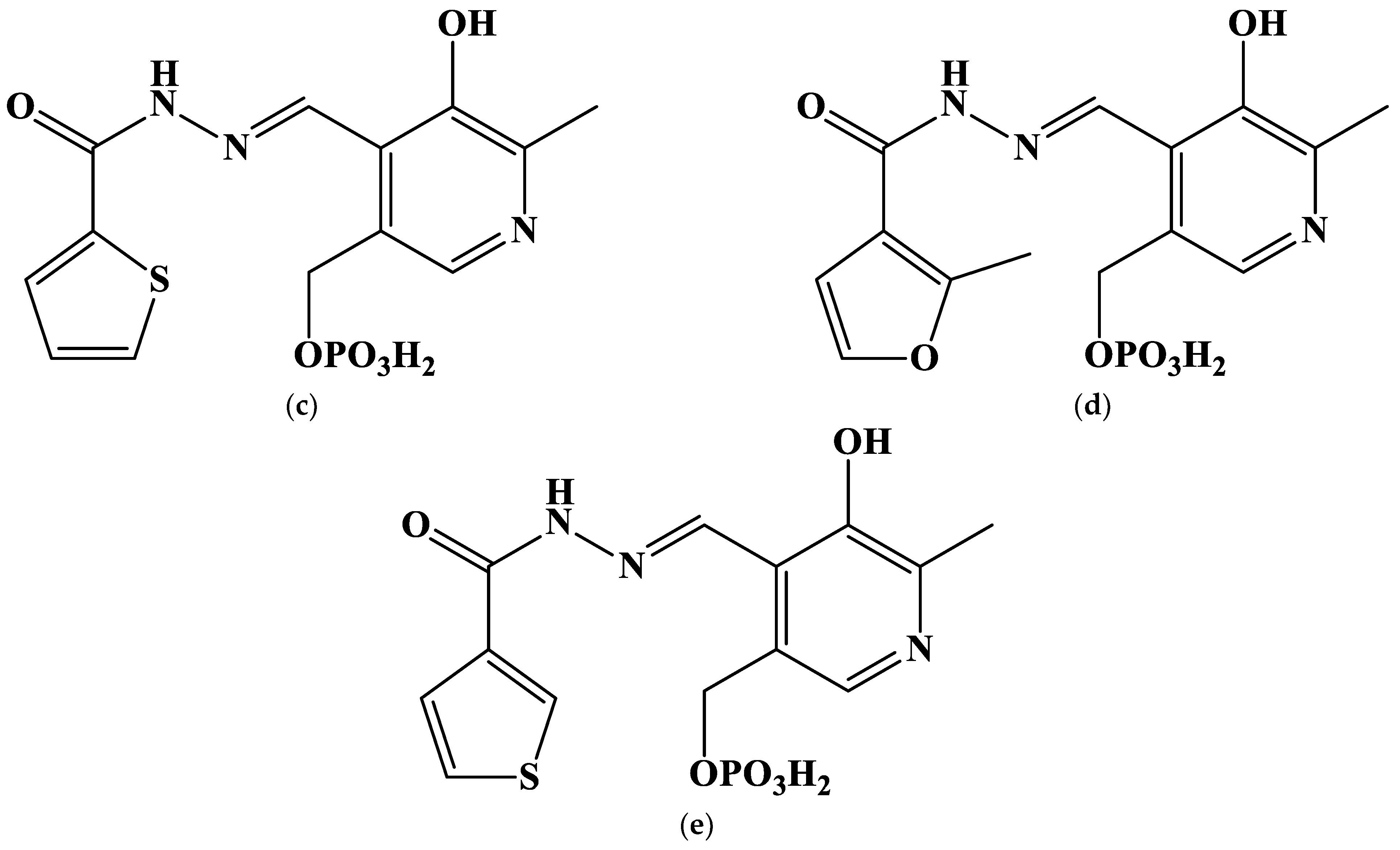
Our previous contribution reports on the stability of different protonated complexes of gold(III) with five hydrazones derived from pyridoxal 5′-phosphate [37]. These complexes were found to be relatively stable and formed with a high yield under physiological conditions. The authors of [37] also set several questions. One of them is why the electron absorption spectra of different protonated complexes are that similar to each other and those of free ligands. In the present contribution, we aim to answer it. To do so, it is necessary (1) to obtain the geometrical parameters of gold(III) complexes with the same hydrazones derived from pyridoxal 5′-phosphate (Figure 1); (2) to describe structural peculiarities of the coordination compounds; and (3) to compute UV-Vis spectra of gold(III) complexes and to compare them with those given in paper [37]. This goal is achieved using the density functional theory (DFT) calculations.
A brief review of the available literature data on DFT calculations of Au3+ complexes shows that gold(III) is prone to having square planar coordination, while the electron structure of the complexes is diverse [1,38,39,40,41,42,43]. Metal ions can contribute to LUMO [38,39,40], both HOMO and LUMO [1,41], or neither [42,43]. Ligand to metal charge transfer was reported in the paper [40]. The counter-ions complementing the coordination sphere of cation up to the square also often participate in frontier molecular orbital formation. This means that gold(III) complexes can show different UV-Vis spectra, even in complexes with the ligands possessing the same donor atoms (nitrogen and oxygen).

2. Results and Discussion

2.1. The Search of Probable Molecular Models

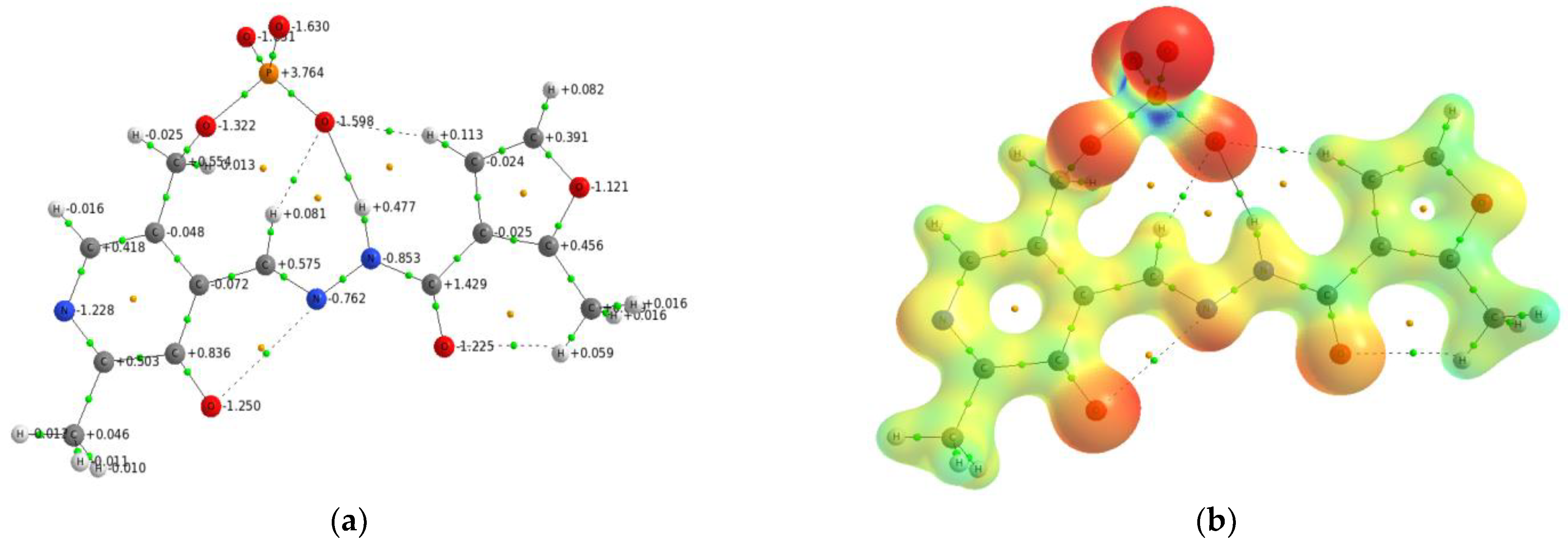
The search for probable molecular models of gold(III) complexes with the hydrazones derived from pyridoxal 5′-phosphate is based on our previous experience with a Zn(II) complex with pyridoxal 5′-phosphate 2-methyl-3-furoylhydrazone [44]. To obtain a demonstrative picture of complexation mechanism, we used the PLP-F3H hydrazone with a charge of –3e and analyzed the electronic density distribution for the most preferable geometry for cation coordination. Figure 2 demonstrates the molecular graph and molecular electrostatic potential (MEP) of [PLP-F3H]3− hydrazone.
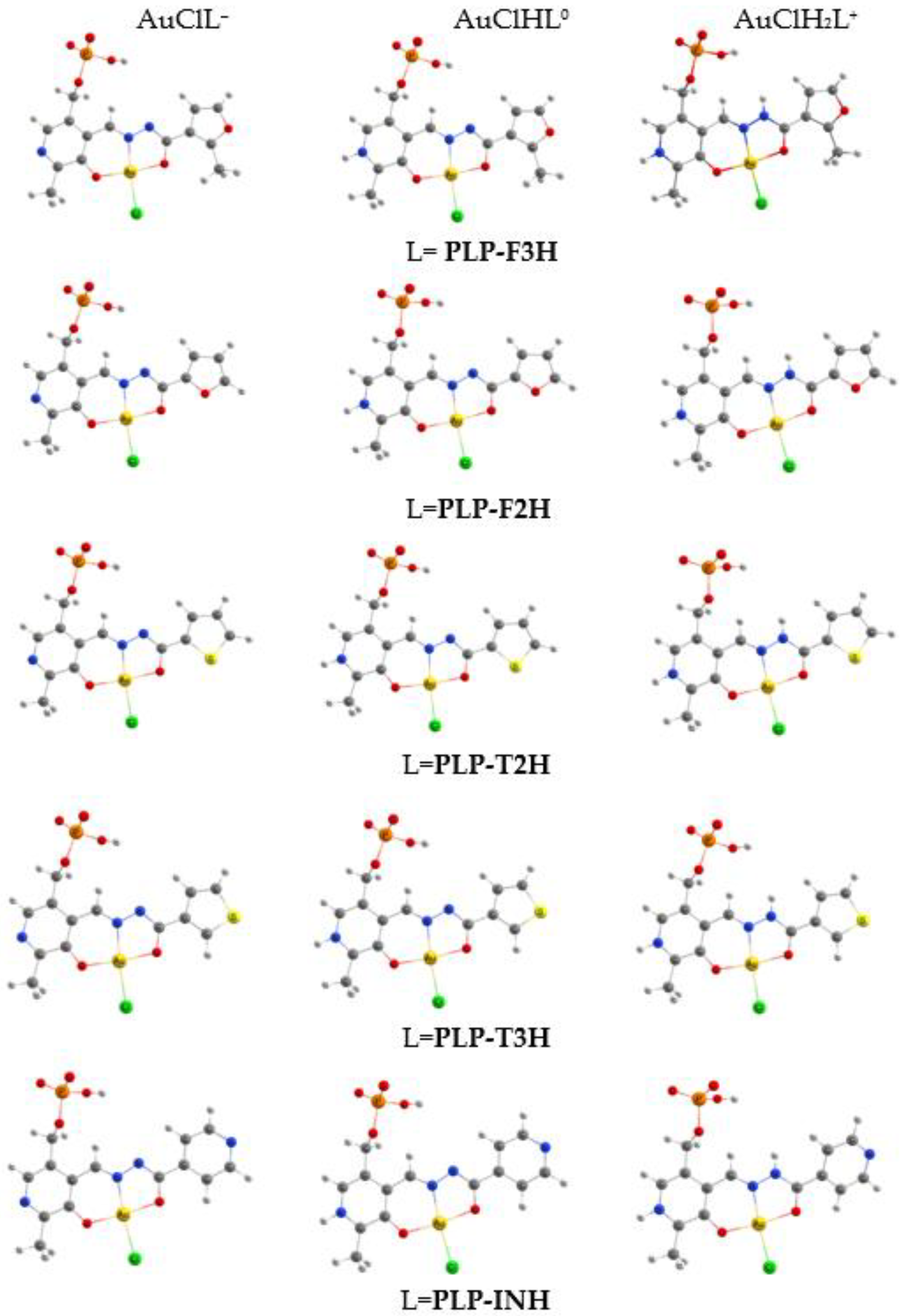
As can be seen from the MEP distribution, the phosphate group was energetically more preferable to attracting the metal cation. However, the [PLP-F3H]3− hydrazone possessed three donor sites (O…N…O) that formed the chelate cavity that was the most reliable area to pick up the metal cation and form the stable complex. Gold(III) has been reported to form the complexes with Schiff bases in a similar way [45]. On the basis of this assumption, all molecular models are considered chelate complexes of gold(III) with hydrazones derived from pyridoxal 5′-phosphate. It should be noted that the hydrazones derived from pyridoxal 5′-phosphate (Figure 1) are denoted for simplicity as L, both here and below. The calculations of geometrical parameters of gold(III) complexes were carried out for both the AuClL molecule and for monoprotonated AuClHL0 and bis-protonated AuClH2L+ ones. To take into account the usage of HAuCl4 as a gold precursor for the synthesis of gold(III) complexes, the calculated molecular models are represented in Figure 3 as the hydrazone chelate complex, where the chloride anion is extra-coordinated to the gold cation.

2.2. The Geometry of Gold(III) Complexes

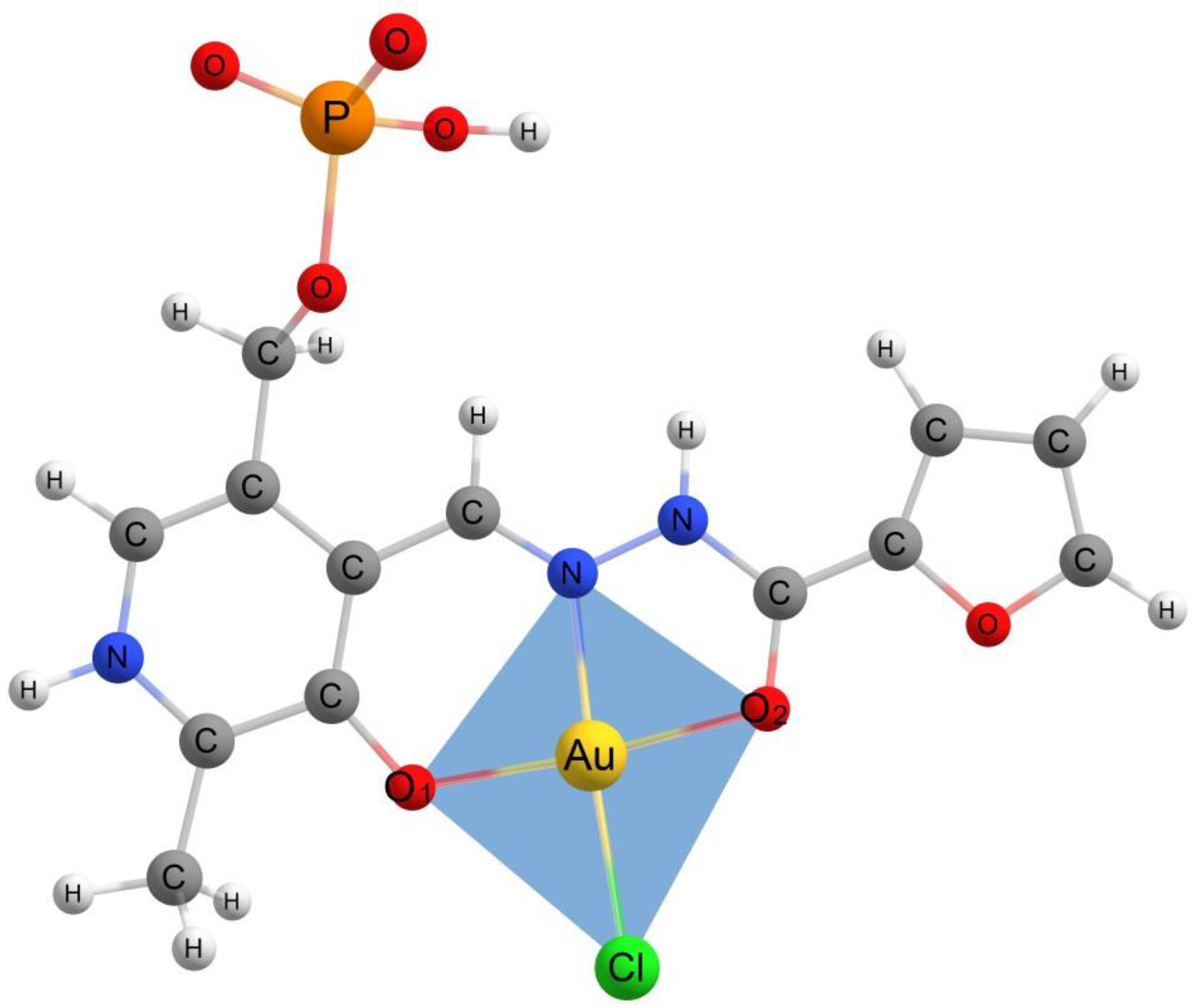
The Cartesian coordinates of C1 symmetry models of gold(III) complexes with the hydrazones derived from pyridoxal 5′-phosphate are presented in Tables S1, S4, S7, S10, and S13. To characterize the geometry of obtained molecules and study the influence of peripheral substitutions to coordination cavity, all considered molecular models were tested using SHAPE [46,47,48] software (version 7.4) to find the closest ideal reference polygons or polyhedra describing the AuO2NCl fragment. As a quantitative criterion, SHAPE calculated the continuous shape measure (CShM). According to the test, the best fit was obtained for the D4h square planar polygon. In the series of AuClL→AuClHL0→AuClH2L+, the value of CShM was decreasing. This result was in good accordance with the well-established square planar geometry of gold(III) complexes (see e.g., [1,38,39,40,41,42,43,49]). The bis-protonated AuClH2L+ species in all cases demonstrated the minimum distortion of the AuO2NCl fragment from the ideal square planar configuration. The serial protonation of AuClL species led to electronic density rearrangement that made the AuO2NCl fragment more symmetrical. The planar tetragon inscribed in the AuO2NCl fragment is presented in Figure 4 for the structure of the bis-protonatedfuroyl-2-hydrazide gold(III) complex. It possessed the least CShM = 0.630 (the closer to zero, the better this parameter can take the value of several units or even tens).
The selected equilibrium distances of the AuO2NCl fragment of the considered gold(III) hydrazone complexes are provided in Table 1.
According to DFT calculations, the geometry of the AuO2NCl coordination tetragon changed slightly along the series from AuCl(PLP-F3H) to AuCl(PLP-INH) (see Table 1 from the left to the right). The most significant change was observed for the Au–O2 bond distance as it was located closely to the position of the substituted moiety, and, thus, was affected the most. From AuCl(PLP-T3H) to AuCL(PLP-INH) bis-protonated forms, the Au–O2 bond distance increased by 0.009 Å. As a result, the coordination cavity size O1…O2 was enlarged by 0.006 Å, while the rest of the changes in bond distances did not exceed 0.004 Å. The most drastic alteration of the AuO2NCl structural parameters took place in series of AuClL→AuClHL0→AuClH2L+, viz., the systematical shortening of the Au–Cl distance was about 0.04 Å, while the elongation of the Au–N bond was more than 0.01 Å. It is interesting to note that the single protonation of hydrazone ligand L AuClL→AuClHL0 was accompanied by the contraction of the coordination cavity size and the decreasing of O1…O2 by 0.009 Å; however, the next protonation step AuClHL0→AuClH2L+ led to the expanding of the cavity by more than 0.01 Å (see Table 1). All changes of the geometrical parameters were the consequence of the electronic density redistribution. To study the effect in detail, the analysis of the electron density distribution function ρ(r) is provided.

2.3. The Electron Density Distribution Analysis

As mentioned above, the influence of ligand substituted fragments on the coordination tetragon of AuO2NCl in gold(III) hydrazone complexes is insignificant compared to the protonation effect within a single molecule. Therefore, as an example, the series AuClL→AuClHL0→AuClH2L+ with L = PLP-F3H was selected to explore the electron density distribution peculiarities and, as a consequence, the nature of chemical bonds between gold(III) and hydrazones derived from pyridoxal 5′-phosphate. The main topological characteristics of electron density distribution function ρ(r) in the AuO2NCl fragment of AuCl(PLP-F3H) complex are provided in Table 2.
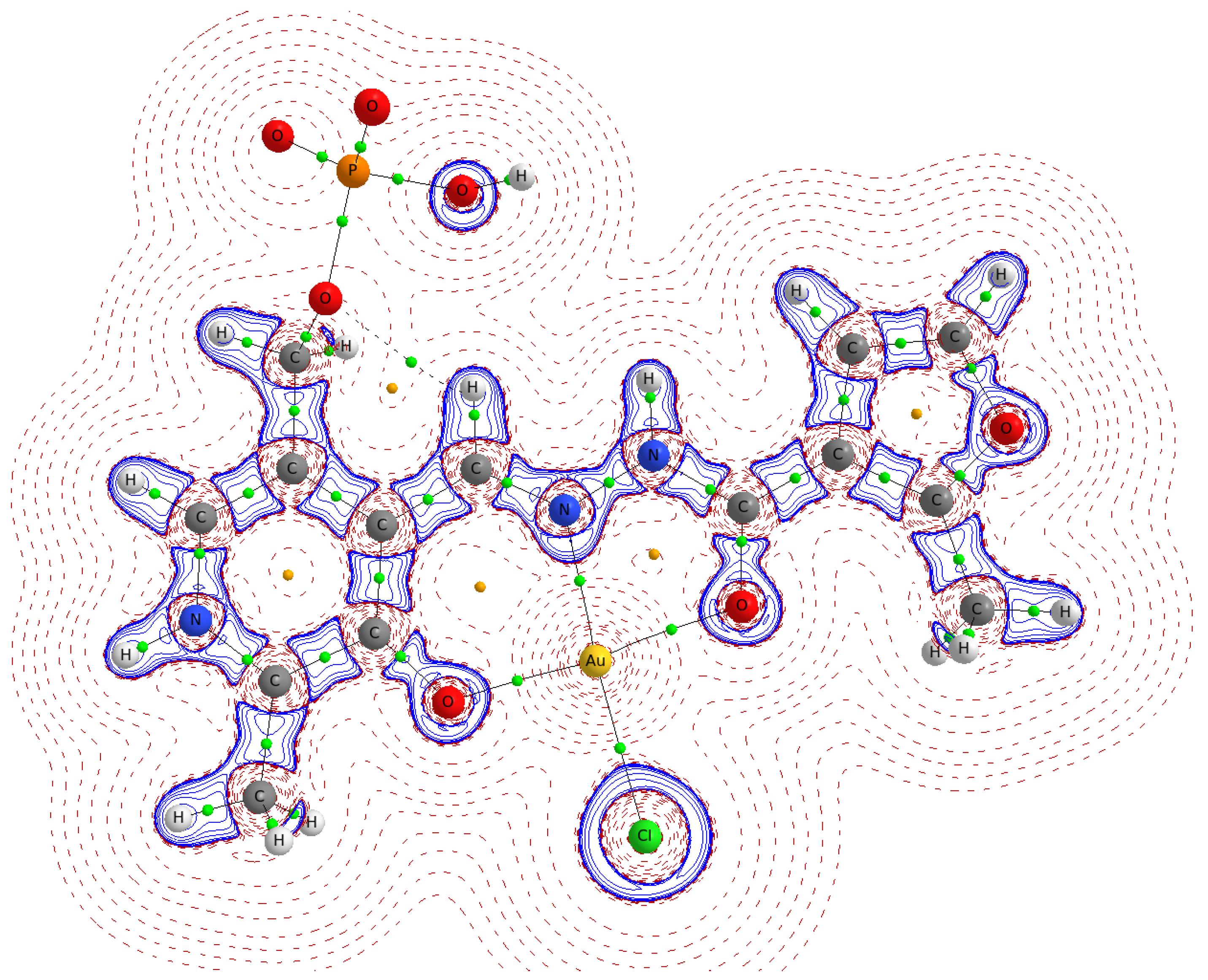
To visualize the topological peculiarities of ρ(r), the molecular graph and Laplacian map in the plane of coordination cavity ONO are shown in Figure 5 for the bis-protonated AuCl(PLP-F3H) complex.
The Laplacian map (Figure 5) displays regions of local charge depletion (∇2ρb > 0) and concentration (∇2ρb < 0) for the AuCl(PLP-F3H) complex. The strongest charge depletion took place between the gold, chloride, and PLP-F3H ligand, which is also supported by a relatively small positive value of Laplacian ∇2ρb in the bond critical points (BCP) of the AuO2NCl fragment. Additionally, the ratio value |λ1/λ3| < 1 means the electron density was shifted from Au towards N, Cl, and O atoms (see Table 2). Despite this shifting, the relatively high electron density ρb characterizing the Au–N bond and negative value of total electronic energy Hb in BCP corresponded to an intermediate type of interatomic interaction (according to the terminology used in QTAIM by Bader). Such an interaction is typical for coordination and covalent polar bonds and has been studied in particular for the AuCl∙PPh3 complex in the crystal phase [50]. The slight deformation of local charge along the bond paths from N, Cl, and O atoms to Au corresponds to electron lone pairs of these atoms directed to the gold atom.
The redistribution of the electron density in the AuO2NCl fragment can be evaluated from the changes in atomic charges. The atomic charges of the AuO2NCl fragment in the series of AuClL→AuClHL0→AuClH2L+ are provided in Table 3 for the AuCl(PLP-F3H) complex.
The sequential protonation led to the decrease in the electron density in the AuO2NCl fragment. In particular, it can be seen from the increase in the positive charge on gold and the decrease in the negative charge on chlorine (Table 3). It is noteworthy that the Coulombic attraction between Au and Cl atoms (which could be estimated as q(Au) ∙ q(Cl)/re2) remained almost unaltered (slightly decreasing) in the series of AuClL→AuClHL0→AuClH2L+; however, the Au–Cl distance was noticeably shortened. This shortening can be explained by the reduction of electrostatic repulsion between negatively charged oxygen atoms O1 and O2, which was caused by the decrease in the chlorine negative charge and the total negative charge of O1 and O2. It allowed the chlorine ion to come closer to Au (Table 1). On the other hand, the observed elongation of the Au–N distance accompanying the shrinkage of the Au-Cl bond cannot be interpreted simply as the electrostatic interactions only. If this were true, the Au-N bond would become shorter; however, it was elongated (Table 1). The changes in the angle O1–Au–O2 (see the atom numbering in Figure 4) were negligible during the protonation steps, which meant that the Au–N elongation was caused by nitrogen shifting from Au. The protons effectively pulled the electron density, which led to the changes in the covalent bond lengths in the hydrazone ligand and, in particular, in the position of the N atom involved in the Au–N bond.

2.4. The UV–Vis Absorption Spectra Calculations

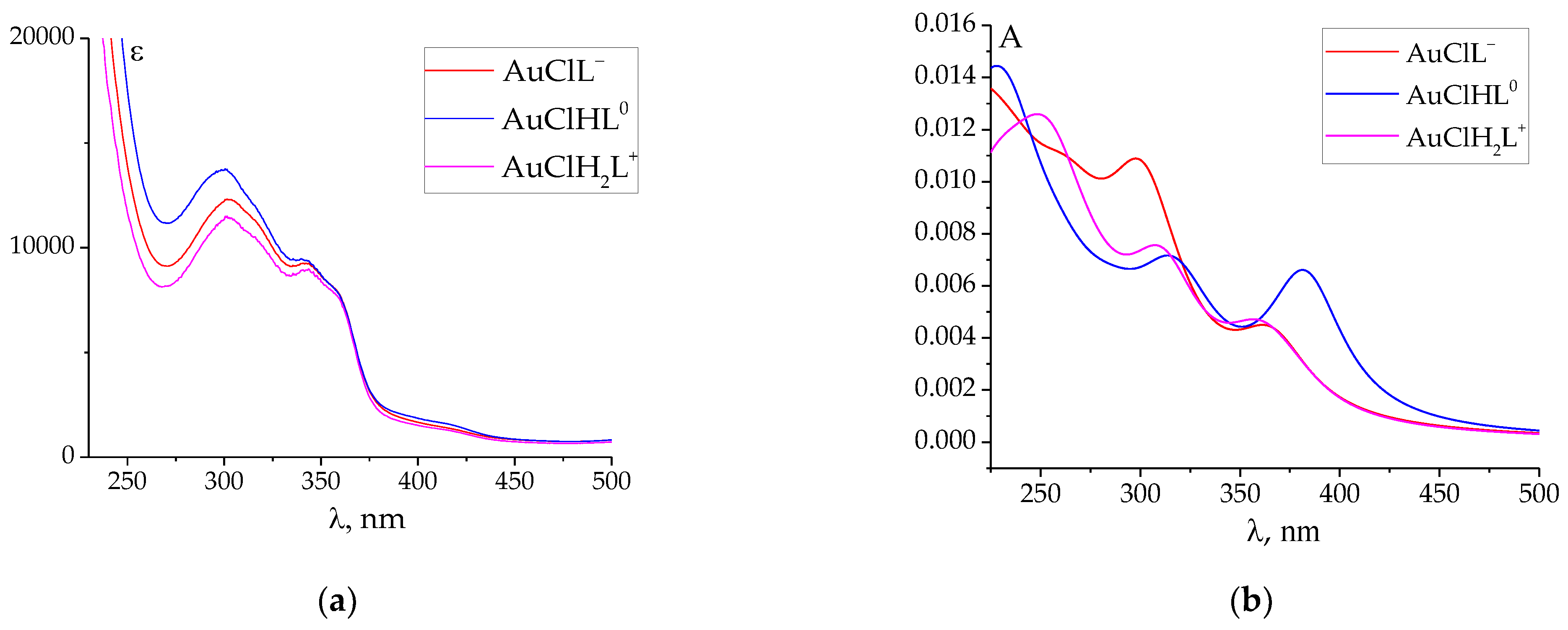
To determine the chemical validity of the proposed gold(III) hydrazone complexes, the theoretical UV–Vis absorption spectra were calculated and compared to the experimental ones. The energies of vertical electronic transitions and oscillator strengths were calculated by the TD DFT method CAM-B3LYP for all considered models and can be found in the Supplementary Materials. To simulate the shape of the experimental UV–Vis absorption spectra in range 225–500 nm, the individual bands were described by Lorentz curves with a half-width of 47 nm. As a result, in Figure 6, the observed and simulated UV–Vis absorption spectra were presented for the AuCl(PLP-F3H) complex. For other models, the observed and the simulated UV–Vis absorption spectra are provided in the Supplementary Materials (Figure S1). It can be seen from the data in Figure 6 that the positions of the absorption band of the simulated spectra were very close to the observed absorption maxima in the vicinity of 300 and 350 nm. In the series AuClL→AuClHL0→AuClH2L+, the best fitting in all cases was obtained for AuClL and bis-protonated AuClH2L+ forms. The results of TDDFT calculations allow for an assignment of the experimental absorption bands. Table 4 summarizes the data on the vertical electronic transitions and oscillator strengths corresponding to the absorption band about 300 and 350 nm for all discussed complexes of gold(III) with hydrazones.
It should be noted that the vertical electronic transitions were of complex composition. The effect of substitution led to weak changes of the electronic structure of the ligand, as is shown in Section 2.2. The same tendency was observed according to TD DFT spectra along the series from AuCl(PLP-F3H) to AuCl(PLP-INH) (see Table 4 from up to down) as the vertical electronic transitions energies (λcal) varied within the narrow range. On the other hand, the qualitative changes in spectra accompanied the protonation process AuClL→AuClHL0→AuClH2L+ according to our results. First, AuClL→AuClHL0 led to the alteration of HOMO and LUMO composition. The shapes of the selected molecular orbitals (MO) for the considered complexes of gold(III) with hydrazones derived from pyridoxal 5′-phosphate are provided in Figure S2. The AuClL species possessed HOMO that fully consisted of p atomic orbitals of the ligand without any contribution from the phosphate group or gold ions, while LUMO was a combination of gold d-orbitals and p-orbitals of N, Cl, and O atoms (referred to as dAu-p), forming the coordination cavity. As a result, there was no HOMO→LUMO transition for AuClL−1 because such a transition has low probability. The first protonation step AuClL→AuClHL0 led to the energy shift in MO. In particular, LUMO dAu-p became LUMO + 1, and following this, a HOMO→LUMO transition was observed in the spectra as a π→π * excitation into a S2 electronic state (see Table 4). The second protonation step resulted in the swapping of LUMO and LUMO + 1, which made a HOMO→LUMO transition for bis-protonated AuClH2L+1 species unlikely again. To make the discussion above more clear, the HOMO/LUMO energy level diagrams of gold(III) hydrazone complexes are presented in Figure S3.
According to the MO composition for models AuClL and AuClHL0, the electronic transitions in the vicinity of 300 and 350 nm are indicative of a π→π * character without metal-to-ligand charge transfer. In the case of bis-protonated AuClH2L+ species, the transition into S4 electronic state (Table 4) occurred due to the electronic excitation, which involves the LUMO dAu-p and occupied MO consisting of gold d-orbital and p-orbitals of Cl atoms (referred to as dAu-Cl; see Figure S2). Therefore, according to our modeling, the electronic transition in the vicinity of 300 nm in bis-protonated AuClH2L+ species could not be assigned to the clear π→π * transition.
According to the data (Table 4), it is possible to estimate the quality of the agreement between the simulated spectra (λcal) and observed absorption maxima (λexp max) in the vicinity of 300 and 350 nm. Figure S4 presents a diagram of absolute values of the deviations |λcal − λexp max| ascending for all those reviewed in the excited states shown in Table 4.
As can be seen from the diagram (Figure S4), the majority (21 cases) of deviations were less than 20 nm (which correspond to the relative error less than 5%). The minority (four cases) exceeded 35 nm (relative error was higher than 10%), wherein these four cases corresponded to the S2 excited states of monoprotonated AuClHL0 species (Table 4). In other words, the AuClHL0 absorption spectra were calculated with the least precision. However, the maximum value of deviation was 45.63 nm (corresponds to the relative error of 13.2%). Thus, in general, the TD DFT calculated spectra and observed spectral data were in satisfactory agreement.
The data in Table 4 show that the calculated UV-Vis spectra of different protonated species of the gold(III) complex (AuClL, AuClHL0, and AuClH2L+, where L remains constant) are similar to each other. It also holds for the comparison between monoanions, neutral species, or monocations containing different hydrazones. The most probable reason for this similarity is the dominant contribution of ligand molecular orbitals into the MO of the complex, where electron transitions take place.

3. Materials and Methods

Calculation Details

The calculations of probable molecular models of gold(III) complexes with the hydrazones derived from pyridoxal 5′-phosphate were carried out for the singlet electronic state using the Gaussian 09W program package [51]. The equilibrium geometrical parameters and normal mode frequencies (Tables S2, S5, S8, S11, and S14) were calculated using the hybrid DFT computational method B3LYP [52]. The energies of vertical electronic transitions and oscillator strengths (UV-Vis spectra; see Tables S3, S6, S9, S12, S15) were calculated using the TDDFT [53] method CAM-B3LYP [54]. The two-component relativistic effective core potential (ECP60MDF) [55] was applied for the inner electronic shells of Au (1s22s22p63s23p63d104s24p64d104f14). The valence shells (5s25p65d106s1) were described by the (41s37p25d2f1g/5s5p4d2f1g) basis set cc-pVTZ-PP [56]. The H, C, N, O, P, and Cl atomic electronic shells were described by the all-electron cc-pVTZ basis set [57]. To take into account the solvent effect of water, all calculations were performed using the polarizable continuum model (PCM) [58]. The visualization of ball-and-stick models and molecular orbitals was performed using the ChemCraft program [59]. The topological analysis of electron density distribution function ρ(r) in the terms of Bader’s Quantum Theory Atoms in Molecules (QTAIM) [60] was carried out using the AIMAll Professional software (version 19.10.12) [61].

4. Conclusions

The different protonated species of gold(III) complexes with five hydrazones derived from pyridoxal 5’-phosphate were studied using density functional theory methods. The complexes studied included completely deprotonated hydrazone molecules, as well as monoprotonated and bis-protonated ligands. The geometry of the complexes was optimized at the DFT/B3LYP level, and the gold coordination sphere was found to be closest to a square planar geometry. The analysis of bond critical points revealed that the gold and N and O atoms, forming the chelating cavity of hydrazones, were bound due to interatomic interactions that were intermediate between ionic and covalent. The main result of this study confirmed that the calculated UV-Vis spectra of different protonated species of gold(III) complexes were similar to each other, as reported previously for experimental electron absorption spectra. The dominant contribution of ligand molecular orbitals into the MO of the complex, where electron transitions take place, is likely the reason for this similarity. We also observed an interesting change in the calculated UV-Vis spectra caused by the first step of protonation, which enables electron transition between frontier MOs, whereas in monoanionic or monocationic complexes, the HOMO→LUMO transition is hindered.

Supplementary Materials

The following supporting information can be downloaded at https://www.mdpi.com/article/10.3390/ijms24098412/s1.

Author Contributions

Conceptualization, O.A.P. and G.A.G.; methodology, O.A.P.; software, O.A.P.; validation, M.N.Z. and K.V.G.; formal analysis, O.A.P.; investigation, O.A.P., M.N.Z., K.V.G. and G.A.G.; resources, G.A.G.; data curation, M.N.Z., K.V.G. and G.A.G.; writing—original draft preparation, O.A.P. and G.A.G.; writing—review and editing, G.A.G.; visualization, O.A.P.; supervision, G.A.G.; project administration, G.A.G.; funding acquisition, G.A.G. All authors have read and agreed to the published version of the manuscript.

Funding

This research was funded by Russian Science Foundation, grant number 22-73-10009, https://rscf.ru/project/22-73-10009; accessed on 6 May 2023.

Institutional Review Board Statement

Not applicable.

Informed Consent Statement

Not applicable.

Data Availability Statement

Data are contained within the article or Supplementary Material.

Acknowledgments

We thank G. V. Girichev and A. V. Belyakov for their help in conducting quantum chemical calculations on computing resources of the Department of Physics of ISUCT and Saint Petersburg State Institute of Technology (Technical University).

Conflicts of Interest

The authors declare no conflict of interest.

Sample Availability

Not applicable.

References

  1. Radisavljević, S.; Ćoćić, D.; Jovanović, S.; Šmit, B.; Petković, M.; Milivojević, N.; Planojević, N.; Marković, S.; Petrović, B. Synthesis, Characterization, DFT Study, DNA/BSA-Binding Affinity, and Cytotoxicity of Some Dinuclear and Trinuclear Gold(III) Complexes. J. Biol. Inorg. Chem. 2019, 24, 1057–1076. [Google Scholar] [CrossRef] [PubMed]
  2. Parish, R.V.; Howe, B.P.; Wright, J.P.; Mack, J.; Pritchard, R.G.; Buckley, R.G.; Elsome, A.M.; Fricker, S.P. Chemical and Biological Studies of Dichloro(2-((Dimethylamino)Methyl)Phenyl)Gold(III). Inorg. Chem. 1996, 35, 1659–1666. [Google Scholar] [CrossRef] [PubMed]
  3. Parish, R.V.; Mack, J.; Hargreaves, L.; Wright, J.P.; Buckley, R.G.; Elsome, A.M.; Fricker, S.P.; Theobald, B.R.C. Chemical and Biological Reactions of Diacetato[2-(Dimethylaminomethyl)-Phenyl]Gold(III), [Au(O2CMe)2(Dmamp)]. J. Chem. Soc. Dalton Trans. 1996, 1, 69–74. [Google Scholar] [CrossRef]
  4. Buckley, R.G.; Elsome, A.M.; Fricker, S.P.; Henderson, G.R.; Theobald, B.R.C.; Parish, R.V.; Howe, B.P.; Kelland, L.R. Antitumor Properties of Some 2-[(Dimethylamino)Methyl]Phenylgold(III) Complexes. J. Med. Chem. 1996, 39, 5208–5214. [Google Scholar] [CrossRef]
  5. Bertrand, B.; Williams, M.R.M.; Bochmann, M. Gold(III) Complexes for Antitumor Applications: An Overview. Chem. Eur. J. 2018, 24, 11840–11851. [Google Scholar] [CrossRef]
  6. Tong, K.-C.; Hu, D.; Wan, P.-K.; Lok, C.-N.; Che, C.-M. Anticancer Gold(III) Compounds With Porphyrin or N-Heterocyclic Carbene Ligands. Front. Chem. 2020, 8, 587207. [Google Scholar] [CrossRef]
  7. Gurba, A.; Taciak, P.; Sacharczuk, M.; Młynarczuk-Biały, I.; Bujalska-Zadrożny, M.; Fichna, J. Gold(III) Derivatives in Colon Cancer Treatment. Int. J. Mol. Sci. 2022, 23, 724. [Google Scholar] [CrossRef]
  8. Abyar, F.; Tabrizi, L. New Cyclometalated Gold (III) Complex Targeting Thioredoxin Reductase: Exploring as Cytotoxic Agents and Mechanistic Insights. Biometals 2020, 33, 107–122. [Google Scholar] [CrossRef]
  9. Büssing, R.; Karge, B.; Lippmann, P.; Jones, P.G.; Brönstrup, M.; Ott, I. Gold(I) and Gold(III) N-Heterocyclic Carbene Complexes as Antibacterial Agents and Inhibitors of Bacterial Thioredoxin Reductase. ChemMedChem 2021, 16, 3402–3409. [Google Scholar] [CrossRef]
  10. Saggioro, D.; Rigobello, M.P.; Paloschi, L.; Folda, A.; Moggach, S.A.; Parsons, S.; Ronconi, L.; Fregona, D.; Bindoli, A. Gold(III)-Dithiocarbamato Complexes Induce Cancer Cell Death Triggered by Thioredoxin Redox System Inhibition and Activation of ERK Pathway. Chem. Biol. 2007, 14, 1128–1139. [Google Scholar] [CrossRef]
  11. Quero, J.; Cabello, S.; Fuertes, T.; Mármol, I.; Laplaza, R.; Polo, V.; Gimeno, M.C.; Rodriguez-Yoldi, M.J.; Cerrada, E. Proteasome versus Thioredoxin Reductase Competition as Possible Biological Targets in Antitumor Mixed Thiolate-Dithiocarbamate Gold(III) Complexes. Inorg. Chem. 2018, 57, 10832–10845. [Google Scholar] [CrossRef]
  12. Haeubl, M.; Reith, L.M.; Gruber, B.; Karner, U.; Müller, N.; Knör, G.; Schoefberger, W. DNA Interactions and Photocatalytic Strand Cleavage by Artificial Nucleases Based on Water-Soluble Gold(III) Porphyrins. J. Biol. Inorg. Chem. 2009, 14, 1037–1052. [Google Scholar] [CrossRef]
  13. Radisavljević, S.; Đeković Kesić, A.; Ćoćić, D.; Puchta, R.; Senft, L.; Milutinović, M.; Milivojević, N.; Petrović, B. Studies of the Stability, Nucleophilic Substitution Reactions, DNA/BSA Interactions, Cytotoxic Activity, DFT and Molecular Docking of Some Tetra- and Penta-Coordinated Gold(iii) Complexes. New J. Chem. 2020, 44, 11172–11187. [Google Scholar] [CrossRef]
  14. Glišić, B.Đ.; Djuran, M.I. Gold Complexes as Antimicrobial Agents: An Overview of Different Biological Activities in Relation to the Oxidation State of the Gold Ion and the Ligand Structure. Dalton Trans. 2014, 43, 5950–5969. [Google Scholar] [CrossRef]
  15. Kanthecha, D.N.; Bhatt, B.S.; Patel, M.N.; Vaidya, F.U.; Pathak, C. DNA Interaction, Anticancer, Cytotoxicity and Genotoxicity Studies with Potential Pyrazine-Bipyrazole Dinuclear µ-Oxo Bridged Au(III) Complexes. Mol. Divers. 2022, 26, 2085–2101. [Google Scholar] [CrossRef]
  16. Al-Khodir, F.A.I.; Refat, M.S. Synthesis, Spectroscopic, Thermal and Anticancer Studies of Metal-Antibiotic Chelations: Ca(II), Fe(III), Pd(II) and Au(III) Chloramphenicol Complexes. J. Mol. Struct. 2016, 1119, 157–166. [Google Scholar] [CrossRef]
  17. Radulović, N.S.; Stojanović, N.M.; Glišić, B.Đ.; Randjelović, P.J.; Stojanović-Radić, Z.Z.; Mitić, K.V.; Nikolić, M.G.; Djuran, M.I. Water-Soluble Gold(III) Complexes with N-Donor Ligands as Potential Immunomodulatory and Antibiofilm Agents. Polyhedron 2018, 141, 164–180. [Google Scholar] [CrossRef]
  18. Hussain, I.; Ullah, A.; Khan, A.U.; Khan, W.U.; Ullah, R.; Almoqbil, A.; Naser, A.A.S.; Mahmood, H.M. Synthesis, Characterization and Biological Activities of Hydrazone Schiff Base and Its Novel Metals Complexes. Sains Malays 2019, 48, 1439–1446. [Google Scholar] [CrossRef]
  19. Abou Melha, K.S.A.; Al-Hazmi, G.A.A.; Refat, M.S. Synthesis of Nano-Metric Gold Complexes with New Schiff Bases Derived from 4-Aminoantipyrene, Their Structures and Anticancer Activity. Russ. J. Gen. Chem. 2017, 87, 3043–3051. [Google Scholar] [CrossRef]
  20. Alibrahim, K.A.; Al-Saif, F.A.; Bakhsh, H.A.; Refat, M.S. Synthesis, Physicochemical, and Biological Studies of New Pyridoxine HCl Mononuclear Drug Complexes of V(III), Ru(III), Pt(II), Se(IV), and Au(III) Metal Ions. Russ. J. Gen. Chem. 2018, 88, 2400–2409. [Google Scholar] [CrossRef]
  21. Ratia, C.; Sueiro, S.; Soengas, R.G.; Iglesias, M.J.; López-Ortiz, F.; Soto, S.M. Gold(III) Complexes Activity against Multidrug-Resistant Bacteria of Veterinary Significance. Antibiotics 2022, 11, 1728. [Google Scholar] [CrossRef] [PubMed]
  22. Đurović, M.D.; Bugarčić, Ž.D.; van Eldik, R. Stability and Reactivity of Gold Compounds—From Fundamental Aspects to Applications. Coord. Chem. Rev. 2017, 338, 186–206. [Google Scholar] [CrossRef]
  23. Zou, T.; Lum, C.T.; Lok, C.-N.; Zhang, J.-J.; Che, C.-M. Chemical Biology of Anticancer Gold(III) and Gold(I) Complexes. Chem. Soc. Rev. 2015, 44, 8786–8801. [Google Scholar] [CrossRef] [PubMed]
  24. Patanjali, P.; Kumar, R.; Sourabh; Kumar, A.; Chaudhary, P.; Singh, R. Reviewing Gold(III) Complexes as Effective Biological Operators. MGC 2018, 17, 35–52. [Google Scholar] [CrossRef]
  25. Warżajtis, B.; Glišić, B.Đ.; Živković, M.D.; Rajković, S.; Djuran, M.I.; Rychlewska, U. Different Reaction Products as a Function of Solvent: NMR Spectroscopic and Crystallographic Characterization of the Products of the Reaction of Gold(III) with 2-(Aminomethyl)Pyridine. Polyhedron 2015, 91, 35–41. [Google Scholar] [CrossRef]
  26. Palanichamy, K.; Sreejayan, N.; Ontko, A.C. Overcoming Cisplatin Resistance Using Gold(III) Mimics: Anticancer Activity of Novel Gold(III) Polypyridyl Complexes. J. Inorg. Biochem. 2012, 106, 32–42. [Google Scholar] [CrossRef]
  27. Lum, C.T.; Wong, A.S.-T.; Lin, M.C.; Che, C.-M.; Sun, R.W.-Y. A Gold(III) Porphyrin Complex as an Anti-Cancer Candidate to Inhibit Growth of Cancer-Stem Cells. Chem. Commun. 2013, 49, 4364–4366. [Google Scholar] [CrossRef]
  28. Al-Jaroudi, S.S.; Monim-ul-Mehboob, M.; Altaf, M.; Al-Saadi, A.A.; Wazeer, M.I.M.; Altuwaijri, S.; Isab, A.A. Synthesis, Spectroscopic Characterization, Electrochemical Behavior and Computational Analysis of Mixed Diamine Ligand Gold(III) Complexes: Antiproliferative and in Vitro Cytotoxic Evaluations against Human Cancer Cell Lines. Biometals 2014, 27, 1115–1136. [Google Scholar] [CrossRef]
  29. Radisavljević, S.; Bratsos, I.; Scheurer, A.; Korzekwa, J.; Masnikosa, R.; Tot, A.; Gligorijević, N.; Radulović, S.; Rilak Simović, A. New Gold Pincer-Type Complexes: Synthesis, Characterization, DNA Binding Studies and Cytotoxicity. Dalton Trans. 2018, 47, 13696–13712. [Google Scholar] [CrossRef]
  30. Murphy, T.B.; Johnson, D.K.; Rose, N.J.; Aruffo, A.; Schomaker, V. Structural Studies of Iron(III) Complexes of the New Iron-Binding Drug, Pyridoxal Isonicotinoyl Hydrazone. Inorg. Chim. Acta 1982, 66, L67–L68. [Google Scholar] [CrossRef]
  31. Back, D.F.; Manzoni de Oliveira, G.; Roman, D.; Ballin, M.A.; Kober, R.; Piquini, P.C. Synthesis of Symmetric N,O-Donor Ligands Derived from Pyridoxal (Vitamin B6): DFT Studies and Structural Features of Their Binuclear Chelate Complexes with the Oxofilic Uranyl and Vanadyl(V) Cations. Inorg. Chim. Acta 2014, 412, 6–14. [Google Scholar] [CrossRef]
  32. Murašková, V.; Szabó, N.; Pižl, M.; Hoskovcová, I.; Dušek, M.; Huber, Š.; Sedmidubský, D. Self Assembly of Dialkoxo Bridged Dinuclear Fe(III) Complex of Pyridoxal Schiff Base with C C Bond Formation—Structure, Spectral and Magnetic Properties. Inorg. Chim. Acta 2017, 461, 111–119. [Google Scholar] [CrossRef]
  33. Gamov, G.A.; Zavalishin, M.N. La3+, Ce3+, Eu3+, and Gd3+ Complex Formation with Hydrazones Derived from Pyridoxal 5’-Phosphate in a Neutral Tris–HCl Buffer. Russ. J. Inorg. Chem. 2021, 66, 1561–1568. [Google Scholar] [CrossRef]
  34. Brittenham, G.M. Pyridoxal Isonicotinoyl Hydrazone.: Effective Iron Chelation after Oral Administration. Ann. N. Y. Acad. Sci. 1990, 612, 315–326. [Google Scholar] [CrossRef]
  35. Shtyrlin, N.V.; Khaziev, R.M.; Shtyrlin, V.G.; Gilyazetdinov, E.M.; Agafonova, M.N.; Usachev, K.S.; Islamov, D.R.; Klimovitskii, A.E.; Vinogradova, T.I.; Dogonadze, M.Z.; et al. Isonicotinoyl Hydrazones of Pyridoxine Derivatives: Synthesis and Antimycobacterial Activity. Med. Chem. Res. 2021, 30, 952–963. [Google Scholar] [CrossRef]
  36. Ahmed, M.A.; Zhernakov, M.A.; Gilyazetdinov, E.M.; Bukharov, M.S.; Islamov, D.R.; Usachev, K.S.; Klimovitskii, A.E.; Serov, N.Y.; Burilov, V.A.; Shtyrlin, V.G. Complexes of NiII, CoII, ZnII, and CuII with Promising Anti-Tuberculosis Drug: Solid-State Structures and DFT Calculations. Inorganics 2023, 11, 167. [Google Scholar] [CrossRef]
  37. Kuranova, N.N.; Yarullin, D.N.; Zavalishin, M.N.; Gamov, G.A. Complexation of Gold(III) with Pyridoxal 5′-Phosphate-Derived Hydrazones in Aqueous Solution. Molecules 2022, 27, 7346. [Google Scholar] [CrossRef]
  38. Behera, P.K.; Maity, L.; Kisan, H.K.; Dutta, B.; Isab, A.A.; Chandra, S.K.; Dinda, J. Gold(I) and Gold(III) Complexes Supported by a Pyrazine / Pyrimidine Wingtip N-Heterocyclic Carbene: Synthesis, Structure and DFT Studies. J. Mol. Struct. 2021, 1223, 129253. [Google Scholar] [CrossRef]
  39. Ramadan, R.M.; Noureldeen, A.F.H.; Abo-Aly, M.M.; El-Medani, S.M. Spectroscopic, DFT Analysis, Antimicrobial and Cytotoxicity Studies of Three Gold(III) Complexes. Inorg. Nano-Metal Chem. 2021, 52, 213–225. [Google Scholar] [CrossRef]
  40. Li, L.; Bai, F.; Zhang, H.; Li, H. DFT/TDDFT Investigation on Bis-Cyclometalated Alkynylgold(III) Complex: Comparison of Absorption and Emission Properties. Sci. China Chem. 2013, 56, 641–647. [Google Scholar] [CrossRef]
  41. Gabbiani, C.; Casini, A.; Messori, L.; Guerri, A.; Cinellu, M.A.; Minghetti, G.; Corsini, M.; Rosani, C.; Zanello, P.; Arca, M. Structural Characterization, Solution Studies, and DFT Calculations on a Series of Binuclear Gold(III) Oxo Complexes: Relationships to Biological Properties. Inorg. Chem. 2008, 47, 2368–2379. [Google Scholar] [CrossRef] [PubMed]
  42. Sankarganesh, M.; Raja, J.D.; Revathi, N.; Solomon, R.V.; Kumar, R.S. Gold(III) Complex from Pyrimidine and Morpholine Analogue Schiff Base Ligand: Synthesis, Characterization, DFT, TDDFT, Catalytic, Anticancer, Molecular Modeling with DNA and BSA and DNA Binding Studies. J. Mol. Liq. 2019, 294, 111655. [Google Scholar] [CrossRef]
  43. Ayoub, N.A.; Browne, A.R.; Anderson, B.L.; Gray, T.G. Cyclometalated Gold(III) Trioxadiborrin Complexes: Studies of the Bonding and Excited States. Dalton Trans. 2016, 45, 3820–3830. [Google Scholar] [CrossRef] [PubMed]
  44. Zavalishin, M.N.; Gamov, G.A.; Pimenov, O.A.; Pogonin, A.E.; Aleksandriiskii, V.V.; Usoltsev, S.D.; Marfin, Y.S. Pyridoxal 5′-Phosphate 2-Methyl-3-Furoylhydrazone as a Selective Sensor for Zn2+ Ions in Water and Drug Samples. J. Photochem. Photobiol. A Chem. 2022, 432, 114112. [Google Scholar] [CrossRef]
  45. González-Arellano, C.; Corma, A.; Iglesias, M.; Sánchez, F. Homogeneous and Heterogenized Au(III) Schiff Base-Complexes as Selective and General Catalysts for Self-Coupling of Aryl Boronic Acids. Chem. Commun. 2005, 15, 1990–1992. [Google Scholar] [CrossRef]
  46. Pinsky, M.; Avnir, D. Continuous Symmetry Measures. 5. The Classical Polyhedra. Inorg. Chem. 1998, 37, 5575–5582. [Google Scholar] [CrossRef]
  47. Casanova, D.; Cirera, J.; Llunell, M.; Alemany, P.; Avnir, D.; Alvarez, S. Minimal Distortion Pathways in Polyhedral Rearrangements. J. Am. Chem. Soc. 2004, 126, 1755–1763. [Google Scholar] [CrossRef]
  48. Cirera, J.; Ruiz, E.; Alvarez, S. Shape and Spin State in Four-Coordinate Transition-Metal Complexes: The Case of the D6 Configuration. Chem. Eur. J. 2006, 12, 3162–3167. [Google Scholar] [CrossRef]
  49. Jouhannet, R.; Dagorne, S.; Blanc, A.; Frémont, P. Chiral Gold(III) Complexes: Synthesis, Structure, and Potential Applications. Chem. Eur. J. 2021, 27, 9218–9240. [Google Scholar] [CrossRef]
  50. Borissova, A.O.; Korlyukov, A.A.; Antipin, M.Y.; Lyssenko, K.A. Estimation of Dissociation Energy in Donor−Acceptor Complex AuCl·PPh3 via Topological Analysis of the Experimental Electron Density Distribution Function. J. Phys. Chem. A 2008, 112, 11519–11522. [Google Scholar] [CrossRef]
  51. Frisch, M.J.; Trucks, G.W.; Schlegel, H.B.; Scuseria, G.E.; Robb, M.A.; Cheeseman, J.R.; Scalmani, G.; Barone, V.; Petersson, G.A.; Nakatsuji, H.; et al. Gaussian 09, Revision A.02 2016. Available online: https://www.scienceopen.com/document?vid=6be7271f-f651-464b-aee6-ef20b0743b6b (accessed on 18 April 2023).
  52. Stephens, P.J.; Devlin, F.J.; Chabalowski, C.F.; Frisch, M.J. Ab Initio Calculation of Vibrational Absorption and Circular Dichroism Spectra Using Density Functional Force Fields. J. Phys. Chem. 1994, 98, 11623–11627. [Google Scholar] [CrossRef]
  53. Scalmani, G.; Frisch, M.J.; Mennucci, B.; Tomasi, J.; Cammi, R.; Barone, V. Geometries and Properties of Excited States in the Gas Phase and in Solution: Theory and Application of a Time-Dependent Density Functional Theory Polarizable Continuum Model. J. Chem. Phys. 2006, 124, 094107. [Google Scholar] [CrossRef]
  54. Yanai, T.; Tew, D.P.; Handy, N.C. A New Hybrid Exchange–Correlation Functional Using the Coulomb-Attenuating Method (CAM-B3LYP). Chem. Phys. Lett. 2004, 393, 51–57. [Google Scholar] [CrossRef]
  55. Figgen, D.; Rauhut, G.; Dolg, M.; Stoll, H. Energy-Consistent Pseudopotentials for Group 11 and 12 Atoms: Adjustment to Multi-Configuration Dirac–Hartree–Fock Data. Chem. Phys. 2005, 311, 227–244. [Google Scholar] [CrossRef]
  56. Peterson, K.A.; Puzzarini, C. Systematically Convergent Basis Sets for Transition Metals. II. Pseudopotential-Based Correlation Consistent Basis Sets for the Group 11 (Cu, Ag, Au) and 12 (Zn, Cd, Hg) Elements. Theor. Chem. Acc. 2005, 114, 283–296. [Google Scholar] [CrossRef]
  57. Kendall, R.A.; Dunning, T.H.; Harrison, R.J. Electron Affinities of the First-row Atoms Revisited. Systematic Basis Sets and Wave Functions. J. Chem. Phys. 1992, 96, 6796–6806. [Google Scholar] [CrossRef]
  58. Caricato, M. Absorption and Emission Spectra of Solvated Molecules with the EOM–CCSD–PCM Method. J. Chem. Theory Comput. 2012, 8, 4494–4502. [Google Scholar] [CrossRef]
  59. Chemcraft—Graphical Program for Visualization of Quantum Chemistry Computations. Available online: https://www.chemcraftprog.com/ (accessed on 18 April 2023).
  60. Bader, R.F.W. The Quantum Mechanical Basis of Conceptual Chemistry. Monatsh. Chem. 2005, 136, 819–854. [Google Scholar] [CrossRef]
  61. AIMAll. Available online: https://aim.tkgristmill.com/ (accessed on 18 April 2023).
Figure 1. Structures of the studied hydrazones derived from pyridoxal 5′-phosphate and (a) isoniazid (PLP-INH); (b) furoyl-2-hydrazide (PLP-F2H); (c) thiophene-2-carbohydrazide (PLP-T2H); (d) 2-methylfuroyl-3-hydrazide (PLP-F3H); (e) thiophene-3-carbohydrazide (PLP-T3H).
Ijms 24 08412 g001aIjms 24 08412 g001b
Figure 2. (a) The molecular graph with atomic charges for [PLP-F3H]3− hydrazone. The bond critical points are green, and the ring critical points are yellow. (b) The molecular electrostatic potential (MEP) is mapped on the isodensity surface (0.08 a.u.) for [PLP-F3H]3− hydrazone within the range of −0.459 a.u. (red) to +0.824 a.u. (blue).
Ijms 24 08412 g002
Figure 3. The C1 symmetry molecular models of gold(III) complexes with the hydrazones derived from pyridoxal 5′-phosphate.
Ijms 24 08412 g003
Figure 4. The inscribed in AuO2NCl fragment planar tetragon of the bis-protonatedfuroyl-2-hydrazide gold(III) complex and oxygen atoms numbered.
Ijms 24 08412 g004
Figure 5. Molecular graph and Laplacian map in the plane of coordination cavity ONO for the bis-protonated AuCl(PLP-F3H) complex. The solid lines (blue) correspond to local charge accumulation (∇2ρb < 0), while the dashed lines (red) represent a local charge depletion (∇2ρb > 0). The bond critical points are green, and ring critical points are yellow.
Ijms 24 08412 g005
Figure 6. (a) The observed UV–Vis absorption spectra for the protonated species of the AuCl(PLP-F3H) complex (taken from [37]). (b) The simulated UV–Vis absorption spectra for protonated species of the AuCl(PLP-F3H) complex in the range of 225–500 nm. The theoretical individual bands were described by Lorentz curves with a half-width of 47 nm.
Ijms 24 08412 g006
Table 1. Equilibrium distances (Å) of AuO2NCl fragment into gold(III) hydrazone complexes obtained by DFT calculations.
DistanceAuCl(PLP-F3H)AuCl(PLP-F2H)AuCl(PLP-T3H)AuCl(PLP-T2H)AuCl(PLP-INH)
r(Au–Cl)2.334 *2.3322.3322.3332.331
2.3242.3222.3222.3232.320
2.2942.2932.2922.2942.292
r(Au–N)1.9871.9891.9871.9891.987
1.9931.9941.9921.9951.992
2.0002.0001.9992.0002.001
r(Au–O1)1.9811.9781.9811.9791.979
1.9831.9811.9841.9811.982
1.9711.9701.9711.9711.969
r(Au–O2)2.0032.0062.0032.0052.005
1.9921.9951.9921.9931.994
2.0152.0172.0142.0162.023
r(O1…O2)3.9803.9813.9803.9803.981
3.9713.9723.9723.9713.972
3.9833.9833.9823.9833.988
* The three rows up to down for each distance corresponding to AuClL, AuClHL0, and AuClH2L+ forms.
Table 2. Topological parameters of ρ(r) in bond critical points of the AuO2NCl fragment into the AuCl(PLP-F3H) complex.
Interactionreρb2ρbλ1λ2λ3εδGbVbHb
Au–Cl2.334 *0.662+4.123−0.097−0.094+0.3630.0320.885+0.0761−0.1095−0.0334
2.3240.680+4.036−0.102−0.098+0.3680.0310.906+0.0770−0.1122−0.0352
2.2940.732+3.773−0.113−0.111+0.3810.0210.977+0.0796−0.1201−0.0405
Au–N1.9871.007+7.919−0.199−0.190+0.7170.0470.828+0.1506−0.2195−0.0689
1.9930.995+7.982−0.194−0.187+0.7120.0420.814+0.1499−0.2174−0.0675
2.0000.957+8.939−0.178−0.174+0.7230.0260.769+0.1554−0.2185−0.0631
Au–O11.9810.917+10.112−0.179−0.178+0.7770.0030.819+0.1609−0.2174−0.0565
1.9830.903+10.467−0.174−0.173+0.7810.0070.793+0.1626−0.2171−0.0545
1.9710.937+10.236−0.185−0.184+0.7940.0060.823+0.1644−0.2231−0.0587
Au–O22.0030.879+9.761−0.172−0.168+0.7450.0270.767+0.1534−0.2060−0.0526
1.9920.903+9.892−0.179−0.175+0.7640.0230.784+0.1577−0.2132−0.0555
2.0150.843+9.794−0.163−0.160+0.7290.0190.725+0.1496−0.1979−0.0483
* The three rows up to down for each distance corresponding to AuClL, AuClHL0, and AuClH2L+ forms; re is the equilibrium distance (Å); ρb is the electron density (eÅ–3); ∇2ρb is the Laplacian (eÅ–5); λ1, λ2, and λ3 are electron density Hessian matrix eigenvalues (a.u.); ε is the bond ellipticity; δ is the electron delocalization index; Gb is the kinetic energy density (a.u.); Vb is the potential energy density (a.u.); Hb is the total electronic energy density (a.u.) as a sum of Gb and Vb.
Table 3. The atomic charges (q) according to the QTAIM schemes of the AuO2NCl fragment in the AuCl(PLP-F3H) complex.
Protonated Speciesq(Au)q(Cl)q(N)q(O1)q(O2)
AuClL+1.137−0.499−0.751−1.024−1.024
AuClHL0+1.165−0.472−0.729−1.023−1.010
AuClH2L++1.219−0.396−0.725−0.991−1.012
Table 4. Selected vertical electronic transitions (UV–Vis absorption spectra) calculated using the TDDFT/CAM-B3LYP method for gold(III) complexes with hydrazones derived from pyridoxal 5′-phosphate in aqueous solution.
ComplexProtonated FormExcited Stateλcal (nm)λexp max (nm)Oscillator Strength (f)Composition *Character
AuCl(PLP-F3H)AuLS3365.64 3400.2094112 → 115 (48%), 98 → 114 (16%)π→π *
S6301.07 3010.538112 → 115 (59%), 111 → 115 (30%)π→π *
AuHL0S2382.48 3410.4084113 → 114 (61%), 112 → 114 (34%)π→π *
S6315.34 3010.2776112 → 114 (36%), 109 → 114 (29%)π→π *
AuH2L+S4359.363430.178105 → 114(50%), 111 → 115 (26%)dAu-Cl → dAu-p
S6310.783020.3531113 → 115 (45%), 111 → 115 (35%)π→π *
Au(PLP-F2H)AuLS3371.033450.3073109 → 111 (87%)π→π *
S6308.573200.5777108 → 111 (82%)π→π *
AuHL0S2390.633450.5444109 → 110 (88%), 107 → 110 (12%) π→π *
S6316.813190.3263107 → 110 (78%), 104 → 110 (11%)π→π *
AuH2L+S4362.113470.2398101 → 110(50%), 108 → 111 (40%)dAu-Cl → dAu-p
S6305.773210.7109106 → 111 (64%), 108 → 112 (13%)π→π *
Au(PLP-T3H)AuLS3366.183440.2188113 → 115 (94%)π→π *
S6300.153050.5659112 → 115 (84%)π→π *
AuHL0S2378.943430.4116113 → 114 (80%), 110 → 114 (10%)π→π *
S6309.813060.4295110 → 114 (76%), 107 → 114 (13%)π→π *
AuH2L+S4359.53440.1836104 → 114 (49%), 112 → 115 (39%)dAu-Cl → dAu-p
S7298.893060.3441109 → 115 (32%), 111 → 115 (20%)π→π *
Au(PLP-T2H)AuLS3370.673580.2977113 → 115 (92%)π→π *
S6308.233120.5955112 → 115 (86%)π→π *
AuHL0S2388.133460.5268113 → 114 (89%)π→π *
S6315.743080.3514110 → 114 (65%), 111 → 114 (14%)π→π *
AuH2L+S4361.333490.2318104 → 114 (53%), 112 → 115 (42%)dAu-Cl → dAu-p
S6308.23200.6769110 → 115 (56%), 112 → 116 (18%)π→π *
Au(PLP-INH)AuLS3367.673580.1659112 → 114 (88%)π→π *
S6298.853050.4482111 → 114 (90%), 103 → 113 (10%)π→π *
AuHL0S2371.423470.2778112 → 113 (93%)π→π *
S6304.513070.3935108 → 113 (49%), 109 → 113 (18%)π→π *
AuH2L+S4361.583460.1556111 → 114 (56%), 103 → 113 (23%)dAu-Cl → dAu-p
S12280.33070.1657110 → 114 (37%), 108 → 114 (18%)π→π *
* The composition includes the first two most significant (%) transitions. The transitions with contribution less than 10% were omitted.
Disclaimer/Publisher’s Note: The statements, opinions and data contained in all publications are solely those of the individual author(s) and contributor(s) and not of MDPI and/or the editor(s). MDPI and/or the editor(s) disclaim responsibility for any injury to people or property resulting from any ideas, methods, instructions or products referred to in the content.