Article

In Vivo and In Vitro Characterization of Close Analogs of Compound KA-11, a New Antiseizure Drug Candidate

1
Department of Experimental Pharmacology, Institute of Rural Health, Jaczewskiego 2, 20-950 Lublin, Poland
2
Department of Medicinal Chemistry, Faculty of Pharmacy, Jagiellonian University Medical College, Medyczna 9, 30-688 Krakow, Poland
3
Department of Animal Physiology and Pharmacology, Institute of Biological Sciences, Faculty of Biology and Biotechnology, Maria Curie-Skłodowska University, Akademicka 19, 20-033 Lublin, Poland
4
Department of Technology and Biotechnology of Drugs, Jagiellonian University Medical College, Medyczna 9 St., 30-688 Krakow, Poland
*
Author to whom correspondence should be addressed.
Int. J. Mol. Sci. 2023, 24(9), 8302; https://doi.org/10.3390/ijms24098302
Submission received: 29 March 2023 / Revised: 27 April 2023 / Accepted: 30 April 2023 / Published: 5 May 2023
(This article belongs to the Special Issue Epilepsy Research and Antiepileptic Drugs)

Abstract

:
Epilepsy is a neurological disorder involving a number of disease syndromes with a complex etiology. A properly matched antiseizure drug (ASD) gives remission in up to 70% of patients. Nevertheless, there is still a group of about 30% of patients suffering from drug-resistant epilepsy. Consequently, the development of new more effective and/or safer ASDs is still an unmet clinical need. Thus, our current studies were focused on the structural optimization/modifications of one of the leading compounds, KA-11, aiming at the improvement of its antiseizure activity. As a result, we designed and synthesized two close analogs with highly pronounced drug-like physicochemical properties according to in silico predictions, namely KA-228 and KA-232, which were subsequently tested in a panel of animal seizure models, i.e., MES, 6 Hz (32 mA), scPTZ and ivPTZ. Among these compounds, KA-232, which was designed as a water-soluble salt, was distinctly more effective than KA-228 and assured similar antiseizure protection as its chemical prototype KA-11. With the aim of a more detailed characterization of both new molecules, in vitro binding tests were performed to evaluate the potential mechanisms of action. Furthermore, KA-232 was also evaluated in several ADME-Tox studies, and the results obtained strongly supported its drug-like potential. The proposed chemical modification of KA-11 enabled the identification of new pharmacologically active chemotypes, particularly water-soluble KA-232, which, despite the lack of better efficacy than the leading compound, may be used as a chemical prototype for the development of new ASDs, as well as substances potentially active in other neurological or neurodegenerative conditions.

1. Introduction

The problem of drug resistance affects approximately one-third of patients suffering from epilepsy [1,2,3,4]. Because of this, many multidisciplinary studies are currently focused on the development of new antiseizure drug (ASD) candidates (based on the existing ASDs), characterized by stronger antiseizure properties, better absorption/bioavailability, minimal/zero side effects and a multimodal mechanism of action. The introduction of such modifications to the chemical structure of currently available medications may contribute to the development of new, improved ASDs [5,6,7]. Over the past several years, we have been conducting intensive research on a new hybrid compound, a 2-(2,5-dioxopyrrolidin-1-yl)propanamide derivative, compound KA-11 (also named C-11), which, as a hybrid molecule, combines structural fragments of three ASDs: ethosuximide, levetiracetam and lacosamide. The results of our previous in vitro studies and animal models of epilepsy clearly showed that the hybrid KA-11 has strong antiseizure and neuroprotective properties in acute mouse seizure models: maximal electroshock seizures (MES), subcutaneous pentylenetetrazole (scPTZ) seizures, pilocarpine (PILO) seizures and the 6 Hz seizure model [6,8]. Moreover, we have shown that KA-11 suppresses seizures after repeated administration of PTZ in the mouse kindling model of epilepsy [9]. Subsequent studies using this compound have shown that chronic administration of KA-11 does not negatively affect the proliferation of neural stem cells in mice [10]. Furthermore, KA-11 significantly stimulated the proliferation migration and differentiation of newborn cells into neurons and astrocytes, as well as protected the cognitive functions in the mouse PILO-induced status epilepticus (PILO-induced SE) [11]. Finally, our last studies using KA-11 in combination with several selected ASDs in the mouse model of an MES test indicated a significant pharmacodynamic impact of KA-11 on the antiseizure action of two ASDs, lacosamide and valproate, with no adverse effects among the tested drugs [8]. Moreover, in silico studies indicated that KA-11 meets the drug-likeness criteria resulting from Lipinski’s [12] and Veber’s [13] rules, as well as the prediction of gastrointestinal absorption and brain penetration (according to Instant JChem by the ChemAxon software version 21.4.0).
Favorable in vivo, in vitro and in silico results obtained for KA-11 so far prompted us to design close analogs modified in the pyrrolidine-2,5-dione (succinimide) ring, namely KA-228 and KA-232 (Figure 1). It should be emphasized that this is the first attempt at structural combinations focused on succinimide in the series of compounds represented by the leading compound KA-11.
The rationale for the proposed structural modifications assumes (i) an improvement in efficacy in all seizure models by the preparation of water-soluble hydrochloride (KA-232) and, as a result, a hypothetically better pharmacokinetic profile (i.e., higher concentration in the brain) and (ii) an exchange of the pyrrolidine-2,5-dione ring to pyrrolidine-2-one moiety with the aim of increasing protection in the 6 Hz (32 mA) seizure model. Notably, the pyrrolidine-2-one core ring is present in the structure of levetiracetam which proved to be especially effective in 6 Hz (32 mA) seizures. Furthermore, the exchange of the succinimide ring to pyrrolidine-2-on lowers the planarity of this fragment which in turn may contribute to better water solubility and an improved pharmacokinetic profile. Obtaining favorable results from the conducted studies, namely the similar or improved antiseizure activity for KA-228 or KA-232 vs. parent compound KA-11, may contribute substantially to the development of new drug candidates effective in various types of epileptic seizures or the identification of new chemical prototype(s) for further and extensive chemical and pharmacological studies, namely a series of pyrrolidine-2-one (KA-228 analogs) or water-soluble salts (KA-232 congeners).

2. Results and Discussion

2.1. Synthesis of Target Compounds KA-232 and KA-228

The water-soluble salt KA-232 and pyrrolidin-2-one analog KA-228 were prepared according to the method depicted in Scheme 1. In the first step, the N-(tert-butoxycarbonyl)-DL-alanine was coupled in the presence of DCC (N,N′-dicyclohexylcarbodiimide) with 1-(3-(trifluoromethyl)phenyl)piperazine to give Boc-protected alanine derivative 1. Removal of the Boc protecting group in the presence of trifluoroacetic acid (TFA) yielded amine 2. The first and second steps were common for both KA-232 and KA-228. In the case of salt derivative KA-232, amine 2 was converted to amido-acid derivative 3 in the condensation reaction with maleic anhydride. The monounsaturated pyrrolidine-2,5-dione derivative 4 was obtained by applying the hexamethyldisilazane (HMDS)-promoted cyclization reaction of 3. The final compound KA-232 was obtained in the addition reaction of dimethylamine with intermediate 4. This reaction was performed in benzene at room temperature. Finally, KA-232 was converted into water-soluble hydrochloride by dissolving it in a 2M methanolic hydrochloric acid solution. The resulting product was obtained in the form of a white solid substance, which was isolated by concentrating the organic solvent under reduced pressure and washing it with diethyl ether. In the case of the pyrrolidin-2-one derivative KA-228, the primary amine 2 was coupled with 4-bromobutanoic acid in the presence of DCC to yield derivative 5. Next, compound 5 was cyclized in the presence of base sodium hydride, to give the desired product KA-228.

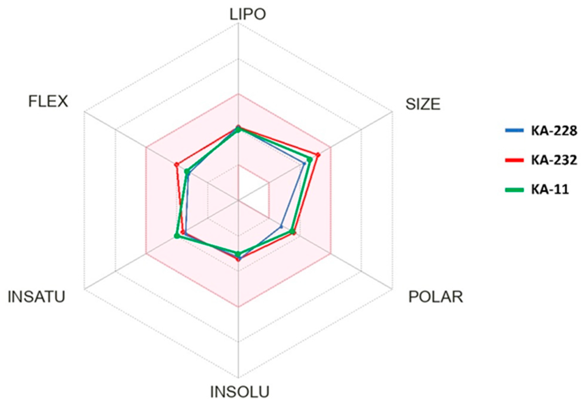
2.2. In Silico Determination of Selected Physicochemical Descriptors

The physicochemical properties of KA-232 and KA-228 were determined based on Lipinski’s and Veber’s rules using the online tool SwissAdme website [14] (Table 1).
Lipinski’s and Veber’s rules are utilized to assess the drug-like characteristics of a chemical compound, which help to determine whether it possesses physicochemical properties favorable for oral absorption in humans. The criteria of Lipinski’s rules are MW ≤ 500 Da, log P ≤ 5, HBD ≤ 5 and HBA ≤ 10, and those of Veber’s rules are NBR ≤ 10 and TPSA ≤ 140 Ǻ2 [12,13]. Consequently, according to Table 1, both compounds meet the requirements of both the aforementioned rules. The SwissAdme website also offers radar charts that take into account six physicochemical properties (lipophilicity, size, polarity, solubility, flexibility and saturation) to depict the relationship between the chemical structure of a molecule and its oral bioavailability. These radar charts display color-coded dots that represent the physicochemical properties of KA-232, KA-228 and their parent compound KA-11. The acceptable range of physicochemical parameters, as determined by Lipinski’s and Veber’s rules, is indicated by the pink area on the charts (Figure 2). Based on this analysis, it can be concluded that KA-232 and KA-228 fulfill the drug-likeness criteria set forth by Lipinski’s and Veber’s rules.
Additionally, KA-232 and KA-228, like their parent compound KA-11, meet the requirements of central nervous system multiparameter optimization (CNS MPO) using Instant JChem by the ChemAxon software version 21.4.0. The CNS MPO score is a well-established algorithm that evaluates six key physicochemical properties: ClogP, calculated distribution coefficient at pH 7.4 (ClogD), TPSA, HBD, MW and most basic center (pKa). Each property has a value between 0 and 1, thus giving the maximum collective score of 6. A higher CNS MPO score is preferred, and a score ≥ 4.0 is generally used as a cut-off point for selecting compounds for hit finding in CNS drug discovery programs [15]. It is important to note that KA-232 and KA-228 scores were >5.5.
In addition to low efficacy or high toxicity, the low bioavailability from the gastrointestinal (GI) tract or weak penetration through the blood–brain barrier (BBB) is one of the reasons for many failures in the development of CNS-active drugs. Therefore, the SwissAdme website offers a tool called ‘BOILED-Egg’ (Figure 3), which predicts how well a substance is absorbed from the GI tract and can penetrate the BBB. According to this model, KA-232 and KA-228 have a high likelihood, similar to their parent compound KA-11, to be well absorbed from the GI tract and to effectively penetrate through the BBB.

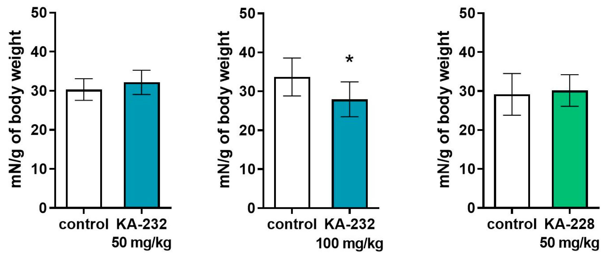
2.3. Antiseizure and Safety Evaluations In Vivo

Using animal models of seizures has been recognized as the most important approach for the discovery and development of new ASDs due to their high predictability and good translation of results from rodents to humans. Notably, all clinically relevant ASDs have been identified by applying this paradigm. Interestingly, so far, only padsevonil which is a dual-acting drug (SV2/GABAA ligand), is an example of rationally designed ASDs (development discontinued in Phase 2B) [16]. Consequently, the newly obtained compounds KA-228 and KA-232 were tested in a panel of well-established seizure models, namely the MES, 6 Hz (32 mA), scPTZ and intravenous ivPTZ tests. Compounds effective in these models may provide protection from different seizures in humans such as tonic–clonic, focal myoclonic and absence seizures. Apart from the antiseizure evaluations, both compounds KA-228 and KA-232 were tested in the chimney test and the grip strength test with the aim of assessing their safety profile in mice.
In the MES test, KA-228 and KA-232 administered alone protected the animals, in a dose-dependent manner, from tonic–clonic seizures (Figure 4). Their ED50 values are presented in Table 2. The ED50 value for KA-232 was significantly lower in comparison to the KA-11 ED50 value obtained previously by Kaminski et al. [6] (61.80 mg/kg vs. 88.40 mg/kg, respectively). In turn, the ED50 value for KA-228 was slightly higher in comparison to KA-11; however, the difference was not statistically significant.
A mouse MES seizure model was previously used to evaluate the impact of KA-11 on the protective efficacy of several selected antiseizure drugs [8]. The obtained results indicated that KA-11 at an ineffective dose (30 mg/kg, intraperitoneal i.p. injection) significantly enhanced the anticonvulsant action of lacosamide and valproate without affecting their metabolism, and it affected the animal’s behavior when used in combination. Considering the beneficial effect of KA-11 on the anticonvulsant activity of other ASDs, it might be worth considering similar studies for the KA-232 analog.
A favorable anticonvulsant effect for the KA-232 analog in the mouse MES model certainly confirms the importance of the optimization of the already known compounds aiming at the improvement of their efficacy in the MES model which mimics human tonic–clonic seizures, among others [17].
Further in vivo studies showed that both KA-228 and KA-232 administered alone protected the animals, in a dose-dependent manner, from psychomotor seizures in the 6 Hz (32 mA) model of human focal epilepsy [18] (Figure 5 and Table 2). However, the ED50 value for KA-228 was significantly higher in comparison to the previously evaluated value of KA-11 by Kamiński [6] (61.80 mg/kg vs. 21.00 mg/kg, respectively). On the other hand, the ED50 value for KA-232 was 26.50 mg/kg and was not significantly different from the ED50 for KA-11 in this experimental seizure model.
According to the obtained results, we can observe different anticonvulsant effectiveness of KA-228 and KA-232 depending on the type of applied electrical stimulation (MES and 6 Hz). These distinctions may result from a different mechanism of seizure induction in both experimental models (myoclonic seizures in MES and psychomotor seizures in 6 Hz), as well as from a difference in the structure of the tested compounds, which may reflect their pharmacological properties.
Based on the main assumption in the process of modifying KA-11, namely that the compound increases antiseizure effectiveness, both analogs did not show an improvement over the KA-11 compound in the 6 Hz model; nevertheless, it should be stressed that water-soluble analog KA-232 was almost as effective as KA-11.
Based on the satisfying protection profile of KA-228 and especially KA-232 in the MES seizure model in the next step of pharmacological investigations, both compounds were tested in chemically induced seizures, namely scPTZ and ivPTZ models. Consequently, KA-228 and KA-232 administered alone protected the animals, in a dose-dependent manner, from clonic seizures in the scPTZ model of absence epilepsy (Figure 6). The ED50 value for KA-228 was significantly higher in comparison to the ED50 value of KA-11, previously studied by Kamiński et al. [6] (95.40 mg/kg vs. 59.90 mg/kg, respectively, Table 2). In turn, the ED50 for KA-232 was very similar to the ED50 for the chemical prototype, KA-11 (Table 2). Thus, in sum, the proposed structural modifications did not improve the protection of KA-228 and KA-232 in the scPTZ model vs. KA-11. Nevertheless, both new compounds were effective in this test. The results obtained suggest that further chemical optimizations may hopefully lead to the identification of substances with improved activity in the scPTZ model.
Table 2. Antiseizure and acute adverse effects of KA-228 and KA-232 in the seizure models and the chimney test in mice after i.p. administration.
CompoundED50 MES (mg/kg) ± SDED50 6 Hz
(mg/kg) ± SD
ED50 PTZ
(mg/kg) ± SD
TD50
(mg/kg) ± SD
TI
KA-11
[6]
88.4 ± 34.1621.0 ± 32.19 59.9 ± 19.55>1500>16.97 (MES)
>71.43 (6 Hz)
>25.04 (PTZ)
KA-22892.5 ± 16.0076.4 ± 40.76 ****95.4 ± 39.78 *131.40 ± 36.241.42 (MES)
1.72 (6 Hz)
1.38 (PTZ)
KA-23261.8 ± 26.94 **26.5 ± 33.4659.3 ± 78.19162.80 ± 31.202.63 (MES)
6.14 (6 Hz)
2.75 (PTZ)
VPA
[8,19]
355.2 ± 108.1110.5 ± 42.8176.2 ± 81.8430.7 ± 47.11.21 (MES)
3.90 (6 Hz)
2.44 (PTZ)
Results are ED50 (± SD) values of KA-228 and KA-232 that protected 50% of the mice from MES, 6 Hz, PTZ-induced seizures. * p < 0.05, ** p < 0.01, **** p < 0.0001 vs. KA-11-treated animals (one-way ANOVA and post hoc Dunnett’s test). The results for KA-11 were used from Kamiński et al. [6] (see compound 11). TI, Therapeutic Index (TD50/ED50). The results for KA-11 were used from Kaminski et al. [6]; see compound 11. ED50 and TD50 of valproic acid (VPA) were used from Zagaja et al. [8,19] and Kaminski et al. [20].
The ivPTZ test was used to further characterize the effect of KA-228 and KA-232 on seizure susceptibility in mice. As shown in Figure 7, KA-228 administered at a dose of 50 mg/kg significantly raised the threshold for the tonic extension of forelimbs. However, it did not cause any significant changes in the thresholds for the first myoclonic twitch and generalized clonus with the loss of the righting reflex. Additionally, due to the unfavorable ED50 results in the above-mentioned acute seizure models (MES, 6 Hz, scPTZ), the KA-228 analog was not tested in the ivPTZ test at a dose of 100 mg/kg.
KA-232 injected at a dose of 50 mg/kg did not significantly affect the thresholds for the onset of all three studied endpoints; however, at a dose of 100 mg/kg, it slightly raised the thresholds for myoclonic and clonic seizures (Student’s t test: p < 0.001 and p < 0.0001, respectively).
In the PTZ-induced seizure models, clonic seizures are followed by the tonic extension of the forelimb. Forelimb tonus is usually (but not always) followed by the tonic extension of hindlimbs and death caused by respiratory arrest [21]. Interestingly, hindlimb tonus did not occur in 5 out of 9 mice treated with KA-232 at 50 mg/kg (Fisher’s exact test: p < 0.01 vs. control group), whereas KA-232 at 100 mg/kg completely abolished the forelimb tonus in 7 out of 11 mice (Fisher’s exact test: p < 0.01 vs. control group). In the four remaining mice, the forelimb tonus was not followed by hindlimb tonus.
Referring to the results for KA-11 (100 mg/kg) obtained by Socała and coworkers [22], which indicated a significant increase in the thresholds for all of the studied endpoints (myoclonic twitch, generalized clonus and forelimb tonus), we can conclude that the modifications did not result in a more favorable antiseizure efficiency for KA-228 and KA-232.
To assess the safety profile and potential acute adverse effects of the studied compounds, we performed a chimney test evaluating motor coordination in mice (see TD50 values in Table 2). The obtained results indicated that both KA-228 and KA-232 analogs, unlike KA-11 [6], disturbed motor performance in mice (131.40 ± 36.24, 162.80 ± 31.20 vs. >1500, respectively). In turn, the therapeutic index (TI) value in all models of epilepsy was more favorable for the KA-232 than for the KA-228 analog. Bearing in mind that the TI for an effective and safe drug should be higher than 2 [8], KA-232 with TIs of 2.63 (MES test), 6.14 (6 Hz, 32 mA test) and 2.75 (scPTZ test) is worth further advanced preclinical research (including subsequent structural modifications), especially since VPA, the most common first-line drug used in epilepsy, revealed distinctly lower TI values, especially in the MES and the 6 Hz (32 mA) seizure models [8].
KA-228 and KA-232 at a dose of 50 mg/kg did not produce any significant effects on the neuromuscular strength measured in the grip strength test. Similarly, no statistically significant abnormalities in muscle strength in mice were observed for KA-11 after a single administration at a dose of 30 mg/kg by Zagaja et al. [8]. However, KA-232 at a dose of 100 mg/kg slightly decreased the grip strength (Student’s t test: p < 0.05, Figure 8). Thus, it appears that KA-232 at higher doses may produce acute adverse effects such as impairment of neuromuscular strength.
In sum, on the basis of the above in vivo data, it can be concluded that compound KA-232, which was designed as a water-soluble derivative of the leading compound KA-11, offers a wide spectrum of antiseizure activities, as well as a satisfying safety profile (nevertheless, not better than KA-11). Certainly, the greatest advantage of KA-232 compared to KA-11 is water solubility, and thus better and faster absorption (bioavailability), and in turn expected improved effectiveness. When responding to seizures immediately, such drugs, especially if their mechanism of action is known, have a great chance to quickly stop seizures. However, this rapid and more effective absorption, as well as its potentially higher concentration in the brain may also reflect undesirable consequences, including an increased motor dysfunction, resulting from, e.g., interaction with off-targets. Nevertheless, a better understanding of this issue certainly requires a more detailed pharmacokinetic/pharmacodynamics analysis.

2.4. In Vitro Binding Assays

Dysfunction of both sodium and calcium ion channels is known to be one of the crucial reasons behind the pathogenesis of seizures [23,24]. Consequently, these ion channels are molecular targets for several clinically important ASDs such as lacosamide, lamotrigine, carbamazepine, oxcarbazepine and compounds currently under preclinical and clinical studies [25]. Thus, for KA-228 and KA-232, we tested herein their binding profile toward the aforementioned ion channels at concentrations of 10 and 100 µM (Table 3). Notably, KA-232, which revealed potent activity in the in vivo seizure models, was also evaluated in a panel of additional assays including ionotropic or metabotropic receptors involved in the mechanism of action of known ASDs (i.a., GABAA, AMPA, NMDA, etc.) at a concentration of 10 µM. Apart from in vitro studies focused on explaining the mechanism of action, KA-232 was additionally tested for its interaction with the potassium channel (hERG) which is recognized as one of the most important ‘off-targets’ responsible for harmful arrhythmogenic activity. Thus, the interaction of potential drug candidates with hERG must be excluded at an early stage of development.
As presented in Table 3, both new compounds showed only moderate binding to the sodium (site 2) channel (KA-232) or Cav1.2 channel (KA-228) at the high concentration of 100 µM; however, they were devoid of activity at the lower concentration of 10 µM. Furthermore, a more comprehensive in vitro characterization of KA-232 revealed that this molecule did not interact with other molecular targets for ASDs, namely NMDA, AMPA, GABAA, 5-HT1A receptors, the Cav2.2 channel and the hERG channel (as ‘off-target’). Notably, the latter result indicates a low risk of the proarrhythmic activity of KA-232. As the binding studies were carried out at higher and lower concentrations (100 and 10 µM, respectively), it seems unlikely that the antiseizure activity of the KA-232 compound, as well as the model compound KA-11 (tested at 100 µM), is mediated by influence on any of the ion channels or receptors tested. Furthermore, our studies on a series of structurally related compounds exclude with high probability the influence of KA-228 and KA-232 on the GAT-1 transporter for GABA, Cav3.2 channels and SV2A protein (not tested herein) [7,9,26,27,28,29,30]. Bearing in mind that all compounds, namely KA-228, KA-232 and the parent molecule KA-11, contain in their structures a phenylpiperazine fragment, which is one of the best-known pharmacophores for serotoninergic receptor (5-HTxR) ligands [6], it seems possible that antiseizure activity may be mediated by their influence on serotoninergic neurotransmission in the CNS. The serotoninergic mechanism of action is characteristic for selected ASDs or ASD candidates, such as naluzotan (5-HT1AR agonist) or lorcaserin (5-HT2CR agonist) [25]. Nevertheless, the obtained 5-HT1AR binding results (Table 3) showed no interaction for KA-232, nor its chemical prototype KA-11, at 10 µM and 100 µM, respectively. Further in vitro studies also proved that KA-232 did not inhibit the TRPV1 channel at 10 µM in an antagonist functional assay. It should be stressed herein that according to the subject’s literature [31], the transient receptor potential cation channel, subfamily V member 1 (TRPV1), which is a nonselective (Ca2+ and Na+) cation channel, is involved in pain, psychiatric and epileptic disorders. Thus, in sum, all the in vitro assays performed for KA-232 (and KA-228) did not enable the identification of a convincing mechanism of action responsible for their antiseizure activity. Therefore, in our opinion, more detailed and especially functional studies (i.e., electrophysiology) are necessary to elucidate the precise pharmacodynamics of KA-228, KA-232 and KA-11.

2.5. In Vitro Absorption Distribution Metabolism Excretion and Toxicity (ADME-Tox) Studies

Taking into account the in vitro binding assays indicating more beneficial results for KA-232, the selected ADME-Tox parameters of this analog were evaluated in vitro and compared to the data obtained previously for KA-11 [6]. The results are presented in Table 4.
In general, both compound KA-232 and parent molecule KA-11 showed satisfying and comparable drug-like properties. The high permeability of KA-232 was sustained. The calculated Pe value (10.20 × 10⁻⁶ cm/s) was actually lower than that estimated for its derivative KA-11 but similar to the highly permeable reference caffeine (10.44 × 10⁻⁶) (Table 4). However, after 5 h of incubation in a PAMPA plate, some products of KA-232 degradation at the pyrrolidine-2,5-dione moiety were observed (see Supporting Material Figures S1 and S2).
Around 73% of KA-232 remained after 120 min of incubation with HLMs (Table 5). This result is similar to the data obtained previously for KA-11. Three main metabolites were identified as the products of hydroxylation and/or the degradation/oxidation of pyrrolidine-2,5-dione. Moreover, the observed products of KA-232 degradation in a physiological buffer in the PAMPA assay were also confirmed (see Supporting Material Figures S3–S7).
Both compounds showed similar, slight effects on CYP3A4 activity only at the highest used doses of 10 and 25 µM. Similarly, a slight induction of CYP2D6 activity was observed: for KA-11, it was at concentrations of 1 and 10 µM, whereas for KA-232, it was at 0.1 and 1 µM (Figure 9A,B) (Table 4).
The statistically significant hepatotoxic effect was determined for both compounds only at the highest used dose of 100 µM. Moreover, the decrease in HepG2 cell viability in the presence of 100 µM of KA-232 was approximately 78% of control, whereas at the 100× lower concentration of the reference cytostatic drug doxorubicin, this effect was determined as only 21% of control (Figure 9C). Interestingly, no neurotoxic effect of KA-232 on neuroblastoma SH-SY5Y cell line was noted at all used doses (Figure 9D) (Table 4).
Overall, the ADME-Tox studies revealed that KA-232 possesses a favorable membrane permeability and a satisfying safety profile in the hepatotoxicity, neurotoxicity and CYPS inhibition/induction studies. These in vitro results together with the potent in vivo antiseizure activity shown in the MES test may justify further and more detailed development of new, more potent and/or safer KA-232 analogs.

3. Materials and Methods

3.1. Chemistry

All chemicals and solvents were purchased from commercial suppliers and were used without further purification. Melting points (mp.) were determined in open capillaries on a Büchi 353 melting point apparatus (Büchi Labortechnik, Flawil, Switzerland). Thin-layer chromatography (TLC) and gradient ultra-performance liquid chromatography (UPLC) were used to assess the purity and homogeneity of the compounds. TLC was carried out on silica gel 60 F254 pre-coated aluminum sheets (Macherey-Nagel, Düren, Germany), using the following developing systems: (S1–Dichloromethane methanol DCM:MeOH (9:0.3; v/v), S2–DCM:MeOH (9:0.5; v/v)). Spot detection: UV light (λ = 254 nm). UPLC and liquid chromatography–mass spectrometry (LC-MS) were obtained on Waters ACQUITY™ TQD system (Waters, Milford, CT, USA) with the MS-TQ detector and UV-Vis-DAD eλ detector. The ACQUITY UPLC BEH C18, 1.7 μm (2.1 × 100 mm), column was used with VanGuard Acquity UPLC BEH C18, 1.7 μm (2.1 × 5 mm) (Waters, Milford, CT, USA). Standard solutions (1 mg/mL) of each compound were prepared in analytical-grade acetonitrile (MeCN)/water mixture (1:1; v/v). Conditions applied were as follows: eluent A (water/0.1% HCOOH), eluent B (MeCN/0.1% HCOOH), a flow rate of 0.3 mL/min, a gradient of 5–100% B over 10 min and an injection volume of 10 μL. The UPLC retention times (tR) are given in min. Preparative column chromatography was performed using silica gel 60 (particle size 0.063–0.200; 70–230 Mesh ATM) purchased from Merck (Darmstadt, Germany). Elemental analyses (C, H and N) for final compounds were carried out by a micro method using the elemental Vario EI III Elemental analyzer (Hanau, Germany). The results of elemental analyses were within ±0.4% of the theoretical values. Proton nuclear magnetic resonance (1H NMR) and carbon nuclear magnetic resonance (13C NMR) spectra were obtained in a JEOL-500 spectrometer (JEOL USA, Inc., Peabody, MA, USA), in CDCl3 operating at 500 MHz (1H NMR) and 126 MHz (13C NMR). Chemical shifts are reported in δ values (ppm) relative to TMS δ = 0 (1H), as internal standard. The J values are expressed in Hertz (Hz). Signal multiplicities are represented by the following abbreviations: s (singlet), br s (broad singlet), d (doublet), dd (double doublet), ddd (double double doublet), dt (doublet of triplets), t (triplet), q (quartet), quin (quintet), m (multiplet).

3.1.1. Synthetic Procedure for Boc-Protected Compound (Tert-butyl-(1-oxo-1-(4-(3-(trifluoromethyl)phenyl)piperazin-1-yl)propan-2-yl)carbamate (1))

To the DCM (100 mL) solution of N-(tert-butoxycarbonyl)-DL-alanine (5 g, 26.4 mmol, 1 eq) was successively added DCC (8.18 g, 39.6 mmol, 1.5 eq) dissolved in 15 mL of DCM. After stirring (15 min), the 1-(3-(trifluoromethyl)phenyl)piperazine (6.08 g, 26.4 mmol, 1.0 eq) was added dropwise, and the reaction was stirred at room temperature for 2 h. The DCM was evaporated in vacuo, and the product was purified by column chromatography using a DCM:MeOH–9:0.3 (v/v) mixture as a solvent system.
Tert-butyl-(1-oxo-1-(4-(3-(trifluoromethyl)phenyl)piperazin-1-yl)propan-2-yl)carbamate (1).
Light oil. Yield: 86% (9.12 g); TLC: Rf = 0.70 (S1); UPLC (purity > 99%): tR = 7.32 min. C19H26F3N3O3 (401.43). LC-MS (ESI): m/z calcd for C19H26F3N3O3 (M+H)+ 401.19, found 402.2.

3.1.2. Synthetic Procedure for Amine (2-Amino-1-(4-(3-(trifluoromethyl)phenyl)piperazin-1-yl)propan-1-one (2))

The DCM (30 mL) solution of 1 (9.0 g, 22.4 mmol, 1 eq) was treated with TFA (7.69 g, 5.00 mL, 67.2 mmol, 3 eq) and stirred at room temperature for 6 h. Next, DCM was evaporated in vacuo. The oil residue obtained was suspended in water (30 mL), and 25% ammonium hydroxide was carefully added to pH = 8. The aqueous phase was extracted using DCM (3 × 50 mL), dried over anhydrous Na2SO4 and concentrated to dryness. Compound 2 was obtained as an oil and used for further reactions without purification.
2-Amino-1-(4-(3-(trifluoromethyl)phenyl)piperazin-1-yl)propan-1-one (2). Light oil. Yield: 96% (6.5 g); UPLC (purity > 99%): tR = 4.23 min. C14H18F3N3O (301.31). LC-MS (ESI): m/z calcd for C14H18F3N3O (M+H)+ 302.14, found 302.3.

3.1.3. Synthetic Procedure for Unsaturated Maleiamic Acid (4-Oxo-4-((1-oxo-1-(4-(3-(trifluoromethyl)phenyl)piperazin-1-yl)propan-2-yl)amino)but-2-enoic acid (3))

Maleic anhydride (0.98 g 10.0 mmol, 1 eq) was added to a solution of 2 (3.00 g, 10.0 mmol, 1 eq) in AcOEt (100 mL) and stirred for 4 h. After this time, the solvent was distilled off to dryness. The compound was obtained as solid after washing with diethyl ether (Et2O).
4-Oxo-4-((1-oxo-1-(4-(3-(trifluoromethyl)phenyl)piperazin-1-yl)propan-2-yl)amino)but-2-enoic acid (3). White solid. Yield: 81% (3.22 g); UPLC (purity 96%): tR = 5.82 min. C18H20F3N3O4 (399.37). LC-MS (ESI): m/z calcd for C18H20F3N3O4 (M+H)+ 400.14, found 400.2.

3.1.4. Synthetic Procedure for Unsaturated Pyrrolidine-2,5-Dione Derivative (1-(1-Oxo-1-(4-(3-(trifluoromethyl)phenyl)piperazin-1-yl)propan-2-yl)-1H-pyrrole-2,5-dione (4))

ZnCl2 (1.05 g, 7.8 mmol, 1 eq) was added to the suspension of 3 (3.10 g, 7.8 mmol, 1 eq) in dry 1,4-dioxane (100 mL), and the mixture was heated to 80 °C. Then, solution of HMDS (2.41 g, 11.7 mmol, 1.5 eq) was added dropwise over 30 min. The reaction was continued with stirring in reflux for about 8 h, then cooled and concentrated under reduced pressure. After distilling off the solvent, the oily residue was dissolved in DCM and extracted with 0.1 M HCl (3 × 50 mL), water (3 × 50 mL) and saturated NaCl solution (3 × 50 mL). The organic layer was dried over anhydrous Na2SO4 and then evaporated to dryness. The crude product was purified by column chromatography. The compound was obtained as solid after washing with Et2O.
1-(1-Oxo-1-(4-(3-(trifluoromethyl)phenyl)piperazin-1-yl)propan-2-yl)-1H-pyrrole-2,5-dione (4). White solid. Yield: 80% (2.37 g); TLC: Rf = 0.42 (S2); UPLC (purity > 99%) tR = 6.41 min. C18H18F3N3O3 (381.36). LC-MS (ESI): m/z calcd for C18H18F3N3O3 (M+H)+ 382.13 found 382.2.

3.1.5. Synthetic Procedure for Target Water-Soluble Compound 3-(Dimethylamino)-1-(1-oxo-1-(4-(3-(trifluoromethyl)phenyl)piperazin-1-yl)propan-2-yl)pyrrolidine-2,5-dione hydrochloride (KA-232)

The dimethylamine solution in tetrahydrofuran (THF, 6.1 mmol, 1 eq) was added to a solution of 4 (2.30 g, 6.1 mmol, 1 eq) in dry benzene (80 mL). The reaction was continued with stirring for about 8 h, then concentrated under reduced pressure. The crude product was purified by column chromatography. The compound was then converted into hydrochloride salt by treating the compound with a 2M methanolic hydrochloric acid solution. The compound was obtained as solid after washing with Et2O.
3-(Dimethylamino)-1-(1-oxo-1-(4-(3-(trifluoromethyl)phenyl)piperazin-1-yl)propan-2-yl)pyrrolidine-2,5-dione hydrochloride (KA-232). White solid. Yield: 84% (2.16 g); mp. 141.4–143.5 °C; TLC: Rf = 0.55 (S2); UPLC (purity: >99%): tR = 4.91 min. LC-MS (ESI): m/z calcd for C20H25F3N4O3 (M+H)+ (426.19), found 427.3. 1H NMR (500 MHz, DMSO-d6) δ 1.38–1.50 (m, 3H), 2.83 (s, 6H), 3.06–3.10 (m, 1H), 3.16 (br s, 1H), 3.21–3.32 (m, 4H), 3.43–3.51 (m, 3H), 3.61 (br s, 1H), 4.64–4.73 (m, 1H), 4.96–5.14 (m, 1H), 7.08 (d, J = 7.3 Hz, 1H), 7.18 (br s, 1H), 7.22 (dd, J = 7.7, 4.6 Hz, 2H), 7.40 (t, J = 8.0 Hz, 1H), 11.91–12.20 (m, 1H). For free base 1H NMR (500 MHz, CDCl3) δ 1.64 (dd, J = 7.3, 1.6 Hz, 3H), 2.39 (d, J = 10.5 Hz, 6H), 2.59–2.72 (m, 1 H), 2.82 (dt, J = 18.3, 9.0 Hz, 1H), 3.20 (br s, 4H), 3.43–3.73 (m, 3H), 3.81 (dd, J = 9.0, 5.2 Hz, 2H), 5.01 (quin, J = 7.4 Hz, 1H), 7.00–7.16 (m, 3H), 7.35 (t, J = 8.0 Hz, 1H). 13C NMR (126 MHz, CDCl3) δ 15.1, 31.0, 41.5 (d, J = 1.1 Hz), 47.8, 53.5, 62.5, 113.0 (d, J = 3.9 Hz), 117.0 (d, J = 3.9 Hz), 119.5 124.2, (d, J = 272.5 Hz), 129.9, 131.7 (q, J = 31.9 Hz), 151.0, 167.35, 174.6, 176.1. Anal. calcd for C20H26ClF3N4O3 (462.90): C: 51.89, H: 5.66, N: 12.10; Found C: 51.97, H: 5.41, N: 12.34.

3.1.6. Synthetic Procedure for 2-((4-Bromobutyl)amino)-1-(4-(3-(trifluoromethyl)phenyl)piperazin-1-yl)propan-1-one (5)

To the DCM (50 mL) solution of 4-bromobutanoic acid (1.67 g, 10.0 mmol, 1.0 eq), was successively added DCC (3.10 g, 15.0 mmol, 1.5 eq) dissolved in 15 mL of DCM. After stirring (15 min), intermediate compound 2 (3.00 g, 10.0 mmol, 1 eq) was added, and the reaction was stirred at room temperature for 2 h. The DCM was evaporated in vacuo, and the product was purified by column chromatography using a DCM:MeOH–9:0.3 (v/v) mixture as a solvent system.
2-((4-bromobutyl)amino)-1-(4-(3-(trifluoromethyl)phenyl)piperazin-1-yl)propan-1-one (5)
White solid. Yield: 85% (3.69 g); TLC: Rf = 0.51 (S1); UPLC (purity: 91%) tR = 4.37 min. C18H25BrF3N3O(436.32). LC-MS (ESI): m/z calcd for C18H25BrF3N3O (M+H)+ 437.1 found 437.6.

3.1.7. Synthetic Procedure for Pyrrolidin-2-One Derivative 1-(1-Oxo-1-(4-(3-(trifluoromethyl)phenyl)piperazin-1-yl)propan-2-yl)pyrrolidin-2-one (KA-228)

NaH (0.40 g, 16.5 mmol, 2 eq) was added to a solution of 5 (3.60 g, 8.25 mmol, 1 eq) in anhydrous THF. The reaction mixture was stirred for 3 h and next concentrated under reduced pressure. The oily residue was dissolved in 0.1 M HCl (50 mL) and extracted with DCM (3 × 50 mL). The organic layer was dried over anhydrous Na2SO4 and evaporated to dryness. The crude product was purified by column chromatography using DCM:MeOH (9:0.5; v/v) solvent system. After washing with Et2O, the compound was obtained as a white solid.
1-(1-oxo-1-(4-(3-(trifluoromethyl)phenyl)piperazin-1-yl)propan-2-yl)pyrrolidin-2-one (KA-228)
White solid. Yield: 88% (2.68 g); mp. 87.1–88.4 °C; TLC: Rf = 0.48 (S2); UPLC (purity: >99%): tR = 5.82 min. LC-MS (ESI): m/z calcd for C18H22F3N3O2 (M+H)+ (370.17), found 370.3. 1H NMR (500 MHz, CDCl3) δ 1.32 (d, J = 6.9 Hz, 3H), 1.96–2.09 (m, 2H), 2.28–2.48 (m, 2H), 2.96–3.17 (m, 2H), 3.25 (ddd, J = 8.8, 6.7, 3.0 Hz, 2H), 3.32–3.51 (m, 2H), 3.59–3.72 (m, 2H), 3.73–3.97 (m, 2H), 5.16 (q, J = 6.9 Hz, 1H), 7.03 (dd, J = 8.3, 2.3 Hz, 1H), 7.07 (s, 1H), 7.10 (d, J = 7.6 Hz, 1H), 7.35 (t, J = 8.0 Hz, 1H). 13C NMR (126 MHz, CDCl3) δ 14.8, 18.2, 31.0, 41.9, 43.3, 45.2, 46.1, 48.9, 49.5, 112.7 (q, J = 3.9 Hz), 116.8 (d, J = 3.8 Hz), 119.3 (d, J = 1.2 Hz), 124.2 (q, J = 272.4 Hz), 129.8, 131.7 (q, J = 31.8 Hz), 150.6, 169.1, 174.3. Anal. calcd for C18H22F3N3O2 (369.39): C: 58.53, H: 6.00, N: 11.38; Found C: 58.97, H: 6.11, N: 11.45.

3.2. In Silico Determination of Selected Physicochemical Descriptors

Physicochemical properties of KA-228 and KA-232 and BOILED-Egg predictive model were determined using the online tool SwissADME website [14].

3.3. In Vivo Studies

3.3.1. Animals and Experimental Conditions

The experiments were conducted on adult male albino Swiss mice weighing 22–26 g. The animals were kept in colony cages under standardized laboratory conditions: natural light–dark cycle 12/12 h, temperature 20–24 °C, air humidity 45–65% and free access to tap water and food (chow pellets). After 7 days of adaptation to laboratory conditions, the animals were randomly assigned to experimental groups consisting of 8 mice. The experimental procedures described herein were approved by the Local Ethics Committee in Lublin, Poland (approval numbers: 71/2020 and 13/2021), and were conducted in accordance with EU Directive 2010/63/EU for animal experiments. All efforts were made to minimize animal suffering and to use only the number of animals necessary to produce reliable scientific data according to the 3Rs rule [32].

3.3.2. Drugs

The following drugs were used: KA-228 and KA-232 (synthesized by the Department of Medicinal Chemistry from the Jagiellonian University Medical College, Faculty of Pharmacy); pentylenetetrazole, PTZ (Sigma-Aldrich, Saint Louis, MO, USA); 0.5% solution of tetracaine hydrochloride (Sigma-Aldrich, Saint Louis, MO, USA).
All substances (except PTZ and tetracaine hydrochloride dissolved in water for injections) were suspended in a 1% solution of Tween 80 (Sigma-Aldrich, Saint Louis, MO, USA) in water for injections (Baxter, Warszawa, Poland) or 0.9% NaCl. All drugs were injected intraperitoneally (i.p.) 30 min before all seizure models with 1 mL syringes as a single injection, in a volume of 10 mL/kg. The pretreatment time (30 min) before testing for KA-228 and KA-232 was chosen based on the time to peak of maximum anticonvulsant activity of KA-11 from our previous studies [6,8,22].
All reference drugs used in ADME-Tox in vitro assays (caffeine, ketoconazole, quinidine and doxorubicin) were purchased from Sigma-Aldrich (St. Louis, MO, USA) and dissolved in DMSO (10 mM).

3.3.3. Maximal Electroshock Seizure (MES) Test

The tonic–clonic seizures in mice were evoked by an electric stimulus (an alternating current 25 mA, 50 Hz, maximum output voltage 500 V, 0.2 s) generated by a rodent shocker (Hugo Sachs Elektronik, Freiburg, Germany) and delivered via ear-clip electrodes. The animals were administered with increasing doses of KA-228 and KA-232, and the anticonvulsant activity of each drug was evaluated as the ED50 value (median effective dose of the tested drug, which protects 50% of mice against seizures). This experimental procedure has been described in detail in our previous studies [33,34].

3.3.4. 6-Hz (32 mA) Seizure Model

The 6-Hz-induced psychomotor seizures in mice were evoked by current (6 Hz, 0.2 ms rectangular pulse width, 32 mA, 3 s duration) generated by an S48 Square Pulse Stimulator and CCU1 Constant Current Unit (Grass Technologies, West Warwick, RI, USA). The animals were administered with increasing doses of KA-228 and KA-232, and the anticonvulsant activity of each drug was evaluated as the ED50. This experimental procedure has been described in detail in our previous studies [19,35].

3.3.5. scPTZ-Induced Convulsions

The PTZ-induced clonic seizures in mice were determined after sc administration of PTZ at a dose of 100 mg/kg. The animals were administered with increasing doses of KA-228 and KA-232, and the anticonvulsant activity of each drug was evaluated as the ED50 value. This experimental procedure has been described in detail in our previous studies [20,36].

3.3.6. Timed ivPTZ Seizure Threshold Test

The timed ivPTZ seizure threshold test was carried out according to the method described by Socała et al. [22]. KA-228 and KA-232 were suspended in a 1% solution of Tween 80 and administered i.p. 30 min before the test. In the experiment with KA-232, two separate control groups were used because the effects of KA-232 at a dose of 50 and 100 mg/kg were determined on different days.

3.3.7. Behavioral Tests

Chimney Test

The chimney test of Boissier et al. [37] was used to quantify the acute adverse-effect potential of KA-228 and KA-232 on motor performance in mice. The acute neurotoxic effects of KA-228 and KA-232 were expressed as median toxic doses (TD50 values in mg/kg), at which these substances impaired motor coordination and balance in 50% of the tested mice. This experimental procedure has been described in detail in our previous studies [30,31].

Grip Strength Test

The grip strength test was carried out according to the methodology described by Socała et al. [22]. Neuromuscular strength was measured just before the ivPTZ seizure threshold test to limit the number of animals used.

3.4. In Vitro Binding Assays

Binding studies were carried out commercially in Eurofins Laboratories (Poitiers, France or New Taipei City, Taiwan). The experimental conditions and references are listed in Table 5 and Table 6.

3.5. In Vitro ADME-Tox Studies

The assays and protocols used for evaluating the KA-232 ADME-Tox parameters were similar to those described previously for compound KA-11 [6]. Pre-coated PAMPA Plate System Gentest™ was purchased from Corning (Tewksbury, MA, USA). Human liver microsomes (HLMs) were provided by Sigma-Aldrich (St. Louis, MO, USA). The in silico studies of metabolism were performed with MetaSite 6.0.1 software (Molecular Discovery Ltd., Hertfordshire, UK). The influence on human cytochromes was estimated with use of CYP3A4 and CYP2D6 P450-Glo™ kits (Promega, Madison, WI, USA). Cell-based safety tests were performed with hepatoma HepG2 (HB-8065™) and neuroblastoma SH-SY5Y (CRL-2266™) cell lines obtained from ATCC® (American Type Culture Collection, Manassas, VA, USA). The CellTiter 96® AQueous Non-Radioactive Cell Proliferation Assay (Promega, Madison, WI, USA) was used for determination of cell viability. The luminescent signal and the absorbances were measured by using a microplate reader EnSpire PerkinElmer (Waltham, MA, USA). The LC/MS/MS analyses used in PAMPA and metabolic stability assays were obtained on Waters ACQUITY™ TQD system (Waters, Milford, CT, USA).

3.6. Statistical Analyses

The ED50 or TD50 values for tested substances in the acute animal models of seizures were calculated using the computer log-probit analysis [49]. Subsequently, the ED50 and TD50 values (±SEM) were statistically analyzed using one-way analysis of variance (ANOVA) followed by the post hoc Dunnett’s test for multiple comparisons. Differences among values were considered statistically significant if p < 0.05. GraphPad Prism version 8 for Windows (GraphPad Software, San Diego, CA, USA) was used as a statistical software program. The results obtained from the acute ivPTZ test and grip strength test were evaluated by a Student’s t test. Fisher’s exact probability test was used to analyze protection from tonic forelimb and hindlimb extension in the ivPTZ test.

4. Conclusions

The ‘me-too’ strategy is one of the most effective chemical tools in medicinal chemistry, which enables the discovery of new drug candidates by applying small structural modifications of the leading molecule. Thus, in the current studies, we proposed two targeted and rational structural modifications of the leading compound KA-11, with the aim of obtaining more effective or safer derivatives.
Based on the in vivo results, it may be concluded that both (KA-228 and KA-232) modifications (within the imide ring) proved to be antiseizure active compounds; nevertheless, their efficacy and safety profile was not more favorable compared to parent KA-11. However, it should be stressed that KA-232 (a water-soluble hydrochloride salt) was distinctly more potent in the MES test vs. KA-11, and therefore this compound should be considered for more extensive chemical and pharmacological development, including efficacy and safety assessment, as well as pharmacokinetic characterization.
In addition, the favorable results obtained for the leading compound KA-11 (named C11) in the PILO-induced SE model in mice [11], showing strong protection of cognitive functions and stimulation of neurogenesis, certainly encourage the continuation and planning of more advanced studies for the parent KA-11, as well as the KA-232 analog using an animal model of chronic epilepsy. Bearing in mind that KA-11 as a hybrid molecule combines the structural fragment of levetiracetam, it would certainly be worth using the PILO model of temporal lobe epilepsy, where this ASD shows strong antiseizure effectiveness. These studies would provide valuable data on the anticonvulsant and neuroprotective effects of chronic treatment with KA-11 and KA-232.
Extremely important in ASD treatment is that, depending on the type of the mechanism, as well as the multidirectional way of action, it can be used in various neurological diseases both with and without symptoms of seizures [50,51,52]. Thus, it seems reasonable to extend the preclinical development of compounds discovered by our team to other neurological or neurodegenerative conditions regardless of convulsive symptoms such as neuropathic pain, Alzheimer’s and Parkinson’s diseases, migraine headaches, bipolar disorders or anxiety. When developing new potential candidates for ASDs, we place a great emphasis on compounds having multiple molecular targets and the lack of side effects. Taking into account the long-term process of therapy, such drugs could be successfully used in other neurological diseases.
Undoubtedly, KA-232, due to its solubility in water, may be of great interest in further preclinical studies, such as models of status epilepticus, where the time and route (i.v.) of compound administration play a significant role in the antiseizure activity.

Supplementary Materials

The following supporting information can be downloaded at https://www.mdpi.com/article/10.3390/ijms24098302/s1.

Author Contributions

Conceptualization, M.A.-M. and K.K.; methodology, M.A.-M., M.Z., A.S., J.S.-R., M.A., M.J., K.S., K.C., G.L., N.K. and K.K.; software, M.Z., K.S., G.L., M.A. and M.J.; investigation, M.A.-M., M.Z., J.S.-R., A.S., M.A., M.J., K.S., K.C., G.L., N.K. and K.K.; writing—original draft preparation, M.A.-M.; writing—review and editing, M.A.-M., P.W., K.S. and K.K. All authors have read and agreed to the published version of the manuscript.

Funding

The research was funded by the National Science Centre, Poland, grant UMO-2017/27/B/NZ7/00249.

Institutional Review Board Statement

The experimental protocols and procedures listed below also conform to the Guide for the Care and Use of Laboratory Animals, were approved by the Local Ethics Committee in Lublin, Poland (approval numbers: 71/2020 and 13/2021), and were conducted in accordance with EU Directive 2010/63/EU for animal experiments.

Informed Consent Statement

Not applicable.

Data Availability Statement

No new data were created.

Acknowledgments

We thank Dorota Nieoczym for skillful technical assistance during in vivo experiments.

Conflicts of Interest

The authors declare no conflict of interest. The funders had no role in the design of the study; in the collection, analyses or interpretation of data; in the writing of the manuscript; or in the decision to publish the results.

Abbreviations

ASDantiseizure drug
MES testmaximal electroshock seizure test
6 Hz test6 Hertz test
scPTZ testsubcutaneous pentylenetetrazole test
ivPTZ testintravenous pentylenetetrazole test
ADME-ToxAbsorption Distribution Metabolism Elimination and Toxicology
PILOpilocarpine
PILO-induced SEpilocarpine induced status epilepticus
DCC(N,N′-dicyclohexylcarbodiimide)
TFAtrifluoroacetic acid
HMDShexamethyldisilazane
MWmolecular weight
Log Plipholicity values
HBDhydrogen bond donors
HBAhydrogen bond acceptors
NBRnumber of rotatable bonds
TPSAtopological polar surface area
LIPOlipophilicity
POLARpolarity
INSOLUsolubility
FLEXflexibility
INSATUsaturation of the molecule
CNScentral nervous system
MPOmultiparameter optimization
GIgastrointestinal
HIAhuman intestinal absorption
BBBblood-brain barrier
PGP+/−poliglicoprotein
ED50effective dose
TD50Toxicity dose
SDStandard Deviation
TITherapeutic Index
VPAvalproic acid
i.p.intraperitoneal injection
GABAAγ-aminobutyric acid A
AMPAα-amino-3-hydroxy-5-methyl-4-isoxazolepropionic acid
NMDAN-methyl-D-aspartic acid
hERGhuman ether-à-go-go-related gene
TRPV1Transient receptor potential vanilloid 1
VR1Vanilloid type 1 receptor
5-HT1Aserotonin 1A
GAT-1GABA transporter 1
SV2Asynaptic Vesicle Glycoprotein 2A
PAMPAparallel artificial membrane permeability
HLMsHuman liver microsomes
CYP3A4Cytochrome P450 3A4
CYP2D6Cytochrome P450 2D6
HepG2Hepatoblastoma cell line
SH-SY5YHuman Neuroblastoma cell line

References

  1. Tang, F.; Hartz, A.M.S.; Bauer, B. Drug-Resistant Epilepsy: Multiple Hypotheses, Few Answers. Front. Neurol. 2017, 8, 301. [Google Scholar] [CrossRef] [PubMed]
  2. Fattorusso, A.; Matricardi, S.; Mencaroni, E.; Dell’Isola, G.B.; Di Cara, G.; Striano, P.; Verrotti, A. The Pharmacoresistant Epilepsy: An Overview on Existant and New Emerging Therapies. Front. Neurol. 2021, 12, 674483. [Google Scholar] [CrossRef] [PubMed]
  3. Löscher, W.; Potschka, H.; Sisodiya, S.M.; Vezzani, A. Drug Resistance in Epilepsy: Clinical Impact, Potential Mechanisms, and New Innovative Treatment Options. Pharmacol. Rev. 2020, 72, 606–638. [Google Scholar] [CrossRef] [PubMed]
  4. Guery, D.; Rheims, S. Is the mechanism of action of antiseizure drugs a key element in the choice of treatment? Fundam. Clin. Pharmacol. 2021, 35, 552–563. [Google Scholar] [CrossRef]
  5. Trott, O.; Olson, A.J. AutoDock Vina: Improving the speed and accuracy of docking with a new scoring function, efficient optimization, and multithreading. J. Comput. Chem. 2010, 31, 455–461. [Google Scholar] [CrossRef]
  6. Kamiński, K.; Zagaja, M.; Łuszczki, J.J.; Rapacz, A.; Andres-Mach, M.; Latacz, G.; Kieć- Kononowicz, K. Design, synthesis, and anticonvulsant activity of new hybrid compounds derived from 2-(2,5-dioxopyrrolidin-1-yl) propanamides and 2-(2,5 dioxopyrrolidin-1-yl) butanamides. J. Med. Chem. 2015, 58, 5274–5286. [Google Scholar] [CrossRef]
  7. Abram, M.; Jakubiec, M.; Reeb, K.; Cheng, M.H.; Gedschold, R.; Rapacz, A.; Mogilski, S.; Socała, K.; Nieoczym, D.; Szafarz, M.; et al. Discovery of (R)-N-Benzyl-2-(2,5-dioxopyrrolidin-1-yl)propanamide [(R)-AS-1], a Novel Orally Bioavailable EAAT2 Modulator with Drug-like Properties and Potent Antiseizure Activity In Vivo. J. Med. Chem. 2022, 65, 11703–11725. [Google Scholar] [CrossRef]
  8. Zagaja, M.; Szewczyk, A.; Szala-Rycaj, J.; Raszewski, G.; Chrościńska-Krawczyk, M.; Abram, M.; Kamiński, K.; Andres-Mach, M. C-11, a New Antiepileptic Drug Candidate: Evaluation of the Physicochemical Properties and Impact on the Protective Action of Selected Antiepileptic Drugs in the Mouse Maximal Electroshock-Induced Seizure Model. Molecules 2021, 26, 3144. [Google Scholar] [CrossRef]
  9. Socała, K.; Mogilski, S.; Pieróg, M.; Nieoczym, D.; Abram, M.; Szulczyk, B.; Lubelska, A.; Latacz, G.; Doboszewska, U.; Wlaź, P.; et al. KA-11, a novel pyrrolidine- 2,5-dione derived broad-spectrum anticonvulsant: Its antiepileptogenic, antinociceptive properties and in vitro characterization. ACS Chem. Neurosci. 2019, 10, 636–648. [Google Scholar] [CrossRef]
  10. Andres-Mach, M.; Szewczyk, A.; Zagaja, M.; Luszczki, J.; Maj, M.; Rola, R.; Abram, M.; Kaminski, K. Evaluation of the impact of compound C11 a new anticonvulsant candidate on cognitive functions and hippocampal neurogenesis in mouse brain. Neuropharmacology 2020, 163, 107849. [Google Scholar] [CrossRef]
  11. Andres-Mach, M.; Szewczyk, A.; Zagaja, M.; Szala-Rycaj, J.; Lemieszek, M.; Maj, M.; Abram, M.; Kaminski, K. Preclinical assessment of a new hybrid compound C11 efficacy on neurogenesis and cognitive functions after pilocarpine induced status epilepticus in mice. Int. J. Mol. Sci. 2021, 22, 3240. [Google Scholar] [CrossRef]
  12. Lipinski, C.A.; Lombardo, F.; Dominy, B.W.; Feeney, P.J. Experimental and computational approaches to estimate solubility and permeability in drug discovery and development settings. Adv. Drug Deliv. Rev. 2001, 46, 3–26. [Google Scholar] [CrossRef] [PubMed]
  13. Veber, D.F.; Johnson, S.R.; Cheng, H.Y.; Smith, B.R.; Ward, K.W.; Kopple, K.D. Molecular properties that influence the oral bioavailability of drug candidates. J. Med. Chem. 2002, 45, 2615–2623. [Google Scholar] [CrossRef]
  14. Swiss ADME Website. Available online: http://www.swissadme.ch/ (accessed on 10 January 2021).
  15. Wager, T.T.; Hou, X.; Verhoest, P.R.; Villalobos, A. Central Nervous System Multiparameter Optimization Desirability: Application in Drug Discovery. ACS Chem. Neurosci. 2016, 7, 767–775. [Google Scholar] [CrossRef]
  16. Löscher, W. Single-Target Versus Multi-Target Drugs Versus Combinations of Drugs with Multiple Targets: Preclinical and Clinical Evidence for the Treatment or Prevention of Epilepsy. Front. Pharmacol. 2021, 12, 730257. [Google Scholar] [CrossRef]
  17. Castel-Branco, M.M.; Alves, G.L.; Figueiredo, I.V.; Falcão, A.C.; Caramona, M.M. The maximal electroshock seizure (MES) model in the preclinical assessment of potential new antiepileptic drugs. Methods Find Exp. Clin. Pharmacol. 2009, 31, 101–106. [Google Scholar] [CrossRef] [PubMed]
  18. Bialer, M.; White, H.S. Key factors in the discovery and development of new antiepileptic drugs. Nat. Rev. Drug Discov. 2010, 9, 68–82. [Google Scholar] [CrossRef] [PubMed]
  19. Zagaja, M.; Zagaja, A.; Szala-Rycaj, J.; Szewczyk, A.; Lemieszek, M.K.; Raszewski, G.; Andres-Mach, M. Influence of Umbelliferone on the Anticonvulsant and Neuroprotective Activity of Selected Antiepileptic Drugs: An In Vivo and In Vitro Study. Int. J. Mol. Sci. 2022, 23, 3492. [Google Scholar] [CrossRef]
  20. Kamiński, K.; Socała, K.; Zagaja, M.; Andres-Mach, M.; Abram, M.; Jakubiec, M.; Pieróg, M.; Nieoczym, D.; Rapacz, A.; Gawel, K.; et al. N-Benzyl-(2,5-dioxopyrrolidin-1-yl)propanamide (AS-1) with Hybrid Structure as a Candidate for a Broad-Spectrum Antiepileptic Drug. Neurotherapeutics 2020, 17, 309–328. [Google Scholar] [CrossRef]
  21. Van Erum, J.; Van Dam, D.; De Deyn, P.P. PTZ-induced seizures in mice require a revised Racine scale. Epilepsy Behav. 2019, 95, 51–55. [Google Scholar] [CrossRef] [PubMed]
  22. Socała, K.; Nieoczym, D.; Kowalczuk-Vasilev, E.; Wyska, E.; Wlaź, P. Increased seizure susceptibility and other toxicity symptoms following acute sulforaphane treatment in mice. Toxicol. Appl. Pharmacol. 2017, 326, 43–53. [Google Scholar] [CrossRef] [PubMed]
  23. Nicita, F.; Spalice, A.; Raucci, U.; Iannetti, P.; Parisi, P. The possible use of the L-type calcium channel antagonist verapamil in drug-resistant epilepsy. Expert Rev. Neurother. 2016, 16, 9–15. [Google Scholar] [CrossRef] [PubMed]
  24. Kumar, P.; Kumar, D.; Jha, S.K.; Jha, N.K.; Ambasta, R.K. Ion Channels in Neurological Disorders. Adv. Protein Chem. Struct. Biol. 2016, 103, 97–136. [Google Scholar]
  25. Löscher, W.; Klein, P. The Pharmacology and Clinical Efficacy of Antiseizure Medications: From Bromide Salts to Cenobamate and Beyond. CNS Drugs 2021, 35, 935–963. [Google Scholar] [CrossRef] [PubMed]
  26. Góra, M.; Czopek, A.; Rapacz, A.; Giza, A.; Koczurkiewicz-Adamczyk, P.; Pękala, E.; Obniska, J.; Kamiński, K. Design, Synthesis and Biological Activity of New Amides Derived from 3-Benzhydryl and 3-sec-Butyl-2,5-dioxo-pyrrolidin-1-yl-acetic Acid. ChemMedChem 2021, 16, 1619–1630. [Google Scholar] [CrossRef]
  27. Góra, M.; Czopek, A.; Rapacz, A.; Dziubina, A.; Głuch-Lutwin, M.; Mordyl, B.; Obniska, J. Synthesis, Anticonvulsant and Antinociceptive Activity of New Hybrid Compounds: Derivatives of 3-(3-Methylthiophen-2-yl)-pyrrolidine-2,5-dione. Int. J. Mol. Sci. 2020, 21, 5750. [Google Scholar] [CrossRef]
  28. Abram, M.; Rapacz, A.; Latacz, G.; Szulczyk, B.; Kalinowska-Tłuścik, J.; Otto-Ślusarczyk, D.; Struga, M.; Kamiński, R.M.; Kamiński, K. Asymmetric synthesis and in vivo/in vitro characterization of new hybrid anticonvulsants derived from (2,5-dioxopyrrolidin-1-yl)phenylacetamides. Bioorg. Chem. 2021, 109, 104751. [Google Scholar] [CrossRef]
  29. Abram, M.; Zagaja, M.; Mogilski, S.; Andres-Mach, M.; Latacz, G.; Baś, S.; Łuszczki, J.J.; Kieć-Kononowicz, K.; Kamiński, K. Multifunctional Hybrid Compounds Derived from 2-(2,5-Dioxopyrrolidin-1-yl)-3-methoxypropanamides with Anticonvulsant and Antinociceptive Properties. J. Med. Chem. 2017, 60, 8565–8579. [Google Scholar] [CrossRef]
  30. Abram, M.; Jakubiec, M.; Rapacz, A.; Mogilski, S.; Latacz, G.; Szulczyk, B.; Szafarz, M.; Socała, K.; Nieoczym, D.; Wyska, E.; et al. Identification of New Compounds with Anticonvulsant and Antinociceptive Properties in a Group of 3-substituted (2,5-dioxo-pyrrolidin-1-yl)(phenyl)-Acetamides. Int. J. Mol. Sci. 2021, 22, 13092. [Google Scholar] [CrossRef]
  31. Yang, F.; Sivils, A.; Cegielski, V.; Singh, S.; Chu, X.P. Transient Receptor Potential (TRP) Channels in Pain, Neuropsychiatric Disorders, and Epilepsy. Int. J. Mol. Sci. 2023, 24, 4714. [Google Scholar] [CrossRef]
  32. Fitzi-Rathgen, J. The 3Rs and replacement methods—Better research, less animal harm. ALTEX 2019, 36, 671–673. [Google Scholar] [CrossRef] [PubMed]
  33. Zagaja, M.; Andres-Mach, M.; Patrzylas, P.; Pyrka, D.; Szpringer, M.; Florek-Łuszczki, M.; Żółkowska, D.; Skalicka-Woźniak, K.; Łuszczki, J.J. Influence of xanthotoxin (8-methoxypsoralen) on the anticonvulsant activity of various novel antiepileptic drugs against maximal electroshock-induced seizures in mice. Fitoterapia 2016, 115, 86–91. [Google Scholar] [CrossRef]
  34. Zagaja, M.; Andres-Mach, M.; Skalicka-Woźniak, K.; Rękas, A.R.; Kondrat-Wróbel, M.W.; Gleńsk, M.; Łuszczki, J.J. Assessment of the Combined Treatment with Umbelliferone and Four Classical Antiepileptic Drugs Against Maximal Electroshock-Induced Seizures in Mice. Pharmacology 2015, 96, 175–180. [Google Scholar] [CrossRef] [PubMed]
  35. Zagaja, M.; Bryda, J.; Szewczyk, A.; Szala-Rycaj, J.; Łuszczki, J.J.; Walczak, M.; Kuś, K.; Andres-Mach, M. Xanthotoxin enhances the anticonvulsant potency of levetiracetam and valproate in the 6-Hz corneal stimulation model in mice. Fundam. Clin. Pharmacol. 2022, 36, 133–142. [Google Scholar] [CrossRef]
  36. Andres-Mach, M.; Zolkowska, D.; Barcicka-Klosowska, B.; Haratym-Maj, A.; Florek-Luszczki, M.; Luszczki, J.J. Effect of ACEA—A selective cannabinoid CB1 receptor agonist on the protective action of different antiepileptic drugs in the mouse pentylenetetrazole-induced seizure model. Prog. Neuropsychopharmacol. Biol. Psychiatry 2012, 39, 301–309. [Google Scholar] [CrossRef]
  37. Boissier, J.R.; Tardy, J.; Diverres, J.C. Une nouvelle methode simple pour explorer l’action tranquilisante: Le test de la cheminee. Med. Exp. 1960, 3, 81–84. [Google Scholar]
  38. Brown, G.B. 3H-batrachotoxinin-A benzoate binding to voltage-sensitive sodium channels: Inhibition by the channel blockers tetrodotoxin and saxitoxin. J. Neurosci. 1986, 6, 2064–2070. [Google Scholar] [CrossRef]
  39. Gould, R.J.; Murphy, K.M.; Snyder, S.H. [3H]nitrendipine-labeled calcium channels discriminate inorganic calcium agonists and antagonists. Proc. Natl. Acad. Sci. USA 1982, 79, 3656–3660. [Google Scholar] [CrossRef] [PubMed]
  40. Schoemaker, H.; Langer, S.Z. [3H]diltiazem binding to calcium channel antagonists recognition sites in rat cerebral cortex. Eur. J. Pharmacol. 1985, 111, 273–277. [Google Scholar] [CrossRef]
  41. Reynolds, I.J.; Snowman, A.M.; Snyder, S.H. (-)-[3H] desmethoxyverapamil labels multiple calcium channel modulator receptors in brain and skeletal muscle membranes: Differentiation by temperature and dihydropyridines. J. Pharmacol. Exp. Ther. 1986, 237, 731–738. [Google Scholar]
  42. Sills, M.A.; Fagg, G.; Pozza, M.; Angst, C.; Brundish, D.E.; Hurt, S.D.; Wilusz, E.J.; Williams, M. [3H]CGP 39653: A new N-methyl-D-aspartate antagonist radioligand with low nanomolar affinity in rat brain. Eur. J. Pharmacol. 1991, 192, 19–24. [Google Scholar] [CrossRef]
  43. Murphy, D.E.; Snowhill, E.W.; Williams, M. Characterization of quisqualate recognition sites in rat brain tissue using DL-[3H]alpha-amino-3-hydroxy-5-methylisoxazole-4-propionic acid (AMPA) and a filtration assay. Neurochem. Res. 1987, 12, 775–781. [Google Scholar] [CrossRef] [PubMed]
  44. Wagner, J.A.; Snowman, A.M.; Biswas, A.; Olivera, B.M.; Snyder, S.H. Omega-conotoxin GVIA binding to a high-affinity receptor in brain: Characterization, calcium sensitivity, and solubilization. J. Neurosci. 1988, 8, 3354–3359. [Google Scholar] [CrossRef]
  45. Wang, X.K. Pharmacological study on recombinant human GABA-A receptor complex containing alpha5 (leucine155 to valine) combined with beta3gamma2s subunits. Acta Pharmacol. Sin. 2001, 22, 521–523. [Google Scholar] [PubMed]
  46. Available online: https://www.eurofinsdiscovery.com/catalogmanagement/viewItem/271110 (accessed on 10 January 2021).
  47. Huang, X.P.; Mangano, T.; Hufeisen, S.; Setola, V.; Roth, B.L. Identification of human Ether-à-go-go related gene modulators by three screening platforms in an academic drug-discovery setting. Assay Drug Dev. Technol. 2010, 8, 727–742. [Google Scholar] [CrossRef] [PubMed]
  48. Phelps, P.T.; Anthes, J.C.; Correll, C.C. Cloning and functional characterization of dog transient receptor potential vanilloid receptor-1 (TRPV1). Eur. J. Pharmacol. 2005, 513, 57–66. [Google Scholar] [CrossRef]
  49. Litchfield, J.T., Jr.; Wilcoxon, F. A simplified method of evaluating dose–effect experiments. J. Pharmacol. Exp. Ther. 1949, 96, 99–113. [Google Scholar]
  50. Johannessen Landmark, C. Antiepileptic drugs in non-epilepsy disorders: Relations between mechanisms of action and clinical efficacy. CNS Drugs 2008, 22, 27–47. [Google Scholar] [CrossRef]
  51. Johannessen Landmark, C.; Larsson, P.G.; Rytter, E.; Johannessen, S.I. Antiepileptic drugs in epilepsy and other disorders—A population-based study of prescriptions. Epilepsy Res. 2009, 87, 31–39. [Google Scholar] [CrossRef]
  52. Baftiu, A.; Johannessen Landmark, C.; Rusten, I.R.; Feet, S.A.; Johannessen, S.I.; Larsson, P.G. Changes in utilisation of antiepileptic drugs in epilepsy and non-epilepsy disorders—A pharmacoepidemiological study and clinical implications. Eur. J. Clin. Pharmacol. 2016, 72, 1245–1254. [Google Scholar] [CrossRef] [PubMed]
Figure 1. Design strategy and structures of new analogs of KA-11KA-232 and KA-228.
Ijms 24 08302 g001
Scheme 1. Synthesis of target compounds KA-232 and KA-228.
Ijms 24 08302 sch001
Figure 2. The bioavailability radar of compounds KA-232, KA-228 and parent KA-11 (LIPO, lipophilicity; POLAR, polarity; INSOLU, solubility; FLEX, flexibility; INSATU, saturation of the molecule). Pink area means range of optimal physicochemical parameters.
Ijms 24 08302 g002
Figure 3. The BOILED-Egg predictive model for KA-232, KA-228 and parent KA-11 (marked as red dots). The white region represents the range of physicochemical properties favorable for absorption from GI tract (see human intestinal absorption HIA in legend), and the yellow region (see BBB in legend) is the range of physicochemical properties’ favorable penetration through BBB. PGP+, potential substrate for P-glycoprotein (PGP) efflux; PGP−, low risk of interaction with PGP.
Ijms 24 08302 g003
Figure 4. Effects of KA-11, KA-228 and KA-232 in the MES model in mice. The graph illustrates dose–response functions (sigmoidal curves) for the anticonvulsant activity of the studied compounds. Each data point represents the percentage of mice protected from seizures (n = 8 mice/data point). Sigmoidal curves are the result of the least squares fitting of the dose–response function for each compound separately. Points of intersections with the dashed line at 50 represent the approximate ED50 values of compounds. R2—a coefficient of determination. The results for KA-11 were used from Kaminski et al. [6] (see compound 11).
Ijms 24 08302 g004
Figure 5. Effects of KA-11, KA-228 and KA-232 in the 6 Hz (32 mA) test in mice. The graphs illustrate dose–response functions (sigmoidal curves) for the anticonvulsant activity of the studied compounds. Each data point represents the percentage of mice protected from seizures (n = 8 mice/data point). Sigmoidal curves are the result of the least squares fitting of dose–response function for each compound separately. Points of intersections with the dashed line at 50 represent the approximate ED50 values of compounds. R2—a coefficient of determination. The results for KA-11 were used from Kaminski et al. [6] (see compound 11).
Ijms 24 08302 g005
Figure 6. Effects of KA-11, KA-228 and KA-232 in PTZ-induced clonic seizures in mice. The graphs illustrate dose–response functions (sigmoidal curves) for the anticonvulsant activity of the studied compounds. Each data point represents the percentage of mice protected from seizures (n = 8 mice/data point). Sigmoidal curves are the result of the least squares fitting of dose–response function for each compound separately. Points of intersections with the dashed line at 50 represent the approximate ED50 values of compounds. R2—a coefficient of determination. The results for KA-11 were used from Kaminski et al. [6] (see compound 11).
Ijms 24 08302 g006
Figure 7. Effects of KA-11, KA-228 and KA-232 on the threshold for the first myoclonic twitch (A), generalized clonus (B) and forelimb tonus (C) in the ivPTZ test in mice. KA-228 and KA-232 were administered i.p., 30 min before seizure induction. Control animals received vehicle. Data are presented as means ± SD (mg/kg PTZ; n = 9–14 animals; the n value varies between groups because some animals were excluded from the study due to unsuccessful catheterization of the vein). The statistical significance for KA-228 and KA-232 was evaluated by a Student’s t test. Data for KA-11 were used from Socała et al. [17] and re-analyzed using one-way ANOVA followed by Dunnett’s post hoc test. * p < 0.05, ** p  <  0.01, *** p  <  0.001, **** p  <  0.0001 vs. the vehicle-treated group (GraphPad Prism 8).
Ijms 24 08302 g007
Figure 8. Effects of KA-228 and KA-232 on neuromuscular strength in mice. KA-228 and KA-232 were administered i.p., 30 min before the test. Control animals received vehicle. Data are presented as mean ± SD grip strengths in millinewtons per gram of mouse body weight (mN/g); n = 10 animals. The statistical significance was evaluated using the Student’s t test: * p < 0.05 vs. the vehicle-treated group (GraphPad Prism 8).
Ijms 24 08302 g008
Figure 9. The effect of KA-232 and the reference drug ketoconazole (KE) on CYP3A4 activity (A). The effect of KA-232 and the reference drug quinidine (QD) on CYP2D6 activity (B). The effect of KA-232 and the reference cytostatic drug doxorubicin (DX) on the HepG2 cell line (C) and the SH-SY5Y cell line (D) viability. Statistical significance (* p < 0.05, **** p < 0.0001) was analyzed by Graph Pad Prism 8 software using one-way ANOVA and Bonferroni’s Multiple Comparison Post Test.
Ijms 24 08302 g009
Table 1. Drug-like parameters estimated according to Lipinski’s and Veber’s rules.
CompoundLipinski RuleVeber RuleCNS MPO
MW a
≤500
Log P b
≤5
HBD c
≤5
HBA d
≤10
NBR e
≤10
TPSA f
≤140 Å2
KA-232426.431.8207664.175.53
KA-228369.382.4105543.865.93
KA-11 [8]383.371.9806560.935.83
a MW: molecular weight; b Log P lipophilicity values; c HBD: hydrogen bond donors; d HBA: hydrogen bond acceptors; e NBR: number of rotatable bonds; f TPSA: topological polar surface area. Data for KA-11 were used from Zagaja et al. [8].
Table 3. In vitro binding assays for KA-228, KA-232 and chemical prototype KA-11.
Binding StudiesSource% Inhibition of Control Specific Binding
(Concentration (µM)) a
KA-228KA-232KA-11
Na+ channel (site 2)Rat cerebral
cortex
15.5 (100)
−2.9 (10)
26.2 (100)
3.8 (10)
37.4 (100) *
Calcium Cav1.2 channels (dihydropyridine site,
antagonist radioligand)
Rat cerebral
cortex
32.5 (100)
8.7 (10)
18.5 (100)
−6.3 (10)
64.2 (100) *
Calcium Cav1.2 channels (diltiazem site, antagonist radioligand) Rat cerebral
cortex
NT17.5 (100)
−1.8 (10)
44.9 (100) #
Calcium Cav1.2 channel (verapamil site, antagonist radioligand) Rat cerebral
cortex
NT−2.8 (10)1.5 (100) #
Glutamate (NMDA, nonselective, antagonist radioligand, [3H]
Ifenprodil radioligand)
Rat cerebral
cortex
NT15.4 (10)13.0 (100) #
Glutamate (AMPA, nonselective, antagonist radioligand, [3H]
AMPA radioligand)
Rat cerebral
cortex
NT3.7 (10)−2.9 (100) #
Calcium Cav2.2 channel (antagonist radioligand) Rat cerebral
cortex
NT10.6 (10)13.9 (100) *
GABAA122) (agonist
radioligand)
Human recombinant
receptor
NT−6.7 (10)−28.1 (100) #
5-HT1A (agonist radioligand) Human recombinant
receptor
NT9.6 (10)19.5 (100) #
Potassium channel (hERG)Human
recombinant
HEK-293 cells
NT18.3 (100)15.0 (100) **
Functional studiesSource% Inhibition of control specific binding
(concentration (µM)) a
KA-228KA-232KA-11
TRPV1 (VR1)
(antagonist effect)
Human recombinant CHO cells.NT10.5 (10)33.0 (100) **
a Results showing activity higher than 50% are considered to represent significant effects of the tested compounds; results showing an inhibition between 25% and 50% are indicative of moderate effect; results showing an inhibition lower than 25% are not considered significant and mostly attributable to variability in the sign around the control level. Data for KA-11 from * Kamiński [6] (see compound 11), # Socała et al. [9]. ** not published before. NT—not tested.
Table 4. Results of the in vitro ADME-Tox studies for KA-232 and chemical prototype KA-11.
ADME-Tox Assay In VitroKA-232KA-11
PAMPA
Pe (10−6 cm/s] ± SD
10.20 ± 1.65 a27.1 ± 3.8 b [9]
Metabolic stability
(% remaining after incubation with HLMs)
73.8173.49 [6]
CYP3A4 activity (% of control ± SD at 10 μM)86.77 ± 5.7199.21 ± 4.9 [6]
CYP2D6 activity (% of control ± SD at 10 μM)100.96 ± 2.57122.30 ± 2.02 [9]
HepG2 viability
(% of control ± SD at 10 μM)
89.44 ± 10.2299.78 ± 2.18 [9]
SH-SY5Y viability
(% of control ± SD at 10 μM)
99.44 ± 5.89not tested
a The result for reference caffeine in performed parallel artificial membrane permeability (PAMPA) assay for KA-11 = 15.1 ± 0.4 [9]. b The result for reference caffeine in performed PAMPA assay for KA-232 = 10.44 ± 1.88.
Table 5. Experimental conditions used in in vitro binding assays.
Assay/Ion ChannelsSourceLigandConc.
(nM)
Kd
(nM)
Non-SpecificIncubationDetection MethodRef.
Na+ channel
(site 2) (antagonist radioligand)
rat cerebral
cortex
[3H]batracho-
toxin
1091veratridine
(300 µM)
60 min
37 °C
Scintillation
counting
[38]
Calcium Cav1.2 channel (dihydropyridine site) (antagonist radioligand)rat cerebral
cortex
[3H]nitrendi-
pine
0.250.27nitrendipine
(1 µM)
90 min
RT
Scintillation
counting
[39]
Calcium Cav1.2 channel (diltiazem site) (benzothiazepines) (antagonist radioligand)rat cerebral
cortex
[3H]diltiazem1552diltiazem
(10 µM)
120 min
RT
Scintillation
counting
[40]
Calcium Cav1.2 channel (verapamil site, antagonist radioligand)rat cerebral
cortex
[3H](-)-
desmethoxy-
verapamil
0.414methoxyvera-
pamil (10 µM)
60 min
RT
Scintillation
counting
[41]
Glutamate (NMDA, nonselective, antagonist radioligandrat cerebral
cortex
[3H]ifenprodil226ifenprodil (10 µM)120 min
4 °C
Scintillation
counting
[42]
Glutamate (AMPA, nonselective, antagonist radioligand)rat cerebral
cortex
[3H]AMPA518L-glutamic acid (1000 µM)90 min
4 °C
Scintillation
counting
[43]
Calcium Cav2.2 channel (antagonist radioligand)rat cerebral
cortex
[125I]ω-conotoxin GVIA0.010.051w-conotoxin GVIA (0.1 µM)30 min
4 °C
Scintillation
counting
[44]
GABAA (α1,β2,γ2) (agonist
radioligand)
human recombinant
receptor
[3H]muscimol1530muscimol (10 µM)120 min
RT
Scintillation
counting
[45]
5-HT1A (agonist radioligand)human recombinant
receptor
[3H]8-OH-DPAT1.52metergoline
(10 µM)
60 min
RT
Scintillation
counting
[46]
Potassium channel (hERG)human
recombinant
HEK-293 cells
[3H]dofetilide36.6dofetilide (10 µM)60 min
RT
Scintillation
counting
[47]
Table 6. The experimental conditions used in in vitro functional assays.
Assay/Ion ChannelSourceControl ActivatorControl InhibitorMeasured
Component
IncubationDetection MethodRef.
TRPV1 (VR1)(h)
(antagonist effect)
human
recombinant
(CHO cells)
capsaicin
(30 nM)
capsazepineintracellular [Ca2+]RTFluorimetry[48]
Disclaimer/Publisher’s Note: The statements, opinions and data contained in all publications are solely those of the individual author(s) and contributor(s) and not of MDPI and/or the editor(s). MDPI and/or the editor(s) disclaim responsibility for any injury to people or property resulting from any ideas, methods, instructions or products referred to in the content.

