
New Evidence on BPA’s Role in Adipose Tissue Development of Proinflammatory Processes and Its Relationship with Obesity

 , , , ,
Abstract
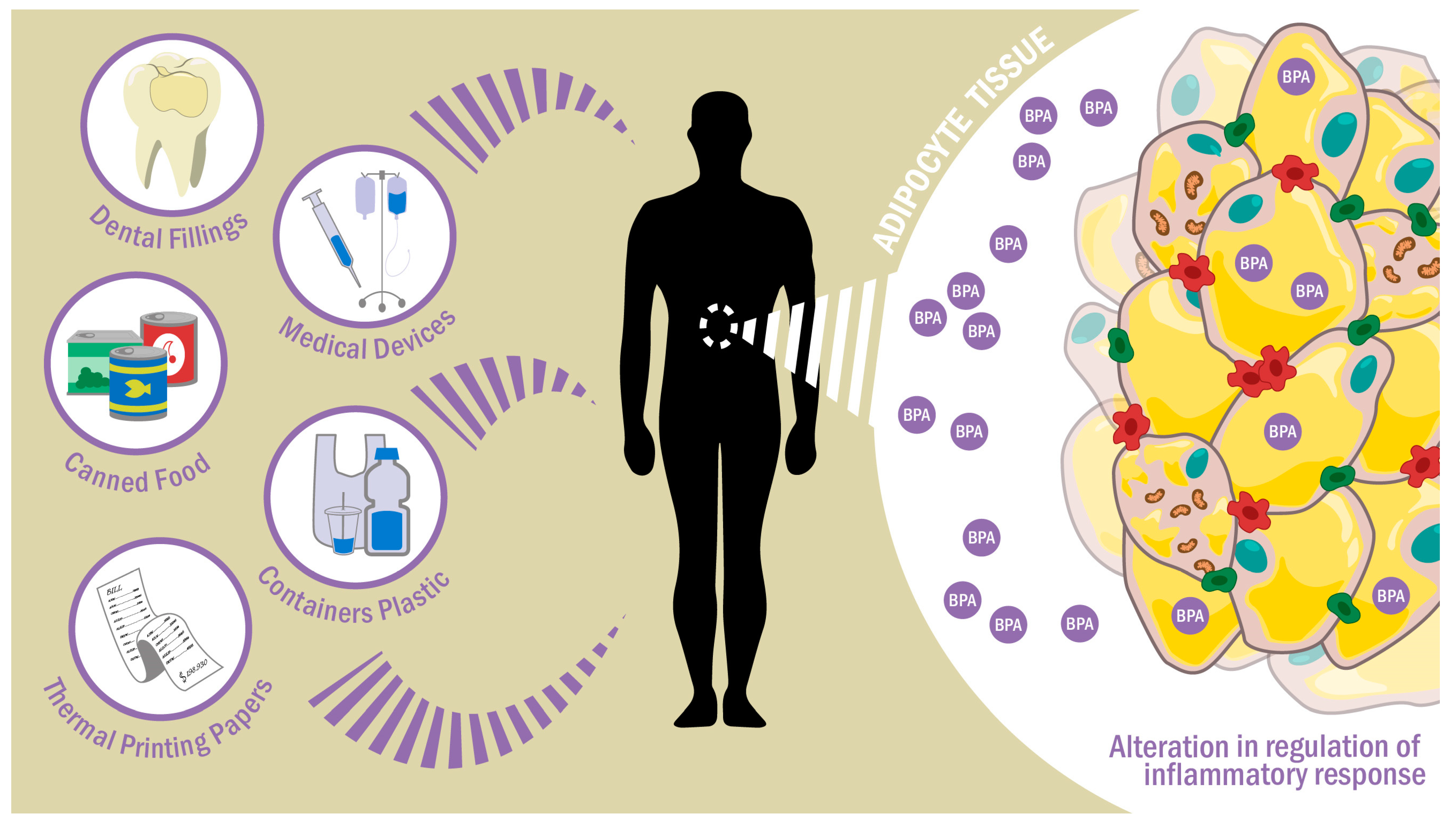
1. Introduction
2. BPA as an Endocrine Disruptor
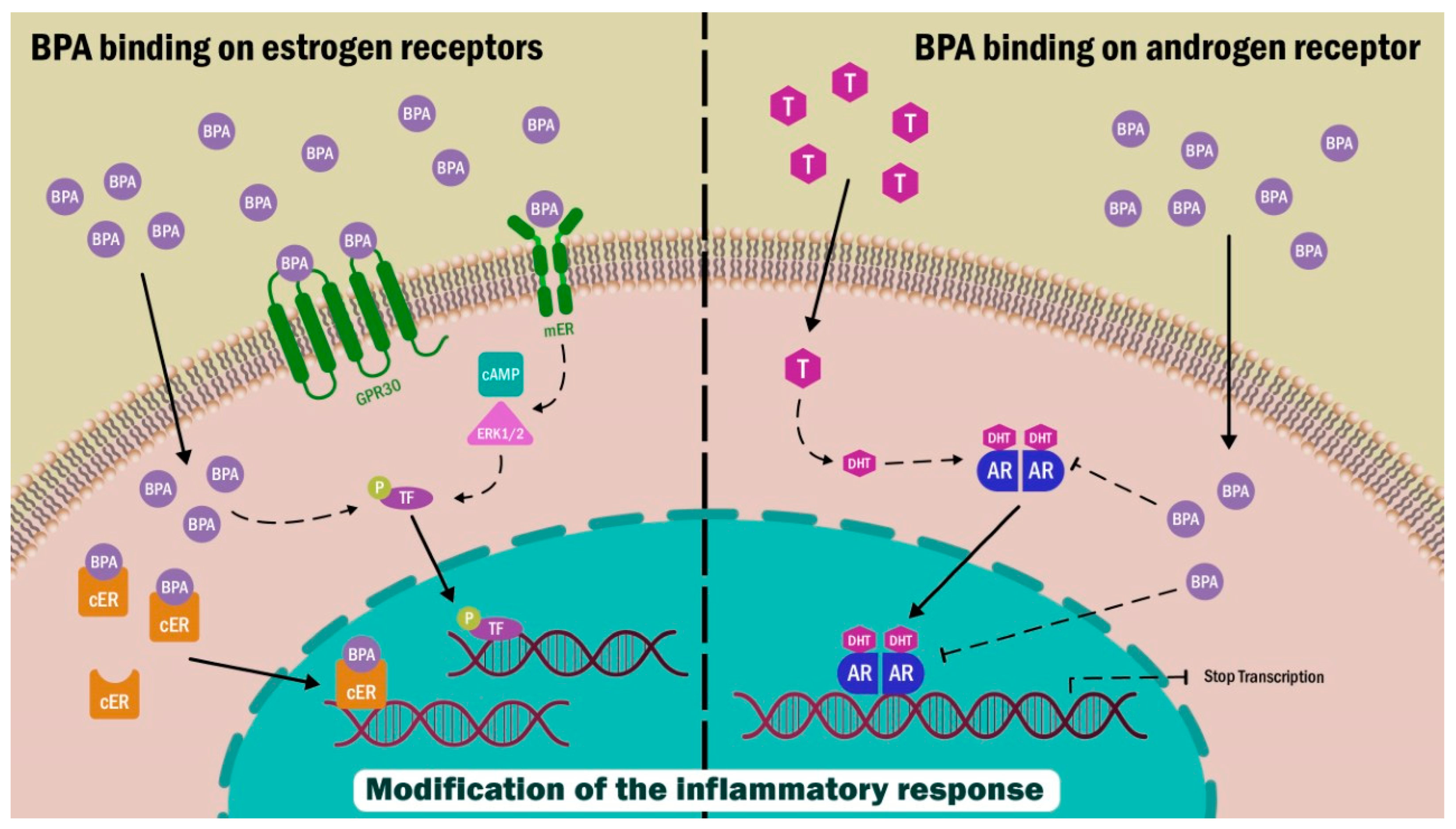
2.1. Estrogen α and β Receptors
2.2. GPR30 Receptor
2.3. Androgen Receptor (AR)
3. Inflammatory Processes in Adipose Tissue
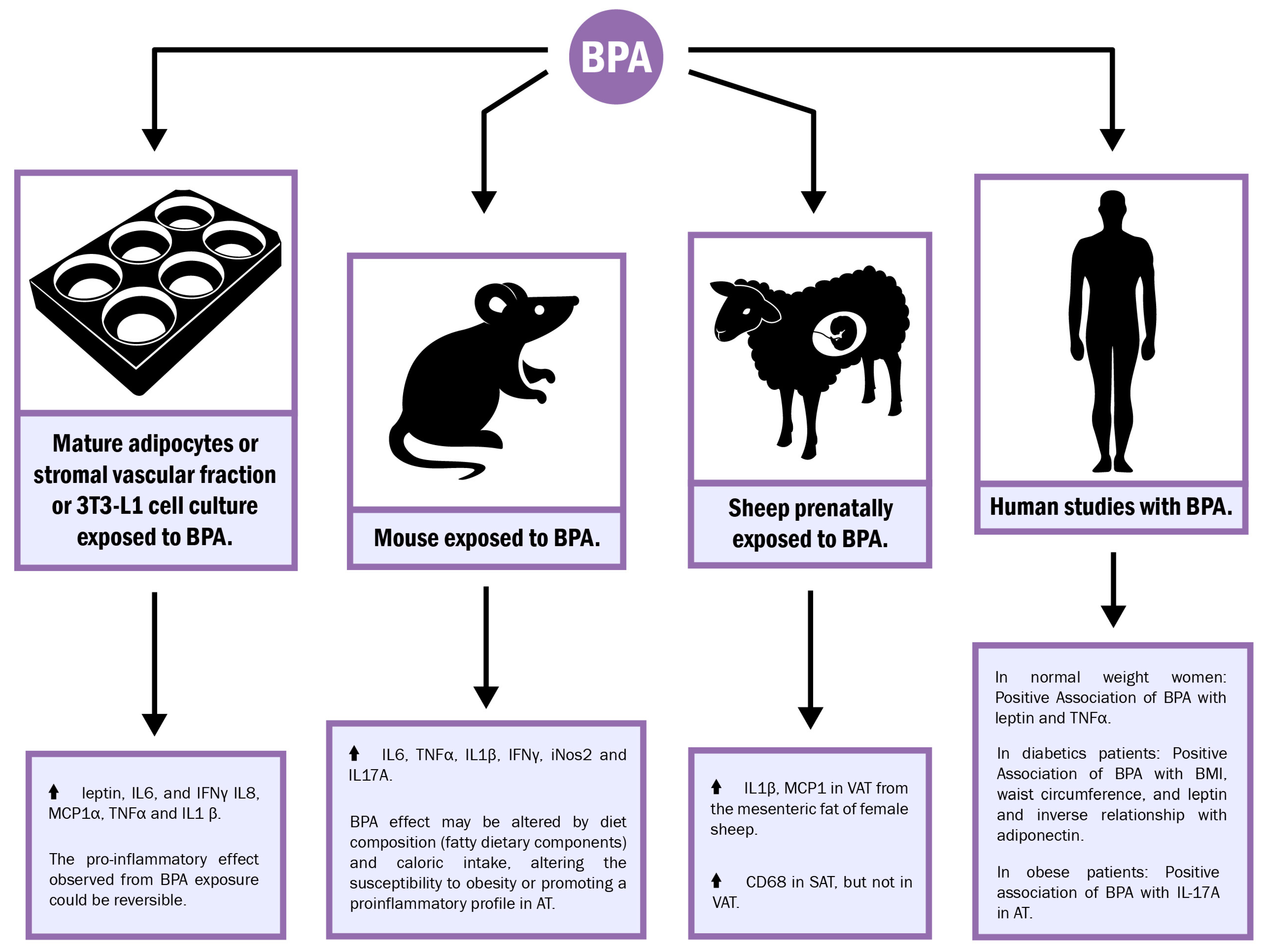
4. Influence of BPA on Inflammatory Signals in Adipose Tissue

| References (Year) | Type of Participants (n: Number of Participants) | Levels Found (ng/g Tissue) | Detection Method | Country |
|---|---|---|---|---|
| Venisse et al. (2019) [68] | Patients during breast or prostate surgery (n: 5). | Range: 1.19–8.73. | LC-MS/MS | France |
| Artacho-Cordón et al. (2017) [64] | Patients undergoing trauma surgery (n: 14). | Mean: 0.60. | HPLC | Spain |
| Reeves et al. (2018) [69] | - Breast cancer mastectomy patients: (n: 36); - Control group: reduction mammoplasty patients (n: 14). | Mean (SD): - Breast cancer mastectomy patients: - Among all samples 0.19 (0.35); - Among samples with detectable BPA 0.71 (0.31); - Control: - Among all samples 0.26 (0.37); - Among samples with detectable BPA 0.66 (0.27). | HPLC-ESI-MS/MS | USA |
| Keshavarz-Maleki et al. (2021) [21] | - Breast cancer mastectomy patients (n: 41); - Control group: mammoplasty patients (n: 11). | - Cancerous patients 4.20 ± 2.40; - Control group: 1.80 ± 1.05. | ELISA assay | Iran |
| Salamanca-Fernández et al. (2020) [70] | Sub-cohort of the Spanish European Prospective Investigation into Cancer and Nutrition (EPIC) (n: 4812): - Breast cancer cases (n: 547); - Prostate cancer cases (n: 575); - Sub-cohort participants (n: 3690). | Geometric mean: - Breast cancer sub-cohort: 1.10; - Breast cancer cases: 1.12; - Prostate cancer sub-cohort: 1.29; - Prostate cancer cases: 1.33. | UHPLC-MS/MS | Spain |
5. BPA’s Inflammatory Action and Cellular Mechanisms Involved
6. Experiments in Fetal Programming: Effect of BPA on Adipose Tissue and Inflammation during Gestation
7. Conclusions
Author Contributions
Funding
Institutional Review Board Statement
Informed Consent Statement
Data Availability Statement
Conflicts of Interest
References
- Fenichel, P.; Chevalier, N.; Brucker-Davis, F. Bisphenol A: An Endocrine and Metabolic Disruptor. Ann. Endocrinol. 2013, 74, 211–220. [Google Scholar] [CrossRef] [PubMed]
- Rubin, B.S.; Schaeberle, C.M.; Soto, A.M. The Case for BPA as an Obesogen: Contributors to the Controversy. Front. Endocrinol. 2019, 10, 30. [Google Scholar] [CrossRef]
- Legeay, S.; Faure, S. Is Bisphenol A an Environmental Obesogen? Fundam. Clin. Pharmacol. 2017, 31, 594–609. [Google Scholar] [CrossRef] [PubMed]
- Mackay, H.; Abizaid, A. Hormones and Behavior Review Article A Plurality of Molecular Targets: The Receptor Ecosystem for Bisphenol-A (BPA). Horm. Behav. 2017, 101, 59–67. [Google Scholar] [CrossRef] [PubMed]
- Agriculture and Environment Research Unit and Toxicology Research Group, University of Hertfordshire, UK. Implementation of the Evidence-Based Risk Assessment for the Re-Evaluation of Bisphenol A: Preparatory Work on Mode of Action Studies in Mammalian, Human and/or In Vitro Models. EFSA Support. Publ. 2021, 18, 6995E. [Google Scholar] [CrossRef]
- Almeida, S.; Raposo, A.; Almeida-González, M.; Carrascosa, C. Bisphenol A: Food Exposure and Impact on Human Health. Compr. Rev. Food Sci. Food Saf. 2018, 17, 1503–1517. [Google Scholar] [CrossRef]
- Geens, T.; Aerts, D.; Berthot, C.; Bourguignon, J.; Goeyens, L.; Lecomte, P.; Maghuin-Rogister, G.; Pironnet, A.; Pussemier, L.; Scippo, M.; et al. A Review of Dietary and Non-Dietary Exposure to Bisphenol-A. Food Chem. Toxicol. 2012, 50, 3725–3740. [Google Scholar] [CrossRef]
- Sasso, A.F.; Pirow, R.; Andra, S.S.; Church, R.; Nachman, R.M.; Linke, S.; Kapraun, D.F.; Schurman, S.H.; Arora, M.; Thayer, K.A.; et al. Pharmacokinetics of Bisphenol A in Humans Following Dermal Administration. Environ. Int. 2020, 144, 106031. [Google Scholar] [CrossRef]
- Karrer, C.; Roiss, T.; von Goetz, N.; Gramec Skledar, D.; Peterlin Masic, L.; Hungerbühler, K. Physiologically Based Pharmacokinetic (PBPK) Modeling of the Bisphenols BPA, BPS, BPF, and BPAF with New Experimental Metabolic Parameters: Comparing the Pharmacokinetic Behavior of BPA with Its Substitutes. Environ. Health. Perspect. 2018, 126, 077002. [Google Scholar] [CrossRef]
- Thayer, K.A.; Doerge, D.R.; Hunt, D.; Schurman, S.H.; Twaddle, N.C.; Churchwell, M.I.; Garantziotis, S.; Kissling, G.E.; Easterling, M.R.; Bucher, J.R.; et al. Pharmacokinetics of Bisphenol A in Humans Following a Single Oral Administration. Environ. Int. 2015, 83, 107–115. [Google Scholar] [CrossRef]
- Jackson, E.; Shoemaker, R.; Larian, N.; Cassis, L. Adipose Tissue as a Site of Toxin Accumulation. Compr. Physiol. 2017, 7, 1085–1135. [Google Scholar] [CrossRef] [PubMed]
- Komarowska, M.D.; Grubczak, K.; Czerniecki, J.; Hermanowicz, A.; Hermanowicz, J.M.; Debek, W.; Matuszczak, E. Identi Fi Cation of the Bisphenol A (BPA) and the Two Analogues BPS and BPF in Cryptorchidism. Front. Endocrinol. 2021, 12, 694669. [Google Scholar] [CrossRef] [PubMed]
- Rebai, I.; Fernandes, J.; Azzouz, M.; Benmohammed, K.; Bader, G.; Benmbarek, K.; Cunha, S.C. Urinary Bisphenol Levels in Plastic Industry Workers. Environ. Res. 2021, 202, 111666. [Google Scholar] [CrossRef] [PubMed]
- Ayar, G.; Yalcin, S.S.; Emeksiz, S.; Yırün, A.; Balcı, A.; Kocer-Gumusel, B.; Erkekoğlu, P. The Association between Urinary BPA Levels and Medical Equipment among Pediatric Intensive Care Patients. Environ. Toxicol. Pharmacol. 2021, 83, 103585. [Google Scholar] [CrossRef] [PubMed]
- Aktag, E.; Yurdakök, K.; Yalçın, S.; Kandemir, N. Urinary Bisphenol A Levels in Prepubertal Children with Exogenous Obesity According to Presence of Metabolic Syndrome. J. Pediatr. Endocrinol. Metab. 2021, 34, 495–502. [Google Scholar] [CrossRef]
- Chang, A.; Ridpath, A.; Carpenter, J.; Kieszak, S.; Sircar, K.; Espinosa-Bode, A.; Nelson, D.; Martin, C. Urine Bisphenol A and Arsenic Levels in Residents of the Cheyenne River Sioux Tribe, South Dakota, with and without Diabetes. J. Med. Toxicol. 2020, 16, 276–283. [Google Scholar] [CrossRef]
- Mohsen, M.A.; Zaki, S.; Youssef, M.; El-Din, E.M.S.; AbuShady, M.M.; Hussein, J.; Hussein, J. May Detectable Urinary Bisphenol A among Children Be Associated with Cardiovascular Risk Factor? Biosci. Res. 2018, 15, 1243–1250. [Google Scholar]
- Omran, G.; Gaber, H.; Mostafa, N.; Abdel-Gaber, R.; Salah, E. Potential Hazards of Bisphenol A Exposure to Semen Quality and Sperm DNA Integrity among Infertile Men. Reprod. Toxicol. 2018, 81, 188–195. [Google Scholar] [CrossRef]
- Manfo, F.; Harthé, C.; Nantia, E.; Dechaud, H.; Tchana, A.; Zabot, M.; Pugeat, M.; Fewou Moundipa, P. Bisphenol A Differentially Affects Male Reproductive Function Biomarkers in a Reference Population and Agro Pesticides Users from Djutitsa, Cameroon. Toxicol. Ind. Health 2019, 35, 324–335. [Google Scholar] [CrossRef]
- Youssef, M.M.; El-din, E.M.S.; Abushady, M.M.; El-baroudy, N.R.; Abd, T.A.; Armaneus, A.F.; El Refay, A.S.; Hussein, J.; Medhat, D.; Latif, Y.A. Urinary Bisphenol A Concentrations in Relation to Asthma in a Sample of Egyptian Children. Hum. Exp. Toxicol. 2018, 37, 1180–1186. [Google Scholar] [CrossRef]
- Abo El-atta, H.M.; Adel- Naby, M.; Ehawary, M. Bisphenol-A and Risk of Obesity Among A Sample of Egyptian Children: Role of Adiponectin as Biomarker of Exposure. Mansoura J. Forensic Med. Clin. Toxicol. 2018, 26, 39–52. [Google Scholar] [CrossRef]
- Jiménez-díaz, I.; Artacho-cordón, F.; Vela-soria, F.; Belhassen, H.; Arrebola, J.P.; Fernández, M.F.; Ghali, R.; Hedhili, A.; Olea, N. Urinary Levels of Bisphenol A, Benzophenones and Parabens in Tunisian Women: A Pilot Study. Sci. Total Environ. 2016, 15, 81–88. [Google Scholar] [CrossRef] [PubMed]
- Keshavarz-Maleki, R.; Kaviani, A.; Omranipour, R.; Gholami, M.; Khoshayand, M.; Ostad, S.; Sabzevari, O. Bisphenol-A in Biological Samples of Breast Cancer Mastectomy and Mammoplasty Patients and Correlation with Levels Measured in Urine and Tissue. Sci. Rep. 2021, 11, 18411. [Google Scholar] [CrossRef]
- Wu, A.; Franke, A.; Wilkens, L.; Tseng, C.; Conroy, S.; Li, Y.; Sangaramoorthy, M.; Polfus, L.; DeRouen, M.; Caberto, C.; et al. Risk of Breast Cancer and Pre-Diagnostic Urinary Excretion of Bisphenol A, Triclosan, and Parabens: The Multiethnic Cohort Study. Int. J. Cancer 2021, 151, E15. [Google Scholar] [CrossRef]
- Durmaz, E.; Asci, A.; Erkekoglu, P.; Balcı, A.; Bircan, I.; Kocer-Gumusel, B. Urinary Bisphenol A Levels in Turkish Girls with Premature Thelarche. Hum. Exp. Toxicol. 2018, 37, 1007–1016. [Google Scholar] [CrossRef]
- Radwan, M.; Wielgomas, B.; Dziewirska, E.; Radwan, P.; Kałuzny, P.; Klimowska, A.; Hanke, W.; Jurewicz, J.; Hanke, W.; Jurewicz, J. Urinary Bisphenol A Levels and Male Fertility. Am. J. Mens. Health 2018, 12, 2144–2151. [Google Scholar] [CrossRef] [PubMed]
- Lee, J.; Choi, K.; Park, J.; Moon, H.; Choi, G.; Lee, J.J.; Suh, E.; Kim, H.; Cho, G.; Kim, S.; et al. Bisphenol A Distribution in Serum, Urine, Placenta, Breast Milk, and Umbilical Cord Serum in a Birth Panel of Mother—Neonate Pairs. Sci. Total Environ. 2017, 626, 1494–1501. [Google Scholar] [CrossRef]
- Adoamnei, E.; Mendiola, J.; Vela-soria, F.; Fernández, M.F.; Olea, N.; Jørgensen, N.; Swan, S.H.; Torres-cantero, A.M. Urinary Bisphenol A Concentrations Are Associated with Reproductive Parameters in Young Men. Environ. Res. 2018, 161, 122–128. [Google Scholar] [CrossRef]
- Benson, T.E.; Gaml-sørensen, A.; Ernst, A.; Brix, N.; Hougaard, K.S.; Hærvig, K.K.; Ellekilde Bonde, J.; Tøttenborg, S.S.; Lindh, C.H.; Ramlau-hansen, C.H.; et al. Urinary Bisphenol A, F and S Levels and Semen Quality in Young Adult Danish Men. Int. J. Environ. Res. Public Health 2021, 18, 1742. [Google Scholar] [CrossRef]
- Mínguez-Alarcón, L.; Messerlian, C.; Bellavia, A.; Gaskins, A.J.; Chiu, Y.; Ford, J.B.; Azevedo, A.R.; Petrozza, J.C.; Calafat, A.M.; Hauser, R.; et al. Urinary Concentrations of Bisphenol A, Parabens and Phthalate Metabolite Mixtures in Relation to Reproductive Success among Women Undergoing In Vitro Fertilization. Environ. Int. 2019, 126, 355–362. [Google Scholar] [CrossRef]
- González, N.; Cunha, S.C.; Monteiro, C.; Fernandes, J.O.; Marques, M.; Domingo, J.L.; Nadal, M. Quantification of Eight Bisphenol Analogues in Blood and Urine Samples of Workers in a Hazardous Waste Incinerator. Environ. Res. 2019, 176, 108576. [Google Scholar] [CrossRef]
- Hong, X.; Zhou, Y.; Zhu, Z.; Li, Y.; Li, Z.; Zhang, Y.; Hu, X.; Zhu, F.; Wang, Y.; Fang, M.; et al. Environmental Endocrine Disruptor Bisphenol A Induces Metabolic Derailment and Obesity via Upregulating IL-17A in Adipocytes. Environ. Int. 2023, 172, 107759. [Google Scholar] [CrossRef]
- Wiraagni, A.I.; Mohd, M.A.; bin Abd Rash, R.; Mohamad Haron, D.E. Validation of a Simple Extraction Procedure for Bisphenol A Identification from Human Plasma. PLoS ONE 2019, 14, e0221774. [Google Scholar] [CrossRef]
- Yamamoto, J.; Minatoya, M.; Sasaki, S.; Araki, A.; Miyashita, C.; Matsumura, T.; Kishi, R. Quantifying Bisphenol A in Maternal and Cord Whole Blood Using Isotope Dilution Liquid Chromatography/Tandem Mass Spectrometry and Maternal Characteristics Associated with Bisphenol A. Chemosphere 2016, 164, 25–31. [Google Scholar] [CrossRef]
- Pednekar, P.P.; Gajbhiye, R.K.; Patil, A.D.; Surve, S.V.; Datar, A.; Balsarkar, G.; Chuahan, A.; Vanage, G. Estimation of Plasma Levels of Bisphenol-A & Phthalates in Fertile & Infertile Women by Gas Chromatography-Mass Spectrometry. Indian J. Med. Res. 2018, 148, 734–742. [Google Scholar] [CrossRef]
- Mas, S.; Bosch-Panadero, E.; Abaigar, P.; Camarero, V.; Mahillo, I.; Civantos, E.; Sanchez-Ospina, D.; Ruiz-Priego, A.; Egido, J.; Ortiz, A.; et al. Influence of Dialysis Membrane Composition on Plasma Bisphenol A Levels during Online Hemodiafiltration. PLoS ONE 2018, 13, e0193288. [Google Scholar] [CrossRef]
- Kolatorova, L.; Vitku, J.; Hampl, R.; Adamcova, K.; Skodova, T.; Simkova, M.; Parizek, A.; Starka, L.; Duskova, M. Exposure to Bisphenols and Parabens during Pregnancy and Relations to Steroid Changes. Environ. Res. 2018, 163, 115–122. [Google Scholar] [CrossRef]
- Jin, H.; Zhu, J.; Chen, Z.; Hong, Y.; Cai, Z. Occurrence and Partitioning of Bisphenol Analogues in Adults’ Blood from China. Environ. Sci. Technol. 2017, 52, 812–820. [Google Scholar] [CrossRef]
- Zbucka-krętowska, M.; Łazarek, U.; Miltyk, W.; Sidorkiewicz, I.; Pierzyński, P.; Milewski, R.; Wołczyński, S.; Czerniecki, J. Simultaneous Analysis of Bisphenol A Fractions in Maternal and Fetal Compartments in Early Second Trimester of Pregnancy. J. Perinat. Med. 2019, 47, 765–770. [Google Scholar] [CrossRef]
- Cambien, G.; Venisse, N.; Migeot, V.; Rabouan, S.; Belmouaz, M.; Binson, G.; Albouy-llaty, M.; Ayraud-thevenot, S. Simultaneous Determination of Bisphenol A and Its Chlorinated Derivatives in Human Plasma: Development, Validation and Application of a UHPLC-MS/MS Method. Chemosphere 2020, 242, 125236. [Google Scholar] [CrossRef]
- Shen, Y.; Dong, Y.; Lu, Q.; Xu, J.; Wu, Y.; Yun, S.; Ren, M. Phenolic Environmental Estrogens in Urine and Blood Plasma from Women with Uterine Leiomyoma: Epidemiological Survey. J. Obstet. Gynaecol. Res. 2016, 42, 440–445. [Google Scholar] [CrossRef]
- Ho, K.; Yuen, K.; Yau, M.; Murphy, M.; Wan, Y.; Fong, B.; Tam, S.; Giesy, J.; Leung, K.; Lam, M. Glucuronide and Sulfate Conjugates of Bisphenol A: Chemical Synthesis and Correlation between Their Urinary Levels and Plasma Bisphenol A Content in Voluntary Human Donors. Arch. Environ. Contam. Toxicol. 2017, 73, 410–420. [Google Scholar] [CrossRef]
- Lin, C.; Chien, C.; Tsai, M.; Hsieh, C.; Hsieh, W.; Chen, P. Prenatal Phenolic Compounds Exposure and Neurobehavioral Development at 2 and 7 Years of Age. Sci. Total Environ. 2017, 15, 801–810. [Google Scholar] [CrossRef]
- Dodds, E.C.; Lawson, W. Synthetic Strogenic Agents without the Phenanthrene Nucleus. Nature 1936, 137, 996. [Google Scholar] [CrossRef]
- Acconcia, F.; Pallottini, V.; Marino, M. Molecular Mechanisms of Action of BPA. Dose Response Int. J. 2015, 13, 1559325815610582. [Google Scholar] [CrossRef]
- Thomas, P.; Pang, Y.; Filardo, E.J.; Dong, J. Identity of an Estrogen Membrane Receptor Coupled to a G Protein in Human Breast Cancer Cells. Endocrinology 2005, 146, 624–632. [Google Scholar] [CrossRef]
- Lee, H.J.; Chattopadhyay, S.; Gong, E.; Ahn, R.S.; Lee, K. Antiandrogenic Effects of Bisphenol A and Nonylphenol on the Function of Androgen Receptor. Toxicol. Sci. 2003, 75, 40–46. [Google Scholar] [CrossRef]
- Sun, H.; Xu, X.; Xu, L.; Song, L.; Hong, X.; Chen, J.; Cui, L.; Wang, X. Antiandrogenic Activity of Pyrethroid Pesticides and Their Metabolite in Reporter Gene Assay. Chemosphere 2007, 66, 474–479. [Google Scholar] [CrossRef]
- Chen, S.; Zhou, D.; Hsin, L.; Kanaya, N.; Wong, C.; Yip, R.; Sakamuru, S.; Xia, M.; Yuan, Y.; Witt, K.; et al. AroER Tri-Screen Is a Biologically Relevant Assay for Endocrine Disrupting Chemicals Modulating the Activity of Aromatase and/or the Estrogen Receptor. Toxicol. Sci. 2014, 139, 198–209. [Google Scholar] [CrossRef]
- Amir, S.; Tahir, S.; Shah, A.; Mamoulakis, C.; Docea, A.O.; Makrigiannakis, A.; Tsatsakis, A. Endocrine Disruptors Acting on Estrogen and Androgen Pathways Cause Reproductive Disorders through Multiple Mechanisms: A Review. Int. J. Environ. Res. Public Health 2021, 18, 1464. [Google Scholar] [CrossRef]
- Choe, S.S.; Huh, J.Y.; Hwang, I.J.; Kim, J.I.; Kim, J.B. Adipose Tissue Remodeling: Its Role in Energy Metabolism and Metabolic Disorders. Front. Endocrinol. 2016, 7. [Google Scholar] [CrossRef]
- Kawai, T.; Autieri, M.V.; Scalia, R. Adipose Tissue Inflammation and Metabolic Dysfunction in Obesity. Am. J. Physiol. Cell Physiol. 2021, 320, C375–C391. [Google Scholar] [CrossRef]
- Reilly, S.M.; Saltiel, A.R. Adapting to Obesity with Adipose Tissue Inflammation. Nat. Rev. Endocrinol. 2017, 13, 633–643. [Google Scholar] [CrossRef] [PubMed]
- Ellulu, M.S.; Patimah, I.; Khaza’ai, H.; Rahmat, A.; Abed, Y. Obesity & Inflammation: The Linking Mechanism & the Complications. Arch. Med. Sci. 2017, 13, 851–863. [Google Scholar] [CrossRef] [PubMed]
- Baker, R.G.; Hayden, M.S.; Ghosh, S. NF-κB, Inflammation, and Metabolic Disease. Cell Metab. 2011, 13, 11–22. [Google Scholar] [CrossRef]
- Araiza, V.; Mendoza, M.; Castro, K.; Cruz, M.; Rueda, K.; De Leon, C.; Morales Montor, J. Bisphenol A: An Endocrine-Disruptor Compound That Modulates the Immune Response to Infections. Front. Biosci. Landmark 2021, 26, 346–362. [Google Scholar] [CrossRef]
- Gonzalez, J.E.; Pertuz-cruz, S.L.; Caicedo-ortega, N.H.; Rojas-gomez, D.M. Adipogenesis Regulation and Endocrine Disruptors: Emerging Insights in Obesity. BioMed Res. Int. 2020, 2020, 7453786. [Google Scholar] [CrossRef]
- Ariemma, F.; D’Esposito, V.; Liguoro, D.; Oriente, F.; Cabaro, S.; Liotti, A.; Cimmino, I.; Longo, M.; Beguinot, F.; Formisano, P.; et al. Low-Dose Bisphenol-A Impairs Adipogenesis and Generates Dysfunctional 3T3-L1 Adipocytes. PLoS ONE 2016, 11, e0150762. [Google Scholar] [CrossRef]
- Valentino, R.; D’Esposito, V.; Passaretti, F.; Liotti, A.; Cabaro, S.; Longo, M.; Perruolo, G.; Oriente, F.; Beguinot, F.; Formisano, P. Bisphenol-A Impairs Insulin Action and Up-Regulates Inflammatory Pathways in Human Subcutaneous Adipocytes and 3T3-L1 Cells. PLoS ONE 2013, 8, e82099. [Google Scholar] [CrossRef]
- Longo, M.; Zatterale, F.; Naderi, J.; Nigro, C.; Oriente, F. Low-Dose Bisphenol-A Promotes Epigenetic Changes at Ppar γ Promoter in Adipose Precursor Cells. Nutrients 2020, 12, 3498. [Google Scholar] [CrossRef]
- Yang, M.; Chen, M.; Wang, J.; Xu, M.; Sun, J.; Ding, L.; Lv, X.; Ma, Q.; Bi, Y.; Liu, R.; et al. Bisphenol A Promotes Adiposity and Inflammation in a Nonmonotonic Dose-Response Way in 5-Week-Old Male and Female C57BL/6J Mice Fed a Low-Calorie Diet. Endocrinology 2016, 157, 2333–2345. [Google Scholar] [CrossRef]
- Jain, J.; Gupta, N.; Mathur, R.; Nimesh, S.; Mathur, S.K. A Study on Impact of BPA in the Adipose Tissue Dysfunction (Adiposopathy) in Asian Indian Type 2 Diabetes Mellitus Subjects. Indian J. Clin. Biochem. 2020, 35, 451–457. [Google Scholar] [CrossRef]
- Stahlhut, R.W.; Welshons, W.V.; Swan, S.H. Bisphenol A Data in NHANES Suggest Longer than Expected Half-Life, Substantial Nonfood Exposure, or Both. Environ. Health Perspect. 2009, 117, 784–789. [Google Scholar] [CrossRef]
- Artacho-Cordón, F.; Arrebola, J.P.; Nielsen, O.; Hernández, P.; Skakkebaek, N.E.; Fernández, M.F.; Andersson, A.M.; Olea, N.; Frederiksen, H. Assumed Non-Persistent Environmental Chemicals in Human Adipose Tissue; Matrix Stability and Correlation with Levels Measured in Urine and Serum. Environ. Res. 2017, 156, 120–127. [Google Scholar] [CrossRef]
- Ahmed, F.; Sarsenbayeva, A.; Katsogiannos, P.; Aguer, C.; Pereira, M.J. The Effects of Bisphenol A and Bisphenol S on Adipokine Expression and Glucose Metabolism in Human Adipose Tissue. Toxicology 2020, 445, 152600. [Google Scholar] [CrossRef]
- Pyo, M.Y.; Kim, H.J.; Back, S.K.; Yang, M. Downregulation of Peritoneal Macrophage Activity in Mice Exposed to Bisphenol A During Pregnancy and Lactation. Arch. Pharm. Res. 2007, 30, 1476–1481. [Google Scholar] [CrossRef] [PubMed]
- Valentino, R.; D’Esposito, V.; Ariemma, I.; Cimmino, I.; Beguinot, F.; Formisano, P. Bisphenol A Environmental Exposure and the Detrimental Effects on Human Metabolic Health: Is It Necessary to Revise the Risk Assessment in Vulnerable Population? J. Endocrinol. Investig. 2016, 39, 259–263. [Google Scholar] [CrossRef]
- Venisse, N.; Cambien, G.; Robin, J.; Rouillon, S.; Nadeau, C.; Charles, T.; Rabouan, S.; Migeot, V.; Dupuis, A. Development and Validation of an LC—MS/MS Method for the Simultaneous Determination of Bisphenol A and Its Chlorinated Derivatives in Adipose Tissue. Talanta 2019, 204, 145–152. [Google Scholar] [CrossRef]
- Reeves, K.W.; Schneider, S.; Xue, J.; Kannan, K.; Mason, H.; Johnson, M.; Makari-Judson, G.; Díaz, M. Bisphenol-A in Breast Adipose Tissue of Breast Cancer Cases and Controls. Environ. Res. 2018, 167, 735–738. [Google Scholar] [CrossRef] [PubMed]
- Salamanca-fernández, E.; Rodríguez-Barranco, M.; Arrebola, J.P.; Vela, F.; Díaz, C.; Chirlaque, M.D.; Colorado-Yohar, S.; Jiménez-Zabala, A.; Irizar, A.; Guevara, M.; et al. Bisphenol-A in the European Prospective Investigation into Cancer and Nutrition Cohort in Spain: Levels at Recruitment and Associated Dietary Factors. Environ. Res. 2020, 182, 109012. [Google Scholar] [CrossRef] [PubMed]
- Cimmino, I.; Oriente, F.; D’Esposito, V.; Liguoro, D.; Liguoro, P.; Ambrosio, M.; Cabaro, S.; D’Andrea, F.; Beguinot, F.; Formisano, P.; et al. Low-Dose Bisphenol-A Regulates Inflammatory Cytokines through GPR30 in Mammary Adipose Cells. J. Mol. Endocrinol. 2019, 63, 273–283. [Google Scholar] [CrossRef] [PubMed]
- Apostolakis, S.; Vogiatzi, K.; Amanatidou, V.; Spandidos, D.A. Interleukin 8 and Cardiovascular Disease. Cardiovasc. Res. 2009, 84, 353–360. [Google Scholar] [CrossRef]
- Qazi, B.S.; Tang, K.; Qazi, A. Recent Advances in Underlying Pathologies Provide Insight into Interleukin-8 Expression-Mediated Inflammation and Angiogenesis. Int. J. Inflam. 2011, 2011, 908468. [Google Scholar] [CrossRef]
- Cimini, F.; Barchetta, I.; Porzia, A.; Mainiero, F.; Costantino, C.; Bertoccini, L.; Ceccarelli, V.; Morini, S.; Baroni, M.; Lenzi, A.; et al. Circulating IL—8 Levels Are Increased in Patients with Type 2 Diabetes and Associated with Worse Inflammatory and Cardiometabolic Profile. Acta Diabetol. 2017, 54, 961–967. [Google Scholar] [CrossRef]
- Mittal, M.; Siddiqui, M.R.; Tran, K.; Reddy, S.P.; Malik, A.B. Reactive Oxygen Species in Inflammation and Tissue Injury. Antioxid. Redox Signal. 2014, 20, 1126–1167. [Google Scholar] [CrossRef] [PubMed]
- Piao, X.; Liu, Z.; Yangyang, L.; Yao, D.; Sun, L.; Wang, B.; Ma, Y.; Wang, L.; Zhang, Y. Spectrochimica Acta Part A: Molecular and Biomolecular Spectroscopy Investigation of the Effect for Bisphenol A on Oxidative Stress in Human Hepatocytes and Its Interaction with Catalase. Spectrochim. Acta A Mol. Biomol. Spectrosc. 2019, 221, 117149. [Google Scholar] [CrossRef] [PubMed]
- Teijeiro, A.; Garrido, A.; Ferre, A.; Perna, C.; Djouder, N. Inhibition of the IL-17A Axis in Adipocytes Suppresses Diet-Induced Obesity and Metabolic Disorders in Mice. Nat. Metab. 2021, 3, 496–512. [Google Scholar] [CrossRef]
- Callan, A.; Hinwood, A.; Heffernan, A.; Eaglesham, G.; Mueller, J.; Odlan, J. Urinary Bisphenol A Concentrations in Pregnant Women. Int. J. Hyg. Environ. Health 2013, 216, 641–644. [Google Scholar] [CrossRef]
- Rubin, B.S.; Murray, M.K.; Damassa, D.A.; King, J.C.; Soto, A.M. Perinatal Exposure to Low Doses of Bisphenol A Affects Body Weight, Patterns of Estrous Cyclicity, and Plasma LH Levels. Environ. Health Perspect. 2001, 109, 675–680. [Google Scholar] [CrossRef]
- Veiga-Lopez, A.; Moeller, J.; Sreedharan, R.; Singer, K.; Lumeng, X.; Ye, W.; Pease, A.; Padmanabhan, V. Developmental Programming: Interaction between Prenatal BPA Exposure and Postnatal Adiposity on Metabolic Variables in Female Sheep. Am. J. Physiol. Endocrinol. Metab. 2016, 310, E238–E247. [Google Scholar] [CrossRef]
- Dou, J.F.; Puttabyatappa, M.; Padmanabhan, V.; Bakulski, K.M. Developmental Programming: Transcriptional Regulation of Visceral and Subcutaneous Adipose by Prenatal Bisphenol-A in Female Sheep. Chemosphere 2020, 255, 127000. [Google Scholar] [CrossRef] [PubMed]
- Puttabyatappa, M.; Martin, J.D.; Andriessen, V.; Stevenson, M.; Zeng, L.; Pennathur, S.; Padmanabhan, V. Developmental Programming: Changes in Mediators of Insulin Sensitivity in Prenatal Bisphenol A-Treated Female Sheep. Reprod. Toxicol. 2019, 85, 110–122. [Google Scholar] [CrossRef] [PubMed]
- Malaisé, Y.; Menard, S.; Cartier, C.; Gaultier, E.; Lasserre, F.; Lencina, C.; Harkat, C.; Geoffre, N.; Lakhal, L.; Castan, I.; et al. Gut Dysbiosis and Impairment of Immune System Homeostasis in Perinatally-Exposed Mice to Bisphenol A Precede Obese Phenotype Development. Sci. Rep. 2017, 7, 14472. [Google Scholar] [CrossRef] [PubMed]


| References (Year) | Type of Participants (n: Number of Participants) | Levels Found | Detection Method | Country |
|---|---|---|---|---|
| Rebai et al. (2021) [13] | Workers in a plastics industry located in an industrial zone (n: 170). | Average (μg/g creatinine): 3.70. | GC/MS | Algeria |
| Ayar et al. (2021) [14] | Patients of pediatric intensive care unit: urine samples (n: 115) of children (n: 40). | Mean (μg/g creatinine): 189.2
- First day of hospitalization: 29.5; - The seventh day of hospitalization: 41.1; - After 30 days of hospitalization (or when the patients were discharged): 104.8. | HPLC | Turkey |
| Aktağ et al. (2021) [15] | Prepubertal children: - Prepubertal children with exogenous obesity (n: 36); - Prepubertal children with exogenous obesity and metabolic syndrome (n: 27); - Control group age- and sex-matched healthy children with no significant underlying medical conditions (n: 34). | Median and (mean ± SD) (μg/g-creatinine): - Prepubertal children with exogenous obesity without metabolic syndrome: 15.0 (25.0 ± 24.2); - Prepubertal children with exogenous obesity and metabolic syndrome: 32.1 (46 ± 39.4); - Control group: 5.0 (6.0 ± 4.6). | LC-MS/MS | Turkey |
| Chang et al. (2020) [16] | Adults in the Cheyenne River Sioux tribe: American Indians/Alaskan natives (n: 276): - Participants with diabetes (n: 138); - Control group without diabetes matched using age (n: 138). | Geometric mean (μg/L): - Total participants: 1.83; - Participants with diabetes: 1.90; - Control: 1.77. | HPLC-MS/MS | USA |
| Mohsen et al. (2018) [17] | Children randomly selected from primary and preparatory schools (6–16 years old) (n: 167): - Boys (n: 95); - Girls (n: 72). | Median (ng/mL): - Boys: 0.60; - Girls: 0.67. | HPLC-MS/MS | Egypt |
| Omran et al. (2018) [18] | Infertile patients presented to the andrology unit (n: 50): - Oligoasthenoteratospermia cases group (n: 16); - Asthenospermia cases group (n: 22); - Asthenoteratospermia cases group (n: 12); - Matched controls with normal semen parameters (n: 50). | Median (µg/g creatinine): - Total infertile cases group: 21.59; - Oligoasthenoteratospermia cases group: 18.16; - Asthenoteratospermia cases group: 19.28; - Asthenoteratospermia cases group: 31.23; - Control group: 19.31. | HPLC | Egypt |
| Manfo et al. (2019) [19] | Adults between 18 and 59 years of age (n: 81): - Townsmen in urban area: (n: 37); - Farmers using agro-pesticides in rural area: (n: 44). | Arithmetic mean (µg/g creatinine): - All participants: 2.18 ± 1.97; - Townsmen in urban area: 2.16; - Farmers using agro-pesticides in rural area: 2.20. | RIA | Cameroon |
| Youssef et al. (2018) [20] | Children aged 3–8 years (n: 97): - Asthmatic children (n: 45); - Healthy controls (n: 52). | Median (ng/mL): - Pediatric asthmatic patients: 1.56; - Control group: 0.790. | HPLC-MS/MS | Egypt |
| Abo El-Atta et al. (2018) [21] | Children/adolescents 2–18 years of age: - Study group: obese children (BMI ≥ 95th percentile) (n: 40); - Control group: normal-weight children (BMI 5th–85th percentile) (n: 40). | Median (min–max) (μg/g creatinine): - Obese children: 121.89 (39.22–586.97); - Control group: 14.92 (<LOD − 34.94). | HPLC | Egypt |
| Jiménez-Díaz et al. (2016) [22] | Healthy population of women aged 18 years or older (n: 34). | Geometric mean (ng/mL): 0.44. Mean (ng/mL): 1.12. | UHPLC-MS/MS | Tunisia |
| Keshavarz-Maleki et al. (2021) [23] | - Breast cancer mastectomy patients (n: 41); - Control group: reduction mammoplasty patients with similar BMI to cases group (n: 11). | Mean ± SD (ng/mL): - Breast cancer mastectomy patients: 2.12 ± 1.48; - Control group: 0.91 ± 0.42. | ELISA | Iran |
| Wu et al. (2021) [24] | Multiethnic cohort (1993–2014): - Postmenopausal women with breast cancer aged 45–75 years: - African American (n: 48); - Latino (n: 77); - Native Hawaiian (n: 155); - Japanese American (n: 478); - White: (n: 274); - Individually matched (n: 1030). | Geometric means (ng/g creatinine):
- Whites (n = 547): 1.48; - Japanese Americans (n = 956): 1.07; - Native Hawaiians (n = 309): 1.26; - African Americans (n = 97): 0.77; - Latinos (n = 157) 0.92. | LC/HRAM-MS | USA |
| Durmaz et al. (2018) [25] | - Newly diagnosed girls with premature thelarche nonobese (aged 4–8 years) (n: 25). - Control group: healthy girls of comparable age with no history of premature thelarche or any other endocrine disorder and no secondary sexual characteristics in their physical exam (n: 25). | Median µg/g (creatinine): - Newly diagnosed girls with premature thelarche, non-obese: 3.21; - Control group: 1.62. | HPLC | Turkey |
| Radwan et al. (2018) [26] | Males attending infertility clinic for diagnostic purposes with normal semen concentration (n: 315). | Median: 1.87 µg/L, 1.63 µg/g creatinine | GC/MS | Poland |
| Lee et al. (2019) [27] | Pregnant women who had babies with normal gestation age, neonatal weight, and information on birth outcome: - Neonatal urine (n: 152); - Maternal urine (n: 224). | Median (ng/mL): - Neonatal urine: 4.75; - Maternal urine: 2.86. | HPLC-MS/MS and GC-MS | Korea |
| Adoamnei et al. (2018) [28] | Healthy, young university students (18–23 years old) (n: 215). | Unadjusted median (ng/mL): 2.8. | UHPLC-MS/MS | Spain |
| Benson et al. (2021) [29] | Men 18–20 years of age from the Fetal Programming of Semen Quality cohort (n: 556). | Pseudo percentiles (ng/mL): 5th: 0.22, 50th: 1.30, 95th: 9.90. | LC-MS/MS | Denmark |
| Mínguez-Alarcón et al. (2019) [30] | Women undergoing in vitro fertilization treatment (between 18 and 45 years old) (n: 420). | Geometric means (µg/L): 1.14. | IDMS | USA |
| Gonzalez et al. (2019) [31] | Workers of a hazardous waste incinerator (n: 29): 11 women and 18 men. | Mean (µg/L): 0.86. | GC/MS | Spain |
| Hong et al. (2023) [32] | Obese patients and healthy individuals (n: 289): - Obesity cases: participants aged above 16 and below 65 years old, body mass index (BMI) ≥ 27.5 kg/m2; - Control group: participants aged above 16 and below 65 years old with body max index < 24.0 kg/m2 (n: 152). | Median (µg/g creatinine): - Obesity cases: 4.33; - Control group: 1.37. | LC-MS/MS | China |
| References (Year) | Type of Participants (n: Number of Participants) | Levels Found | Detection Method | Country |
|---|---|---|---|---|
| Wiraagni et al. (2019) [33] | Healthy volunteers (n: 150): - Males (n: 43); - Females (n: 107). | Observed BPA Levels (ng/mL): Range: 0 to 76.80 Mean: 2.22 ± 9.91 - Males: 0.29; - Females: 2.99; - Less than 33 years of age: 0.847; - 33 years of age and older: 5.852; - Subjects with tap water as source of drinking: 2.882; - Subjects with mineral water as source of drinking: 0.318. | LC-MS/MS | Malaysia |
| Yamamoto et al. (2016) [34] | Women at 23–35 weeks of gestation and those who delivered between 2002 and 2005: - Maternal blood (n: 59); - Cord blood (n: 285), | Geometric mean (ng/mL): - Maternal blood 0.051; - Cord blood 0.046; Mean (ng/mL): - Maternal blood 0.063; - Cord blood 0.057. | ID-LC/MS/MS | Japan |
| Pednekar et al. (2018) [35] | Women between 20 and 40 years of age, attending infertility outpatient department, diagnosed with infertility (n: 45): - Polycystic ovary syndrome (n: 31); - Endometriosis (n: 11); - Polycystic ovary syndrome and endometriosis (n: 3); - Married women between 20 and 40 years of age with proven fertility and no evidence of any gynecological disorders, who achieved pregnancy naturally and delivered recently (within one year) (n: 34). | Mean (ng/mL): - Women with infertility: 4.66 ± 3.52; - Polycystic ovary syndrome: 5.80 ± 3.05; - Endometriosis: 4.59 ± 1.22; - Polycystic ovary syndrome and endometriosis (3): 13.17; - Fertile women group: 2.64 ± 3.99. | GC-MS | India |
| Mas et al. (2018) [36] | Online hemodiafiltration patients using BPA-free (polynephron) or BPA-containing (polysulfone) dialyzers in a crossover design with two arms after a run-in period of at least 6 months with the same membrane: - Patients with BPA-free high-flux polynephron (polynephron) membranes (n: 36); - Patients with high-flux polysulfone (Helixone®) dialyzers that contain BPA (n: 36); - Patients on conventional hemodialysis (n: 10); - Healthy controls (n: 10). | Mean (ng/mL): - Patients using BPA-free (polynephron): 8.79 ± 7.97; - Patients with high-flux polysulfone: 23.42 ± 20.38; - Patients on conventional hemodialysis: 98.96 ± 120.75; - Healthy controls: < 2. | High-sensitivity ELISA | Spain |
| Kolatorova et al. (2018) [37] | Healthy pregnant women of 33 ± 4.1 years (week 37 of pregnancy) (n: 27). | Median with lower and upper quartiles (ng/mL): 0.059 (0.023, 0.084). | LC-MS/MS | Czech Republic |
| Jin et al. (2017) [38] | Participants of healthy general population, without any evidence of occupational exposure to bisphenols: - Women (n: 9); - Men (n: 10); - Total (n: 19). | Mean (range) (ng/mL): - Women: 0.75 ± 0.12, (0.60–0.88); - Men: 0.60 ± 0.17, range: (0.41–0.85); - Total: 0.67 ± 0.16 (0.41–0.88). | LC-MS/MS | China |
| Komarowska et al. (2021) [12] | - Children with congenital unilateral cryptorchidism aged 1–4 years (n: 98); - Healthy boys without any disorders of the testes at a comparable age of 1–4 years (n: 19). | Median (ng/mL): - Children with congenital unilateral cryptorchidism: 9.95; - Control group: 5.54. | GC-MS | Poland |
| Zbucka-Krętowska et al. (2019) [39] | Women undergoing routine amniocentesis between the 15th and 18th weeks of gestation carrying fetuses with a normal karyotype (n: 52). | Mean (ng/mL): 8.69, range: 4.3–55.3. | GC-MS | Poland |
| Cambien et al. (2019) [40] | Patients suffering from end-stage renal disease and hospitalized (n: 10). | Range (ng/mL): 0.266–86.831. | UHPLC–MS/MS | France |
| Shen et al. (2016) [41] | - Women with uterine leiomyoma (n: 300); - Control group with no use of hormone drugs during the 3 months prior to the study and free of reproductive-system-related tumors and other estrogen-dependent diseases (breast cancer, endocrine system diseases, etc.) (n: 300). | Media (mean ± SD) (ng/mL): - Women with uterine leiomyoma: 11.19 (16.7 ± 13.9); - Control group: 4.31 (8.62 ± 11.8). | HPLC-MS/MS | China |
| Ho et al. (2017) [42] | Voluntary human donors (n: 140) age range from 18 to 96 years: - Men (n: 66); - Women (n: 64). | Geometric mean (range) (ng/mL): - Total: 0.53 (range: N.D.–10.43); - Women: 0.59 (N.D.–8.99); - Men: 0.47 (N.D.–10.43). | LC-MS/MS | China |
| Lin et al. (2017) [43] | - Mother–child pairs with 2-year-old children (n: 208): girls (n: 91), boys (n: 117); - Mother–child pairs with 7-year-old children (n: 148): girls (n: 70), boys (n: 78). | Median (ng/mL): - 2-year-old children: 3.2; girls: 2.9; boys: 3.3; - 7-year-old children: 3.2; girls: 1.2; boys: 4.0. | UPLC-MS-MS | Taiwan |
Disclaimer/Publisher’s Note: The statements, opinions and data contained in all publications are solely those of the individual author(s) and contributor(s) and not of MDPI and/or the editor(s). MDPI and/or the editor(s) disclaim responsibility for any injury to people or property resulting from any ideas, methods, instructions or products referred to in the content. |
© 2023 by the authors. Licensee MDPI, Basel, Switzerland. This article is an open access article distributed under the terms and conditions of the Creative Commons Attribution (CC BY) license (https://creativecommons.org/licenses/by/4.0/).
Share and Cite
González-Casanova, J.E.; Bermúdez, V.; Caro Fuentes, N.J.; Angarita, L.C.; Caicedo, N.H.; Rivas Muñoz, J.; Rojas-Gómez, D.M. New Evidence on BPA’s Role in Adipose Tissue Development of Proinflammatory Processes and Its Relationship with Obesity. Int. J. Mol. Sci. 2023, 24, 8231. https://doi.org/10.3390/ijms24098231
González-Casanova JE, Bermúdez V, Caro Fuentes NJ, Angarita LC, Caicedo NH, Rivas Muñoz J, Rojas-Gómez DM. New Evidence on BPA’s Role in Adipose Tissue Development of Proinflammatory Processes and Its Relationship with Obesity. International Journal of Molecular Sciences. 2023; 24(9):8231. https://doi.org/10.3390/ijms24098231
Chicago/Turabian StyleGonzález-Casanova, Jorge Enrique, Valmore Bermúdez, Nelson Javier Caro Fuentes, Lissé Chiquinquirá Angarita, Nelson Hernando Caicedo, Jocelyn Rivas Muñoz, and Diana Marcela Rojas-Gómez. 2023. "New Evidence on BPA’s Role in Adipose Tissue Development of Proinflammatory Processes and Its Relationship with Obesity" International Journal of Molecular Sciences 24, no. 9: 8231. https://doi.org/10.3390/ijms24098231
APA StyleGonzález-Casanova, J. E., Bermúdez, V., Caro Fuentes, N. J., Angarita, L. C., Caicedo, N. H., Rivas Muñoz, J., & Rojas-Gómez, D. M. (2023). New Evidence on BPA’s Role in Adipose Tissue Development of Proinflammatory Processes and Its Relationship with Obesity. International Journal of Molecular Sciences, 24(9), 8231. https://doi.org/10.3390/ijms24098231