Review

Functional Surface Coatings on Orthodontic Appliances: Reviews of Friction Reduction, Antibacterial Properties, and Corrosion Resistance

1
Department of Orthodontics, School and Hospital of Stomatology, Peking University, Beijing 100081, China
2
National Center of Stomatology & National Clinical Research Center for Oral Diseases & National Engineering Laboratory for Digital and Material Technology of Stomatology & Beijing Key Laboratory for Digital Stomatology & Research Center of Engineering and Technology for Computerized Dentistry Ministry of Health & NMPA Key Laboratory for Dental Materials, Beijing 100081, China
*
Authors to whom correspondence should be addressed.
Int. J. Mol. Sci. 2023, 24(8), 6919; https://doi.org/10.3390/ijms24086919
Submission received: 29 January 2023 / Revised: 19 March 2023 / Accepted: 31 March 2023 / Published: 7 April 2023
(This article belongs to the Special Issue Recent Process Design and Development Strategies for Dental Materials)

Abstract

:
Surface coating technology is an important way to improve the properties of orthodontic appliances, allowing for reduced friction, antibacterial properties, and enhanced corrosion resistance. It improves treatment efficiency, reduces side effects, and increases the safety and durability of orthodontic appliances. Existing functional coatings are prepared with suitable additional layers on the surface of the substrate to achieve the abovementioned modifications, and commonly used materials mainly include metal and metallic compound materials, carbon-based materials, polymers, and bioactive materials. In addition to single-use materials, metal-metal or metal-nonmetal materials can be combined. Methods of coating preparation include, but are not limited to, physical vapor deposition (PVD), chemical deposition, sol-gel dip coating, etc., with a variety of different conditions for preparing the coatings. In the reviewed studies, a wide variety of surface coatings were found to be effective. However, the present coating materials have not yet achieved a perfect combination of these three functions, and their safety and durability need further verification. This paper reviews and summarizes the effectiveness, advantages and disadvantages, and clinical perspectives of different coating materials for orthodontic appliances in terms of friction reduction, antibacterial properties, and enhanced corrosion resistance, and discusses more possibilities for follow-up studies as well as for clinical applications in detail.

1. Introduction

Surface coating technology relies on various metals, polymers, and composite materials by preparing a suitable additional layer on the surface of the substrate and applying different coatings to the surface of the brackets and archwires to modify their surface morphology, mechanical properties, and antibacterial properties [1]. This is currently an important way to improve the performance of orthodontic appliances, enabling clinical effects such as friction reduction, antibacterial effects, and corrosion resistance.
It is necessary to reduce the friction between the archwire and brackets, improve the antibacterial properties of orthodontic appliances, and increase their corrosion and wear resistance using surface coatings, in order to improve treatment efficiency, reduce the consumption of anchorage, reduce the incidence of caries and periodontal disease caused by orthodontics, and increase the safety and durability of orthodontic attachments. Some studies have shown that friction can offset 12–60% of the orthodontic force during orthodontic treatment [2], reducing the efficiency of tooth movement and increasing the consumption of anchorage, which is detrimental to the treatment [2,3]. Moreover, a white spot lesion (WSL) is often observed on the enamel surface around orthodontic attachments [4,5,6], and plaque-induced gingivitis [7] appears occasionally. These are due to difficulties in the mechanical removal of plaque and increased plaque retention [8]. Furthermore, metal orthodontic appliances are susceptible to corrosion in the intraoral environment [9,10,11]. In addition to electrochemical corrosion, chewing, brushing teeth, and archwires sliding along the brackets, the mechanical forces on the surfaces also accelerate the process of corrosion [10,12], causing the release of various metal ions—especially nickel (Ni) ions [13,14,15]. These metal ions act as allergens, increasing the likelihood of metal allergy for orthodontic patients [16,17]. Allergic reaction of the oral mucosa to nickel can cause distress and pain to patients; this is significantly more common in females than in males, especially between the ages of 16 and 35 years. The discomfort to patients includes burning sensation of the oral mucosa, cheilitis, parageusia, periodontitis, and even erythema multiforme and popular perioral rash [17]. These conditions can reduce patients’ quality of life and oral health, which can adversely affect treatment compliance and therapeutic effects. Thus, surface coatings are important to be put into clinical use.
However, the existing coating materials and methods for orthodontic fixed appliances are numerous and complicated, while the purpose and focus point of the coatings differ. Based on the above considerations, this article mainly reviews and summarizes the effects, advantages, disadvantages, and clinical prospects of different coating materials with respect to friction reduction, antibacterial effects, and corrosion resistance, in order to guide the selection and preparation of suitable surface coatings for clinical applications. Coatings on brackets and archwires can be seen in Figure 1.

2. Friction Reduction Coatings

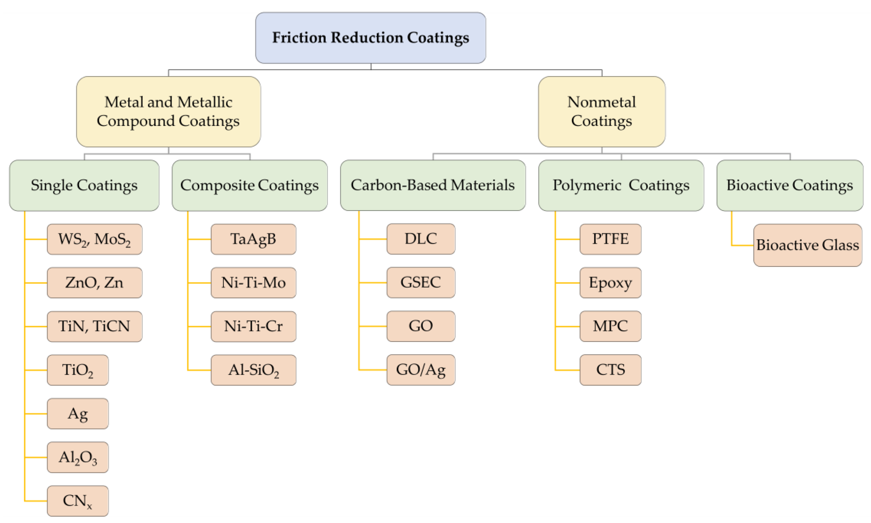
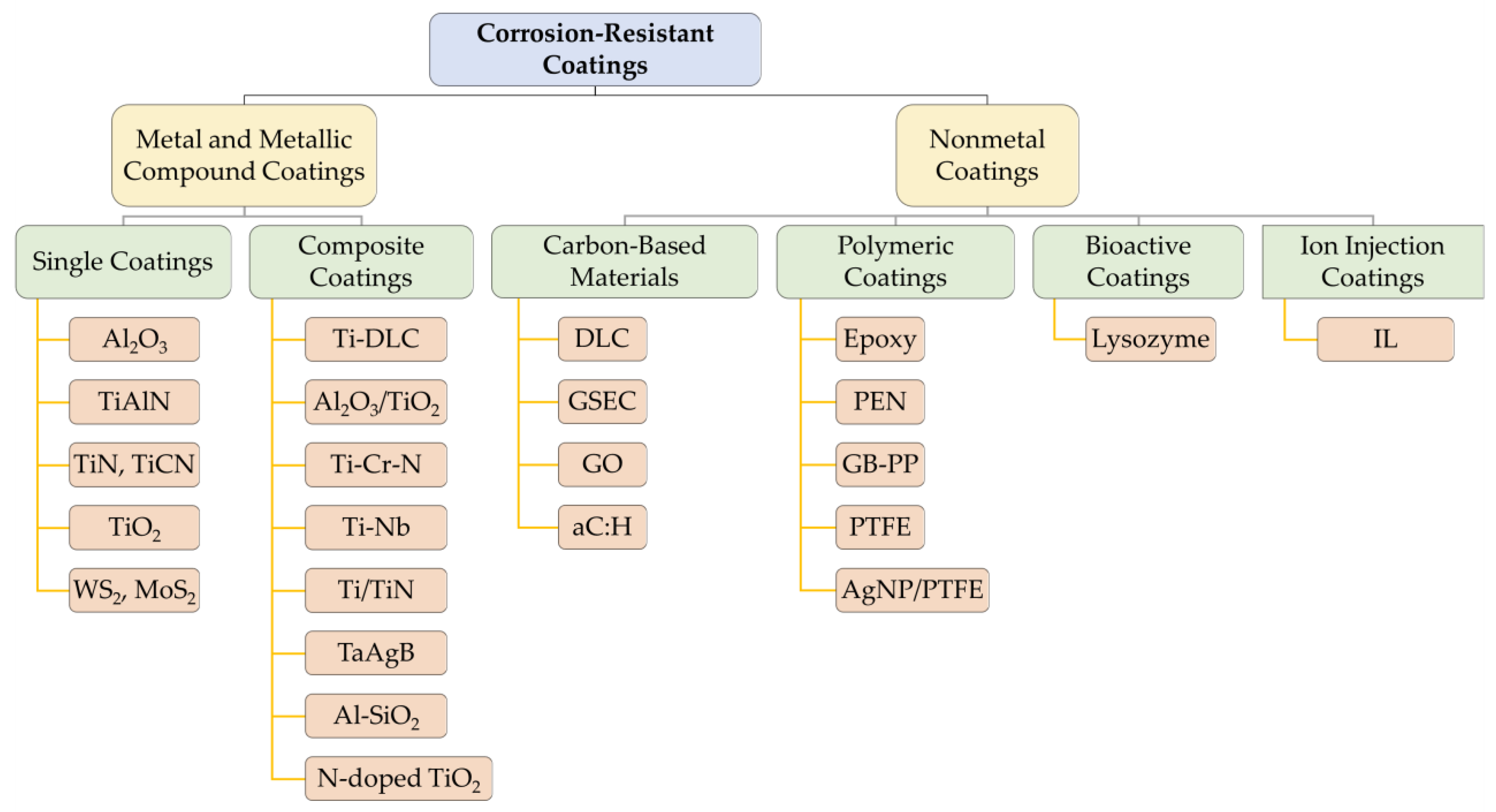
Existing orthodontic friction reduction coatings can be divided into two major categories—metallic and non-metallic—and each major category can be divided into various single coatings and composite coatings. Among metal and metallic compound friction reduction coatings, tungsten disulfide (WS2) was the first to be applied [22]. Since then, silver (Ag) coatings and metallic compound coatings, such as zinc oxide (ZnO) [23,24,25,26,27], titanium nitride (TiN) [28,29,30,31,32,33], and aluminum oxide (Al2O3) [30], have also received good attention. Nonmetals, including carbon-based materials [34], polymers [35,36,37,38,39], and bioactive materials [40], are relatively new coating materials. In addition, preparing nanoparticles and depositing them on orthodontic attachment surfaces via magnetron sputtering [30,33], evaporation [41], and immersion are also common techniques. These materials play an antifriction role mainly by filling the grooves on the surface of the appliances and forming a lubricating layer, which effectively reduces the dynamic and static friction between the archwire and the brackets [42]. The main friction reduction coating materials discussed in this section are shown in Figure 2.

2.1. Inorganic Fullerene-like Nanoparticles of Tungsten Disulfide Coatings

A. Katz et al. used nickel-phosphorus (Ni-P) electroless films impregnated with inorganic fullerene-like nanoparticles of tungsten disulfide (IF-WS2) to coat stainless steel (SS) archwires and showed that the IF-WS2 coating significantly reduced archwire friction and mitigated adverse complications [22]. Similarly, the results of M. Redlich et al. showed a 54% reduction in the friction of the coated archwire compared to the uncoated SS archwires [42]. However, despite the biocompatibility of IF-WS2, its cytotoxicity has not been studied in detail [42], which may have a negative impact on the organization. In 2019, a study applied molybdenum disulfide (MoS2) and tungsten disulfide (WS2) coatings to orthodontic SS archwires via electrodeposition and found that both materials reduced the friction between the archwires and the brackets under dry conditions [18]. However, when fixed appliances are placed inside the oral cavity, the environment to which they are exposed is wet. The wettability and durability of WS2 and MoS2 has been discussed in other fields [43,44,45], which needs to be taken into account when considering their application in the dental field. Therefore, the ability of MoS2 and WS2 coatings to reduce friction on archwires and brackets in wet environments—for instance, when immersed in artificial saliva or applied in animals’ oral cavities—needs to be investigated further.
WS2 nanoparticles can form a homogeneous and thickness-appropriate coating on the surface of the archwires and brackets; they can also have a similar effect in combination with Ni and Ni-P [22]. The film of nanoparticles peels off when exposed to friction, forming a solid lubricant film between the interfaces, thereby reducing the friction—especially when the pressure is high [42]. Therefore, WS2 coatings are more suitable apply during the alignment process where the contact angle is large, i.e., the tipping and uprighting type of tooth movement [22]. Still, it is worth noting that large-angle bending can damage the coating and, therefore, WS2 is not suitable for large-angle bending in clinical applications [42,46]. In addition, it has demonstrated good biocompatibility in preliminary studies, but its cytotoxicity is still unproven, and there are no experiments simulating the intraoral environment, so subsequent studies need to focus on these aspects.

2.2. Zinc and Zinc Compound Coatings

ZnO is of interest because of its low toxicity and good biocompatibility. A human safety review showed that nanostructured ZnO is safe for humans [47], and its suitability for biomedical applications has led to an increasing number of studies on ZnO nanocoatings for reducing friction on fixed appliances. Mojghan Kachoei et al. conducted an in vitro study and found that coating SS archwires with ZnO nanoparticles resulted in a 39–51% [23] (using deposition-precipitation) or 64% [24] (using the sol-gel technique) reduction in friction force on the coated archwires compared to uncoated archwires, and this positive effect could be retained under variations in the angle between the slot and the archwire (0–10°) [23]. Another experiment showed an even better result: when the angle increased, its ability to reduce friction force was enhanced [25]. However, a similar study conducted by Ahmad Behroozian et al. [26] showed that there was no significant reduction in friction force after coating ZnO nanoparticles on SS archwires, but coating ceramic brackets with ZnO nanoparticles could reduce friction force. More interestingly, having both SS archwires and ceramic brackets coated with ZnO nanoparticles did not contribute to the reduction in friction force [26]. This phenomenon hints that when using SS archwires and ceramic brackets as a pair, there is no need to apply ZnO coatings on both the archwires and the brackets, as this would increase the cost and decrease the friction-reducing effect. In addition to SS, nickel titanium (NiTi) archwires also need reduced friction due to their wide usage. Coating ZnO nanoparticles on NiTi archwires using the chemical deposition method reduces surface friction by nearly 21% [25], while using electrochemical deposition reduces friction force by 34% [27].
ZnO NPs not only provide good biocompatibility and antibacterial properties, but also have better friction reduction than compact ZnO [23], so they should be prepared in the form of nanoparticles when the ZnO coating is performed. In addition, ZnO coatings prepared using the sol-gel technique can be applied chairside [24], making this one of the most feasible preparation methods in the clinic. Currently, many studies have demonstrated its ability to exert good friction reduction on 0.019 × 0.025 SS archwires [23,26]. In fact, it has worked well in various sizes and materials of archwires and brackets, including ceramic brackets, while its utility in self-ligating brackets has not yet been determined. However, pairing ZnO-coated SS archwires with ZnO-coated ceramic brackets is not recommended, as it is not effective in reducing friction [26].
The use of physical vapor deposition (PVD) for zinc (Zn) plating on the surface of SS archwires is thought to significantly reduce surface friction. Whether the angle between the archwire and the slot is 0° or 10°, the change in angle does not significantly affect this result [23]. In addition, tensile strength and three-point bending strength also increase significantly after Zn plating, indicating that Zn plating has a good effect on changing the mechanical properties of SS archwires and is one of the potentially available materials [48]. One of the advantages of Zn coating over ZnO coating is that fewer cracks are formed, and the mechanical properties are better maintained after bending, which indicates that it can be used in clinical situations where the archwire is to be bent at large angles.

2.3. Zirconia Compound Coatings

In clinical medicine, zirconium oxide (ZrO2) nanoparticle coatings are used on a variety of implant surfaces, such as artificial joints, skin implants, interventional catheters, and other surgical instruments, due to their high stability, corrosion resistance, and excellent biocompatibility [49,50,51]. Therefore, their use in orthodontic appliances has been investigated for performance improvements. However, among the NiTi, SS, and beta-titanium (TMA) archwires, ZrO2 could only be applied to the TMA wire, and no coating was observed on the other two archwires using scanning electron microscopy (SEM) [52]. Moreover, ZrO2 on the surface of TMA archwires did not have a significant effect on friction reduction, although it increased the surface smoothness of the archwires [52]. This differs from the outcomes of orthopedic implants studied in another field, which applied zirconia nanomaterials to titanium alloys and obtained lower friction [53]. This alloy has a different composition from TMA archwires, which could be the reason for the different results. Therefore, the composition of the substrate also needs to be taken into consideration when selecting the coating material. Based on the above conclusions, the clinical application of ZrO2 coatings in orthodontic appliances is of low value.

2.4. Titanium and Titanium Compound Coatings

The use of titanium compound coatings in orthodontics is becoming more widespread. Ion-plated TiN coatings have the characteristics of high hardness, wear resistance, corrosion resistance, and surface lubrication. They can form a passivated titanium dioxide (TiO2) layer on their surface, so TiN coatings are commonly used on various dental instruments and materials [54,55,56]. In an earlier study, the friction force of TiN-coated SS brackets coated via the hollow cathode discharge method and of uncoated SS archwires before and after corrosion in fluorinated solutions was investigated. It was found that the friction force of the coated brackets was not significantly lower than that of the uncoated brackets, whether before or after corrosion [28]. It has also been shown that TiN coated via ion beam-assisted deposition (IBAD) on SS materials can even increase the friction force [29]. Interestingly, a recent study by Arici N. et al. concluded that coating TiN using radio frequency magnetron sputtering on SS brackets can reduce the coefficient of friction (CoF) by 50% when applied in combination with uncoated SS archwires, while the combination of TiN-coated archwires and uncoated brackets increases the CoF whether the material of the archwires is NiTi or SS [30]. That is, when TiN is coated on SS brackets and archwires, there can be different influences on friction force. The different conclusions might be related to different coating methods or other conditions. Thus, more research is needed in order to determine and verify the reasons.
The effectiveness of TiN in reducing friction on the surface of SS archwires and brackets is still controversial. However, based on some of the studies where it was considered effective, it is easy to see that its clinical application requires pairing with specific brackets or archwires. For example, uncoated brackets are not recommended when TiN-coated archwires are used [30]. This suggests that attention should be paid to the application of TiN coatings when selecting brackets and archwires.
The current trend tends to combine TiN with other application materials. TiN-derived coating materials such as titanium carbonitride (TiCN) [31,32], multilayer titanium nitride/titanium (TiN/Ti) [33], and nickel-titanium-molybdenum (Ni-Ti-Mo) composite coatings [57] not only have a more exact friction reduction effect than TiN, but are also more beneficial to clinical application due to other advantages.
In a recent study, Suciu V et al. used direct-current (DC) reactive magnetron sputtering to cover the surface of SS archwires with TiN and TiCN films and varied the ambient nitrogen content during the film fabrication process to investigate the effects of different nitrogen contents on the properties of the produced films. They found that the higher the nitrogen and carbon contents, the lower the CoF. In particular, the TiCN coating could reduce the CoF to 0.30 and make the CoF more stable, while having better hardness and corrosion resistance than TiN. Therefore, overall, the TiCN film had better surface properties than TiN in the tested samples. More interestingly, the researchers also found that the colors of TiN and TiCN films have decorative aesthetic properties, so these two coatings may be among the future directions for the development of aesthetic friction reduction archwires [31]. Similarly, Jie Zhang et al. found that TiN and TiCN coatings on SS surfaces can reduce surface friction, especially in artificial saliva [32]. Liyuan Sheng et al. prepared different layers of TiN/Ti multilayer coatings on titanium-aluminum-vanadium alloy with 6% aluminum and 4% vanadium (Ti6Al4V) substrates with the same deposition time via high-power direct-current reactive magnetron sputtering, and they found that the CoF of 1- and 2-layer TiN/Ti multilayer coatings maintained an increasing trend, but the CoF of 4-, 8-, and 12-layer TiN/Ti multilayer coatings showed a decrease [33]. Thus, TiCN is superior to TiN in terms of friction reduction, hardness, corrosion resistance, and aesthetic properties, offering better clinical prospects. For multilayer coatings, researchers also found that in order to obtain the best clinical results, it is necessary to restrict the TiN/Ti coatings to no more than two layers.
The above studies are all about SS archwires and brackets, while there are relatively few studies about titanium compound coatings on orthodontic archwires made from other materials. A study by Haruki Sugisawa et al. showed that TiN coatings can reduce the friction of NiTi and SS archwires, increase the tensile strength and stiffness of SS archwires, and reduce the elasticity of NiTi archwires [58]. In addition, Vinod Krishnan et al. attempted to apply a titanium aluminum nitride (TiAlN) coating to β-titanium orthodontic archwires using PVD to investigate its effect on friction, but the results showed no significant reduction compared to uncoated archwires [59]. Not many further studies on the usage of TiAlN coatings in orthodontic appliances have been followed up. In addition, a recent study investigated TiO2 and silicon dioxide (SiO2) nanoparticles on the surface of chromium-nickel (Cr-Ni) archwires and found that TiO2 did not show a significant ability to reduce friction [60]. Conversely, SiO2 can be used to reduce friction between the brackets and archwires under both dry and wet conditions, while making the surface of the archwires smoother than uncoated ones [60]. A systematic review and meta-analysis suggested that nanoparticle-coated orthodontic archwires can be considered to significantly reduce frictional resistance [61]. In this regard, it can be presumed that TiO2 nanoparticles are also theoretically able to reduce the friction force. In fact, the reason for the inability of TiO2 nanocoatings to significantly reduce friction may be that the coating is thicker than other coatings [60]. Therefore, the pressure on the inner wall of the slot is greater, resulting in greater friction force. In addition, the deformation and peeling of the surface morphology are also among the reasons [62]. Similarly, the application of TiO2 coatings on SS brackets using PVD does not reduce friction [63].
Therefore, the TiO2 coating may not exhibit the desired ability to reduce friction, because of its thickness [60]. However, is still worth developing as a very versatile material and, therefore, has a certain research value. In future research, other coating methods to reduce the thickness could be considered. Based on the consideration of film thickness, the preparation of a thinner film of TiO2 nanocoatings could be attempted in future studies. Recent studies have proven that low-pressure metal-organic chemical vapor deposition (LPMOCVD) can control the film thickness by adjusting the deposition time [64]. In fact, this method of controlling the thickness of the coating can be applied to many deposition-based coating methods; thus, future research could take it into consideration when finding the most suitable coating conditions.
In addition to titanium compounds, composite coatings of titanium with other metals are also being investigated. In the study of Yılmaz H et al. (2021), nickel ions and TiO2 nanoparticles were used as standard coating materials on the surface of NiTi archwires, while the addition of 0.0015 g/dm3 sodium molybdate (Na2MoO4) on this basis led to the formation of a Ni-Ti-Mo coating on the surface of the archwires; thus, a reduction in the CoF from 0.288 to 0.252 was observed. However, although the addition of a certain amount of molybdenum ion (Mo6+) to the bath solution can reduce the surface friction of the archwires after the formation of the coating, the reduction in surface friction gradually becomes less effective as the concentration of Mo6+ increases. The standard NiTi coating does not reduce friction, and the addition of different concentrations of chromium (Cr) to the standard coating material to form a nickel-titanium-chromium (Ni-Ti-Cr) coating also does not reduce friction. [57]
In summary, the trends of Ti compound coatings are nitriding [30], incorporation of other metal ions [57], multi-layered materials [33], and low-thickness materials [64]. Most of these are still some distance away from being applied in clinical practice, but attempts can be made to find the most suitable preparation conditions and methods to obtain coating materials with satisfactory properties in all respects. The biggest disadvantage of this type of coating, however, may be its high price [65]. If the price of orthodontic supplies is increased by the application of the coating, then there is no advantage compared to other coatings with similar properties, and patients are less likely to choose to use these materials. Therefore, although the materials are excellent in various respects, in order to put them into use, there is still a need to find ways to reduce the cost of their production.

2.5. Silver and Silver Compound Coatings

Nanosilver coatings can reduce the surface roughness of SS brackets [63] or NiTi archwires [66], but the change in friction between brackets and archwires is not obvious [63]. Moreover, silver (Ag) plating on 0.019 × 0.025-inch coated SS archwires significantly reduces the friction between the brackets and the archwires, while 0.017 × 0.025-inch SS archwires do not significantly reduce the friction [67]. The 0.019 × 0.025-inch SS archwires, whether coated or not, have higher friction than the 0.017 × 0.025-inch SS archwires. The play between 0.019 × 0.025-inch SS archwires and the slots is smaller, so the pressure is higher when the slots are of the same size. After applying the coating, the friction coefficient of the surface should be the same regardless of the size of the archwires, so the alteration of the friction force of the archwires subjected to more pressure may be relatively more obvious when the other conditions are the same [67]. However, this explanation has not yet been clearly confirmed. Therefore, more research is still needed in order to confirm its capabilities. Still, considering the antibacterial properties of silver [63], nanosilver coatings are still a popular research topic for surface coatings of orthodontic appliances, even without clear friction reduction properties.
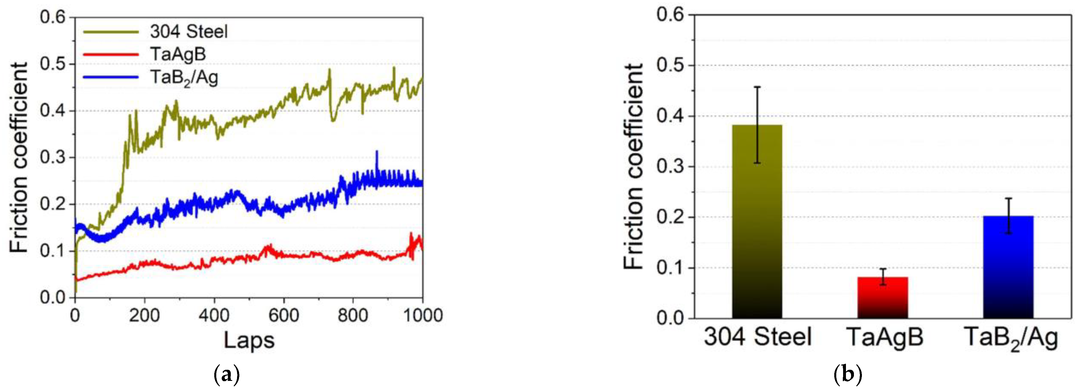
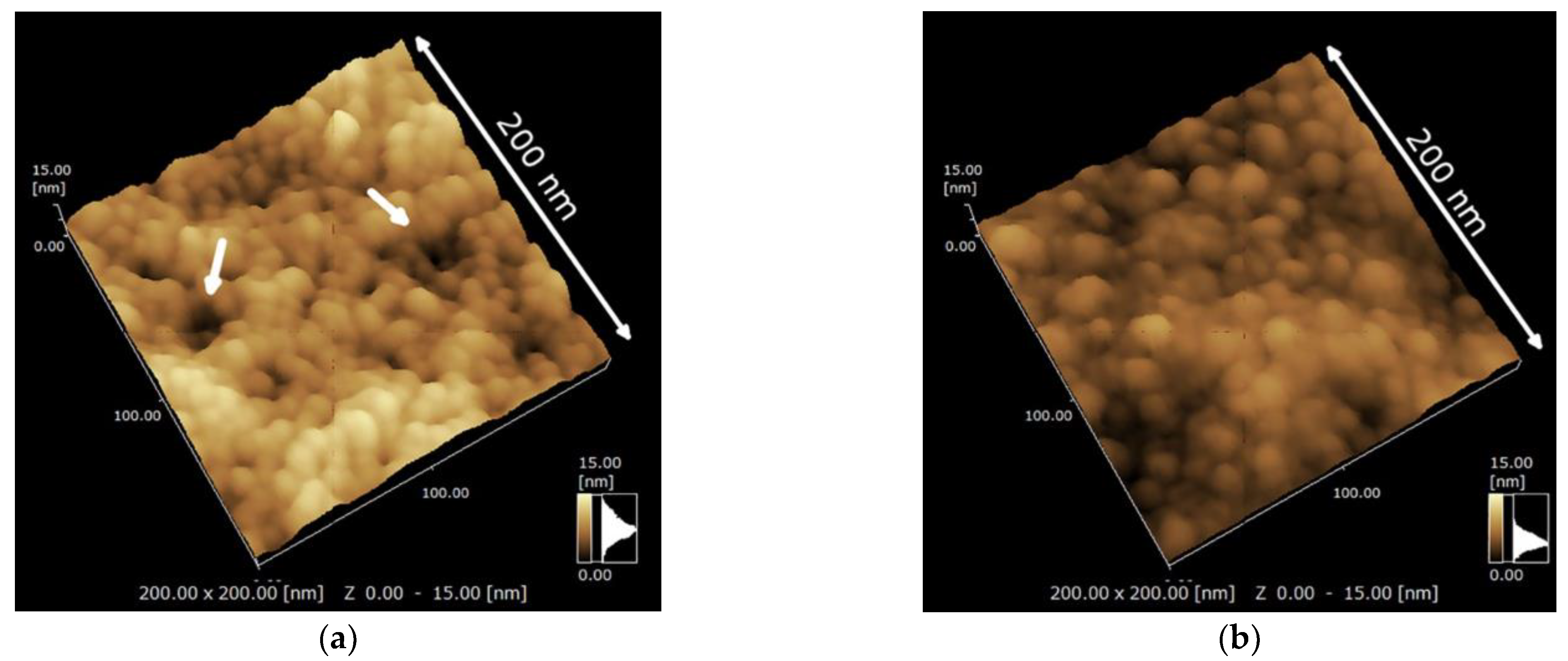
Currently, there are only a few types of materials related to silver used in the surface coating of orthodontic appliances, which are generally silver nanoparticles (AgNPs). Interestingly, Jia Wang et al. [68] successfully prepared silver-doped tantalum boride (TaAgB) solid solution coatings on SS blocks using magnetron sputtering technology with Ag doping. They verified the effectiveness of the coating in reducing the CoF of the SS material to 0.08, as shown in Figure 3, and suggested that it could be used for orthodontic archwires and brackets because of its good hardness, strength, and biocompatibility. Therefore, as a surface coating material with low friction, good antibacterial properties, high wear resistance, and high corrosion resistance, it has strong potential value for application on fixed appliances.
As a precious metal, silver coatings are also a relatively high-value material. However, nanosilver coatings have multifunctional properties that may be accepted by the general public even if their price is high [69]. Therefore, it is necessary to develop more friction reduction, antibacterial, and other properties of nanosilver coatings.

2.6. Aluminum and Aluminum Compound Coatings

Al2O3 coatings are oxide coatings that are common in various fields. Research by Nursel Arici et al. [30] in 2021 demonstrated the friction reduction effect of Al2O3 coatings in detail. When an Al2O3 coating is applied on the surface of SS brackets, whether the material of the combined archwire is made of SS or NiTi, and regardless of whether the archwires have an Al2O3 coating or not, the CoF can be reduced significantly. Among all combinations, the combination of Al2O3-coated SS brackets with the same coated archwire was able to obtain the lowest CoF. That is, the combination of coated SS brackets-coated SS archwire is better than other combinations. It is also worth mentioning that in almost all cases involving coated archwire-uncoated bracket combinations, an increase in CoF was observed for both NiTi and SS archwires, being much higher than for both uncoated combinations. This phenomenon may be related to the peeling of the coating [30]. Therefore, when testing friction, the pressure of the coating environment, time, immersion conditions, and peeling should also be taken into account to obtain more accurate conclusions.
Others have combined aluminum (Al) and SiO2 to form Al-SiO2 composite coatings. The excellent friction reduction ability of SiO2 has been mentioned above [60]. In contrast, the friction reduction properties of Al have not been adequately studied. A reduction in the CoF is observed in Al-SiO2-coated NiTi and SS archwires compared to the uncoated ones [70]. Therefore, Al-SiO2 composite coatings may also be used to reduce the friction of orthodontic archwires in the future.
Al compound coatings are effective in reducing friction, retain high aesthetic value, and are relatively inexpensive [30]. However, studies about them are still scarce, especially since their performance changes after long-term use are not confirmed. In addition, Al2O3 needs to be considered in clinical applications for the combination of coated and uncoated archwires and brackets. Combining coated archwires with uncoated brackets is not recommended, but it is possible to use coated brackets only [30]. Ideally, both the brackets and the archwire should be coated, which will result in the best friction reduction.

2.7. Other Metal and Metallic Compound Coatings

Nursel Arici et al. [30] noted that the CrN coating was ineffective in reducing friction between the SS brackets and the SS or NiTi archwires, whether both were coated or only one was coated. SEM images after friction tests showed that the CrN coating peels off from the surface. Therefore, residual coating material at the contact area between the surfaces may be responsible for the increase in CoF. This phenomenon is not found in the application of hard chrome carbide plating (HCCP) coatings. HCCP coatings significantly reduce the friction of SS archwires, and mechanical properties such as flexural strength and flexural modulus remain unchanged [21]. HCCP is a promising coating material that can potentially be used clinically to reduce friction. However, its shortcomings include aesthetics that need to be improved and biocompatibility that has not been explicitly tested.
Another common metal coating is rhodium. As a coating based on aesthetic considerations, it is generally applied to archwires. Some recent studies have begun to explore its usage beyond aesthetics. A study by Tahereh Hosseinzadeh Nik et al. found that the frictional force generated during sliding between ceramic brackets and rhodium-plated SS archwires was significantly less than that of ceramic brackets and SS archwires. Even after immersion in 0.05% sodium fluoride (NaF) mouthwash, the increase in friction was much less in the former than in the latter [71]. This indicates that rhodium plating can be used not only as a method to increase the aesthetic effect of the archwires, but also to reduce the friction between the archwires and the brackets, constituting a method to increase the efficiency of the orthodontic treatment and maintain good performance even when the patient uses NaF mouthwash. In addition, they demonstrated that rhodium-plated SS archwires showed a smaller increase in roughness than uncoated SS archwires after treatment in mouthwash [71]. Of course, the correlation between surface roughness and friction remains controversial. Some think that the two can be directly correlated [72,73], while others do not [35,74], so it is not yet possible to conclude that the smaller increase in friction of rhodium-plated archwires is related to the smaller increase in surface roughness. Friction-reducing coatings made of other metallic monomers are also gradually being investigated. Recent studies have attempted to prepare niobium (Nb), tantalum (Ta), and vanadium (V) coatings on 316 SS substrates using the plasma sputtering method and found that V coatings have an effect on the roughness of the SS surface [75]. However, other experiments on the tribology of these new materials have not yet been conducted.

2.8. Carbon-Based Coatings

Carbon-based coating materials mainly contain diamond-like carbon (DLC) and graphene.
DLC is a diamond-like carbon film that exhibits extremely high hardness, a low coefficient of friction, chemical inertness, and high corrosion resistance [34]. Research has demonstrated the biomedical applications of DLC films. For example, implants with protective films can extend the life of the implant by reducing corrosion and wear, which can benefit patients [76].
DLC-coated SS brackets exhibit lower static [77] and dynamic friction [78], and their effect is better than that of TiN-coated brackets [79]. However, some studies have also found no reduction in friction on the surface of DLC-coated SS brackets [80]. In addition to brackets, DLC-coated SS archwires [78,80,81,82] or NiTi archwires [83,84] can be applied to reduce friction. Hao Zhang et al. concluded that the use of the plasma-enhanced chemical vapor deposition (PECVD) method to cover the surface of SS archwires with a DLC coating can reduce the coefficient of dynamic friction by 40.71% and significantly improve the surface hardness of the archwire, as well as reducing friction and exhibiting good biocompatibility [82]. It has also been shown that the reduction in friction is effective whether the angle between the archwire and the bracket is 0° or 10° [81].
Furthermore, the application of DLC via different coating methods—such as plasma-enhanced chemical vapor deposition (PECVD) [79,82,85], plasma-based ion implantation/deposition (PBIID) [81,84,86], and mirror confinement electron cyclotron resonance (MCECR) plasma sputtering [80,83]—has shown similar friction-reducing effects. It has also been shown that for DLC-coated SS brackets, the size and material of the archwire, the contact angle, and the dryness or humidity of the experimental conditions have no influence on the friction reduction effect of DLC coatings [77]. However, although changes in the above conditions have no significant influence on the effect of DLC coatings in reducing friction, the effect of the thickness of the coating is still not negligible. Muguruma et al. [78] investigated the mechanical properties of DLC coatings of different thicknesses. They found that a thin DLC coating reduces surface friction, which is the same result as previously described, yet the relatively thick DLC layer tends to break from the DLC-steel interface. In addition, thicker layers have a negative effect on friction force. DLC coatings also have the advantage of being resistant to high-fluorine environments. Even at high fluoride ion concentrations, the friction force remains relatively stable, i.e., there is no significant increase [83].
DLC coatings with different properties were prepared under different conditions as DLC-1 and DLC-2. DLC-1 uses a 10 kV target voltage, an acetylene-toluene gas environment, and deposits for 3 min to form a structure with a diamond-rich outer surface and a graphite-rich inner surface; DLC-2 uses a 7 kV target voltage, a toluene gas environment, and deposits for 4 min to form a structure with a graphite-rich outer surface and a diamond-rich inner surface. The results showed that DLC-2 produces significantly lower friction than the uncoated case when wet and at an angle of 10°, which was not observed for DLC-1. The reasons for this were that partial cracks in the DLC-1 coating led to increased friction, while no cracks or breakages of the coating were observed in DLC-2. Therefore, we can conclude that the DLC-2 coating is more valuable than DLC-1 in practical applications because of its good flexibility and adhesion [87].
In addition to pure DLC, fluorine-doped DLC (F-DLC) and silicon-doped DLC (Si-DLC) coatings can also reduce friction on the surface of SS brackets. The F-DLC-coated brackets exhibit low static friction between the brackets and the archwire under moist conditions, which is lower than that under dry conditions. However, doping DLC with fluorine or silicon results in a significant reduction in surface hardness. Even though F-DLC is most effective in reducing the friction between the archwire and the brackets in humid conditions, this disadvantage needs to be taken into account in practical applications [85]. Titanium-doped DLC (Ti-DLC) coatings are also ideal friction-reducing films for orthodontic brackets, as they exhibit a lower CoF than TiCN and TiN in most cases, under both artificial saliva and dry conditions [32].
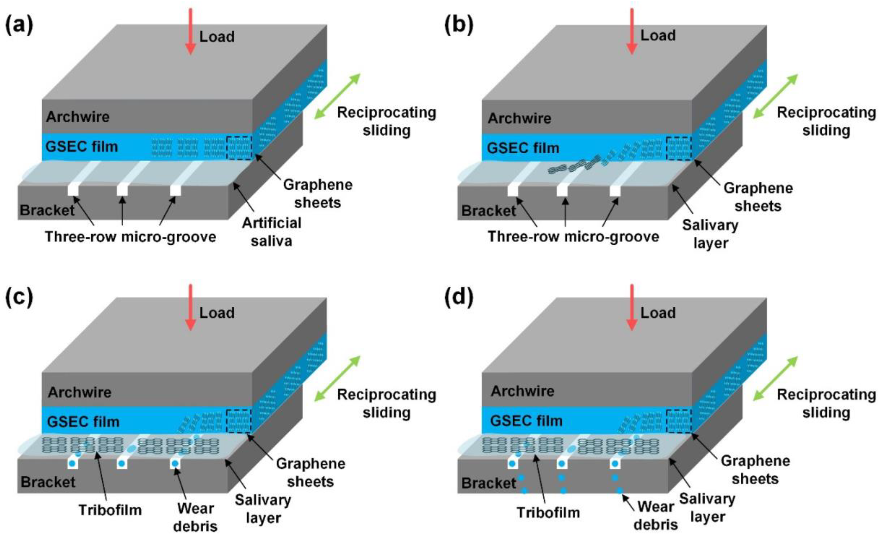
Recently, research on graphene coatings has gradually emerged. Commonly used graphene materials include graphene oxide and graphene-sheet-embedded carbon (GSEC). Graphene oxide (GO) coatings can reduce the CoF of NiTi alloy [88,89], while the addition of AgNPs and the formation of graphene oxide/silver nanoparticle (GO/Ag) coatings can further reduce CoF [88]. In artificial saliva, the GSEC surface coating of SS archwires coated with MCECR reduced the CoF to 0.12 [90]. Pengfei Wang et al. later discovered ways to make the friction on the surface of GSEC coatings even lower. In this study, they found that the thickness of the coating increased as the GSEC film deposition time increased. After 80 min of deposition, both stable and low friction coefficients could be obtained, even reaching a level of less than 0.10 when running against three-row microgroove-textured SS brackets in an artificial saliva environment [46]. The possible mechanisms are shown in Figure 4. Although immersion causes a rebound in CoF, after 30 days of immersion, the CoF remains below 0.30, consistently exhibiting very good friction reduction [90]. This illustrates the effectiveness of the graphene coating in reducing the friction on the surface of the archwire.
In summary, carbon-based friction-reducing coatings have good overall durability and biocompatibility [82], and DLC coatings are resistant to corrosion by fluoride-containing mouthwashes [83]. The coating method and conditions are less restrictive compared to metal coatings, but there are strict requirements for the thickness of the coating layers, requiring a thickness that is both thin and effective [87]. GSEC coatings can be used for up to one month, and the optimal coating deposition length has been determined and may be ready for clinical application soon.

2.9. Polymeric and Bioactive Coatings

Teflon (polytetrafluoroethylene (PTFE)), an aesthetic white coating for dental instruments, has been shown to provide a smoother surface and reduce frictional resistance and frictional damage [35,36,37,38,39] on NiTi [91] or SS archwires [92]—especially when paired with metal slots in ceramic brackets [38]—in both dry and wet conditions. However, when combined with ceramic brackets, PTFE- and epoxy-coated NiTi archwires will increase the friction between the archwires and the brackets [93]. This is probably because ceramics have a rougher texture and a more porous surface than stainless steel. Another research shows the contrast, which whether combined with ceramic or metal brackets, PTFE-, or epoxy-coated SS archwires always show higher friction resistance [94]. However, the latest study by K. Ranjan and R. Bhat et al. contradicts this finding; the study concludes that whether paired with SS brackets, ceramic brackets, or ceramic brackets with metal slots, the PTFE-coated archwire produces lower frictional resistance compared to uncoated SS archwires [92]. In addition, research has investigated suitable preparation conditions. PTFE coatings prepared at 200 °C offer lower friction, higher wear resistance, lower coloration, and higher resistance to microbial adhesion than those prepared at 380 °C [95].
In addition to PTFE, other polymer surface coatings have been used to explore the ability to reduce friction on fixed appliances, such as chitosan (CTS) [24], 2-methacryloyloxyethyl phosphorylcholine (MPC), parylene, and epoxy.
Elhelbawy N. et al. investigated CTS as a newer and potentially useful coating material for reducing friction on fixed appliance surfaces in comparison to ZnO. This comparison revealed no significant difference in the degree of friction reduction between CTS and ZnO (64 and 53%, respectively). Interestingly, when CTS and ZnO were applied to the brackets or archwires alone, and the matching archwires or brackets were not coated, the degree of friction reduction was similar to that of having both brackets and archwires coated with CTS or ZnO, suggesting that coating both the brackets and the archwires did not significantly improve this aspect of friction reduction [24].
In a recent study, Ryo Kunimatsu et al. found a promising application of MPC in fixed appliances. They covered SS archwires with an MPC coating and performed tensile tests using a bracket-archwire combination to demonstrate the coating’s ability to reduce friction. Furthermore, they directly measured the efficiency of tooth movement using an in vitro experimental tooth movement model at 50 g and 100 g traction force, and finally confirmed that the SS archwire coating with MPC improves the efficiency of tooth movement by reducing the occurrence of friction [96]. This experimental design has provided ideas for subsequent studies. Simply confirming that a certain surface coating can reduce friction is not enough to fully prove its clinical value. Its positive effect on improving tooth movement efficiency needs to be further determined through model experiments, animal experiments, and even clinical trials.
Chin-Yu Lin et al. [97] coated SS archwires with parylene, epoxy, or PTFE and tested the friction between the archwires and ceramic brackets after immersion in water for different lengths of time. Compared to the other groups, the epoxy-coated archwires presented higher or no significant difference in friction resistance and were the only coated archwires whose maximal resistance to sliding at a 3° contact angle (MRS3) was consistently lower than that of the uncoated control group. Parylene, on the other hand, had relatively high friction at all water immersion times (0 to 4 weeks) and angles (0 to 3°). Therefore, epoxy-coated archwires may even be a better choice than PTFE.
In addition to polymers, bioactive glass (BG) coatings have recently been prepared on the surface of SS archwires using electrophoretic deposition. Although BG has higher hardness, elastic modulus, and surface smoothness, it failed to reduce the friction in the experiment [40]. Overall, studies on the application of bioactive coatings on fixed appliances are extremely rare and could perhaps be explored more in the future.
Carbon-based materials [82], natural polymeric materials [24], and bioactive materials [40] are safer than metal and metallic compound materials and can be used to replace other coating materials that are potentially cytotoxic [42]. CTS coatings can replace ZnO and are relatively more convenient and cost-effective, as they do not require coating on both the archwire and the bracket surface [24]. Aesthetic coatings such as PTFE are suitable for both SS and NiTi archwires, but when used with ceramic brackets, it is better to choose ceramic brackets with metal slots [92] and other commonly used polymeric aesthetic archwires. Among the other common aesthetic polymer coatings on the market, only epoxy shows a stronger friction reduction effect than PTFE [97]. However, the popularity of its use and the difficulty of its preparation have not been conclusively proven, so the most suitable polymer aesthetic coating for clinical friction reduction is still PTFE.
Recent studies on friction reduction coatings for orthodontic appliances are summarized in Table 1.
Table 1. Recent studies on friction reduction coatings for orthodontic appliances.
Ref.Coating
Materials
Coating
Technique
SubstrateStudy TypeRoughnessFriction Reduction EffectivenessWear
Mechanism
Wear
Resistance
Other
Effectiveness
[67]AgDirect current sputteringSS archwiresIn vitro (dry condition)NACompared to uncoated archwires, the friction force of 0.019 × 0.025-inch SS archwires reduced 1 N after coating (p = 0.032), but no significant difference between coated and uncoated 0.017 × 0.025-inch SS archwires (p = 0.854).NANANA
[30]Al2O3Radio frequency magnetron sputteringSS brackets, NiTi archwires, SS archwiresIn vitro (dry condition)Surface roughness decreased with the coating process.
Ra = 331.53 nm, Rq = 426.17 nm (coated brackets);
Ra = 361.64 nm, Rq = 466.01 nm (coated NiTi archwires);
Ra = 95.86 nm, Rq = 128.01 nm (coated SS archwires)
NiTi archwires: When both brackets and archwires were coated and only brackets were coated, CoF reduced from 0.316 to 0.238 and 0.251, respectively. CoF increased from 0.316 to 0.400 when only archwires were coated.
SS archwires: When both brackets and archwires were coated, only brackets were coated, and only archwires were coated, CoF reduced from 0.552 to 0.227, 0.2,35, and 0.445, respectively.
No peeling offThe coatings did not peel off after friction, thermal, and brushing tests.NA
[70]Al-SiO2Magnetron sputteringNiTi and SS archwiresIn vitro (in artificial saliva)NACoF of NiTi archwires and SS archwires reduced from 0.68 to 0.46 and from 0.58 to 0.45, respectively.NACorrosion-resistantCorrosion-resistant effectiveness
Biocompatibility
[40]BGElectrophoretic depositionSS archwiresIn vitro (dry condition)Sa increased from 0 to 0.46–0.79.Friction forces showed no significant reduction.NANone of the coatings were damaged after three-point bending and they sustained good interfacial adhesion.Aesthetic effect
[84]CF4Plasma-based ion implantation and depositionNiTi archwiresIn vitro (dry condition)NAFriction forces reduced from 129.48 to 104.97 gf, which showed no significant reduction.NANANA
[84]CH4Plasma-based ion implantation and depositionNiTi archwiresIn vitro (dry condition)NAFriction forces reduced from 129.48 to 87.30 gf, which showed no significant reduction.NANANA
[29]CNxIon beam-assisted deposition304L SS disksIn vitro (friction test: NA; antibacterial test: in bacterial suspension)The films slightly reduce the surface roughness parameter. Ra reduced from 0.181 to 0.140 μmCoF reduced from 0.431 to 0.188 (p < 0.05).NANAAntibacterial Effectiveness
Biocompatibility
[30]CrNRadio frequency magnetron sputteringSS brackets, NiTi archwires, SS archwiresIn vitro (dry condition)Surface roughness decreased with the coating process.
Ra = 276.85 nm, Rq = 360.24 nm (coated brackets);
Ra = 354.35 nm, Rq = 454.66 nm (coated NiTi archwires);
Ra = 190.28 nm, Rq = 229.26 nm (coated SS archwires)
NiTi archwires: When both brackets and archwires were coated, only brackets were coated, and only archwires were coated, CoF increased from 0.316 to 0.443, 0.324, and 0.505, respectively.
SS archwires: When both brackets and archwires were coated and only archwires were coated, CoF increased from 0.552 to 0.598 and 0.586, respectively. CoF reduced from 0.552 to 0.410 when only brackets were coated.
Peeling offLarge areas of peeling could be seen after friction, thermal, and brushing tests.NA
[24]CTS (NPs)Sol-gel dip coatingSS archwires and SS bracketsIn vitroNAFriction force decreased by ~53%.NANANA
[84]DLCPlasma-based ion implantation and depositionNiTi archwiresIn vitro (dry condition)NAThe friction force reduced from 129.48 to 86.13 gf (p = 0.039).NANANA
[87]DLCPlasma-based ion implantation and depositionSS archwiresIn vitro (in artificial saliva and dry condition)NAWhen coated with DLC-2, the static friction reduced from 2.39 to 2.09 N in artificial saliva and from 2.49 to 2.25 N under dry conditions, and the kinetic friction reduced from 2.37 to 1.99 N in artificial saliva and from 2.55 to 2.21 N under dry conditions, which showed a significantly lower frictional force than the uncoated archwires, while DCL-1 showed no significant difference compared with uncoated samples.RuptureNo rupture was observed for the DLC-2 condition after the drawing-friction testing.NA
[97]EpoxyNA (commercial)SS archwiresIn vitro (in distilled-deionized water)NAThe average resistance under 0° bracket-wire angle reduced from 1.63 to 1.13 N immediately after being coated. The average resistance under a 3° bracket-wire angle reduced from 5.12 to 4.27 N immediately after being coated.NANADurability (>4 weeks)
[94]EpoxyNA (commercial)SS archwiresIn vitro (in artificial saliva and dry condition)NAThe friction force increased from 3.00–9.00 N to 16.00–20.50 N in both wet and dry conditions.NANANA
[93]EpoxyNA (commercia)NiTi archwiresIn vitro (dry condition)NAFriction forces increased from 49.287 to 53.316 gf.NANANA
[89]GOSilane couplingNiTi archwiresIn vitro (friction test: NA; antibacterial test: in bacterial suspension; corrosion test: artificial saliva)The surface of the samples coated with 2 mg/mL GO concentrations was smooth with a uniformly coated area.CoF reduced from ~0.9 to 0.2–0.4.Grooves in the same direction as the glidingThe samples coated with 0.5 mg/mL GO concentrations had fewer grooves, but a small amount of wear debris was present.Antibacterial effectiveness
Corrosion-resistant effectiveness
Biocompatibility
[88]GO/Ag (NPs)Electrophoretic depositionNiTi alloyIn vitro (dry condition)Ra ranged from 50.72–69.93 nm.CoF reduced from 0.060 to 0.006, and increased coating time led to lower CoF.NANANA
[46,90]GSECElectron cyclotron resonance plasma sputteringSS archwiresIn vitro (in artificial saliva)NACoF reduced to ~0.10 and remained under 0.30 after 30 days.Peeling offThe corresponding wear rate was strongly decreased from 4.84 × 10⁻⁶ to 0.11 × 10⁻⁶ mm3/Nm.NA
[21]HCCPElectroplatingSS archwiresIn vitro (in PBS solution and dry condition)There were very small protrusions on the surfaces of the coated archwires, while the surfaces of uncoated archwires were smooth.The friction force reduced from 147.15 to 124.61 gf (p = 0.0076) and from 143.55 to 121.41 gf (p = 0.04) under dry and wet conditions, respectively.ScratchesAfter the friction test, scratches were seen on the surfaces on the coated surfaces.Aesthetic effect
[18]Ni+MoS2 (NPs)Electrochemical co-depositionSS archwiresIn vitro (in artificial saliva and dry condition)NADry conditions: CoF reduced from 0.58–1.43 to 0.50–1.19.
In artificial saliva: CoF reduced from 0.95–2.52 to 0.94–2.35.
NANANA
[18]Ni+WS2 (NPs)Electrochemical co-depositionSS archwiresIn vitro (in artificial saliva and dry condition)NADry conditions: CoF reduced from 0.58–1.43 to 0.42–1.06.
In artificial saliva: CoF reduced from 0.95–2.52 to 0.66–1.46.
NANANA
[57]Ni-Ti-CrChronopotentiometryNiTi archwiresIn vitro (in artificial saliva)NACoF showed no significant reduction.NANACorrosion-resistant effectiveness
Durability
[57]Ni-Ti-MoChronopotentiometryNiTi archwiresIn vitro (in artificial saliva)NACoF reduced from 0.288 to 0.252–0.265.NANACorrosion-resistant effectiveness
Durability (>60 days)
[97]ParyleneNA (commercial)SS archwiresIn vitro (in distilled-deionized water)NAThe average resistance under 0° bracket-wire angle increased from 1.63 to 5.39 N immediately after being coated. The average resistance under the 3° bracket-wire angle increased from 5.12 to 11.38 N immediately after being coated.NANADurability
[97]PTFENA (commercial)SS archwiresIn vitro (in distilled-deionized water)NAThe average resistance under 0° bracket-wire angle reduced from 1.63 to 1.15 N immediately after being coated. The average resistance under a 3° bracket-wire angle showed no significant change immediately after being coated.NANADurability
[95]PTFESprayingSS, Ni-Ti, and β-titanium archwiresIn vitroRa of coated archwires increased from 0.02–0.21 to 0.53–0.58 μm.The friction force of coated archwires reduced from 123.94–152.61 to 102.98–124.40 gf compared to uncoated archwires and 200 °C coating resulted in less friction against brackets than did the conventional 380 °C coating.ScratchesPTFE-coating at 200 °C resulted in good microbial adhesion and tolerance of wear.Durability (>3 months)
Aesthetic effect
[94]PTFENA (commercial)SS archwiresIn vitro (in artificial saliva and dry condition)NAThe friction force was higher than uncoated archwires but lower than epoxy and rhodium-coated archwires in wet and dry conditions.
Dry conditions: The friction force increased from 3.80–9.00 to 5.50–12.80 N.
In artificial saliva: The friction force increased from 3.00–8.20 to 3.80–11.60 N.
NANANA
[93]PTFENA (commercial)NiTi archwiresIn vitro (dry condition)NAFriction forces increased from 49.287 to 61.427 gf.NANANA
[92]PTFENA (commercial)SS archwiresIn vitroThe surface roughness increased after the friction test.When compared with ceramic with a metal slot, the friction force reduced from 1.61 to 1.03 N after the archwires were coated with PTFE.NANANA
[21]RhodiumNA (commercial)SS archwiresIn vitro (in PBS and dry condition)There were protrusions on the surfaces of the coated archwires, while the surfaces of uncoated archwires were smooth.The friction force increased from 147.15 to 216.29 gf (p < 0.001) and from 143.55 to 210.21 gf (p < 0.001) under dry and wet conditions, respectively.ScratchesAfter the friction test, scratches were seen on the surfaces on the coated surfaces.Aesthetic effect
[94]RhodiumNA (commercial)SS archwiresIn vitro (in artificial saliva and dry condition)NAThe friction force increased from 3.00–9.00 N to 17.30–21.70 N in both wet and dry conditions.NANANA
[71]RhodiumNA (commercial)SS archwiresIn vitro (in artificial saliva and 0.05% NaF mouthwash)Surface roughness showed no significant reduction with the coating process.The friction force was reduced from 2.22 to 1.49 N in artificial saliva and reduced from 2.72 to 2.17 N in 0.05% sodium fluoride mouthwash.NANANA
[60]SiO2 (NPs)NA (commercial)Cr-Ni archwiresIn vitro (in artificial saliva and dry condition)Surface roughness decreased with the coating process. Ra reduced from 0.673 to 0.040 μm.Friction force reduced from 0.51 to 0.38 N and from 0.56 to 0.44 N under wet and dry conditions, respectively.NANANA
[68]TaAgBMagnetron sputtering with Ag-doping304 SS sheetsIn vitro (in artificial saliva)NACoF reduced from 0.38 to 0.08.No serious wear phenomenonThe wear rate was 6.51 × 10⁻¹⁵ m3/Nm.Antibacterial effectiveness
Biocompatibility
[32]TiCNPVD316L SS platesIn vitro (in artificial saliva and dry condition)Ra = 29.86 nm; root mean square roughness: Rq = 39.09 nmCoF reduced from 0.20 to less than 0.06 under dry conditions with loads of 5 N.NANANA
[31]TiCNDirect current reactive magnetron sputtering316L SS and (100)-oriented Si substratesIn vitro (in artificial saliva)NACoF reduced to less than 0.30, and the addition of N2 and C in the Ti matrix further lowered the CoF.NAThe lowest wear rate was 5.6 × 10⁻⁶ mm3/NmCorrosion-resistant effectiveness
Aesthetic effect
[32]Ti-DLCPVD316L SS platesIn vitro (in artificial saliva and dry condition)The surface of the Ti-DLC film has many nanocrystal clusters that cause substantial pitting and layer defects. Ra = 10.40 nm; Rq = 13.43 nmCoF remained less than 0.04 under both dry and artificial saliva conditions.NANANA
[29]TiNIon beam-assisted deposition304L SS disksIn vitro (friction test: NA; antibacterial test: in bacterial suspension)The films slightly reduce the surface roughness parameter. Ra reduced from 0.181 to 0.162 μm.CoF increased from 0.431 to 0.469 (p < 0.05).NANAAntibacterial Effectiveness
Biocompatibility
[58]TiNIon platingSS and NiTi archwiresIn vitro (friction test: NA; corrosion test: in 0.9% NaCl solution)Ra of TiN-coated SS archwire increased from 0.023 to 0.046 µm. Ra of TiN-coated NiTi archwire remained at 0.001 µm.At angles of 0 degrees, the friction forces of the TiN-coated NiTi archwires reduced, but SS archwires showed no significant difference. At angles of 10 degrees, the friction forces of the TiN-coated SS and NiTi archwires were both reduced.NANACorrosion-resistant effectiveness
[32]TiNPVD316L SS platesIn vitro (in artificial saliva and dry condition)The surface of the TiN film has some particles and pinholes. Ra = 26.59 nm; Rq = 32.62 nmCoF reduced from 0.08 to less than 0.03 under dry conditions with loads of 5 N.NANANA
[30]TiNRadio frequency magnetron sputteringSS brackets, NiTi, and SS archwiresIn vitro (dry condition)Ra = 354.12 nm, Rq = 458.74 nm (coated brackets);
Ra = 391.99 nm, Rq = 534.36 nm (coated NiTi archwires);
Ra = 105.15 nm, Rq = 132.67 nm (coated SS archwires)
NiTi archwires: When both brackets and archwires were coated, only brackets were coated, and only archwires were coated, CoF increased from 0.316 to 0.399, 0.331, and 0.446, respectively.
SS archwires: When both brackets and archwires were coated and only brackets were coated, CoF reduced from 0.552 to 0.372 and 0.237, respectively. CoF increased from 0.552 to 0.818 when only archwires were coated.
Peeling offAfter friction, thermal, and brushing tests, the coatings peeled off in some small areas.NA
[31]TiNDirect current reactive magnetron sputtering316 L SS and (100)-oriented Si substratesIn vitro (in artificial saliva)NACoF was reduced to less than 0.30, and the addition of N2 in the Ti matrix further lowered the CoF.NAThe lowest wear rate was 7.8 × 10⁻⁶ mm3/NmCorrosion-resistant effectiveness
[29]TiO2Ion beam-assisted deposition304L SS disksIn vitro (friction test: NA; antibacterial test: in bacterial suspension)NANANAThe TiO2 film wore out in a few seconds.Antibacterial effectiveness
Biocompatibility
[60]TiO2 (NPs)NA (commercial)Cr-Ni archwiresIn vitro (in artificial saliva and dry condition)Surface roughness decreased with the coating process. Ra reduced from 0.673 to 0.042 μm.Friction force showed no significant reduction or even increased from 0.56 to 0.61 N compared to uncoated archwires.NANANA
[48]ZnPVDSS archwiresIn vitroNAThe friction force reduced from 2.98 to 2.03 N (p = 0.001) and from 3.51 to 1.72 N (p < 0.0001) at 0° and 10° angles, respectively.Cracks and scratchesThe scratches and cracks could be seen clearly.NA
[46]ZnO (NPs)Sol-gel dip coatingSS archwires and SS bracketsIn vitroNAFriction force decreased by ~64%.NANANA
[27]ZnO (NPs)Electrochemical depositionNiTi archwiresIn vitro (friction test: NA; antibacterial test: on nutrient agar plates)NAFriction force decreased by 34%.NANAAntibacterial effectiveness
[75]Niobium (NPs)Plasma sputtering316L SSIn vitroRa reduced from 157.8 to 133.3–156.1 nm after being coated (p > 0.05).Potential friction reduction effectivenessNANANA
[75]Tantalum (NPs)Plasma sputtering316L SSIn vitroRa reduced from 157.8 to 110–130.6 nm after being coated (p > 0.05).Potential friction reduction effectivenessNANANA
[75]Vanadium (NPs)Plasma sputtering316L SSIn vitroRa reduced from 157.8 to 83.4–96.8 nm after being coated (p = 0.002).Potential friction reduction effectivenessNANANA
Ag: silver; NA: not applicable; SS: stainless steel; NiTi: nickel titanium; Ra: average roughness; Rq: root mean square deviation; CoF: coefficient of friction; Al-SiO2: aluminum-silicon dioxide; BG: bioactive glass; Sa: mean surface roughness; CF4: tetrafluoromethane; CNx: carbon nitride; NPs: nanoparticles; CTS: chitosan; DLC: diamond-like carbon; DLC-1: a DLC structure with a diamond-rich outer surface and a graphite-rich inner surface; DLC-2: a DLC structure with a graphite-rich outer surface and a diamond-rich inner surface; GO/Ag: graphene oxide/silver; GSEC: graphene-sheet-embedded carbon; HCCP: hard chrome carbide plating; PBS: phosphate-buffered solution; Ni: nickel; MoS2: molybdenum disulfide; WS2: tungsten disulfide; Ni-Ti-Mo: nickel-titanium-molybdenum; Ni-Ti-Cr: nickel-titanium-chromium; PTFE: polytetrafluoroethylene; SEM: scanning electron microscopy; SiO2: silicon dioxide; Cr-Ni: chromium-nickel; TaAgB: silver-doped tantalum boride; TiCN: titanium carbonitride; PVD: physical vapored deposition; Si: silicon; N2: nitrogen; C: carbon; Ti: titanium; Ti-DLC: titanium-doped DLC; TiN: titanium nitride; TiO2: titanium dioxide; ZnO: zinc oxide; Zn: zinc; NaF: sodium fluoride.

3. Antibacterial Coatings

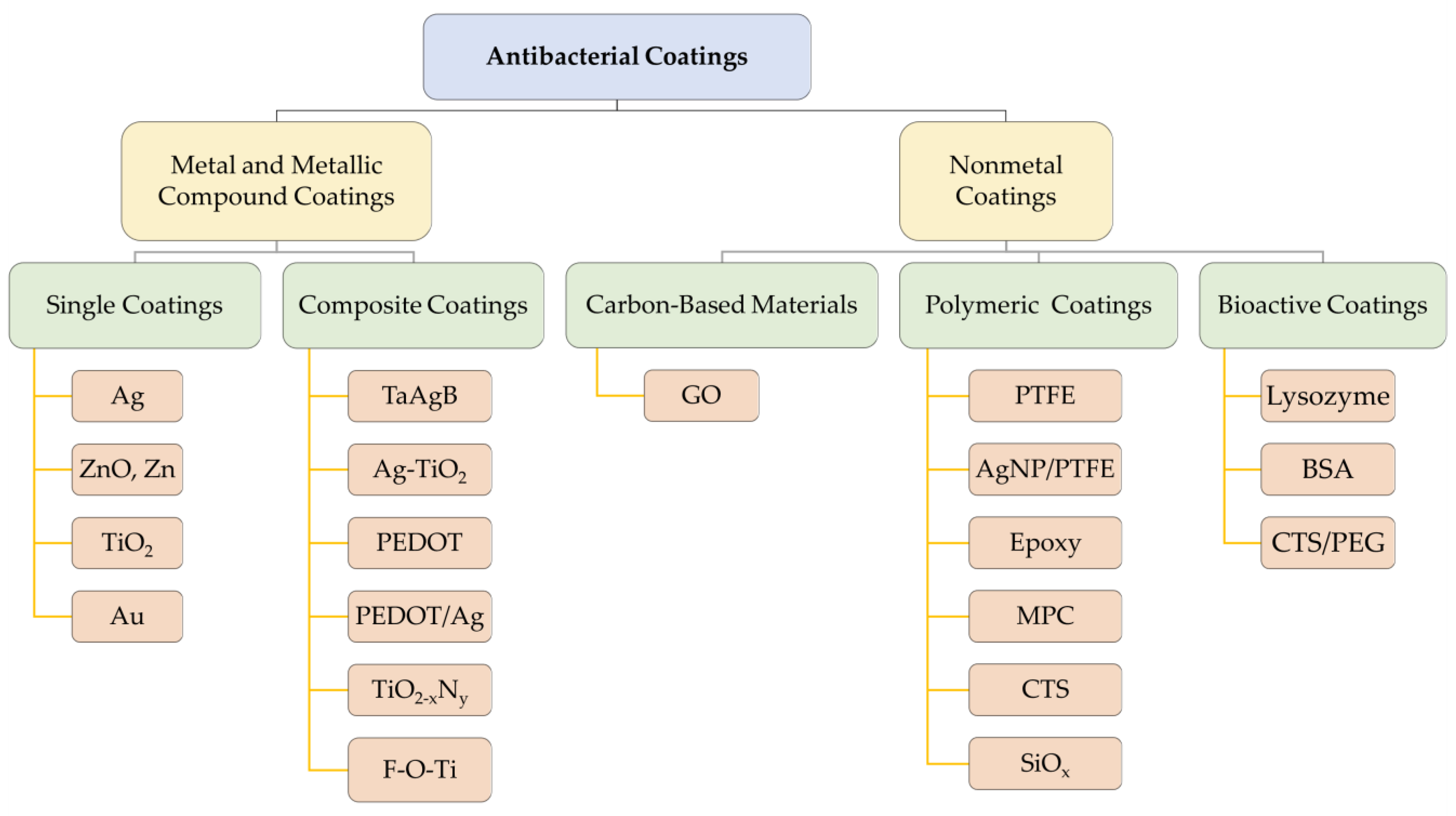
Among orthodontic antimicrobial coatings, the most valued are Ag and ZnO coatings, as they have been shown to exert good antimicrobial activity and are safe for humans in other areas of medicine [98,99]. Recently, TiO2 photosensitive antimicrobial coatings, polymeric coatings originally used for aesthetic purposes, and bioactive lysozyme coatings have also gradually entered the trend and have been explored for their potential as antibacterial coatings. As for the research on the antimicrobial properties of carbon-based material coatings, only Dai et al. have investigated this topic, and the material used was GO [89]. This study found that when the GO concentration was low, the GO coating could not completely cover the NiTi substrate, and the tribological and anticorrosion properties were barely improved, while the antibacterial properties—although statistically significantly different—only reduced the survival of Streptococcus mutans by 20%. Increasing the GO concentration enhanced the antimicrobial properties of the GO coatings, but the biocompatibility of the GO-coated NiTi substrate decreased [89]. Based on the above results, the GO concentration should be controlled within a suitable range when making GO coatings, and the recommended value of this concentration has not been determined at present, while the biocompatibility of GO coatings to the human body has also not been clearly confirmed thus far. In contrast, Ag and ZnO coatings not only have reliable antimicrobial properties, but also have been applied in various medical disciplines with a higher guarantee of safety. Therefore, the earliest Ag and ZnO may still be the most suitable materials for antibacterial coatings. The main antibacterial coating materials discussed in this section are shown in Figure 5.

3.1. Silver and Silver Compound Coatings

Silver has a wide range of antibacterial properties, protecting the surface of the material from microbial adhesion through different mechanisms, and has a certain antibacterial effect [100,101]. AgNPs can continuously release silver ions, which cause damage to bacterial cell walls and cell membranes. They can also penetrate cells and interfere with the synthesis of proteins and deoxyribonucleic acid (DNA). Inactivation of respiratory enzymes and the production of reactive oxygen species in bacterial cells are other mechanisms that can kill the bacteria. In addition, silver nanoparticles themselves can adhere to the cell wall and the membrane surface, directly causing cell membrane denaturation and perforation, and disrupting the signal transduction of bacterial cells [99,102]. In addition to the direct killing and inhibition of bacteria, AgNPs also regulate inflammatory responses, further inhibiting the reproductive survival of microorganisms [103]. Therefore, it can be assumed that by applying a silver coating to orthodontic appliances, bacterial colonization of the dental surfaces can be reduced, reducing plaque and caries formation during orthodontic treatment.
In a study by Mhaske et al., the authors found that silver plating applied by thermal evaporation on the surface of SS and NiTi archwires reduced the adhesion of Lactobacillus acidophilus on the surface of archwires and showed antibacterial activity against Lactobacillus acidophilus [41]. AgNPs coated on SS archwire via hydrothermal synthesis also showed significant inhibition of Staphylococcus aureus and Streptococcus mutans [104].
Nanosilver-coated SS brackets also have significant antibacterial activity. Valiollah Arash et al. prepared silver particles via an electroplating method and verified the antibacterial activity of silver plating against Streptococcus mutans using direct contact tests and disk diffusion tests, concluding that its antibacterial activity could last for up to 30 days after application [105]. Tania Ghasemi et al. demonstrated direct inhibition of Streptococcus mutans reproduction by preparing nanosilver coatings on the surface of brackets using PVD [63]. In addition to its antiadhesive and antibacterial properties against Streptococcus mutans and Streptococcus distortus [106], the nanosilver coating was also effective against Staphylococcus aureus and Escherichia coli [107]. Moreover, this effect was not limited to SS brackets, but also applied to ceramic and cobalt-chromium (Co-Cr) alloy brackets [107].
In addition to in vitro tests, there have also been animal studies demonstrating the antibacterial activity of silver-coated brackets. Gamze Metin-Gürsoy et al. found that nanosilver-coated orthodontic brackets can inhibit Streptococcus mutans in dental plaque and produce low levels of nanosilver ion release in saliva, as well as reducing smooth-surface caries. However, they did not have any significant effect on the incidence of occlusal caries [108]. This is the only study to date in which surface-coated fixed appliances have been applied in animals’ oral cavities; thus, we need more animal experiments in further studies.
Different surface coating methods for silver have different effects on the improvement of antibacterial properties. In a clinical trial, Viktoria Meyer-Kobbe et al. first investigated the use of plasma-immersion ion implantation and deposition (PIIID) in SS brackets. They concluded that the antibacterial effect of PIIID silver-modified surfaces is as significant as that of electroplated silver layers and PVD silver coatings, reducing biofilm volume and surface coverage, and producing an even stronger bactericidal performance [109]. Coating of SS brackets with silver using PIIID has proven good antibacterial properties in clinical applications, and direct evidence is next needed to show its ability to reduce the incidence of white spots and plaque gingivitis caused by orthodontic appliances. In addition to this, TaAgB also reduces the attachment and growth of Streptococcus mutans on the SS surface. This is evidenced by the results observed via SEM; not only was there a reduction in Streptococcus mutans on the TaAgB surface, but the morphology of the bacteria was also altered, further confirming its antibacterial activity [68].
Silver compound coatings have been studied for many years. To increase the corrosion resistance of silver coatings, a hard coating of silver-platinum (Ag-Pt) alloy on stainless steel surfaces has been developed and demonstrated significant antibacterial efficacy against Streptococcus mutans and Aggregatibacter actinomycetemcomitans, as well as good biocompatibility [110]. However, this study did not use a bracket as the substrate, but rather an SS block. This design may not fully simulate the bracket when it is performing its function in the oral cavity, as the morphology and ligation of the brackets and archwires are not taken into account.
To further increase the antibacterial properties of the silver coating, some studies have applied Ag together with TiO2 to SS brackets. They demonstrated better antiadhesive and antibacterial properties against Streptococcus mutans and Porphyromonas gingivalis than Ag alone, helping to prevent dental caries and plaque accumulation, with satisfactory biocompatibility [111].
Both Ag and ZnO nanoparticle-coated SS brackets exhibit antibacterial effects against Streptococcus mutans and Lactobacillus acidophilus, and the antibacterial effects are maintained for at least 3 months. In addition, silver/zinc oxide nanoparticle composite-coated brackets had a stronger antibacterial effect on Streptococcus mutans and Lactobacillus acidophilus compared to silver nanoparticles and zinc oxide nanoparticles alone [112].
Other silver particle composite coatings are still being explored. A 2021 study concluded that a silver-polymer-coated archwire was not antibacterially effective. Its colony counts did not differ significantly from those of uncoated archwires in 0% sucrose and 3% sucrose environments [113]. Of course, this study only added a single species of bacteria (Streptococcus mutans) as the test bacteria, whereas in reality, the environment and strain composition in the oral cavity are more complex. To verify the accuracy of this result, further experiments under conditions that more closely simulate the real conditions of the oral cavity are needed.
A composite nanocoating was synthesized by Bor-Shiunn Lee et al. [114]. They applied a layer-by-layer deposition method with materials consisting of polydopamine, functionalized poly(3,4-ethylenedioxythiophene) (PEDOT), and AgNPs, and demonstrated the antifouling and antibacterial properties of this coating when applied to the surface of stainless steel materials. Its release of Ag ions does not have a harmful effect on humans, as it is biocompatible. This may be used in the future as a coating for orthodontic brackets and archwires.
Silver has been used as an antibacterial material for a long time and has also been shown to be biocompatible on numerous occasions [115]. Silver coatings are antibacterial against a number of species, including cariogenic and periodontal pathogenic bacteria, and provide a definite resistance to smooth-surface caries [108]. The long-lasting antibacterial effect allows for the use of brackets, which are an infrequently replaced component [112]. However, the shortcomings of silver coatings are their poor corrosion resistance, low hardness, and high price. The upside is that silver composite coatings with other materials have similar antibacterial effects [112], so silver composite coatings can be developed to discover potential improvements in other properties.

3.2. Titanium and Titanium Compound Coatings

TiO2, as a photosensitive material, is capable of generating hydroxyl (OH) radicals and reactive oxygen species (ROS) when irradiated by ultraviolet (UV) light, which are highly reactive when exposed to organic compounds [116,117]. Based on this principle, the antibacterial properties of TiO2 have been receiving attention.
Several studies have confirmed that SS and NiTi archwires coated with TiO2 show antiadhesive effects against Streptococcus mutans [118,119,120,121], as well as bactericidal effects on Streptococcus mutans [118,119,120,121], Porphyromonas gingivalis [119], Candida albicans, and Enterococcus faecalis [120]. For brackets, TiO2 coatings also show antiadhesive and antibacterial properties against Streptococcus mutans [122,123], Candida albicans [122], and Lactobacillus acidophilus [124].
Rutile, anatase, and brookite are crystal structures in which TiO2 exists. Anatase is formed by anodic oxidation (AO), while rutile is formed by thermal oxidation (TO). Studies have shown that anatase films applied to titanium (Ti) and titanium silver (TiAg) plates are more effective than rutile films against Streptococcus mutans [125]. In contrast, Roshen Daniel Baby et al. applied TiO2 coatings with different crystal structures to SS brackets but came to the opposite conclusion. Their results showed that both structures of TiO2 have antibacterial effects against Streptococcus mutans, but the rutile phase has a stronger bactericidal effect and more significant cytotoxicity than the anatase phase [126]. All of these considerations mean that rutile should be avoided regardless of which crystal type has the better antibacterial effect, as its cytotoxicity reduces the biocompatibility of the material.
Recently, there have been some clinical studies of TiO2 coatings. Keerthi Venkatesan et al. showed that TiO2 nanoparticle coatings on NiTi archwires have good antiadhesive and antibacterial effects against Streptococcus mutans. However, by the end of 1 month, 60% of the TiO2 coating was lost, and the roughness of the archwire was similar to that of the uncoated one [127]. Nevertheless, the adhesion of Streptococcus mutans to TiO2-coated archwires was still lower compared to uncoated archwires, which can be attributed to the antibacterial properties of the nanoparticles [127]. In addition, the TiO2 nanoparticle coating on the SS archwire also showed an effect against Streptococcus mutans in the first and third weeks [128]. Whether on NiTi or SS archwires, the TiO2 coating was effective in reducing the initial bacterial adhesion [127,128]. However, although the TiO2 coating was shown to have a direct antiadhesive and bactericidal effect, its effect on the prevention of enamel demineralization was not significant [127]. Therefore, more experiments are needed to prove its preventive effects against enamel demineralization and periodontal disease in practice.
In addition to the TiO2 coating applied directly to the surface of the material, the surface of Ti or titanium alloys also oxidizes to form a TiO2 layer, which is responsible for the coatings of titanium compound materials having certain antibacterial properties [125]. As mentioned already, Ag is also an effective antibacterial agent, so it has also been investigated to be added to Ti to prepare TiAg alloys, which it is hoped will have a stronger antibacterial effect than a Ti coating alone. For Streptococcus mutans, TiO2 coatings on TiAg alloy plates have a significantly stronger and faster antibacterial effect than TiO2 coatings on Ti plates, suggesting that the addition of Ag to Ti can result in a synergistic enhancement of the effect against Streptococcus mutans [125]. However, for Lactobacillus acidophilus, although both showed antibacterial activity, the TiAg-coated samples showed no difference in resistance to Lactobacillus acidophilus compared to Ti [129], which means that the TiAg coating did not have stronger activity against Lactobacillus acidophilus than the Ti coating. Nevertheless, irrespective of their antibacterial effects, neither of them exhibited cytotoxicity [129]. Therefore, both materials have the potential for application on fixed appliances, although further research is needed to determine whether the addition of Ag will give the desired antibacterial effect. Ti coatings and TiAg coatings have not yet been applied to archwires or brackets, which is a gap for the time being. In addition, apart from the clinical studies, the above studies have not taken into account the mechanical forces to which the archwires are subjected in practice. Thus, the ability of the coatings to withstand mechanical forces has not been tested.
The shortcoming of TiO2 is that it only produces hydroxyl radicals under UV light irradiation. When UV light is filtered and only visible light is used, the photocatalytic activity of TiO2 is poor [130]. On the other hand, the nitrogen-doped (N-doped) TiO2 thin film substrate, as a visible-light-sensitive photocatalyst, has a significant bactericidal rate under visible-light irradiation [121]. Nitrogen-doped titanium dioxide (TiO2−xNy) films coated on the surface of SS brackets using RF magnetron sputtering produced 95.19%, 91.00%, 69.44%, and 98.86% inhibition against Streptococcus mutans, Lactobacillus acidophilus, Actinomyces viscous, and Candida albicans, respectively [131]. In addition, the coating exhibited strong antibacterial properties against Streptococcus mutans over a 90-day timeframe. In other words, surface coating of SS brackets with N-doped TiO2 films inhibits Streptococcus mutans for up to 3 months [132]. To obtain coatings with the highest visible-light photocatalytic activity and antibacterial activity against Lactobacillus acidophilus and Candida albicans, a sputtering temperature of 300 °C for 180 min, an aigon (Ar):nitrogen (N) ratio of 30:1, and an annealing temperature set at 450 °C can be performed [133]. The preparation of TiO2 nanofilms using PVD has also been examined and has shown similar results on the antibacterial effect of TiO2, regardless of whether the coating thickness was 60 μm or 100 μm [63]. In some studies, however, the visible-light exposure time was set to 24 h [121,131]. This condition is impossible to achieve in practical applications. Therefore, simulation experiments that are closer to the clinical situation, animal experiments, or clinical trials are needed to further demonstrate the effectiveness of N-doped TiO2 coatings in improving the antibacterial properties of fixed appliances. As shown in a randomized controlled clinical trial by Avula Monica et al. [134], N-doped TiO2-coated SS brackets are effective in reducing the extent of the increase in Streptococcus mutans concentration when exposed to natural visible light and dental surgical light. This effect holds for up to 60 days and is better at 30 days after the placement of orthodontic appliances than at 60 days. However, even this latest related experiment did not directly detect and document enamel demineralization around the brackets or gingival inflammation. This will need to be followed up in future studies.
When TiO2 is applied in combination with other materials, it is found that Ag applied with TiO2 on SS brackets has better antiadhesive and antibacterial properties against Streptococcus mutans and Porphyromonas gingivalis [111].
Titanium nitride is also considered to have an antibacterial effect, although TiN is not as antibacterial as TiO2 and carbon nitride (CNx) [29]. However, the study by Licia Pacheco Teixeira et al. concluded that neither TiN nor titanium nitride doped with calcium phosphate (TNCP) films interfere with the attachment of Streptococcus mutans to the surface of the bracket, meaning that this coating has no antiadhesive effect and does not reduce the growth of Streptococcus mutans [135]. Moreover, the use of TiN as the surface coating may increase the surface friction [29]. In summary, the utility of TiN as a surface coating for orthodontic materials needs to be further verified.
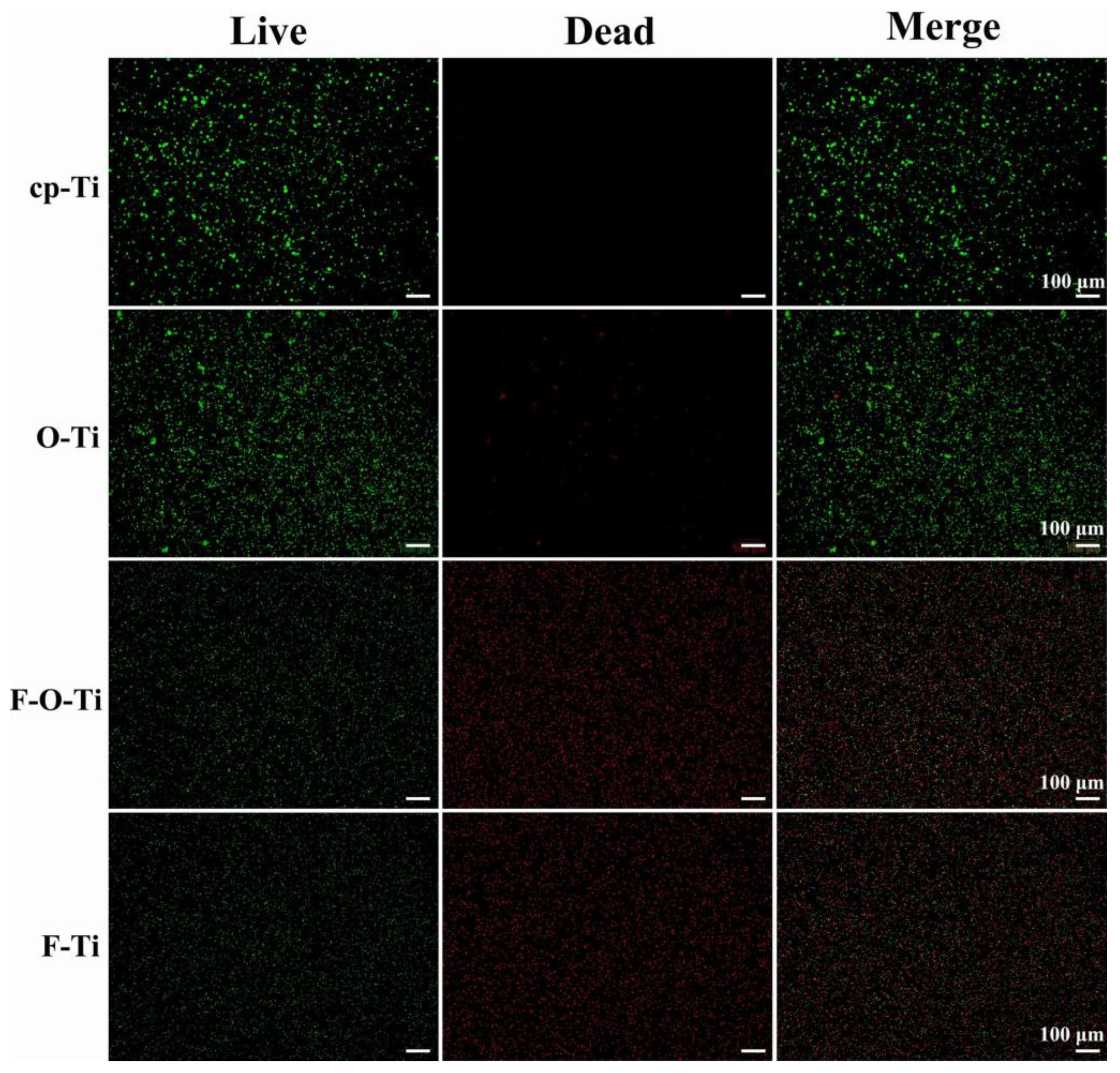
Mian Chen et al. [136] used titanium treated with plasma-enhanced fluorine (F) and oxygen (O) mono/double chemical vapor deposition to obtain nanofunctional coatings with improved antibacterial properties and biocompatibility. The results showed that the fluorine-deposited samples can effectively exterminate Staphylococcus aureus with sufficient antibacterial properties, as shown in Figure 6. More importantly, the F and O double-deposited (F-O-Ti) coatings exhibited better sustained antibacterial performance than the F single-deposited coatings after 7 days of immersion in 0.9% sodium chloride (NaCl) solution. This result tells us that this type of material may be seen as an effective antibacterial coating for clinical applications.
Titanium compound coatings rely mainly on the photoreactivity of TiO2 for their antibacterial action [130]. The advantages include their ability to resist caries and periodontal pathogenic bacteria [111,131,136], as well as their biocompatibility [125]. TiO2 coatings are probably best used for short-term applications only, as they reduce initial bacterial adhesion [134] and undergo rapid peeling [127], making them unsuitable for long-term use. In addition, the increased friction of such coatings is detrimental to therapeutic efficiency, and the need for light causes inconvenience in their use [130]. Titanium has composite applications with silver [125], nitrogen [121], fluorine, and oxygen [136], which enhance its antibacterial properties, but the durability and high friction have not been addressed thus far. Currently, difficulties remain in clinical application.

3.3. Zinc and Zinc Compound Coatings

ZnO nanoparticles reduce biofilm formation, with low toxicity as well as good biocompatibility [137], making them suitable for biomedical applications. In a 2015 study, Baratali Ramazanzadeh et al. found that ZnO-nanoparticle-coated brackets prepared via spray pyrolysis had antibacterial effects, but they were not as effective as copper oxide (CuO) nanoparticle coatings and CuO-ZnO composite coatings, both of which reduced the number of Streptococcus mutans to 0 after 2 h [138]. However, the coatings were not tested for biocompatibility. If the addition of CuO leads to elevated cytotoxicity beyond the acceptable threshold for biocompatibility, it will be difficult to apply it directly in clinical applications, even if the antibacterial properties are good enough. Unlike CuO, ZnO has proven to be biocompatible [25]. Therefore, it is safe to use for a wide range of biomedical materials.
It has been shown that ZnO nanoparticle coatings on the surface of NiTi archwires have antibacterial effects against Streptococcus mutans [25,139], Staphylococcus aureus, Streptococcus pyogenes, and Escherichia coli [27]. Different coating methods can be used to obtain ZnO particles with different physicochemical properties, such as chemical precipitation, chemical vapor deposition (CVD), electrostatic spinning, polymer composite coating, and the sol-gel method. Comparisons also reveal that the smaller the particles, the larger the specific surface area of the particles, and the more the antibacterial performance is improved [139,140]. In addition, the color of the ZnO coating is white or light gold and does not tarnish in the short term, resulting in a higher aesthetic quality [25,138].
However, a study in 2021 concluded that the antibacterial effect of ZnO coatings was not significant [123]. In this study, the investigators prepared a photocatalytic ZnO coating on the surface of SS brackets via magnetron sputtering, and the results showed that the antibacterial effect of ZnO was not satisfactory. Based on these contradictory findings, more experiments are needed in order to determine the feasibility of the clinical application of ZnO nanoparticle coatings on orthodontic archwires or brackets.
In addition to the composite application of ZnO with CuO, it can also be used to form a coating with Ag nanoparticles. Noha K. Zeidan et al. [112] made a composite coating of ZnO with silver nanoparticles and applied it to SS brackets, finding that the antibacterial effect against Streptococcus mutans and Lactobacillus acidophilus exceeded that of the application of ZnO alone. Considering the antibacterial effectiveness of silver and the mechanical properties of ZnO, the combination of the two can bring the advantages of the coating to a more powerful level.
In summary, ZnO can be used as a multifunctional coating in clinical applications. It is antibacterial, friction reducing [26], and biocompatible [25], and it is safe and aesthetically pleasing to use [25,138]. On the downside, its corrosion resistance and durability for long-term application are not yet confirmed, and the antibacterial effect is still controversial. However, its application in combination with Ag nanoparticles can provide similar antibacterial performance and is more effective than ZnO alone [112]. In addition, future studies could focus on how to obtain finer ZnO nanoparticles in the preparation of coatings to enhance the antibacterial properties.

3.4. Other Metal and Metallic Compound Coatings

For antibacterial surface coatings prepared from other metallic materials, such as rhodium and gold, attempts have also been made on fixed appliances, although many research gaps remain.
There is no consensus on the effect of rhodium plating on the surface of archwire in terms of antibacterial properties. Some believe that the rhodium-coated NiTi archwires have increased antibacterial properties [141], while others consider that the risk of biofilm retention on rhodium-coated NiTi archwires is as high as that of uncoated NiTi archwires [113].
In 2014, a gold-plated aesthetic archwire was shown to reduce the surface roughness of SS archwires, but it caused no reduction in the adhesion of Streptococcus mutans. Moreover, surface free energy (SFE) may have a positive correlation with bacterial adhesion. If the SFE of the coating is the same as that of the archwire, the antiadhesive effect may not be significant, just as the SFE of gold-plated SS archwires is similar to that of SS archwires [141]. However, another study on other coatings suggested that surface roughness is positively correlated with biofilm adhesion [142]. This contradiction may be due to different experimental designs, or to the varying nature of different coatings and their antibacterial mechanisms. According to the “attachment point theory”, biofoulers (in the context of this review, bioorganisms) have more attachment points on rough surfaces [143]. This theory may partially explain why some believe that bacteria are more likely to be attached on rough surfaces. However, smooth surfaces are not necessarily more resistant to adhesion than surfaces with undulations. Some researchers, inspired by bionics, have tried to create nanofolds on the surface of the substrate. These folds are like ripples with a certain wavelength and amplitude. When these two parameters are appropriate, the adhesion of biofoulers to the surface can be reduced. That is, although the surface is not completely smooth, its antiadhesion properties are significantly better than even that of a smooth surface when the gap between the folds is slightly smaller than the size of the bacteria [144]. This suggests that preparing nanofolds of suitable wavelengths and amplitudes on the surfaces of archwires and brackets might be a possible way to resist bacterial adhesion.
In addition, other gold materials—such as 4,6-diamino-2-pyrimidinethiol-modified gold nanoparticles (AuDAPT) [145] and quaternary ammonium (QA)-modified gold nanoclusters (QA-GNCs) [146]—can be used as antibacterial coatings for invisible orthodontic appliances. AuDAPT and QA-GNC have resistance to Porphyromonas gingivalis [145] and Streptococcus mutans [146], respectively, and they have shown excellent biocompatibility, but they have not yet been investigated in fixed appliances.

3.5. Polymeric and Bioactive Materials

Epoxy has been clinically demonstrated to have antibacterial properties as an aesthetic coating on the surface of NiTi archwires [147]; however, this study took the plaque directly from the surface of the archwire during orthodontic treatment and counted the number of bacterial colonies in the culture, without distinguishing different strains. Two other in vitro experiments focused on Streptococcus mutans and showed that epoxy coating on the surface of SS or NiTi archwires reduces the adhesion of Streptococcus mutans and Streptococcus distans to the archwires in the short term [141,148]. Regarding the mechanism of this antiadhesive effect, the authors suggested that it was due to a reduction in the SFE of the archwire, rather than a change in surface roughness [141]. Conversely, Deise C. Oliveira et al. found that the amount of biofilm adhesion was significantly higher on epoxy-coated than on uncoated NiTi archwires. In addition, there was no significant difference in the number of bacterial colonies between coated and uncoated archwires [113]. This indicates that the epoxy coating does not increase the antibacterial properties of the NiTi archwire surface, and it may even decrease the antiadhesive properties. Based on these contradictory findings, multiple replicate experiments are needed in the future to determine the accuracy of the results. Efforts are also needed to make the in vitro experimental conditions more closely resemble the intraoral physiological environment, or to conduct animal and clinical experiments.
Another polymeric aesthetic coating material that has been used for a long time is polytetrafluoroethylene, also known as PTFE. It has been shown in clinical trials to reduce the amount of plaque biofilm adhesion on NiTi archwires [147] and SS brackets [149], and it is effective on every surface of the SS bracket [149]. Additionally, PTFE coatings prepared at low temperatures have better resistance to microbial adhesion than those prepared at high temperatures [95]. Still, these antibacterial and antiadhesive effects cannot be attributed to a reduction in initial bacterial adhesion [150]. However, similar to epoxy, it has been suggested that PTFE does not reduce the amount of biofilm adhesion on the surface of NiTi archwires. Interestingly, both results came from the same research [113], and only this single study reached the opposite conclusion from the previous ones. More similar studies may be needed in the future to verify the effects of PTFE and epoxy coatings on the antibacterial properties of NiTi archwire surfaces. Furthermore, the combined usage of AgNPs and PTFE through a similar method has been investigated, showing better antibacterial properties compared to applying PTFE coatings alone.
Regarding other polymeric coatings, Adauê S Oliveira et al. [151] prepared silicon oxides (SiOx) hydrophobic/superhydrophobic coatings on SS and ceramic bracket surfaces via a sol-gel process. Hydrophobic coatings were made using cetyltrimethoxysilane diluted in ethanol, termed HS1; superhydrophobic coatings were made using 1H,1H,2H,2H-perfluorodecyltriethoxysilane diluted in dimethyl sulfoxide, termed HS2. The results showed that both HS1 and HS2 reduced the biofilm accumulation on the surface of the bracket in 24 h, and the superhydrophobic HS2 was more effective than the hydrophobic HS1. Based on the results of this study, when applying SiOx coatings to increase the antibacterial properties of the brackets, HS2 superhydrophobic coatings can be prepared to obtain the best results [151]. The oral environment is moist, so the liquid–air interface formed between the hydrophobic surface and saliva prevents bacteria from adhering and forming biofilms [152], which explains the excellent antimicrobial properties of hydrophobic and superhydrophobic coatings. The abovementioned study was published in 2015, and there have been no subsequent studies on the surface coating of orthodontic brackets or archwires that present prospects worth exploring.
MPC polymer coatings inhibit the adhesion of Streptococcus mutans to SS archwires [96]. CTS has been used as an antibacterial material in the resin for bonding orthodontic attachments, with the purpose of controlling enamel demineralization, but it has not been tested for its antibacterial properties as a surface coating for orthodontic attachments, which is a direction that could be developed in the future [24]. As another polymer, polyethylene glycol (PEG)-coated SS archwires exhibited the best antiadhesive properties at a molecular mass of 5000, maintaining very low bacterial adhesion even after 10 h [153].
Bioactive materials such as lysozyme are also increasingly being used in orthodontics. New lysozyme-coated orthodontic composite archwires (CAWs) exhibit antibacterial properties in artificial saliva—mainly against Staphylococcus aureus [154]. In addition, the corrosion resistance and biocompatibility of the lysozyme coating are relatively considerable [154]. However, other strains of bacteria in the oral cavity—such as Streptococcus mutans, which often causes caries, and Porphyromonas gingivalis, which causes periodontal disease—have not been studied. Nevertheless, lysozyme as a bioactive material provides inspiration for follow-up studies. More bioactive materials can be explored for surface modification in the surface coating of fixed appliances. However, although lysozyme coatings have been shown to have an antibacterial effect against Staphylococcus aureus, their effect on Gram-negative bacteria is weaker than on Gram-positive bacteria, so there is a risk of disrupting the balance of the intraoral flora when applied to fixed orthoses. Further animal experiments are needed in order to confirm their clinical value.
There are also bioactive antimicrobial coatings that are based on antiadhesion. The protein molecule bovine serum albumin (BSA) significantly reduced bacterial adhesion on the surfaces of SS, ceramic, and resin brackets and SS archwires, with a maximum decrease of more than 95% on the surfaces of brackets, probably due to the BSA-mediated reduction in surface free energy [155]. In addition, in 2020, Peng et al. developed an antimicrobial hydrogel using a combination of PEG and chitosan (CTS/PEG) [156]. Archwires coated with these bioactive materials showed a significant increase in both antiadhesive and antimicrobial properties; thus, their possible uses in dental applications are considerable.
Recent studies have found that coatings of polydopamine (PDA) and blue fluorescent hollow carbon dots (HCDs) can maintain more than 50% antimicrobial performance against Escherichia coli and Streptococcus mutans for 14 days. The fluorescent nature of HCDs may also provide clinical visualization, allowing for timely replacement of defective coatings. In addition, the combination of low drug resistance, low toxicity, and high biosafety will allow this bioactive material to go far in the future [20].
Recent studies on antibacterial coatings for orthodontic appliances are summarized in Table 2.
Table 2. Recent studies on antibacterial coatings for orthodontic appliances.
Ref.Coating
Materials
Coating
Technique
SubstrateStudy TypeRoughnessAntibacterial EffectivenessOther
Effectiveness
[109]AgGalvanic, PVD, PIIID, and depositionSS bracketsClinical trialRa for the untreated bracket material was 0.04 µm, Ra for the galvanic coating was 0.12 µm, Ra for the PVD coating was 0.08 µm, and Ra for the PIIID procedure coating was 0.06 µm.The biofilm volume per test specimen for the control was 7.24 × 108 µm3. For the galvanically applied silver coating, the biofilm volume decreased to 2.62 × 107 µm3, for the PVD coating to 4.44 × 107 µm3, and the PIIID procedure to 3.82 × 107 µm3. The reduction of the biofilm volume compared to the control was statistically significant for all surface modifications. The percentage surface coverage per test specimen was 64.40% for the unmodified control and decreased to 16.97% for the galvanic silver surface, 23.81% for the PVD coating, and 23.63% for the PIIID-modified surface.NA
[112]Ag (NPs)PVDSS bracketsIn vitro (in artificial saliva)NAThe inhibition percent of Streptococcus mutans and Lactobacillus acidophilus were 27.60% and 62.02%, respectively.Durability (>3 months)
[106]Ag (NPs)NA (commercial)NAIn vitro (in suspensions of microorganisms)NAThe silver coating decreased the adhesion of both Streptococcus mutans and Streptococcus sanguinis to the orthodontic bracketsNA
[104]Ag (NPs)Hydrothermal synthesisSS archwiresIn vitro (in suspensions of Streptococcus mutans and Staphylococcus aureus)It was not possible to observe changes in roughness after coating.Microbial adhesion and biofilm formation of Staphylococcus aureus and Streptococcus mutans reduced.Aesthetic effect
[107]Ag (NPs)Synthesized in situSS and ceramic bracketsIn vitro (on Mueller–Hinton agar plates)NAThe inhibitory halos obtained by the in vitro evaluation of the antibacterial effect, in terms of brackets with silver nanoparticles with Staphylococcus aureus and Escherichia coli, showed an excellent inhibition of microbial growth compared to the bracket control, with a diameter between 9 and 10 mm.NA
[140]Ag (NPs)Aqueous reductionNiTi, CuNiTi, SS archwires, and SS bracketsIn vitro (in suspensions of Streptococcus mutans)Roughness values were increased in SS wires (7.094 × 10³ + 1 nm), followed by NiTi wires (6.234 × 10³ + 1 nm), and the lowest roughness value for CuNiTi wires (3.116 × 10³ + 1 nm).Smaller Ag nanoparticles (16.7 μg/mL) had consistently better antimicrobial inhibition effects against the Streptococcus mutans strain compared to larger Ag nanoparticles (66.8 μg/mL), showing significant differences between them. The coated brackets had significantly better antiadherence activity (4.3 CFU/mL for smaller particles and 5 CFU/mL for larger particles) than the uncoated brackets (356 CFU/mL). Both sizes of Ag nanoparticles had statistically good adherence inhibition of the Streptococcus mutans strain for all types of orthodontic wires (SS = 26.1–52.6 CFU/mL, NiTi = 15.1–49.6 CFU/mL, and CuNiTi = 89.1–287.8 CFU/mL) compared with the control groups (SS = 346.7 CFU/mL, NiTi = 342.3 CFU/mL, and CuNiT = 376.2 CFU/mL).NA
[112]Ag/ZnO (NPs)PVDSS bracketsIn vitro (in artificial saliva)NAThe inhibition percent of Streptococcus mutans and Lactobacillus acidophilus were 45.32% and 80.29%, respectively.Durability (>3 months)
[113]Ag + polymerNA (commercial)NiTi archwiresIn vitro (in suspensions of microorganisms + sucrose)NANo significant reduction in bacterial adhesion (0% sucrose) or biofilm accumulation (3% sucrose) was found when assessed by colony counting of Streptococcus mutans.NA
[157]AgNP/PTFELayer-by-layer deposition316L SS platesIn vitro (in suspensions of Escherichia coli)The surface roughness increased from 59.4 ± 6.1 nm to 158.1 ± 2.7 nm (deposition time of 6 h) and 177.3 ± 5.1 nm (deposition time of 12 h), respectively.Coatings with 6 and 12 h deposition time could inhibit by ~75% and ~90% bacterial growth over the initial 3 days, respectively. After 7 days, coatings with 6 and 12 h deposition time still exhibited significant antibacterial activity, reducing by ~40% and ~50% of bacterial growth, respectively.Corrosion-resistant effectiveness
Biocompatibility
[158]Al2O3 (NPs)Atomic layer deposition316L SSIn vitro (in simulation body fluid solution)NAThe mean colonies forming units per milliliter were reduced by 20%; the diffusion zone was ~4 mm.Corrosion-resistant effectiveness
Biocompatibility
[158]Al2O3/TiO2 MultilayerAtomic layer deposition316L SSIn vitro (in simulation body fluid solution)NAThe mean colonies forming units per milliliter were reduced by 40%; the diffusion zone was >6 mm.Corrosion-resistant effectiveness
[155]BSAChemical depositionSS, ceramic, and resin brackets; SS archwiresIn vitro (in suspensions of Streptococcus mutans)The adsorbed BSA molecule was not uniform on the surface, thereby leading to slight surface roughness.After integrating BSA molecules, the three kinds of brackets all showed more than 95.0% reduction in Streptococcus mutans adhesion (i.e., 98.3% for SS, 96.3% for ceramic, and 95.2% for resin). Compared with bare archwires, only a few bacteria (~7.5%) could be found on the BSA-coated archwires’ surface, even after incubation in bacterial suspension for 300 min; and the optimal BSA concentration was 10 mg/mL.NA
[156]CTS/PEG hydrogelCombining silane chemistry and subsequent copolymerizationSS archwiresIn vitro (in suspensions of Streptococcus mutans)Rq of CTS/PEG-coated SS archwires increased from 0.26 to 1.57 nm.This biointerface showed superior activity in early-stage adhesion inhibition (98.8%, 5 h) and displayed remarkably long-lasting colony-suppression activity (93.3%, 7 d).Durability (>7 days)
Wear resistance
Biocompatibility
[29]CNxIon beam-assisted deposition304L SS disksIn vitro (friction test: NA; antibacterial test: in bacterial suspension)The films slightly reduce the surface roughness parameter. Ra reduced from 0.181 to 0.140 μmBacteria density reduced from 13.002 × 10³ /mm2 to 4.030 × 10³ /mm2 (p < 0.05).Friction reduction effectiveness
Biocompatibility
[113]EpoxyNA (commercial)NiTi archwiresIn vitro (in suspensions of microorganisms + sucrose)NANo significant reduction in bacterial adhesion (0% sucrose) or biofilm accumulation (3% sucrose) was found when assessed by colony counting of Streptococcus mutans.NA
[142]EpoxyNA (commercial)NiTi archwiresIn vitro (in suspensions of Streptococcus mutans and Streptococcus sobrinus)Ra = 1.29 μm (higher than uncoated archwires)Epoxy-coated wires demonstrated an increased adhesion of Streptococcus mutans (5.55 CFU/cm2) and Streptococcus sobrinus (4.64 CFU/cm2).NA
[136]F-O-TiPlasma-enhanced fluorine and oxygen mono/dual CVDCommercially available pure titanium with 99.9% purityIn vitro (in suspensions of Staphylococcus aureus and artificial saliva)A large amount of convex texture with 100–200 nm size distributes uniformly all over the surface.The antibacterial rates were higher than 90%.Corrosion-resistant effectiveness
Durability (~7 days)
Biocompatibility
[89]GOSilane couplingNiTi archwiresIn vitro (friction test: NA; antibacterial test: in bacterial suspension; corrosion test: artificial saliva)The surface of the samples coated with 2 mg/mL GO concentrations was smooth with a uniformly coated area.The bacterial CFU values for Streptococcus mutans were 0.77 (samples coated with 0.5 mg/mL GO concentrations), 0.40 (samples coated with 2 mg/mL GO concentrations), and 0.23 (samples coated with 5 mg/mL GO concentrations) relative to that on bare NiTi (1.00). The number of live bacteria decreased, and the number of dead bacteria increased, indicating that GO coating could effectively resist adherent bacteria. This effect had a positive correlation with GO concentration, indicating the concentration-dependent antibacterial ability of these GO-coated surfaces.Friction reduction effectiveness
Corrosion-resistant effectiveness
Wear resistance
Biocompatibility
[154]LysozymeLiquid phase depositionCAWsIn vitro (in suspensions of Staphylococcus aureus and artificial saliva)The surface roughness increased after being coated according to two- and three-dimensional atomic force micrographs.When coated with 20, 40, and 60 g/L lysozyme, Live/dead bacteria staining of Staphylococcus aureus reduced from 90% to 82%, 59%, and 61%, respectively.Corrosion-resistant effectiveness
Durability (~2 weeks)
Wear resistance
Biocompatibility
[134]N-doped TiO2Radio frequency magnetron sputteringSS bracketsClinical trialNACoated archwires (38.54 and 36.84) showed greater Ct values than uncoated wires (34.71 and 31.89) at 30 d and 60 d, respectively. Greater Ct values indicate lower Streptococcus mutans adhesion. Therefore, coated wires demonstrated lower Streptococcus mutans adhesion when compared with uncoated wires.Durability
[132]N-doped TiO2Radio frequency magnetron sputteringSS bracketsIn vitro (in suspensions of Streptococcus mutans)NAThe coating decreased Streptococcus mutans colonies from 401.21 to 37.82 CFU/mL.Durability (>90 days)
[114]PEDOT/AgLayer-by-layer deposition316L SS platesIn vitro (in suspensions of Streptococcus mutans and Escherichia coli)NAThe antiadhesive and antibacterial activity against Streptococcus mutans and Escherichia coli significantly increased.Biocompatibility
[20]PDA and HCDsElectrostatic adsorptionSS bracketsIn vitro (in the soaking solution of the archwires)NAAfter soaking in artificial saliva for 7 and 14 days, the coatings formed by 50 μL and 100 μL of HCDs showed antibacterial rates against Escherichia coli and Streptococcus mutans that could still reach more than 50%, but the antibacterial properties of the coatings formed by 150 μL of HCDs were much weaker than those of the above two groups.Durability (>7 days)
Biocompatibility
[113]PTFENA (commercial)NiTi archwiresIn vitro (in suspensions of microorganisms + sucrose)NANo significant reduction in bacterial adhesion (0% sucrose) or biofilm accumulation (3% sucrose) was found when assessed by colony counting of Streptococcus mutans.NA
[142]PTFENA (commercial)NiTi archwiresIn vitro (in suspensions of Streptococcus mutans and Streptococcus sobrinus)Ra = 0.74 μm (higher than uncoated archwires)PTFE-coated wires demonstrated an increased adhesion of Streptococcus mutans (4.76 CFU/cm2) and Streptococcus sobrinus (3.73 CFU/cm2).NA
[157]PTFENA316L SS platesIn vitro (in suspensions of Escherichia coli)The surface roughness increased from 59.4 ± 6.1 nm to 134.7 ± 3.9 nm after being coated.The PTFE coating only demonstrated short-term antiadhesive activity, reducing by ~45% biomass adhesion on the first day as compared with 316L SS, while coatings with 6 h and 12 h deposition time inhibited by ~65% and ~80% of biomass formation.Corrosion-resistant effectiveness
Biocompatibility
[113]RhodiumNA (commercial)NiTi archwiresIn vitro (in suspensions of microorganisms + sucrose)NANo significant reduction in bacterial adhesion (0% sucrose) or biofilm accumulation (3% sucrose) was found when assessed by colony counting of Streptococcus mutans.NA
[142]RhodiumNA (commercial)NiTi archwiresIn vitro (in suspensions of Streptococcus mutans and Streptococcus sobrinus)Ra = 0.34 μm (higher than uncoated archwires)Rhodium-coated wires demonstrated an increased adhesion of Streptococcus mutans (3.85 CFU/cm2) and Streptococcus sobrinus (2.79 CFU/cm2).NA
[68]TaAgBMagnetron sputtering with Ag-doping304 SS sheetsIn vitro (in artificial saliva)NAThe amount of Streptococcus mutans adhesion on the TaAgB surface was significantly reduced and the morphology changed according to the SEM image.Friction reduction effectiveness
Biocompatibility
[29]TiNIon beam-assisted deposition304L SS disksIn vitro (friction test: NA; antibacterial test: in bacterial suspension)The films slightly reduce the surface roughness parameter. Ra reduced from 0.181 to 0.162 μm.Bacteria density reduced from 13.002 × 10³ /mm2 to 3.888 × 10³ /mm2 (p < 0.05).Friction reduction effectiveness
Biocompatibility
[135]TiNCathodic cage depositionSS bracketsIn vitro (in suspensions of Streptococcus mutans)NAThe presence of coatings did not influence the formation of the Streptococcus mutans biofilm (p = 0.06).NA
[29]TiO2Ion beam-assisted deposition304L SS disksIn vitro (friction test: NA; antibacterial test: in bacterial suspension)NABacteria density reduced from 13.002 × 10³ /mm2 to 1.368 × 10³ /mm2 (p < 0.05).Wear resistance
Biocompatibility
[123]TiO2Radio frequency magnetron sputteringSS bracketsIn vitro (in suspensions of Streptococcus mutans)NAThe “weight increase” reduced from 1.0100 × 10³ mg to 0.6750 × 10³ mg, which showed increased antiadhesive activity against Streptococcus mutans. The survival rate reduced from 3548.3350 to 2895.0000 CFU/mL, which showed increased antibacterial activity against Streptococcus mutans.NA
[127]TiO2Radio frequency magnetron sputteringNiTi archwiresClinical trialRa reduced from 1591.08 to 746.14 nm.Coated archwires (30.97) showed greater Ct values than uncoated wires (37.00). Greater Ct values indicate lower Streptococcus mutans adhesion. Therefore, coated wires demonstrated lower Streptococcus mutans adhesion when compared with uncoated wires. This difference was statistically significant (p = 0.0005).Durability (~1 month)
[128]TiO2PVDSS archwiresClinical trialNAThe coating decreased Streptococcus mutans colonies from 5122.0 to 1400.0 and from 1141.8 to 297.7 CFU/mL in the first and the third week, respectively.Durability (>3 weeks)
Biocompatibility
[158]TiO2 (NPs)Atomic layer deposition316L SSIn vitro (in simulation body fluid solution)NAThe mean colonies forming units per milliliter were reduced by 15%; the diffusion zone was ~2 mm.Corrosion-resistant effectiveness
Biocompatibility
[135]TNCPCathodic cage depositionSS bracketsIn vitro (in suspensions of Streptococcus mutans)NAThe presence of coatings did not influence the formation of the Streptococcus mutans biofilm (p = 0.06).NA
[123]ZnORadio frequency magnetron sputteringSS bracketsIn vitro (in suspensions of Streptococcus mutans)NAThe antiadhesive and antibacterial activity against Streptococcus mutans showed no significant increase.NA
[112]ZnO (NPs)PVDSS bracketsIn vitro (in artificial saliva)NAThe inhibition percent of Streptococcus mutans and Lactobacillus acidophilus were 17.54% and 28.85%, respectively.Durability (>3 months)
[139]ZnO (NPs)CVD, chemical precipitation method, polymer composite coating, sol-gel synthesis, the electrospinning processNiTi archwiresIn vitro (in suspensions of Streptococcus mutans)The sizes of the NPs were 59–61 nm, 30–150 nm, and 28 nm when coated during CVD, chemical precipitation method, and sol-gel synthesis process, respectively. Electrospinning gave a branch of fibers gathered together in a network, and polymer composite coating showed a nonuniform and excursive surface.The highest Streptococcus mutans antibacterial effect with 98%, 96%, and 93% microbial cell reduction belonged to CVD, precipitation method, and sol-gel synthesis, respectively, and the lowest cell reduction was seen in the electrospinning method (72%)NA
[27]ZnO (NPs)Electrochemical depositionNiTi archwiresIn vitro (friction test: NA; antibacterial test: on nutrient agar plates)NAThe inhibition zone of Staphylococcus aureus, Streptococcus pyogenes, and Escherichia coli was formed around all ZnO nanoparticles coated archwires, with diameters of 4.25, 6.25, and 3.57 mm, respectively.Friction reduction effectiveness
[159]ZrO2EBPVD316L SSIn vitro (in artificial saliva and artificial saliva containing NaF)Ra reduced from 10 to 3 nm after being coated.The bacterial adhesion reduced from 8 to 5.5 × 10³ CFU/cm2.Corrosion-resistant effectiveness
Wear resistance
Biocompatibility
NA: not applicable; PIIID: plasma immersion ion implantation; CuNiTi: copper-nickel-titanium; CFU: colony-forming unit; Ag/ZnO: silver/zinc oxide; AgNP/PTFE: nanosilver/polytetrafluoroethylene; Al2O3: aluminum oxide; Al2O3/TiO2: aluminum oxide/titanium dioxide; BSA: bovine serum albumin; CTS/PEG: chitosan/polyethylene glycol; F-O-Ti: fluorine and oxygen double-deposited titanium; GO: graphene oxide; CAWs: orthodontic composite arch wires; Ct: cycle threshold; N: nitrogen; PEDOT/Ag: poly(3,4-ethylenedioxythiophene)/silver; PDA: polydopamine; HCDs: blue fluorescent carbon dots; TNCP: titanium nitride doped with calcium phosphate; CVD: chemical vapor deposition; ZrO2: zirconium oxide; EBPVD: electron beam-physical vapor deposition.

4. Corrosion-Resistant Coatings

Research on corrosion-resistant coatings has focused on nitride- and carbonized-titanium compound materials, as well as diamond-like material coatings. Although other carbon-based materials such as graphene are also available, their effectiveness is still not clearly recognized. In particular, a common phenomenon in corrosion-resistant coatings is that their durability has not been confirmed by long-term experiments. Other properties of aesthetic coated polymers such as epoxy and PTFE have recently been investigated in the literature, demonstrating their favorable corrosion resistance for orthodontic archwires. This versatility gives more space for the development of polymeric aesthetic coatings. The main corrosion-resistant coating materials discussed in this section are shown in Figure 7.

4.1. Titanium and Titanium Compound Coatings

NiTi alloys have a tendency to corrode in chlorine- and fluorine-containing solutions [160], as does stainless steel. Some surface coatings can enhance their corrosion resistance, but coatings might wear and peel off after a period of application, resulting in increased roughness and reduced aesthetics [161]. Therefore, the surface coating of fixed appliances needs to be resistant to wear as well as corrosion in order to provide better durability.
As early as 2002, a study concluded that the corrosion resistance of ion-plated titanium nitride (TiN) SS brackets in artificial saliva was not significantly better than that of uncoated SS brackets [162]. Later, the opposite conclusion was reached, suggesting that TiN-plated SS brackets had increased corrosion resistance [28] and wear resistance [163] in artificial saliva. However, at that time, the corrosion resistance of TiN coatings in fluorine-containing solutions had not been confirmed in studies [28]. It was not until Xue-shun Yuan et al. prepared N-doped TiO2 films on SS brackets via radio frequency magnetron sputtering that they were demonstrated to increase the corrosion resistance of SS brackets in both artificial saliva and 1.23% acidulated phosphate fluoride (APF) solution [164]. In addition to brackets, TiN-coated SS archwires have shown good corrosion resistance in both acidic saliva [19] and electrochemical corrosion experiments [58].
Recently, it has been found that TiN coatings have favorable wear resistance but are more likely to fracture due to bending [165]. However, for coatings on SS substrates, some new materials also have higher corrosion resistance than TiN. These coatings include titanium-diamond-like carbon (Ti-DLC) [166] and TiCN [31], and the corrosion resistance of TiCN increases with the nitrogen content and carbon content used in the preparation process [31]. In addition to SS, the use of TiCN coatings also reduced the amount of ions released from nickel-chromium-molybdenum (Ni-Cr-Mo) alloy substrates [167] and the mass loss of nickel-chromium (Ni-Cr) alloy substrates [168], indicating that the coatings improved the corrosion resistance of various metallic materials.
In addition to carbon, aluminum compounded with nitrogen and titanium also has a good effect. TiAlN coatings applied by cathodic arc physical vapor deposition (CA-PVD) are also capable of reducing the corrosive effect of fluoride on TMA wire and are more effective than tungsten carbide/carbon (WC/C) coatings [59].
For NiTi brackets, multilayer TiN/Ti coatings deposited by pulsed bias arc ion plating (PBAIP) have higher corrosion resistance than a single layer of TiN in artificial saliva [169]. For NiTi archwires, TiN coatings formed by nitriding [170,171] or electroplating [19] reduce the precipitation of metal ions, and ion-plated TiN coatings show a visible reduction in the area of corrosion spots on the surface of the archwire [58]. TiN/Ti coatings formed by PVD not only improve the corrosion resistance of the archwire, but also its resistance to friction loss [172]. Liu, Jia-Kuang et al. [172] further discovered that in TiN/Ti coatings, the TiN layer provides protection against mechanical damage, while the Ti layer improves the corrosion resistance. In multilayer TiN/Ti, the improvement of wear resistance is similar for a single layer, two layers, and four layers, and when the number of layers continues to increase, the wear resistance of the substrate is reduced instead [33]. Therefore, it is recommended to use multilayer TiN/Ti coatings with four or fewer layers to enhance wear resistance and corrosion resistance.
It has been shown that the corrosion resistance of composite archwires (CAWs) can be significantly improved by coating them with TiO2 nanocrystalline films—especially by using N-doped TiO2 nanocrystalline films [121]. However, a study by Kielan-Grabowska Z et al. [173] showed that TiO2- and Ag-doped TiO2-coated SS materials have reduced corrosion resistance. In addition, although Al2O3 coatings are rarely used alone for orthodontic brackets and archwires, they can be coated together with TiO2. Muna Khethier Abbass et al. prepared nanocoated films of Al2O3, TiO2, and multilayer aluminum oxide/titanium dioxide (Al2O3/TiO2) on SS surfaces via atomic layer deposition (ALD) and found that all three films could effectively enhance corrosion resistance, and that multiple layers are better than a single layer, while Al2O3 is better than TiO2 [158]. However, in a recent study, researchers formed nonporous, nickel-free TiO2 coatings with a thickness of 50 nm via pulse anodization, as shown in Figure 8, which exhibited good hydrophilicity and corrosion protection [174]. To date, there has been no study comparing the differences in the performance of this nonporous nickel-free TiO2 coating with compared with N-doped TiO2 film and Al2O3/TiO2 coating, which would be necessary for the consideration of practical applications.
In addition to titanium nitrides and oxides, metallic titanium and other composite coatings are also under research. Firstly, titanium sputter-coated NiTi archwire has good adhesion and corrosion resistance even after 30 days of exposure to artificial saliva [175]. Secondly, titanium-chromium-nitrogen (Ti-Cr-N) coatings obtained via radio frequency reactive sputtering deposition annealed at 400 and 700 °C can reduce the chromium released from SS substrates by ~67% in artificial saliva, and the effect is better at 400 °C than at 700 °C [176]. Another example is that titanium-niobium (Ti-Nb) coatings prepared via laser melting methods on NiTi substrates reduce the release of nickel ions [177]. These are surface coatings that can potentially be applied to orthodontic fixed appliances to prevent wear and metal sensitization of orthodontic attachments.
As TiO2 is not the most optimal material for corrosion-resistant coatings, TiN is relatively more worthy of research [158,173]. TiN coatings have the advantage of being effective in enhancing the corrosion resistance of brackets and archwires in both chloride- and fluoride-containing solutions [164], but they also have disadvantages, such as the tendency to fracture when bending and the potential adverse effect on friction [165]. Based on these conditions, TiN can be used as a corrosion-resistant coating for fixed appliances in patients who normally use mouthwash. In addition, carbon or other metallic materials can be added to the material, or a double-layer titanium/titanium nitride (Ti/TiN) coating can be prepared to compensate for the deficiencies of TiN applied alone [33]. In terms of coating method, a coating method that can be applied chairside is preferable, so that the archwire can bend first before coating to prevent undesirable coating peeling and archwire corrosion.

4.2. Carbon-Based Coatings

Around 15 years ago, S. Kobayashi et al. [76] and Yasuharu Ohgoe et al. [178] used ion beam plating to prepare DLC films on the surface of NiTi archwires. They found that the DLC coating was able to reduce the release of nickel ions in physiological saline at 80–85 °C by as much as 80% after 5 days of immersion [178], while still remaining corrosion resistant after 14 days [76]. Additionally, the DLC-coated NiTi archwire still reduced the nickel ion concentration in the solution by 16.7% after 6 months of immersion in physiological saline at 37 °C [179]. DLC coating not only enhances the corrosion resistance of NiTi archwire in warm saline, but also alleviates the toxic effects of nickel ion release on cells, and shows good biocompatibility [178,179]. It also has excellent friction resistance and will not peel off the surface of the archwire easily [76]. Similarly, on the SS filament surface, DLC coatings can be deposited to provide comparable friction reduction and corrosion resistance [82].
The corrosion of metals by fluorine ions should not be neglected either. Therefore, a study investigated the performance of MCECR plasma sputtering coatings in high-fluoride-ion environments. The results indicated that the DLC coating of the NiTi archwire under a high-fluorine environment reduced the change in the surface roughness of the archwire due to corrosion by 91.3% [83], verifying the outstanding corrosion resistance of DLC coatings. DLC-coated NiTi archwire prepared by the same method has also been shown to have strong friction resistance, significantly reducing the micromotion wear of the archwire [80]. There is still room for improvement of this excellent performance of DLC coatings. For example, deep cryogenic treatment (DCT) at temperatures from −120 to −196 °C can improve adhesion strength from 30.08 N to up to 40.54 N [180]. Therefore, this post-treatment method may be a potential way to improve wear resistance.
Graphene is valued for its excellent mechanical properties in dental material applications. Both graphene oxide (GO) and GO/Ag coatings prepared by Viritpon Srimaneepong et al. using electrophoretic deposition (EPD) on the surface of NiTi alloys had lower corrosion rates than uncoated NiTi alloys, indicating that they can enhance the corrosion resistance of NiTi alloys, as shown in Figure 9 [181]. GO can also improve the wear resistance of NiTi archwires. Dai, Danni et al. found that many grooves caused by wear were produced on the surface of uncoated NiTi archwire, but the width of grooves on the surface of GO-coated archwire was less [89]. In addition, a GO coating concentration of 2 mg/mL resulted in less wear and a smoother surface after mechanical friction compared to GO coatings at concentrations of 0.5 or 5 mg/mL [89]. This indicates that coating with GO at a concentration of 2 mg/mL results in better wear resistance. Therefore, this is the reference value when GO coatings are applied to the surface of NiTi archwires.
GSEC is another form of graphene as a surface coating, which also has good wear resistance. As observed by Pan et al. [90], GSEC-coated SS archwire produces significantly fewer wear marks than bare SS archwire after being subjected to friction [90], while the wear rate can reach a minimum of 0.11 × 10⁻⁶ mm3/Nm [46].
Fróis, António et al. coated SS brackets and archwires with hydrogenated forms of diamond-like carbon (aC:H) sputter coating via the magnetron sputtering method. They demonstrated its chemical and mechanical inertness and ability to reduce the pitting corrosion that occurs on SS substrates. However, the leaching of metal ions in the aC:H coating was higher than that in the uncoated SS [182]. What should be considered in this case, if it is to be applied, is the need to reduce either the damage to the surface morphology or the metal allergy due to the leaching of metal ions. However, studies on this coating are still insufficient, and more research should be carried out in the future.
Among the carbon-based coatings, DLC, GO, and GSEC have similar corrosion resistance advantages. However, DLC shows the best clinical application prospects. The DLC coating is not only durable [179], but also allows fixed aligners to withstand certain thermal and fluoridated environments [83]. Specifically, it reduces the corrosion that occurs on the archwire and brackets under the influence of a hotter diet or fluoride mouthwash. DLC coatings, in turn, have the ability to reduce friction [80] and are more biocompatible [178,179]. Although the application of DLC coatings leads to a decrease in the surface hardness of the attachments, this can be enhanced and the corrosion resistance further increased by means of DCT [180].

4.3. Other Metal and Metallic Compound Coatings

Some other metal and metallic compound coatings have corrosion resistance, but are mainly used for friction reduction, such as WS2 [43], ZnO [24,25,26], and TiN [29,31,32]. The potential of the remaining metallic materials as corrosion-resistant coatings is still not enough for clinical applications, so there is still space for corrosion-resistant coatings to be developed using metallic materials. Among all metal coatings, only TaAgB has good performance in friction reduction, antibacterial activity, and corrosion resistance, as a multifunctional coating with good application prospects [68].
IF-WS2 has been applied for a long time to reduce the friction on the surface of archwires and has been proven to have good wear resistance. In a study by Redlich, Meir et al. [183], after 100 cycles under dry conditions, very little surface wear was observed on the Ni + IF-WS2-coated SS archwire, and no peeling of the coating was observed—as opposed to the uncoated or Ni-coated archwires, which were severely worn after only 10 cycles [183]. This indicates that WS2 is highly resistant to wear and tear. In recent years, Antonio Gracco et al. have come to similar conclusions and have further verified that WS2 has good resistance to wear even under wet conditions [18]. Additionally, tungsten disulfide (MoS2) has similar properties to WS2 [18]. Both of them have good performance in reducing friction and wear. Therefore, the use of either MoS2 or WS2 as the surface coating of SS archwires can maintain the durability of their effects.
Solid TaAgB solution coating on SS blocks have high hardness, smoothness, and wear resistance, with a wear rate of only 6.51 × 10⁻¹⁵m3/Nm [68]. Based on its low-friction and antibacterial properties described above, it could be applied in the future as a multifunctional coating for orthodontic fixed appliances.
Rhodium is commonly used as an aesthetic coating and, in addition to its aesthetic properties, also has the property of reducing friction on the surface of the archwire, as well as potential antibacterial properties. However, there is more distrust in the corrosion resistance of rhodium. Katić Višnja et al. [170] and Milena Carolina de Amorim et al. [184] both suggested that rhodium coating reduces the corrosion resistance and electrochemical resistance of NiTi archwires in artificial saliva, making them more susceptible to localized pitting corrosion on the surface. Lina M. Escobar et al. [185] described rhodium-plated NiTi archwire as having a significantly rougher surface and many scratches on its surface after testing. This indicates that the wear resistance of rhodium plating is also less than satisfactory.
Although copper-coated NiTi archwire has sufficient corrosion resistance [186], the biocompatibility of copper and copper oxide coatings is yet to be confirmed, due to the cytotoxicity of copper ions, among other characteristics. Until then, its use in clinical settings requires caution. Gold-plated SS archwire is resistant to friction damage, although it is not durable under bending [165]. The performance characteristics of these materials have not been completely evaluated, and more research is needed.
Zinc oxide [187], boron-doped hydroxyapatite (B-HAp) [188], and composites of both materials [189] have been well studied as surface coatings for titanium alloys. Their resistance to corrosion, wear and tear, and peeling has been demonstrated. Zirconium oxide and zirconium oxide-silica composite coatings have also been shown to have excellent corrosion resistance. The former was used to coat a stainless steel metal block surface using the electron-beam physical vapor deposition (EBPVD) technique [159], while the latter was deposited on a titanium metal block surface using the sol-gel method [190]. However, the above materials have not been verified in terms of corrosion and wear resistance in orthodontic archwires and brackets, but are more focused on friction reduction and antibacterial properties. More research should be conducted in the future on their performance in terms of wear resistance and corrosion resistance.

4.4. Polymeric and Bioactive Coatings

Epoxy resin has been proven to be effective in improving the corrosion resistance of NiTi archwires in artificial saliva [191], exhibiting a significant decrease in their electrochemical corrosion tendency [184,191] and nickel ion release [184,186]. Furthermore, its ability to reduce the release of nickel ions has been confirmed in a double-blind randomized clinical trial [81].
The corrosion resistance of PTFE is stronger than that of epoxy resin [191]. The corrosion rate of a PTFE-coated NiTi archwire was 10 times lower than that of uncoated NiTi substrates [192]. However, PTFE may exhibit slight cytotoxicity to fibroblasts—approximately 36% [185]. Therefore, careful consideration is needed when choosing this material. If a high level of biocompatibility is required, it is typically better to choose epoxy resins. Recently, Zhang, Shuai et al. immobilized PTFE nanoparticles in a sol-gel matrix and dip-coated them onto 316L SS via a mussel-inspired method, followed by AgNPs deposition. The nanosilver/polytetrafluoroethylene (AgNP/PTFE) coating obtained in this way can be used on metal implant surfaces because of its good antibacterial and anticorrosion properties [157]. This coating has not been applied in orthodontics thus far and could be subjected to trials.
Polyethylene naphthalate (PEN)-coated SS archwires exhibit virtually no chromium, manganese, iron, or nickel ion leaching in HCl compared to uncoated archwires, and they consistently provide significant corrosion resistance over a bending range of 0–120° [165]. It can be assumed that the PEN coating on the SS surface has good corrosion resistance and durability to withstand large-angle bending of the archwire.
The research applications of superhydrophobic coatings are also on the rise. Cheng-Wei Lin et al. prepared a double-layer grid-blasted plasma-polymerized (GB-PP) superhydrophobic coating on an SS surface. Even after a period of toothbrush cleaning action or food chewing, the water contact angle (WCA) still exceeded 90°, indicating that the hydrophobic properties are always maintained. Moreover, its surface morphology and microstructure were still similar to those at the beginning of the coating, indicating its good durability [193].
There are a few bioactive materials used in orthodontic appliances, such as lysozyme. Longwen He et al. [154] applied a lysozyme coating to a CAW surface via liquid-phase deposition and examined its multiple properties. After 2 weeks of immersion in artificial saliva at 37 °C, the substrate with 40 g/L lysozyme showed the least corrosion pitting, cracking, and roughness. Therefore, the best concentration to use as a corrosion- and wear-resistant coating for CAW surfaces may be 40 g/L. However, no similar studies have been conducted for common SS and NiTi archwires and brackets. In the future, we may try to widen the application range of such coatings.
Studies on corrosion- and friction-resistant polymers and bioactive surface coatings are relatively new and few in number. Epoxy coatings have better corrosion resistance and friction reduction than PTFE [191], but they have not been compared to other polymeric coatings. As no obvious defects have been found so far, epoxy is one of the most promising multifunctional coatings while the performance of other polymeric coatings is not conclusive in all respects. However, the various studied polymeric coatings have good friction and corrosion resistance, which may lead to them becoming a research trend in the future.

4.5. Ion Injection Coatings

In 2021, Rasha A. Ahmed et al. [194] formed an ionic liquid (IL) coating on the surface of a kind of nickel-titanium-cobalt shape memory alloy (Ni47Ti49Co4) archwire and demonstrated the excellent corrosion resistance of the coating through electrochemical experiments and 0.1% sodium fluoride (NaF) immersion experiments. Moreover, the addition of 0.015–0.05% albumin to any IL concentration could further improve the corrosion resistance of the coated surface through the deposition of albumin on the surface of coated or uncoated archwires. However, this is the only study on the corrosion and wear resistance of IL coatings, and it is also a relatively recent development. This field is still in a nascent state and deserves more exploration.
Recent studies on corrosion-resistant coatings for orthodontic appliances are summarized in Table 3.
Table 3. Recent studies on corrosion-resistant coatings for orthodontic appliances.
Ref.Coating
Materials
Coating
Technique
SubstrateStudy TypeRoughnessCorrosion Resistant EffectivenessOther
Effectiveness
[182]aC:HReactive magnetron sputteringSS brackets, tubes, and bandsIn vitro (in Fusayama-Meyer artificial saliva)The coating presented a low Ra of ~7 nm.Right after 7 days of immersion, the Cr release rate from coated samples was ~40% higher than that from uncoated samples, but was similar at day 30. Ni release from coatings was ~10% lower than uncoated samples after 7 days but ~55% higher after 30 days. Finally, Fe release was ~15% lower and similar to uncoated samples after 7 and 30 days, respectively. However, there were no segregation, metallic inclusion, delamination, or detachments on coated samples after 30 days of immersion.Wear resistance
Durability (>30 days)
[157]AgNP/PTFELayer-by-layer deposition316L SS platesIn vitro (in suspensions of Escherichia coli)The surface roughness increased from 59.4 ± 6.1 nm to 158.1 ± 2.7 nm (deposition time of 6 h) and 177.3 ± 5.1 nm (deposition time of 12 h).The Icorr values reduced from 2.01 × 10⁻⁶ to 1.58–2.51 × 10⁻⁷ A/cm2, indicating enhanced corrosion protection. However, the corrosion protection was still lower than the PTFE coatings.Antibacterial effectiveness
Durability (>7 days)
Biocompatibility
[158]Al2O3 (NPs)Atomic layer deposition316L SSIn vitro (in simulation body fluid solution)NAThe corrosion rate calculated by current density was 1.913 mpy for an uncoated sample and reduced after coated to 0.203 mpy for titania film, 0.174 mpy for alumina film, and 0.164 mpy for multilayer. The corrosion resistance was effectively enhanced by thin films, multilayer proved to be more corrosion protection than single layers, and Al2O3 had better corrosion resistance than TiO2.Antibacterial effectiveness
Biocompatibility
[158]Al2O3/TiO2 MultilayerAtomic layer deposition316L SSIn vitro (in simulation body fluid solution)NAThe corrosion rate calculated by current density was 1.913 mpy for an uncoated sample and reduced after coated to 0.203 mpy for titania film, 0.174 mpy for alumina film, and 0.164 mpy for multilayer. The corrosion resistance was effectively enhanced by thin films, multilayer proved to be more corrosion protection than single layers, and Al2O3 had better corrosion resistance than TiO2.Antibacterial effectiveness
[70]Al-SiO2Magnetron sputteringNiTi and SS archwiresIn vitro (in artificial saliva)NAThe Icorr values decreased from 23.72 to 1.21 μA/cm2 and from 0.22 to 0.06 μA/cm2 after coating with Al-SiO2 on the NiTi archwires and SS archwires, respectively.Friction reduction effectiveness
Wear resistance
Biocompatibility
[188]B-HApElectrophoretic depositionNiTi alloyIn vitro (in simulated body fluid)NAThe corrosion rate of 10 wt.% B-HAp and 15 wt.% B-Hap coatings reduced from 0.055 to 0.046 and 0.036 mpy, respectively, which showed better corrosion-resistant effectiveness than other concentrations and only-HAp coatings.Adhesion strength (up to 20–30 Mpa)
[184]EpoxyNA (commercial)NiTi archwiresIn vitro (in artificial saliva)NAThe Ni release was reduced from 8.36 to 0.57 mg/L.Durability (>30 days)
[136]F-O-TiPlasma-enhanced fluorine and oxygen mono/dual chemical vapor depositionCommercially available pure titanium with 99.9% purityIn vitro (in suspensions of Staphylococcus aureus and artificial saliva)A large amount of convex texture with 100–200 nm size distributes uniformly all over the surface.Icorr reduced from 0.22 to 0.09 μA/cm2Antibacterial effectiveness
Durability (~7 days)
Biocompatibility
[193]GB-PPPulsed-direct current plasma-enhanced chemical vapor depositionAISI 304 SSIn vitro (in modified Fusayama artificial saliva)Ra increased from 0.062 to 10.64 μm after being coated.The coating particles were retained on surfaces even after being worn 500 times by a toothbrush, peanut, and nougat, while the uncoated substrates showed some scratching and surface pitting or cavity after the wear tests, especially by peanut and nougat.Wear resistance
Durability
[89]GOSilane couplingNiTi archwiresIn vitro (friction test: NA; antibacterial test: in bacterial suspension; corrosion test: artificial saliva)The surface of the samples coated with 2 mg/mL GO concentrations was smooth with a uniformly coated area.When coated with 0.5, 2, or 5 mg/mL GO concentrations, nickel ion release decreased from 20.75 to 19.75, 18.00, and 17.75 μg/L cm2, weight loss decreased from 0.31% to 0.29%, 0.25%, and 0.23%, Icorr decreased from 0.696 to 0.547, 0.381 and 0.504 μA/cm2, upon application of 4 mm dislocation, respectively.Friction reduction effectiveness
Antibacterial effectiveness
Wear resistance
Biocompatibility
[181]GOElectrophoretic depositionNiTi alloyIn vitro (in 3.5% NaCl solution)Ra increased from 7.55 to 11.67 nm after being coated.Icorr reduced from 0.158 to 0.017 μA/cm2, and Ecorr increased from −0.170 to 0.031 V vs. SCE, which exhibited good corrosion resistance.Biocompatibility
[181]GO/AgElectrophoretic depositionNiTi alloyIn vitro (in 3.5% NaCl solution)Ra increased from 7.55 to 18.43 nm after being coated.Icorr reduced from 0.158 to 0.002 μA/cm2, and Ecorr increased from −0.170 to 0.008 V vs. SCE, which exhibited good corrosion resistance.Biocompatibility
[154]LysozymeLiquid phase depositionCAWsIn vitro (in suspensions of Staphylococcus aureus and artificial saliva)The surface roughness increased after being coated according to two- and three-dimensional atomic force micrographs.When coated with 20, 40, and 60 g/L lysozyme, the copper ion release of the archwires reduced from 0.225 to 0.20, 0.1,5, and 0.125 μg, respectively.Antibacterial effectiveness
Wear resistance
Durability (>2 weeks)
Biocompatibility
[174]Ni-free oxide layerPulsed anodizationNiTi plateIn vitro (in PBS solution)Ra of the anodized surfaces, calculated from the SPM image, was 1.78 nm for the current-anodized surface and 1.16 nm for the pulse-anodized surface. The current-anodized surface included tiny pores (~10 nm), where the depth was determined as ~10 nm; these pores did not appear on the pulse-anodized surface.The pores on the surface reduced, and Ni release of a pulse-anodized surface at 168 h reduced from ~0.55 to ~0.2 μg·cm−2; however, Ni release of a current-anodized surface at 168 h increased from ~0.55 to ~1.0 μg·cm−2.Durability (>168 h)
Biocompatibility
[57]Ni-Ti-CrChronopotentiometryNiTi archwiresIn vitro (in artificial saliva)NAEcorr of electrodes of coated archwires (−427–328 mV) were higher than the uncoated archwires (−447 mV), which represented the corrosion-resistant effectiveness of the coatings.Friction reduction effectiveness
Durability (>60 days)
[57]Ni-Ti-MoChronopotentiometryNiTi archwiresIn vitro (in artificial saliva)NAEcorr of electrodes of coated archwires (−395−366 mV) were higher than the uncoated archwires (−447 mV), which represented the corrosion-resistant effectiveness of the coatings.Friction reduction effectiveness
Durability (>60 days)
[157]PTFENA316L SS platesIn vitro (in suspensions of Escherichia coli)The surface roughness increased from 59.4 ± 6.1 nm to 134.7 ± 3.9 nm after being coated.The PTFE coating exhibited the best substrate protection as the Icorr parameter was over one order of magnitude lower in value than the 316L SS substrate.Antibacterial effectiveness
Biocompatibility
[185]PTFENA (commercial)NiTi archwiresIn vitro (using before-use and after-use samples)NAA significant reduction (17.2%) in the percentage of Ni was observed between before- and after-use archwires, which exhibited an unsatisfactory corrosion resistance.Wear resistance
Durability (>2 months)
Biocompatibility
[185]RhodiumNA (commercial)NiTi archwiresIn vitro (using before-use and after-use samples)NAA significant reduction (9.6%) in the percentage of Ni was observed between before- and after-use archwires, which exhibited an unsatisfactory corrosion resistance.Wear resistance
Durability (>2 months)
Biocompatibility
[184]RhodiumNA (commercial)NiTi archwiresIn vitro (in artificial saliva)NAThe Ni release was reduced from 8.36 to 1.52 mg/L.Durability (>30 days)
[31]TiCNDirect current reactive magnetron sputtering316L SS and (100)-oriented Si substratesIn vitro (in artificial saliva)NAThe corrosion resistance of TiCN increased with the nitrogen content and carbon content of the preparation process when the N2 flux was larger than 2.5 sccm.Friction reduction effectiveness
Aesthetic effect
[166]TiCNMulti-arc ion plating316L SS platesIn vitro (in artificial saliva)NAThe TiCN film showed a corrosion current density of ~7 μA/cm2, which showed better corrosion resistance than TiN, but not as good as Ti-DLC.NA
[167]TiCNMagnetron sputteringNi-Cr-Mo disksIn vitro (dry condition)Ra increased from 0.33 to 0.38–0.45 μm after being coated.There were no visible areas with an elevated content of nickel or chromium, which proved that the base had not been revealed.Wear resistance
[168]TiCNMagnetron sputteringNi-Cr alloyIn vitro (in neutral salt spray and seawater acetic acid)NAThe mass loss of the examined samples reduced from 0.37 to ~0.20 mg/mm2 × 10−4.Durability (>30 days)
[176]Ti-Cr-NRadiofrequency reactive sputtering304 SSIn vitro (in artificial saliva)The RMS roughness increased from 5 to 105–20 nm after being coated.Ti-Cr-N coatings reduced 304 SS’s release of chromium species by ~67%, and annealed at 400 °C displayed higher corrosion resistance.Durability (>90 days)
[166]Ti-DLCMulti-arc ion plating316L SS platesIn vitro (in artificial saliva)The surface roughness was 10.4 nm after being coated.The Ti-DLC film showed the lowest corrosion current density (~4.577 μA/cm2) and thickness reduction (~0.12 μm) in different electrolytes compared to TiN and TiCN.Wear resistance
[31]TiNDirect current reactive magnetron sputtering316 L SS and (100)-oriented Si substratesIn vitro (in artificial saliva)NAThe corrosion resistance of TiN decreased with the nitrogen content and carbon content used in the preparation process.Friction reduction effectiveness
Wear resistance
[58]TiNIon platingSS and NiTi archwiresIn vitro (friction test: NA; corrosion test: in 0.9% NaCl solution)Ra of TiN-coated SS archwire increased from 0.023 to 0.046 µm. Ra of TiN-coated NiTi archwire remained at 0.001 µm.Corrosion resistance increased in coated archwires. The pitting corrosion was reduced compared to uncoated archwires. The breakdown potential of the non-coated and the TiN-coated SS wire was 0.46 and 0.61 V (p < 0.05), and that of the non-coated and the TiN-coated Ni-Ti wire was 1.20 V and more than 2.0 V, respectively.Friction reduction effectiveness
[166]TiNMulti-arc ion plating316L SS platesIn vitro (in artificial saliva)NAThe TiN film showed a corrosion current density of ~8.5 μA/cm2, which showed lower corrosion resistance than TiCN and Ti-DLC.NA
[19]TiNHollow cathode dischargeSS and NiTi archwiresIn vitro (in physiological saline, sterile water, 35% hydrochloric acid, and 88% lactic acid)When SS archwires were coated, Ra increased from ~0.025 to ~0.04 μm; when NiTi archwires were coated, Ra increased from ~0.14 to ~0.15 μm.The acid-mediated corrosion and the elution of Ni ions from the wire surface reduced from 2.38, 3.37, 215, 499.84, and 2.29 μg/L to 1.35, 4.16, 13536.28, and 1.44 μg/L after being immersed for 30 min in sterile water, physiological saline, 35% hydrochloric acid, and 88% lactic acid, respectively.NA
[33]TiN/TiHigh-power magnetron sputtering depositionTi6Al4V plateIn vitroThe average grain size was 9.1–11.6 μm after being coated.The 1-layer, 2-layer, and 4-layer TiN/Ti multilayer coatings had similar wear rates which were less than 1/20 of the wear rate of the Ti6Al4V substrate, while the wear rates of the 8-layer and 12-layer TiN/Ti multilayer coatings were higher than that of the Ti6Al4V substrate.Wear resistant (1-layer, 2-layer, and 4-layer TiN/Ti multilayer coatings)
[177]Ti-NbLaser claddingCold-rolled NiTi alloyIn vitro (in simulated body fluid)NAIcorr reduced from 272.4 to 163.7 nA/cm2 and Ecorr increased from −0.184 to −0.128 V, which exhibited a good corrosion resistance.NA
[173]TiO2Sol-gel dip-coatingSS archwiresIn vitro (in Ringer’s solution)NAIcorr increased from 0.007 to 39.9 μA/cm2 and Ecorr reduced from −162 to −300 mV, which exhibited an unsatisfactory corrosion resistance.NA
[158]TiO2 (NPs)Atomic layer deposition316L SSIn vitro (in simulation body fluid solution)NAThe corrosion rate calculated by current density was 1.913 mpy for an uncoated sample and reduced after coated to 0.203 mpy for titania film, 0.174 mpy for alumina film, and 0.164 mpy for multilayer. The corrosion resistance was effectively enhanced by thin films, multilayer proved to be more corrosion protection than single layers, and Al2O3 had better corrosion resistance than TiO2.Antibacterial effectiveness
Biocompatibility
[173]TiO2/AgSol-gel dip-coatingSS archwiresIn vitro (in Ringer’s solution)NAIcorr increased from 0.007 to 30.0 μA/cm2 and Ecorr reduced from −162 to −285 mV, which exhibited an unsatisfactory corrosion resistance.Wear resistance
[159]ZrO2EBPVD316L SSIn vitro (in artificial saliva and artificial saliva containing 0.2% or 2% of NaF)Ra reduced from 10 to 3 nm after being coated.Icorr reduced from 0.34–9.27 to 0.04–0.46 μA/cm2; Ecorr increased from −0.08775 to −0.0739, from −0.2122 to −0.1267, and from −0.2831 to −0.1714 mV when immersed in artificial saliva, artificial saliva containing 0.2% NaF, and artificial saliva containing 2% of NaF, respectively, which exhibited a good corrosion resistance.Antibacterial effectiveness
Wear resistance
Biocompatibility
NA: not applicable; Ecorr: corrosion potential values; Icorr: corrosion current density; aC:H: hydrogenated forms of diamond-like carbon; Cr: chromium; Fe: ferrum; B-HAp: boron-doped hydroxyapatite; NaCl: sodium chloride; GB-PP: grid-blasted plasma-polymerized; AISI: American Iron and Steel Institute; SPM: scanning probe microscope; Ni-Cr: nickel-chromium; Ti-Cr-N: titanium-chromium-nitrogen; RMS: root mean square; TiN/Ti: titanium nitride/titanium; Ti6Al4V: titanium-aluminum-vanadium alloy with 6% aluminum and 4% vanadium; Ti-Nb: titanium-niobium; TiO2/Ag: titanium dioxide/silver.

5. Discussion

It is necessary to modify the surface of orthodontic accessories—especially brackets and archwires—using surface coatings. This article reviews previous surface coatings and modification effects related to friction reduction, antibacterial properties, and corrosion resistance.
From the perspective of a research mindset, there are numerous types of functional coatings for the surface of orthodontic attachments. They have different methods and conditions of coating, but all have certain functions under different experimental conditions. This may seem like another new kind of coating has been discovered to choose from; however, in fact, it makes little sense to keep discovering new materials without comparing a range of materials and coating methods horizontally under the same conditions. Therefore, a detailed comparison of newly discovered coating materials with existing materials should be performed in various aspects in order to tailor the most appropriate coating materials for clinical applications. In addition, different coating methods and conditions have different effects on the properties and effects of the coating. When the coating temperature, duration, and/or raw material ratio are changed, the nature of the coating also changes [133]. With this in mind, the best conditions for preparing a certain functional coating can be found by changing the coating process. Moreover, there have been few relevant animal experiments and clinical trials, making it challenging to determine the clinical value of coatings. The use of animals for validation of the effectiveness and safety of various surface coatings is essential, and clinical trials should also be conducted to document what is really happening in clinical applications in more detail.
In clinical practice, it is expected that a single coating on the surface of a fixed orthodontic appliance will meet all of the performance requirements for friction reduction, antibacterial activity, and corrosion resistance. However, it is difficult to find a single coating that can satisfy all three conditions at the same time. A few coatings, such as TaAgB and PTFE, offer relatively comprehensive performance improvements [68,92,97,147,192]. However, studies on these materials are scarce, and there is not enough clear and stable clinical evidence. Recently, new coating materials such as tantalum nitride-copper (TaCuN) solid solution [195] have also been verified with excellent modification properties, in addition to novel coatings in other field, such as ferrum-aluminum/dizinc magnesium (Fe-Al/MgZn2) multilayer [196] and nanographene piece + carbon dots and nickel-tungsten (Ni-W) co-deposition coatings [197], but they have not been tested on brackets or archwires. Moreover, the durability of the coatings needs to be further improved. As a long-term fixed intraoral orthodontic accessory, the brackets need to maintain their surface properties for months or more than a year, and the coatings are likely to be damaged and peeled off by sliding [62]. This suggests that functional coatings need to be able to remain effective in the mouth for a long period, in addition to the abovementioned properties. Therefore, coatings with high durability are needed.
The antifriction effect of GSEC and epoxy can last up to 30 days [90,97]. For antibacterial coatings, N-doped TiO2 inhibited Streptococcus mutans for up to 90 days in vitro and up to 60 days in clinical trials [132,134], making it the most durable material in the scope of this review. Ag and ZnO have also been validated for a period of 3 months [112]. TiO2 nanoparticles were effective within 3 weeks [128], and lysosome coating lasted 2 weeks [154]. For titanium sputter coating, corrosion can be reduced within 30 days [175]. Likewise, DLC will have this effect for 14 days [76]. Most of the remaining materials have not been confirmed by long-term experiments and cannot be used in the comparison. Therefore, sustainability aspects should be an important research direction in the future, with considerable significance for the life and validity of the coating.
In a comprehensive view, it is necessary to formulate and compare various surface coating materials and technologies in order to obtain surface coatings that combine multiple functions, are proven to be safe and effective in clinical trials, and have good durability to increase efficiency and reduce risks in the orthodontic treatment process. Among all functional coatings, the best friction-reducing coatings are TiN, TaAgB, and GO, which not only have remarkably low CoF, but also have advantages such as antibacterial properties, wear resistance, corrosion resistance, and good biocompatibility [30,32,58,68,89,181]. The next choices are TiCN, GSEC, CNx, and ZnO, which do not have as many additional advantages, but also have excellent friction reduction properties [27,29,31,32,46,90,112]. However, CrN [30], epoxy [93], and parylene [97] are least recommended as friction-reducing coatings because they have been shown in recent studies not to reduce surface friction and sometimes even increase it. Among the antimicrobial coatings, AgNP/PTFE coatings [157], CTS/PEG coatings [156], and F-O-Ti coatings [136] are the most recommended because they have the advantages of corrosion resistance, durability, and biocompatibility, on the basis of antimicrobial rates of over 90%. Ag and ZnO may not be as strong as the former coatings in terms of wear resistance, aesthetics, and durability. However, Ag and ZnO, as classic antimicrobial coatings, have also been repeatedly proven to have excellent antimicrobial properties and biocompatibility, and have been put into clinical application for a longer time and with good results [109,139]. Interestingly, Al compound and composite coatings generally perform better in corrosion-resistant coatings, such as Al2O3 coatings [30,158], Al2O3/TiO2 multi-layer coatings [158], and Al-SiO2 coatings [70]. In particular, Al-SiO2 coatings not only significantly reduce the corrosion on the surface of substrates, but also have the advantages of friction reduction, wear resistance, and biocompatibility [70]. This kind of multilayer and composite material is also gradually becoming a research trend in recent years, which might be suitable for application.
The materials and methods already available are adequate, but there are still directions that need to be studied. Some future research prospects are presented here. First, the control of the thickness of the coatings: on the basis of ensuring the modification effect, the thickness of the coating should be reduced as much as possible, which not only saves material but also lessens the adverse effect of the coating’s thickness on the friction [60,78]. Second, it is necessary to verify the clinical effect that the coating can eventually bring—for example, the improvement of the friction-reducing coatings in terms of practical orthodontic efficiency, the contribution of the antimicrobial coatings to avoiding the demineralization of the tooth surface and the inflammation of the periodontal tissues, and the ability of the anticorrosion coatings to reduce patients’ metal-ion allergies. However, the biggest problem with animal experiments is the difficulty of controlling the experimental conditions [198]. Therefore, if the safety of the materials can be confirmed, preclinical trials can be considered directly, without animal experiments. Finally, the long-term effects of various coatings are uncertain. For orthodontic treatment, they can be as short as a few months or as long as a few years. Orthodontic appliances—especially brackets—are present in the oral environment from start to finish, so the coatings on the bracket surfaces need to maintain the longest-lasting modified effect. In fact, laser cladding may offer greater advantages than chemical, electrochemical, and physical deposition methods in terms of coating thickness and permanence. These processes have little effect on the dimensions of the substrates and are less susceptible to peeling off [199]. In this regard, more efforts could be made in laser cladding as a coating method in the future. There are still some limitations to this review. Our discussion in this case is directed only to the surface coating of orthodontic fixed appliances, while other surface modification methods have not been discussed extensively. In the case of orthodontic appliances, the main focus is only on the coating of the two main components—brackets and archwires. Other components of orthodontic appliances, such as ligature wires, buccal tubes, and orthodontic band strips, have not been discussed in depth. Of course, functional surface coatings for orthodontic bands have recently emerged, such as AgNPs coatings with enhanced antimicrobial properties [200]. Going forward, each attachment for fixed appliances should be taken into account for future improvements.

6. Conclusions

In order to increase treatment efficiency and reduce side effects, functional surface coatings of orthodontic fixed appliances are receiving increasingly more attention. Over the past decade, research on friction-reducing, antibacterial, and corrosion-resistant coatings has grown, better materials have been discovered, and methods and conditions have been explored to optimize the performance of the materials. However, the current coating materials have not yet achieved the perfect integration of these three functions, and most of the research is limited to in vitro experiments. Thus, there is still a certain distance to go before clinical applications, so further research is needed in the future.

Author Contributions

Conceptualization, B.H., X.L. and R.Z.; validation, X.L. and B.H.; data curation, R.Z.; writing—original draft preparation, R.Z.; writing—review and editing, R.Z., X.L. and B.H.; visualization, R.Z.; supervision, X.L. and B.H.; project administration, B.H. and X.L.; funding acquisition, X.L. and B.H. All authors have read and agreed to the published version of the manuscript.

Funding

This research was funded by the Beijing Municipal Natural Science Foundation—Haidian Original Innovation Joint Fund, Key project, L222001; the Program for New Clinical Techniques and Therapies of Peking University School and Hospital of Stomatology, grant number PKUSSNCT-21B11; and the National Natural Science Foundation of China, grant numbers 51972005, U21A2055, 82101074, and 51672009.

Institutional Review Board Statement

Not applicable.

Informed Consent Statement

Not applicable.

Data Availability Statement

Not applicable.

Conflicts of Interest

The authors declare no conflict of interest.

References

  1. Arango, S.; Peláez-Vargas, A.; García, C. Coating and Surface Treatments on Orthodontic Metallic Materials. Coatings 2013, 3, 1–15. [Google Scholar] [CrossRef]
  2. Lee, S.M.; Hwang, C.-J. A comparative study of frictional force in self-ligating brackets according to the bracket-archwire angulation, bracket material, and wire type. Korean J. Orthod. 2015, 45, 13–19. [Google Scholar] [CrossRef] [PubMed] [Green Version]
  3. Braun, S.; Bluestein, M.; Moore, B.K.; Benson, G. Friction in perspective. Am. J. Orthod. Dentofac. Orthop. 1999, 115, 619–627. [Google Scholar] [CrossRef] [PubMed]
  4. Julien, K.C.; Buschang, P.H.; Campbell, P.M. Prevalence of white spot lesion formation during orthodontic treatment. Angle Orthod. 2013, 83, 641–647. [Google Scholar] [CrossRef]
  5. Øgaard, B.; Rølla, G.; Arends, J. Orthodontic appliances and enamel demineralization: Part 1. Lesion development. Am. J. Orthod. Dentofac. Orthop. 1988, 94, 68–73. [Google Scholar] [CrossRef]
  6. Featherstone, J.D.B. The Continuum of Dental Caries—Evidence for a Dynamic Disease Process. J. Dent. Res. 2004, 83, 39–42. [Google Scholar] [CrossRef]
  7. Wu, Y.; Cao, L.; Cong, J. The periodontal status of removable appliances vs fixed appliances: A comparative meta-analysis. Medicine 2020, 99, e23165. [Google Scholar] [CrossRef]
  8. Beberhold, K.; Sachse-Kulp, A.; Schwestka-Polly, R.; Hornecker, E.; Ziebolz, D. The Orthodontic Plaque Index: An oral hygiene index for patients with multibracket appliances. Orthod. Art Pract. Dentofac. Enhanc. 2012, 13, 94–99. [Google Scholar]
  9. House, K.; Sernetz, F.; Dymock, D.; Sandy, J.R.; Ireland, A.J. Corrosion of orthodontic appliances—Should we care? Am. J. Orthod. Dentofac. Orthop. 2008, 133, 584–592. [Google Scholar] [CrossRef] [PubMed]
  10. Chaturvedi, T.P.; Upadhayay, S.N. An overview of orthodontic material degradation in oral cavity. Indian J. Dent. Res. 2010, 21, 275–284. [Google Scholar] [CrossRef]
  11. Eliades, T.; Athanasiou, A.E. In vivo aging of orthodontic alloys: Implications for corrosion potential, nickel release, and biocompatibility. Angle Orthod. 2002, 72, 222–237. [Google Scholar]
  12. Chaturvedi, T.P. Corrosion Behaviour of Orthodontic Alloys—A Review; Banaras Hindu University: Varanasi, India, 2008. [Google Scholar]
  13. Pulikkottil, V.J.; Chidambaram, S.; Bejoy, P.U.; Femin, P.K.; Paul, P.; Rishad, M. Corrosion resistance of stainless steel, nickel-titanium, titanium molybdenum alloy, and ion-implanted titanium molybdenum alloy archwires in acidic fluoride-containing artificial saliva: An in vitro study. J. Pharm. Bioallied Sci. 2016, 8, S96–S99. [Google Scholar] [CrossRef] [PubMed]
  14. Martín-Cameán, A.; Jos, A.; Puerto, M.; Calleja, A.; Iglesias-Linares, A.; Solano, E.; Cameán, A.M. In vivo determination of aluminum, cobalt, chromium, copper, nickel, titanium and vanadium in oral mucosa cells from orthodontic patients with mini-implants by Inductively coupled plasma-mass spectrometry (ICP-MS). J. Trace Elem. Med. Biol. 2015, 32, 13–20. [Google Scholar] [CrossRef] [PubMed]
  15. Kovac, V.; Poljsak, B.; Bergant, M.; Scancar, J.; Mezeg, U.; Primozic, J. Differences in Metal Ions Released from Orthodontic Appliances in an In Vitro and In Vivo Setting. Coatings 2022, 12, 190. [Google Scholar] [CrossRef]
  16. Rahilly, G.; Price, N. Nickel allergy and orthodontics. J. Orthod. 2003, 30, 171–174. [Google Scholar] [CrossRef] [PubMed]
  17. Noble, J.; Ahing, S.I.; Karaiskos, N.E.; Wiltshire, W.A. Nickel allergy and orthodontics, a review and report of two cases. Br. Dent. J. 2008, 204, 297–300. [Google Scholar] [CrossRef] [PubMed]
  18. Gracco, A.; Dandrea, M.; Deflorian, F.; Zanella, C.; De Stefani, A.; Bruno, G.; Stellini, E. Application of a Molybdenum and Tungsten Disulfide Coating to Improve Tribological Properties of Orthodontic Archwires. Nanomaterials 2019, 9, 753. [Google Scholar] [CrossRef] [Green Version]
  19. Ito, A.; Kitaura, H.; Sugisawa, H.; Noguchi, T.; Ohori, F.; Mizoguchi, I. Titanium Nitride Plating Reduces Nickel Ion Release from Orthodontic Wire. Appl. Sci. 2021, 11, 9745. [Google Scholar] [CrossRef]
  20. Wang, Y.; Ding, C.; Ge, Z.; Li, Z.; Chen, L.; Guo, X.; Dong, G.; Zhou, P. A novel antibacterial and fluorescent coating composed of polydopamine and carbon dots on the surface of orthodontic brackets. J. Mater. Sci. Mater. Med. 2023, 34, 10. [Google Scholar] [CrossRef]
  21. Usui, T.; Iwata, T.; Miyake, S.; Otsuka, T.; Koizumi, S.; Shirakawa, N.; Kawata, T. Mechanical and frictional properties of aesthetic orthodontic wires obtained by hard chrome carbide plating. J. Dent. Sci. 2018, 13, 151–159. [Google Scholar] [CrossRef] [PubMed]
  22. Katz, A.; Redlich, M.; Rapoport, L.; Wagner, H.D.; Tenne, R. Self-lubricating coatings containing fullerene-like WS2 nanoparticles for orthodontic wires and other possible medical applications. Tribol. Lett. 2006, 21, 135–139. [Google Scholar] [CrossRef]
  23. Kachoei, M.; Eskandarinejad, F.; Divband, B.; Khatamian, M. The effect of zinc oxide nanoparticles deposition for friction reduction on orthodontic wires. Dent. Res. J. 2013, 10, 499–505. [Google Scholar]
  24. Elhelbawy, N.; Ellaithy, M. Comparative evaluation of Stainless-steel wires and brackets coated with nanoparticles of Chitosan or Zinc oxide upon friction: An in vitro study. Int. Orthod. 2021, 19, 274–280. [Google Scholar] [CrossRef] [PubMed]
  25. Kachoei, M.; Nourian, A.; Divband, B.; Kachoei, Z.; Shirazi, S. Zinc-oxide nanocoating for improvement of the antibacterial and frictional behavior of nickel-titanium alloy. Nanomedicine 2016, 11, 2511–2527. [Google Scholar] [CrossRef] [PubMed]
  26. Behroozian, A.; Kachoei, M.; Khatamian, M.; Divband, B. The effect of ZnO nanoparticle coating on the frictional resistance between orthodontic wires and ceramic brackets. J. Dent. Res. Dent. Clin. Dent. Prospect. 2016, 10, 106. [Google Scholar] [CrossRef]
  27. Hammad, S.M.; El-Wassefy, N.A.; Shamaa, M.S.; Fathy, A. Evaluation of zinc-oxide nanocoating on the characteristics and antibacterial behavior of nickel-titanium alloy. Dent. Press J. Orthod. 2020, 25, 51–58. [Google Scholar] [CrossRef]
  28. Kao, C.-T.; Guo, J.-U.; Huang, T.-H. Comparison of friction force between corroded and noncorroded titanium nitride plating of metal brackets. Am. J. Orthod. Dentofac. Orthop. 2011, 139, 594–600. [Google Scholar] [CrossRef]
  29. Zhang, M.; Liu, X.; Shang, H.; Lin, J. Comparison of TiN and CNx coatings on orthodontic stainless steel: Tribological and biological evaluation. Surf. Coat. Technol. 2019, 362, 381–387. [Google Scholar] [CrossRef]
  30. Arici, N.; Akdeniz, B.S.; Oz, A.A.; Gencer, Y.; Tarakci, M.; Arici, S. Effectiveness of medical coating materials in decreasing friction between orthodontic brackets and archwires. Korean J. Orthod. 2021, 51, 270–281. [Google Scholar] [CrossRef]
  31. Suciu, V.; Ferreira, A.; Correa, M.A.; Vaz, F.; Munteanu, D. Structural, Mechanical, and Decorative Properties of Sputtered TiN and Ti (N, C) Films for Orthodontic Applications; an In Vitro Study. Materials 2021, 14, 5175. [Google Scholar] [CrossRef]
  32. Zhang, J.; Lou, J.; He, H.; Xie, Y. Comparative Investigation on the Tribological Performances of TiN, TiCN, and Ti-DLC Film-Coated Stainless Steel. JOM 2019, 71, 4872–4879. [Google Scholar] [CrossRef]
  33. Sheng, L.; Xiao, Y.; Jiao, C.; Du, B.; Li, Y.; Wu, Z.; Shao, L. Influence of layer number on microstructure, mechanical properties and wear behavior of the TiN/Ti multilayer coatings fabricated by high-power magnetron sputtering deposition. J. Manuf. Process. 2021, 70, 529–542. [Google Scholar] [CrossRef]
  34. Bewilogua, K.; Hofmann, D. History of diamond-like carbon films—From first experiments to worldwide applications. Surf. Coat. Technol. 2014, 242, 214–225. [Google Scholar] [CrossRef]
  35. Husmann, P.; Bourauel, C.; Wessinger, M.; Jäger, A. The frictional behavior of coated guiding archwires. J. Orofac. Orthop. /Fortschr. Kieferorthopädie 2002, 63, 199–211. [Google Scholar] [CrossRef]
  36. Farronato, G.; Maijer, R.; Carìa, M.P.; Esposito, L.; Alberzoni, D.; Cacciatore, G. The effect of Teflon coating on the resistance to sliding of orthodontic archwires. Eur. J. Orthod. 2012, 34, 410–417. [Google Scholar] [CrossRef] [Green Version]
  37. Katta, A.; Vannala, V.; Navaneethakrishnan, K.K.; Kandasamy, R.; Venkatachalam, B.; Arafath, M. Coated, uncoated stainless steel ligatures versus Self ligation-In the perspective of friction. Pak. Orthod. J. 2013, 5, 60–66. [Google Scholar]
  38. Sukh, R.; Singh, G.K.; Tandon, P.; Singh, G.P.; Singh, A. A comparative study of frictional resistance during simulated canine retraction on typodont model. J. Orthod. Sci. 2013, 2, 61–66. [Google Scholar] [CrossRef]
  39. Abbas, A.A.; Alhuwaizi, A.F. The effect of wire dimension, type and thickness of coating layer on friction of coated stainless-steel arch wires. Int. J. Med. Res. Health Sci. 2018, 7, 115–121. [Google Scholar]
  40. Kawaguchi, K.; Iijima, M.; Muguruma, T.; Endo, K.; Mizoguchi, I. Effects of bioactive glass coating by electrophoretic deposition on esthetical, bending, and frictional performance of orthodontic stainless steel wire. Dent. Mater. J. 2020, 39, 593–600. [Google Scholar] [CrossRef] [PubMed] [Green Version]
  41. Mhaske, A.R.; Shetty, P.C.; Bhat, N.S.; Ramachandra, C.S.; Laxmikanth, S.M.; Nagarahalli, K.; Tekale, P.D. Antiadherent and antibacterial properties of stainless steel and NiTi orthodontic wires coated with silver against Lactobacillus acidophilus—An in vitro study. Prog. Orthod. 2015, 16, 40. [Google Scholar] [CrossRef] [PubMed] [Green Version]
  42. Redlich, M.; Katz, A.; Rapoport, L.; Wagner, H.D.; Feldman, Y.; Tenne, R. Improved orthodontic stainless steel wires coated with inorganic fullerene-like nanoparticles of WS2 impregnated in electroless nickel–phosphorous film. Dent. Mater. 2008, 24, 1640–1646. [Google Scholar] [CrossRef]
  43. Chow, P.K.; Singh, E.; Viana, B.C.; Gao, J.; Luo, J.; Li, J.; Lin, Z.; Elías, A.L.; Shi, Y.; Wang, Z.; et al. Wetting of Mono and Few-Layered WS2 and MoS2 Films Supported on Si/SiO2 Substrates. ACS Nano 2015, 9, 3023–3031. [Google Scholar] [CrossRef]
  44. Kozbial, A.; Gong, X.; Liu, H.; Li, L. Understanding the Intrinsic Water Wettability of Molybdenum Disulfide (MoS2). Langmuir 2015, 31, 8429–8435. [Google Scholar] [CrossRef]
  45. Wang, L.; Jibin, P.U.; Wang, H.; Zeng, C.; Xue, Q. Tantalum-Doped Molybdenum Disulfide/Tungsten Disulfide Multi-Layer Film as Well as Preparation Method and Use Thereof. U.S. Patent Application US20220341023A1, 27 October 2022. [Google Scholar]
  46. Wang, P.; Luo, X.; Qin, J.; Pan, Z.; Zhou, K. Effect of Graphene Sheets Embedded Carbon Films on the Fretting Wear Behaviors of Orthodontic Archwire–Bracket Contacts. Nanomaterials 2022, 12, 3430. [Google Scholar] [CrossRef] [PubMed]
  47. Schilling, K.; Bradford, B.; Castelli, D.; Dufour, E.; Nash, J.F.; Pape, W.; Schulte, S.; Tooley, I.; van den Bosch, J.; Schellauf, F. Human safety review of “nano” titanium dioxide and zinc oxide. Photochem. Photobiol. Sci. 2010, 9, 495–509. [Google Scholar] [CrossRef] [PubMed]
  48. Karandish, M.; Pakshir, M.; Moghimi, M.; Jafarpour, D. Evaluating the Mechanical Properties of Zinc-Coated Stainless Steel Orthodontic Wires Using Physical Vapor Deposition. Int. J. Dent. 2021, 2021, 1–8. [Google Scholar] [CrossRef] [PubMed]
  49. Deshmukh, S.B.; Bari, R.H. Nanostructured ZrO2 thin films deposited by spray pyrolysis techniques for ammonia gas sensing application. Int. Lett. Chem. Phys. Astron. 2015, 56, 120–130. [Google Scholar] [CrossRef]
  50. Sollazzo, V.; Pezzetti, F.; Scarano, A.; Piattelli, A.; Bignozzi, C.A.; Massari, L.; Brunelli, G.; Carinci, F. Zirconium oxide coating improves implant osseointegration in vivo. Dent. Mater. 2008, 24, 357–361. [Google Scholar] [CrossRef]
  51. Manicone, P.F.; Iommetti, P.R.; Raffaelli, L. An overview of zirconia ceramics: Basic properties and clinical applications. J. Dent. 2007, 35, 819–826. [Google Scholar] [CrossRef] [PubMed]
  52. Golshah, A.; Feyli, S.A. Effect of zirconium oxide nano-coating on frictional resistance of orthodontic wires. J. Orthod. Sci. 2022, 11, 35. [Google Scholar] [CrossRef]
  53. Davidson, J.A. Zirconium Oxide Coated Prosthesis for Wear and Corrosion Resistance. U.S. Patent Application US5037438A, 6 August 1991. [Google Scholar]
  54. Zhang, Z.X.; Fu, B.F.; Zhang, D.Y.; Cheng, Y.; Sheng, L.Y.; Lai, C.; Xi, T.F. Safety and efficacy of nano lamellar TiN coatings on nitinol atrial septal defect occluders in vivo. Mater. Sci. Eng. C 2013, 33, 1355–1360. [Google Scholar] [CrossRef] [PubMed]
  55. Kim, H.; Kim, C.Y.; Kim, D.W.; Lee, I.S.; Lee, G.H.; Park, J.C.; Lee, S.J.; Lee, K.Y. Wear performance of self-mating contact pairs of TiN and TiAlN coatings on orthopedic grade Ti-6Al-4V. Biomed. Mater. 2010, 5, 044108. [Google Scholar] [CrossRef]
  56. Steele, J.G.; McCabe, J.F.; Barnes, I.E. Properties of a titanium nitride coating for dental instruments. J. Dent. 1991, 19, 226–229. [Google Scholar] [CrossRef] [PubMed]
  57. Yılmaz, H.; Zortuk, F.B.; Özyılmaz, A.T. Evaluation of the surface properties of modified Ni-Ti arch wires. Int. Dent. Res. 2021, 11, 167–175. [Google Scholar] [CrossRef]
  58. Sugisawa, H.; Kitaura, H.; Ueda, K.; Kimura, K.; Ishida, M.; Ochi, Y.; Kishikawa, A.; Ogawa, S.; Takano-Yamamoto, T. Corrosion resistance and mechanical properties of titanium nitride plating on orthodontic wires. Dent. Mater. J. 2018, 37, 286–292. [Google Scholar] [CrossRef] [PubMed] [Green Version]
  59. Krishnan, V.; Krishnan, A.; Remya, R.; Ravikumar, K.K.; Nair, S.A.; Shibli, S.M.A.; Varma, H.K.; Sukumaran, K.; Kumar, K.J. Development and evaluation of two PVD-coated β-titanium orthodontic archwires for fluoride-induced corrosion protection. Acta Biomater. 2011, 7, 1913–1927. [Google Scholar] [CrossRef]
  60. da Silveira, R.E.; Elias, C.N.; do Amaral, F.L.B. Assessment of frictional resistance and surface roughness in orthodontic wires coated with two different nanoparticles. Microsc. Res. Tech. 2022, 85, 1884–1890. [Google Scholar] [CrossRef]
  61. Maliael, M.T.; Jain, R.K.; Srirengalakshmi, M. Effect of Nanoparticle Coatings on Frictional Resistance of Orthodontic Archwires: A Systematic Review and Meta-analysis. World J. Dent. 2022, 13, 417–424. [Google Scholar]
  62. Syed, S.S.; Kulkarni, D.; Todkar, R.; Bagul, R.S.; Parekh, K.; Bhujbal, N. A novel method of coating orthodontic archwires with nanoparticles. J. Int. Oral Health JIOH 2015, 7, 30. [Google Scholar]
  63. Ghasemi, T.; Arash, V.; Rabiee, S.M.; Rajabnia, R.; Pourzare, A.; Rakhshan, V. Antimicrobial effect, frictional resistance, and surface roughness of stainless steel orthodontic brackets coated with nanofilms of silver and titanium oxide: A preliminary study. Microsc. Res. Tech. 2017, 80, 599–607. [Google Scholar] [CrossRef]
  64. Jung, S.-C.; Kim, S.-J.; Imaishi, N.; Cho, Y.-I. Effect of TiO2 thin film thickness and specific surface area by low-pressure metal–organic chemical vapor deposition on photocatalytic activities. Appl. Catal. B Environ. 2005, 55, 253–257. [Google Scholar] [CrossRef]
  65. Zhang, L.-C.; Attar, H. Selective Laser Melting of Titanium Alloys and Titanium Matrix Composites for Biomedical Applications: A Review.  Adv. Eng. Mater. 2015, 18, 463–475. [Google Scholar] [CrossRef]
  66. Krishnan, M.; Seema, S.; Tiwari, B.; Sharma, H.S.; Londhe, S.; Arora, V. Surface characterization of nickel titanium orthodontic arch wires. Med. J. Armed Forces India 2015, 71, S340–S345. [Google Scholar] [CrossRef] [PubMed] [Green Version]
  67. Shah, P.K.; Sharma, P.; Goje, S.K. Comparative Evaluation of Frictional Resistance of Silver-Coated Stainless Steel Wires with Uncoated Stainless Steel Wires: An In vitro Study. Contemp. Clin. Dent. 2018, 9, S331–S336. [Google Scholar] [CrossRef] [PubMed]
  68. Wang, J.; Yu, D.; Li, Y.; Miao, K.; Bao, X.; Hu, M.; Zhang, K. Tribological and biological assessments of TaAgB solid solution coatings for orthodontic treatment. Appl. Surf. Sci. 2022, 597, 153704. [Google Scholar] [CrossRef]
  69. Hasan, K.M.F.; Xiaoyi, L.; Shaoqin, Z.; Horváth, P.G.; Bak, M.; Bejó, L.; Sipos, G.; Alpár, T. Functional silver nanoparticles synthesis from sustainable point of view: 2000 to 2023—A review on game changing materials. Heliyon 2022, 8, e12322. [Google Scholar] [CrossRef]
  70. Wu, H.; Yang, J.; Yan, Y.; Zheng, B.; Algahefi, A.L.; Ma, S.; Liu, Y. Study of Al–SiO2 Aesthetic Composite Coating on Orthodontic Metal Archwire. Coatings 2022, 12, 746. [Google Scholar] [CrossRef]
  71. Nik, T.H.; Ghadirian, H.; Hooshmand, T.; Kharazifard, M.J.; Nasiri, M.; Mahd, M.J. Effect of 0.05% Sodium Fluoride Mouthwash on Surface Roughness and Friction between Ceramic Brackets and Rhodium-Coated and Uncoated Stainless Steel Wires. Front. Dent. 2019, 16, 121–129. [Google Scholar] [CrossRef] [PubMed]
  72. Choi, S.; Hwang, E.Y.; Park, H.K.; Park, Y.G. Correlation between frictional force and surface roughness of orthodontic archwires. Scanning 2015, 37, 399–405. [Google Scholar] [CrossRef] [PubMed]
  73. Kusy, R.P.; Whitley, J.Q.; Mayhew, M.J.; Buckthal, J.E. Surface roughness of orthodontic archwires via laser spectroscopy. Angle Orthod. 1988, 58, 33–45. [Google Scholar] [PubMed]
  74. Kusy, R.P.; Whitley, J.Q. Effects of surface roughness on the coefficients of friction in model orthodontic systems. J. Biomech. 1990, 23, 913–925. [Google Scholar] [CrossRef]
  75. Aldabagh, D.J.; Alzubaydi, T.L.; Alhuwaizi, A.F. Surface Characterization of Stainless Steel 316L Coated with Various Nanoparticle Types. Int. J. Biomater. 2023, 2023, 3997281. [Google Scholar] [CrossRef]
  76. Kobayashi, S.; Ohgoe, Y.; Ozeki, K.; Sato, K.; Sumiya, T.; Hirakuri, K.K.; Aoki, H. Diamond-like carbon coatings on orthodontic archwires. Diam. Relat. Mater. 2005, 14, 1094–1097. [Google Scholar] [CrossRef]
  77. Akaike, S.; Hayakawa, T.; Kobayashi, D.; Aono, Y.; Hirata, A.; Hiratsuka, M.; Nakamura, Y. Reduction in static friction by deposition of a homogeneous diamond-like carbon (DLC) coating on orthodontic brackets. Dent. Mater. J. 2015, 34, 888–895. [Google Scholar] [CrossRef] [Green Version]
  78. Muguruma, T.; Iijima, M.; Nagano-Takebe, F.; Endo, K.; Mizoguchi, I. Frictional Properties and Characterization of a Diamond-Like Carbon Coating Formed on Orthodontic Stainless Steel. J. Biomater. Tissue Eng. 2017, 7, 119–126. [Google Scholar] [CrossRef]
  79. Huang, T.-H.; Guo, J.-U.; Kao, C.-T. A comparison of the friction associated with diamond-like carbon (DLC) or titanium nitride (TiN) plating metal brackets. Surf. Coat. Technol. 2010, 205, 1917–1921. [Google Scholar] [CrossRef]
  80. Kang, T.; Huang, S.-Y.; Huang, J.-J.; Li, Q.-H.; Diao, D.-F.; Duan, Y.-Z. The effects of diamond-like carbon films on fretting wear behavior of orthodontic archwire-bracket contacts. J. Nanosci. Nanotechnol. 2015, 15, 4641–4647. [Google Scholar] [CrossRef]
  81. Muguruma, T.; Iijima, M.; Brantley, W.A.; Mizoguchi, I. Effects of a diamond-like carbon coating on the frictional properties of orthodontic wires. Angle Orthod. 2011, 81, 141–148. [Google Scholar] [CrossRef] [PubMed] [Green Version]
  82. Zhang, H.; Guo, S.; Wang, D.; Zhou, T.; Wang, L.; Ma, J. Effects of nanostructured, diamondlike, carbon coating and nitrocarburizing on the frictional properties and biocompatibility of orthodontic stainless steel wires. Angle Orthod. 2016, 86, 782–788. [Google Scholar] [CrossRef] [Green Version]
  83. Huang, S.Y.; Huang, J.J.; Kang, T.; Diao, D.F.; Duan, Y.Z. Coating NiTi archwires with diamond-like carbon films: Reducing fluoride-induced corrosion and improving frictional properties. J. Mater. Sci. Mater. Med. 2013, 24, 2287–2292. [Google Scholar] [CrossRef] [PubMed]
  84. Tantiwinyupong, N.; Chintavalakorn, R.; Santiwong, P.; Khantachawana, A. Frictional and Mechanical Properties of Surface Modified Nickel-Titanium Orthodontic Archwires. Key Eng. Mater. 2019, 801, 39–43. [Google Scholar] [CrossRef]
  85. Akaike, S.; Kobayashi, D.; Aono, Y.; Hiratsuka, M.; Hirata, A.; Hayakawa, T.; Nakamura, Y. Relationship between static friction and surface wettability of orthodontic brackets coated with diamond-like carbon (DLC), fluorine-or silicone-doped DLC coatings. Diam. Relat. Mater. 2016, 61, 109–114. [Google Scholar] [CrossRef]
  86. Muguruma, T.; Iijima, M.; Brantley, W.A.; Nakagaki, S.; Endo, K.; Mizoguchi, I. Frictional and mechanical properties of diamond-like carbon-coated orthodontic brackets. Eur. J. Orthod. 2011, 35, 216–222. [Google Scholar] [CrossRef] [PubMed]
  87. Muguruma, T.; Iijima, M.; Kawaguchi, M.; Mizoguchi, I. Effects of sp2/sp3 Ratio and Hydrogen Content on In Vitro Bending and Frictional Performance of DLC-Coated Orthodontic Stainless Steels. Coatings 2018, 8, 199. [Google Scholar] [CrossRef] [Green Version]
  88. Rokaya, D.; Srimaneepong, V.; Qin, J.; Siraleartmukul, K.; Siriwongrungson, V. Graphene oxide/silver nanoparticle coating produced by electrophoretic deposition improved the mechanical and tribological properties of NiTi alloy for biomedical applications. J. Nanosci. Nanotechnol. 2019, 19, 3804–3810. [Google Scholar] [CrossRef] [PubMed]
  89. Dai, D.; Zhou, D.; He, L.; Wang, C.; Zhang, C. Graphene oxide nanocoating for enhanced corrosion resistance, wear resistance and antibacterial activity of nickel-titanium shape memory alloy. Surf. Coat. Technol. 2022, 431, 128012. [Google Scholar] [CrossRef]
  90. Pan, Z.; Zhou, Q.; Wang, P.; Diao, D. Robust low friction performance of graphene sheets embedded carbon films coated orthodontic stainless steel archwires. Friction 2021, 10, 142–158. [Google Scholar] [CrossRef]
  91. Mousavi, S.M.; Shamohammadi, M.; Rastegaar, Z.; Skini, M.; Rakhshan, V. Effect of esthetic coating on surface roughness of orthodontic archwires. Int. Orthod. 2017, 15, 312–321. [Google Scholar] [CrossRef]
  92. Bhat, K.R.R.; Ahmed, N.; Joseph, R.; Abrar, A.Y. Comparative Evaluation of Frictional Resistance between Different Types of Ceramic Brackets and Stainless Steel Brackets with Teflon-Coated Stainless Steel and Stainless Steel Archwires: An In-Vitro Study. Cureus 2022, 14, e24161. [Google Scholar] [CrossRef] [PubMed]
  93. Al-Ghroosh, D.H.; Basim, A.; Nahidh, M.; Ghazi, A. Assessment of Static Friction Generated from Different Aesthetic Archwires (In-Vitro Study). J. Pharm. Sci. Res. 2018, 10, 3310. [Google Scholar]
  94. Joseph, J.N.; Ammayappan, P.; Sankar, H.; Yashwant, V.A.; Alexander, L. Comparison of frictional resistance of different esthetic archwires in different esthetic brackets in dry and wet fields: An in vitro study. J. Dent. Res. Rev. 2021, 8, 176. [Google Scholar] [CrossRef]
  95. Kameda, T.; Sato, H.; Oka, S.; Miyazaki, A.; Ohkuma, K.; Terada, K. Low temperature polytetrafluoroethylene (PTFE) coating improves the appearance of orthodontic wires without changing their mechanical properties. Dent. Mater. J. 2020, 39, 721–734. [Google Scholar] [CrossRef]
  96. Kunimatsu, R.; Tsuka, Y.; Nakajima, K.; Sumi, K.; Yoshimi, Y.; Kado, I.; Inada, A.; Kiritoshi, Y.; Tanimoto, K. The Influence of 2-Methacryloyloxyethyl Phosphorylcholine Polymer Materials on Orthodontic Friction and Attachment of Oral Bacteria. Materials 2022, 15, 5770. [Google Scholar] [CrossRef] [PubMed]
  97. Lin, C.Y.; Kim, J.S.; Lin, E.T.; Lin, E.T. Prolonged water immersion alters resistance to sliding of aesthetic orthodontic coated wires. Orthod. Craniofac. Res. 2021, 24, 111–120. [Google Scholar] [CrossRef]
  98. Moradpoor, H.; Safaei, M.; Mozaffari, H.R.; Sharifi, R.; Imani, M.M.; Golshah, A.; Bashardoust, N. An overview of recent progress in dental applications of zinc oxide nanoparticles. RSC Adv. 2021, 11, 21189–21206. [Google Scholar] [CrossRef] [PubMed]
  99. Yin, I.X.; Zhang, J.; Zhao, I.S.; Mei, M.L.; Li, Q.; Chu, C.H. The Antibacterial Mechanism of Silver Nanoparticles and Its Application in Dentistry. Int. J. Nanomed. 2020, 15, 2555–2562. [Google Scholar] [CrossRef] [Green Version]
  100. Monteiro, D.R.; Gorup, L.F.; Takamiya, A.S.; Ruvollo-Filho, A.C.; de Camargo, E.R.; Barbosa, D.B. The growing importance of materials that prevent microbial adhesion: Antimicrobial effect of medical devices containing silver. Int. J. Antimicrob. Agents 2009, 34, 103–110. [Google Scholar] [CrossRef] [PubMed]
  101. Rivero, P.J.; Urrutia, A.; Goicoechea, J.; Zamarreño, C.R.; Arregui, F.J.; Matías, I.R. An antibacterial coating based on a polymer/sol-gel hybrid matrix loaded with silver nanoparticles. Nanoscale Res. Lett. 2011, 6, 305. [Google Scholar] [CrossRef] [Green Version]
  102. Sun, Q.; Zhang, L.; Bai, R.; Zhuang, Z.; Zhang, Y.; Yu, T.; Peng, L.; Xin, T.; Chen, S.; Han, B. Recent Progress in Antimicrobial Strategies for Resin-Based Restoratives. Polymers 2021, 13, 1590. [Google Scholar] [CrossRef] [PubMed]
  103. Dakal, T.C.; Kumar, A.; Majumdar, R.S.; Yadav, V. Mechanistic Basis of Antimicrobial Actions of Silver Nanoparticles. Front. Microbiol. 2016, 7, 1831. [Google Scholar] [CrossRef] [PubMed] [Green Version]
  104. Gonçalves, I.S.; Viale, A.B.; Sormani, N.N.; Pizzol, K.E.D.C.; Araujo-Nobre, A.R.d.; Oliveira, P.C.S.d.; Barud, H.G.d.O.; Antonio, S.G.; Barud, H.d.S. Antimicrobial orthodontic wires coated with silver nanoparticles. Braz. Arch. Biol. Technol. 2020, 63. [Google Scholar] [CrossRef]
  105. Arash, V.; Keikhaee, F.; Rabiee, S.M.; Rajabnia, R.; Khafri, S.; Tavanafar, S. Evaluation of antibacterial effects of silver-coated stainless steel orthodontic brackets. J. Dent. Tehran Univ. Med. Sci. 2016, 13, 49–54. [Google Scholar]
  106. Jasso-Ruiz, I.; Velazquez-Enriquez, U.; Scougall-Vilchis, R.J.; Morales-Luckie, R.A.; Sawada, T.; Yamaguchi, R. Silver nanoparticles in orthodontics, a new alternative in bacterial inhibition: In vitro study. Prog. Orthod. 2020, 21, 24. [Google Scholar] [CrossRef] [PubMed]
  107. Jasso-Ruiz, I.; Velazquez-Enriquez, U.; Scougall-Vilchis, R.J.; Lara-Carrillo, E.; Toral-Rizo, V.H.; López-Castañares, R.; Morales-Luckie, R.A. Synthesis and characterization of silver nanoparticles on orthodontic brackets: A new alternative in the prevention of white spots. Coatings 2019, 9, 480. [Google Scholar] [CrossRef] [Green Version]
  108. Metin-Gürsoy, G.; Taner, L.; Akca, G. Nanosilver coated orthodontic brackets: In vivo antibacterial properties and ion release. Eur. J. Orthod. 2017, 39, 9–16. [Google Scholar] [CrossRef] [PubMed] [Green Version]
  109. Meyer-Kobbe, V.; Doll, K.; Stiesch, M.; Schwestka-Polly, R.; Demling, A. Comparison of intraoral biofilm reduction on silver-coated and silver ion-implanted stainless steel bracket material: Biofilm reduction on silver ion-implanted bracket material. J. Orofac. Orthop./Fortschr. Kieferorthopädie 2018, 80, 32–43. [Google Scholar] [CrossRef] [Green Version]
  110. Ryu, H.-S.; Bae, I.-H.; Lee, K.-G.; Hwang, H.-S.; Lee, K.-H.; Koh, J.-T.; Cho, J.-H. Antibacterial effect of silver-platinum coating for orthodontic appliances. Angle Orthod. 2012, 82, 151–157. [Google Scholar] [CrossRef] [Green Version]
  111. Fatani, E.J.; Almutairi, H.H.; Alharbi, A.O.; Alnakhli, Y.O.; Divakar, D.D.; Muzaheed; Alkheraif, A.A.; Khan, A.A. In vitro assessment of stainless steel orthodontic brackets coated with titanium oxide mixed Ag for anti-adherent and antibacterial properties against Streptococcus mutans and Porphyromonas gingivalis. Microb. Pathog. 2017, 112, 190–194. [Google Scholar] [CrossRef] [PubMed]
  112. Zeidan, N.K.; Enany, N.M.; Mohamed, G.G.; Marzouk, E.S. The antibacterial effect of silver, zinc-oxide and combination of silver/zinc oxide nanoparticles coating of orthodontic brackets (an in vitro study). BMC Oral Health 2022, 22, 1–14. [Google Scholar] [CrossRef] [PubMed]
  113. Oliveira, D.C.; Thomson, J.J.; Alhabeil, J.A.; Toma, J.M.; Plecha, S.C.; Pacheco, R.R.; Cuevas-Suárez, C.E.; Piva, E.; Lund, R.G. In vitro Streptococcus mutans adhesion and biofilm formation on different esthetic orthodontic archwires. Angle Orthod. 2021, 91, 786–793. [Google Scholar] [CrossRef]
  114. Lee, B.-S.; Lin, Y.-C.; Hsu, W.-C.; Hou, C.-H.; Shyue, J.-J.; Hsiao, S.-Y.; Wu, P.-J.; Lee, Y.-T.; Luo, S.-C. Engineering Antifouling and Antibacterial Stainless Steel for Orthodontic Appliances through Layer-by-Layer Deposition of Nanocomposite Coatings. ACS Appl. Bio Mater. 2020, 3, 486–494. [Google Scholar] [CrossRef] [PubMed] [Green Version]
  115. Chen, H.; Zhang, Y.; Yu, T.; Song, G.; Xu, T.; Xin, T.; Lin, Y.; Han, B. Nano-Based Drug Delivery Systems for Periodontal Tissue Regeneration. Pharmaceutics 2022, 14, 2250. [Google Scholar] [CrossRef] [PubMed]
  116. Cho, M.; Chung, H.; Choi, W.; Yoon, J. Linear correlation between inactivation of E. coli and OH radical concentration in TiO2 photocatalytic disinfection. Water Res. 2004, 38, 1069–1077. [Google Scholar] [CrossRef] [PubMed]
  117. Bai, R.; Peng, L.; Sun, Q.; Zhang, Y.; Zhang, L.; Wei, Y.; Han, B. Metallic Antibacterial Surface Treatments of Dental and Orthopedic Materials. Materials 2020, 13, 4594. [Google Scholar] [CrossRef]
  118. Chhattani, S.; Shetty, P.C.; Laxmikant, S.M.; Ramachandra, C.S. In vitro assessment of photocatalytic titanium oxide surface-modified stainless steel and nickel titanium orthodontic wires for its antiadherent and antibacterial properties against Streptococcus mutans. J. Indian Orthod. Soc. 2014, 48, 82–87. [Google Scholar] [CrossRef]
  119. Chun, M.-J.; Shim, E.; Kho, E.-H.; Park, K.-J.; Jung, J.; Kim, J.-M.; Kim, B.; Lee, K.-H.; Cho, D.-L.; Bai, D.-H.; et al. Surface modification of orthodontic wires with photocatalytic titanium oxide for its antiadherent and antibacterial properties. Angle Orthod. 2007, 77, 483–488. [Google Scholar] [CrossRef]
  120. Özyildiz, F.; Uzel, A.; Hazar, A.S.; Güden, M.; Ölmez, S.; Aras, I.; Karaboz, I. Photocatalytic antimicrobial effect of TiO2 anatase thin-film-coated orthodontic arch wires on 3 oral pathogens. Turk. J. Biol. 2014, 38, 289–295. [Google Scholar] [CrossRef]
  121. Liu, J.; Lou, Y.; Zhang, C.; Yin, S.; Li, H.; Sun, D.; Sun, X. Improved corrosion resistance and antibacterial properties of composite arch-wires by N-doped TiO2 coating. RSC Adv. 2017, 7, 43938–43949. [Google Scholar] [CrossRef] [Green Version]
  122. Özyıldız, F.; Güden, M.; Uzel, A.; Karaboz, I.; Akil, O.; Bulut, H. Antimicrobial activity of TiO2-coated orthodontic ceramic brackets against Streptococcus mutans and Candida albicans. Biotechnol. Bioprocess Eng. 2010, 15, 680–685. [Google Scholar] [CrossRef] [Green Version]
  123. Math, M.; Shah, A.G.; Gangurde, P.; Karandikar, A.G.; Gheware, A.; Jadhav, B.S. In-vitro Comparative Assessment of Antibacterial and Anti-adherent Effect of Two Types of Surface Modificants on Stainless Steel Orthodontic Brackets against Streptococcus mutans. J. Indian Orthod. Soc. 2021, 56, 282–289. [Google Scholar] [CrossRef]
  124. Shah, A.G.; Shetty, P.C.; Ramachandra, C.S.; Bhat, N.S.; Laxmikanth, S.M. In vitro assessment of photocatalytic titanium oxide surface modified stainless steel orthodontic brackets for antiadherent and antibacterial properties against Lactobacillus acidophilus. Angle Orthod. 2011, 81, 1028–1035. [Google Scholar] [CrossRef] [PubMed]
  125. Choi, J.-Y.; Chung, C.J.; Oh, K.-T.; Choi, Y.-J.; Kim, K.-H. Photocatalytic antibacterial effect of TiO2 film of TiAg on Streptococcus mutans. Angle Orthod. 2009, 79, 528–532. [Google Scholar] [CrossRef] [PubMed] [Green Version]
  126. Baby, R.D.; Subramaniam, S.; Arumugam, I.; Padmanabhan, S. Assessment of antibacterial and cytotoxic effects of orthodontic stainless steel brackets coated with different phases of titanium oxide: An in-vitro study. Am. J. Orthod. Dentofac. Orthop. 2017, 151, 678–684. [Google Scholar] [CrossRef]
  127. Venkatesan, K.; Kailasam, V.; Padmanabhan, S. Evaluation of titanium dioxide coating on surface roughness of nickel-titanium archwires and its influence on Streptococcus mutans adhesion and enamel mineralization: A prospective clinical study. Am. J. Orthod. Dentofac. Orthop. 2020, 158, 199–208. [Google Scholar] [CrossRef] [PubMed]
  128. Mollabashi, V.; Farmany, A.; Alikhani, M.Y.; Sattari, M.; Soltanian, A.R.; Kahvand, P.; Banisafar, Z. Effects of TiO2-coated stainless steel orthodontic wires on Streptococcus mutans bacteria: A clinical study. Int. J. Nanomed. 2020, 15, 8759–8766. [Google Scholar] [CrossRef]
  129. Choi, J.Y.; Kim, K.H.; Choy, K.C.; Oh, K.T.; Kim, K.N. Photocatalytic antibacterial effect of TiO2 film formed on Ti and TiAg exposed to Lactobacillus acidophilus. J. Biomed. Mater. Res. 2006, 80, 353–359. [Google Scholar]
  130. Domínguez-Espíndola, R.B.; Bruguera-Casamada, C.; Silva-Martínez, S.; Araujo, R.M.; Brillas, E.; Sirés, I. Photoelectrocatalytic inactivation of Pseudomonas aeruginosa using an Ag-decorated TiO2 photoanode. Sep. Purif. Technol. 2019, 208, 83–91. [Google Scholar] [CrossRef]
  131. Cao, B.; Wang, Y.; Li, N.; Liu, B.; Zhang, Y. Preparation of an orthodontic bracket coated with an nitrogen-doped TiO2-xNy thin film and examination of its antimicrobial performance. Dent. Mater. J. 2013, 32, 311–316. [Google Scholar] [CrossRef] [Green Version]
  132. Salehi, P.; Babanouri, N.; Roein-Peikar, M.; Zare, F. Long-term antimicrobial assessment of orthodontic brackets coated with nitrogen-doped titanium dioxide against Streptococcus mutans. Prog. Orthod. 2018, 19, 35. [Google Scholar] [CrossRef] [Green Version]
  133. Cao, S.; Liu, B.; Fan, L.; Yue, Z.; Liu, B.; Cao, B. Highly antibacterial activity of N-doped TiO2 thin films coated on stainless steel brackets under visible light irradiation. Appl. Surf. Sci. 2014, 309, 119–127. [Google Scholar] [CrossRef]
  134. Monica, A.; Padmanabhan, S. The effect of nitrogen-doped titanium dioxide–modified stainless steel brackets on Streptococcus mutans: A randomized clinical trial. Angle Orthod. 2022, 92, 396–401. [Google Scholar] [CrossRef] [PubMed]
  135. Teixeira, L.P.; Gontijo, L.C.; Franco Júnior, A.R.; Pereira, M.F.; Schuenck, R.P.; Malacarne-Zanon, J. Evaluation of antimicrobial potential and surface morphology in thin films of titanium nitride and calcium phosphate on orthodontic brackets. Am. J. Orthod. Dentofac. Orthop. 2021, 160, 209–214. [Google Scholar] [CrossRef] [PubMed]
  136. Chen, M.; Li, H.; Wang, X.; Qin, G.; Zhang, E. Improvement in antibacterial properties and cytocompatibility of titanium by fluorine and oxygen dual plasma-based surface modification. Appl. Surf. Sci. 2019, 463, 261–274. [Google Scholar] [CrossRef]
  137. Mobeen, N.; Duraisamy, S.; Ravi, K. Evaluation of the Ion release from nanoparticles coated orthodontic brackets-In vitro Study. Int. J. Orthod. Rehabil. 2022, 13, 10–21. [Google Scholar] [CrossRef]
  138. Ramazanzadeh, B.; Jahanbin, A.; Yaghoubi, M.; Shahtahmassbi, N.; Ghazvini, K.; Shakeri, M.; Shafaee, H. Comparison of antibacterial effects of ZnO and CuO nanoparticles coated brackets against Streptococcus mutans. J. Dent. 2015, 16, 200–205. [Google Scholar]
  139. Gholami, M.; Esmaeilzadeh, M.; Kachoei, Z.; Kachoei, M.; Divband, B. Influence of Physical Dimension and Morphological-Dependent Antibacterial Characteristics of ZnO Nanoparticles Coated on Orthodontic NiTi Wires. BioMed Res. Int. 2021, 2021, 6397698. [Google Scholar] [CrossRef]
  140. Espinosa-Cristóbal, L.F.; López-Ruiz, N.; Cabada-Tarín, D.; Reyes-López, S.Y.; Zaragoza-Contreras, A.; Constandse-Cortéz, D.; Donohué-Cornejo, A.; Tovar-Carrillo, K.; Cuevas-González, J.C.; Kobayashi, T. Antiadherence and Antimicrobial Properties of Silver Nanoparticles against Streptococcus mutans on Brackets and Wires Used for Orthodontic Treatments. J. Nanomater. 2018, 2018, 9248527. [Google Scholar] [CrossRef] [Green Version]
  141. Kim, I.-H.; Park, H.-S.; Kim, Y.K.; Kim, K.-H.; Kwon, T.-Y. Comparative short-term in vitro analysis of mutans streptococci adhesion on esthetic, nickel-titanium, and stainless-steel arch wires. Angle Orthod. 2014, 84, 680–686. [Google Scholar] [CrossRef]
  142. Asiry, M.A.; AlShahrani, I.; Almoammar, S.; Durgesh, B.H.; Al Kheraif, A.A.; Hashem, M.I. Influence of epoxy, polytetrafluoroethylene (PTFE) and rhodium surface coatings on surface roughness, nano-mechanical properties and biofilm adhesion of nickel titanium (Ni-Ti) archwires. Mater. Res. Express 2018, 5, 026511. [Google Scholar] [CrossRef]
  143. Vellwock, A.E.; Yao, H. Biomimetic and bioinspired surface topographies as a green strategy for combating biofouling: A review. Bioinspir. Biomim. 2021, 16, 041003. [Google Scholar] [CrossRef]
  144. Vellwock, A.E.; Su, P.; Zhang, Z.; Feng, D.; Yao, H. Reconciling the Conflict between Optical Transparency and Fouling Resistance with a Nanowrinkled Surface Inspired by Zebrafish’s Cornea. ACS Appl. Mater. Interfaces 2022, 14, 7617–7625. [Google Scholar] [CrossRef]
  145. Zhang, M.; Liu, X.; Xie, Y.; Zhang, Q.; Zhang, W.; Jiang, X.; Lin, J. Biological safe gold nanoparticle-modified dental aligner prevents the Porphyromonas gingivalis biofilm formation. ACS Omega 2020, 5, 18685–18692. [Google Scholar] [CrossRef]
  146. Xie, Y.; Zhang, M.; Zhang, W.; Liu, X.; Zheng, W.; Jiang, X. Gold nanoclusters-coated orthodontic devices can inhibit the formation of Streptococcus mutans biofilm. ACS Biomater. Sci. Eng. 2020, 6, 1239–1246. [Google Scholar] [CrossRef]
  147. Raji, S.H.; Shojaei, H.; Ghorani, P.S.; Rafiei, E. Bacterial colonization on coated and uncoated orthodontic wires: A prospective clinical trial. Dent. Res. J. 2014, 11, 680–683. [Google Scholar]
  148. Al-Lami, A.A.; Al-Sheakli, I.I. Quantitative assessment of Mutans Streptococci adhesion to coated and uncoated orthodontic archwires: In vitro study. J. Baghdad Coll. Dent. 2014, 26, 156–162. [Google Scholar]
  149. Demling, A.; Elter, C.; Heidenblut, T.; Bach, F.-W.; Hahn, A.; Schwestka-Polly, R.; Stiesch, M.; Heuer, W. Reduction of biofilm on orthodontic brackets with the use of a polytetrafluoroethylene coating. Eur. J. Orthod. 2010, 32, 414–418. [Google Scholar] [CrossRef] [PubMed] [Green Version]
  150. Fuchslocher Hellemann, C.; Grade, S.; Heuer, W.; Dittmer, M.P.; Stiesch, M.; Schwestka-Polly, R.; Demling, A.P. Three-dimensional analysis of initial biofilm formation on polytetrafluoroethylene in the oral cavity. J. Orofac. Orthop./Fortschr. Kieferorthopädie 2013, 74, 458–467. [Google Scholar] [CrossRef]
  151. Oliveira, A.S.; Kaizer, M.R.; Azevedo, M.S.; Ogliari, F.A.; Cenci, M.S.; Moraes, R.R. (Super) hydrophobic coating of orthodontic dental devices and reduction of early oral biofilm retention. Biomed. Mater. 2015, 10, 065004. [Google Scholar] [CrossRef] [PubMed]
  152. Zhang, X.; Bai, R.; Sun, Q.; Zhuang, Z.; Zhang, Y.; Chen, S.; Han, B. Bio-inspired special wettability in oral antibacterial applications. Front. Bioeng. Biotechnol. 2022, 10, 1001616. [Google Scholar] [CrossRef] [PubMed]
  153. Peng, L.; Chang, L.; Liu, X.; Lin, J.; Liu, H.; Han, B.; Wang, S. Antibacterial property of a polyethylene glycol-grafted dental material. ACS Appl. Mater. Interfaces 2017, 9, 17688–17692. [Google Scholar] [CrossRef]
  154. He, L.; Cui, Y.; Zhang, C. The corrosion resistance, cytotoxicity, and antibacterial properties of lysozyme coatings on orthodontic composite arch wires. RSC Adv. 2020, 10, 18131–18137. [Google Scholar] [CrossRef] [PubMed]
  155. Liu, X.; Peng, L.; Meng, J.; Zhu, Z.; Han, B.; Wang, S. Protein-mediated anti-adhesion surface against oral bacteria. Nanoscale 2018, 10, 2711–2714. [Google Scholar] [CrossRef]
  156. Peng, L.; Chang, L.; Si, M.; Lin, J.; Wei, Y.; Wang, S.; Liu, H.; Han, B.; Jiang, L. Hydrogel-Coated Dental Device with Adhesion-Inhibiting and Colony-Suppressing Properties. ACS Appl. Mater. Interfaces 2020, 12, 9718–9725. [Google Scholar] [CrossRef]
  157. Zhang, S.; Liang, X.; Gadd, G.M.; Zhao, Q. A sol–gel based silver nanoparticle/polytetrafluorethylene (AgNP/PTFE) coating with enhanced antibacterial and anti-corrosive properties. Appl. Surf. Sci. 2021, 535, 147675. [Google Scholar] [CrossRef]
  158. Abbass, M.K.; Ajeel, S.A.; Wadullah, H.M. Biocompatibility, Bioactivity and Corrosion Resistance of Stainless Steel 316L Nanocoated with TiO2 and Al2O3 by Atomic Layer Deposition Method. J. Phys. Conf. Ser. 2018, 1032, 012017. [Google Scholar] [CrossRef] [Green Version]
  159. Kaliaraj, G.S.; Vishwakarma, V.; Kirubaharan, K.; Dharini, T.; Ramachandran, D.; Muthaiah, B. Corrosion and biocompatibility behaviour of zirconia coating by EBPVD for biomedical applications. Surf. Coat. Technol. 2018, 334, 336–343. [Google Scholar] [CrossRef]
  160. Li, X.; Wang, J.; Han, E.-h.; Ke, W. Influence of fluoride and chloride on corrosion behavior of NiTi orthodontic wires. Acta Biomater. 2007, 3, 807–815. [Google Scholar] [CrossRef]
  161. da Silva, D.L.; Mattos, C.T.; Simão, R.A.; de Oliveira Ruellas, A.C. Coating stability and surface characteristics of esthetic orthodontic coated archwires. Angle Orthod. 2013, 83, 994–1001. [Google Scholar] [CrossRef] [PubMed]
  162. Kao, C.-T.; Ding, S.-J.; Chen, Y.-C.; Huang, T.-H. The anticorrosion ability of titanium nitride (TiN) plating on an orthodontic metal bracket and its biocompatibility. J. Biomed. Mater. Res. 2002, 63, 786–792. [Google Scholar] [CrossRef] [PubMed]
  163. Zuo, J.; Xie, Y.; Zhang, J.; Wei, Q.; Zhou, B.; Luo, J.; Wang, Y.; Yu, Z.M.; Tang, Z.G. TiN coated stainless steel bracket: Tribological, corrosion resistance, biocompatibility and mechanical performance. Surf. Coat. Technol. 2015, 277, 227–233. [Google Scholar] [CrossRef]
  164. Yuan, X.-S.; Wang, Y.; Cao, L.; Cao, B.-C.; Liang, J. Effects of N-Doped TiO2 Thin Films on Corrosion Resistance of Stainless Steel Orthodontic Brackets in Artificial Saliva. Corrosion 2015, 71, 784–794. [Google Scholar] [CrossRef] [PubMed]
  165. Ito, A.; Kitaura, H.; Noguchi, T.; Ohori, F.; Mizoguchi, I. Analysis of Coating Loss from Coated Stainless Steel Orthodontic Wire. Appl. Sci. 2022, 12, 9497. [Google Scholar] [CrossRef]
  166. Lou, J.; Gao, Z.; Zhang, J.; He, H.; Wang, X. Comparative Investigation on Corrosion Resistance of Stainless Steels Coated with Titanium Nitride, Nitrogen Titanium Carbide and Titanium-Diamond-like Carbon Films. Coatings 2021, 11, 1543. [Google Scholar] [CrossRef]
  167. Banaszek, K.; Klimek, L.; Dąbrowski, J.R.; Jastrzębski, W. Fretting Wear in Orthodontic and Prosthetic Alloys with Ti(C,N) Coatings. Processes 2019, 7, 874. [Google Scholar] [CrossRef] [Green Version]
  168. Banaszek, K.; Maślanka, M.; Semenov, M.; Klimek, L. Corrosive Studies of a Prosthetic Ni-Cr Alloy Coated with Ti(C,N) Type Layers. Materials 2022, 15, 2471. [Google Scholar] [CrossRef] [PubMed]
  169. Liu, C.; Chu, P.K.; Lin, G.; Yang, D. Effects of Ti/TiN multilayer on corrosion resistance of nickel–titanium orthodontic brackets in artificial saliva. Corros. Sci. 2007, 49, 3783–3796. [Google Scholar] [CrossRef]
  170. Katić, V.; Ćurković, H.O.; Semenski, D.; Baršić, G.; Marušić, K.; Špalj, S. Influence of surface layer on mechanical and corrosion properties of nickel-titanium orthodontic wires. Angle Orthod. 2014, 84, 1041–1048. [Google Scholar] [CrossRef] [Green Version]
  171. Gil, F.J.; Solano, E.; Mendoza, A.; Peña, J. Inhibition of Ni Release from NiTi and NiTiCu Orthodontic Archwires by Nitrogen Diffusion Treatment. J. Appl. Biomater. Biomech. 2004, 2, 151–155. [Google Scholar]
  172. Liu, J.-K.; Liu, I.-H.; Liu, C.; Chang, C.-J.; Kung, K.-C.; Liu, Y.-T.; Lee, T.-M.; Jou, J.-L. Effect of titanium nitride/titanium coatings on the stress corrosion of nickel–titanium orthodontic archwires in artificial saliva. Appl. Surf. Sci. 2014, 317, 974–981. [Google Scholar] [CrossRef]
  173. Kielan-Grabowska, Z.; Bącela, J.; Zięty, A.; Seremak, W.; Gawlik-Maj, M.; Kawala, B.; Borak, B.; Detyna, J.; Sarul, M. Improvement of Properties of Stainless Steel Orthodontic Archwire Using TiO2:Ag Coating. Symmetry 2021, 13, 1734. [Google Scholar] [CrossRef]
  174. Ohtsu, N.; Yamasaki, K.; Taniho, H.; Konaka, Y.; Tate, K. Pulsed anodization of NiTi alloy to form a biofunctional Ni-free oxide layer for corrosion protection and hydrophilicity. Surf. Coat. Technol. 2021, 412, 127039. [Google Scholar] [CrossRef]
  175. Anuradha, P.; Varma, N.K.S.; Balakrishnan, A. Reliability performance of titanium sputter coated Ni–Ti arch wires: Mechanical performance and nickel release evaluation. Bio-Med. Mater. Eng. 2015, 26, 67–77. [Google Scholar] [CrossRef]
  176. Kosari Mehr, A.; Kosari Mehr, A.; Babaei, R. Performance of reactively co-sputtered titanium chromium nitride films in artificial saliva: Corrosion protection and reduction in the release of potentially toxic elements. Surf. Coat. Technol. 2021, 427, 127855. [Google Scholar] [CrossRef]
  177. Hu, J.; Ren, Y.; Huang, Q.; He, H.; Liang, L.; Liu, J.; Li, R.; Wu, H. Microstructure and Corrosion Behavior of Ti-Nb Coatings on NiTi Substrate Fabricated by Laser Cladding. Coatings 2021, 11, 597. [Google Scholar] [CrossRef]
  178. Ohgoe, Y.; Kobayashi, S.; Ozeki, K.; Aoki, H.; Nakamori, H.; Hirakuri, K.K.; Miyashita, O. Reduction effect of nickel ion release on a diamond-like carbon film coated onto an orthodontic archwire. Thin Solid Film. 2006, 497, 218–222. [Google Scholar] [CrossRef]
  179. Kobayashi, S.; Ohgoe, Y.; Ozeki, K.; Hirakuri, K.; Aoki, H. Dissolution effect and cytotoxicity of diamond-like carbon coatings on orthodontic archwires. J. Mater. Sci. Mater. Med. 2007, 18, 2263–2268. [Google Scholar] [CrossRef] [PubMed]
  180. Peng, J.; Liao, J.; Peng, Y.; Xiao, Y.; Huang, J.; Li, L. Enhancement of sp3 C Fraction in Diamond-like Carbon Coatings by Cryogenic Treatment. Coatings 2022, 12, 42. [Google Scholar] [CrossRef]
  181. Srimaneepong, V.; Rokaya, D.; Thunyakitpisal, P.; Qin, J.; Saengkiettiyut, K. Corrosion Resistance of Graphene oxide/Silver Coatings on Ni–Ti alloy and Expression of IL-6 and IL-8 in Human Oral Fibroblasts. Sci. Rep. 2020, 10, 3247. [Google Scholar] [CrossRef] [Green Version]
  182. Fróis, A.; Evaristo, M.; Santos, A.C.; Louro, C.S. Salivary pH Effect on Orthodontic Appliances: In Vitro Study of the SS/DLC System. Coatings 2021, 11, 1302. [Google Scholar] [CrossRef]
  183. Redlich, M.; Gorodnev, A.; Feldman, Y.; Kaplan-Ashiri, I.; Tenne, R.; Fleischer, N.; Genut, M.; Feuerstein, N. Friction reduction and wear resistance of electro-co-deposited inorganic fullerene-like WS2 coating for improved stainless steel orthodontic wires. J. Mater. Res. 2008, 23, 2909–2915. [Google Scholar] [CrossRef]
  184. de Amorim, M.C.; da Rocha Gomes, S.; da Silva, B.P.; Aoki, I.V.; Basting, R.T. Surface Micromorphology, Ion Release and Resistance to Corrosion of Orthodontic Wires Aesthetic Coating Subject to Degradation. J. Bio- Tribo-Corros. 2021, 8, 22. [Google Scholar] [CrossRef]
  185. Escobar, L.M.; Rivera, J.R.; Arbelaez, E.; Torres, L.F.; Villafañe, A.; Díaz-Báez, D.; Mora, I.; Lafaurie, G.I.; Tanaka, M. Comparison of Cell Viability and Chemical Composition of Six Latest Generation Orthodontic Wires. Int. J. Biomater. 2021, 2021, 8885290. [Google Scholar] [CrossRef] [PubMed]
  186. Khaneh Masjedi, M.; Niknam, O.; Haghighat Jahromi, N.; Javidi, P.; Rakhshan, V. Effects of Fixed Orthodontic Treatment Using Conventional, Copper-Included, and Epoxy-Coated Nickel-Titanium Archwires on Salivary Nickel Levels: A Double-Blind Randomized Clinical Trial. Biol. Trace Elem. Res. 2016, 174, 27–31. [Google Scholar] [CrossRef] [PubMed]
  187. Trino, L.D.; Dias, L.F.G.; Albano, L.G.S.; Bronze-Uhle, E.S.; Rangel, E.C.; Graeff, C.F.O.; Lisboa-Filho, P.N. Zinc oxide surface functionalization and related effects on corrosion resistance of titanium implants. Ceram. Int. 2018, 44, 4000–4008. [Google Scholar] [CrossRef] [Green Version]
  188. Aksoy, M.E.; Aksakal, B.; Aslan, N.; Dikici, B. Boron-Doped Hydroxyapatite Coatings on NiTi Alloys Using the Electrophoretic Deposition Method: Enhanced Corrosion and Adhesion Performances. J. Mater. Eng. Perform. 2021, 30, 7365–7375. [Google Scholar] [CrossRef]
  189. Mehrvarz, A.; Khalil-Allafi, J.; Etminanfar, M.; Mahdavi, S. The study of morphological evolution, biocorrosion resistance, and bioactivity of pulse electrochemically deposited Hydroxyapatite/ZnO composite on NiTi superelastic alloy. Surf. Coat. Technol. 2021, 423, 127628. [Google Scholar] [CrossRef]
  190. Catauro, M.; Barrino, F.; Bononi, M.; Colombini, E.; Giovanardi, R.; Veronesi, P.; Tranquillo, E. Coating of Titanium Substrates with ZrO2 and ZrO2-SiO2 Composites by Sol-Gel Synthesis for Biomedical Applications: Structural Characterization, Mechanical and Corrosive Behavior. Coatings 2019, 9, 200. [Google Scholar] [CrossRef] [Green Version]
  191. Krishnan, M.; Seema, S.; Kumar, A.V.; Varthini, N.P.; Sukumaran, K.; Pawar, V.R.; Arora, V. Corrosion resistance of surface modified nickel titanium archwires. Angle Orthod. 2014, 84, 358–367. [Google Scholar] [CrossRef]
  192. Mareci, D.; Earar, K.; Zetu, I.; Bolat, G.; Crimu, C.; Istrate, B.; Munteanu, C.; Matei, M.N. Comparative electrochemical behaviour, of uncoated and coated Ni Ti, for dental orthodontic wires. Mater. Plast. 2015, 52, 150–153. [Google Scholar]
  193. Lin, C.-W.; Chung, C.-J.; Chou, C.-M.; He, J.-L. In vitro wear tests of the dual-layer grid blasting-plasma polymerized superhydrophobic coatings on stainless steel orthodontic substrates. Thin Solid Film. 2019, 687, 137464. [Google Scholar] [CrossRef]
  194. Ahmed, R.A.; Farghali, R.A.; Alshahrani, W.A. Influence of Fluoride and/or Bovine Albumin on Electrochemical Properties of Bare and ionic liquid-Coated Ni47Ti49Co4 Orthodontic archwires in artificial saliva solution. Int. J. Electrochem. Sci. 2021, 16, 2. [Google Scholar] [CrossRef]
  195. Yu, D.; Miao, K.; Li, Y.; Bao, X.; Hu, M.; Zhang, K. Sputter-deposited TaCuN films: Structure, tribological and biomedical properties. Appl. Surf. Sci. 2021, 567, 150796. [Google Scholar] [CrossRef]
  196. Il-Jeong, P.; Kim, M.-S.; Suk-Kyu, L.E.E. Highly Corrosion-Resistant Plated Steel Sheet Having Excellent Plating Adhesion and Resistance to Liquid Metal Embrittlement. U.S. Patent Application US11530470B2, 27 September 2018. [Google Scholar]
  197. Cui, H.-Z.; Zhang, G.-S.; Tian, S.-C.; Wei, N.; Tian, J.; Wang, X.-Z. A Kind of Layered Bionic Wear-and Corrosion-Resistant Anti-Friction Coating and Preparation Method and Application. CN Patent Application CN109208044B, 9 June 2020. [Google Scholar]
  198. Singh, V.P.; Pratap, K.; Sinha, J.; Desiraju, K.; Bahal, D.; Kukreti, R. Critical evaluation of challenges and future use of animals in experimentation for biomedical research. Int. J. Immunopathol. Pharmacol. 2016, 29, 551–561. [Google Scholar] [CrossRef] [Green Version]
  199. Zhao, Y.; Zhang, T.; Chen, L.; Yu, T.; Sun, J.; Guan, C. Microstructure and mechanical properties of Ti–C–TiN-reinforced Ni204-based laser-cladding composite coating. Ceram. Int. 2021, 47, 5918–5928. [Google Scholar] [CrossRef]
  200. Bahrami, R.; Pourhajibagher, M.; Badiei, A.; Masaeli, R.; Tanbakuchi, B. Evaluation of the cell viability and antimicrobial effects of orthodontic bands coated with silver or zinc oxide nanoparticles: An in vitro study. Korean J. Orthod. 2023, 53, 16–25. [Google Scholar] [CrossRef] [PubMed]
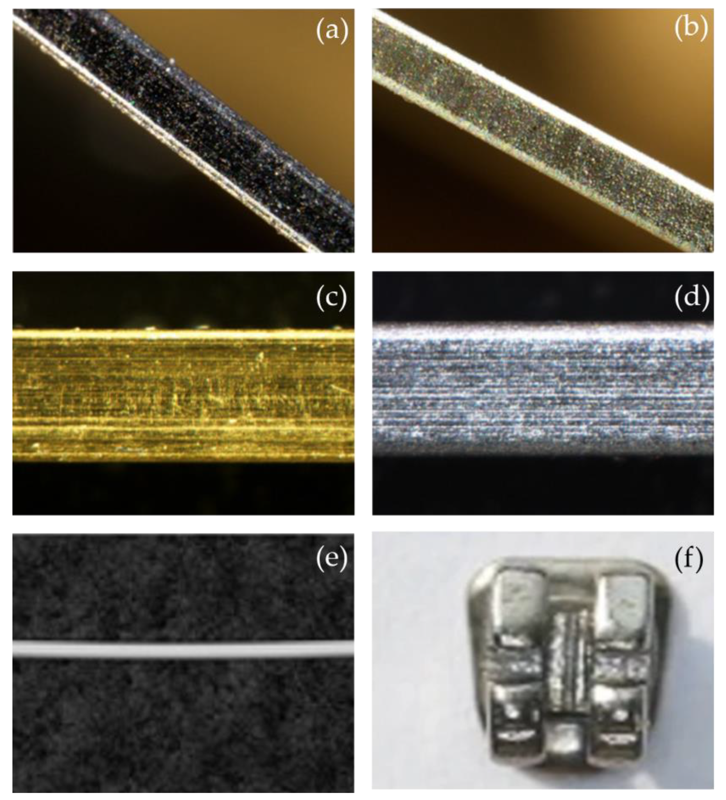
Figure 1. Functional coatings that have been applicated on brackets and archwires. (a) nickel (Ni) + molybdenum disulfide (MoS2)-coated stainless steel (SS) archwire; (b) Ni + tungsten disulfide (WS2)-coated SS archwire; (c) titanium nitride (TiN)-coated SS archwire; (d) TiN-coated nickel-titanium (NiTi) archwire; (e) polymer-coated wires (microcoated stainless steel wire®, G&H Wire Company, Franklin, IN, USA); (f) polydopamine and blue fluorescent hollow carbon dots (PDA-HCD) brackets. Adapted with permission from [18,19,20,21].
Ijms 24 06919 g001
Figure 2. Summary of friction reduction coating materials. WS2: tungsten disulfide; MoS2: molybdenum disulfide; ZnO: zinc oxide; Zn: zinc; TiN: titanium nitride; TiCN: titanium carbonitride; TiO2: titanium dioxide; Ag: silver; Al2O3: aluminum oxide; CNx: carbon nitride; TaAgB: silver-doped tantalum boride; Ni-Ti-Mo: nickel-titanium-molybdenum; Ni-Ti-Cr: nickel-titanium-chromium; Al-SiO2: aluminum-silicon dioxide; DLC: diamond-like carbon; GSEC: graphene-sheet-embedded carbon; GO: graphene oxide; GO/Ag: graphene oxide/silver; PTFE: polytetrafluoroethylene; MPC: 2-methacryloyloxyethyl phosphorylcholine; CTS: chitosan.
Ijms 24 06919 g002
Figure 3. (a) Dynamic friction curves for TaAgB coating, TaB2/Ag coating, and uncoated 304 steel. (b) Mean friction coefficient for TaAgB coating, TaB2/Ag coating, and uncoated 304 steel. TaB2: tantalum boride. Adapted with permission from [68]. Copyright 2022, Elsevier.
Ijms 24 06919 g003
Figure 4. The low-friction and low-wear mechanisms of the GSEC-film-coated stainless steel archwires running against three-row microgroove-textured stainless steel brackets in an artificial saliva environment. (a) Initial state of the contact combination, (b) stable low friction with the formation of graphene-rich tribofilm and salivary adsorbed layer, (c) accumulation of wear debris detached from the GSEC film with microgroove, and (d) flow out of wear debris with artificial saliva from the microgroove. Adapted with permission from [46].
Ijms 24 06919 g004
Figure 5. Summary of antibacterial coating materials. Au: gold; Ag-TiO2: silver-titanium dioxide; PEDOT: poly(3,4-ethylenedioxythiophene); PEDOT/Ag: poly(3,4-ethylenedioxythiophene)/silver; TiO2−xNy: nitrogen-doped titanium dioxide; F-O-Ti: fluorine and oxygen double-deposited titanium; AgNP/PTFE: nanosilver/polytetrafluoroethylene; SiOx: silicon oxides; BSA: bovine serum albumin; CTS/PEG: chitosan/polyethylene glycol.
Ijms 24 06919 g005
Figure 6. Live (green) and dead (red) staining of Staphylococcus aureus on different samples after culturing for 1 day. Adapted with permission from [136]. Copyright 2022, Elsevier.
Ijms 24 06919 g006
Figure 7. Summary of antibacterial coating materials. TiAlN: titanium aluminum nitride; Ti-DLC: titanium-diamond-like carbon; Al2O3/TiO2: aluminum oxide/titanium dioxide; Ti-Cr-N: titanium-chromium-nitrogen; Ti-Nb: titanium-niobium; Ti/TiN: titanium/titanium nitride; N: nitrogen; aC:H: hydrogenated forms of diamond-like carbon; PEN: polyethylene naphthalate; GB-PP: grid-blasted plasma-polymerized; IL: ionic liquid.
Ijms 24 06919 g007
Figure 8. Scanning probe microscope (SPM) images of (a) current-anodized and (b) pulse-anodized NiTi surfaces. The white arrows show the pores on the surfaces of the coating. Adapted with permission from [174]. Copyright 2022, Elsevier.
Ijms 24 06919 g008
Figure 9. (ac) Scanning electron microscope (SEM) images, (df) energy-dispersive spectroscopy (EDS) analysis, and (gi) atomic force microscope (AFM) images of the bare NiTi, GO-coated NiTi, and GO/Ag-coated NiTi alloy, respectively. Adapted with permission from [181].
Ijms 24 06919 g009
Disclaimer/Publisher’s Note: The statements, opinions and data contained in all publications are solely those of the individual author(s) and contributor(s) and not of MDPI and/or the editor(s). MDPI and/or the editor(s) disclaim responsibility for any injury to people or property resulting from any ideas, methods, instructions or products referred to in the content.