Article

Abundance of Transgene Transcript Variants Associated with Somatically Active Transgenic Helitrons from Multiple T-DNA Integration Sites in Maize

1
Biotechnology Research Institute, Chinese Academy of Agricultural Sciences, Beijing 100081, China
2
College of Agronomy, Qingdao Agricultural University, Qingdao 266109, China
*
Author to whom correspondence should be addressed.
Int. J. Mol. Sci. 2023, 24(7), 6574; https://doi.org/10.3390/ijms24076574
Submission received: 8 March 2023 / Revised: 27 March 2023 / Accepted: 28 March 2023 / Published: 31 March 2023
(This article belongs to the Special Issue Molecular Genetics and Plant Breeding 3.0)

Abstract

:
Helitrons, a novel type of mysterious DNA transposons discovered computationally prior to bench work confirmation, are components ubiquitous in most sequenced genomes of various eukaryotes, including plants, animals, and fungi. There is a paucity of empirical evidence to elucidate the mechanism of Helitrons transposition in plants. Here, by constructing several artificial defective Helitron (dHel) reporter systems, we aim to identify the autonomous Helitrons (aHel) in maize genetically and to demonstrate the transposition and repair mechanisms of Helitrons upon the dHel-GFP excision in maize. When crossing with various inbred lines, several transgenic lines produced progeny of segregated, purple-blotched kernels, resulting from a leaky expression of the C1 gene driven by the dHel-interrupted promoter. Transcription analysis indicated that the insertion of different dHels into the C1 promoter or exon would lead to multiple distinct mRNA transcripts corresponding to transgenes in the host genome. Simple excision products and circular intermediates of dHel-GFP transposition have been detected from the leaf tissue of the seedlings in F1 hybrids of transgenic lines with corresponding c1 tester, although they failed to be detected in all primary transgenic lines. These results revealed the transposition and repair mechanism of Helitrons in maize. It is strongly suggested that this reporter system can detect the genetic activity of autonomic Helitron at the molecular level. Sequence features of dHel itself, together with the flanking regions, impact the excision activity of dHel and the regulation of the dHel on the transcription level of the host gene.

1. Introduction

Helitrons are DNA transposons that were first discovered by the computational analysis of eukaryote genomes and shown to be ubiquitous in fungi, plants, and animals with later experimental confirmations [1,2,3,4,5,6]. Helitrons lack the typical structural features of classic DNA transposons, such as terminal inverted repeats (TIRs) or target site duplications (TSDs) upon insertion, making it more challenging for precise annotation. Helitrons possess few constant structural and sequence features instead, including conserved 5′-TC and 3′-CTRR termini as well as palindromic sequences near the 3′ terminus. Helitrons insert mainly into the AT host dinucleotides. Furthermore, the continuous, incremental improvements of algorithms have allowed the identification of an increasing number of Helitrons from unceasing genomic sequencing efforts [7,8,9,10]. In addition, bioinformatic analysis established that Helitrons also exhibit sequence conservation over a 30-bp stretch from either their 5′ or 3′ terminus, which is oftentimes one of the major criteria for grouping novel Helitron elements [11,12,13,14].
The variation caused by Helitron activity in plants mainly consists of insertional mutations and consequent haplotype diversity from colinearity violation of genic Helitron transposons. Helitron transposons tend to insert themselves into the regulatory regions of genes, such as in promoters [15,16,17], introns [7,18], and 3′ untranslated regions (3′ UTRs) [19], which often leads to a loss of function in the affected gene and changes in traits. For instance, a 6.5-kb Helitron element was identified in the proximal promoter region of barren stalk1 (ba1-ref), leading to defective developments of axillary meristems in maize [15]. The insertion of Helitrons can also create new functions and traits in the host, which is an important driving force of genome variability and evolution [20].
The identification of structurally similar Helitron transposons in a growing number of genomes [18,21] lends further support to the hypothesis that aHel elements are genetically active in diverse genomes [10,22,23,24,25,26]. In plants, Helitron transposons appear to be exclusively non-autonomous and sometimes carry other gene fragments with protein-coding potential outside of the open reading frame encoding a defective RepHel (replication initiation protein and helicase domain) transposase [8,9,15,17,27].
Due to their lack of significant structural and sequence features compared to other transposable elements, it is extremely challenging that the identification of Helitron transposons in a given genome relies heavily on thorough mining and annotation of the genome. The only plausible conserved sequences at both termini and 3′-end stem-loop structure are the main basis for the identification of Helitrons [11,12,13,28,29,30,31,32]. Notably, the development of high-throughput sequencing technologies and a better understanding of Helitron structure have facilitated the discovery of many non-autonomous Helitron transposons and a few suspected examples of aHel in plant and animal genomes [1,2,3,5,13,26]. Recently, Grabundzija et al. have successfully assembled an autonomous Helitron transposon, named Helraiser, from Myotis lucifugus [10,33], demonstrating transposon activities in vitro via the previously proposed transposition mechanism of rolling-circle replication (RCR). Furthermore, Kosek et al. reported the cryo-electron microscopy structure of the Helraiser transposase and demonstrated the tightly packed assembly formed by monomeric Helitron transposase to bury the covalently attached cleaved end and protect it until the second end becomes available [34].
Nevertheless, in maize (Zea mays L.), Li and Dooner [35] detected the unexpected somatic excision of various non-autonomous Helitrons from multiple genetic loci differed in inbred lines. Among them, the 6-kb HelA2 Helitron transposon was excised, leaving footprints of a variable number of TA repeats at the original insertion site. These results suggested that Helitrons may also transpose through a cut-and-paste mechanism in addition to the RCR transposition mechanism. Helitrons are widespread DNA transposons in eukaryotes; however, their distribution was significantly different among organisms. For instance, the total number and classifications of Helitron transposons in rice (Oryza sativa) differ from maize genomes, as revealed by bioinformatic analysis with the HelitronScanner [13]. There are hundreds of tandemly arrayed and truncated CentHel elements in rice; however, the number of CentHel in the same category in maize was fewer than six. Thereby, the cut-and-paste transposition mechanism [35] may be more prevalent in maize than in rice [36]. While the molecular structure of autonomous Helitrons and their replicative transposition via rolling-circle replication mechanism—particularly in animals—has been intensively studied, further investigations are much need to verify the prevalence of the cut-and-paste transposition mechanism and genetically define and molecularly clone the autonomous Helitrons, practically in plants.
Here, we constructed several maize Helitron systems to demonstrate their transposition activity of the cut-and-paste mechanism, excision footprint repairing mechanism, and sequence preferences upon reinsertion of excised Helitrons. In this artificial system, green fluorescent protein (GFP) is driven by the promoter of an endosperm-specific maize gene and flanked by both 5′-end and 3′-end conserved terminal sequences from either high-copy-number or genetically active non-autonomous Helitrons in maize. To phenotypically monitor the transposition activity of aHel in maize, the dHel-GFP elements are placed in a gene cassette of Colored aleurone 1 (C1), an indispensable transcription factor responsible for pigmentation in aleurone of maize kernels. We transformed the construct into a c1 donor inbred line via Agrobacterium tumefaciens–mediated transformation, identified GFP-positive transformants and crossed to other c1 maize inbred lines or hybrids from different inbred lines. The somatic excision of dHel-GFP transposons was monitored by the occurrence of the segregation of purple-spotted kernels resulting from the presence of aHel and the existence of a cut-and-paste transposition mechanism. These genetically identified aHel could then be cloned in the corresponding segregating populations for further characterization of their sequence structures, the genetic basis of either the rolling-circle-replication or cut-and-paste transposition mechanism, their repair mechanism upon excision, and their sequence preference for new insertion sites. Tackling these questions will lead to a greater understanding of Helitron biology and its application in genome engineering. Furthermore, the cloning of autonomous Helitrons in maize and the generation of efficient mutant collection from dHel-GFP insertions will spur the development of reverse genetic resources in maize functional genomic studies.

2. Results

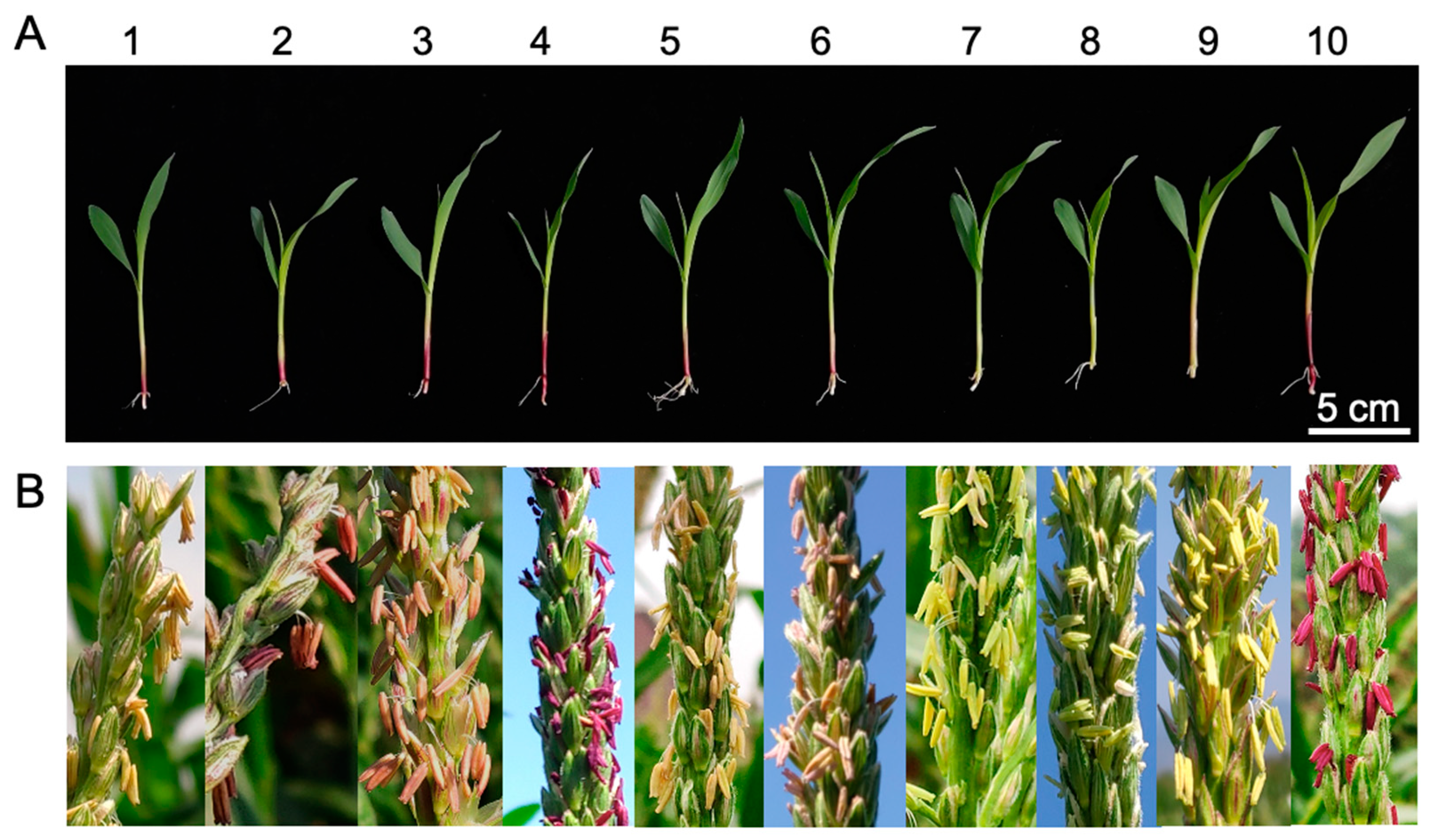
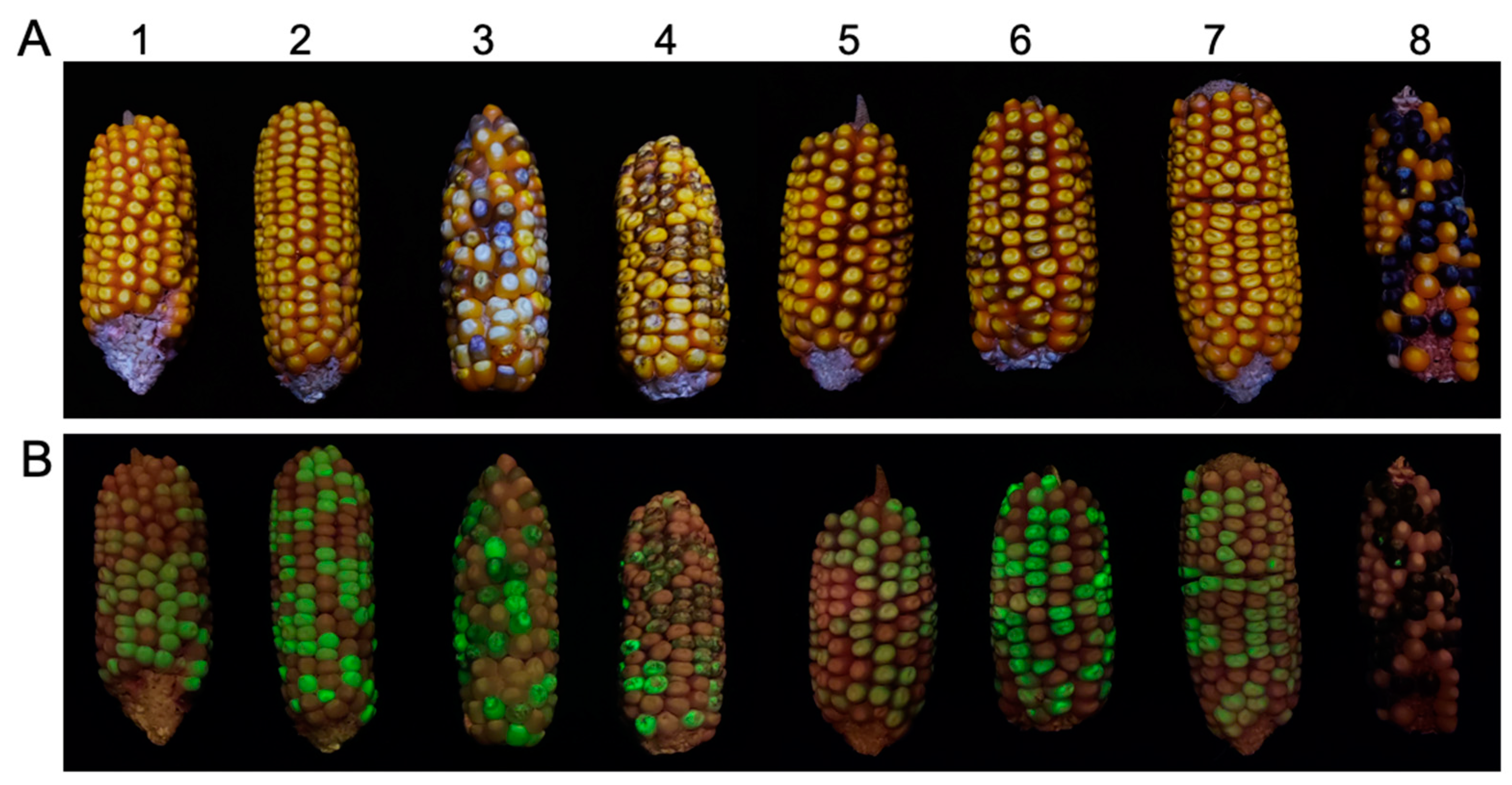
2.1. Phenotypic Variation Correlated with Transgene Expression in Primary Transformants and Their Hybrids with c1 Tester Lines

We created several DNA constructs to answer long-standing questions regarding autonomous Helitron transposons in maize by combining genetic analysis and transgenic approaches to detect the genetic behaviors of transformed defective Helitrons in various genetic backgrounds upon crossing with numerous inbred lines (Figure 1). Surprisingly, we found that the majority of T2 c1-dHel-GFP transgenic seedlings show various degrees of purple pigmentation on their coleoptiles (Figure 2(A1–A6), Table 1, Table S1), in contrast to the colorless coleoptiles of seedlings of either donor lines or non-transgenic segregants, except for the transgenic lines from construct S4-Hel1-4, of which dHel insertion site is right at the translation start site of C1 gene (Figure 1B, Figure 2(A7)). Meanwhile, T2 c1-dHel-GFP transgenic anthers show a varied intensity of purple pigmentation upon anther emergence except for the transgenic lines from construct S4-Hel1-4 which represents its donor line of B104, in line with the pigmentation patterns on the coleoptiles (Figure 2B), regardless of the shared colorless kernel phenotype in all T2 ears (Figure S1).
When crossing the T2 transgenic lines to the c1 tester line, the F1 progenies possess interesting kernel phenotypes. For instance, all F1 kernels derived from crosses between the c1 tester and transgenic plants harboring the S3-Hel1613 transgene (F1-S3-Hel1613 thereafter) or F1-S4-Hel1-4 are colorless, regardless of if they are GFP segregants. On the other hand, we detected pale purple seeds, which also showed green fluorescence, in the F1 test ears of F1-S1-Hel1-4, F1-S2-Hel1-4, F1-S3-Hel1-4, F1-S3-Hel1158, and F1-S3-HelA2 (Figure 3).
Additionally, in the BC1F1 populations for assessing the presence of autonomous Helitrons in tested inbred lines (Table S2), purple kernels are being segregated from crosses with inbred lines of Ky228, HP72-11, 4722, Va102, Va35, Chang7-2, CML 277, A554, P39, IA5125, and C13, respectively. However, the purple kernel phenotype is independent of green fluorescence, suggesting the presence of a wild-type C1 gene in these inbred lines, which makes it unlikely precise excision in the transgenic c1-dHel-GFP constructs. Moreover, unilateral cross incompatibility (UCI) occurs between the BC1 generation (as pollen donor) and the inbred lines of CML 220, CML 321, HP72-11, and HP301, an expected result elucidated in recent publications [37]. However, no purple-spotted kernels are found on the BC1F1 segregants when all four Ga1-S inbred lines crossed with transgenic BC1 plants.
In the panel of tested inbred lines, CML 11, CML 328, and CML 331 are known for carrying C1-I allele [38], when crossing with W22, BC1-S1-Hel1-4, BC1-S2-Hel1-4, BC1-S3-Hel1-4, BC1-S4-Hel1-4, BC1-S3-Hel1613, BC1-S3-Hel1158, and BC1-S3-HelA2, all BC1F1 hybrid seeds are colorless as expected. When crossing with other inbred lines in our testing panel, the kernel pigmentation phenotype of the resulting BC1F1 segregants was similar to the kernel phenotype in F1 populations (T2 transgenic line × c1 tester) as exampled in Figure 3. Both BC1F1-S3-Hel1613 and BC1F1-S4-Hel1-4 progenies are colorless kernels without purple spots, regardless of the green, fluorescent kernel phenotype segregation. However, green, fluorescent BC1F1 progenies of transformants from the other five constructs are all purple-mottled phenotypes.

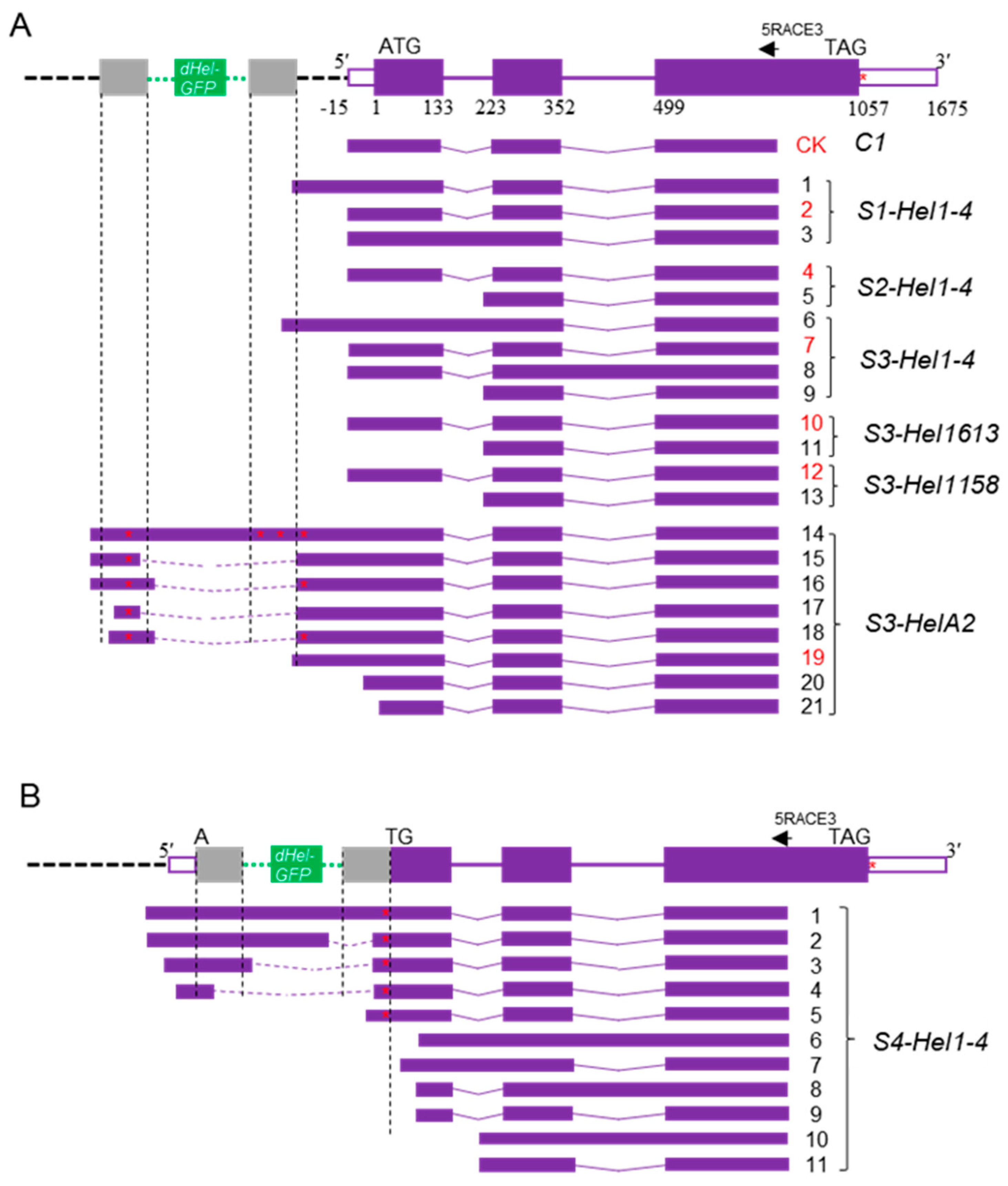
2.2. Transcript Spectrum of c1 Transgenes from Variable dHel-GFP Insertion Sites

The unexpected pigmentation phenotypes in the coleoptile and anthers of T2 transformants from several c1-dHel-GFP constructs (Figure 2) or among the F1 kernels of T2 × c1 tester and BC1F1 kernel segregants (Figure 3, Table 1) ushered in scrutinizing the C1 transcripts from c1-dHel-GFP transgenes via RT-PCR and rapid amplification of cDNA ends (RACE) with total RNA extracted from T2 transgenic seedlings of all seven constructs (Table S3). RT-PCR assays showed an unpredicted expression of the c1-dHel-GFP transgene in T2 transformants from all seven constructs as the positive C1 transgene control and the predictable absence of the c1 expression in the negative controls of B104 and HiII donor lines (Figure S2). Notably, RT-PCR products from the S4-Hel1-4 construct transgene are much longer than those from all other six constructs. Further sequence analysis of the cloned RT-PCR products was in line with the purple pigmentation phenotype in both coleoptile and anthers of the corresponding transgenic lines.
To elucidate the molecular bases of the difference between pigmentation phenotypes of transgenic lines from various constructs, we constructed C1 cDNA using both the 3′ RACE and 5′ RACE protocols. The generation of C1 cDNA from the positive C1 transgene control by the RACE protocols confirms the presence of C1 transcripts in the tested tissue from the transgenic plants (Figure 4). Nucleotide sequence analysis of the C1 cDNA has defined the 5′ end and the 3′ end of each of the clones. All sequenced clones of the 5′ end from the positive C1 transgene control were identical to the endogenous C1 cds (AF320613 and AF320614) in terms of the transcription start site and the intron-exon splicing sites (Figure 3B and Figure 4A). At the 3′ end, we identified two major transcripts resulting from alternative polyadenylation differed by 54 bp (Figure 4A and Figure S3), 618 bp and 564 bp downstream of the stop codon, respectively (Figure S4).
Three types of shorter 3′ UTR sequences were detected across the seven c1-dHel-GFP transgenic lines (Figure 4A and Figure S3). Sequence analysis showed that they mainly differed from the positive C1 transgene control or one another at the alterable polyadenylation sites (Table S4). The majority of polyadenylation sites of the clones generated by the RACE protocol spanned an area of 400 nucleotides annotated as an hAT element (Figure S4). Nevertheless, the detected 3′-end coding regions were intact from all transgenic lines, suggesting that none of the dHel-GFP insertions affected the transcription of the 3′-end of the transgene.
In contrast to the multiple 3′ RACE products resulting from alternative polyadenylation, the 5′ RACE products detected in the transgenic lines were greatly influenced by dHel-GFP insertions at either the promoter region or the translation starting site (Figure 1B, Table 1 and Figure 5B). The 5′ RACE sequence analysis confirms that the 5′ UTR of transgenic lines from constructs of S2-Hel1-4, S3-Hel1-4, S3-Hel1158 and S3-Hel1613 is 15 bp in length (Figure 5A, transcript variants No.4, 7, 10 and 12, Table S5), identical to the positive C1 transgene control (Figure 5A CK) and the canonical 5′ UTR of the endogenous C1 transcript. Additionally, other uncanonical variants of transcripts are readily detected, including possible novel upstream promoter (Figure 5A, transcript variant No.6), intron retention (Figure 5A, transcript variants No.6 and 8), additional exonic sequences (Figure 5A, transcript variants No.5, 9, 11, and 13, Table S5) due to alternative splicing, and unlikely to be translated to functional proteins.
The canonical 5′ UTR is also identified from transcripts of S1-Hel1-4 transgenic lines (Figure 5A, transcript variant No.2, Table S5), in addition to transcripts characterized with retained intron 1 (Figure 5A, transcript variant No.3) or transcripts initiated possibly in the 3′ end of Hel1-4 and transcribed at 16-bp upstream of the insertion site without altering both introns-exon splicing sites in the downstream region (Figure 5A, transcript variant No.1, Table S5).
Several novel transcripts of splicing variants were cloned with the amplicon mixture from S3-HelA2 transgenic line sharing the same site (AT di-nucleotides) as in the S3-Hel1613, S3-Hel1158, and S3-Hel1-4 lines. The major amplicon was a mixture of multiple splicing variants with similar lengths but different sequence compositions from the S3-HelA2 transformant (Figure 4(B3), Table S5). The eight variants of sequenced transcripts, they share the same downstream intron-exon junctions in the c1 coding region as the wild type C1 gene. They mainly differed from each other by the first one or two novel exons resulting from initiating possibly in a novel promoter region (thus, in the transgenic C1 promoter) further upstream of the 20-bp upstream of HelA2-GFP insertion site (Figure 5A, transcript variants No.14, 15 and 16).
Among them, the longest transcript, although much less abundant than the major amplicons, includes the entire HelA2-GFP element and the downstream c1 transcripts featured by the canonical intron-exon splicing junctions, generating a new type of chimeric transcript (Figure 5A, transcript variant No.14, Table S5). Two alternative splicing transcripts may share the same novel promoter with the longest transcript since their first exon is identical to the 5′-end of the longest transcript. However, they differ from each other with an extra intron sharing the same 3′ splice site (right at the 3′-end of HelA2-GFP, CTAG) but not the 5′splice sites (47 bp apart) inside the HelA2-GFP segment (Figure 5A, transcript variants No.15 and 16, Table S5). Two other alternative splicing transcripts, initiating right in the LTS (14 bp apart) from other possible novel promotors, share the same intron-exon splicing junctions as two previously described transcripts, respectively (Figure 5A, transcript variants No.17 and 18, Table S5). Moreover, the RTS could also be a novel promotor since one transcript initiated from 14 bp upstream of the 3′-end CTAG (Figure 5A, transcript variants No.19, Table S5). The sequences of the two shortest transcript variants detected from the S3-HelA2 transformants are different at the transcription start sites that are 3-bp apart around the ATG translation start codon, although they are much less abundant in the 5′ RACE amplicons.
The most heterogeneous transcript variants are isolated from S4-Hel1-4 transgenic lines (Figure 3B and Figure 4B), in which the dHel-GFP insertion site was in between an AT dinucleotide of the C1 start codon (Figure 1B). The most abundant amplicon is the uncanonical variant of transcripts from additional exonic sequences as previously described in other transgenic lines transformed with various constructs (Figure 4, lane4; Figure 5A, transcript variants No.5, 9, 11, 13, Figure 5B transcript variant No.11 and Table S5). In addition, other ten uncanonical variants of transcripts are readily detected. For instance, the second most abundant amplicon is a transcript variant of intron retention sharing the same transcription start site as the most abundant amplicon (Figure 5B transcript variant No.10 and Table S5). The transcription start sites of four transcript variants, featured with the prevalent intron retention variations, are at three different nucleotide positions in the first exon, such as 3, 67, and 70, respectively (Figure 5B, transcript variants No.6–9, Table S5). Similar to the findings from other constructs, either the same Hel1-4 or the different HelA2, the RTS of S4-Hel1-4 could also be a novel promotor since B7 transcript initiated from nucleotide position 3, evidenced with another transcript variant initiating from 67 bp upstream of the 3′-end CTAG (Figure 5B, transcript variant No.5, Table S5). All remaining four transcript variants may share the same promotor as the canonical C1 transcript, but their transcription start sites are at a region of 12 bp to 68 bp upstream of the 5′-end TC, leading to uncanonical transcript variants of intron retention (Figure 5 B, transcript variant No.1), additional exonic sequences due to alternative splicing (Figure 5B, transcript variants No.1–4, Table S5). All detected uncanonical variants of transcripts from S4-Hel1-4 transgenic lines are unlikely to be translated to functional proteins, consistent with the colorless kernel phenotype.
A total of five of the six transgenic c1 alleles showed relatively consistent transcript patterns in spite of their apparent difference at the insertion site of dHel-GFP in the promoter region (S1-Hel1-4, S2-Hel1-4, S3-Hel1-4, S3-Hel1613, and S3-Hel1158). Most transcripts were still functionally equivalent to the control C1 transcript unless they lacked the first exon or retained part of the first intron.
Surprisingly, the main types of transcripts seen in the transgenic lines S3-Hel1158, S3-Hel1613, and S3-Hel1-4 were the same as C1, whereas the S3-HelA2 transgenic line produced a variety of new transcripts encoding a protein with partial C1 function. The above four transgenes share the same insertion site for the dHel-GFP cassette and have similar 5′ and 3′ end sequences, but S3-HelA2 generated completely different transcript isoforms from the other three transgenes, perhaps reflecting the Helitron transposon incorporated into the constructs.
The main 5′ RACE products detected from S3-HelA2 transgenic lines are quite unique compared with transgenic lines from all other constructs (Figure 4B lane 3). Two major transcripts are initiated from either 20-bp upstream of the HelA2 insertion site or right inside the 5′-end (30-bp) of the HelA2 element (Figure 5A, transcript variants No.15 and 18, Table S5). These two C1 transcripts differ at their 5′ splicing sites since both 3′ splicing sites were the same and corresponded exactly to the 3′ end (AG) of HelA2 (Figure 5A, transcript variants No.15 and 18). Other minor transcripts differ from each other with varied lengths due to various transcriptional start sites (TSSs) around the HelA2 insertion site (Figure 5A, transcript variants No.14–21), multiple 5′ splicing sites (Figure 5A, transcript variants No.15–18), the intron retention of the whole HelA2 element (Figure 5A, transcript variant No.14, Table S5). The majority of the C1 transcripts in the S3-HelA2 transgenic line include the TATA box region and the C1 promoter (Figure 5A, transcript variants No.14–19); however, the open reading frames of these transcripts had multiple translation termination codons prior to the canonical translation start site of C1 and led to non-functional open reading frame upstream of the original C1 open reading frame.
Recently, the identification of evolutionarily conserved features correlated with promoter expression levels in TSSs has become an important area in understanding the control of transcription initiation and regulation in a plant, including the identification of novel patterns in the dinucleotides composing the initiator element [39]. To gain a glimpse into the alteration of general sequence patterns surrounding the TSS resulting from Helitron insertions, 5′ RACE assay showed that Helitron insertions generally did not affect the patterns in the dinucleotides composing the initiator element since highly expressed transgenes were enriched most commonly for CA, followed by CG, TG, which perfectly agrees with recent research [39]. This occurred regardless of the presence of the TATA box and the varied content of CG nucleotide between the TATA box and TSS, the initiator element (Figure 1B, Table S6).

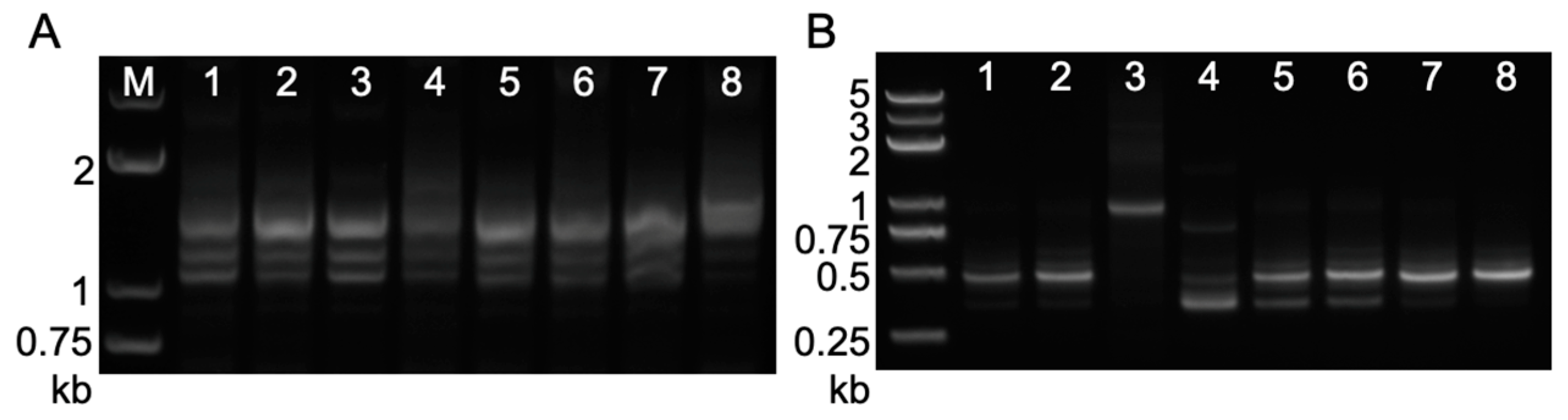
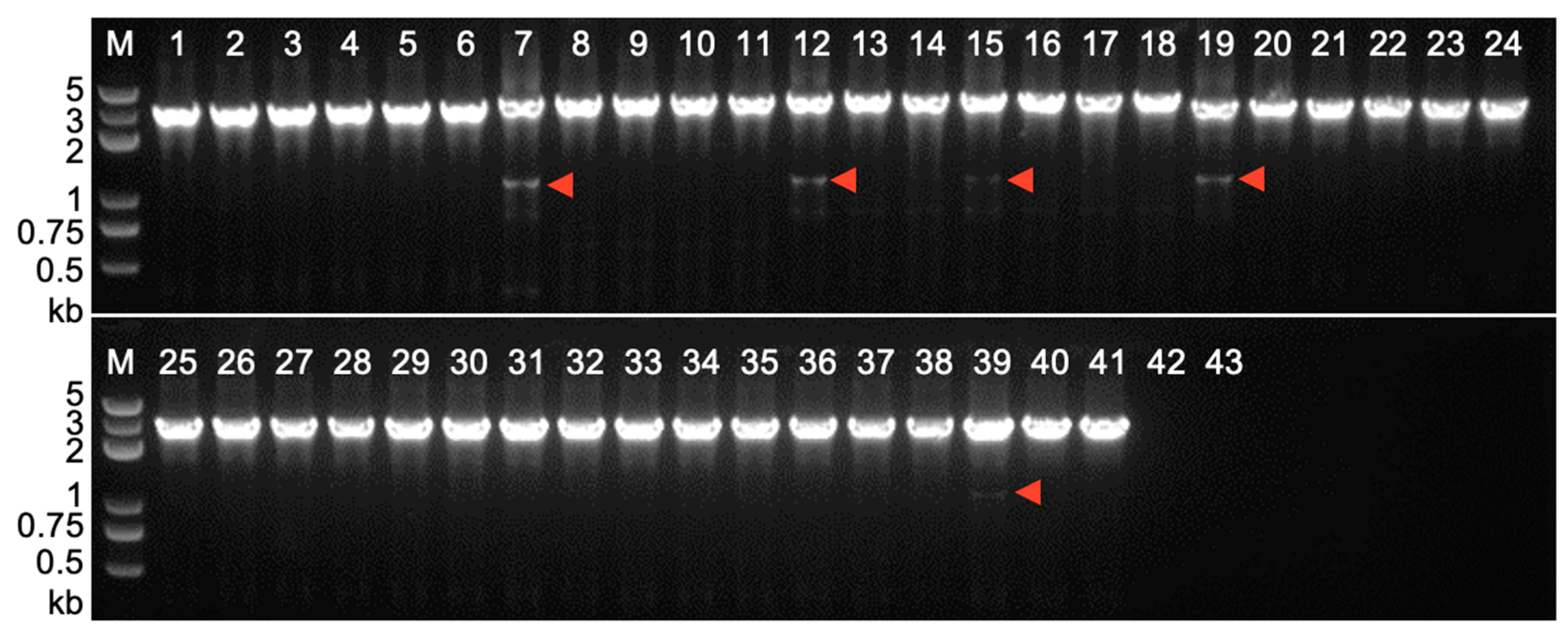
2.3. Footprint Analysis on Somatic Excision of Helitrons via the Cut-And-Paste Mechanism in Transgenic Lines and Hybrids with Maize Inbred Lines

Somatic excision of Helitrons has been demonstrated via PCR amplification in previous research on a previously unfavorable cut-and-paste transposition mechanism [35]. To address the possibility of genetic activity of putative autonomous Helitrons in maize, we utilized nested PCR to detect occasional somatic excision events from T2 leaves of all seven different transformants (Figure 1A, Table S3). However, we have not amplified PCR products with expected sizes of the corresponding somatic excision from the c1-dHel-GFP cassette in any one of the seven T2 transgenic lines, suggesting an absence of autonomous Helitrons activities from the genetic background of the donor lines (Figure S5).
When the same PCR strategy was applied to DNA samples of leave tissue from various F1 hybrids of T2 transgenic lines with the c1 tester line, we were instead able to amplify putative somatic excision products from some transgenic segregants, such as S1-Hel1-4 (Figure 6, lane 39), S3-Hel1158 (Figure 6, lane 7), S3-HelA2 (Figure 6, lanes 12 and 15), and S4-Hel1-4 (Figure 6, lane 19). Similar PCR amplicons of putative somatic excision products were also detected from several other segregants of two out of those four transgenic lines, namely S1-Hel1-4 F1 hybrids and S4-Hel1-4 F1 hybrids (Figure S6). However, we were failed to detect somatic excision products from DNA samples of leave tissue at the V2 seedling stage of more than 30 segregants of F1 hybrids between other T2 transgenic lines, S3-Hel1613, S2-Hel1-4, or S3-Hel1-4, with the c1 tester line, respectively (Table 2). This suggests that the excision frequency of dHel-GFP is extremely low, in the case of S2-Hel1-4 or S3-Hel1-4, or an absence of autonomous Helitrons in both parental lines, in the case of S3-Hel1613.
The cloning of PCR amplicons of putative somatic excision products and the subsequent sequence analysis revealed that all somatic excisions were identical to the wild-type sequence (Table 3). The direct sequencing of the gel-purified PCR amplicons of putative somatic excision products (Figure S6) showed the same footprint as fp0. The unique excision footprint of fp0 has been reported and deemed conserved among certain Helitron families sharing a similar repairing mechanism upon somatic excision [12,35].

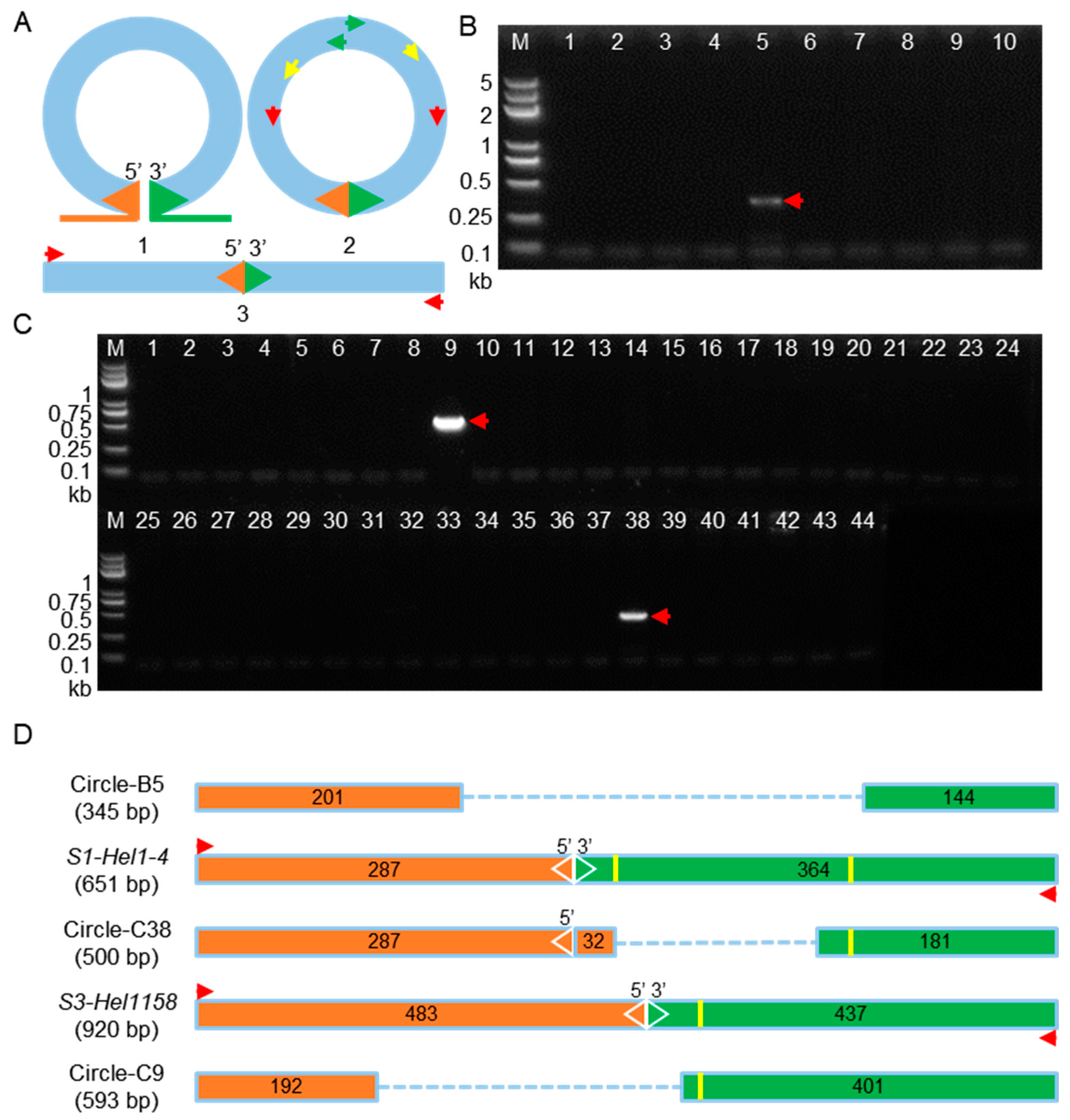
2.4. Circular Intermediates Detection in Rolling-Circle Transposition of Helitrons from Transgenic Lines and Their Hybrid Populations

Recently, Grabundzija et al. constructed an active Helitron (“Helraiser”) from the genome of Myotis lucifugus and experimentally demonstrated the RCR transposition mechanism by sequence confirmation of the PCR amplified circular transposon intermediate containing an RTS-LTS configuration and the reinsertion of Helraiser in vitro [33]. To address the possibility of the existence of circular intermediates of dHel-GFP elements undergoing the RCR transposition, PCR strategy was applied to the same set of DNA samples of leaf tissue at V2 seedling stage from corresponding transgenic lines as for the somatic excision analysis (Table S3 and Figure 7A).
We have not amplified any circular dHel intermediates with an expected sequence configuration of both intact LTSs and RTSs by nested PCR in either T2 transgenic individuals or their F1 hybrids with the c1 tester line from all seven constructs (Figure S7, Table S7). Alternatively, we detected other structure derivatives of expected circular dHel intermediates from T2 individuals transformants of S1-Hel1-4 (Figure 7B lane 5) and individuals transformants from F1 hybrids of S1-Hel1-4 and S3-Hel1158 (Figure 7C lanes 38 and 9, respectively).
The sequence analysis of such circular dHel intermediates revealed that these amplicons featured deletions at either or both ends of dHel-GFP elements (Figure 7D). In detail, the PCR fragment amplified in T2 transformant from S1-Hel1-4 is 345 bp in length, composed of 201-bp LTS and 144-bp RTS, which is much shorter than the expected length of 651-bp (287-bp LTS and 364-bp RTS), due to an internal deletion of the junction sequences of 86 bp from the LTS and 220 bp from the RTS including two stem-loop structures at the 3′-end of S1-Hel1-4 RTS (Figure 7D, Circle-B5). Furthermore, no microhomology is presented at the junction site, which is frequently found during transposition. In contrast, the circular dHel-GFP intermediate amplified in F1 individual of the same S1-Hel1-4 transformant is 500 bp in length, composed of an intact LTS and its adjacent sequence of 32 bp, and 181-bp remanent RTS due to an adjacent deletion of 183 bp proximal RTS. Notably, only one of the two stem-loop structures (TTTTTACCAAAAAA) was retained in the 181-bp remanent RTS ((Figure 7D, Circle-C38; Table S8). Moreover, we detected an imperfect microhomology (CTCAA/CTCACA) at the circularization junction between LTS and RTS. Finally, the circular dHel intermediate amplified from the F1 individual of S3-Hel1158 transformant is 593 bp in length, composed of 192-bp LTS and 401-bp RTS, which is much shorter than the expected length of 920-bp (483-bp LTS and 437-bp RTS), due to an imbalanced internal deletion of the junction sequences of 291-bp from the LTS and 35-bp from the RTS excluding the solo stem-loop structure at the 3′-end of S3-Hel1158 RTS (TAAAAAATCTTGAAATTTTTTTA, Figure 7D, Circle-C9, and Table S8). In addition, a dinucleotide microhomology (GT) was presented at the circularization junction between LTS and RTS of Circle-C9.

3. Discussion

3.1. Helitron Transposon Families and Their Insertion Sites in the Promoter Region Alter Gene Expression through Linked Processes of Transcription or Splicing

The initial objective of this research was to genetically identify the autonomous Helitron transposons in maize by constructing reporter systems of non-autonomous dHel elements to generate transgenic lines for genetic tests. We build seven c1-dHel-GFP constructs featured by four diverged Helitron families inserted at four dispersed sites in the promoter region of the C1 gene to augment the possibility of identifying the genetic activities of mysterious autonomous Helitrons in maize (Figure 1, Table 1, Tables S1 and S2). In total, 34 transgenic events transformed with seven constructs were analyzed for phenotyping on various tissues of the individual primary transformants or their segregating populations genetically (Table 1, Table 2 and Table S1, Figure 1, Figure 2 and Figure S1), and were characterized for T-DNA integration sites, transgene copy numbers and expression pattern molecularly (Figure 3 and Figure 4, Figures S2–S4, Tables S3–S6).
The C1 promoter has been one of the hotspots for gene function characterization in maize even in plant [40,41,42]. Different C1 expression rates can be correlated with promoter sequence alterations in maize [43]. In current study, all four dHel insertion sites −93, −49, −47, and +1 relative to the start codon ATG) are located downstream of the previously identified cis-regulatory elements (Figure 1), such as abscisic acid (ABA) response factors and the Viviparous-1 (Vp-1) transcription factors at positions −157 to −130 relative to the transcription start site [43]. Compared to non-transgenic wild-type controls, the coleoptiles or anthers of T2 transgenic seedlings, segregants of the c1 tester line crossing with transformants from constructs of S3-Hel1613, S3-Hel1158, S3-HelA2, S1-Hel1-4, S2-Hel1-4, and S3-Hel1-4, are anthocyanin pigmented, while the coleoptile of S4-Hel1-4 T2 transgenic seedlings is in light green or transparent (Figure 1 and Figure 2, Table 1). Furthermore, the discrepancy between purple-pigmented coleoptile in T2 S3-Hel1613 seedlings and their colorless kernel phenotype indicated that the C1 transgene expression in the aleurone layer might be blocked by the insertion of Hel1613-GFP but not in coleoptiles tissues, due to a possible change in the expression pattern of temporal and spatial specificity of the C1 transgene.
We employed both RT-PCR and RACE assays to decipher the unforeseen pigmentation pattern due to possible expression of the c1-dHel-GFP alleles in the transformation constructs by detecting the expression pattern and cloning the full-length cDNA of the C1 transcripts transcribed from each dHel-GFP transgene (Figure 3 and Figure 4, Figures S2–S4, Tables S3–S6). RT-PCR results are extremely supportive of the pigmentation pattern of different transformants, such as the colorless kernel and the light green or transparent coleoptile phenotype of the S4-Hel1-4 T2 transgenic seedlings. However, further RACE assays show abundant transcript variants of the S4-Hel1-4 T2 transgenic seedlings indicating the normal functioning of the c1 promoter. All other three insertion sites of Hel1-4 were free of interfering with the normal function of the c1 promoter, which was evidenced by the similar pigmentation phenotypes and expression patterns of C1 transgene from T2 transgenic segregants from transformation constructs of S3-Hel1613, S3-Hel1158, and S3-HelA2, regardless of the abundance in transcription initiation variants and splicing patterns resulting from the dHel-GFP insertions in the promoter region (Figure 5, Table S5).
Combining the seed and coleoptile phenotypes, we hypothesize that dHel-GFP insertions into the C1 promoter region did not completely inactivate the C1 gene. Instead, it allowed the production of at least some canonical C1 transcripts or 5′-truncated transcripts, as seen with the S2-HelA2 transgene that appeared to retain some C1 function.
The BC1F1 hybrids between the inbred lines CML 11, CML 328, and CML 331 and S2-HelA2 transgenic plants also exhibited a block in anthocyanin biosynthesis in seeds. Just like the canonical C1 transcript, the C1 transcripts derived from the S2-HelA2 transgene also failed to induce anthocyanin biosynthesis in these BC1F1 hybrids, suggesting that the observed variation in the 5′ UTR of C1 transcripts from S2-HelA2 does not change the inhibitory effect of upstream inhibitors on anthocyanin biosynthesis.
Based on the transcripts and phenotypes described in this study, we propose that multiple transcript variants from the host gene will appear when the insertion site of dHel-GFP is located in the promoter region of the host gene, even if the insertion site is conserved between transgenes, due to the differences in non-conserved sequences between different dHels. These variant transcripts will affect the expression of downstream target genes and ultimately lead to phenotypic differences.
The promoter of the C1 locus or, in general, the 5′ region of the gene was under strong selection pressure, and several conserved or functional important sequence motifs have been characterized experimentally [43]. In our constructs, three dHel-GFP insertion sites are embedded between the insertion site of an Enl-induced C1 promoter mutation c-m668613 [44] and the TATA box for a desirably strong excision and reversion frequency without depleting the function of the c1 gene, since previous report showed that SNPs or small indels causing depletion of the C1 function in certain mutants [43]. However, the majority of dHel insertions purely affected splicing of the primary transcripts from the transgene in the transformants. Although, occasionally, the dHel functions as a novel promoter to drive the transcription of the transgene in the constructs, such as in the case of S3-HelA2 [45]. Furthermore, the Helitron insertion in different construct has no substantial effects on the patterns in the dinucleotides composing the initiator element, which is important and highly conserved to the control of transcription initiation and regulation in plant [39], but on the alternative splicing and aberrant splicing (Table S6).

3.2. Coexistence of RCR/Cut-And-Paste Somatic Transposition Mechanism Catalyzed by Autonomous Helitrons in Maize

The postulated rolling circle mode of Helitrons transposition has been demonstrated from the recent reconstruction of Helraiser, an active Helitron, from the genome of Myotis lucifugus, evidenced by molecular detection and sequence confirmation of excised circular transposon intermediate that contains an RTS-LTS junction [10,33]. We have applied direct PCR and inverse PCR on transgenic segregants from 34 events transformed with seven constructs to detect excision footprints and excised circular intermediate containing RTS-LTS junctions (Figure 5 and Figure 6). First of all, PCR-amplified somatic excision products were detected from the F1 segregation population of T2 transgenic segregants with the c1 tester line but not in T2 transgenic segregants (Figures S5 and S6). Among 165 transgenic segregants of 34 events transformed with seven constructs, only 12 segregants from four constructs (S1-Hel1-4, S4-Hel1-4, S3-Hel1158 and S3-HelA2) tested positive for footprint analysis (Table 2), representing three different Helitron families all inserted into AT dinucleotide. Sequencing analysis revealed that all footprints are fp0 (Table 3), notwithstanding the type of Helitrons or their insertion sites, which was not rare in endogenous dHel transpositions [12,35]. Nevertheless, our results support the existence of genetically active autonomous Helitrons and their catalytic activities to excise non-autonomous dHel transposons in maize. This cut-and-paste mode might be shared between somatic tissues and germ cells, although the excision frequency in either of them would be devilishly limited.
Circularized DNA transposons, such as circularized Ac/Ds, circular extrachromosomal forms of Mu, circularized TED or dTED, and circular intermediates of Helraiser, are common in plants and animals [10,46,47,48]. To test whether RCR mode, which is more favorable to explain the colorless kernel phenotype in transgenic segregants such as from constructs of S4-Hel1-4 or S3-Hel1613, plays an important role in the dHel transposition in current c1-dHel-GFP systems, inverse PCR was applied to detect circular intermediates in the process of RCR transposition. Among the four constructs in which somatic excision activities have been demonstrated from the corresponding transformed segregants via footprints analysis, two of them (S1-Hel1-4 and S3-Hel1158) tested positive for amplification circular dHel-GFP intermediates with fused LTS and RTS in the corresponding T2 or F1 segregants. All amplified circular dHel-GFP intermediates are not the perfect fusion of LTS and RTS due to deletions from one or both termini featured with stretches of microhomology at the deletion junctions (Tables S7 and S8). The footprint analysis of somatic excision and the circular intermediates of dHel-GFP from the transgenic individuals with varied genetic backgrounds suggest the strong coexistence of rolling-circle-replication and cut-and-paste transposition mechanisms in Helitron transposition systems in maize.

4. Materials and Methods

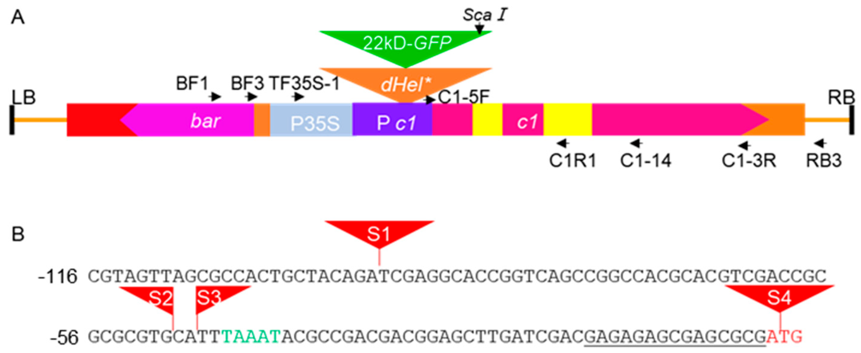
4.1. Development of the Reporter System

To develop reporter systems for Helitron transposition activity in maize, artificial defective Helitron transposons labeled by GFP were embedded in a maize C1 gene cassette (Figure 1A) and used for Agrobacterium transformation.
Four dHel transposons were selected to construct reporter systems for the following different reasons. The 6.0-kb HelA2, containing sequence fragments of three genes, was shown to excise from its original site in the chromosome 5S of several inbred lines [27,49], leaving multifarious TA repeats as somatic excision footprints [35]. Hel1-4 is present in the inbred lines of H99 and A188, but the somatic excision products have only been verified from H99. Hel1158 is one of the Cornucopious Helitrons, the high copy-number Helitron families in maize [31]. Hel1613 is a novel family of high copy-number Helitron other than Cornucopious newly identified by manual sequence annotation in maize genomes of B73 and other inbred lines.
Hel1-4 is 469 bp in length and has been demonstrated to be capable of excising somatically in H99 [35]. In the Hel1-4-GFP constructs, the Hel1-4 element was split into two parts for insertion of the GFP cassette, the 233-bp left terminal sequence (LTS) and the 236-bp right terminal sequence (RTS) according to the sequence conservation among elements in the same subfamily. The configuration of other dHel-GFP constructs is listed in Table S1.
A GFP expression cassette, driven by the 22-kD α zein promoter and ending with the cauliflower mosaic virus (CaMV) 35S terminator, was flanked by the LTS and RTS of each dHel. The C1 coding sequence comprised a 15-bp 5′ untranslated region (UTR) and a 431-bp promoter fragment with a TATA box [42]. The individual dHel-GFP cassette was inserted into the C1 promoter or exon region and led to the inactivation of the transgenic C1 copy. Following a bioinformatics analysis, three sites upstream of the TATA box were designated as insertion sites of dHel-GFP (S1, S2, and S3). The fourth site, S4, was at the C1 translation start codon (Figure 1B). The exact insertion sites of all seven constructs are given in Table 1 and Figure 1B. These constructs were transformed via Agrobacterium transformation method into maize inbred line B104, with an exception of S3-HelA2 into HiⅡ hybrid line.

4.2. Genetic Activity Detection of dHel in BC1F1 Population from Test Crosses with Inbred Lines

The R and C1 genes are indispensable in the anthocyanin biosynthesis pathway in the maize aleurone layer [50]. The lack of either the functional C1 or R1 gene results colorless kernels due to no anthocyanin accumulation in aleurone cells. Either transformation donor line of B104 inbred line or Hi II hybrid line is a c1 r1 double mutant; therefore, progenies (T2) from crosses of c1-dHel-GFP primary transformants(T1) with corresponding donor lines are all colorless kernels regardless of the potential existence of aHels in it (Figure S1). Our c1 tester line contains a functional R1-st gene; when crossing with it, the transgenic copy of c1-dHel-GFP is feasibly discriminated from the endogenous c1 gene by purple pigmentation phenotypes resulting from somatic excisions of dHel-GFP in the mutable c1-dHel-GFP catalyzed by aHels.
We first cross the T2 transgenic lines to the c1 tester line to observe the F1 seed phenotype. Then, we crossed the BC1 generation (F1 × c1 tester) of all transgenic lines with the inbred lines listed in Table S2 to assess the possibility of the presence of autonomous Helitrons in these genetic backgrounds via an expected spotting phenotype segregating in the BC1F1 populations.
C1-I, an allele of the C1 locus specifying a colorless phenotype in the aleurone layer, suppresses the wild-type C1 allele in the heterozygous condition [51]. Since C1-I does not affect the expression of the C1 allele in heterozygous C1/C1-I seeds, it may negatively interfere with C1 on a structural level [52]. Some of the inbred lines in Table S2 may have C1-I that are unlike candidates for detection of genetic activities of autonomous Helitrons in current experimental systems.

4.3. PCR Amplification of Somatic Excision Products and Circular dHel Intermediates

The dHel-GFP somatic excision products were examined by nested PCR on genomic DNA of the leaf blade from the third leaf of seedlings at the V2 stage with primer combinations of BF1/RB3, BF3/C1-14, and TF35S-1/C1R1 (Figure 1A, Table S3). Circular dHels were examined by nested PCR on the same set of genomic DNA samples using primer combinations of GFP-R3/GFP-R4, GFP-R1/GFP-4R, and 22ZEIN-1/GFP-R35S (color-coded arrows in Figure 7A, Table S3). All PCR amplifications were performed on a Bio-Rad CFX T100 instrument according to the 2 × Phanta Max Master Mix instructions (Vazyme, P525, Nanjing, China).

4.4. Molecular Characterization of Variant Transcripts from Transgenes

Rapid amplification of cDNA ends (RACE) is an effective method to rapidly amplify the 5′ and 3′ ends of cDNA from low-abundance transcripts based on PCR. The total RNA was prepared from the same batch of maize seedling leaves with the Transzol reagent following the user manual (TransGen Biotech, Beijing, China). The DNA was degraded by DNase I (GenStar, Beijing, China) following the manufacturer’s protocol prior to RACE PCR. The 5′ and 3′ cDNA of the c1 transgene was cloned by HiScript-TS 5′/3′ RACE Kit (Vazyme, RA101, Nanjing, China) following the manufacturer’s protocol.

5. Conclusions

Taking together, c1-dHel-GFP transgenic systems presented here are likely profitable in pursuing the genetic activity of autonomous Helitrons and elucidating the transposition mechanisms, RCR or cut-and-paste, of the transformed dHels catalyzed by the aHels at the molecular level. Meantime, complications of unexpected phenotypes of c1-dHel-GFP transgenic systems have been introduced serendipitously, and our research on the transcription alteration of the transgenes may aggrandize the functional characterization of the regulatory promoter regions, normally enriched with SNPs, indels or transposon insertions in plant. It is worthwhile to construct novel c1-dHel-GFP cassette in which dHel is inserted in the exons of the c1 gene to lessen the intricacy of the entire system for an inspiring breakthrough in Helitron genetics.

Supplementary Materials

The following supporting information can be downloaded at: https://www.mdpi.com/article/10.3390/ijms24076574/s1.

Author Contributions

Conceptualization, C.L. and Y.L.; Methodology, C.L., Y.L. and C.C.; Validation, C.L. and Y.L.; Formal analysis, F.L. and Y.Z.; Writing—original draft preparation, C.L.; Writing—review and editing, F.L., Q.Y., Y.Z., L.Z. and Y.L.; Supervision, Y.L.; Project administration, Y.L.; Funding acquisition, Y.L. All authors have read and agreed to the published version of the manuscript.

Funding

This research was funded by the National Natural Science Foundation of China (31871642 and 32072011), the 2020 Research Program of Sanya Yazhou Bay Science and Technology City (2020-02-005), the CAAS Elite Youth Program Grant, the Qingdao Agricultural University High-Level Talent Special Support Program Grant, and the Xinjiang High-Level Talent “Tianchi” Support Program Grant.

Institutional Review Board Statement

Not applicable.

Informed Consent Statement

Not applicable.

Data Availability Statement

Not applicable.

Acknowledgments

We are grateful to Hugo K. Dooner for constructing the backbone and helping with the experimental design. We thank anonymous reviewers for their comments on the manuscript and lab members Jinsheng Tan, Guantao Zhao, and Haoran Yu for their help in the laboratory and fieldwork.

Conflicts of Interest

The authors declare no conflict of interest.

References

  1. Kapitonov, V.V.; Jurka, J. Rolling-circle transposons in eukaryotes. Proc. Natl. Acad. Sci. USA 2001, 98, 8714–8719. [Google Scholar] [CrossRef] [PubMed] [Green Version]
  2. Galagan, J.E.; Calvo, S.E.; Cuomo, C.; Ma, L.J.; Wortman, J.R.; Batzoglou, S.; Lee, S.I.; Basturkmen, M.; Spevak, C.C.; Clutterbuck, J.; et al. Sequencing of Aspergillus nidulans and comparative analysis with A. fumigatus and A. oryzae. Nature 2005, 438, 1105–1115. [Google Scholar] [CrossRef] [PubMed] [Green Version]
  3. Pritham, E.J.; Feschotte, C. Massive amplification of rolling-circle transposons in the lineage of the bat Myotis lucifugus. Proc. Natl. Acad. Sci. USA 2007, 104, 1895–1900. [Google Scholar] [CrossRef] [PubMed] [Green Version]
  4. Coates, B.S.; Hellmich, R.L.; Grant, D.M.; Abel, C.A. Mobilizing the Genome of Lepidoptera through Novel Sequence Gains and End Creation by Non-autonomous Lep1 Helitrons. DNA Res. 2011, 19, 11–21. [Google Scholar] [CrossRef] [PubMed]
  5. Cocca, E.; De Iorio, S.; Capriglione, T. Identification of a novel helitron transposon in the genome of Antarctic fish. Mol. Phylogenet. Evol. 2011, 58, 439–446. [Google Scholar] [CrossRef]
  6. Tollis, M.; Boissinot, S. The transposable element profile of the anolis genome: How a lizard can provide insights into the evolution of vertebrate genome size and structure. Mob. Genet. Elem. 2011, 1, 107–111. [Google Scholar] [CrossRef] [Green Version]
  7. Lal, S.K.; Giroux, M.J.; Brendel, V.; Vallejos, C.E.; Hannah, L.C. The maize genome contains a helitron insertion. Plant Cell 2003, 15, 381–391. [Google Scholar] [CrossRef] [Green Version]
  8. Brunner, S.; Pea, G.; Rafalski, A. Origins, genetic organization and transcription of a family of non-autonomous helitron elements in maize. Plant J. 2005, 43, 799–810. [Google Scholar] [CrossRef]
  9. Xu, J.H.; Messing, J. Maize haplotype with a helitron-amplified cytidine deaminase gene copy. BMC Genet. 2006, 7, 52. [Google Scholar] [CrossRef] [Green Version]
  10. Grabundzija, I.; Messing, S.A.; Thomas, J.; Cosby, R.L.; Bilic, I.; Miskey, C.; Gogol-Doring, A.; Kapitonov, V.; Diem, T.; Dalda, A.; et al. A Helitron transposon reconstructed from bats reveals a novel mechanism of genome shuffling in eukaryotes. Nat. Commun. 2016, 7, 10716. [Google Scholar] [CrossRef] [Green Version]
  11. Tempel, S.; Giraud, M.; Lavenier, D.; Lerman, I.C.; Valin, A.S.; Couee, I.; El Amrani, A.; Nicolas, J. Domain organization within repeated DNA sequences: Application to the study of a family of transposable elements. Bioinformatics 2006, 22, 1948–1954. [Google Scholar] [CrossRef] [Green Version]
  12. Du, C.G.; Caronna, J.; He, L.M.; Dooner, H.K. Computational prediction and molecular confirmation of Helitron transposons in the maize genome. BMC Genom. 2008, 9, 51. [Google Scholar] [CrossRef] [Green Version]
  13. Xiong, W.; He, L.; Lai, J.; Dooner, H.K.; Du, C. HelitronScanner uncovers a large overlooked cache of Helitron transposons in many plant genomes. Proc. Natl. Acad. Sci. USA 2014, 111, 10263–10268. [Google Scholar] [CrossRef] [Green Version]
  14. Hu, K.; Xu, K.; Wen, J.; Yi, B.; Shen, J.; Ma, C.; Fu, T.; Ouyang, Y.; Tu, J. Helitron distribution in Brassicaceae and whole Genome Helitron density as a character for distinguishing plant species. BMC Bioinform. 2019, 20, 354. [Google Scholar] [CrossRef] [Green Version]
  15. Gupta, S.; Gallavotti, A.; Stryker, G.A.; Schmidt, R.J.; Lal, S.K. A novel class of Helitron-related transposable elements in maize contain portions of multiple pseudogenes. Plant Mol. Biol. 2005, 57, 115–127. [Google Scholar] [CrossRef]
  16. Chuck, G.; Meeley, R.; Irish, E.; Sakai, H.; Hake, S. The maize tasselseed4 microRNA controls sex determination and meristem cell fate by targeting Tasselseed6/indeterminate spikelet1. Nat. Genet. 2007, 39, 1517–1521. [Google Scholar] [CrossRef]
  17. Gao, C.; Zhou, G.; Ma, C.; Zhai, W.; Zhang, T.; Liu, Z.; Yang, Y.; Wu, M.; Yue, Y.; Duan, Z.; et al. Helitron-like transposons contributed to the mating system transition from out-crossing to self-fertilizing in polyploid Brassica napus L. Sci. Rep. 2016, 6, 33785. [Google Scholar] [CrossRef] [Green Version]
  18. Choi, J.D.; Hoshino, A.; Park, K.I.; Park, I.S.; Iida, S. Spontaneous mutations caused by a Helitron transposon, Hel-It1, in morning glory, Ipomoea tricolor. Plant J. 2007, 49, 924–934. [Google Scholar] [CrossRef]
  19. Tsukamoto, T.; Hauck, N.R.; Tao, R.; Jiang, N.; Iezzoni, A.F. Molecular and genetic analyses of four nonfunctional S haplotype variants derived from a common ancestral S haplotype identified in sour cherry (Prunus cerasus L.). Genetics 2010, 184, 411–427. [Google Scholar] [CrossRef] [Green Version]
  20. Liu, Q.; Deng, S.; Liu, B.; Tao, Y.; Xu, M. A helitron-induced RabGDIα variant causes quantitative recessive resistance to maize rough dwarf disease. Nat. Commun. 2020, 11, 495. [Google Scholar] [CrossRef] [Green Version]
  21. Morgante, M.; Brunner, S.; Pea, G.; Fengler, K.; Zuccolo, A.; Rafalski, A. Gene duplication and exon shuffling by helitron-like transposons generate intraspecies diversity in maize. Nat. Genet. 2005, 37, 997–1002. [Google Scholar] [CrossRef] [PubMed]
  22. Yang, L.; Bennetzen, J.L. Distribution, diversity, evolution, and survival of Helitrons in the maize genome. Proc. Natl. Acad. Sci. USA 2009, 106, 19922–19927. [Google Scholar] [CrossRef] [PubMed] [Green Version]
  23. Yang, L.; Bennetzen, J.L. Structure-based discovery and description of plant and animal Helitrons. Proc. Natl. Acad. Sci. USA 2009, 106, 12832–12837. [Google Scholar] [CrossRef] [PubMed] [Green Version]
  24. Du, J.; Grant, D.; Tian, Z.; Nelson, R.T.; Zhu, L.; Shoemaker, R.C.; Ma, J. SoyTEdb: A comprehensive database of transposable elements in the soybean genome. BMC Genom. 2010, 11, 113. [Google Scholar] [CrossRef] [PubMed] [Green Version]
  25. Hollister, J.D.; Smith, L.M.; Guo, Y.L.; Ott, F.; Weigel, D.; Gaut, B.S. Transposable elements and small RNAs contribute to gene expression divergence between Arabidopsis thaliana and Arabidopsis lyrata. Proc. Natl. Acad. Sci. USA 2011, 108, 2322–2327. [Google Scholar] [CrossRef] [Green Version]
  26. Thomas, J.; Phillips, C.D.; Baker, R.J.; Pritham, E.J. Rolling-circle transposons catalyze genomic innovation in a mammalian lineage. Genome Biol. Evol. 2014, 6, 2595–2610. [Google Scholar] [CrossRef] [Green Version]
  27. Wang, Q.H.; Dooner, H.K. Remarkable variation in maize genome structure inferred from haplotype diversity at the bz locus. Proc. Natl. Acad. Sci. USA 2006, 103, 17644–17649. [Google Scholar] [CrossRef] [Green Version]
  28. Volfovsky, N.; Haas, B.J.; Salzberg, S.L. A clustering method for repeat analysis in DNA sequences. Genome Biol. 2001, 2, RESEARCH0027. [Google Scholar] [CrossRef]
  29. Price, A.L.; Jones, N.C.; Pevzner, P.A. De novo identification of repeat families in large genomes. Bioinformatics 2005, 21 (Suppl. S1), i351–i358. [Google Scholar] [CrossRef] [Green Version]
  30. Flutre, T.; Duprat, E.; Feuillet, C.; Quesneville, H. Considering transposable element diversification in de novo annotation approaches. PLoS ONE 2011, 6, e16526. [Google Scholar] [CrossRef]
  31. Du, C.G.; Fefelova, N.; Caronna, J.; He, L.M.; Dooner, H.K. The polychromatic Helitron landscape of the maize genome. Proc. Natl. Acad. Sci. USA 2009, 106, 19916–19921. [Google Scholar] [CrossRef] [Green Version]
  32. Feschotte, C.; Pritham, E.J. A cornucopia of Helitrons shapes the maize genome. Proc. Natl. Acad. Sci. USA 2009, 106, 19747–19748. [Google Scholar] [CrossRef] [Green Version]
  33. Grabundzija, I.; Hickman, A.B.; Dyda, F. Helraiser intermediates provide insight into the mechanism of eukaryotic replicative transposition. Nat. Commun. 2018, 9, 1278. [Google Scholar] [CrossRef] [Green Version]
  34. Kosek, D.; Grabundzija, I.; Lei, H.; Bilic, I.; Wang, H.; Jin, Y.; Peaslee, G.F.; Hickman, A.B.; Dyda, F. The large bat Helitron DNA transposase forms a compact monomeric assembly that buries and protects its covalently bound 5′-transposon end. Mol. Cell 2021, 81, 4271–4286. [Google Scholar] [CrossRef]
  35. Li, Y.; Dooner, H.K. Excision of Helitron transposons in maize. Genetics 2009, 182, 399–402. [Google Scholar] [CrossRef] [Green Version]
  36. Xiong, W.; Dooner, H.K.; Du, C. Rolling-circle amplification of centromeric Helitrons in plant genomes. Plant J. 2016, 88, 1038–1045. [Google Scholar] [CrossRef]
  37. Wang, Z.; Zhao, G.; Yang, Q.; Gao, L.; Liu, C.; Ru, Z.; Wang, D.; Jia, J.; Cui, D. Helitron and CACTA DNA transposons actively reshape the common wheat—AK58 genome. Genomics 2022, 114, 110288. [Google Scholar] [CrossRef]
  38. Chaikam, V.; Nair, S.K.; Babu, R.; Martinez, L.; Tejomurtula, J.; Boddupalli, P.M. Analysis of effectiveness of R1-nj anthocyanin marker for in vivo haploid identification in maize and molecular markers for predicting the inhibition of R1-nj expression. Theor. Appl. Genet. 2015, 128, 159–171. [Google Scholar] [CrossRef]
  39. Murray, A.; Mendieta, J.P.; Vollmers, C.; Schmitz, R.J. Simple and accurate transcriptional start site identification using Smar2C2 and examination of conserved promoter features. Plant J. 2022, 112, 583–596. [Google Scholar] [CrossRef]
  40. Paz-Ares, J.; Wienand, U.; Peterson, P.A.; Saedler, H. Molecular cloning of the c locus of Zea mays: A locus regulating the anthocyanin pathway. EMBO J. 1986, 5, 829–833. [Google Scholar] [CrossRef]
  41. Cone, K.C.; Burr, F.A.; Burr, B. Molecular analysis of the maize anthocyanin regulatory locus C1. Proc. Natl. Acad. Sci. USA 1986, 83, 9631–9635. [Google Scholar] [CrossRef] [PubMed] [Green Version]
  42. Paz-Ares, J.; Ghosal, D.; Saedler, H. Molecular analysis of the C1-I allele from Zea mays: A dominant mutant of the regulatory C1 locus. EMBO J. 1990, 9, 315–321. [Google Scholar] [CrossRef] [PubMed]
  43. Scheffler, B.; Franken, P.; Schutt, E.; Schrell, A.; Saedler, H.; Wienand, U. Molecular analysis of C1 alleles in Zea mays defines regions involved in the expression of this regulatory gene. Mol. Gen. Genet. 1994, 242, 40–48. [Google Scholar] [CrossRef] [PubMed]
  44. Reddy, A.R.; Peterson, P.A. Transposable elements of maize--genetic basis of pattern differentiation of some mutable c alleles of the enhancer system. Mol. Gen. Genet. MGG 1983, 192, 21–31. [Google Scholar] [CrossRef]
  45. Li, Y.; Dooner, H.K. Helitron Proliferation and Gene-Fragment Capture. In Plant Transposable Elements: Impact on Genome Structure and Function; Grandbastien, M.-A., Casacuberta, J.M., Eds.; Springer: Berlin/Heidelberg, Germany, 2012; pp. 193–217. [Google Scholar] [CrossRef]
  46. Li, Y.; Harris, L.; Dooner, H.K. TED, an autonomous and rare maize transposon of the mutator superfamily with a high gametophytic excision frequency. Plant Cell 2013, 25, 3251–3265. [Google Scholar] [CrossRef] [Green Version]
  47. Sundaresan, V.; Freeling, M. An extrachromosomal form of the Mu transposons of maize. Proc. Natl. Acad. Sci. USA 1987, 84, 4924–4928. [Google Scholar] [CrossRef] [Green Version]
  48. Gorbunova, V.; Levy, A.A. Circularized Ac/Ds transposons: Formation, structure and fate. Genetics 1997, 145, 1161–1169. [Google Scholar] [CrossRef]
  49. Lai, J.S.; Li, Y.B.; Messing, J.; Dooner, H.K. Gene movement by Helitron transposons contributes to the haplotype variability of maize. Proc. Natl. Acad. Sci. USA 2005, 102, 9068–9073. [Google Scholar] [CrossRef] [Green Version]
  50. Ludwig, S.R.; Habera, L.F.; Dellaporta, S.L.; Wessler, S.R. Lc, a member of the maize R gene family responsible for tissue-specific anthocyanin production, encodes a protein similar to transcriptional activators and contains the myc-homology region. Proc. Natl. Acad. Sci. USA 1989, 86, 7092–7096. [Google Scholar] [CrossRef] [Green Version]
  51. Coe, E.H. Spontaneous Mutation of the Aleurone Color Inhibitor in Maize. Genetics 1962, 47, 779. [Google Scholar] [CrossRef]
  52. Paz-Ares, J.; Ghosal, D.; Wienand, U.; Peterson, P.A.; Saedler, H. The regulatory c1 locus of Zea mays encodes a protein with homology to myb proto-oncogene products and with structural similarities to transcriptional activators. EMBO J. 1987, 6, 3553–3558. [Google Scholar] [CrossRef]
Figure 1. Schematic representation of the T-DNA construct used in Agrobacterium transformation. (A) Schematic diagram of the c1-dHel-GFP T-DNA construct used in Agrobacterium-mediated transformation of Hi II (c1) or B104 (c1) embryos. LB, left border; RB, right border. Horizontal arrows, primers for PCR and RT-PCR; vertical arrow, ScaI restriction site. (B) Sequence features of dHel-GFP insertion sites in the c1 gene. The start codon is in red. The 5′ UTR is underlined. The TATA box is in green. The positions of the dHel-GFP inserts tested are shown as red triangles numbered from S1 to S4. The S2 site is a GC dinucleotide, whereas all other sites are typical AT dinucleotides.
Ijms 24 06574 g001
Figure 2. T2 transgenic seedlings and their anther phenotypes. (A) Representative T2 seedling phenotypes. (B) Representative anthers of T2 transgenic plants. 1, S3-Hel1613; 2, S3-Hel1158, 3, S3-HelA2; 4, S1-Hel1-4; 5, S2-Hel1-4; 6, S3-Hel1-4; 7, S4-Hel1-4; 8, B104 donor line; 9, Hi Ⅱ donor line; 10, positive C1 transgenic line.
Ijms 24 06574 g002
Figure 3. Kernel phenotype of test-cross ears. (A) Representative kernel pigmentation phenotypes of test-cross ears of the c1 tester line with primary transformants from various constructs. Anthocyanin pigmentation is varied in test-cross ears from primary transformants harboring individual c1-dHel-GFP constructs crossed to the c1 tester. (B) Representative kernel phenotype of GFP fluorescence segregation from the same test-cross ears as in (A) detecting under blue light illumination. 1, S3-Hel1613; 2, S3-Hel1158; 3, S3-HelA2; 4, S1-Hel1-4; 5, S2-Hel1-4; 6, S3-Hel1-4; 7, S4-Hel1-4; 8, positive C1 transgenic line.
Ijms 24 06574 g003
Figure 4. Results of RACE for C1 transcripts from T2 transgenic leaf tissue. (A) 3′ RACE and (B) 5′RACE results for the C1 transcript from total RNA extracted from leaf tissue of T2 transgenic plants. 1, S3-Hel1613; 2, S3-Hel1158; 3, S3-HelA2; 4, S4-Hel1-4; 5, S2-Hel1-4; 6, S3-Hel1-4; 7, S1-Hel1-4; 8, positive C1 transgenic line.
Ijms 24 06574 g004
Figure 5. Schematic representation of C1 transcript variants detected by 5′ RACE from T2 transgenic leaf tissue. (A) c1 transcripts detected in total RNA extracted from leaf tissue of T2 transgenic plants harboring dHel insertions in the c1 promoter region (sites S1 to S3), putative wild type C1 transcripts are labeled with red numbers; (B) c1 transcripts detected in total RNA extracted from leaf tissue of T2 transgenic plants harboring the dHel insertion at the translation start site of the c1 gene (site S4). GFP promoter (α-zein promoter) and terminator (CAMV 35S terminator) are indicated by green dotted lines. LTS and RTS of dHel are indicated by dark vertical dashed lines. C1 exons and introns are indicated by purple boxes and lines respectively. The corresponding intron-exon junction sites are labeled with numbers beneath. C1 promoter region is indicated by black dotted line. 5′ and 3′ UTR are represented by open boxes. Alternative splicing events at non-canonical sites are shown with broken lines, whereas splicing events at canonical sites are shown with solid lines. dHels are indicated by grey boxes, and GFP is indicated by green boxes. Horizontal arrows, primer for 5′ RACE. Red asterisks, stop codons.
Ijms 24 06574 g005
Figure 6. PCR amplification of somatic excision products for dHel in F1 segregants from test-crosses between different c1-dHel-GFP T2 transgenic plants and the c1 tester line. PCR primer pairs are shown in Figure 1A. Lanes 1–6, S3-Hel1613; lanes 7–11, S3-Hel1158; lanes 12–18, S3-HelA2; lanes 19–23, S4-Hel1-4; lanes 24–28: S2-Hel1-4; lanes 29–34, S3-Hel1-4; lanes 35–41, S1-Hel1-4; lanes 42–43: F1 individuals from a non-transgenic line crossed to the c1 tester. Red arrows, PCR products with the expected size for dHel excision products.
Ijms 24 06574 g006
Figure 7. Schematic representation and PCR detection of circular dHel intermediates from leaf tissues of transgenic plants transformed with c1-dHel-GFP constructs. (A) Schematic diagram of circular dHel-GFP (1) and nested-PCR primer combinations (pairs of color-coded arrows in 2) to amplify somatic circular dHel intermediates (3); (B) Lanes 1–10, T2 transgenic lines from S1-Hel1-4 construct; (C) F1 segregants from a test-cross between different c1-dHel-GFP T2 transgenic plants and the c1 tester line. Lanes 1–6, S3-Hel1613; lanes 7–11, S3-Hel1158; lanes 12–18, S3-HelA2; lanes 19–23, S4-Hel1-4; lanes 24–28, S2-Hel1-4; lanes 29–34, S3-Hel1-4; lanes 35–41, S1-Hel1-4; Lanes 42–44, F1 individuals from a cross between a non-transgenic line and the c1 tester. Red arrows, PCR products of putative circular dHel intermediates; (D) Schematic representation of sequence features of circular intermediates from (B) and (C) (red arrows). The expected circular dHel intermediates (A3) are inverse PCR amplification of a fused 5′-end upstream sequence with 3′-end downstream sequence color-coded as orange and green, respectively. Red arrows are PCR primers, light blue dotted lines are internal deletions, vertical yellow bars are palindromic sequences upstream of the 3′-end CTAG, and the sequence configurations are labeled with numbers in the color-coded rectangles.
Ijms 24 06574 g007
Table 1. Sequence features of dHel-GFP constructs and pigmentation phenotypes of T2 transgenic plants and F1 segregants of test cross population.
ConstructSite (nt)dHel-GFP (bp)Donor LineT1 EventsT1 Single CopyT2
Coleoptiles
T2
Anthers
F1 Kernel
S1-Hel1-4−931723B10431PurplePMottled
S2-Hel1-4−491723B10454PurpleLP *Mottled
S3-Hel1-4−471723B10454PurpleLPMottled
S4-Hel1-411723B10462ColorlessColorlessColorless
S3-Hel1613−471781B10454PurpleLPColorless
S3-Hel1158−471996B10454PurpleLPMottled
S3-HelA2−472038HiⅡ54PurpleLPMottled
Note: Single copy T2 events were used for RACE. All T2 events and corresponding F1 were used for Helitron somatic excision footprint analysis. * LP, light purple.
Table 2. Somatic excision activity in F1 segregants from different c1-dHel-GFP T2 transgenic plants.
ConstructsExamined Segregants Segregants with Detected SET-DNA Copy Numbers in Transformants
S1-Hel1-41462 (Arrayed in tandem)
S2-Hel1-43601
S3-Hel1-4340/
S4-Hel1-41531
S3-Hel1613340/
S3-Hel11581511
S3-HelA21721
Table 3. Sequence of full and empty sites from dHel-GFP constructs.
dHel-GFP Construct    dHel 5′-End Junction                  dHel 3′-End Junction
S3-Hel1158ACGTCGACCGCGCGCGTGCATCTATACTAT…AACCGACTAGTTTAAATACGCCGACGACGG
S3-Hel1158 (e)ACGTCGACCGCGCGCGTGCA---------------------------------------------TTTAAATACGCCGACGACGG
S3-HelA2ACGTCGACCGCGCGCGTGCATCTCTACTAC…ACTCACCTAGTTTAAATACGCCGACGACGG
S3-HelA2 (e)ACGTCGACCGCGCGCGTGCA---------------------------------------------TTTAAATACGCCGACGACGG
S1-Hel1-4GTTAGCGCCACTGCTACAGATCTATACTAC…ACCTAACTAGTCGAGGCACCGGTCAGCCGG
S1-Hel1-4 (e)GTTAGCGCCACTGCTACAGA---------------------------------------------TCGAGGCACCGGTCAGCCGG
S4-Hel1-4CGACGAGAGAGCGAGCGCGATCTATACTAC…ACCTAACTAGTGGGGAGGAGGGCGTGTTGC
S4-Hel1-4 (e)CGACGAGAGAGCGAGCGCGA---------------------------------------------TGGGGAGGAGGGCGTGTTGC
Note: Helitron sequences are in bold; e: empty site.
Disclaimer/Publisher’s Note: The statements, opinions and data contained in all publications are solely those of the individual author(s) and contributor(s) and not of MDPI and/or the editor(s). MDPI and/or the editor(s) disclaim responsibility for any injury to people or property resulting from any ideas, methods, instructions or products referred to in the content.

