Review

Lupin, a Unique Legume That Is Nodulated by Multiple Microsymbionts: The Role of Horizontal Gene Transfer

1
Department of Soil. Plant and Environmental Quality, Institute of Agricultural Sciences, ICA-CSIC, 28006 Madrid, Spain
2
Laboratory of Biodiversity and Valorization of Arid Areas Bioresources, BVBAA, Faculty of Sciences, University of Gabès, Erriadh, Zrig, Gabès 6072, Tunisia
*
Author to whom correspondence should be addressed.
Int. J. Mol. Sci. 2023, 24(7), 6496; https://doi.org/10.3390/ijms24076496
Submission received: 28 February 2023 / Revised: 25 March 2023 / Accepted: 27 March 2023 / Published: 30 March 2023

Abstract

:
Lupin is a high-protein legume crop that grows in a wide range of edaphoclimatic conditions where other crops are not viable. Its unique seed nutrient profile can promote health benefits, and it has been proposed as a phytoremediation plant. Most rhizobia nodulating Lupinus species belong to the genus Bradyrhizobium, comprising strains that are phylogenetically related to B. cytisi, B. hipponenese, B. rifense, B. iriomotense/B. stylosanthis, B. diazoefficiens, B. japonicum, B. canariense/B. lupini, and B. retamae/B. valentinum. Lupins are also nodulated by fast-growing bacteria within the genera Microvirga, Ochrobactrum, Devosia, Phyllobacterium, Agrobacterium, Rhizobium, and Neorhizobium. Phylogenetic analyses of the nod and nif genes, involved in microbial colonization and symbiotic nitrogen fixation, respectively, suggest that fast-growing lupin-nodulating bacteria have acquired their symbiotic genes from rhizobial genera other than Bradyrhizobium. Horizontal transfer represents a key mechanism allowing lupin to form symbioses with bacteria that were previously considered as non-symbiotic or unable to nodulate lupin, which might favor lupin’s adaptation to specific habitats. The characterization of yet-unstudied Lupinus species, including microsymbiont whole genome analyses, will most likely expand and modify the current lupin microsymbiont taxonomy, and provide additional knowledge that might help to further increase lupin’s adaptability to marginal soils and climates.

1. Introduction

Fabaceae or Leguminoseae (nom. cons.) constitute a large plant family, widespread all over the world, that includes more than 19,700 species [1,2]. Many legume species, including all the agronomically important legume crops, establish symbiotic associations with soil bacteria that are able to fix atmospheric nitrogen (N2) in specific symbiotic organs called nodules. The legume-nodulating bacteria, known as rhizobia, are gram-negative bacteria that have the ability to induce the formation of a de novo organ in the roots and/or stems of leguminous plants [3]. They are capable of fixing atmospheric N2 owing to the microaerobic environment that is created inside the nodule. Most rhizobia belong to the alpha subgroup of the phylum Pseudomonadota (formerly Proteobacteria). The class α-Proteobacteria comprises most of the N2-fixing genera that nodulate legumes, while some symbiotic genera belong to the class β-Proteobacteria. The great diversity of legume species is reflected in their wide variety of nodulating microsymbionts.
Ribosomal ribonucleic acid (rRNA) is the primary component of ribosomes, and is essential to all cells. rRNA is a ribozyme that facilitates protein synthesis in the ribosomes [4]. The 16S rRNA gene is present in all bacteria, and phylogenetic analyses based on these gene sequences are used to determine genetic relationships among organisms. Including conserved and variable regions, the 16S rRNA gene has a considerable size (around 1500 bp), which provides sufficient information for taxonomy. This gene presents a high level of conservation at the species level in such a way that the sequence variations observed in this gene can provide a precise indicator of evolution. For these characteristics, the 16S rRNA gene is considered as a primary discriminative taxonomic marker to classify a strain at the genus and often at the species level, and represents one of the preferred genetic methods for bacterial phylogenetic analysis [5,6,7], including the phylogenetic classification of legume microsymbionts [8,9,10].
Simultaneously with the development of molecular biology techniques, bioinformatics tools, and whole genome sequencing, a large number of 16S rRNA gene sequences are available in gene databases. The DNA DataBank of Japan (DDBJ), the European Nucleotide Archive (ENA), and the National Center for Biotechnology Information (NCBI) gene databases, which are part of the International Nucleotide Sequence Database Collaboration (INSDC), constitute a potent tool for scientists, as 16S rRNA gene sequences are available for the construction of phylogenetic trees and identification of species [11]. Phylogenetic trees are useful for structuring taxonomic classifications, and they provide information about events that might have occurred during evolution, showing lines of evolutionary descent for diverse species, organisms, or even genes. Phylogenetic tree analyses affiliate new bacterial isolates with their closest described strains [9,10,12]. Despite the fact that the aforementioned databases exchange data on a daily basis, multiple sequence uploads have often led to lack of agreement on the dependability of 16S rRNA gene sequence data [13,14].
Phylogenetic analysis of the nearly full-length sequences of the 16S rRNA genes of the majority of representative microsymbiont strains has allowed for the identification of 18 genera and around 238 species of bacteria that nodulate legumes [15]. Within the class α-Proteobacteria, the family Rhizobiaceae comprises the genera Rhizobium, Pararhizobium, Allorhizobium, Ensifer (syn. Sinorhizobium), Neorhizobium, and Shinella. The genera Aminobacter, Mesorhizobium, and Phyllobacterium are included in the family Phyllobacteriaceae. The genera Microvirga and Methylobacterium are part of the family Methylobacteriaceae. The genera Bradyrhizobium, Blastobacter, and Photorhizobium constitute the family Bradyrhizobiaceae. The genus Ochrobactrum is specific to the Brucellaceae. In the family Hyphomicrobiaceae, only Devosia has been reported to nodulate Fabaceae [12]. In the class β-Proteobacteria, the genus Cupriavidus, along with Burkholderia, are part of the family Burkholderiaceae [16]. Bacteria of the γ-Proteobacteria group have also been detected in legume nodules [17,18]; however, later reports have questioned their ability to nodulate legumes [19].
Effective nodulation and nitrogen fixation are supported by the nod and nif gene clusters, respectively. Nitrogen is one of the most important nutrients for plant growth. Nodule organogenesis is induced by the rhizobial-secreted Nod factors, which are signal molecules controlled by the nod genes that are recognized by the host plant and lead to the formation of the symbiotic nodule [20]. This suggests that nod genes are responsible for determining the specificity of the legume–rhizobial symbiosis [21]. The nif genes, which codify for the nitrogenase enzyme complex subunits and several regulatory proteins, are essential for nitrogen fixation [22]. In consequence, while 16S rRNA gene sequences are key for the classification of bacterial species and a reliable marker for phylogenetic analyses, a multigene analysis combining the 16S rRNA and nod and nif genes [23] tends to be used to approximate the systematic characteristics of rhizobia.
The nif and nod genes are often carried on symbiotic islands in the chromosome or in plasmids, which are easily transferred horizontally between different bacterial species within and across genera [24,25]. This transfer of nod and nif genes between bacteria in the rhizosphere can confer the ability to nodulate legumes to non-rhizobial species [26,27]. It has been suggested that stressful environments, in general, may favor horizontal gene transfer, as the acquisition of certain genes might represent an adaptive advantage. Furthermore, bacteria can engage in horizontal gene transfer to rapidly disseminate traits in a population, thus providing indiscriminate benefits to their neighbors, by means of a behavior that can be considered a method of bacterial cooperation [28]. In this manner, the transfer of symbiotic genes to bacteria that are well-adapted to local soil conditions allows these microorganisms to become rhizobial symbionts of originally incompatible legumes [29]. The horizontal transfer of symbiotic genes most often leads to phylogenies that differ from those of the core genome of the receiving bacteria [21,24].
There is abundant evidence that horizontal gene transfer occurs commonly in nature, involving all kinds of transposable elements and all classes of living organisms [30,31]. Even phylogenetically distant species may experience the transference of transposable elements to/from their genomes through transfer vectors such as viruses, bacteria, or other parasites [32]. There are many examples in the literature of how horizontal gene transfer has driven significant evolutionary changes, or helped organisms to adapt to abiotic stress [33]. There are also some examples of horizontal gene transfer helping plants to adapt to biotic stress: for example, when symbiotic genes are acquired by bacteria with PGPR traits [34]. It is well known that biofilms facilitate horizontal gene transfer between bacteria and that soil bacteria are often organized in biofilms on root, litter, and soil particles [35]. Therefore, biofilms may play an important role in the movement of transposable elements between soil organisms.
Initially identified within a species, biological variants, commonly named biovars, have been described in diverse bacterial species. A biovar represents a specific group of bacterial strains which are distinguishable from other strains in the same species on the basis of biochemical or physiological characteristics. Biovars were described for the first time in Rhizobium leguminosarum by Jordan [36] in a taxonomical revision of rhizobial species. In rhizobia, biovars and symbiotic variants, or symbiovars, are used to differentiate distinct subgroups, usually within a genus, comprising species that nodulate the same legume [37]. Legume host range specificity is encoded in rhizobial genetic elements that can be disseminated by horizontal transfer; thus, symbiovars can be shared by rhizobial species belonging to different genera [9,37].
Until the present century, lupin was assumed to be a microsymbiont-specific legume. However, owing to the evolution of molecular taxonomy, nowadays, lupin can be considered as a promiscuous legume that can be nodulated by different Bradyrhizobium lineages [38,39,40], and by bacteria within the genera Phyllobacterium [41,42], Ochrobactrum [27], Rhizobium, Neorhizobium, Agrobacterium [43], Microvirga [42,44], and Devosia [12]. The aim of this review was to collect the existing information on the diversity and taxonomy of the rhizobial bacteria associated with Lupinus spp. nodules. Phylogenetic trees, including the published 16S rRNA, nodC, and nifH gene sequences of lupin microsymbionts, will be analyzed. The incongruences between the classifications derived from the 16S rRNA gene and the symbiotic gene sequences analyses, and the symbiovars that can be defined thereafter, will be discussed. The importance of lupin, a unique legume with specific characteristics, as summarized below, substantiates the significance of the present review. Moreover, the fact that different lupin-nodulating bacteria might be able to modulate or even determine some legume agronomic traits, such as its adaptability to different soils, its tolerance to abiotic stress, or the nutrient profile of its grain adds relevance to the current knowledge on lupin microsymbionts, and to the discussion on the role that horizontal gene transfer appears to play.

2. Lupin, a Unique Legume in So Many Ways

The legume genus Lupinus (Papilionideae: Genisteae) comprises 200–500 species of perennial and annual shrubs and herbs with an amphi-Atlantic distribution [1]. Lupinus species are distributed in Europe; the Near East; North and East Africa; and North, Central, and South America. Less than 13 species are native of the Old World, mainly surrounding the Mediterranean basin, while American species are much more numerous [45,46]. Lupins have been grown since antiquity and represent an important pulse crop due to the highly effective N2-fixing symbiosis they establish with rhizobia, as well as their adaptation to different climates and soils.
Lupins are mainly employed as a source of protein in both human and animal nutrition. The high protein content of lupin seeds (up to 44% dry weight) is similar to that of soybean, and lupin has been proposed as a viable alternative to soybean cultivation in Europe [47]. Lupin seeds possess a high nutritional value and unique phytochemical profile and biological activities [48,49]. Among the lupin species, L. albus (white lupin), L. luteus (yellow lupin), L. angustifolius (blue lupin), and L. mutabilis (tarwi) have gained agricultural market importance and have become part of the modern agriculture and food systems for high-protein production [47]. Lupinus species have also been studied as medicinal plants [50] and used in traditional medicine to treat various diseases, including urinary tract infections, heart conditions, or skin disorders [51]. Due to the presence of essential fatty acids, specific proteins, and amino acids, as well as minerals, alkaloids, and dietary fiber in lupin seeds, component fractionation allows for extracts to be obtained that display promising anticarcinogenic, antidiabetic, antihypertensive, anti-inflammatory, antimicrobial, and antioxidant activities [51]. The phenolic compounds and flavonoid constituents in lupin seed extracts have important antioxidant activity [51,52]. Lupin protein hydrolysates present anti-inflammatory properties [53]. The antifungal and antibacterial activities have been demonstrated in different studies that report an inhibitory effect toward various bacterial and fungal pathogens [54,55].
Lupin plants have also been shown to have great potential for the recovery of degraded soils and in heavy metal phytoremediation [56,57,58,59]. Lupinus albus appears to be an especially well-suited species to withstanding heavy metal stress, and white lupin plants inoculated with a mercury-tolerant Bradyrhizobium strain have been reported to present increased mercury tolerance [59]. Additionally, its adaptive mechanisms make white lupin a choice crop for acidic soils affected by aluminum toxicity [60]. Nitrogen and phosphorus are, together with potassium, the two principal nutrients needed by plants, and are essential to ensure crop yield. In phosphate-deficient soils, some lupin species, including white lupin, are able to modify their root architecture to form cluster roots, which are bottlebrush-like structures consisting of hundreds of short rootlets [61]. Cluster roots display enhanced synthesis and secretion of acid phosphatases, organic acids, flavonoids, and proton efflux. Such modifications lead to the mobilization of the soil’s unavailable phosphorus. Therefore, lupin arises as a choice crop for nutrient-poor soils, as it reduces the need for both N and P fertilizers [61]. Lupin’s ability to adapt to acid, nutrient-poor, or moderately polluted soils, and to arid and cold climates, enables lupin cultivation in affected areas, where other more demanding crops, such as soybeans, are not economically viable [62]. Identifying lupin cultivars tolerant to a variety of abiotic stresses may allow for the expansion of lupin cultivation to a broader range of edaphoclimatic conditions [47].
Lupin nodules are also unique. According to the host legume and independently of the microsymbiont species, nodules can be determinate or indeterminate [21,63]. Determinate nodules present a transient meristem, while indeterminate nodules maintain meristematic activity throughout their lifespan. All genera examined in the Fabaceae subfamilies Mimosoideae and Caesalpinioideae, and most tribes within the subfamily Papilionoideae, form indeterminate nodules, while the papilionoid species within the tribes Desmodieae, Dalbergieae, Psoraleeae, Phaseoleae, and some genera in the Loteae form determinate nodules [63]. The lupinoid nodule is a subtype of indeterminate nodule, first identified in lupin [64], and to our knowledge has only been described in another legume genus, Listia (Papilionideae: Crotalarieae) [65]. The lupinoid nodule is characterized by the presence of lateral meristems that allow the nodule to encircle the root. Another characteristic of the lupinoid nodule is that the infected cells remain mitotically active and the symbiosomes, much like other cell organelles, are equally distributed between the daughter cells [66,67]. The infection process that leads to lupin nodule organogenesis is also uncommon, as rhizobia infection does not occur through an infection thread, as happens in most legumes. Rhizobia enter the root intercellularly and specifically infect a cell beneath a root hair [66]. Remarkably, in the work in which the infection process was first described, and despite the fact that lupin was thought to be a microsymbiont selective legume exclusively nodulated by Bradyrhizobium sp. (Lupinus), a Mesorhizobium loti strain was used that was able to elicit nodules, but unable to form effective nitrogen-fixing nodules [66].

3. Rhizobia That Nodulate Lupins, Many More Than Initially Expected

Lupin is nowadays described as a promiscuous plant host, as it can be nodulated by many different species and genotypes of symbiotic bacteria [40]. Until recently, lupins were assumed to be exclusively nodulated by slow-growing rhizobia of the genus Bradyrhizobium, initially referred to as Bradyrhizobium sp. (Lupinus) or B. lupini [68,69]. Numerous species under the genus Bradyrhizobium have thereafter been described as lupin microsymbionts: B. lupini [3,42,70,71], B. canariense, B. japonicum [38,42,70,72,73,74,75], B. valentinum [76], B. elkanii [77], B. cytisi [70], B. diazoefficiens [70], and B. hipponense [78]. Figure 1 shows the phylogenetic analysis of the 16S rRNA gene sequence of some examples of different species of Lupinus spp. microsymbionts and their closest relatives. Recently, two new putative Bradyrhizobium genospecies within the genistearum symbiovar were identified [79,80]. As could be expected, all lupin-nodulating species in the genus Bradyrhizobium are phylogenetically close, according to a 16S rRNA gene sequence-derived tree (Figure 1).
Lupinus mariae-josephi, the last Lupinus species discovered in the Old World, is an endemic species, growing in alkaline soils in Eastern Spain [81]. Remarkably, this species, unlike other lupins, thrives in high-pH lime soils [82]. The phylogenetic analysis of 16S rRNA gene sequences of the rhizobia isolated from L. mariae-josephi nodules evidenced a new species, B. valentinum, belonging to a distinct evolutionary lineage that also includes B. jicamae [39,83], and which is phylogenetically distant from any other lineage of lupin microsymbionts isolated in the Iberian Peninsula. Considering that most New World lupin species microsymbionts have not yet been characterized, the number of new Bradyrhizobium species able to nodulate lupins might substantially increase in the future. The Bradyrhizobium strains isolated in Europe from Lupinus spp. also prevail in the soils of South Africa and Western Australia as a result of their introduction with lupin seeds [74]. Housekeeping gene trees reveal a relatively high level of diversity among Old World lupin-nodulating bradyrhizobia, but most isolates are placed in the B. japonicum lineage, and some are closely related to B. canariense [72,74].
Despite the prevalence of slow-growing Bradyrhizobium species, fast-growing bacteria belonging to the genera Phyllobacterium [41,42,71], Ochrobactrum [27], Microvirga [42,44,71,84], Rhizobium, Neorhizobium, Agrobacterium [43], and Devosia [12] have also been described as lupin microsymbionts. Burkholderia, a genus that nodulates other legume species, has also been isolated from L. perennis nodules [85]; however, its symbiotic effectiveness after re-inoculation has yet to be confirmed. Phylogenetic analyses based on the 23S and 16S rRNA gene sequences of fast-growing bacteria isolated from L. honoratus nodules affiliated them to the genus Ochrobactrum, and classified a new species (O. lupini) [27]. They were able to nodulate L. albus and fix nitrogen. A Phyllobacterium strain, isolated from Trifolium pretense and classified as a new species (P. trifolii), was reported to nodulate L. albus [41]. A Phyllobacterium strain was also isolated from L. micranthus nodules [42]. Two studies have reported that Microvirga sp. strains were able to nodulate L. micranthus [42,71] and L. cosentinii [44]. The 16S rRNA gene sequence of those isolates showed high nucleotide identity (up to 98%) with other species in the genus Microvirga. The isolates were classified as a new species (M. tunisiensis) [9]. Microvirga bacteria have also been isolated from L. texensis nodules and classified as a new species (M. lupini) [84] according to a phylogenetic analysis based on 16S rRNA gene sequences. Msaddak et al. [12] reported that Devosia sp. was able to nodulate L. micranthus. A phylogenetic analysis of the concatenated dnaK, atpD, and recA gene sequences led to the classification of various bacterial strains isolated from L. albus nodules in Tunisia as belonging to the genera Agrobacterium, Rhizobium, and Neorhizobium. Although the nodA and nodC genes could not be amplified, infectivity tests showed nodulation and substantial increases in the dry weight of shoots [43].
In summary, the phylogenetic analysis based on 16S rRNA gene sequences (Figure 1) included the available sequences of Lupinus microsymbionts and grouped them into five different clades. Within the genus Bradyrhizobium, eight subgroups can be differentiated, related to B. cytisi, B. hipponenese, B. rifense, B. iriomotense/stylosanthis, B. diazoefficiens, B. japonicum, B. canariense /B. lupini, and the extra-slow-growing B. retamae and B. valentinum, respectively. The strains in the Microvirga cluster showed good identity with different Microvirga species. In the Phyllobacterium cluster, Phyllobacterium sp. LmiT21 and P. trifolii grouped with P. endophyticum. Ochrobactrum lupini was close to O. cytisi, and Devosia sp. LanTb5 grouped with other Devosia spp.
Besides nodulating bacteria, legume nodules often harbor other non-nodulating endophytic bacteria whose role in symbiosis is not clearly defined [3]. Some nodule endophytes have been described as plant growth-promoting rhizobacteria (PGPR). To cite a few examples of lupin nodule endophytes, a Paenibacillus glycanilyticus strain isolated from L. luteus nodules has been reported to modulate the lipidic and phenolic profile in white lupin, which points to PGPR inoculation as a possible strategy to enhance the nutritional quality of lupin seeds by increasing the quantity and quality of lipids and enhancing their phenolic profile [86]. Paenibacillus has also been reported as a nodule endophyte in L. albus [87]. Bacteria isolated from L. luteus and L. angustifolius have been reported to cluster with strains in the genera Rahnella, Serratia, Raoultella, and Stenotrophomonas [88]. Most of the endophytes isolated from L. mutabilis nodules belong to the family Bacillaceae [89]. A potentially new species of Micromonospora has been isolated from L. angustifolius nodules, and a nifH-like gene which was amplified from Micromonospora that presented a 99% identity with the nifH gene of Frankia alni, an actinobacterium that induces nitrogen-fixing nodules in plants within the genus Alnus [90]. Micromonospora bacteria have also been identified in nodules formed by A. glutinosa, A. viridis, and other actinorhizal plants [91], where they may have acquired the nifH gene from Frankia. Horizontal transfer of symbiotic genes from symbiotic to non-symbiotic nodule bacteria has also been reported in different legumes [92,93]. The physical proximity of nodule endophytes with symbiotic bacteria appears to make them likely recipients of symbiotic genes by horizontal transfer.

4. Horizontal Transfer of nod and nif Genes: On How to Become a Lupin Microsymbiont

Depending on the rhizobial genus, symbiotic genes are located in the chromosome (Bradyrhizobium), in “symbiotic islands” within the chromosome (Mesorhizobium), or in plasmids (Rhizobium, Ensifer) [94]. These genes are easily interchangeable among bacteria by lateral or horizontal gene transfer [95], and they can be used to define symbiovars within one or several rhizobial species according to the legume host specificity [37]. Nodulation (nod) genes are involved in nodule organogenesis in plant roots and stems. They are exclusive of symbiotic bacteria, and the nodulation (nodA, nodB, nodC, and nodD) gene-based phylogenies are normally similar to each other, but substantially differ from the 16S rRNA gene phylogeny [96].
Previous works have shown that the nod gene phylogenies somewhat correlate with the host legume [9,96,97]. The nodC gene is the most common nodulation gene marker, as this nod gene is essential for nodulation in all rhizobial species. On the contrary, nif genes are present in many bacteria besides rhizobia, and are not unique to bacteria that are able to fix nitrogen. Nevertheless, it is not clear whether nif genes are an evolutionary part of the rhizobial genome or belong to the common bacterial gene pool [98]. It has been reported that the phylogeny of the nifH gene is similar to that of the 16S rRNA gene, which suggests that both genes are likely to share the same evolutionary history [98,99]. However, when horizontal transfer of nif genes has taken place, a phylogenetic discordance appears [100]. As indicated above, there are various bacteria previously considered as non-symbiotic that have been described as effective lupin microsymbionts, which appear to have acquired their nod and nif genes most likely through horizontal gene transfer [9,12,84].

4.1. nodC, an Essential Gene for Nodulation and Host Specificity

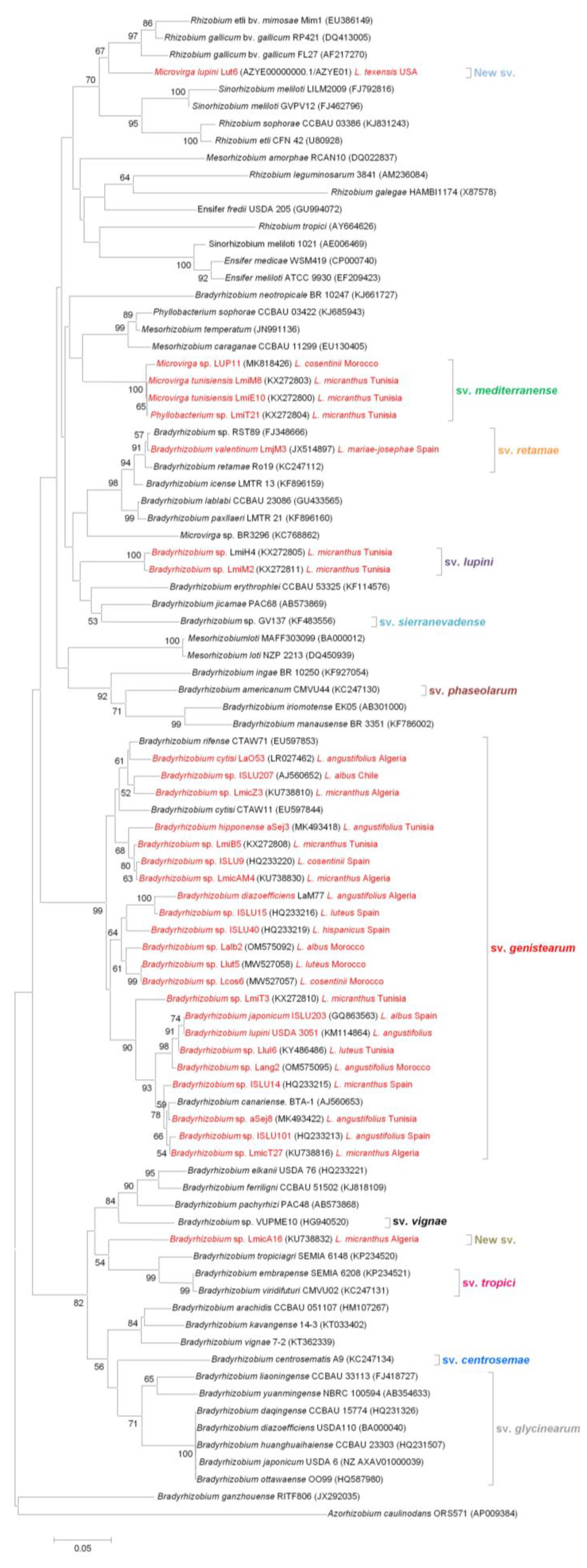
The symbiotic nodC gene encodes for N-acetylglucosaminyltransferase, a protein required for Nod factor assembly. Nod factors are molecules that determine host specificity and are indispensable for nodulation of compatible host legumes [23,37,101]. The nodC gene has been used to define the host range and symbiovars within the several rhizobial genera [3,9,37]. Nod genes are often considered not useful to classify species of rhizobia due to the possibility of natural horizontal gene transfer among bacterial strains in the soil. However, they prove useful for defining symbiovars. The nodC gene-based phylogeny of lupin-nodulating symbiotic rhizobia is presented in Figure 2. This analysis of the available nodC sequences from Lupinus spp. isolates allows for the identification of six different symbiovars: four of them include Bradyrhizobium isolates, one includes the Phyllobacterium and Microvirga strains, and one includes M. lupini. The majority of lupin-nodulating bacteria belong to the sv. genistearum, first described by Vinuesa et al. [73], which also includes some strains that nodulate other legume genera in the tribe Genisteae. The sv. genistearum is phylogenetically divergent from the rest of Bradyrhizobium symbiovars, which are integrated by Bradyrhizobium species that nodulate other legumes, namely sv. glycinearum [73], sv. retamae [102], sv. vignae [103], sv. sierranevadense [104], sv. centrosemae [105], sv. tropici [105], and sv. phaseolarum [105]. L. mariae-josephi-nodulating rhizobia, isolated in Spain, represent a new symbiotic lineage affiliated to sv. retamae, which also includes B. retamae [39]. Interestingly, all other rhizobial strains isolated from Lupinus spp. in the Iberian Peninsula belong to the genera B. japonicum and B. canariense, within sv. genistearum. Based on their symbiotic characteristics and nodC sequences, three strains isolated from L. micranthus nodules in Tunisia allowed for two new symbiovars to be defined: sv. lupini, which comprises two Bradyrhizobium isolates, and sv. mediterranense, which includes Microvirga and Phyllobacterium species [9]. Finally, Bradyrhizobium sp. LmicA16 and M. lupini Lut6 seem to represent to two new symbiovars, which appear distant from all other defined symbiovars. M. lupini is close to Rhizobium and Sinorhizobium (syn. Ensifer) strains, suggesting that its nodC was obtained from these rhizobial species through horizontal transfer. This is in agreement with a previous study in which a nodA gene sequence phylogenetic analysis placed M. lupini in the same clade with Rhizobium, Mesorhizobium, and Sinorhizobium species [84]. The nodC gene sequences of Ochrobactrum lupini and Phyllobacterium lupini are not available. However, their nodD gene sequences show a high similarity to the nodD gene of Rhizobium species [27,41], suggesting that their nodulation genes could have been acquired by horizontal gene transfer from this rhizobial genus.
When we compare the phylogenetic trees of the nodC gene (Figure 2) and the 16S rRNA gene (Figure 1), we can appreciate some consistencies and inconsistencies. Most lupin-nodulating Bradyrhizobium species group together within sv. genistearum, which suggests that they share a common evolutionary history or that a horizontal transfer of symbiotic genes between different Bradyrhizobium species associated with lupin and other Genisteae legumes has occurred. However, B. valentinum, as well as Bradyrhizobium sp. strains LmiH4, LmiT2, and LmicA16, which appear relatively close in the 16S rRNA tree, belong to three quite distant symbiovars according to their nodC sequences, all of them still close to bradyrhizobia that nodulate other legumes. Unlike the species in the genus Bradyrhizobium, non-rhizobial species isolated from lupin nodules appear to have obtained their symbiotic genes from other rhizobial genera via horizontal transfer [29]. Some Phyllobacterium and Microvirga strains share a common symbiovar in the proximity of Mesorhizobium species, suggesting that they acquired their symbiotic genes from rhizobia within the genus Mesorhizobium. These strains were isolated from L. micranthus in Tunisia and Morocco, so they share host plant and have close geographical origins. However, Microvirga lupini, isolated from L. texensis nodules in Texas, represents a completely different symbiovar. According to the nodC tree, it could have acquired this gene from phylogenetically close Rhizobium or Ensifer species.

4.2. nifH, a Gene Required for Effective Nitrogen Fixation

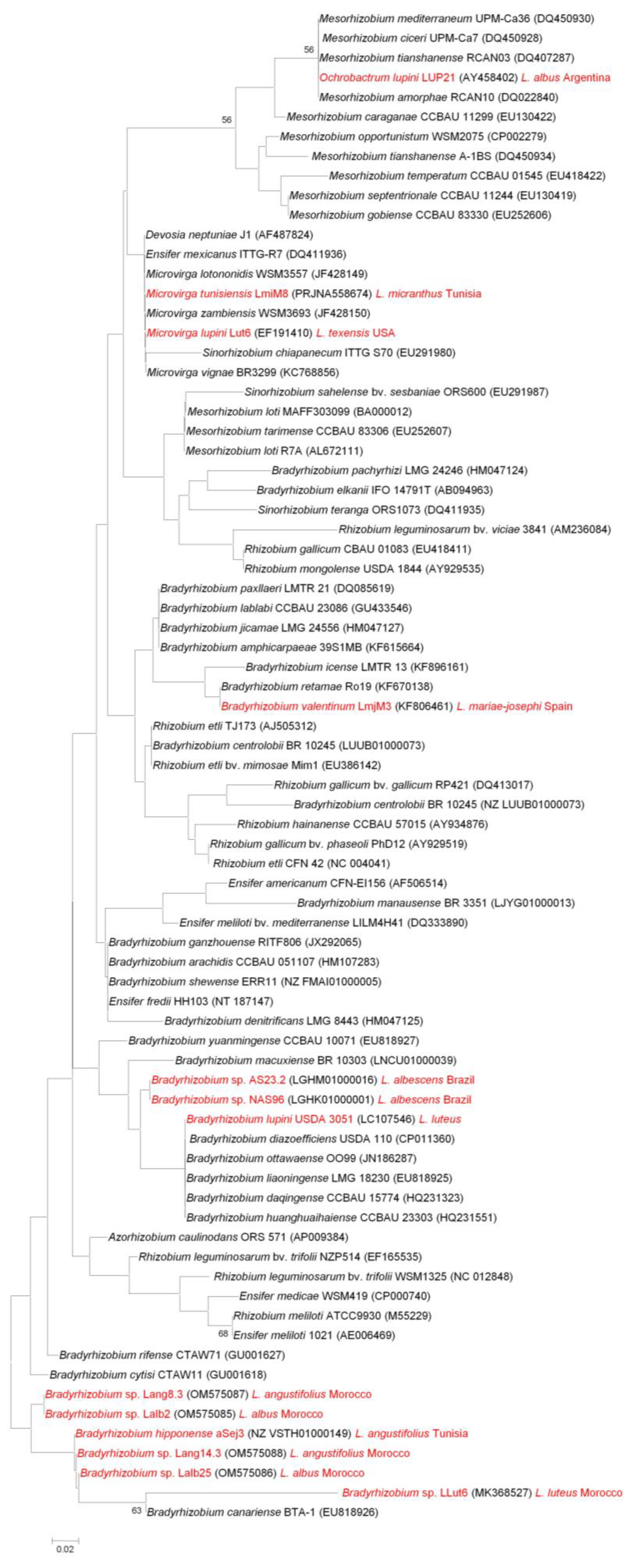
The nif genes, which codify for different nitrogenase enzyme complex subunits and several regulatory proteins, are required for nitrogen fixation [22]. They are present in rhizobia and in other bacteria capable of nitrogen fixation, including other symbiotic bacteria, such as Frankia; endophytes, such as Azospirillum; heterotrophic free-living bacteria, such as Azotobacter; and cyanobacteria, such as Anabaena, but they are also found in bacteria that are not able to fix nitrogen. The nifH symbiotic gene is one of the essential genes required to synthesize the enzyme nitrogenase, and it is considered as a highly conserved gene [106]. The nifH gene-based phylogenetic tree shown in Figure 3 includes the available nifH sequences of lupin-nodulating rhizobia and the nifH sequences with the highest similarity. Most strains associated with the genus Bradyrhizobium were isolated from lupin species growing in the Old World. American lupin microsymbionts isolated from L. albescens are somewhat distant from the Old World group. This is in agreement with a recent report in which a phylogenetic analysis of the nifD gene sequences of bradyrhizobia present in Brazilian soils was performed [107]. This analysis clustered the indigenous and the Old World-imported lupin-nodulating Bradyrhizobium strains in different clades. The fact that Old World genistoids are predominantly nodulated by Old World bradyrhizobia in American soils suggests that high symbiotic specificity is the reason why microsymbionts have been codispersed and adapted to the soils where their legume hosts have been introduced [107]. B. lupini appears closer to the American strains than to the Old World group, while B. valentinum is distant from the rest of lupin microsymbionts, and clusters together with B. retamae in concordance with the nodC gene phylogenetic analysis (Figure 2).
Regarding lupin-nodulating bacteria not belonging to the genus Bradyrhizobium, Microvirga tunisiensis groups together with M. lupini despite the fact that their respective nodC gene sequences seem to have different origins (Figure 2). They appear close to M. lotononidis and M. zambiensis, isolated from Listia angolensis nodules, a species within the only other genus besides Lupinus that forms lupinoid nodules. This suggests that Microvirga species share their own nifH gene. A phylogenetic analysis based on the nifD and nifH gene sequences of M. lupini, M. lotononidis, and M. zambiensis showed that these species are closely related to Rhizobium etli [84]. The nifH sequence of Ochrobactrum lupini isolated from L. honoratus nodules [27] shows high similarity to those of several Mesorhizobium species, which suggests that O. lupini obtained its symbiotic genes by horizontal transfer from Mesorhizobium.

5. Conclusions

Most Lupinus spp. microsymbionts isolated all over the world belong to the Bradyrhizobium lineage. While all lupin-nodulating rhizobia were initially referred to as Bradyrhizobium sp. (Lupinus) and sometimes as B. lupini, the advancement of molecular biology, bioinformatics tools, and gene sequence-based taxonomy has allowed for the identification of numerous species under the Bradyrhizobium sp. (Lupinus) umbrella, as shown in the phylogenetic analysis based on 16S rRNA gene sequences (Figure 1). This analysis groups lupin microsymbionts into five different groups. The largest one corresponds to the genus Bradyrhizobium, and comprises eight subgroups. Four additional clades include the fast-growing bacteria Microvirga, Phyllobacterium, Ochrobactrum, and Devosia. Bacteria associated with the genera Agrobacterium, Rhizobium, and Neorhizobium have also been described as lupin microsymbionts according to a concatenated phylogenetic analysis which used the sequences of housekeeping genes. However, the 16S RNA gene sequences of these strains are not yet available.
It is a known fact that, in general, the phylogeny of symbiotic genes is not in congruence with that of core genes (16S rRNA and housekeeping genes) [21]. Symbiotic genes’ phylogenies can be used to characterize isolates at the symbiovar level [37]. Divergences between phylogenies indicate that horizontal gene transfer of symbiotic genes might have occurred between species and/or genera. Full genome sequence analysis of symbiotic bacteria will likely increase our understanding of the legume–rhizobia interactions, and, most probably, will lead to a modification of the taxonomic criteria currently accepted for the definition of rhizobial species and genera. In fact, the taxonomy of bacteria that induce nodules in Lupinus spp. has been rearranged in recent years with the definition of new species and genera, the reclassification of some species in new genera, and the definition of new symbiovars. Moreover, a significant increase in the number of genera and species of lupin microsymbionts is expected to occur in the future, considering that most studies to date are mostly limited to Old World lupin species, while between 200 and 500 New World Lupinus species remain to be characterized.
The phylogenetic analysis of nodC sequences (Figure 2) suggests the existence of six different symbiovars: four of them include Bradyrhizobium isolates, one includes Phyllobacterium and Microvirga strains, and one includes Microvirga lupini. The majority of Lupinus spp. symbiotic bacteria in the genus Bradyrhizobium which are related to B. lupini and to the phylogenetically close B. cytisi/B. rifense lineages belong to the symbiovar genistearum. However, some lupin-nodulating Bradyrhizobium species appear to constitute new distant symbiotic lineages, namely sv. retamae and sv. lupini, and, possibly, a new symbiovar. Symbiovar mediterranense comprises Microvirga and Phyllobacterium strains, and M. lupini appears to represent another new symbiovar. Despite their taxonomic distance, it seems that the nodC symbiotic genes of all bradyrhizobia that nodulate lupin are more closely related to other Bradyrhizobium species than to other rhizobial genera. Contrary to lupin-nodulating bradyrhizobia, it appears that other lupin microsymbiont species have acquired their nod genes from several rhizobial genera other than Bradyrhizobium. The nifH gene-based phylogenetic tree (Figure 3) suggests that within the genus Bradyrhizobium, this gene seems to somehow be associated with the geographical origin of the strains. In the nifH tree, all Microvirga species cluster together, suggesting that, contrary to the foreign origin of their nodC gene, they share a common nifH gene which might not have been obtained from other rhizobia through horizontal transfer. The nifH gene of Ochrobactrum lupini is probably the result of a horizontal transfer event from Mesorhizobium species.
Several studies have reported the isolation of Paenibacillus, Micromonospora, Rahnella, Serratia, Raoultella, and Stenotrophomonas strains from lupin nodules; however, these bacterial isolates have not been confirmed to form effective nitrogen-fixing nodules. Most of these nodule endophytes have been reported to have PGPR traits. The horizontal gene transfer from symbiotic bacteria to endophytes represents a one-step evolution that might lead to the emergence of new symbiotic bacteria; therefore, it is ecologically and evolutionarily important [108]. Despite the physical proximity of endophytes and symbiotic bacteria in lupin nodules, it seems unlikely that they gain nodulating and nitrogen-fixing capacities from bradyrhizobia, as no horizontal transfer of symbiotic genes from Bradyrhizobium to other lupin-nodulating bacteria has been described to date. However, the transfer of nifH from soybean-nodulating B. japonicum to endophytic Bacillus has been reported [109], and still, the possibility exists that lupin nodule endophytes acquire symbiotic genes when sharing nodule occupancy with lupin microsymbionts other than Bradyrhizobium.
Overall, the data that are available so far and included in the present review suggest that horizontal transfer of specific symbiotic genes from different rhizobial genera represents a key mechanism, allowing lupin to form symbioses with selected bacteria initially considered as non-symbiotic or unable to nodulate lupin. These new lupin microsymbionts are probably better adapted to particular soil and climate conditions. Thus, the dissemination of indiscriminate symbiotic genes appears to be part of a bacterial cooperation strategy which benefits a biodiverse population rather than a particular species, and allows for the development of strain-specific legume–rhizobia symbioses in particular habitats, which would not be viable otherwise. A recent review [29] reported on the occurrence and importance of horizontal transfer of symbiotic genes within and between rhizobial genera, addressing the diversity of microsymbionts that are able to nodulate different legume genera. The authors did not report, however, on lupin and its nodulating bacteria. Lupin is a singular legume crop in many ways, and is, foremost, a promising yet underexploited plant protein source. It also presents great potential as a medicinal plant, owing to its seed particular protein, lipidic, and phenolic profiles. Its application in the remediation of poor and degraded soils and heavy metal-contaminated soils has also been proposed as very promising. Well-adapted microsymbionts have been shown to increase lupin tolerance to heavy metals, and certain inoculants have been reported to improve the nutritional and antioxidant characteristics of lupin seeds. In the present review, we summarized the current knowledge on the diversity of microsymbionts that nodulate Lupinus spp., and the important role that horizontal transfer seems to play. Our conclusions are in agreement with those of Andrews et al. [29]. Deeper insight into the nature of lupin microsymbionts, including comprehensive whole genome analyses, will undoubtedly provide new knowledge that might prove useful for the purpose of increasing lupin’s stress tolerance, adaptability to marginal soils and climates, grain quality, and crop yields.

Author Contributions

Conceptualization, A.M. and J.J.P.; writing—original draft preparation, A.M.; writing—review and editing, A.M., M.M., M.A.Q., M.M.L. and J.J.P.; supervision, J.J.P.; funding acquisition, M.M., M.M.L. and J.J.P. All authors have read and agreed to the published version of the manuscript.

Funding

This research was funded by the Ministry of High Education and Scientific Research, Tunisia, the Agencia Estatal de Investigación, AEI, Spain, (grant numbers AGL2017-88381-R and PID2021-125371OB-I00), and the Agencia Estatal Consejo Superior de Investigaciones Científicas, CSIC, Spain (grant number COOPB20610).

Acknowledgments

We apologize to all colleagues whose work we have not cited due to space restrictions.

Conflicts of Interest

The authors declare no conflict of interest.

References

  1. Sprent, J.I.; Ardley, J.K.; James, E.K. Biogeography of nodulated legumes and their nitrogen-fixing symbionts. New Phytol. 2017, 215, 40–56. [Google Scholar] [CrossRef] [PubMed] [Green Version]
  2. Azani, N.; Babineau, M.; Bailey, C.D.; Banks, H.; Barbosa, A.R.; Pinto, R.B.; Boatwright, J.S.; Borges, L.M.; Brown, G.K.; Bruneau, A.; et al. A new subfamily classification of the Leguminosae based on a taxonomically comprehensive phylogeny. Taxon 2017, 66, 44–77. [Google Scholar] [CrossRef] [Green Version]
  3. Peix, A.; Ramírez-Bahena, M.H.; Velázquez, E.; Bedmar, E.J. Bacterial associations with legumes. CRC Crit. Rev. Plant Sci. 2015, 34, 17–42. [Google Scholar] [CrossRef]
  4. Acinas, S.G.; Marcelino, L.A.; Klepac-Ceraj, V.; Polz, M.F. Divergence and redundancy of 16S rRNA sequences in genomes with multiple rrn operons. J. Bacteriol. 2004, 186, 2629–2635. [Google Scholar] [CrossRef] [Green Version]
  5. Gornung, E. Twenty years of physical mapping of major ribosomal RNA genes across the teleosts: A review of research. Cytogenet. Genome Res. 2013, 141, 90–102. [Google Scholar] [CrossRef]
  6. Idris, A.B.; Hassan, H.G.; Salaheldin Ali, M.A.; Eltaher, S.M.; Idris, L.B.; Altayb, H.N.; Abass, A.M.; Ibrahim, M.M.A.; Ibrahim, E.A.M.; Hassan, M.A. Molecular phylogenetic analysis of 16S rRNA sequences identified two lineages of Helicobacter pylori strains detected from different regions in Sudan suggestive of differential evolution. Int. J. Microbiol. 2020, 2020, 8825718. [Google Scholar] [CrossRef]
  7. Janda, J.M.; Abbott, S.L. 16S rRNA gene sequencing for bacterial identification in the diagnostic laboratory: Pluses, perils, and pitfalls. J. Clin. Microbiol. 2007, 45, 2761–2764. [Google Scholar] [CrossRef] [Green Version]
  8. Caputo, A.; Fournier, P.E.; Raoult, D. Genome and pan-genome analysis to classify emerging bacteria. Biol. Direct 2019, 14, 5. [Google Scholar] [CrossRef] [Green Version]
  9. Msaddak, A.; Rejili, M.; Durán, D.; Rey, L.; Palacios, J.M.; Imperial, J.; Ruiz-Argüeso, T.; Mars, M. Definition of two new symbiovars, sv. lupini and sv. mediterranense, within the genera Bradyrhizobium and Phyllobacterium efficiently nodulating Lupinus micranthus in Tunisia. Syst. Appl. Microbiol. 2018, 41, 487–493. [Google Scholar] [CrossRef]
  10. Rincón, A.; Arenal, F.; González, I.; Manrique, E.; Lucas, M.M.; Pueyo, J.J. Diversity of rhizobial bacteria isolated from nodules of the gypsophyte Ononis tridentata L. growing in Spanish soils. Microb. Ecol. 2008, 56, 223–233. [Google Scholar] [CrossRef]
  11. Fuks, G.; Elgart, M.; Amir, A.; Zeisel, A.; Turnbaugh, P.J.; Soen, Y.; Shental, N. Combining 16S rRNA gene variable regions enables high resolution microbial community profiling. Microbiome 2018, 16, 17. [Google Scholar] [CrossRef] [PubMed] [Green Version]
  12. Msaddak, A.; Rey, L.; Imperial, J.; Palacios, J.M.; Mars, M.; Pueyo, J.J. Phylogenetic analyses of rhizobia isolated from nodules of Lupinus angustifolius in Northern Tunisia reveal Devosia sp. as a new microsymbiont of lupin species. Agronomy 2021, 11, 1510. [Google Scholar] [CrossRef]
  13. Woo, P.C.Y.; Lau, S.K.P.; Teng, J.L.L.; Tse, H.; Yuen, K.Y. Then and now: Use of 16S rDNA gene sequencing for bacterial identification and discovery of novel bacteria in clinical microbiology laboratories. Clin. Microbiol. Infect. 2008, 14, 908–934. [Google Scholar] [CrossRef] [Green Version]
  14. Clarridge, J.E. Impact of 16S rRNA gene sequence analysis for identification of bacteria on clinical microbiology and infectious diseases. Clin. Microbiol. Rev. 2004, 17, 840–862. [Google Scholar] [CrossRef] [PubMed] [Green Version]
  15. Shamseldin, A.; Abdelkhalek, A.; Sadowsky, M.J. Recent changes to the classification of symbiotic, nitrogen-fixing, legume-associating bacteria: A review. Symbiosis 2017, 71, 91–109. [Google Scholar] [CrossRef]
  16. Gyaneshwar, P.; Hirsch, A.M.; Moulin, L.; Chen, W.M.; Elliott, G.N.; Bontemps, C.; De Los Santos, P.E.; Gross, E.; Dos Reis, F.B.; Janet, I.S.; et al. Legume-nodulating betaproteobacteria: Diversity, Host range, and future prospects. Mol. Plant-Microbe Interact. 2011, 24, 1276–1288. [Google Scholar] [CrossRef] [Green Version]
  17. Benhizia, Y.; Benhizia, H.; Benguedouar, A.; Muresu, R.; Giacomini, A.; Squartini, A. Gamma proteobacteria can nodulate legumes of the genus Hedysarum. Syst. Appl. Microbiol. 2004, 27, 462–468. [Google Scholar] [CrossRef]
  18. Shiraishi, A.; Matsushita, N.; Hougetsu, T. Nodulation in black locust by the gammaproteobacteria Pseudomonas sp. and the betaproteobacteria Burkholderia sp. Syst. Appl. Microbiol. 2010, 33, 269–274. [Google Scholar] [CrossRef]
  19. Esma, T.; Ahlem, R.; Boutheina, T.; Soumia, S.; Razika, G.; Hayet, B.; Yacine, B.; Ammar, B. Contribution to the study of the relationship between gammaproteobacteria and rhizobia in legume species of the genus Hedysarum. Legum. Res. 2020, 43, 872–877. [Google Scholar] [CrossRef]
  20. Schultze, M.; Kondorosi, A. Regulation of symbiotic root nodule development. Annu. Rev. Genet. 1998, 32, 33–57. [Google Scholar] [CrossRef]
  21. Andrews, M.; Andrews, M.E. Specificity in legume-rhizobia symbioses. Int. J. Mol. Sci. 2017, 18, 705. [Google Scholar] [CrossRef] [PubMed] [Green Version]
  22. Franche, C.; Lindström, K.; Elmerich, C. Nitrogen-fixing bacteria associated with leguminous and non-leguminous plants. Plant Soil 2009, 321, 35–59. [Google Scholar] [CrossRef]
  23. Laguerre, G.; Nour, S.M.; Macheret, V.; Sanjuan, J.; Drouin, P.; Amarger, N. Classification of rhizobia based on nodC and nifH gene analysis reveals a close phylogenetic relationship among Phaseolus vulgaris symbionts. Microbiology 2001, 147, 981–993. [Google Scholar] [CrossRef] [PubMed] [Green Version]
  24. Remigi, P.; Zhu, J.; Young, J.P.W.; Masson-Boivin, C. Symbiosis within symbiosis: Evolving nitrogen-fixing legume symbionts. Trends Microbiol. 2016, 24, 63–75. [Google Scholar] [CrossRef]
  25. Sullivan, J.T.; Patrick, H.N.; Lowther, W.L.; Scott, D.B.; Ronson, C.W.; Sullivan, J.T.; Patrickt, H.N.; Lowthert, W.L.; Scottt, D.B.; Ronson, C.W. Nodulating strains of Rhizobium loti arise through chromosomal symbiotic gene transfer in the environment. Proc. Natl. Acad. Sci. USA 1995, 92, 8985–8989. [Google Scholar] [CrossRef] [Green Version]
  26. Rivas, R.; Velázquez, E.; Willems, A.; Vizcaíno, N.; Subba-Rao, N.S.; Mateos, P.F.; Gillis, M.; Dazzo, F.B.; Martínez-Molina, E. A new species of Devosia that forms a unique nitrogen-fixing root-nodule symbiosis with the aquatic legume Neptunia natans (L.f.) Druce. Appl. Environ. Microbiol. 2003, 68, 5217–5222. [Google Scholar] [CrossRef] [Green Version]
  27. Trujillo, M.E.; Willems, A.; Abril, A.; Planchuelo, A.M.; Rivas, R.; Ludeña, D.; Mateos, P.F.; Martínez-Molina, E.; Velázquez, E. Nodulation of Lupinus albus by strains of Ochrobactrum lupini sp. nov. Appl. Environ. Microbiol. 2005, 71, 1318–1327. [Google Scholar] [CrossRef] [Green Version]
  28. Lee, I.P.A.; Eldakar, O.T.; Gogarten, J.P.; Andam, C.P. Bacterial cooperation through horizontal gene transfer. Trends Ecol. Evol. 2022, 37, 223–232. [Google Scholar] [CrossRef]
  29. Andrews, M.; De Meyer, S.; James, E.K.; Stępkowski, T.; Hodge, S.; Simon, M.F.; Young, J.P.W. Horizontal Transfer of symbiosis genes within and between rhizobial genera: Occurrence and importance. Genes 2018, 9, 321. [Google Scholar] [CrossRef] [Green Version]
  30. Diao, X.; Freeling, M.; Lisch, D. Horizontal transfer of a plant transposon. PLoS Biol. 2006, 4, e5. [Google Scholar] [CrossRef] [Green Version]
  31. Ivancevic, A.M.; Kortschak, R.D.; Bertozzi, T.; Adelson, D.L. Horizontal transfer of BovB and L1 retrotransposons in eukaryotes. Genome Biol. 2018, 19, 85. [Google Scholar] [CrossRef] [PubMed] [Green Version]
  32. Gilbert, C.; Feschotte, C. Horizontal acquisition of transposable elements and viral sequences: Patterns and consequences. Curr. Opin. Genet. Dev. 2018, 49, 15–24. [Google Scholar] [CrossRef]
  33. Caneschi, W.L.; Sanchez, A.B.; Felestrino, É.B.; de Carvalho Lemes, C.G.; Cordeiro, I.F.; Fonseca, N.P.; Villa, M.M.; Vieira, I.T.; Moraes, L.Â.G.; de Almeida Barbosa Assis, R.; et al. Serratia liquefaciens FG3 isolated from a metallophyte plant sheds light on the evolution and mechanisms of adaptive traits in extreme environments. Sci. Rep. 2019, 9, 18006. [Google Scholar] [CrossRef] [PubMed] [Green Version]
  34. Ashrafi, S.; Kuzmanović, N.; Patz, S.; Lohwasser, U.; Bunk, B.; Spröer, C.; Lorenz, M.; Elhady, A.; Frühling, A.; Neumann-Schaal, M.; et al. Two new Rhizobiales species isolated from root nodules of common sainfoin (Onobrychis viciifolia) show different plant colonisation strategies. Microbiol. Spec. 2022, 10, e0109922. [Google Scholar] [CrossRef] [PubMed]
  35. Burmølle, M.; Hansen, L.H.; Sørensen, S.J. Establishment and early succession of a multispecies biofilm composed of soil bacteria. Microb. Ecol. 2007, 54, 352–362. [Google Scholar] [CrossRef] [PubMed]
  36. Jordan, D.C. Family III. Rhizobiaceae Conn. In Bergey’s Manual of Systematic Bacteriology; Krieg, N.R., Holt, J.G., Eds.; Williams & Wilkins: Baltimore, ND, USA, 1938; Volume 1, pp. 234–254. [Google Scholar]
  37. Rogel, M.A.; Ormeño-Orrillo, E.; Martinez Romero, E. Symbiovars in rhizobia reflect bacterial adaptation to legumes. Syst. Appl. Microbiol. 2011, 34, 96–104. [Google Scholar] [CrossRef]
  38. Bourebaba, Y.; Durán, D.; Boulila, F.; Ahnia, H.; Boulila, A.; Temprano, F.; Palacios, J.M.; Imperial, J.; Ruiz-Argüeso, T.; Rey, L. Diversity of Bradyrhizobium strains nodulating Lupinus micranthus on both sides of the Western Mediterranean: Algeria and Spain. Syst. Appl. Microbiol. 2016, 39, 266–274. [Google Scholar] [CrossRef]
  39. Sánchez-Cañizares, C.; Rey, L.; Durán, D.; Temprano, F.; Sánchez-Jiménez, P.; Navarro, A.; Polajnar, M.; Imperial, J.; Ruiz-Argüeso, T. Endosymbiotic bacteria nodulating a new endemic lupine Lupinus mariae-josephi from alkaline soils in Eastern Spain represent a new lineage within the Bradyrhizobium genus. Syst. Appl. Microbiol. 2011, 34, 207–215. [Google Scholar] [CrossRef] [Green Version]
  40. Stępkowski, T.; Banasiewicz, J.; Granada, C.E.; Andrews, M.; Passaglia, L.M.P. Phylogeny and phylogeography of rhizobial symbionts nodulating legumes of the tribe Genisteae. Genes 2018, 9, 163. [Google Scholar] [CrossRef] [Green Version]
  41. Valverde, A.; Velázquez, E.; Fernández-Santos, F.; Vizcaíno, N.; Mateos, P.F.; Martínez-Molina, E.; Igual, J.M.; Willems, A. Phyllobacterium trifolii sp. nov., nodulating Trifolium and Lupinus in Spanish soils. Int. J. Syst. Evol. Microbiol. 2005, 55, 1985–1989. [Google Scholar] [CrossRef] [Green Version]
  42. Msaddak, A.; Rejili, M.; Durán, D.; Rey, L.; Imperial, J.; Palacios, J.M.; Ruiz-Argüeso, T.; Mars, M. Members of Microvirga and Bradyrhizobium genera are native endosymbiotic bacteria nodulating Lupinus luteus in Northern Tunisian soils. FEMS Microbiol. Ecol. 2017, 93, fix068. [Google Scholar] [CrossRef] [PubMed]
  43. Tounsi-Hammami, S.; Le Roux, C.; Dhane-Fitouri, S.; De Lajudie, P.; Duponnois, R.; Ben Jeddi, F. Genetic diversity of rhizobia associated with root nodules of white lupin (Lupinus albus L.) in Tunisian calcareous soils. Syst. Appl. Microbiol. 2019, 42, 448–456. [Google Scholar] [CrossRef] [PubMed]
  44. Missbah El Idrissi, M.; Lamin, H.; ElFaik, S.; Tortosa, G.; Peix, A.; Bedmar, E.J.; Abdelmoumen, H. Microvirga sp. symbiovar mediterranense nodulates Lupinus cosentinii grown wild in Morocco. J. Appl. Microbiol. 2020, 128, 1109–1118. [Google Scholar] [CrossRef] [PubMed]
  45. Hughes, C.; Eastwood, R. Island radiation on a continental scale: Exceptional rates of plant diversification after uplift of the Andes. Proc. Natl. Acad. Sci. USA 2006, 103, 10334–10339. [Google Scholar] [CrossRef] [PubMed] [Green Version]
  46. Aïnouche, A.K.; Bayer, R.J. Phylogenetic relationships in Lupinus (Fabaceae: Papilionoideae) based on internal transcribed spacer sequences (ITS) of nuclear ribosomal DNA. Am. J. Bot. 1999, 86, 590–607. [Google Scholar] [CrossRef] [PubMed]
  47. Lucas, M.M.; Stoddard, F.L.; Annicchiarico, P.; Frías, J.; Martínez-Villaluenga, C.; Sussmann, D.; Duranti, M.; Seger, A.; Zander, P.M.; Pueyo, J.J. The future of lupin as a protein crop in Europe. Front. Plant Sci. 2015, 6, 1–6. [Google Scholar] [CrossRef] [Green Version]
  48. Ohadoma, S.C.; Akah, P.A.; Okolo, C.E. Isolation and characterization of flavonol glycosides from leaves extract of Lupinus arboreus Sims. Pharm. Biosci. J. 2016, 4, 6–9. [Google Scholar] [CrossRef]
  49. Zhang, Z.; Yuan, W.; Wang, P.; Grant, G.; Li, S. Flavonoids from Lupinus texensis and their free radical scavenging activity. Nat. Prod. Res. 2011, 25, 1641–1649. [Google Scholar] [CrossRef]
  50. Leporatti, M.L.; Ghedira, K. Comparative analysis of medicinal plants used in traditional medicine in Italy and Tunisia. J. Ethnobiol. Ethnomed. 2009, 5, 31. [Google Scholar] [CrossRef] [Green Version]
  51. Ishaq, A.R.; El-Nashar, H.A.S.; Younis, T.; Mangat, M.A.; Shahzadi, M.; Ul Haq, A.S.; El-Shazly, M. Genus Lupinus (Fabaceae): A review of ethnobotanical, phytochemical and biological studies. J. Pharm. Pharmacol. 2022, 74, 1700–1717. [Google Scholar] [CrossRef]
  52. Siger, A.; Czubinski, J.; Kachlicki, P.; Dwiecki, K.; Lampart-Szczapa, E.; Nogala-Kalucka, M. Antioxidant activity and phenolic content in three lupin species. J. Food Compos. Anal. 2012, 25, 190–197. [Google Scholar] [CrossRef]
  53. Sachdev, D.O.; Gosavi, D.D.; Salwe, K.J. Evaluation of antidiabetic, antioxidant effect and safety profile of gomutra ark in Wistar albino rats. Anc. Sci. Life 2012, 31, 84–89. [Google Scholar] [CrossRef] [PubMed]
  54. Confortin, T.C.; Todero, I.; Soares, J.F.; Luft, L.; Brun, T.; Rabuske, J.E.; Nogueira, C.U.; Mazutti, M.A.; Zabot, G.L.; Tres, M.V. Extracts from Lupinus albescens: Antioxidant power and antifungal activity in vitro against phytopathogenic fungi. Environ. Technol. 2019, 40, 1668–1675. [Google Scholar] [CrossRef] [PubMed]
  55. Hanania, M.; Radwan, S.; Odeh, S.A.; Qumber, A. Determination of minerals, total phenolic content, flavonoids, antioxidants and antimicrobial activities of ethanolic extract of sweet Lupinus angustifolius of Palestine. Eur. J. Med. Plants 2019, 28, 1–6. [Google Scholar] [CrossRef]
  56. Fernández-Pascual, M.; Pueyo, J.J.; Felipe, M.R.; Golvano, M.P.; Lucas, M.M. Singular features of the Bradyrhizobium-Lupinus symbiosis. Dyn. Soil Dyn. Plant 2007, 1, 1–16. [Google Scholar]
  57. Coba de la Peña, T.; Pueyo, J.J. Legumes in the reclamation of marginal soils, from cultivar and inoculant selection to transgenic approaches. Agron. Sustain. Dev. 2012, 32, 65–91. [Google Scholar] [CrossRef] [Green Version]
  58. Quiñones, M.A.; Fajardo, S.; Fernández-Pascual, M.; Lucas, M.M.; Pueyo, J.J. Nodulated white lupin plants growing in contaminated soils accumulate unusually high mercury concentrations in their nodules, roots and especially cluster roots. Horticulturae 2021, 7, 302. [Google Scholar] [CrossRef]
  59. Quiñones, M.A.; Ruiz-Díez, B.; Fajardo, S.; López-Berdonces, M.A.; Higueras, P.L.; Fernández-Pascual, M. Lupinus albus plants acquire mercury tolerance when inoculated with an Hg-resistant Bradyrhizobium strain. Plant Physiol. Biochem. 2013, 73, 168–175. [Google Scholar] [CrossRef] [Green Version]
  60. Quiñones, M.A.; Lucas, M.M.; Pueyo, J.J. Adaptive mechanisms make lupin a choice crop for acidic soils affected by aluminum toxicity. Front. Plant Sci. 2022, 12, 810692. [Google Scholar] [CrossRef]
  61. Pueyo, J.J.; Quiñones, M.A.; Coba de la Peña, T.; Fedorova, E.E.; Lucas, M.M. Nitrogen and phosphorus interplay in lupin root nodules and cluster roots. Front. Plant Sci. 2021, 12, 1–9. [Google Scholar] [CrossRef]
  62. Gladstones, J.S. Distribution, origin, taxonomy, history and importance. In Lupinus as Crop Plants: Biology, Production and Utilization; Gladstones, J.S., Atkins, C.A., Hamblin, J., Eds.; CAB International: Cambridge, UK, 1998; pp. 1–37. [Google Scholar]
  63. Sprent, J.I.; Ardley, J.K.; James, E.K. From North to South: A latitudinal look at legume nodulation processes. S. Afr. J. Bot. 2013, 89, 31–41. [Google Scholar] [CrossRef] [Green Version]
  64. Corby, H.D.L. Types of rhizobial nodules and their distribution among the Leguminosae. Kirkia 1988, 13, 53–123. [Google Scholar]
  65. Ardley, J.K.; Reeve, W.G.; O’Hara, G.W.; Yates, R.J.; Dilworth, M.J.; Howieson, J.G. Nodule morphology, symbiotic specificity and association with unusual rhizobia are distinguishing features of the genus Listia within the Southern African crotalarioid clade Lotononis s.l. Ann. Bot. 2013, 112, 1–15. [Google Scholar] [CrossRef] [PubMed] [Green Version]
  66. González-Sama, A.; Lucas, M.M.; De Felipe, M.R.; Pueyo, J.J. An unusual infection mechanism and nodule morphogenesis in white lupin (Lupinus albus). New Phytol. 2004, 163, 371–380. [Google Scholar] [CrossRef] [PubMed] [Green Version]
  67. Fedorova, E.E.; De Felipe, M.R.; Pueyo, J.J.; Lucas, M.M. Conformation of cytoskeletal elements during the division of infected Lupinus albus L. nodule cells. J. Exp. Bot. 2007, 58, 2225–2236. [Google Scholar] [CrossRef]
  68. Scholla, M.H.; Moorefield, J.A.; Elkan, G.H. DNA homology between species of the rhizobia. Syst. Appl. Microbiol. 1990, 13, 288–294. [Google Scholar] [CrossRef]
  69. Auling, G.; Busse, J.; Hahn, M.; Hennecke, H.; Kroppenstedt, R.-M.; Probst, A.; Stackebrandt, E. Phylogenetic heterogeneity and chemotaxonomic properties of certain gram-negative aerobic carboxydobacteria. Syst. Appl. Microbiol. 1988, 10, 264–272. [Google Scholar] [CrossRef]
  70. Mellal, H.; Yacine, B.; Boukaous, L.; Khouni, S.; Benguedouar, A.; Castellano-Hinojosa, A.; Bedmar, E.J. Phylogenetic diversity of Bradyrhizobium strains isolated from root nodules of Lupinus angustifolius grown wild in the North East of Algeria. Syst. Appl. Microbiol. 2019, 42, 397–402. [Google Scholar] [CrossRef]
  71. Msaddak, A.; Durán, D.; Reijili, M.; Mars, M.; Ruiz-Argüeso, T.; Imperial, J.; Palacios, J.M.; Rey, L. Diverse bacteria affiliated with the genera Microvirga, Phyllobacterium, and Bradyrhizobium nodulate Lupinus micranthus growing in soils of Northern Tunisia. Appl. Environ. Microbiol. 2017, 83, e02820-16. [Google Scholar] [CrossRef] [Green Version]
  72. Jarabo-Lorenzo, A.; Pérez-Galdona, R.; Donate-Correa, J.; Rivas, R.; Velázquez, E.; Hernández, M.; Temprano, F.; Martínez-Molina, E.; Ruiz-Argüeso, T.; León-Barrios, M. Genetic diversity of bradyrhizobial populations from diverse geographic origins that nodulate Lupinus spp. and Ornithopus spp. Syst. Appl. Microbiol. 2003, 26, 611–623. [Google Scholar] [CrossRef]
  73. Vinuesa, P.; León-Barrios, M.; Silva, C.; Willems, A.; Jarabo-Lorenzo, A.; Pérez-Galdona, R.; Werner, D.; Martínez-Romero, E. Bradyrhizobium canariense sp. nov., an acid-tolerant endosymbiont that nodulates endemic genistoid legumes (Papilionoideae: Genisteae) from the Canary Islands, along with Bradyrhizobium japonicum bv. genistearum, Bradyrhizobium genospecies alpha and Bradyrhizobium genospecies beta. Int. J. Syst. Evol. Microbiol. 2005, 55, 569–575. [Google Scholar] [CrossRef] [PubMed] [Green Version]
  74. Stȩpkowski, T.; Hughes, C.E.; Law, I.J.; Markiewicz, Ł.; Gurda, D.; Chlebicka, A.; Moulin, L. Diversification of lupine Bradyrhizobium strains: Evidence from nodulation gene trees. Appl. Environ. Microbiol. 2007, 73, 3254–3264. [Google Scholar] [CrossRef] [PubMed] [Green Version]
  75. Velázquez, E.; Valverde, A.; Rivas, R.; Gomis, V.; Peix, A.; Gantois, I.; Igual, J.M.; León-Barrios, M.; Willems, A.; Mateos, P.F.; et al. Strains nodulating Lupinus albus on different continents belong to several new chromosomal and symbiotic lineages within Bradyrhizobium. Antonie van Leeuwenhoek 2010, 97, 363–376. [Google Scholar] [CrossRef] [PubMed] [Green Version]
  76. Durán, D.; Rey, L.; Sánchez-Cañizares, C.; Navarro, A.; Imperial, J.; Ruiz-Argueso, T. Genetic diversity of indigenous rhizobial symbionts of the Lupinus mariae-josephae endemism from alkaline-limed soils within its area of distribution in Eastern Spain. Syst. Appl. Microbiol. 2013, 36, 128–136. [Google Scholar] [CrossRef]
  77. Granada, C.E.; Beneduzi, A.; Lisboa, B.B.; Turchetto-Zolet, A.C.; Vargas, L.K.; Passaglia, L.M.P. Multilocus sequence analysis reveals taxonomic differences among Bradyrhizobium sp. symbionts of Lupinus albescens Plants growing in arenized and non-arenized areas. Syst. Appl. Microbiol. 2015, 38, 323–329. [Google Scholar] [CrossRef] [PubMed]
  78. Rejili, M.; Off, K.; Brachmann, A.; Marín, M. Bradyrhizobium hipponense sp. nov., isolated from Lupinus angustifolius growing in the Northern region of Tunisia. Int. J. Syst. Evol. Microbiol. 2020, 70, 5539–5550. [Google Scholar] [CrossRef] [PubMed]
  79. Lamrabet, M.; ElFaik, S.; Laadraoui, C.; Bouhnik, O.; Lamin, H.; Alami, S.; Abdelmoumen, H.; Bedmar, E.J.; El Idrissi, M.M. Phylogenetic and symbiotic diversity of Lupinus albus and L. angustifolius microsymbionts in the Maamora Forest, Morocco. Syst. Appl. Microbiol. 2022, 45, 126338. [Google Scholar] [CrossRef]
  80. Missbah El Idrissi, M.; Bouhnik, O.; ElFaik, S.; Alami, S.; Lamin, H.; Bedmar, E.J.; Abdelmoumen, H. Characterization of Bradyrhizobium spp. nodulating Lupinus cosentinii and L. luteus microsymbionts in Morocco. Front. Agron. 2021, 3, 661295. [Google Scholar] [CrossRef]
  81. Pascual, H. Lupinus mariae-josephi (Fabaceae), nueva y sorprendente especie descubierta en España. An. Jard. Bot. Madr. 2004, 61, 69–72. [Google Scholar] [CrossRef]
  82. Navarro Peris, A.; Fos Martín, S.; Ferrando Pardo, I.; Laguna Lumbreras, E. Localización del endemismo aparentemente extinto Lupinus mariae-josephi. Flora Montiberica 2006, 33, 59–63. [Google Scholar]
  83. Durán, D.; Rey, L.; Navarro, A.; Busquets, A.; Imperial, J.; Ruiz-Argüeso, T. Bradyrhizobium valentinum sp. nov., isolated from effective nodules of Lupinus mariae-josephae, a lupine endemic of basic-lime soils in Eastern Spain. Syst. Appl. Microbiol. 2014, 37, 336–341. [Google Scholar] [CrossRef] [PubMed]
  84. Ardley, J.K.; Parker, M.A.; De Meyer, S.E.; Trengove, R.D.; O’Hara, G.W.; Reeve, W.G.; Yates, R.J.; Dilworth, M.J.; Willems, A.; Howieson, J.G. Microvirga lupini sp. nov., Microvirga lotononidis sp. nov. and Microvirga zambiensis sp. nov. are alphaproteobacterial root-nodule bacteria that specifically nodulate and fix nitrogen with geographically and taxonomically separate legume hosts. Int. J. Syst. Evol. Microbiol. 2012, 62, 2579–2588. [Google Scholar] [CrossRef] [PubMed] [Green Version]
  85. Beligala, D.H.; Michaels, H.J.; Devries, M.; Phuntumart, V. Multilocus sequence analysis of root nodule bacteria associated with Lupinus spp. and Glycine max. Adv. Microbiol. 2017, 07, 790–812. [Google Scholar] [CrossRef] [Green Version]
  86. Ferchichi, N.; Toukabri, W.; Vrhovsek, U.; Angeli, A.; Masuero, D.; Mhamdi, R.; Trabelsi, D. Inoculation of Lupinus albus with the nodule-endophyte Paenibacillus glycanilyticus LJ121 improves grain nutritional quality. Arch. Microbiol. 2020, 202, 283–291. [Google Scholar] [CrossRef] [PubMed]
  87. Carro, L.; Flores-Félix, J.D.; Ramírez-Bahena, M.H.; García-Fraile, P.; Martínez-Hidalgo, P.; Igual, J.M.; Tejedor, C.; Peix, A.; Velázquez, E. Paenibacillus lupini sp. nov., isolated from nodules of Lupinus albus. Int. J. Syst. Evol. Microbiol. 2014, 64, 3028–3033. [Google Scholar] [CrossRef] [PubMed] [Green Version]
  88. Kępczyńska, E.; Karczyński, P. Medicago truncatula root developmental changes by growth-promoting microbes isolated from Fabaceae, growing on organic farms, involve cell cycle changes and WOX5 gene expression. Planta 2020, 251, 25. [Google Scholar] [CrossRef] [PubMed]
  89. Sarmiento, L.H.; Moreno Díaz, P.; Jiménez Dávalos, J. Characterization and evaluation of the PGPR potential of the microflora associated with tarwi (Lupinus mutabilis Sweet). Ecol. Appl. 2020, 19, 65–76. [Google Scholar] [CrossRef]
  90. Trujillo, M.E.; Alonso-Vega, P.; Rodríguez, R.; Carro, L.; Cerda, E.; Alonso, P.; Martínez-Molina, E. The genus Micromonospora is widespread in legume root nodules: The example of Lupinus angustifolius. ISME J. 2010, 4, 1265–1281. [Google Scholar] [CrossRef] [Green Version]
  91. Carro, L.; Pujic, P.; Trujillo, M.E.; Normand, P. Micromonospora is a normal occupant of actinorhizal nodules. J. Biosci. 2013, 38, 685–693. [Google Scholar] [CrossRef]
  92. Muresu, R.; Polone, E.; Sulas, L.; Baldan, B.; Tondello, A.; Delogu, G.; Cappuccinelli, P.; Alberghini, S.; Benhizia, Y.; Benhizia, H.; et al. Coexistence of predominantly nonculturable rhizobia with diverse, endophytic bacterial taxa within nodules of wild legumes. FEMS Microbiol. Ecol. 2008, 63, 383–400. [Google Scholar] [CrossRef] [Green Version]
  93. Zakhia, F.; Jeder, H.; Willems, A.; Gillis, M.; Dreyfus, B.; De Lajudie, P. Diverse bacteria associated with root nodules of spontaneous legumes in Tunisia and first report for nifH-like gene within the genera Microbacterium and Starkeya. Microb. Ecol. 2006, 51, 375–393. [Google Scholar] [CrossRef] [PubMed]
  94. Sprent, J.I. Legume Nodulation. A Global Perspective; Wiley-Blackwell: Chichester, UK, 2009. [Google Scholar] [CrossRef] [Green Version]
  95. Wernegreen, J.J.; Riley, M.A. Comparison of the evolutionary dynamics of symbiotic and housekeeping loci: A case for the genetic coherence of rhizobial lineages. Mol. Biol. Evol. 1999, 16, 98–113. [Google Scholar] [CrossRef] [PubMed]
  96. Aguilar, O.M.; Collavino, M.M.; Mancini, U. Nodulation competitiveness and diversification of symbiosis genes in common beans from the American centers of domestication. Sci. Rep. 2022, 12, 4591. [Google Scholar] [CrossRef] [PubMed]
  97. Mohammed, M.; Jaiswal, S.K.; Dakora, F.D. Distribution and correlation between phylogeny and functional traits of cowpea (Vigna unguiculata L. Walp.)-nodulating microsymbionts from Ghana and South Africa. Sci. Rep. 2018, 8, 18006. [Google Scholar] [CrossRef] [Green Version]
  98. Drew, G.C.; Stevens, E.J.; King, K.C. Microbial evolution and transitions along the parasite–mutualist continuum. Nat. Rev. Microbiol. 2021, 19, 623–638. [Google Scholar] [CrossRef]
  99. Watanabe, T.; Horiike, T. The evolution of molybdenum dependent nitrogenase in cyanobacteria. Biology 2021, 10, 329. [Google Scholar] [CrossRef]
  100. Lau, M.C.Y.; Cameron, C.; Magnabosco, C.; Brown, C.T.; Schilkey, F.; Grim, S.; Hendrickson, S.; Pullin, M.; Lollar, B.S.; van Heerden, E.; et al. Phylogeny and phylogeography of functional genes shared among seven terrestrial subsurface metagenomes reveal N-cycling and microbial evolutionary relationships. Front. Microbiol. 2014, 5, 531. [Google Scholar] [CrossRef]
  101. Barny, M.A.; Schoonejans, E.; Economou, A.; Johnston, A.W.B.; Downie, J.A. The C-terminal domain of the Rhizobium leguminosarum chitin synthase NodC is important for function and determines the orientation of the N-terminal region in the inner membrane. Mol. Microbiol. 1996, 19, 443–453. [Google Scholar] [CrossRef] [Green Version]
  102. Guerrouj, K.; Ruíz-Díez, B.; Chahboune, R.; Ramírez-Bahena, M.-H.; Abdelmoumen, H.; Quiñones, M.A.; El Idrissi, M.M.; Velázquez, E.; Fernández-Pascual, M.; Bedmar, E.J.; et al. Definition of a novel symbiovar (sv. retamae) within Bradyrhizobium retamae sp. nov., nodulating Retama sphaerocarpa and Retama monosperma. Syst. Appl. Microbiol. 2013, 36, 218–223. [Google Scholar] [CrossRef]
  103. Bejarano, A.; Ramírez-Bahena, M.H.; Velázquez, E.; Peix, A. Vigna unguiculata is nodulated in Spain by endosymbionts of Genisteae legumes and by a new symbiovar (vignae) of the genus Bradyrhizobium. Syst. Appl. Microbiol. 2014, 37, 533–540. [Google Scholar] [CrossRef] [Green Version]
  104. Cobo-Díaz, J.F.; Martínez-Hidalgo, P.; Fernández-González, A.J.; Martínez-Molina, E.; Toro, N.; Velázquez, E.; Fernández-López, M. The endemic Genista versicolor from Sierra Nevada National Park in Spain is nodulated by putative new Bradyrhizobium species and a novel symbiovar (sierranevadense). Syst. Appl. Microbiol. 2014, 37, 177–185. [Google Scholar] [CrossRef] [PubMed]
  105. Ramírez-Bahena, M.H.; Flores-Félix, J.D.; Chahboune, R.; Toro, M.; Velázquez, E.; Peix, A. Bradyrhizobium centrosemae (symbiovar centrosemae) sp. nov., Bradyrhizobium americanum (symbiovar phaseolarum) sp. nov. and a new symbiovar (tropici) of Bradyrhizobium viridifuturi establish symbiosis with Centrosema species native to America. Syst. Appl. Microbiol. 2016, 39, 378–383. [Google Scholar] [CrossRef] [PubMed] [Green Version]
  106. Boyd, E.S.; Peters, J.W. New insights into the evolutionary history of biological nitrogen fixation. Front. Microbiol. 2013, 4, 201. [Google Scholar] [CrossRef] [PubMed] [Green Version]
  107. Banasiewicz, J.; Granada, C.E.; Lisboa, B.B.; Grzesiuk, M.; Matuśkiewicz, W.; Bałka, M.; Schlindwein, G.; Vargas, L.K.; Passaglia, L.M.P.; Stępkowski, T. Diversity and phylogenetic affinities of Bradyrhizobium isolates from Pampa and Atlantic forest biomes. Syst. Appl. Microbiol. 2021, 44, 126203. [Google Scholar] [CrossRef] [PubMed]
  108. Barcellos, F.G.; Menna, P.; Batista, J.S.D.S.; Hungria, M. Evidence of horizontal transfer of symbiotic genes from a Bradyrhizobium japonicum inoculant strain to indigenous diazotrophs Sinorhizobium (Ensifer) fredii and Bradyrhizobium elkanii in a Brazilian savannah soil. Appl. Environ. Microbiol. 2007, 73, 2635–2643. [Google Scholar] [CrossRef] [Green Version]
  109. Li, J.H.; Wang, E.T.; Chen, W.F.; Chen, W.X. Genetic diversity and potential for promotion of plant growth detected in nodule endophytic bacteria of soybean grown in Heilongjiang Province of China. Soil Biol. Biochem. 2008, 40, 238–246. [Google Scholar] [CrossRef]
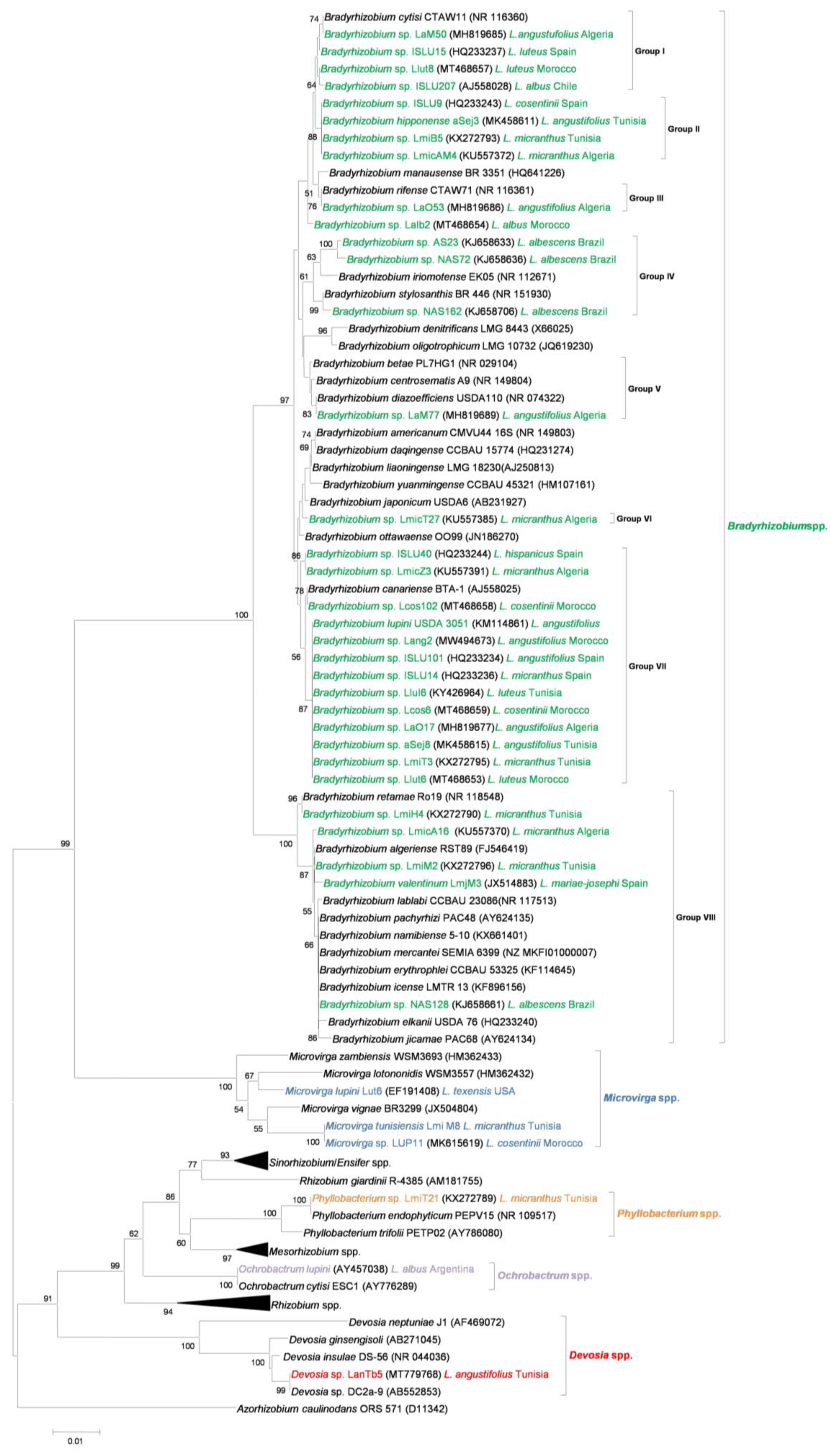
Figure 1. Neighbor-joining phylogenetic analysis of 16S rRNA gene sequence of different species of Lupinus spp. microsymbionts and their closest relatives. Lupin microsymbionts are highlighted in color as follows: Bradyrhizobium spp. in green; Microvirga spp. in blue; Phyllobacterium spp. in orange; Ochrobactrum spp. in yellow; Devosia spp. in red. Values of bootstrap probability greater than 50% are indicated. The host legume and place of isolation are reported when known.
Ijms 24 06496 g001
Figure 2. Neighbor-joining phylogenetic analysis of nodC gene sequences of different species of Lupinus spp. microsymbionts (highlighted in red) and their closest relatives. Bootstrap probability values greater than 50% are indicated.
Ijms 24 06496 g002
Figure 3. Neighbor-joining phylogenetic analysis of nifH gene sequences of different species of Lupinus spp. microsymbionts (highlighted in red) and their closest relatives. Values of bootstrap probability greater than 50% are indicated.
Ijms 24 06496 g003
Disclaimer/Publisher’s Note: The statements, opinions and data contained in all publications are solely those of the individual author(s) and contributor(s) and not of MDPI and/or the editor(s). MDPI and/or the editor(s) disclaim responsibility for any injury to people or property resulting from any ideas, methods, instructions or products referred to in the content.