Communication

Exploiting the Integrated Valorization of Eucalyptus globulus Leaves: Chemical Composition and Biological Potential of the Lipophilic Fraction before and after Hydrodistillation

by
Cátia. S. D. Oliveira
1,
Patrícia Moreira
2,3,
Maria T. Cruz
2,3,
Cláudia M. F. Pereira
2,4,
Artur M. S. Silva
5,
Sónia A. O. Santos
1,* and
Armando J. D. Silvestre
1
1
CICECO—Aveiro Institute of Materials, Department of Chemistry, University of Aveiro, 3810-193 Aveiro, Portugal
2
CNC—Center for Neuroscience and Cellular Biology, University of Coimbra, 3004-504 Coimbra, Portugal
3
Faculty of Pharmacy, University of Coimbra, 3000-548 Coimbra, Portugal
4
Faculty of Medicine, University of Coimbra, 3000-548 Coimbra, Portugal
5
LAQV-REQUIMTE, Department of Chemistry, University of Aveiro, 3810-193 Aveiro, Portugal
*
Author to whom correspondence should be addressed.
Int. J. Mol. Sci. 2023, 24(7), 6226; https://doi.org/10.3390/ijms24076226
Submission received: 27 February 2023 / Revised: 22 March 2023 / Accepted: 24 March 2023 / Published: 25 March 2023

Abstract

:
E. globulus leaves have been mainly exploited for essential oil recovery or for energy generation in industrial pulp mills, neglecting the abundance of valuable families of extractives, namely, triterpenic acids, that might open new ways for the integrated valorization of this biomass. Therefore, this study highlights the lipophilic characterization of E. globulus leaves before and after hydrodistillation, aiming at the integrated valorization of both essential oils and triterpenic acids. The lipophilic composition of E. globulus leaves after hydrodistillation is reported for the first time. Extracts were obtained by dichloromethane Soxhlet extraction and analyzed by gas chromatography-mass spectrometry. In addition, their cytotoxicity on different cell lines representative of the innate immune system, skin, liver, and intestine were evaluated. Triterpenic acids, such as betulonic, oleanolic, betulinic and ursolic acids, were found to be the main components of these lipophilic extracts, ranging from 30.63–37.14 g kg−1 of dry weight (dw), and representing 87.7–89.0% w/w of the total content of the identified compounds. In particular, ursolic acid was the major constituent of all extracts, representing 46.8–50.7% w/w of the total content of the identified compounds. Other constituents, such as fatty acids, long-chain aliphatic alcohols and β-sitosterol were also found in smaller amounts in the studied extracts. This study also demonstrates that the hydrodistillation process does not affect the recovery of compounds of greatest interest, namely, triterpenic acids. Therefore, the results establish that this biomass residue can be considered as a promising source of value-added bioactive compounds, opening new strategies for upgrading pulp industry residues within an integrated biorefinery context.

Graphical Abstract

1. Introduction

In the last decades, the growing concern about the finitude of fossil resources, as well as the environmental impact resulting from their massive use, associated with greenhouse gas (GHG) emissions that result in climate change and global warming [1,2,3] have been pushing industry to develop fully sustainable processes and particularly those based on renewable raw materials. The exploitation of value-chains based on biomass has led to the emergence of the biorefinery concept, which still requires the development of efficient fractionation processes, allowing for the integrated exploitation of all biomass fractions and ensuring benefits to society and the environment, including their economic viability [3,4].
At the same time, in the last decades, the demand for new alternatives to petrochemical products has aroused great interest in the search for high-value compounds from renewable feedstocks, such as forest biomass. The forest activity, associated with the pulp and paper sector has a great impact on the world economy. In fact, Portugal is considered the 3rd largest European producer of pulp fibers [5].
Eucalyptus globulus is one of the most widely cultivated species in Portugal. According to the 6th National Forest Inventory (IFN6) published in 2019, eucalyptus is the forest species that occupies the largest planted area, approximately 844 kha, which corresponds to 26% of the Portuguese forest area [6]. The large-scale exploitation of E. globulus wood for pulp production generates large amounts of forest by-products, such as bark, branches and leaves (Figure 1) that have been widely used as solid fuel for power generation [4,7,8]. Nevertheless, the integrated valorization of these residues may represent a significant contribution to the paper and/or forestry sector’s profitability.
E. globulus pulping residues have been scrutinized as a source for added-value applications such as materials, chemicals or biofuels [9,10,11,12,13,14]. In the last years, particular interest has been devoted to the search for bioactive compounds from these resources. Some studies on E. globulus leaves report the presence of different families of compounds, namely, its essential oil (EO) [15,16,17], hydrophilic components, such as phenolic compounds [18,19,20,21], and lipophilic components, such as fatty acids, long-chain aliphatic alcohols, sterols, and triterpenic acids, such as betulinic, betulonic, oleanolic, and ursolic acids [12,22,23,24].
Eucalyptus leaf extracts and EO have long been used in the pharmaceutical, sanitary, agricultural, cosmetic, and food industries because of their beneficial and healthy properties [25,26]. In fact, traditionally, Eucalyptus leaves have been widely used for the treatment of various diseases such as influenza, dysentery, pulmonary tuberculosis, cystitis, diabetes, articular pain, fungal infections, dermatitis, scabies, and burns [25,27].
E. globulus is widely recognized and exploited as a source of EO that is composed mainly of monoterpenes and sesquiterpenes, which are obtained by hydrodistillation or steam distillation with extraction yields ranging from 1.2 to 2.7% (w/w) [15,16,17,28]. Nevertheless, after this process the leaf biomass remains underexploited, despite its richness in other valuable extractives fractions as, for example, triterpenic acids. The effect of the hydrodistillation process on E. globulus leaf extract composition is also unknown; therefore, this study aims to understand the effect of hydrodistillation on the composition of E. globulus leaf lipophilic extractives with emphasis on triterpenic acids, but including also the less abundant fractions of fatty acids and long-chain aliphatic alcohols in order to access the potential of the exploitation of this fraction integrated with EO.
In this vein, the lipophilic fraction of E. globulus leaves before and after hydrodistillation was obtained by Soxhlet extraction, and analyzed by gas chromatography-mass spectrometry (GC–MS) and their compositions were discussed in detail. In order to evaluate the safety of the different extracts to be exploited in different applications, their cytotoxicity was evaluated in cell lines representative of the innate immune system, skin, liver, and intestine, namely, on macrophages (RAW 264), fibroblasts (NIH/3T3), hepatocytes (HepG2) and colon cancer (Caco-2), respectively.

2. Results and Discussion

2.1. Extraction Yields

Lipophilic extracts of E. globulus leaves before and after hydrodistillation were obtained by Soxhlet extraction with dichloromethane (DCM). The extraction yields obtained were 19.5 ± 0.2% and 22.0 ± 0.6% of dw for E. globulus leaf DCM extracts before and after hydrodistillation, respectively. Although the values were approximate, the E. globulus leaves after hydrodistillation (EgLHD) showed a slightly higher extraction yield than before hydrodistillation (EgL), despite the fact that in this latter case EOs (1.7% of dw) were also extracted. These values were significantly higher than those previously reported for E. globulus leaves using non-polar organic solvents [22,29,30]. Rodrigues et al. [22] presented Soxhlet DCM extraction yields of 7.32% of dw and 2.38–2.89% of dw before and after a wax removal pretreatment, respectively. While other studies, also with E. globulus leaves, reported extraction yields of 2.2 and 9% of dw for solid–liquid extractions with hexane and DCM, respectively [24,30]. The lipophilic extractive yield of Eucalyptus leaves after hydrodistillation was only reported by El-Ghorab et al. [29] for a different species, namely, E. camaldulensis, with a value of 7.4 ± 0.6% of dw.

2.2. Chemical Characterization of the Lipophilic Extracts

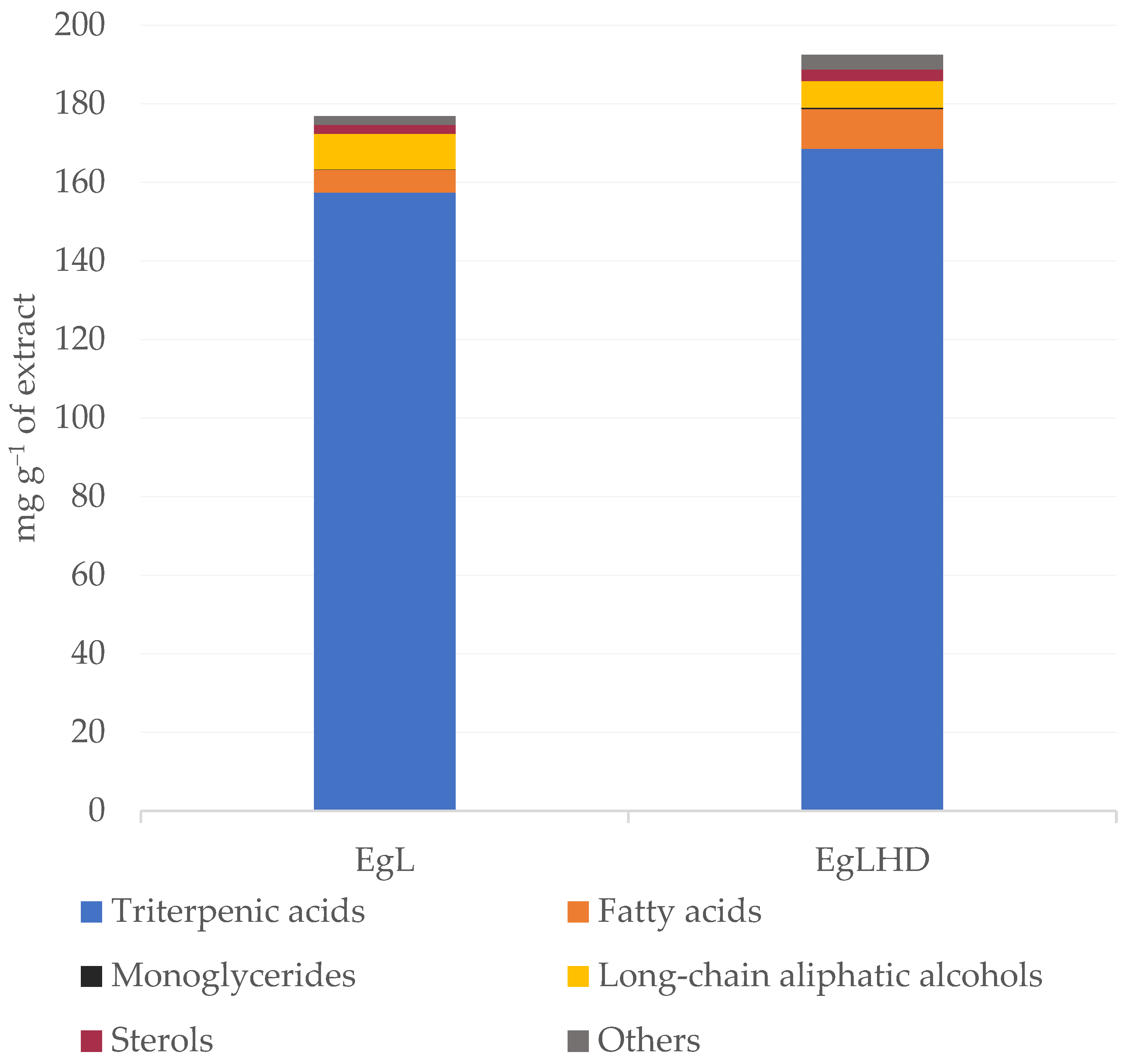
The chemical composition of DCM extracts from E. globulus leaves before and after hydrodistillation was studied in detail by GC–MS. The identification of the lipophilic components and the corresponding quantification in the studied extracts are summarized in Table 1 and Figure 2. This analysis excludes the EO components (mono and sesquiterpenic compounds) which have been the focus of another study [31].
To our knowledge, this is the first study reporting in detail the chemical characterization of the lipophilic fraction of E. globulus leaves before and after hydrodistillation.
In general, both extracts were mainly composed of triterpenic compounds, free fatty acids, and long-chain aliphatic alcohols. One sterol and one monoglyceride were also detected, among other minor components. The total contents of the identified compounds were 34.41 and 42.37 g kg−1 of dw in the EgL and EgLHD extracts, respectively. The higher contents of lipophilic compounds in the EgLHD extract may have been due to the disruption of the leaf’s cellular structures during hydrodistillation, that might have facilitated the extractability of these components.

2.2.1. Triterpenic Compounds

Triterpenic compounds were the predominant lipophilic compounds in the E. globulus leaves extracts (Table 1) and accounted for nearly 87.7–89% of the total content of identified compounds, with the DCM extract from the EgLHD containing the highest amount with 37.14 g kg−1 of dw, and the DCM extract from the EgL with 30.63 g kg−1 of dw, respectively. Ursolic acid was the major triterpenic compound in both extracts, with contents of 17.46 g kg−1 of dw in the EgL and 19.82 g kg−1 of dw in the EgLHD. Previous studies on E. globulus leaves also identified ursolic acid as the majority triterpenic acid [12,22]. Considerable amounts of oleanolic acid were also detected in a range from 5.45 g kg−1 of dw in the EgL to 6.32 g kg−1 of dw in the EgLHD, followed by betulonic acid with 3.61 g kg−1 of dw in the EgL and 6.48 g kg−1 of dw in the EgLHD, and betulinic acid with 1.37 g kg−1 of dw in the EgL to 1.43 g kg−1 of dw in the EgLHD. According to the literature, triterpenic acids possess a wide range of biological activities. Ursolic and oleanolic acids have very low toxicity and are known for their significant antimicrobial, anti-inflammatory and antihyperlipidemic, antitumor, hepatoprotective, and cytotoxic activities, among others [32,33,34,35]. Betulonic acid has shown significant antiviral, antimalarial and anti-leishmanial activities and furthermore, cytotoxic properties against human cancer cell lines (e.g., HT29 colorectal carcinoma cells, KB oral epidermoid carcinoma and HONE-1 nasopharyngeal carcinoma) [36,37,38,39]. Whereas betulinic acid has been shown to exhibit a wide range of biological activities including anti-HIV, antimalarial, anti-inflammatory, antibacterial, anthelmintic and antioxidant properties [32,38,40,41]. This wide range of biological properties associated with the triterpenic acids identified in E. globulus leaves reveals the promising character of this biomass as a raw material for applications in the pharmaceutical, nutraceutical and cosmetic industries.
Additionally, acetyl derivatives of triterpenic acids (e.g., oleanolic, betulinic and ursolic acids) were identified in significant amounts in all the extracts. The most abundant acetylated compound was 3-acetylbetulinic acid, followed by 3-acetyloleanolic and 3-acetylursolic acids. All these triterpenic acids have already been reported as components of E. globulus [8,9,12,22,42].

2.2.2. Fatty Acids

Fatty acids (C14 to C30) were detected in both extracts (Table 1), accounting for 3.3–5.3% of the total compounds identified. Total saturated fatty acids accounted for 1.13 and 2.11 g kg−1 of dw for the EgL and EgLHD, respectively. Hexadecanoic acid was the predominant saturated fatty acid found in the DCM extracts from the EgLHD, with a content of 0.58 g kg−1 of dw, while it only showed a content of 0.11 g kg−1 of dw in the extract of EgL. Hexadecanoic acid is one of the most common saturated fatty acids and also the most prevalent in body lipids [43]. This compound, both in acid and sodium salt form, is widely used in a variety of applications, such as food additives, cosmetic formulations, waterproofing materials, organic synthesis, etc. [44]. Triacontanoic and hexacosanoic acids were the most abundant saturated fatty acids for the DCM extract from the EgL, with contents of 0.39 and 0.33 g kg−1 of dw, respectively. In the extract from the EgLHD, the triacontanoic and hexacosanoic acids showed higher contents (i.e., 0.53 and 0.35 g kg−1 of dw, respectively). Fatty acids are the main constituents of the cell membrane structure with saturated fatty acids being essential for energy, cell membranes, hormone production, and organ cushioning [43].
Regarding the unsaturated fatty acids, the lipophilic extracts presented lower amounts than the saturated fatty acids. The extract from the EgLHD presented larger amounts of unsaturated fatty acids than before hydrodistillation, showing a content of 0.09 g kg−1 of dw, whereas only traces of unsaturated fatty acids were found in the DCM extract from the EgL. The most abundant unsaturated fatty acid was cis-octadec-9-enoic acid with a content of 0.07 g kg−1 of dw in the EgLHD.

2.2.3. Monoglycerides and Sterols

The only monoglyceride detected, and in rather low amounts, was 1-monohexadecanoin, with the highest and lowest values recorded for the EgLHD (0.03 g kg−1 of dw) and EgL (0.01 g kg−1 of dw), respectively (Table 1).
β-Sitosterol was the only sterol identified in the studied E. globulus leaves, accounting for 0.64 g kg−1 of dw in the EgLHD and 0.45 g kg−1 of dw in the EgL (Table 1). β-Sitosterol had already been identified in the leaf waxes of E. globulus, and in other morphological parts such as bark, fruit, wood, etc. [8,9,12]. This phytosterol has a relevant added-value to the extracts, since it is reported to exhibit analgesic, anti-inflammatory, anti-proliferative, hypocholesterolemic, anti-cholesterolemic, anti-helmenthic, anti-diabetic, anti-atherogenic and antibacterial activities against E. coli and Salmonella enterica Typhimurium, among others [44,45]. β-Sitosterol, therefore, is already considered a functional bioactive compound, which can be used in nutraceutical and food products (called functional foods), and the conditions of use of its health claims have already been established in Commission Regulation (EU) No. 686/2014 [46].

2.2.4. Long-Chain Aliphatic Alcohols

Four long-chain aliphatic alcohols (C24 to C30) were also detected in both E. globulus leaf extracts representing about 3.6–5.1% of the total lipophilic compounds identified (Table 1). The DCM extract from the EgL showed the highest amount with 1.76 g kg−1 of dw and the extract from the EgLHD showed the lowest amount (1.51 g kg−1 of dw). Triacontan-1-ol was the major constituent of this lipophilic class in the extracts with contents ranging from 0.86 to 1.01 g kg−1 of dw in the EgLHD and EgL, respectively, while tetracosan-1-ol was the minor aliphatic alcohol (with 0.04 g kg−1 of dw in the EgLHD and 0.06 g kg−1 of dw in the EgL).

2.2.5. Other Compounds

Finally, other minor compounds were also present in the different extracts. A small amount of tyrosol and gallic acid was found in the EgLHD (Table 1), accounting for 0.01 g kg−1 of dw and 0.04 g kg−1 of dw, respectively.
Glycerol was detected in an amount of 0.06 g kg−1 of dw in the EgLHD and traces in the EgL. α-Tocopherol and 1,6-dihydroxy-2-methylanthraquinone were identified in both extracts. In the case of α-tocopherol, also known as vitamin E, it is a lipophilic/liposoluble antioxidant, and reports indicate that it plays an important role in skin protection. Due to its properties, this compound is widely used as a low-cost antioxidant in cosmetic formulations and also as a food preservative [44,47].

2.3. Cell Viability of Lipophilic Extracts

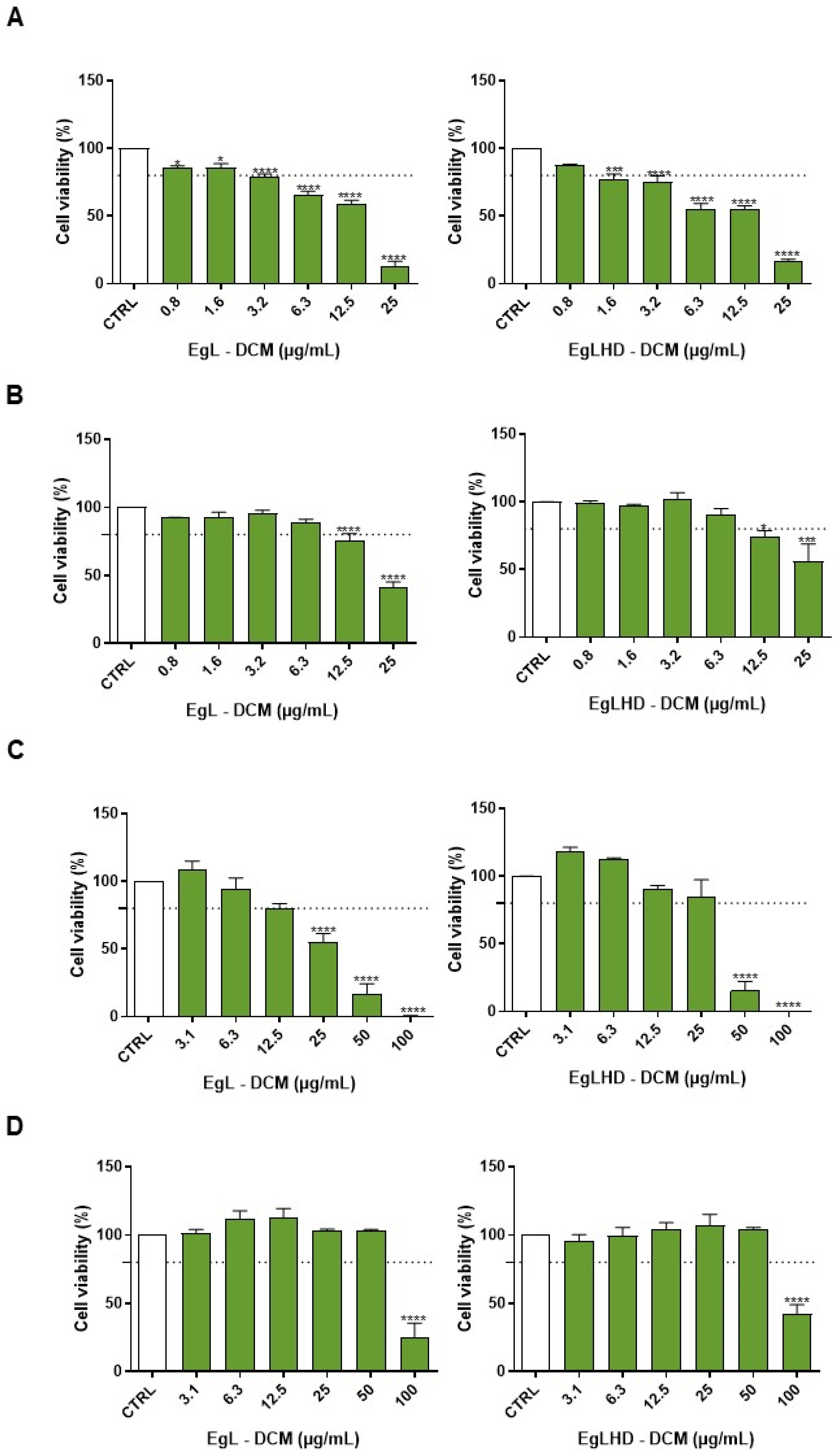
The cytotoxicity of lipophilic extracts from the EgL and EgLHD obtained with DCM was evaluated, by the assessment of the cell viability using the MTT assay, in cell lines representative of the innate immune system, skin, liver, and intestine, namely, macrophages (RAW 264.7), fibroblasts (NIH/3T3), hepatocytes (HepG2), and intestinal cells (Caco-2) (Figure 3). Macrophages (Figure 3A) were the more sensitive cells to the toxicity of the extracts. After a 24 h treatment, the EgL and EgLHD extracts obtained with DCM were devoid of toxicity (i.e., they did not reach 20% of cellular mortality) at concentrations below 1.6 µg mL−1 and 0.8 µg mL−1, respectively. In fibroblasts (Figure 3B), an absence of toxicity was observed after a 24 h treatment with the lipophilic extracts at a concentration below 6.3 µg mL−1 for the EgL and EgLHD. Regarding hepatocytes (Figure 3C), non-toxic effects of the lipophilic extracts were observed at 24 h for concentrations below 12.5 µg mL−1 for the EgL, and 25 µg mL−1 for the EgLHD. In intestinal cells (Figure 3D), which were the cell lines more sensitive to the toxic effects of the extracts, an absence of toxicity was found after a 24 h incubation with both extracts at concentrations below 50 µg mL−1. In general, no differences in terms of toxicity were observed between the lipophilic extracts of E. globulus leaves before or after hydrodistillation. No studies in the literature report the toxicity of lipophilic extracts from eucalyptus; however, there are some studies in the same cells with ursolic acid, which was the major compound found in these extracts. For example, Yang et al. (2015) determined that around 30 µg mL−1 of ursolic acid causes 50% mortality to hepatocytes HepG2 after 24 h exposure [48], while other studies revealed that approximately 20–40 µg mL−1 of ursolic acid is non-toxic to the same cell line. Here, 25 µg mL−1 of the lipophilic extracts, which were composed of 40–44% ursolic acid, were safe to hepatocytes [49,50,51]. In addition, some studies have revealed that ursolic acid showed a low toxicity at approximately 15 µg mL−1 to RAW 264.7 macrophages after 24 h treatment [52], while almost 10 µg mL−1 was devoid of toxicity in peritoneal macrophages [53]. On the other hand, one study also with RAW 264.7 macrophages did not observe toxic effects in concentrations below 5 µg mL−1 after a 48 h incubation [54].
These studies revealed the susceptibility of macrophages to ursolic acid, which is in accordance with our study where macrophages were the cells more sensitive to the lipophilic extracts (at safe concentrations lower than 0.8–6.3 µg mL−1). Fibroblasts were the second cell line more sensitive to the lipophilic extracts, with safe concentrations between 6.3–12.5 µg mL−1. Actually, an in vitro study in the literature demonstrated that concentrations exceeding 10 µg mL−1 influenced HSF fibroblasts viability after a 24 h treatment [55]. Regarding intestinal cells, the more resistant cells to the toxicity of the lipophilic extracts in our study were with safe concentrations from 50 µg mL−1, but a study previously reported that concentrations above 10 µg mL−1 ursolic acid were toxic to intestinal Caco-2 cells [56]. This result is not similar to our study because we observed a low toxicity of the lipophilic extracts in intestinal cells; however, no exposure period of ursolic acid was mentioned in the previous study. A cytotoxicity screening of the lipophilic extracts obtained from E. globulus leaves before and after hydrodistillation was performed for the first time in cell lines representing the immune system, skin, liver, and intestine, disclosing its safe concentrations. The main compound identified in these extracts, namely, ursolic acid, possesses relevant biological effects, including anti-inflammatory, anticancer, antidiabetic, antioxidant, and antibacterial effects [57], and has been involved in a range of pharmacological applications, which are associated with the prevention of several diseases [58], such as skin conditions [59,60,61], liver, and intestinal damage [62,63,64], as well as inflammatory diseases, particularly in diabetes [65,66]. The bioactivities of the lipophilic extracts from E. globulus leaves, however, have not been studied yet; therefore, our study reveals the potential of lipophilic extracts from E. globulus leaves regarding their future incorporation in pharmaceutical formulations, and it supports the argument that their bioactivities should be further investigated.

3. Materials and Methods

3.1. Reagents

The dichloromethane (p.a., ≥99% purity) and ethanol (p.a., ≥99% purity) were supplied by Fisher Scientific (Thermo Fisher Scientific, Waltham, MA, USA). The pyridine (p.a., ≥99.5% purity), N,O-bis(trimethylsilyl)trifluroacetamide (99% purity), trimethylchlorosilane (99% purity), tetracosane (99% purity), hexadecanoic acid (≥99% purity), pentadecan-1-ol (99% purity), stigmasterol (95% purity), and ursolic acid (≥98% purity) were supplied by Sigma Chemical Co (Madrid, Spain). The gallic acid (≥97.5% purity) was purchased from Sigma-Aldrich (Merck, Darmstadt, Germany). The Dulbecco’s Modified Eagle’s Medium (DMEM), sodium bicarbonate, sodium pyruvate, non-essential amino acids, L-glutamine, glucose, phenol red, trypsin-ethylenediamine tetraacetic acid (EDTA) solution 1X and 3-(4,5-dimethylthiazol-2-yl)-2,5-diphenyltetrazolium bromide (MTT) were purchased from Sigma-Aldrich (St. Louis, MO, USA). The fetal bovine serum (FBS), and penicillin-streptomycin were obtained from Gibco (Carlsbad, CA, USA).

3.2. Samples Collection

E. globulus leaves, representative of harvesting biomass residues, were sampled from 6-year-old E. globulus trees, in October 2018, randomly selected from a property of “The Navigator Company”, Braçal (GPS coordinates 40°44′5.388 N, 8°23′53.97 W), in the region of Sever do Vouga, Portugal.

3.3. Hydrodistillation

Following the process of recovery of essential oils used at an industrial scale, a sample of fresh E. globulus leaves was subjected to a hydrodistillation process until the complete extraction of the EO (2–3 h), using the modified Clevenger apparatus [31]. The EO was obtained with a 1.7% yield. The main components of the EO were 1,8-cineole (72.3%), a-pinene (9.4%), E-pinocarveol (3.6%), limonene (2.3%), globulol (1.6), pinocarvone (1.4%) and a-terpinyl acetate (1.2%) [31].
Then, so that the water content did not compromise the extraction of the lipophilic contents, hydrodistilled leaves were air-dried until reaching a constant weight and grounded to obtain a biomass with a particle size less than 2/3 mm before extraction.

3.4. Preparation of Lipophilic Extracts

Samples of E. globulus leaves before and after hydrodistillation (ca. 12 and 10 g of dried biomass, respectively) were submitted, in triplicate, to Soxhlet extractions with DCM for a period of 8 h. The solvent was evaporated to dryness, at a low-pressure and 35 °C and the results were expressed as a percentage of dw. DCM is a very selective solvent to extract lipophilic compounds from biomass [10].

3.5. GC–MS Analysis

Before the GC–MS analysis, aliquots containing about 15 to 20 mg of each dried extract were dissolved in 250 μL of pyridine containing 0.4 mg of tetracosane (internal standard) and then, 250 μL of N,O-bis(trimethylsilyl)trifluoroacetamide and 50  μL of trimethylchlorosilane were added to converting compounds with hydroxyl and carboxyl groups into trimethylsilyl ethers and esters, respectively. The mixture remained at 70 °C for 30 min and the derivatized extracts were analyzed by GC–MS [42,67].
GC–MS analysis were carried out in a GC–MS-QP2010 Ultra (Shimadzu, Kyoto, Japan). The compounds were separated in a DB-1 J&W capillary column (with a 30 m × 0.32 mm inner diameter, and 0.25 µm film thickness), using helium as the carrier gas (40 cm s−1). The chromatographic conditions were as follows: initial temperature, 80 °C for 5 min; temperature rate, 4 °C min−1 up to 260 °C, 2 °C min−1 up to 285 °C, which was maintained for 15 min. The injector temperature was 250 °C, and the transfer-line temperature was 290 °C, while the split ratio was 1:50. The mass spectrometer was operated in the electron impact mode with an energy of 70 eV, and the data were collected at a rate of 1 scan s−1 over a range of m/z 35–900. The ion source was kept at 250 °C [68].
The eluted compounds identification was made by comparing their mass spectra (MS) with the equipment’s mass spectral library (NIST Mass Spectral Library), by comparing the MS fragmentation profiles with data from the literature [12,19,22] and by the co-injection of standards.
For the semi-quantitative analysis, the GC–MS apparatus was calibrated with pure reference standards representative of the main families of compounds present in the lipophilic extracts, namely, hexadecanoic acid, pentadecan-1-ol, stigmasterol, ursolic acid and gallic acid, in relation to tetracosane (the internal standard), which allowed to determine the respective response factors. The compounds were quantified by their peak areas in relation to tetracosane (the internal standard), corrected using the response factors, and their abundance expressed in mg g−1 of extract and g kg−1 of dw of biomass.
Each of the three extracts, prepared from leaves before and after hydrodistillation, was analyzed in duplicate (n = 6). The results presented are the average of the concordant values obtained (with less than a 5% variation between aliquots of the same sample and between tripled extracts of the same type of extraction).

3.6. Cell Culture

The mouse fibroblasts (NIH/3T3, ATCC CRL-1658, Manassas, VA, USA), and human colorectal adenocarcinoma (Caco-2, ATCC HTB-37, Manassas, VA, USA) cell lines were cultured with DMEM (#D5648), supplemented with 10% (v/v) heat-inactivated FBS, 1% (v/v) antibiotic solution (from a 10,000 U mL−1 penicillin, and 10 000 µg mL−1 streptomycin stock), 3.7 g L−1 of sodium bicarbonate and 1 mM sodium pyruvate. The culture medium of the Caco-2 cell line was additionally supplemented with 1% (v/v) non-essential amino acids. The mouse leukaemic macrophages cell line (RAW 264.7, ATCC TIB-71, Manassas, VA, USA) was cultured in DMEM (#D5648) supplemented with 10% (v/v) non-inactivated FBS, a 1% (v/v) penicillin/streptomycin antibiotic solution, 1.5 g L−1 of sodium bicarbonate, and 1 mM sodium pyruvate. The human liver hepatocellular (HepG2, ATCC HB-8065, Manassas, VA, USA) cell line was cultured in DMEM (#D5030) supplemented with 10% (v/v) heat-inactivated FBS, a 1% (v/v) penicillin/streptomycin antibiotic solution, 1.5 g L−1 of sodium bicarbonate, 1 mM sodium pyruvate, 4 mM L-glutamine, 1 g L−1 of glucose and phenol red. The cells were cultivated in 75 cm2 flasks in a humidified 5% CO2-95% air atmosphere at 37 °C, and the medium was changed every 2–3 days. For passage and sub-culturing, the fibroblasts, hepatocytes, and intestinal cells were detached using a trypsin-EDTA solution 1X when the cells reached a 70–80% confluence, while the macrophages were detached with a cell scrape. The cells were sub-cultured over a maximum of ten passages [31].

3.7. Cell Viability Evaluation

For the assessment of cell viability, the MTT reduction assay was performed. RAW 264.7, Caco-2, HepG2, and NIH/3T3 cells were seeded in 96-well plates at a density of 9.6, 5, 2.5 or 1 × 10⁴ cells/well, respectively, and allowed to stabilize for 24 h. The next day, the culture medium was removed and substituted by an exposure medium (i.e., DMEM supplemented with 1% (v/v) FBS). The cells were incubated for 24 h at 37 °C with 0–100 µg mL−1 of lipophilic extracts from E. globulus leaves before and after hydrodistillation obtained with DCM. The extracts were added from stock solutions prepared in DMSO and stored at −20 °C. Cells treated with the medium alone were used as a control. After the incubation period, the medium was aspirated after the incubation period and a solution of 0.5 mg mL−1 MTT prepared in Krebs medium (i.e., 140 mM NaCl, 5 mM KCl, 1 mM NaH2PO4, 1 mM MgCl2, 9.6 mM Glucose, 20 mM HEPES, 1.5 mM CaCl2, and a pH of 7.4) was added. The cells were incubated with MTT at 37 °C for 30 min (RAW 264. 7 cells), 1 h (HepG2 cells), 2 h (Caco-2 cells) or 4 h (NIH/3T3 cells). After that, the MTT solution was aspirated and DMSO was added to dissolve the formed formazan crystals. The absorbance was measured after 10 min of shaking, at 570 nm using a SpectraMax Plus 384 Spectrophotometer (Molecular Devices, San Jose, CA, USA). The results of at least three independent experiments made in triplicate were expressed as a percentage (%) of the absorbance value obtained in the control, which was considered 100%, and were graphically presented as a % of the cell viability versus the concentration of the extracts [69].

Statistical Analysis

The results are represented as the mean ± standard error of the mean (SEM) of the indicated number of experiments. The normality of the data distribution was evaluated using the D’Agostino and Pearson and Shapiro–Wilk normality tests. Statistical comparisons between the groups were performed by a one-way analysis of variance (ANOVA) followed by a Dunnett’s multiple comparison test. Significance was accepted at p values < 0.05. The GraphPad Prism software (8.0.2, GraphPad Software Inc., San Diego, CA, USA) was used to perform the statistical analysis.

4. Conclusions

The present study highlights promising insights into the chemical composition and cytotoxicity of lipophilic extracts of E. globulus leaves before and after hydrodistillation. To our knowledge, this is the first study of the chemical composition of hydrodistilled leaves. The obtained extracts were characterized in detail by GC–MS, allowing the identification and quantification of 31 compounds, including different families of compounds, such as triterpenic compounds, fatty acids, long-chain aliphatic alcohol, only one sterol, β-sitosterol, and other minor compounds. DCM extracts of the leaves before and after hydrodistillation were shown to exhibit valuable bioactive compounds, namely, triterpenic acids (e.g., betulonic, oleanolic, betulinic and ursolic acids) that are associated with numerous biological activities. The majority compound was ursolic acid, with contents ranging from 17.46–19.82 g kg−1 of dw. Interestingly, the extracts of the hydrodistilled leaves showed a higher content of the identified compounds than the non-hydrodistilled leaves.
The non-toxic concentrations of the extracts in intestinal cells, hepatocytes (liver), fibroblasts (skin) and macrophages (innate immune system) were determined. No significant differences in toxicity were observed between the extracts obtained from the leaves before or after hydrodistillation. Macrophages were shown to be the most sensitive cells to the extracts (with safe concentrations less than or equal to 0.8 µg mL−1) and intestinal cells the most resistant (with non-toxic concentrations less than or equal to 50 µg mL−1).
This study highlights the potential of E. globulus leaves, promoting their economic exploitation as a source of bioactive compounds with potential applications in pharmaceutical, nutraceutical and cosmetic formulations, which can only be implemented after the development of sustainable extraction methodologies, as well as a thorough technical–economic evaluation, and finally, an analysis to ensure the ecological impact of its exploitation. Finally, this study indicates that an integrated and sustainable exploitation of the species E. globulus can be considered, combining the exploitation of the leaves, for the recovery of EO and extracts enriched in value-added compounds, along with the exploitation of the wood, which is the main raw material for pulp production.

Author Contributions

Conceptualization, C.M.F.P., S.A.O.S. and A.J.D.S.; Formal analysis, C.S.D.O.; Investigation, C.S.D.O.; Methodology, C.S.D.O. and P.M.; Supervision, M.T.C., C.M.F.P., A.M.S.S., S.A.O.S. and A.J.D.S.; Writing—original draft, C.S.D.O. and P.M.; Writing—review and editing, M.T.C., C.M.F.P., A.M.S.S., S.A.O.S. and A.J.D.S. All authors have read and agreed to the published version of the manuscript.

Funding

This work was carried out under the Project InPacTus—Innovative products and technologies from eucalyptus, Project N.º 21874 (POCI-01-0247-FEDER-021874), funded by Portugal 2020 through the European Regional Development Fund (ERDF) in the frame of COMPETE 2020 nº 246/AXIS II/2017, and the projects CICECO–Aveiro Institute of Materials (UIDB/50011/2020, UIDP/50011/2020 and LA/P/0006/2020), LAQV-REQUIMTE (UIDB/50006/2020 and UIDP/50006/2020) and CIBB (UIDB/04539/2020 and UIDP/04539/2020), financed by national funds through the FCT/MEC (PIDDAC). FCT is also acknowledged for the research contract under the Scientific Employment Stimulus to S. Santos (2021.03348.CEECIND).

Institutional Review Board Statement

Not applicable.

Conflicts of Interest

The authors declare no conflict of interest.

References

  1. Dugmore, T.I.J.; Clark, J.H.; Bustamante, J.; Houghton, J.A.; Matharu, A.S. Valorisation of biowastes for the production of green materials using chemical methods. Top. Curr. Chem. 2017, 375, 46. [Google Scholar] [CrossRef] [PubMed] [Green Version]
  2. De Jong, E.; Higson, A.; Walsh, P.; Wellisch, M. Bio-based chemicals, value added products from biorefineries. In IEA Bioenergy Task 42 Report; IEA Bioenergy: Paris, France, 2012. [Google Scholar]
  3. Waldron, K.W. Advances in Biorefineries: Biomass and Waste Supply Chain Exploitation; Woodhead Publishing: Cambridge, UK, 2014; ISBN 9780857097385. [Google Scholar]
  4. Sadhukhan, J.; Ng, K.S.; Hernandez, E.M. Biorefineries and Chemical Processes: Design, Integration and Sustainability Analysis; John Wiley & Sons, Ltd.: Hoboken, NJ, USA, 2014; ISBN 9781119990864. [Google Scholar]
  5. CELPA. Boletim Estatístico da CELPA—Indústria Papeleira Portuguesa; CELPA: Lisbon, Portugal, 2018. [Google Scholar]
  6. ICNF. IFN6—Principais resultados—relatório sumário. In Instituto da Conservação da Natureza e das Florestas; ICNF: Lisbon, Portugal, 2019; pp. 1–34. [Google Scholar]
  7. Neiva, D.M.; Araújo, S.; Gominho, J.; Carneiro, A.C.O.; Pereira, H. Potential of Eucalyptus globulus industrial bark as a biorefinery feedstock: Chemical and fuel characterization. Ind. Crops Prod. 2018, 123, 262–270. [Google Scholar] [CrossRef]
  8. Freire, C.S.R.; Silvestre, A.J.D.; Neto, C.P.; Cavaleiro, J.A.S. Lipophilic extractives of the inner and outer barks of Eucalyptus globulus. Holzforschung 2002, 56, 372–379. [Google Scholar] [CrossRef]
  9. Freire, C.S.R.; Silvestre, A.J.D.; Neto, C.P. Identification of new hydroxy fatty acids and ferulic acid esters in the wood of Eucalyptus globulus. Holzforschung 2002, 56, 143–149. [Google Scholar] [CrossRef]
  10. Domingues, R.M.A.; Patinha, D.S.J.; Sousa, G.D.A.; Villaverde, J.; Silva, C.M.; Freire, C.S.R.; Silvestre, A.J.D.; Neto, C.P. Eucalyptus biomass residues from agro-forest and pulping industries as sources of high-value triterpenic compounds. Cellul. Chem. Technol. 2011, 45, 475–481. [Google Scholar]
  11. Santos, S.A.O.; Freire, C.S.R.; Domingues, M.R.M.; Silvestre, A.J.D.; Pascoal Neto, C. Characterization of phenolic components in polar extracts of Eucalyptus globulus Labill. bark by high-performance liquid chromatography-mass spectrometry. J. Agric. Food Chem. 2011, 59, 9386–9393. [Google Scholar] [CrossRef]
  12. Domingues, R.M.A.; Sousa, G.D.A.; Freire, C.S.R.; Silvestre, A.J.D.; Neto, C.P. Eucalyptus globulus biomass residues from pulping industry as a source of high value triterpenic compounds. Ind. Crops Prod. 2010, 31, 65–70. [Google Scholar] [CrossRef]
  13. Ko, C.-H.; Wang, Y.-N.; Chang, F.-C.; Chen, J.-J.; Chen, W.-H.; Hwang, W.-S. Potentials of lignocellulosic bioethanols produced from hardwood in Taiwan. Energy 2012, 44, 329–334. [Google Scholar] [CrossRef]
  14. Romaní, A.; Garrote, G.; Parajó, J.C. Bioethanol production from autohydrolyzed Eucalyptus globulus by simultaneous saccharification and fermentation operating at high solids loading. Fuel 2012, 94, 305–312. [Google Scholar] [CrossRef]
  15. Silvestre, A.J.D.; Cavaleiro, J.S.; Delmond, B.; Filliatre, C.; Bourgeois, G. Analysis of the variation of the essential oil composition of Eucalyptus globulus Labill. from Portugal using multivariate statistical analysis. Ind. Crops Prod. 1997, 6, 27–33. [Google Scholar] [CrossRef]
  16. Bachir, R.G.; Benali, M. Antibacterial activity of the essential oils from the leaves of Eucalyptus globulus against Escherichia coli and Staphylococcus aureus. Asian Pac. J. Trop. Biomed. 2012, 2, 739–742. [Google Scholar] [CrossRef] [PubMed] [Green Version]
  17. Ghaffar, A.; Yameen, M.; Kiran, S.; Kamal, S.; Jalal, F.; Munir, B.; Saleem, S.; Rafiq, N.; Ahmad, A.; Saba, I.; et al. Chemical composition and in-vitro evaluation of the antimicrobial and antioxidant activities of essential oils extracted from seven Eucalyptus species. Molecules 2015, 20, 20487–20498. [Google Scholar] [CrossRef] [Green Version]
  18. González-Burgos, E.; Liaudanskas, M.; Viškelis, J.; Žvikas, V.; Janulis, V.; Gómez-Serranillos, M.P. Antioxidant activity, neuroprotective properties and bioactive constituents analysis of varying polarity extracts from Eucalyptus globulus leaves. J. Food Drug Anal. 2018, 26, 1293–1302. [Google Scholar] [CrossRef] [PubMed]
  19. Nile, S.H.; Keum, Y.S. Chemical composition, antioxidant, anti-inflammatory and antitumor activities of Eucalyptus globulus labill. Indian J. Exp. Biol. 2018, 56, 734–742. [Google Scholar]
  20. Boulekbache-Makhlouf, L.; Meudec, E.; Mazauric, J.P.; Madani, K.; Cheynier, V. Qualitative and semi-quantitative analysis of phenolics in Eucalyptus globulus leaves by high-performance liquid chromatography coupled with diode array detection and electrospray ionisation mass spectrometry. Phytochem. Anal. 2013, 24, 162–170. [Google Scholar] [CrossRef] [PubMed]
  21. Dezsi, Ș.; Bădărău, A.; Bischin, C.; Vodnar, D.; Silaghi-Dumitrescu, R.; Gheldiu, A.-M.; Mocan, A.; Vlase, L. Antimicrobial and antioxidant activities and phenolic profile of Eucalyptus globulus Labill. and Corymbia ficifolia (F. Muell.) K.D. Hill & L.A.S. Johnson Leaves. Molecules 2015, 20, 4720–4734. [Google Scholar] [CrossRef] [Green Version]
  22. Rodrigues, V.H.; de Melo, M.M.R.; Portugal, I.; Silva, C.M. Extraction of Eucalyptus leaves using solvents of distinct polarity. Cluster analysis and extracts characterization. J. Supercrit. Fluids 2018, 135, 263–274. [Google Scholar] [CrossRef]
  23. Guzmán, P.; Fernández, V.; Graça, J.; Cabral, V.; Kayali, N.; Khayet, M.; Gil, L. Chemical and structural analysis of Eucalyptus globulus and E. camaldulensis leaf cuticles: A lipidized cell wall region. Front. Plant Sci. 2014, 5, 481. [Google Scholar] [CrossRef] [Green Version]
  24. Singh, A.; Ahmad, A.; Bushra, R. Supercritical carbondioxide extraction of essential oils from leaves of Eucalyptus globulus L., their analysis and application. Anal. Methods 2016, 8, 1339–1350. [Google Scholar] [CrossRef]
  25. Gullón, B.; Muñiz-Mouro, A.; Lú-Chau, T.A.; Moreira, M.T.; Lema, J.M.; Eibes, G. Green approaches for the extraction of antioxidants from eucalyptus leaves. Ind. Crops Prod. 2019, 138, 111473. [Google Scholar] [CrossRef]
  26. Vecchio, M.G.; Loganes, C.; Minto, C. Beneficial and healthy properties of Eucalyptus plants: A great potential use. Open Agric. J. 2016, 10, 52–57. [Google Scholar] [CrossRef] [Green Version]
  27. Coppen, J.J.W. Eucalyptus: The Genus Eucalyptus; Taylor & Francis: London, UK, 2002; ISBN 9780415278799. [Google Scholar]
  28. Barbosa, L.C.A.; Filomeno, C.A.; Teixeira, R.R. Chemical variability and biological activities of Eucalyptus spp. essential oils. Molecules 2016, 21, 1671. [Google Scholar] [CrossRef] [Green Version]
  29. El-Ghorab, A.H.; El-Massry, K.F.; Marx, F.; Fadel, H.M. Antioxidant activity of Egyptian Eucalyptus camaldulensis var. brevirostris leaf extracts. Food/Nahrung 2003, 47, 41–45. [Google Scholar] [CrossRef] [PubMed]
  30. Pereira, V.; Dias, C.; Vasconcelos, M.C.; Rosa, E.; Saavedra, M.J. Antibacterial activity and synergistic effects between Eucalyptus globulus leaf residues (essential oils and extracts) and antibiotics against several isolates of respiratory tract infections (Pseudomonas aeruginosa). Ind. Crops Prod. 2014, 52, 1–7. [Google Scholar] [CrossRef]
  31. Moreira, P.; Sousa, F.J.; Matos, P.; Brites, G.S.; Gonçalves, M.J.; Cavaleiro, C.; Figueirinha, A.; Salgueiro, L.; Batista, M.T.; Branco, P.C.; et al. Chemical composition and effect against skin alterations of bioactive extracts obtained by the hydrodistillation of Eucalyptus globulus leaves. Pharmaceutics 2022, 14, 561. [Google Scholar] [CrossRef] [PubMed]
  32. Domingues, R.; Guerra, A.; Duarte, M.; Freire, C.; Neto, C.; Silva, C.; Silvestre, A. Bioactive triterpenic acids: From agroforestry biomass residues to promising therapeutic tools. Mini Rev. Org. Chem. 2014, 11, 382–399. [Google Scholar] [CrossRef]
  33. Qian, W.; Wang, W.; Zhang, J.; Wang, T.; Liu, M.; Yang, M.; Sun, Z.; Li, X.; Li, Y. Antimicrobial and antibiofilm activities of ursolic acid against carbapenem-resistant Klebsiella pneumoniae. J. Antibiot. 2020, 73, 382–391. [Google Scholar] [CrossRef] [PubMed]
  34. Gutiérrez-Rebolledo, G.A.; Siordia-Reyes, A.G.; Meckes-Fischer, M.; Jiménez-Arellanes, A. Hepatoprotective properties of oleanolic and ursolic acids in antitubercular drug-induced liver damage. Asian Pac. J. Trop. Med. 2016, 9, 644–651. [Google Scholar] [CrossRef] [Green Version]
  35. Somova, L.O.; Nadar, A.; Rammanan, P.; Shode, F.O. Cardiovascular, antihyperlipidemic and antioxidant effects of oleanolic and ursolic acids in experimental hypertension. Phytomedicine 2003, 10, 115–121. [Google Scholar] [CrossRef]
  36. Pavlova, N.I.; Savinova, O.V.; Nikolaeva, S.N.; Boreko, E.I.; Flekhter, O.B. Antiviral activity of betulin, betulinic and betulonic acids against some enveloped and non-enveloped viruses. Fitoterapia 2003, 74, 489–492. [Google Scholar] [CrossRef]
  37. Alakurtti, S.; Bergström, P.; Sacerdoti-Sierra, N.; Jaffe, C.L.; Yli-Kauhaluoma, J. Anti-leishmanial activity of betulin derivatives. J. Antibiot. 2010, 63, 123–126. [Google Scholar] [CrossRef] [PubMed] [Green Version]
  38. De Sá, M.S.; Costa, J.F.O.; Krettli, A.U.; Zalis, M.G.; De Azevedo Maia, G.L.; Sette, I.M.F.; De Amorim Câmara, C.; Filho, J.M.B.; Giulietti-Harley, A.M.; Ribeiro Dos Santos, R.; et al. Antimalarial activity of betulinic acid and derivatives in vitro against Plasmodium falciparum and in vivo in P. berghei-infected mice. Parasitol. Res. 2009, 105, 275–279. [Google Scholar] [CrossRef]
  39. Chiang, Y.M.; Chang, J.Y.; Kuo, C.C.; Chang, C.Y.; Kuo, Y.H. Cytotoxic triterpenes from the aerial roots of Ficus microcarpa. Phytochemistry 2005, 66, 495–501. [Google Scholar] [CrossRef] [PubMed]
  40. Fujioka, T.; Kashiwada, Y.; Kilkuskie, R.E.; Cosentino, L.M.; Bailas, L.M.; Jiang, J.B.; Janzen, W.P.; Chen, I.S.; Lee, K.H. Anti-AIDS agents, 11. Betulinic acid and platanic acid as anti-HIV principles from Syzigium claviflorum, and the anti-HIV activity of structurally related triterpenoids. J. Nat. Prod. 1994, 57, 243–247. [Google Scholar] [CrossRef] [PubMed]
  41. Del Carmen Recio, M.; Giner, R.M.; Manez, S.; Gueho, J.; Julien, H.R.; Hostettmann, K.; Rios, J.L. Investigations on the steroidal anti-inflammatory activity of triterpenoids from Diospyros leucomelas. Planta Med. 1995, 61, 9–12. [Google Scholar] [CrossRef] [PubMed]
  42. Patinha, D.J.S.; Domingues, R.M.A.; Villaverde, J.J.; Silva, A.M.S.; Silva, C.M.; Freire, C.S.R.; Neto, C.P.; Silvestre, A.J.D. Lipophilic extractives from the bark of Eucalyptus grandis x globulus, a rich source of methyl morolate: Selective extraction with supercritical CO2. Ind. Crops Prod. 2013, 43, 340–348. [Google Scholar] [CrossRef]
  43. Cordeiro, N.; Freitas, N.; Faria, M.; Gouveia, M. Ipomoea batatas (L.) Lam.: A rich source of lipophilic phytochemicals. J. Agric. Food Chem. 2013, 61, 12380–12384. [Google Scholar] [CrossRef]
  44. Devappa, R.K.; Rakshit, S.K.; Dekker, R.F.H. Forest biorefinery: Potential of poplar phytochemicals as value-added co-products. Biotechnol. Adv. 2015, 33, 681–716. [Google Scholar] [CrossRef]
  45. Ramos, P.A.B.; Moreirinha, C.; Santos, S.A.O.; Almeida, A.; Freire, C.S.R.; Silva, A.M.S.; Silvestre, A.J.D. Valorisation of bark lipophilic fractions from three Portuguese Salix species: A systematic study of the chemical composition and inhibitory activity on Escherichia coli. Ind. Crop. Prod. 2019, 132, 245–252. [Google Scholar] [CrossRef]
  46. Chanioti, S.; Katsouli, M.; Tzia, C. β-Sitosterol as a functional bioactive. In A Centum of Valuable Plant Bioactives; Mushtaq, M., Anwar, F., Eds.; Academic Press: London, UK, 2021; pp. 193–212. ISBN 9780128229231. [Google Scholar]
  47. Huang, Z.-R.; Lin, Y.-K.; Fang, J.-Y. Biological and pharmacological activities of squalene and related compounds: Potential uses in cosmetic dermatology. Molecules 2009, 14, 540–554. [Google Scholar] [CrossRef]
  48. Yang, X.; Li, Y.; Jiang, W.; Ou, M.; Chen, Y.; Xu, Y.; Wu, Q.; Zheng, Q.; Wu, F.; Wang, L.; et al. Synthesis and biological evaluation of novel ursolic acid derivatives as potential anticancer prodrugs. Chem. Biol. Drug Des. 2015, 86, 1397–1404. [Google Scholar] [CrossRef]
  49. Ramos, A.A.; Lima, C.F.; Pereira, M.L.; Fernandes-Ferreira, M.; Pereira-Wilson, C. Antigenotoxic effects of quercetin, rutin and ursolic acid on HepG2 cells: Evaluation by the comet assay. Toxicol. Lett. 2008, 177, 66–73. [Google Scholar] [CrossRef] [Green Version]
  50. Li, T.; Chen, X.; Liu, Y.; Fan, L.; Lin, L.; Xu, Y.; Chen, S.; Shao, J. pH-Sensitive mesoporous silica nanoparticles anticancer prodrugs for sustained release of ursolic acid and the enhanced anti-cancer efficacy for hepatocellular carcinoma cancer. Eur. J. Pharm. Sci. 2017, 96, 456–463. [Google Scholar] [CrossRef] [Green Version]
  51. Dong, H.; Yang, X.; Xie, J.; Xiang, L.; Li, Y.; Ou, M.; Chi, T.; Liu, Z.; Yu, S.; Gao, Y.; et al. UP12, a novel ursolic acid derivative with potential for targeting multiple signaling pathways in hepatocellular carcinoma. Biochem. Pharmacol. 2015, 93, 151–162. [Google Scholar] [CrossRef]
  52. Zhang, T.-Y.; Li, C.-S.; Li, P.; Bai, X.-Q.; Guo, S.-Y.; Jin, Y.; Piao, S.-J. Synthesis and evaluation of ursolic acid-based 1,2,4-triazolo[1,5-a]pyrimidines derivatives as anti-inflammatory agents. Mol. Divers. 2022, 26, 27–38. [Google Scholar] [CrossRef]
  53. Jang, S.-E.; Jeong, J.-J.; Hyam, S.R.; Han, M.J.; Kim, D.-H. Ursolic acid isolated from the seed of Cornus officinalis ameliorates colitis in mice by inhibiting the binding of lipopolysaccharide to toll-like receptor 4 on macrophages. J. Agric. Food Chem. 2014, 62, 9711–9721. [Google Scholar] [CrossRef]
  54. Kim, M.-H.; Kim, J.N.; Han, S.N.; Kim, H.-K. Ursolic acid isolated from guava leaves inhibits inflammatory mediators and reactive oxygen species in LPS-stimulated macrophages. Immunopharmacol. Immunotoxicol. 2015, 37, 228–235. [Google Scholar] [CrossRef]
  55. Wójciak-Kosior, M.; Paduch, R.; Matysik-Woźniak, A.; Niedziela, P.; Donica, H. The effect of ursolic and oleanolic acids on human skin fibroblast cells. Folia Histochem. Cytobiol. 2011, 49, 664–669. [Google Scholar] [CrossRef] [Green Version]
  56. Qiang, Z.; Ye, Z.; Hauck, C.; Murphy, P.A.; McCoy, J.-A.; Widrlechner, M.P.; Reddy, M.B.; Hendrich, S. Permeability of rosmarinic acid in Prunella vulgaris and ursolic acid in Salvia officinalis extracts across Caco-2 cell monolayers. J. Ethnopharmacol. 2011, 137, 1107–1112. [Google Scholar] [CrossRef] [Green Version]
  57. Mlala, S.; Oyedeji, A.O.; Gondwe, M.; Oyedeji, O.O. Ursolic acid and its derivatives as bioactive agents. Molecules 2019, 24, 2751. [Google Scholar] [CrossRef] [Green Version]
  58. Kashyap, D.; Tuli, H.S.; Sharma, A.K. Ursolic acid (UA): A metabolite with promising therapeutic potential. Life Sci. 2016, 146, 201–213. [Google Scholar] [CrossRef]
  59. Park, H.J.; Jo, D.S.; Choi, D.S.; Bae, J.-E.; Park, N.Y.; Kim, J.-B.; Chang, J.H.; Shin, J.J.; Cho, D.-H. Ursolic acid inhibits pigmentation by increasing melanosomal autophagy in B16F1 cells. Biochem. Biophys. Res. Commun. 2020, 531, 209–214. [Google Scholar] [CrossRef]
  60. Cho, J.; Rho, O.; Junco, J.; Carbajal, S.; Siegel, D.; Slaga, T.J.; DiGiovanni, J. Effect of combined treatment with ursolic acid and resveratrol on skin tumor promotion by 12-O-tetradecanoylphorbol-13-acetate. Cancer Prev. Res. 2015, 8, 817–825. [Google Scholar] [CrossRef] [Green Version]
  61. Samivel, R.; Nagarajan, R.P.; Subramanian, U.; Khan, A.A.; Masmali, A.; Almubrad, T.; Akhtar, S. Inhibitory effect of ursolic acid on ultraviolet B radiation-induced oxidative stress and proinflammatory response-mediated senescence in human skin dermal fibroblasts. Oxid. Med. Cell. Longev. 2020, 2020, 1246510. [Google Scholar] [CrossRef]
  62. Zheng, Y.; Huang, C.; Zhao, L.; Chen, Y.; Liu, F. Regulation of decorin by ursolic acid protects against non-alcoholic steatohepatitis. Biomed. Pharmacother. 2021, 143, 112166. [Google Scholar] [CrossRef]
  63. Wan, S.-Z.; Liu, C.; Huang, C.-K.; Luo, F.-Y.; Zhu, X. Ursolic acid improves intestinal damage and bacterial dysbiosis in liver fibrosis mice. Front. Pharmacol. 2019, 10, 1321. [Google Scholar] [CrossRef]
  64. Hao, W.; Kwek, E.; He, Z.; Zhu, H.; Liu, J.; Zhao, Y.; Ma, K.Y.; He, W.-S.; Chen, Z.-Y. Ursolic acid alleviates hypercholesterolemia and modulates the gut microbiota in hamsters. Food Funct. 2020, 11, 6091–6103. [Google Scholar] [CrossRef]
  65. Li, J.; Li, N.; Yan, S.; Liu, M.; Sun, B.; Lu, Y.; Shao, Y. Ursolic acid alleviates inflammation and against diabetes-induced nephropathy through TLR4-mediated inflammatory pathway. Mol. Med. Rep. 2018, 18, 4675–4681. [Google Scholar] [CrossRef] [Green Version]
  66. Wang, X.; Gong, Y.; Zhou, B.; Yang, J.; Cheng, Y.; Zhao, J.; Qi, M. Ursolic acid ameliorates oxidative stress, inflammation and fibrosis in diabetic cardiomyopathy rats. Biomed. Pharmacother. 2018, 97, 1461–1467. [Google Scholar] [CrossRef]
  67. Freire, C.S.R.; Coelho, D.S.C.; Santos, N.M.; Silvestre, A.J.D.; Pascoal Neto, C. Identification of Δ7 phytosterols and phytosteryl glucosides in the wood and bark of several Acacia species. Lipids 2005, 40, 317–322. [Google Scholar] [CrossRef]
  68. Santos, S.; Trindade, S.; Oliveira, C.; Parreira, P.; Rosa, D.; Duarte, M.; Ferreira, I.; Cruz, M.; Rego, A.; Abreu, M.; et al. Lipophilic fraction of cultivated Bifurcaria bifurcata R. Ross: Detailed composition and in vitro prospection of current challenging bioactive properties. Mar. Drugs 2017, 15, 340. [Google Scholar] [CrossRef] [PubMed] [Green Version]
  69. Sittampalam, G.S.; Coussens, N.B.P.; Brimacombe, K.; Grossman, A.B.; Arkin, M.B.; Auld, D.B.; Austin, C.; Baell, J.B.; Bejcek, B.B.; Chung, T.; et al. Assay Guidance Manual: Quantitative Biology and Pharmacology in Preclinical Drug Discovery; 2004; pp. 1–12. Available online: https://ascpt.onlinelibrary.wiley.com/doi/full/10.1111/cts.12570 (accessed on 11 January 2022).
Figure 1. Eucalyptus globulus leaves.
Ijms 24 06226 g001
Figure 2. Main families of lipophilic compounds identified by GC–MS in dichloromethane (DCM) extracts of E. globulus leaves before (EgL) and after (EgLHD) hydrodistillation.
Ijms 24 06226 g002
Figure 3. Effect of the lipophilic extracts from E. globulus leaves before (EgL) and after (EgLHD) hydrodistillation obtained with dichloromethane (DCM) on the cell viability of RAW 264.7 macrophages (A), NIH/3T3 fibroblasts (B), HepG2 hepatocytes (C), and Caco-2 intestinal (D) cells. The cells were treated for 24 h with 0–100 µg mL−1 of EgL or EgLHD, and the cell viability was evaluated using a 3-(4,5-dimethylthiazol-2-yl)-2,5-diphenyltetrazolium bromide (MTT) reduction assay. Cells treated with the medium alone were used as a control (CTRL). The results were expressed as the percentage (%) of cell viability relative to the CTRL and represent the mean ± standard error of the mean (SEM) of at least three independent experiments performed in triplicate. The statistical analysis was carried out by one-way analysis of variance (ANOVA) followed by a Dunnett’s multiple comparison test. * p < 0.05, *** p < 0.001, and **** p < 0.0001: significantly different compared to the CTRL.
Ijms 24 06226 g003
Table 1. Compounds identified in dichloromethane (DCM) extracts of E. globulus leaves before (EgL) and after (EgLHD) hydrodistillation expressed in mg g−1 of extract and g kg−1 of dw biomass.
Rt(min)Compoundmg g−1 of Extractg kg−1 of dw
EgLEgLHDEgLEgLHD
Triterpenic acids157.42168.5330.6337.14
68.11Betulonic acid18.5629.403.616.48
68.93Oleanolic acid28.0228.705.456.32
69.52Betulinic acid7.056.491.371.43
70.46Ursolic acid89.7389.9317.4619.82
73.093-Acetyloleanolic acid5.114.560.991.00
76.203-Acetylbetulinic acid6.857.141.331.57
77.253-Acetylursolic acid2.092.320.410.51
Fatty acids5.7910.141.132.23
Saturated fatty acids5.799.571.132.11
30.83Tetradecanoic acid0.290.520.060.11
35.74Hexadecanoic acid0.592.610.110.58
38.03Heptadecanoic acidtr0.12tr0.03
40.24Octadecanoic acid0.130.260.030.06
48.26Docosanoic acid0.210.290.040.06
51.94Tetracosanoic acid0.330.470.060.10
55.97Hexacosanoic acid1.681.610.330.35
60.37Octacosanoic acid0.541.290.110.28
65.28Triacontanoic acid2.022.410.390.53
Unsaturated fatty acidstr0.42tr0.09
39.32Octadeca-9,12-dienoic acidtrtrtrtr
39.51cis-Octadec-9-enoic acidtr0.34tr0.07
39.65trans-Octadec-9-enoic acidtr0.08tr0.02
Diacidstr0.15tr0.03
29.37Nonanedioic acidtr0.15tr0.03
Long-chain aliphatic alcohols9.066.851.761.51
50.41Tetracosan-1-ol0.320.190.060.04
54.24Hexacosan-1-ol1.230.960.240.21
58.49Octacosan-1-ol2.331.790.450.39
63.06Triacontan-1-ol5.183.911.010.86
Monoglycerides0.070.290.010.03
47.571-Monohexadecanoin0.070.290.010.03
Sterols2.332.910.450.64
62.61β-Sitosterol2.332.910.450.64
Others2.133.700.420.82
14.14Glyceroltr0.28tr0.06
22.95Tyrosoln.d.0.05n.d.0.01
34.03Gallic acidn.d.0.19n.d.0.04
57.231,6-Dihydroxy-2-methylanthraquinone1.160.990.230.22
58.03α-Tocopherol0.972.190.190.48
TOTAL176.81192.4434.4142.37
Abbreviations: n.d.: not detected; Rt: retention time; tr: traces.
Disclaimer/Publisher’s Note: The statements, opinions and data contained in all publications are solely those of the individual author(s) and contributor(s) and not of MDPI and/or the editor(s). MDPI and/or the editor(s) disclaim responsibility for any injury to people or property resulting from any ideas, methods, instructions or products referred to in the content.