Article

CcNAC1 by Transcriptome Analysis Is Involved in Sudan Grass Secondary Cell Wall Formation as a Positive Regulator

1
National Forage Breeding Innovation Base (JAAS), Key Laboratory for Saline-Alkali Soil Improvement and Utilization (Coastal Saline-Alkali Lands), Ministry of Agriculture and Rural Affairs, Institute of Animal Science, Jiangsu Academy of Agricultural Sciences, Nanjing 210014, China
2
National Center for Soybean Improvement, Key Laboratory of Biology and Genetics and Breeding for Soybean, Ministry of Agriculture, State Key Laboratory of Crop Genetics and Germplasm Enhancement, Nanjing Agricultural University, Nanjing 210095, China
3
Crops Research Institute, Council for Scientific and Industrial Research, Kumasi P.O. Box 3785, Ghana
4
College of Agro-Grassland Science, Nanjing Agricultural University, Nanjing 210095, China
*
Author to whom correspondence should be addressed.
These authors contributed equally to this work as first authors.
Int. J. Mol. Sci. 2023, 24(7), 6149; https://doi.org/10.3390/ijms24076149
Submission received: 3 February 2023 / Revised: 9 March 2023 / Accepted: 18 March 2023 / Published: 24 March 2023
(This article belongs to the Section Molecular Genetics and Genomics)

Abstract

:
Sudan grass is a high-quality forage of sorghum. The degree of lignification of Sudan grass is the main factor affecting its digestibility in ruminants such as cattle and sheep. Almost all lignocellulose in Sudan grass is stored in the secondary cell wall, but the mechanism and synthesis of the secondary cell wall in Sudan grass is still unclear. In order to study the mechanism of secondary cell wall synthesis in Sudan grass, we used an in vitro induction system of Sudan grass secondary cell wall. Through transcriptome sequencing, it was found that the NAC transcription factor CcNAC1 gene was related to the synthesis of the Sudan grass secondary cell wall. This study further generated CcNAC1 overexpression lines of Arabidopsis to study CcNAC1 gene function in secondary cell wall synthesis. It was shown that the overexpression of the CcNAC1 gene can significantly increase lignin content in Arabidopsis lines. Through subcellular localization analysis, CcNAC1 genes could be expressed in the nucleus of a plant. In addition, we used yeast two-hybrid screening to find 26 proteins interacting with CcNAC1. GO and KEGG analysis showed that CcNAC1 relates to the metabolic pathways and biosynthesis of secondary metabolites. In summary, the synthesis of secondary cell wall of Sudan grass can be regulated by CcNAC1.

1. Introduction

Sudan grass (Sorghum sudanense (Piper) Stapf.) is an annual sorghum plant native to Africa. It is widely adapted and used by all kinds of herbivorous livestock and poultry due to its high biological yield, nutrient profile, palatability, and barren tolerance [1]. The main nutrient in Sudan grass, and other herbage for ruminants, is lignocellulose, in which cellulose and hemicellulose can be digested and absorbed by microorganisms in the rumen of ruminants such as cattle and sheep [2]. Lignin maintains plant mechanical strength, promotes water and nutrient transport, and resists environmental stresses [3]. The majority of lignocellulose in Sudan grass is stored in the plant’s secondary cell wall [4]. Therefore, it is important to study the biosynthesis of the secondary cell wall of Sudan grass to improve its digestibility by livestock while increasing its biological yield.
During plant growth, tissue cell differentiation occurs which results in cells undergoing secondary cell wall thickening processes [5]. The initiation of secondary cell wall synthesis requires coordination between plant development and environmental signals, and the regulation of the expression of cellulose, hemicellulose, and lignin synthesis genes through a complex multistage transcriptional regulatory network [6]. Transcription factors include NAC (NAM, ATAF1/2, CUC2) and MYB as core members involved in the synthesis of different cell wall components, processing, and modification of different carbohydrate main and branch chains, and cross-linking and depolymerization between different macromolecules [7].
In plants, two types of NAC domain transcription factors, VND (vascular-related NAC domain) and NST/SND (NAC secondary wall thickening promoting factor/secondary wall-associated NAC domain protein), regulate the secondary wall synthesis of vascular duct cells and fiber cells, respectively [8,9]. In Arabidopsis, AtVND6 can induce epigenous xylem thickening by regulating programmed cell death in conduit cells [10]. AtVND7 regulates the development of vascular tissues by controlling the expression of AtREV (Revoluta) and AtPHB (Phabulosa) genes [11,12]. Overexpression of AtVND7 in Arabidopsis leads to secondary wall thickening in the protoxylem of the plant [13]. AtVNI2 (VND-interacting2) can inhibit the function of AtVND7 by interacting with AtVND7 [14]. In bananas, MnVND6/MnVND7 genes are activated by upstreaming MnSNBE-like sites to regulate xylem secondary cell wall synthesis [15].
The second type of transcription factor, AtNST1/AtNST3 (AtSND1), is a key factor in regulating the secondary wall thickening of xylem fiber cells [8,9]. The expression of AtSND1 relates to the thickening of the secondary wall in the fibers, and inhibition of AtSND1 expression can significantly reduce the secondary wall thickness of the fibers [16]. The expression of synthetic genes such as cellulose, hemicellulose, and lignin were inhibited in nst1nst3 double mutants. The thickening of other intervascular fibers and xylem secondary cell walls were completely inhibited but the xylem duct was not affected [17].
The MYB transcription factor downstream of NAC is a secondary regulator of secondary cell wall synthesis and is involved in regulating the synthesis of components of the secondary cell wall [18]. The upstream NAC transcription factor activates the expression of 14 downstream genes related to lignin and cellulose synthesis by directly regulating AtMYB46 and AtMYB83 [19]. An myb46myb83 double mutant caused no secondary cell wall formation in the plant leading to plant wilt and death [5]. Similarly, in woody plants such as pine (Pinus taeda L.) and eucalyptus (Eucalyptus grandis H.), overexpression of homologous genes of AtMYB46 and AtMYB83 activate the expression of genes relating to secondary cell wall synthesis, thus, regulating the synthesis of lignin and other substances in plants [20,21]. MYB family genes such as AtMYB20 and AtMYB42 are also involved in plant secondary cell wall synthesis by controlling lignin and phenylalanine biosynthesis [22,23], whereas AtMYB52 and AtMYB54 negatively regulate xylem secondary cell wall thickening [24], while AtMYB58 and AtMYB63 activate lignin accumulation in cells [25]. Thus, plants can activate downstreaming of MYB secondary transcription factors through NAC transcription factors, resulting in regulating the expression of downstream transcription factors and secondary cell wall synthesis genes.
With the development of omics technology, including genomics, transcriptomics, proteomics, and metabolomics, researchers are able to understand the function of genes and proteins in plant physiological process. With the reduction of sequencing cost and upgrading of technology, transcriptome analysis based on RNA-seq has become one of the most effective strategies to analyze differential gene expression before and after treatment application [26]. A total of 62 transcription factor members of the NAC (NAM, ATAF1/2 and CUC2) family were obtained and seven PdeNAC genes were preferentially expressed by wood tissue-specific transcriptome analysis in Pinus densiflora (Korean red pine) [27]. In the legume A. auriculiformis and A. mangium, five transcriptionally regulated R2R3-MYB genes are involved in secondary cell wall formation and lignin deposition by sequencing the transcriptome of young stem and inner bark tissue [28]. Overexpression of this gene in Arabidopsis showed that CcNAC1 can positively regulate plant lignin synthesis. At present, there is no reference genome for Sudan grass, and the basic research is weak and lacks sufficient molecular markers. Therefore, it is difficult to conduct research through a traditional map-based cloning strategy coupled with a waiting period to obtain results. Therefore, the establishment of the in vitro secondary cell wall induction system of Sudan grass combined with transcriptome sequencing is a new and faster way for the biosynthesis of the secondary cell wall of Sudan grass. The functional analysis of the CcNAC1 gene can provide a theoretical basis for study of the synthesis of the secondary cell wall of Sudan grass and germplasm resources for genetic engineering breeding for forage quality. Our study provides evidence on the function of CcNAC1 protein in Sudan grass and its regulatory pathway.

2. Results

2.1. Induction of Secondary Cell Wall Establishment by Sudan Grass In Vitro

We found that the callus of Sudan grass does not contain secondary cell walls, but under the induction condition of light and brassinosteroids (BRs) it can produce secondary cell walls. Hence, we established an in vitro induction system of Sudan grass callus secondary cell wall to study its synthesis pathway. We induced the Sudan grass callus to produce secondary cell wall tissue by adding 5 g/L activated carbon and 2 μM BRs to the medium under 16 h/d light treatment (Figure 1A). After 18 days, the production of secondary cell wall reached a plateau (Figure 1B). We then used transcriptome analysis to further explore the synthesis mechanism of Sudan grass secondary cell wall and related genes using the in vitro induced tissue of Sudan grass secondary cell wall.

2.2. Transcriptome of Secondary Cell Wall Synthesis of Sudan Grass

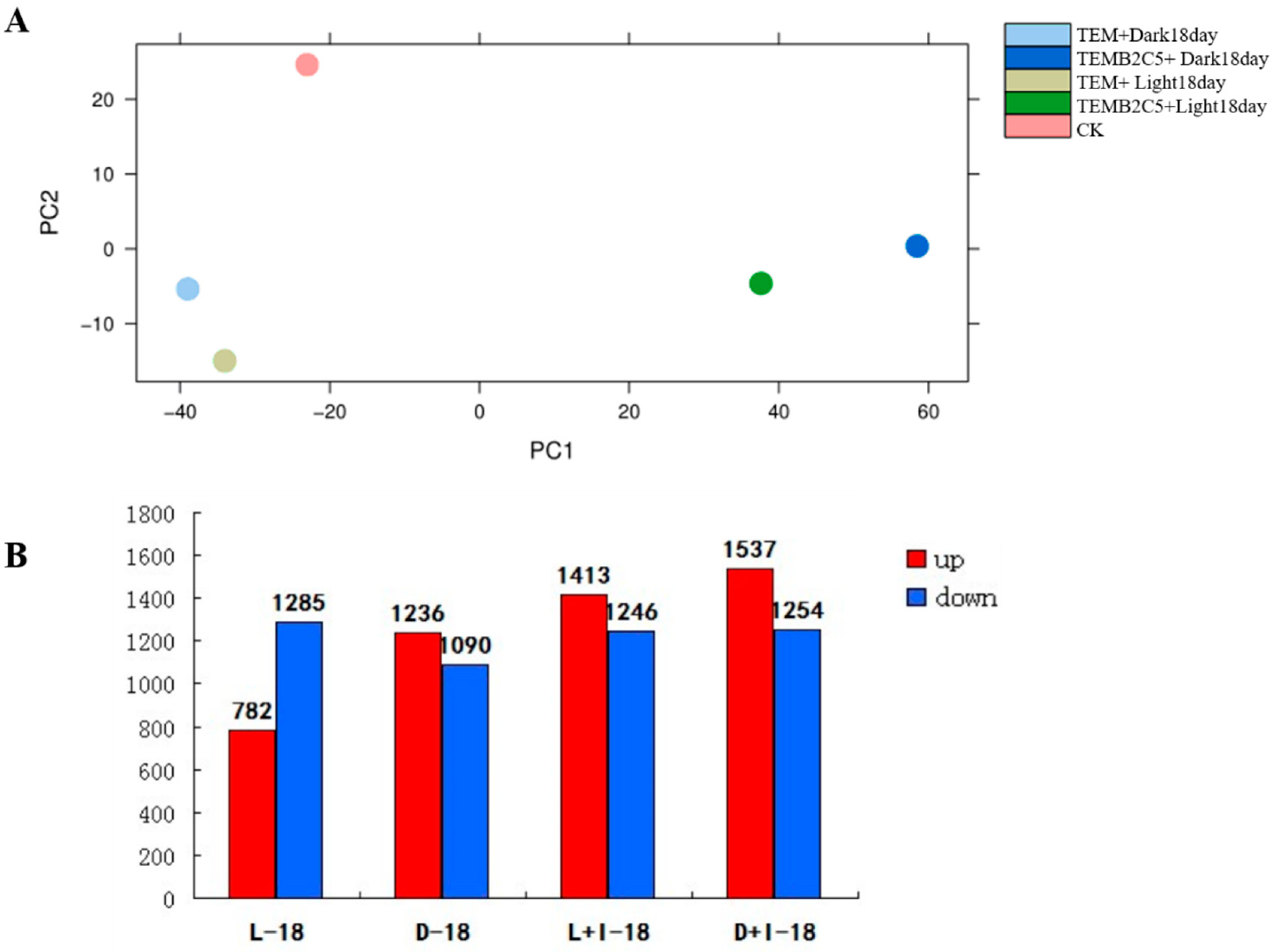
Five cDNA libraries induced by light and hormones in Sudan grass were constructed, and the transcriptome was sequenced. A total of 221,936,940 sequence read fragments (reads) were obtained, including 27,739,721,621 base sequences (bp) (Supplementary Table S1). The reads were sequence assembled to obtain 67,070 single gene clusters (unigene) (Supplementary Figure S1). Principal component analysis (PCA) showed that the effect of light conditions on callus gene expression was less than that induced by hormones (Figure 2A). For instance, CK (control), TEMB2C5+Light18day expression profile had 1413 genes up-regulated and 1246 genes down-regulated; TEMB2C5+Dark18day expression profile had 1537 genes up-regulated and 1254 genes down-regulated; TEM+Light18day expression profile had 782 genes up-regulated and 1285 genes down-regulated, and TEM+Dark18day expression profile had 1236 genes up-regulated and 1090 genes down-regulated (Figure 2B and Supplementary Tables S3–S6).

2.3. Functional Classification of Unigene of Secondary Cell Wall Synthesis of Sudan Grass

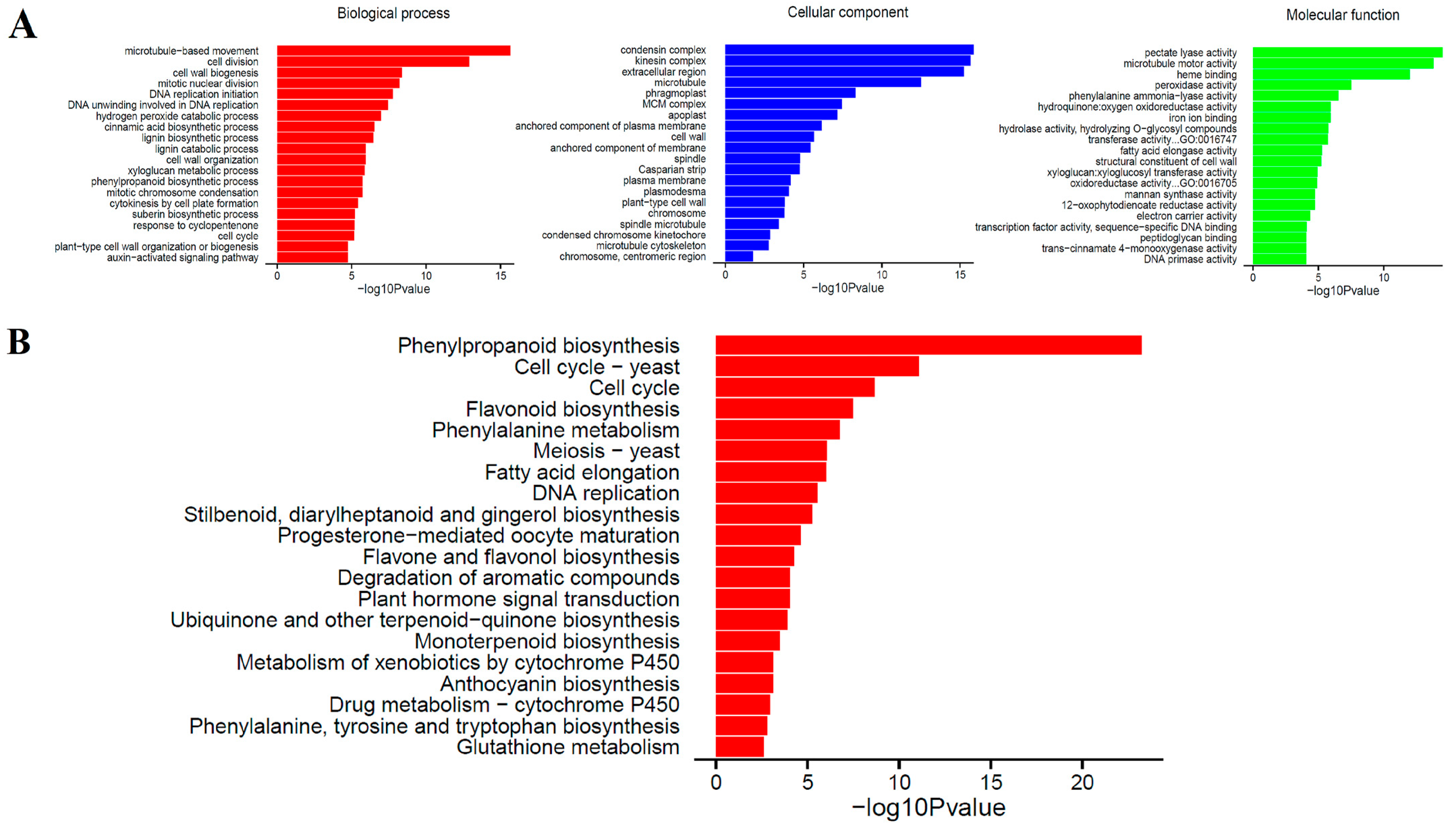
The effect of BRs on secondary cell wall gene expression was stronger than that of light. To further explore gene responses to secondary cell wall synthesis of Sudan grass, we analyzed unigene functional annotations of callus BRs and dark treatment (TEMB2C5+Dark18day vs. CK). According to GO functional annotation, 64 branches could be roughly divided into three categories: biological process (BP), cellular component (CC) and molecular function (MF) (Figure 3A). Most of the Unigenes were related to the condensin complex, kinesin complex, and the extracellular region. The GO functional annotation for TEM+Dark18day vs. CK, TEM+Light18day vs. CK, and TEMB2C5+Light18day vs. CK are shown in Supplementary Figures S2–S4 and Table S7. The results from the KEGG analysis of Unigenes showed that their functions can be roughly divided into 20 categories (Figure 3B). A KOG database comparison analysis revealed that the unigenes can be mapped to 112 metabolic pathway branches according to metabolic pathways, such as phenylpropanoid biosynthesis, flavone and flavonoid biosynthesis, fatty acid elongation, flavonoid synthesis, phenylalanine, tyrosine and tryptophan biosynthesis, plant hormone signal transduction, and degradation of aromatic compounds (Supplementary Table S8). The KEGG pathway analysis for TEM+Dark18day vs. CK, TEM+Light18day vs. CK, and TEMB2C5+Light18day vs. CK are shown in Supplementary Figures S5–S7 and Supplementary Table S8.

2.4. Analysis of CcNACs Family Genes Related to Secondary Cell Wall Synthesis

In the TEMB2C5+Dark18day, TEMB2C5+Light18day and TEM+Light18day treatments, the secondary cell wall in the Sudan grass callus was induced and synthesized. The result shows that 51 genes were upregulated while 180 genes were down regulated in all the three treatments (Figure 4A,B, and Supplementary Tables S9 and S10). In plants, the NAC transcription factor family is an upstream regulator of secondary cell wall synthesis, so this study focused on the analysis of NAC family transcription factors in transcriptome data. Through analysis, it was found that nine different CcNACs transcription factors had common in these three treatments. (Figure 4C). Five of the CcNACs genes were up-regulated under light and BRs treatment, CcNAC1 and CcNAC2 were not expressed in CK but were up-regulated under light and BRs treatment. The expression level of CcNAC1 was higher than that of CcNAC2. The qRT-PCR data were consistent with the transcriptomic data (Figure 4D). Therefore, CcNAC1 was selected for further functional verification.

2.5. CcNAC1 Increases the Lignin Content of Plants

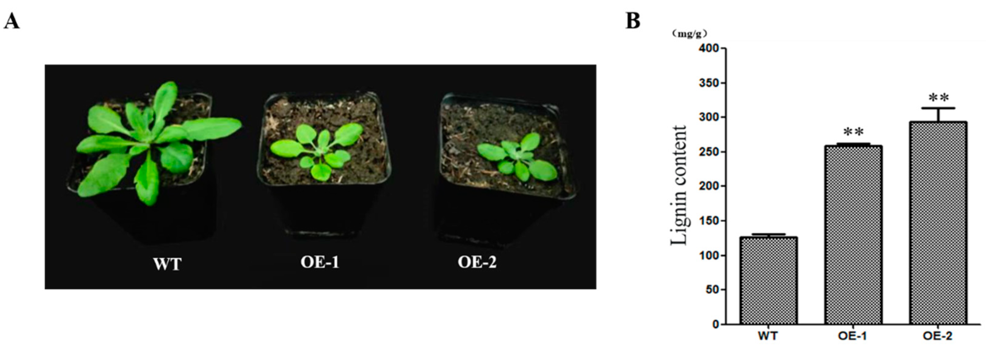
We generated CcNAC1 overexpressing Arabidopsis lines (OE1 and OE2) to investigate the function of CcNAC1 in secondary cell wall synthesis. The overexpressing of CcNAC1 lines (OE1 and OE2) were significantly lower than that of the wild type (WT) (Figure 5A). The overexpression lines and the WT were sampled for their lignin content. The results showed that the lignin contents of the overexpression lines were significantly higher (2.2 times) than that of the WT (2 times) (Figure 5B).

2.6. CcNAC1 Encodes a Nuclear Localization Protein

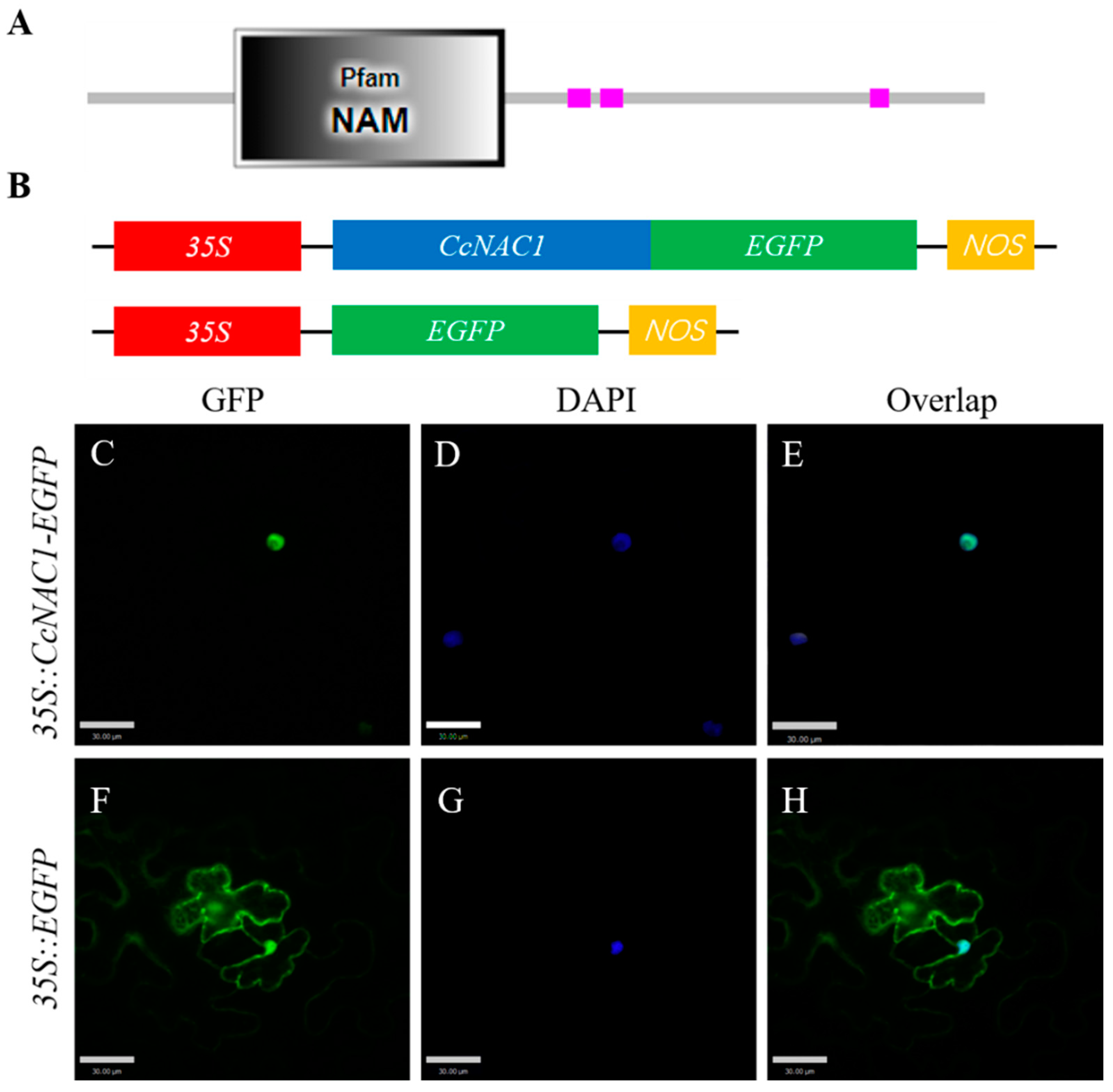
To investigate the mechanism of CcNAC1 protein regulation in plant secondary cell wall synthesis we performed subcellular localization of the functional regions of CcNAC1. Online prediction of the NAC protein domain showed that CcNAC1 was encoded by 341 amino acids, among which 6-130 amino acids were the NAM domain (Figure 6A). It is predicted that CcNAC1 is a nuclear localization protein.
To verify the online prediction results, we constructed the CcNAC1 and EGFP fusion expression vector 35S-CcNAC1-EGFP, which controlled by the 35S promoter (Figure 6B). Transient expression of fusion expression vectors and empty vectors in N. benthamiana leaves was performed to assess CcNAC1 subcellular localization. DAPI was also used as a nuclear marker. The green fluorescent protein (GFP) signal was observed through laser confocal microscopy. The results showed that a GFP signal of an empty vector was detected in the cytoplasm, cell membrane, and nucleus (Figure 6F–H). In contrast, the fusion protein expressed by 35S-CcNAC1-EGFP was only detected in the nucleus (Figure 6C–E). These results demonstrate that CcNAC1 is localized in the nucleus.

2.7. Candidate Interaction Proteins Analysis of CcNAC1

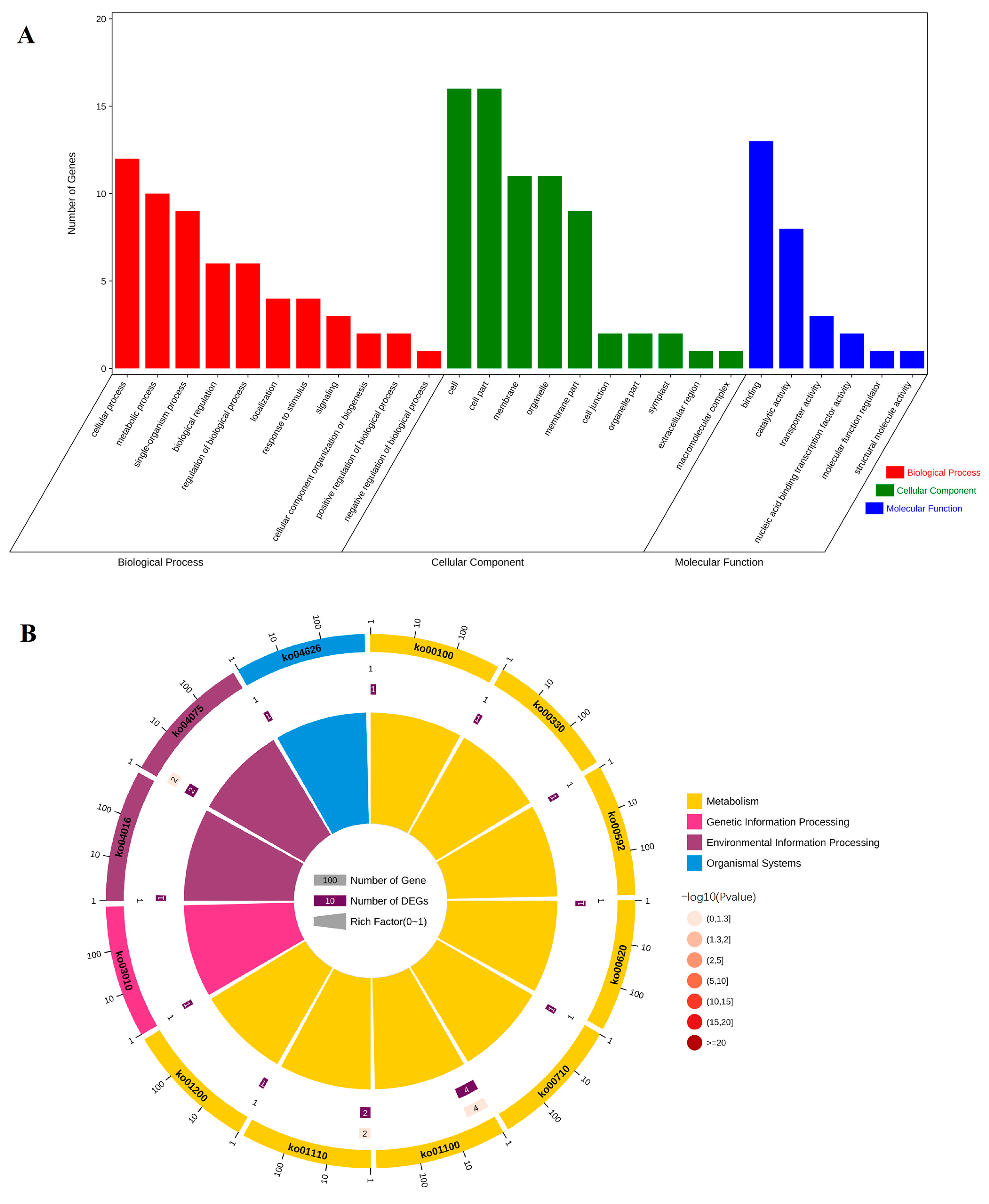
Using NAC as the bait protein to screen the yeast library of Sudan grass secondary cell wall and analyze the molecular biological function of CcNAC1 interacting protein indicated the regulatory pathway of CcNAC1 in the synthesis of Sudan grass secondary cell wall. Through GO annotation, 26 proteins interact with CcNAC1 in secondary cell wall synthesis, and interacting proteins were found to be associated with one or more of the following three GO categories: Biological Process (BP), Cellular Component (CC) and Molecular (Figure 7A). Analysis of the biological processes indicated that the interaction proteins were mainly involved in processes relating to cellular (GO:0009987), metabolic (GO:0008152), single-organism (GO:0044699), biological regulation (GO:0065007), and regulation of biological (GO:0050789), among other processes. Analysis of the cell composition indicated that the interaction proteins were mainly parts the cell (GO:0005623), cell components (GO:0044464), membrane (GO:0016020), organelle (GO:0043226) and membrane components (GO:0044425). Furthermore, a molecular biological function analysis demonstrated that proteins interactions mainly included structural components of binding (GO:0005488), catalytic activity (GO:0003824), and transporter activity (GO:0005215), among others (Supplementary Table S11).
KEGG enrichment analysis helps us to understand the metabolic pathways involved in genes. We performed KEGG enrichment analysis to increase our understanding of gene metabolic pathways in secondary cell wall synthesis in plants. The KEGG enrichment analysis of the 26 candidate interaction proteins of CcNAC1 indicated that the proteins mainly involve 12 pathways (Figure 7B), including metabolic pathways (ko01100), biosynthesis of secondary metabolites (ko01110), plant hormone signal transduction (ko04075), and other regulatory pathways (Supplementary Table S12).

3. Discussion

With improved living standards, demand for animal husbandry products is on the rise. Increasing the fiber content of forage grass is one of the ways to increase the utilization rate of feed for ruminants such as cattle and sheep in order for them to yield high quality products. Sudan grass callus does not contain secondary cell walls under dark conditions, but can produce secondary cell walls under the induction of light and BRs. Therefore, we performed transcriptome sequencing to analyze differentially expressed secondary cell wall regulation to switch on the NAC family during secondary cell wall synthesis [29]. It was found that CcNAC1 was expressed along with the synthesis of the secondary cell wall of Sudan grass. Through functional verification, it was found that CcNAC1 was a positive regulator of the biosynthesis of the secondary cell wall of Sudan grass. Light can induce changes in the structure and play a role in the function of plant xylem and promote the growth of plant secondary cell walls [30]. For instance, in cotton, under shade conditions, the number of vascular bundles and the degree of lignification were decreased [31]. In poplar, dark treatment resulted in thinning of poplar secondary cell walls and reduced the density of the wood [32]. Studies have shown that blue-violet light in sunlight can induce the expression of the AtCRY1 gene and promote the expression of AtMYC2/AtMYC4 [33]. AtMYC2/AtMYC4 can directly bind to the AtNST1 promoter and activate its expression, thereby promoting the synthesis and thickening of secondary cell walls [34]. AtMYB46 regulates secondary cell wall synthesis genes (AtCesA4 (cellulose synthase 4), AtCesA7, and AtCesA8) under the induction of light [35].
BRs play an important role in plant growth and development [36]. Studies have shown that BRs are related to the development of plant xylem [37], and there is a synergistic association between the synthesis of BRs in plants and the development of plant xylem [38]. In binding of BRs to the receptor BRI1 (BRASSINOSTEROID INSENSITIVE 1), SERK3 (BAK1/somatic embryogenesis receptor kinase) is phosphorylated, thereby initiating a signaling cascade reaction. BRs signaling could induce the expression of BZR1 and BES1/BZR2, thereby regulating the gene expression of cell wall biosynthesis such as PMEs [39]. Deficiency in BRs synthesis in plants leads to dwarfism in plant phenotypes, with decrease in lignin and cellulose content [40]. For instance, in poplar (Populus L.), knockout of the BRs receptor by gene editing inhibits the differentiation of secondary cell walls in the xylem, resulting in marked plant dwarf and growth-deficient phenotypes [41]. Studies have shown that in the presence of BRs, the transcription factor AtBZR1 of the BRs pathway binds to the cellulose and enzyme gene AtCesA6 [42], and the AtBES1 transcription factor can combine with other AtCesA family genes to jointly regulate the synthesis of plant secondary cell walls [43]. Therefore, natural light and BRs can induce the synthesis of secondary cell walls in plants. In this study, Sudan grass callus treated with light and BRs showed that BRs induce secondary cell walls formation more than that of light (Figure 2A). Therefore, the ability of BRs to induce the secondary cell wall of Sudan grass callus is stronger than that of light.
In the KEGG pathway analysis, the differential genes were mainly concentrated in the phenylalanine metabolism pathway, flavonoid synthesis, plant-pathogen interaction, plant hormone biosynthesis and signal transduction (Figure 3B). Both lignin and flavonoids belong to a group of phenylpropanoids that share Coumaroyl-CoA in two metabolic pathways [44]. Visible and UV light could activate the expression of flavonoid biosynthetic genes by regulating the R2R3-MYB gene [45]. Moreover, the gene expression level of flavonoid biosynthesis genes is positively correlated with light intensity, but too strong light inhibits the expression of related genes [46]. It could be speculated that the synthesis of secondary cell walls of Sudan grass under light conditions might be related to the biosynthesis pathway of flavonoids induced by light. During the synthetic processes of plant cells, phenylalanine is synthesized into lignin through a series of catalytic reactions under the catalysis of peroxidase [47]. Previous studies have shown that the key enzymes AtADT4 and AtADT5 in the synthesis of flavonoid anthocyanins are related to the synthesis of secondary cell walls. The mutants AtADT4 and AtADT5 resulted in softening of plant stems and phenotypes that could not provide support [48]. When a pathogen invades plants, the secondary cell wall is thickened at the invasion site to block further invasion by the pathogen [49]. In these pathways, plant hormones are involved and play a very important regulatory role [50,51].
The discovery of key regulatory genes for secondary cell wall synthesis in Sudan grass requires the search for genes with common expression trends in treatments with secondary cell wall synthesis. It was clear from the Wenn Diagram that 51 genes were up-regulated and 180 genes were down-regulated in the three secondary cell wall synthesis treatments (TEMB2C5+Dark18day, TEMB2C5+Light18day and TEM+Light18day) (Figure 4A,B, and Supplementary Tables S9 and S10). Therefore, these 231 genes might be involved in regulating secondary cell wall synthesis in Sudan grass. The NAC transcription factors play a very important role in the regulation of secondary cell wall synthesis in plants as a primary transcription factor [8,9]. Ours results on the differentially expressed NAC transcription factors and the expression trend of CcNAC1 and CcNAC2 genes were consistent with the synthesis of secondary cell wall in Sudan grass callus (Figure 4C). Therefore, the expression of these two genes may regulate the synthesis of the secondary cell wall of Sudan grass. The expression of the CcNAC1 gene was higher than that of CcNAC2. So, CcNAC1 was selected for validation. We overexpressed the CcNAC1 gene in Arabidopsis through heterologous expression. The lignin content of 2-month-old plants with CcNAC1 overexpressed in Arabidopsis lines (OE1 and OE2) was significantly higher than that of wild-type plants (Figure 5B), suggesting that the CcNAC1 gene is a positive regulator of secondary cell wall synthesis. The two CcNAC1 overexpression lines (OE1 and OE2) showed dwarf phenotype (Figure 5A). Studies have shown that lignin content directly affects plant growth and development. Plant root development is negatively correlated with lignin content, and excessive lignin accumulation inhibits the development and elongation of roots in the case of Arabidopsis and carrot. Treatment of plants with exogenous IBA resulted in decreased lignin accumulation and increased root length [52]. Lignin content not only affects root development, but also affects fruit size. In peanuts (Arachis hypogaea L.), plants with high lignin content also have a relatively small pod [53]. This means that the dwarf phenotype of plants caused by high lignin content may be caused by the inability of cells to elongate due to excessive lignin in cells.
GO annotation found that some interaction proteins relate to CcNAC1 functions involving in cellular process and cell (Figure 7A). Secondary cell wall synthesis is part of plant morphogenesis. The enrichment of CcNAC1 interacting proteins and cell progression suggest that CcNAC1 and its interacting proteins are involved in the regulation of cell morphogenesis [54]. KEGG analysis showed that CcNAC1 interacting proteins were mainly enriched in metabolic pathways and biosynthesis of secondary metabolites, indicating that CcNAC1 interacting proteins are mainly related to the synthesis of secondary metabolites (Figure 7B). This indicate that NAC interacting proteins involved in the pathway may also be related to secondary cell wall formation [55]. Therefore, CcNAC1 positively regulates secondary cell wall synthesis in plants by regulating the synthesis of related secondary metabolites and participating in cell processes.

4. Materials and Methods

4.1. Plant Materials and Growth Conditions

Sudan grass Sumu No.3 was used as the plant material for different experiments. Sudan grass cultivar Sumu NO.3 and Arabidopsis cultivar Columbia-0 were used in the laboratory. Secondary cell wall induction was carried out in a controlled growth chamber at 25 °C for 18 days from Sudan grass callus that had been subcultured twice. TEM+Light18 day represented Sudan grass callus cultured in the subculture medium (MS basic salt, B5 vitamin, sucrose 30 g/L, agar 8g/L, 2,4-D 2 mg/L and 6-BA 0.05 mg/L, pH = 5.8) and placed in a temperature at 25 °C under long day (16/8 h light/dark) conditions for 18 days. TEM+Dark18 day represented Sudan grass callus cultured in the subculture medium and placed in a temperature at 25 °C in the absence of light conditions for 18 days. TEMB2C5+Light18 day represented Sudan grass callus cultured in the subculture medium supplemented with 5 g/L activated carbon and 2 μM brassinolide (BRs) (MS basic salt, B5 vitamin, sucrose 30 g/L, agar 8g/L, 2,4-D 2 mg/L, 6-BA 0.05 mg/L, activated carbon 5g/L and BRs 0.96 mg/L, pH = 5.8), and placed in a temperature at 25 °C under long day (16/8 h light/dark) conditions for 18 days. TEMB2C5+Dark18 day represented Sudan grass callus cultured in the subculture medium supplemented with 5 g/L activated carbon and 2 μM BRs, and placed in a temperature at 25 °C in the absence of light conditions for 18 day. CK was treated on the 0 day in the subculture medium of Sudan grass callus culture.

4.2. Lignin Dyeing and Content Measurement Method

Phloroglucinol staining was used to stain secondary cell walls of callus of Sudan grass. The Sudan callus was placed in a 10mL centrifuge tube, and 6 M HCl was added. After 30 s, the same amount of phloroglucinol dye solution (phloroglucinol 5 g, and 95% alcohol 100 mL) was added and mixed. The callus was stained for 2 min, and then photographed.
The lignin content of different Arabidopsis lines was determined using a lignin assay kit. Wild-type (WT) lines and CcNAC1 overexpression lines (OE1 and OE2) were grown under normal growth conditions for 2 months. Then, lignin content was determined using a lignin content assay kit (AKSU010U) following the manufacturer’s protocol (Boxbio, Beijing, China). Three independent replicates were assayed and their standard errors (SEs) calculated.

4.3. Phenylalanine Ammonia-Lyase (PAL) Activity Measurement Method

The PAL activity of Sudan grass callus was determined using a PAL activity assay kit. In a controlled growth chamber at 25 °C, the callus of Sudan grass subcultured twice was induced by light with 5 g/L activated carbon and 2 μM BRs treatment (TEMB2C5+Light) and dark treatment (TEM+Dark). Samples were taken from the two treatments every 3 days. PAL activity of samples was determined using a PAL activity assay kit (BC0215) following the manufacturer’s protocol (Solarbio, Beijing, China). Three independent replicates were assayed, and their standard errors (SEs) calculated.

4.4. Transcriptome Sequencing and Analyses

Total RNA of Sudan grass callus was extracted using an mirVana miRNA Isolation Kit (Ambion, Austin, TX, USA). All the RNA samples were pooled to construct a complementary DNA (cDNA) library (Integrity Number (RIN) ≥ 7). The libraries were sequenced on an Illumina Hiseq platform (Illumina HiSeq X Ten) and 125 bp/150 bp paired-end reads were generated. Raw data (raw reads) were processed using Trimmomatic. The reads containing ploy-N and the low-quality reads were removed to obtain clean reads. The clean reads were mapped to SWISSPROT database (http://www.uniprot.org (accessed on 4 February 2016)) location information. The library construction and sequencing were carried out at OE Biotech (Shanghai, China)
In transcriptome sequencing analysis, the fragments per kb per million fragments (FPKM) value was applied to represent the expression level of differentially expressed genes under different samples. Principal component analysis (PCA) of gene expression was used to investigate the distribution of samples, to explore the relationship between samples and to verify the experimental design. Genes with an adjusted p-value < 0.05 and an absolute value of |log2FoldChange| > 1 were assigned as differentially expressed [26].

4.5. Functional Classification of DEGs

Gene Ontology (GO) annotation was obtained from the GO database (https://github.com/tanghaibao/GOatools (accessed on 15 March 2017 and 20 July 2022)). The GO annotation method used the Fisher’s exact test. The GO annotation used four methods, namely Bonferroni, Holm, Sidak, and the false discovery rate, to correct the p-value. The enrichment of GO function was considered significant when then p-value was lower than 0.05. The Kyoto Encyclopedia of Genes and Genomes (KEGG) database was used for the systematic examination of gene roles, contact genomics, and functional information. KEGG software was used, and KOBAS 3.0 (http://kobas.cbi.pku.edu.cn/home.do (accessed on 16 March 2017 and 21 July 2022)). The Benjamini–Hochberg (BH) method was used to correct the p-value. A p-value of 0.05 as the threshold, indicated significant enrichment [26].

4.6. Quantitative Real-Time PCR (qRT–PCR) Verification

Total RNA from Sudan grass and Arabidopsis was extracted using the RNAprep Pure Plant Kit (Tiangen, Beijing, China), and subjected to reverse transcription using PrimeScript™ RT reagent Kit with gDNA Eraser (Perfect Real Time) (TaKaRa, Kyoto, Japan). Gene-specific primers for qRT-PCR were designed in the GenScript Real-time PCR Primer Design (https://www.genscript.com/tools/real-time-pcr-taqman-primer-designtool), which was accessed on 1 June 2018, and are listed in Supplementary Table S2. Quantitative real-time PCR (qRT-PCR) was performed for each cDNA template using ChamQ Universal SYBR qPCR Master Mix (Vazyme, Nanjing, China) with a standard protocol, while the Sudan grass reference gene CcEIF4a served as the internal reference gene. Three independent biological replicates were performed for each sample to ensure statistical credibility. The specificity of the reactions was verified by melting curve analysis. The relative mRNA level for each gene was calculated as follows: ratio = 2−ΔΔCt = 2−[Ct,t-Ct,r] (Ct cycle threshold, Ct,t Ct of the target gene, Ct,r Ct of the control gene). These reactions were conducted using an Applied Biosystems™QuantStudio™5 Real-Time PCR instrument [26].

4.7. Subcellular Localisation of CcNAC1 Protein

The protein domain of CcNAC1, using its amino acid sequence, was predicted through the online software SMART (http://smart.embl-heidelberg.de/smart/set_mode.cgi?NORMAL=1 (accessed on 20 September 2019)). To verify the localisation of the CcNAC1 protein in plant cells, coding CDS sequences of CcNAC1 were introduced into pBinGFP4. The pBinGFP4-CcNAC1 and pBinGFP4 vectors were transformed into Agrobacterium GV3101 strains, and both vectors were transformed into N. benthamiana leaves using the N. benthamiana leaf transient transformation method. The fluorescence of GFP was detected at 488 nm and 405 nm by an upright confocal microscope (Carl Zeiss, Thornwood, NY, USA) at 48–72 h post-inoculation. The excitation wavelengths used were 488 nm for Enhanced Green Fluorescent Protein (EGFP), and 405 nm for 2-(4-Amidinophenyl)-6-indolecarbamidine dihydrochloride (DAPI).

4.8. CcNAC1 Overexpression in Arabidopsis

The Arabidopsis thaliana cultivar, Columbia-0, was used as the plant material in gene function verification experiments. Arabidopsis seeds were cultivated in soil after being vernalized at 4 °C for 5 days, and then grown in a controlled growth chamber at a temperature of 22 °C under long day (16/8 h light/dark) conditions and a relative humidity of 70%. Arabidopsis transformations were performed at the time of full flowering.
The full-length CDS of the CcNAC1 gene was cloned into the pTF101.1 vector under the control of the CaMV 35S promoter (35S-CcNAC1). The primers utilized are listed in Supplementary Table S1. The 35S-CcNAC1 construct was transformed in Arabidopsis cultivar Columbia-0 using Agrobacterium tumefaciens strain EHA101. The selection marker gene (bar) and a 35S promoter were amplified by PCR to verify the positive transgenic plants. Arabidopsis plants were transformed by the floral dip method. The seeds harvested after infection were T1 generation. T1 seeds were collected and selected on MS medium containing 20 mg/L Basta and then tested bar, CcNAC1 and 35S promote by PCR [56]. The seeds harvested from positive plants of T1 generation were T2 generation, and T2 generation plants were identified as positive plants by the same screening method as T1 generation plants. If all T2 generation plants of a line were positive, the line was considered to be homozygous; if separation occurred, the line was considered to be heterozygous. The seeds harvested from T2 homozygous lines were T3 homozygous lines. T3 homozygous lines were used for further study. Samples of Arabidopsis overexpressed lines and the wild type were taken to determine lignin content when each Arabidopsis line was 2 months old.

4.9. Yeast Two-Hybrid Assay

We used the yeast two-hybrid (Y2H) system (Takara, Kyoto, Japan) to screen the yeast library of Sudan grass secondary cell walls, The full-length CDS of CcNAC1 was amplified and cloned into a pGBKT7 vector, yielding pGBKT7-CcNAC1 as bait, while using the Sudan grass callus yeast library plasmids as prey. Then, pGBKT7- CcNAC1 construct and library plasmids were co-transformed into the Y2H Gold yeast strain. Subsequently, these yeast cells were subsequently cultured on SD/-Trp/-Leu plates with 50 µL to calculate transformation efficiency, and others were subsequently cultured on SD/-Trp/-Leu/-Ade/-His+AbA+50 mM 3-AT plates for interaction protein screening. A single yeast clone was picked and grown on the SD/-Trp/-Leu/-Ade/-His+AbA+50 mM 3-AT plates and transferred to an SD/-Trp/-Leu/-Ade/-His+AbA+50 mM 3-AT+X-α-Gal plate for further screening. The blue yeast single clone interacted with proteins of CcNAC1.
The primers and sequences used in this study were from Genscript (China).

5. Conclusions

In this study, the in vitro induction system of the secondary cell wall of Sudan grass was established by treating the callus of Sudan grass with light and brassinosteroids (BRs). Transcriptome analysis of five callus samples with different treatments found that the expression trend of CcNAC1 was consistent with the synthesis of secondary cell walls in Sudan grass callus. Therefore, this gene was selected for further study. Furthermore, using the yeast two-hybrid (Y2H) method, we found that CcNAC1 interacted with many functional proteins relating to WRKY transcription factors, amino acid transporters, and cytochrome p450, among others. Thus, our findings suggest that CcNAC1 is a key regulator of secondary cell wall synthesis in Sudan grass.

Supplementary Materials

The following supporting information can be downloaded at: https://www.mdpi.com/article/10.3390/ijms24076149/s1.

Author Contributions

Y.H. and C.Q. designed and conceived the research. X.Z. and C.Q. created a secondary cell wall induction system for callus of Sudan grass and treated, sampled for RNA-Seq. C.Q. and A.A.-B. analyzed the transcriptome of the callus of Sudan grass. Y.H. designed primer for qRT-PCR and performed qRT-PCR analysis. J.L. and Y.H. transformed Arabidopsis and performed functional verification. Y.H. performed subcellular localization and yeast library screening of target gene. Z.L. and Z.M. planted the transgenic Arabidopsis lines in the greenhouse. Y.H. wrote the draft paper. A.A.-B., C.Q., J.W. and X.Z. revised the paper. All authors have read and agreed to the published version of the manuscript.

Funding

This work was supported by National Natural Science Foundation of China (No. 31901384), Jiangsu Agricultural Science and Technology Innovation Fund (Grant No. CX (21) 3142), Jiangsu Forestry Science and Technology Innovation and Extension Foundation (LYL [2021]23), and Forest and Forage Grass Introduction Project of Kezhou Animal Husbandry and Veterinary Bureau Foundation (KZZB-2022169).

Data Availability Statement

The data that support the findings of this study are available from the corresponding author upon reasonable request.

Conflicts of Interest

The authors declare no conflict of interest. The funders had no role in the design of the study; in the collection, analyses, or interpretation of data; in the writing of the manuscript, or in the decision to publish the results.

References

  1. Kanani, J.; Lukefahr, S.D.; Stanko, R.L. Evaluation of tropical forage legumes (Medicago sativa, Dolichos lablab, Leucaena leucocephala and Desmanthus bicornutus) for growing goats. Small Rumin. Res. 2006, 65, 1–7. [Google Scholar] [CrossRef]
  2. Wolff, M.S.; Elliso, J.M.; Hao, Y.; Cockrum, R.R.; Austin, J.K.; Baraboo, M.; Burch, K.; Lee, J.H.; Maurer, T.; Patil, R.; et al. Diet shifts provoke complex and variable changes in the metabolic networks of the ruminal microbiome. Microbiome 2017, 5, 60–75. [Google Scholar] [CrossRef] [PubMed] [Green Version]
  3. Duan, A.; Yang, X.; Feng, K.; Liu, J.; Xu, Z.; Xiong, A. Genome-wide analysis of NAC transcription factors and their response to abiotic stress in celery (Apium graveolens L.). Comput. Biol. Chem. 2020, 84, 107186. [Google Scholar] [CrossRef]
  4. Huang, C.; Li, L. Understanding of plant cell wall biosynthesis for utilization of lignocellulosic biomass resources. Chin. Sci. Bull. 2016, 61, 3623–3629. [Google Scholar] [CrossRef] [Green Version]
  5. Wang, H.; Dixon, R.A. On-off switches for secondary cell wall biosynthesis. Mol. Plant. 2012, 5, 297–303. [Google Scholar] [CrossRef] [Green Version]
  6. Zhong, R.; Ye, Z. Secondary cell walls: Biosynthesis, patterned deposition and transcriptional regulation. Plant Cell Physiol. 2015, 56, 195–214. [Google Scholar] [CrossRef] [Green Version]
  7. Nakano, Y.; Yamaguchi, M.; Endo, H.; Rejab, N.A.; Ohtani, M. NAC-MYB-based transcriptional regulation of secondary cell wall biosynthesis in land plants. Front. Plant Sci. 2015, 6, 288–306. [Google Scholar] [CrossRef] [Green Version]
  8. Kubo, M.; Udagawa, M.; Nishikubo, N.; Horiguchi, G.; Yamaguchi, M.; Ito, J.; Mimura, T.; Fukuda, H.; Demura, T. Transcription switches for protoxylem and metaxylem vessel formation. Gene Dev. 2005, 19, 1855–1861. [Google Scholar] [CrossRef] [Green Version]
  9. Wang, X.; Wang, D.; Xu, W.; Kong, L.; Ye, X.; Zhuang, Q.; Fan, D.; Luo, K. Histone methyltransferase ATX1 dynamically regulates fiber secondary cell wall biosynthesis in Arabidopsis inflorescence stem. Nucleic Acids Res. 2021, 49, 190–205. [Google Scholar] [CrossRef]
  10. Zhou, J.; Zhong, R.; Ye, Z. Arabidopsis NAC domain proteins, VND1 to VND5, are transcriptional regulators of secondary wall biosynthesis in vessels. PLoS ONE 2014, 9, e105726. [Google Scholar] [CrossRef]
  11. Ruonala, R.; Ko, D.; Helariutta, Y. Genetic networks in plant vascular development. Annu. Rev. Genet. 2017, 51, 335–359. [Google Scholar] [CrossRef]
  12. Ochando, I.; Gonzalez-Reig, S.; Ripoll, J.J.; Vera, A.; Martinez-Laborda, A. Alteration of the shoot radial pattern in Arabidopsis thaliana by a gain-of-function allele of the class Ⅲ HD-Zip gene INCURVATA4. Int. J. Dev. Biol. 2008, 52, 953–961. [Google Scholar] [CrossRef]
  13. Ohashi-Ito, K.; Iwamoto, K.; Fukuda, H. LOB DOMAINCONTAINING PROTEIN 15 positively regulates expression of VND7, a master regulator of tracheary elements. Plant Cell Physiol. 2018, 59, 989–996. [Google Scholar] [CrossRef] [Green Version]
  14. Yamaguchi, M.; Ohtani, M.; Mitsuda, N.; Kubo, M.; Ohme-Takagi, M.; Fukuda, H.; Demura, T. VND-INTERACTING2, a NAC domain transcription factor, negatively regulates xylem vessel formation in Arabidopsis. Plant Cell 2010, 22, 1249–1263. [Google Scholar] [CrossRef] [PubMed] [Green Version]
  15. Negi, S.; Tak, H.; Ganapathi, T.R. Xylem specific activation of 5′ upstream regulatory region of two NAC transcription factors (MusaVND6 and MusaVND7) in banana is regulated by SNBE-like sites. PLoS ONE 2018, 13, e0192852. [Google Scholar] [CrossRef] [PubMed] [Green Version]
  16. Zhong, R.; Demura, T.; Ye, Z. SND1, a NAC domain transcription factor, is a key regulator of secondary wall synthesis in fibers of Arabidopsis. Plant Cell 2006, 18, 3158–3170. [Google Scholar] [CrossRef] [PubMed] [Green Version]
  17. Mitsuda, N.; Iwase, A.; Yamamoto, H.; Yoshida, M.; Seki, M.; Shinozaki, K.; Ohme-Takagi, M. NAC transcription factors, NST1 and NST3, are key regulators of the formation of secondary walls in woody tissues of Arabidopsis. Plant Cell 2007, 19, 270–280. [Google Scholar] [CrossRef] [Green Version]
  18. Zhong, R.; Lee, C.; Ye, Z.H. Evolutionary conservation of the transcriptional network regulating secondary cell wall biosynthesis. Trends Plant Sci. 2021, 15, 625–632. [Google Scholar] [CrossRef]
  19. Kim, W.; Ko, J.; Kim, J.; Kim, J.; Bae, H.; Han, K. MYB46 directly regulates the gene expression of secondary wall-associated cellulose synthases in Arabidopsis. Plant J. 2013, 73, 26–36. [Google Scholar] [CrossRef] [PubMed]
  20. Patzlaff, A.; McInnis, S.; Courtenay, A.; Surman, C.; Newman, L.J.; Smith, C.; Bevan, M.W.; Mansfield, S.; Whetten, R.W.; Sederoff, R.R.; et al. Characterisation of a pine MYB that regulates lignification. Plant J. 2003, 36, 743–754. [Google Scholar] [CrossRef]
  21. Goicoechea, M.; Lacombe, E.; Legay, S.; Mihaljevic, S.; Rech, P.; Jauneau, A.; Lapierre, C.; Pollet, B.; Verhaegen, D.; Chaubet-Gigot, N.; et al. EgMYB2, a new transcriptional activator from eucalyptus xylem, regulates secondary cell. Plant J. 2005, 43, 553–567. [Google Scholar] [CrossRef] [PubMed] [Green Version]
  22. Geng, P.; Zhang, S.; Liu, J.; Zhao, C.; Wu, J.; Cao, Y.; Fu, C.; Han, X.; He, H.; Zhao, Q. MYB20, MYB42, MYB43, and MYB85 regulate phenylalanine and lignin biosynthesis during secondary cell wall formation. Plant Physiol. 2020, 182, 1272–1283. [Google Scholar] [CrossRef] [Green Version]
  23. McCarthy, R.L.; Zhong, R.; Ye, Z. MYB83 is a direct target of SND1 and Acts redundantly with MYB46 in the regulation of secondary cell wall biosynthesis in Arabidopsis. Plant Cell Physiol. 2009, 50, 1950–1964. [Google Scholar] [CrossRef] [PubMed] [Green Version]
  24. Zhong, R.; Lee, C.; Zhou, J.; McCarthy, L.R.; Ye, Z. A battery of transcription factors involved in the regulation of secondary cell wall biosynthesis in Arabidopsis. Plant Cell 2008, 20, 2763–2782. [Google Scholar] [CrossRef] [PubMed] [Green Version]
  25. Zhou, J.; Lee, C.; Zhong, R.; Ye, Z. MYB58 and MYB63 are transcriptional activators of the lignin biosynthetic pathway during secondary cell wall formation in Arabidopsis. Plant Cell 2009, 21, 248–266. [Google Scholar] [CrossRef] [Green Version]
  26. Xuan, H.; Huang, Y.; Zhou, L.; Deng, S.; Wang, C.; Xu, J.; Wang, H.; Zhao, J.; Guo, N.; Xing, H. Key Soybean Seedlings Drought-Responsive Genes and Pathways Revealed by Comparative Transcriptome Analyses of Two Cultivars. Int. J. Mol. Sci. 2022, 23, 2893. [Google Scholar] [CrossRef]
  27. Kim, M.H.; Tran, T.N.A.; Cho, J.S.; Park, E.J.; Lee, H.; Kim, D.G.; Hwang, S.; Ko, J.H. Wood transcriptome analysis of Pinus densiflora identifies genes critical for secondary cell wall formation and NAC transcription factors involved in tracheid formation. Tree Physiol. 2021, 4, 1289–1305. [Google Scholar] [CrossRef]
  28. Wong, M.M.; Cannon, C.H.; Wickneswari, R. Identification of lignin genes and regulatory sequences involved in secondary cell wall formation in Acacia auriculiformis and Acacia mangium via de novo transcriptome sequencing. BMC Genom. 2011, 12, 342–355. [Google Scholar] [CrossRef] [Green Version]
  29. Endo, H.; Yamaguchi, M.; Tamura, T.; Nakano, Y.; Nishikubo, N.; Yoneda, A.; Kato, K.; Kubo, M.; Kajita, S.; Katayama, Y. Multiple classes of transcription factors regulate the expression of VASCULAR-RELATED NAC-DOMAIN7, a master switch of xylem vessel differentiation. Plant Cell Physiol. 2015, 56, 242–254. [Google Scholar] [CrossRef] [Green Version]
  30. Doley, D. Effects of Shade on Xylem Development in Seedings of Eucalyptus Grandis Hill EX.Maiden. New Phytol. 2010, 82, 545–555. [Google Scholar] [CrossRef]
  31. Liu, J.; Meng, Y.; Lv, F.; Chen, J.; Ma, Y.; Wang, Y.; Chen, B.; Zhang, L.; Zhou, Z. Photosynthetic characteristics of the subtending leaf of cotton boll at different fruiting branch nodes and their relationships with lint yield and fiber quality. Front Plant Sci. 2015, 6, 747–757. [Google Scholar] [CrossRef] [Green Version]
  32. Steffens, B.; Rasmussen, A. The Physiology of Adventitious Roots. Plant Physiol. 2016, 170, 603–618. [Google Scholar] [CrossRef] [PubMed] [Green Version]
  33. Zhang, Q.; Xie, Z.; Zhang, R.; Xu, P.; Liu, H.; Yang, H.; Doblin, M.S.; Bacic, A.; Li, L. Blue Light Regulates Secondary Cell Wall Thickening via MYC2/MYC4 Activation of the NST1-Directed Transcriptional Network in Arabidopsis. Plant Cell. 2018, 30, 2512–2528. [Google Scholar] [CrossRef] [PubMed] [Green Version]
  34. Fagard, M.; Desnos, T.; Desprez, T.; Goubet, F.; Refregier, G.; Mouille, G.; McCann, M.; Rayon, C.; Vernhettes, S.; Höfte, H. PROCUSTE1 encodes a cellulose synthase required for normal cell elongation specifically in roots and dark-grown hypocotyls of Arabidopsis. Plant Cell 2000, 12, 2409–2423. [Google Scholar] [CrossRef] [Green Version]
  35. Chen, S.; Sun, W.; Xiong, Y.; Jiang, Y.; Liu, X.; Liao, X.; Zhang, D.; Jiang, S.; Li, Y.; Liu, B.; et al. The Phoebe genome sheds light on the evolution of magnoliids. Hortic Res. 2020, 7, 146–159. [Google Scholar] [CrossRef] [PubMed]
  36. Nagata, N.; Asami, T.; Yoshida, S. Brassinazole, an inhibitor of brassinosteroid biosynthesis, inhibits development of secondary xylem in cress plants (Lepidium sativum). Plant Cell Physiol. 2001, 42, 1006–1011. [Google Scholar] [CrossRef] [PubMed] [Green Version]
  37. Yamamoto, R.; Fujioka, S.; Iwamoto, K.; Demura, T.; Takatsuto, S.; Yoshida, S.; Fukuda, H. Co-regulation of brassinosteroid biosynthesis-related genes during xylem cell differentiation. Plant Cell Physiol. 2007, 48, 74–83. [Google Scholar] [CrossRef] [PubMed] [Green Version]
  38. Wang, C.; Liu, N.; Geng, Z.; Ji, M.; Wang, S.; Zhuang, Y.; Wang, D.; He, G.; Zhao, S.; Zhou, G.; et al. Integrated transcriptome and proteome analysis reveals brassinosteroid-mediated regulation of cambium initiation and patterning in woody stem. Hortic. Res. 2022, 9, uhab048. [Google Scholar] [CrossRef]
  39. Jia, K.; Wang, W.; Zhang, Q.; Jia, W. Cell Wall Integrity Signaling in Fruit Ripening. Int. J. Mol. Sci. 2023, 24, 4054. [Google Scholar] [CrossRef]
  40. Hossain, Z.; McGarvey, B.; Amyot, L.; Gruber, M.; Jung, J.; Hannoufa, A. DIMINUTO 1 affects the lignin profile and secondary cell wall formation in Arabidopsis. Planta 2012, 235, 485–498. [Google Scholar] [CrossRef]
  41. Du, J.; Gerttula, S.; Li, Z.; Zhao, S.; Liu, Y.; Liu, Y.; Lu, M.; Groover, A.T. Brassinosteroid regulation of wood formation in poplar. New Phytol. 2020, 225, 1516–1530. [Google Scholar] [CrossRef] [PubMed]
  42. Yu, X.; Li, L.; Zola, J.; Aluru, M.; Ye, H.; Foudree, A.; Guo, H.; Anderson, S.; Aluru, S.; Liu, P.; et al. A brassinosteroid transcriptional network revealed by genome wide identification of BESI target genes in Arabidopsis thaliana. Plant J. 2011, 65, 634–646. [Google Scholar] [CrossRef] [PubMed]
  43. Xie, L.; Yang, C.; Wang, X. Brassinosteroids can regulate cellulose biosynthesis by controlling the expression of CESA genes in Arabidopsis. J. Exp. Bot. 2011, 62, 4495–4506. [Google Scholar] [CrossRef] [Green Version]
  44. Dong, N.; Lin, H. Contribution of phenylpropanoid metabolism to plant development and plant-environment interactions. J. Integr. Plant Biol. 2021, 63, 180–209. [Google Scholar] [CrossRef] [PubMed]
  45. Matus, T.; Loyola, R.; Vega, A.; Peña-Neira, A.; Bordeu, E.; Arce-Johnson, P.; Alcalde, A. Post-veraison sunlight exposure induces MYB-mediated transcriptional regulation of anthocyanin and flavonol synthesis in berry skins of Vitis vinifera. J. Exp. Bot. 2009, 60, 853–867. [Google Scholar] [CrossRef] [Green Version]
  46. Pandey, A.; Bhushan, A. Ultraviolet-B radiation: A potent regulator of flavonoids biosynthesis, accumulation and functions in plants. Curr. Sci. India 2020, 119, 176–185. [Google Scholar] [CrossRef]
  47. Wang, Y.; Chantreau, M.; Sibout, R.; Hawkins, S. Plant cell wall lignification and monolignol metabolism. Front Plant Sci. 2013, 4, 220–234. [Google Scholar] [CrossRef] [Green Version]
  48. Chen, Q.; Man, C.; Li, D.; Tan, H.; Xie, Y.; Huang, J. Arogenate Dehydratase Isoforms Differentially Regulate Anthocyanin Biosynthesis in Arabidopsis thaliana. Mol. Plant 2016, 9, 1609–1619. [Google Scholar] [CrossRef] [Green Version]
  49. Lee, M.H.; Jeon, H.S.; Kim, S.H.; Chung, J.H.; Roppolo, D.; Lee, H.J.; Cho, H.J.; Tobimatsu, Y.; Ralph, J.; Park, O.K. Lignin-based barrier restricts pathogens to the infection site and confers resistance in plants. EMBO J. 2019, 38, e101948. [Google Scholar] [CrossRef]
  50. Li, X.; Liu, C.; Zhao, Z.; Ma, D.; Zhang, J.; Yang, Y.; Liu, Y.; Liu, H. COR27 and COR28 are Novel Regulators of the COP1–HY5 Regulatory Hub and Photomorphogenesis in Arabidopsis. Plant Cell 2020, 32, 3139–3154. [Google Scholar] [CrossRef]
  51. Sun, Y.; Han, Z.; Tang, J.; Hu, Z.; Chai, C.; Zhou, B.; Chai, J. Structure reveals that BAK1 as a co-receptor recognizes the BRI1-bound brassinolide. Cell Res. 2013, 23, 1326–1329. [Google Scholar] [CrossRef]
  52. Cheng, L.; Zhao, C.; Zhao, M.; Han, Y.; Li, S. Lignin Synthesis, Affected by Sucrose in Lotus (Nelumbo nucifera) Seedlings, Was Involved in Regulation of Root Formation in the Arabidopsis thanliana. Int. J. Mol. Sci. 2022, 23, 2250. [Google Scholar] [CrossRef]
  53. Lv, Z.; Zhou, D.; Shi, X.; Ren, J.; Zhang, H.; Zhong, C.; Kang, S.; Zhao, X.; Yu, H.; Wang, C. Comparative Multi-Omics Analysis Reveals Lignin Accumulation Affects Peanut Pod Size. Int. J. Mol. Sci. 2022, 23, 13533. [Google Scholar] [CrossRef] [PubMed]
  54. Wang, G.; Wang, C.; Liu, W.; Ma, Y.; Dong, L.; Tian, J.; Yu, Y.; Kong, Z. Augmin Antagonizes Katanin at Microtubule Crossovers to Control the Dynamic Organization of Plant Cortical Arrays. Curr. Biol. 2018, 28, 1311–1317. [Google Scholar] [CrossRef] [PubMed] [Green Version]
  55. Duan, L.; Liu, H.; Li, X.; Xiao, J.; Wang, S. Multiple phytohormones and phytoalexins are involved in disease resistance to Magnaporthe oryzae invaded from roots in rice. Physiol. Plant 2014, 152, 486–500. [Google Scholar] [CrossRef] [PubMed]
  56. Xu, J.; Xue, C.; Xue, D.; Zhao, J.; Gai, J.; Guo, N.; Xing, H. Overexpression of GmHsp90s, a heat shock protein 90 (Hsp90) gene family cloning from soybean, decrease damage of abiotic stresses in Arabidopsis thaliana. PLoS ONE 2013, 8, e69810. [Google Scholar] [CrossRef]
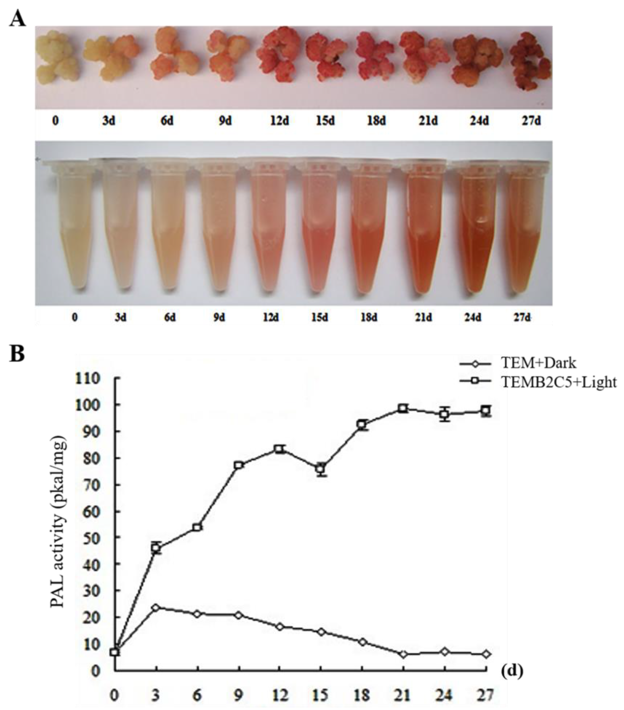
Figure 1. Secondary cell wall induction system of Sudan grass. (A) Specific staining of phloroglucinol lignin after callus induction with 2 μM BRs. (B) Determination of PAL enzyme activity related to lignin synthesis in callus.
Ijms 24 06149 g001
Figure 2. Differentially expressed genes (Unigene) between tested samples. (A) Principal component analysis (PCA) of Unigene expression. (B) Number of Unigenes compared between treated sample and CK sample are shown in red (up-regulated) and blue (down-regulated).
Ijms 24 06149 g002
Figure 3. GO and KEGG analysis of unigenes of TEMB2C5+Dark18day and CK differentially expressed. (A) Diagram showing GO enrichment analysis of differentially expressed Unigene in two samples. BP, biological processes; CC, cellular components; MF, molecular function. (B) KEGG analysis of differentially expressed Unigene in two samples.
Ijms 24 06149 g003
Figure 4. Identification of key CcNACs genes for secondary cell wall synthesis biosynthesis in Sudan grass. (A) Venn diagram of the up-regulated expression by Unigene in four treatments. (B) Venn diagram of the down-regulated expression by Unigene in four treatments. (C) Analysis of the expression levels of nine different CcNACs genes among five expression profiles. (D) Analysis of tissue expression specificity of CcNAC1 gene by qRT-PCR (n = 3). The data represent the means ± SEs. **, p < 0.01 (Student’s t test).
Ijms 24 06149 g004
Figure 5. CcNAC1 positively regulates secondary cell wall synthesis. (A) Performance of wild-type (WT) plants and CcNAC1 overexpression lines (OE1 and OE2) under normal condition. (B) Lignin content of wild-type plants and CcNAC1 overexpression lines 2 months after planting (n = 3); Over 30 plants in each line were used for survival rate analysis. The data represent the means ± SEs. **, p < 0.01 (Student’s t test).
Ijms 24 06149 g005
Figure 6. Subcellular localization of CcNAC1 in tobacco cells. (A) Amino acid domain prediction of CcNAC1 protein. (B) CcNAC1 subcellular localization vector structure. (C) 35S-CcNAC1-EGFP fusion protein. (D) DAPI fluorescent dye. (E) Merged image of C and D. (F) 35S-EGFP fusion protein. (G) DAPI fluorescent dye. (H) Merged image of F and G. Scale bars are 50 μm.
Ijms 24 06149 g006
Figure 7. GO and KEGG analysis of CcNAC1 candidate interacting proteins associated with secondary cell wall synthesis. (A) Diagram showing GO enrichment analysis of CcNAC1 candidate interacting proteins associated with secondary cell wall synthesis. BP, biological processes; CC, cellular components; MF, molecular function. (B) KEGG analysis of CcNAC1 candidate interacting proteins associated with secondary cell wall synthesis.
Ijms 24 06149 g007
Disclaimer/Publisher’s Note: The statements, opinions and data contained in all publications are solely those of the individual author(s) and contributor(s) and not of MDPI and/or the editor(s). MDPI and/or the editor(s) disclaim responsibility for any injury to people or property resulting from any ideas, methods, instructions or products referred to in the content.