Review

Preventive Effect of Pharmaceutical Phytochemicals Targeting the Src Family of Protein Tyrosine Kinases and Aryl Hydrocarbon Receptor on Environmental Stress-Induced Skin Disease

1
School of Food Science and Biotechnology, Kyungpook National University, Daegu 41566, Republic of Korea
2
Department of Microbiology, College of Medicine, Dankook University, Cheonan 31116, Republic of Korea
3
Research Institute of Tailored Food Technology, Kyungpook National University, Daegu 41566, Republic of Korea
*
Author to whom correspondence should be addressed.
These authors contribute equally to this work.
Int. J. Mol. Sci. 2023, 24(6), 5953; https://doi.org/10.3390/ijms24065953
Submission received: 7 February 2023 / Revised: 13 March 2023 / Accepted: 20 March 2023 / Published: 21 March 2023
(This article belongs to the Special Issue Molecular Mechanisms of Anti-inflammatory Phytochemicals 2.0)

Abstract

:
The skin protects our body; however, it is directly exposed to the environment and is stimulated by various external factors. Among the various environmental factors that can threaten skin health, the effects of ultraviolet (UV) and particulate matter (PM) are considered the most notable. Repetitive exposure to ultraviolet and particulate matter can cause chronic skin diseases such as skin inflammation, photoaging, and skin cancer. The abnormal activation of the Src family of protein tyrosine kinases (SFKs) and the aryl hydrocarbon receptor (AhR) in response to UV and/or PM exposure are involved in the development and aggravation of skin diseases. Phytochemicals, chemical compounds of natural plants, exert preventive effects on skin diseases through the regulation of various signaling pathways. Therefore, this review aims to highlight the efficacy of phytochemicals as potential nutraceuticals and pharmaceutical materials for the treatment of skin diseases, primarily by targeting SFK and AhR, and to explore the underlying mechanisms of action. Future studies are essential to validate the clinical potential for the prevention and treatment of skin diseases.

1. Introduction

Skin is the largest organ that covers the body and protects it from environmental stress [1]. It comprises two main layers, i.e., the dermis and epidermis, and keratinocytes constitute 95% of the latter [2]. Keratinocytes originate from epidermal stem cells in the basement membrane of the skin; after their development, they continue to move upward and are finally differentiated to form the stratum corneum, the main structure of the epidermis of the skin [3]. Keratinocytes in the stratum corneum follow a brick-and-mortar pattern of formation. This formation facilitates the prevention of various environmental damages caused by ultraviolet (UV) radiation, pathogenic microbiome, parasites, and surrounding pollutants.
UV is important to human beings for killing pathogens and vitamin D synthesis; however, acute and chronic exposure to UV rays promotes various skin diseases, including edema, erythema, wrinkles, and skin cancer [4]. Using various animal models, it has been demonstrated that skin inflammation [5,6], skin aging [7,8], and skin cancer [9,10] can be induced depending on the type and duration of UV rays. The International Agency for Research on Cancer under the World Health Organization found that diesel exhaust gas adversely affects human health; it was also identified as a first-class carcinogen [11]. Among various environmental pollutants, particulate matter (PM) is the major cause of skin damage [12]. Although particulate matter (PM) hardly penetrates healthy skin, it has been reported that contact with PM causes disease aggravation in people with damaged skin barriers, such as those with atopic dermatitis [13]. Therefore, PM is also recognized as one of the major environmental factors that cause skin damage.
The Src family of protein tyrosine kinases (SFKs) and the aryl hydrocarbon receptor (AhR) play important roles in maintaining homeostasis in various cells. However, hyperactivated SFK and AhR in response to environmental stressors, such as UV and PM, results in the activation of signal transduction related to the induction of various skin diseases [14,15]. Furthermore, SFKs and AhR are major causes responsible for the deterioration of skin health. A variety of external stimuli can stimulate SFKs and AhR, leading to the initiation of signaling cascades. After the activation of signaling responses to various external stimuli, skin diseases such as skin inflammation, psoriasis, atopic dermatitis, and skin cancer can occur [16,17,18,19,20,21]. Acute and chronic UV exposure in rodent skin causes abnormal phosphorylation of Fyn and Src, leading to skin inflammation, photoaging, and skin cancer [22]. In addition, PM directly binds to activated AhR and is subsequently involved in the onset of skin inflammation and skin carcinogenesis [23].
A nutraceutical is a health functional food or dietary supplement that is expected to help prevent or alleviate diseases in addition to the supplementation of basic nutritional components [24]. Phytochemicals are an important source of nutraceuticals and may effectively prevent skin diseases by maintaining skin homeostasis that can be imbalanced through environmental stress such as UV and PM [16,25]. Cellular signaling pathways are critical in maintaining homeostasis, and the hyperactivation of Src and the AhR leads to the onset of skin diseases [17,18,26,27]. Therefore, the consumption of phytochemicals could be a possible strategy to prevent skin diseases by blocking Src- and AhR-induced signaling pathways. The results of this study demonstrated that SFKs and AhR are major targets for preventing environmental stress-induced skin diseases and that pharmaceutical phytochemicals can be used to prevent skin disease through the regulation of SFKs and the AhR. The purpose of this study was to prove that phytochemicals can be effective in preventing skin diseases owing to their ability to control molecular mechanisms. The study results suggested that phytochemicals are a safe pharmaceutical material to maintain skin health in response to environmental pollution.

2. Environmental Factors That Cause Skin Diseases

2.1. UV Irradiation in Skin Diseases

Although there are various environmental factors that can directly affect skin health, UV is the primary factor that disrupts human skin homeostasis. UV is a type of magnetic radiation, and the wavelength of UV is 10–400 nm, which is shorter than that of visible light but longer than that of X-rays. The electromagnetic spectrum of UV is divided into ultraviolet A (UVA), ultraviolet B (UVB), and ultraviolet C (UVC), with wavelengths of 315–400, 280–315, and 100–280 nm, respectively [28]. Most of the UVC used as a source of germicidal lamp is absorbed by the ozone layer in the atmosphere, and most of the UV range reaching the Earth’s surface is UVA [4]. UVB reaches the Earth’s surface at a maximum of 5% even in cloudless and sunny areas. It is considered a complete carcinogen because a single treatment causes skin cancer [29]. Although UVA causes relatively low damage to the skin, compared with UVB, repetitive exposure to UVA has also been reported to cause skin inflammation and skin cancer [30]. The effects of UV irradiation on mice are summarized in Table 1.

2.1.1. Skin Inflammation

Acute UV irradiation causes DNA damage, including pyrimidine dimers and 6–4 photoproducts [33]. If left untreated by the DNA proofreading system of mammalian cells, it leads to aberrant gene expression. As with other inflammation, it is accompanied by redness, swelling, and pain, and these are representative symptoms of erythema. In previous in vivo studies, acute UVB exposure (0.5 J/cm2) resulted in skin inflammation and increased thickness of the epidermis in ICR and SKH-1 hairless mice [16,31].

2.1.2. Skin Aging

Skin aging is categorized into intrinsic and extrinsic aging based on certain triggers. Unlike intrinsic aging caused by individual genetic characteristics, extrinsic aging is caused by environmental factors, mainly UV; thus, extrinsic aging and photoaging are considered the same [34]. It is generally known that repetitive exposure to UV rays results in the decomposition and collapse of collagen in the dermal layer of the skin, causing wrinkle formation, a phenomenon of photoaging [35]. In our previous study, a gradual increase in low-dose UVB irradiation appeared to cause skin photoaging in SKH-1 mice. One minimal erythema dose (MED) was defined as 45 mJ/cm2, and the irradiation dose for mice dorsal skin was increased weekly from 1 MED to 4 MED (0.18 mJ/cm2). Irradiation at 4 MED was maintained until 15 weeks. At the end of the experiment, wrinkle formation, increased epidermal thickness, and hyperplasia of keratinocytes were confirmed to occur on mouse skin [32].

2.1.3. Skin Carcinogenesis

Skin carcinogenesis occurs in three stages: initiation, promotion, and progression [36]. In the initiation stage, acute UV irradiation causes prompt and irreversible DNA damage [37]. The promotion stage includes the clonal expansion of abnormal cell harboring DNA mutation [38]. Chemoprevention, prevention, and delay of carcinogenesis with the use of natural and synthetic medications are also considered promotion stages as they can be reversible with appropriate medication. However, once the neoplasm reaches the progression stage, it becomes difficult to control because cancer cells have a metastatic capacity [39]. Therefore, cancer prevention should take precedence over treatment. Because inflammation is considered a promotion stage in carcinogenesis, diseases such as cancer can be prevented through appropriate relief of inflammation. In a previous study, repetitive exposure to UV rays on the skin caused chronic inflammation and skin cancer in SKH-1 hairless mice. In addition, the pretreatment of natural flavonoid before UV exposure inhibited the development of skin tumors as well as the expression of inflammation-related biological markers [22]. Therefore, controlling inflammation to suppress the cancer progression may be an appropriate cancer prevention method.

2.2. Particulate Matter in Skin Diseases

Air pollution has been reported as one of the causes of worsening skin diseases such as atopic dermatitis and extrinsic aging in several epidemiological studies. Vierkotter et al. suggested that air pollution correlated with a 20% increase in the presence of rough wrinkles and pigment spots on the forehead and cheeks [40]. A study by Hüls et al. clarified a relationship between pigment spots on the cheeks and NO2 [41]. A German cohort study found that the cumulative ozone exposure was correlated with the development of coarse facial wrinkles [42].

2.2.1. Air Pollution

Models of skin damage caused by PM exposure are being constructed; furthermore, standardized PM—a standard reference material available from National Institute of Standards and Technology—is used to establish PM-induced skin damage models. Various mechanisms of action have been reported regarding the toxicity of human-derived skin cells and rodents caused by PM, including the overproduction of reactive oxygen species (ROS) [43], the overexpression of inflammatory proteins such as cyclooxygenase (COX)-2 [44,45], and the overproduction of inflammatory cytokines [46,47].

2.2.2. Cohort Analysis

A recent Cohort of Child Origin of Asthma and Allergic Disease study reported that exposure to fine dust in the early stage of pregnancy can cause fetal skin barrier dysfunction and increased atopic dermatitis induction at the age of 1 year [12]. In addition, a survey conducted on 4907 French children aged 9–11 years reported a correlation between PM10, NO2, NOx, and CO concentrations and eczema outbreaks [48]. In a study conducted on 7030 domestic children aged 6–13 years, a high correlation was observed between atopic dermatitis and parents’ smoking habits [49].

3. The Role of SFKs in Skin Disease

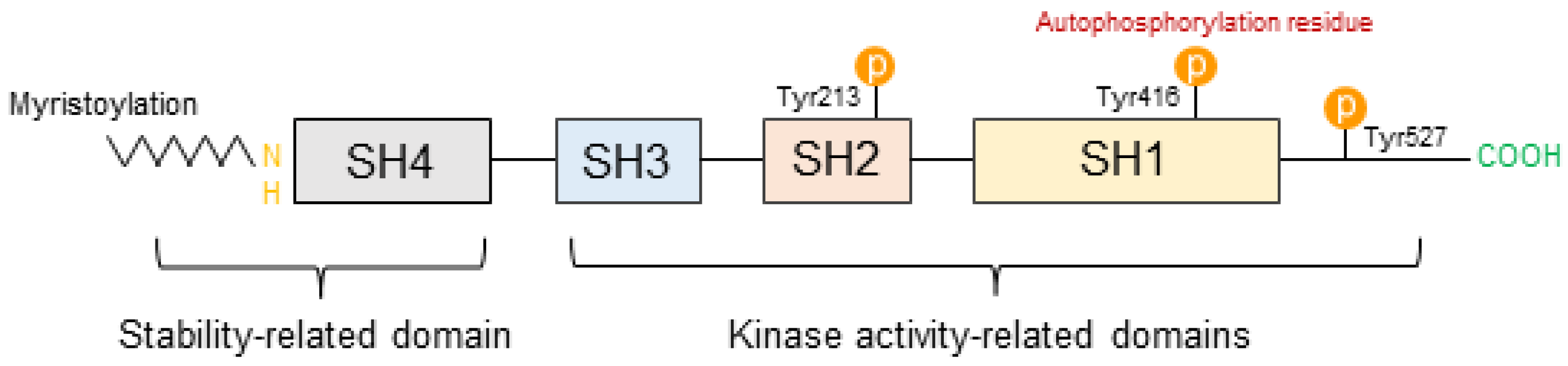
3.1. Structural Characteristics of SFKs

SFKs are important regulators in signaling transduction associated with various cell receptors’ response to the stimulation of environmental stress. Src, Fyn, Yes, Yrk, Blk, Fgr, Hck, Lck, and Lyn are included in SFK [14]. Among them, Src is particularly well known as a proto-oncogene that is closely associated with cancer development and progression in normal mammalian cells. SFKs play a significant role in oncogenesis through the mediation of multiple signaling cascades and are frequently highly activated in tumors [50]. Furthermore, SFKs share the same form of domain structure across all family members and are composed of four Src homology (SH) domains, which perform different functions [51]. Similar to kinases, SFKs have N-terminus and C-terminus at both ends of their structure, including the SH4, SH3, SH2, and SH1 domains. The common structure of SFKs is presented in Figure 1.
The structure of SFK is divided into two predominant domains as follows: stability-related domains and kinase activity-related domains. The SH4 domain is a region located at the N-terminus of SFK and can be myristoylated and/or palmitoylated. Myristoylation and palmitoylation may contribute to the stabilization of the protein structure. Myristoylation is included in the structure of all SFKs whereas palmitoylation is not. Myristoylation is a reaction that adds myristate that consists of 14-carbon fatty acid to the N-terminal glycine residue of SFKs and leads to the formation of a covalent amide bond. Palmitoylation is an addition reaction that adds palmitate to a Cys residue of a protein and leads to the formation of stable amide linkage [52]. Myristoylation and palmitoylation are the main types of protein lipidation, a process in which various lipid and lipid metabolites are produced for transforming proteins in eukaryotic cells. If these are preceded, membrane localization may be possible, and membrane binding becomes stronger than before. Thus, these processes may lead to the regulation of protein trafficking, stability, and aggregation [52,53,54,55].
The SH3 domain consists of approximately 50 amino acids, and this domain is one of the peptide recognition modules; it is related to various signaling pathways, such as cell growth control, cytoskeleton organization, and endocytosis. This domain is important for protein–protein interactions, which may be caused by the assembly of specific protein complexes. The SH3 domain and its specific ligand form specific protein complexes, and its ligand recognition can occur through interaction with polyproline and proline-rich (PxxP) binding sites [56,57].
The SH2 domain specifically combines with phosphorylated tyrosine containing peptide segments. This is a key element in the regulation of tyrosine kinase signaling and involves connecting proteins to signaling systems initiated by tyrosine phosphorylation [56]. Through these processes, the SH2 domain may affect the SFK activities. Aside from the SH2 domain, the SH3 domain and C-terminal are also associated with SFK activities [14].
The SH1 domain is the most preserved among all SFKs, and this domain is located at the C-terminal of the kinase. The SH1 domain is a kinase active site that contains tyrosine phosphorylation activation such as Tyr416 (Tyr419 in human) residue and an ATP-binding pocket. The autophosphorylation of Tyr416 residue is associated with cancer development, whereas Tyr527 phosphorylation prevents cancer. Phosphorylated Tyr527 performs intramolecular interactions with the SH2 domain of SFKs, leading to intramolecular SH3 domain-mediated interactions that inhibit catalytic activity [58]. Thus, the SH1 domain is important as it is closely related to carcinogenesis [53].

3.2. The Role of SFKs in Skin Diseases

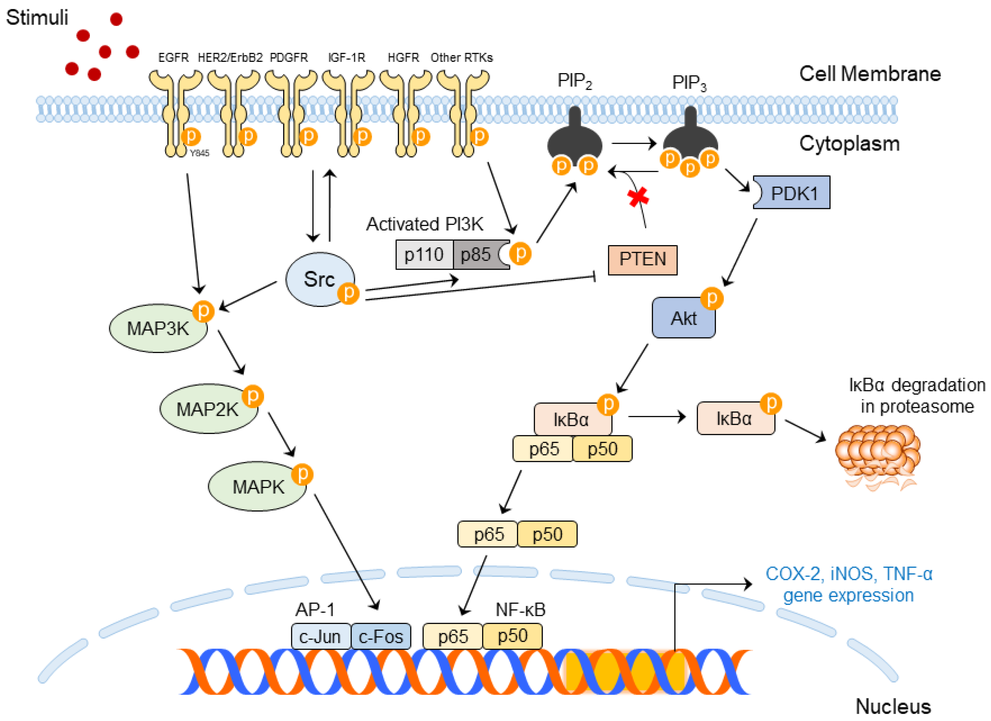
3.2.1. Upstream and Downstream Regulators for SFK Activity

SFK activity is closely related to upstream and downstream regulators of SFKs. For Src, Src activation can be determined through C-terminal Src kinase (CSK) and Src autophosphorylation [50]. CSK homologous kinase phosphorylates Tyr527 residue at the C-terminal of the kinase. When phosphorylated, Tyr527 binds to the SH2 domain, the C-terminal, SH2, and SH3 domains of Src facilitate intramolecular interaction [58]. The SH1 domain cannot facilitate kinase activity with this interaction; thus, Src is converted to an inactive form that is not involved in carcinogenesis [59]. Receptor-type protein tyrosine phosphatase α (RPTPα), dephosphorylates Tyr527, and may cause conformational change in intramolecular interaction. Contrarily, the Tyr416 residue in the SH1 domain is autophosphorylated [60]. Src is fully activated by the autophosphorylation of Tyr416 at the activation loop [61]. Therefore, Tyr416 phosphorylation induces tumorigenesis as opposed to Tyr527.
Src plays important roles by extensively interacting with transmembrane receptor tyrosine kinases (RTKs) through the SH2 and SH3 domains [62]. Epidermal growth factor receptor (EGFR), human epidermal growth factor receptor 2 (HER2/ErbB2), platelet-derived growth factor receptor, insulin-like growth factor-1 receptor (IGF-1R), and c-Met/hepatocyte growth factor receptor are included in RTKs, and RTKs interact with Src and convey the phosphate. After the interaction, Src proceeds as cascades regulated by RTKs and directly transduces signals to its downstream factors. Src has been found to affect phosphoinositide 3-kinases (PI3Ks)/Akt and the signaling transducer and activator of transcription 3 (STAT3). Other receptors located in the membrane, such as integrins and erythropoietin receptor (EpoR), can be related to Src activation [50].
Similarly, other SFKs can be phosphorylated by CSK or be autophosphorylated in specific residues. The activated SFK functions by transmitting signals to the downstream factors [63,64].

3.2.2. Importance of SFK Activities in Skin Diseases

SFK activation induces signaling that leads to various biological functions, and the participation of other cell receptors is also associated with functional determination [59]. Among SFKs, it is well known that Src is closely related to the development of many skin diseases, such as skin inflammation and skin cancer [16,22,65,66].
Many experimental methods have been developed to prove that Src is the main factor affecting skin diseases. Knockdown of the Src gene, the use of Src inhibitors, and the use of Src-related gene knockout mice can be presented as effective ways to determine whether Src plays a crucial role in pathogenesis.
Transfection of HaCaT cells using siRNA targeting Src was performed to demonstrate that Src is a main factor affecting skin inflammation, such as COX-2 expression and mitogen-activated protein kinase (MAPK) phosphorylation. Knockdown of Src mRNA resulted in decreased COX-2 overexpression and blocked MAPK phosphorylation induced by UV irradiation. It also inhibited the phosphorylation of the Tyr845 residue of EGFR known to interact with Src [16]. Src inhibitors can be used for blocking Src activation. As an SFK inhibitor, PP2 (AG 1879) can be treated to the HCT 116 human colon cancer cell lines to confirm the role of Src in wound healing. As a result, the inhibition of Src activity led to a reduction in the phosphorylation of Src Tyr416 and EGFR Tyr845 and affected the downstream factors that facilitate wound healing [67]. Knockout (KO) mice can be used to show that CSK plays a key role in anti-cancer effects. CSK is known to activate Src via phosphorylation of the Tyr527 residue related to anti-cancer effects. In the epidermis of K5-CSK KO mice, Tyr529 (same as Tyr527) phosphorylation was decreased, whereas Tyr418 (same as Tyr416) phosphorylation was increased. In addition, CSK KO mice exhibited apparent epidermal hyperplasia. As the age of the mice increased, hyperplasia spread to the entire skin, and skin thickness also increased. Chronic inflammation was observed in these mice. This evidence showed that Src and CSK play an important role in the regulation of skin inflammation and cancer development [68]. Therefore, targeting SFKs, including Src, can be a powerful method for preventing skin diseases via the regulation of various signaling pathways.

3.2.3. Molecular Mechanism of Src in Skin Diseases

Various factors can activate SFKs, and increased SFKs activity causes inflammation and cancer. Inflammation is a self-limiting reaction that produces anti-inflammatory cytokines against the production of proinflammatory cytokines. However, chronic inflammation can occur if inflammatory factors persist or if the mechanisms required to complete the inflammatory response fail. Chronic inflammation is known to be crucial in malignant tumor development; thus, inflammation and cancer are closely related [69].
Among SFKs, Src has been proven to be an important kinase that induces an inflammatory response due to phosphorylation [67]. Activated Src interacts with EGFR, and EGFR is activated. Activated EGFRs form dimer, and the EGFR dimers stimulate many intracellular signaling pathways [68]. Activated EGFR can stimulate downstream pathways such as the MAPK and phosphatidylinositol 3-kinase (PI3K)/Akt signaling pathways [70].
MAPK cascades are composed of MAPK kinase kinases (MAP3Ks), MAPK kinases (MAP2Ks), and MAPK-linked phosphorylation kinases. MAP3K can be activated by stimuli such as environmental stress, cytokines, and growth factors [71]. Especially, it is well known that UVB [72] and PM can, as environmental factors, mediate MAPK signaling [12]. These stimuli activate MAP3Ks, including Raf, mixed-lineage kinase, and MAPK/extracellular signal-regulated kinase (ERK) kinase kinases, and MAP3Ks deliver phosphate groups to its downstream MAP2Ks. MAPK kinases and PAK1 are MAP2K components. Phosphorylated MAP2K also phosphorylates its downstream MAPK. ERK, Jun N-terminal kinase, and p38 are also MAPK components. Phosphorylated MAPK affects the transcription factors that exist in the cell nucleus [71,73].
The PI3K/Akt signaling pathway consists of a connection between PI3K and Akt. Extracellular stimuli can stimulate PI3K, and then PI3K forms heterodimers composed of the p85 and p110 subunits for stabilization. p85 is an inhibitory adaptor/regulatory subunit. It incorporates signals from intracellular proteins, such as Src, Ras, and protein kinase C (PKC), and RTKs. p85 then offers integration points for the activation of catalytic subunit p110 and its downstream molecules [74,75]. Activated PI3K converts phosphatidylinositol-4,5-bisphosphate (PIP2) into phosphatidylinositol-3,4,5-triphosphate (PIP3) by adding a phosphate group to PIP2. Contrarily, PIP3 may also be dephosphorylated by a negative regulator of PI3K signaling, PTEN, which can be reversed to PIP2. Akt is a downstream of PI3K and binds to PIP3 at the cell membrane. Phosphoinositide-dependent kinase 1 (PDK1) also binds to PIP3 and phosphorylates threonine 308, a kinase domain of Akt, which contributes to the stabilization of the activation loop [74,75,76].
Signaling cascades activate nuclear factor kappa B (NF-κB) or upregulate activator protein (AP)-1 transcription factors that promote inflammation and carcinogenesis. The expression of these transcription factors activates several inflammatory genes encoding tumor necrosis factor (TNF)-α, COX-2, and inducible nitric oxide synthase (iNOS) and subsequently produces TNF-α, prostaglandin E2 (PGE2), and nitric oxide (NO), respectively [10,70,77,78]. These factors can indicate skin diseases [77,79]. COX-2 promotes disease progression ranging from inflammation to cancer; thus, COX-2 is a disease-inducible enzyme [80]. It is also considered a crucial factor that represents skin inflammation and skin carcinogenesis [22]. TNF-α and iNOS can be accompanied by skin inflammatory reactions [70]. Therefore, blocking Src activation is important to exert anti-inflammatory and anti-cancer effects. Src-mediated signaling pathways are presented in Figure 2.

4. Molecular Mechanism of AhR in Skin Diseases

AhR is a transcription factor that is influenced by different environmental stimuli, and their exposure initiates signaling cascades related to inflammation and cancer [81,82]. Interestingly, AhR signaling paradoxically functions as a treatment for skin barrier dysfunction; however, at the same time, it exacerbates skin diseases through ROS generation [19,27].

4.1. AhR Ligands

AhR is located in the cytoplasm, and some low-molecular-weight chemicals are AhR ligands that can promote AhR activation [83]. Ligands with a proper size of three benzene rings are known to bind to AhR. There are several extrinsic substances known as AhR ligands. Extrinsic composites such as toxic pollutant polycyclic aromatic hydrocarbons and 2,3,7,8-tetrachlorodibenzo-p-dioxin (TCDD) are well-known AhR ligands. Extrinsic natural compounds, such as flavonoids, glucosinolates, and indocarbionols, and endogenous AhR ligands that form in the body, such as a tryptophan dimer 6-formylindolo[3,2-b]carbazole (FICZ), can also act as AhR ligands [84,85]. Furthermore, PM activates inflammatory responses in human skin by easily penetrating the skin layer [21]. Therefore, various external factors including environmental pollutants may activate AhR.

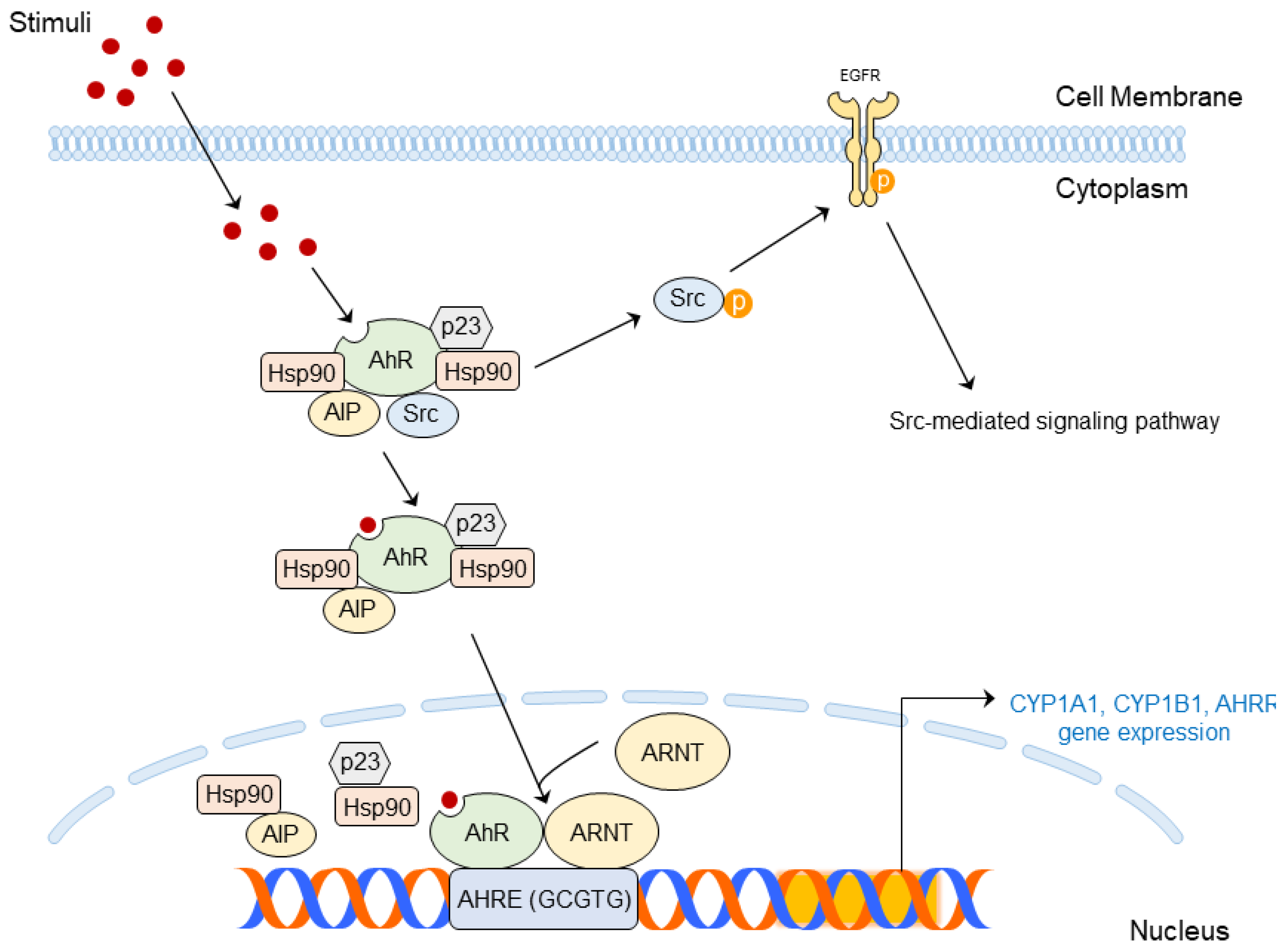
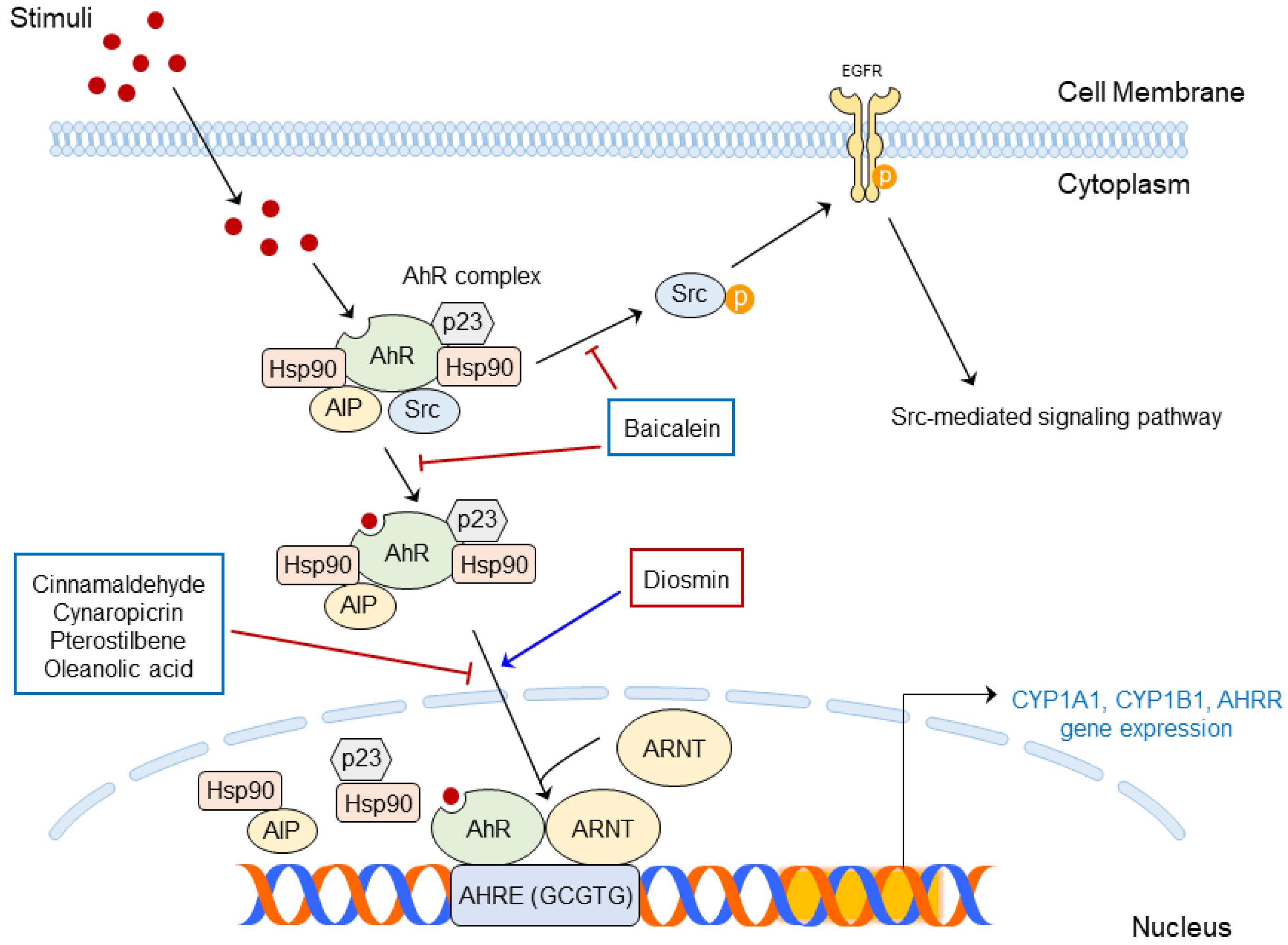
4.2. Structure and Molecular Mechanism of AhR

AhR is a multi-component complex composed of immunophilin-like Ah receptor-interacting protein (AIP, the same as hepatitis B virus X-associated protein 2 [XAP2]), two molecules of 90-kD heat shock protein (HSP90), HSP90 co-chaperone p23, and Src under a normal condition [82,86,87]. Hsp90 and AIP block AhR degradation, and AhR can translocate from the cytoplasm to the nucleus. Then, DNA binding occurs from its translocation [15,82,88].
Translocated AhR combines with the aryl hydrocarbon receptor nuclear translocator (ARNT). AhR and ARNT forms heterodimer, and its C-terminal acts with its promoter, known as aryl hydrocarbon response elements (same as xenobiotic response elements [XREs], 5′-(C)GCGTG-3′). AhR–ARNT recruit coactivator and transcript genes related to xenobiotic metabolism, including cytochrome P450 family 1 subfamily A member 1 (CYP1A1), cytochrome P450 family 1 subfamily B member 1 (CYP1B1), and AhR repressor (AhRR) [15,82,89]. CYP1B1 and CYP1B1 metabolite AhR ligands to mutagenic epoxide intermediates that may lead to ROS generation; then, inflammation may occur due to the ROS production [82,90].
On the other hand, the promotion of the CYP1A1 gene’s expression ironically strengthens the disrupted skin barrier [19]. The AhRR disrupts AhR–ARNT complexes, suppressing inflammatory responses and carcinogenesis induced by AhR signaling [91].
Additionally, the AhR ligand binds to AhR, and the resulting AhR complex releases Src; then, Src interacts with EGFR. Activated EGFR mediates several cellular events, such as MAPK signaling [89,92], and can also produce inflammatory responses, such as COX-2 expression [73,93].
Activated AhR is well known to be closely associated with skin inflammation and skin carcinogenesis [89]. UV radiation produces an AhR ligand FICZ, and exposure to UVB has been demonstrated to mediate AhR signaling [94,95]. PM also affects the activation of AhR and contributes to the outbreak of skin inflammation [96]. Therefore, targeting AhR can be an effective way to treat environmental stress-induced skin diseases at the molecular level.
AhR-mediated signaling pathways affected by various stimuli are summarized in Figure 3.

5. Prevention of Skin Diseases Using SFKs and/or AhR-Targeted Phytochemicals

Targeting SFKs and/or AhR is an effective way to prevent skin diseases, including inflammation and skin carcinogenesis caused by different signaling cascades, as described above. It has been demonstrated that phytochemicals prevent skin diseases caused by environmental factors such as UVB [9,16,17,26,27,97]. Therefore, research into phytochemicals targeting SFKs and/or AhR may be a key strategy for alleviating skin diseases.

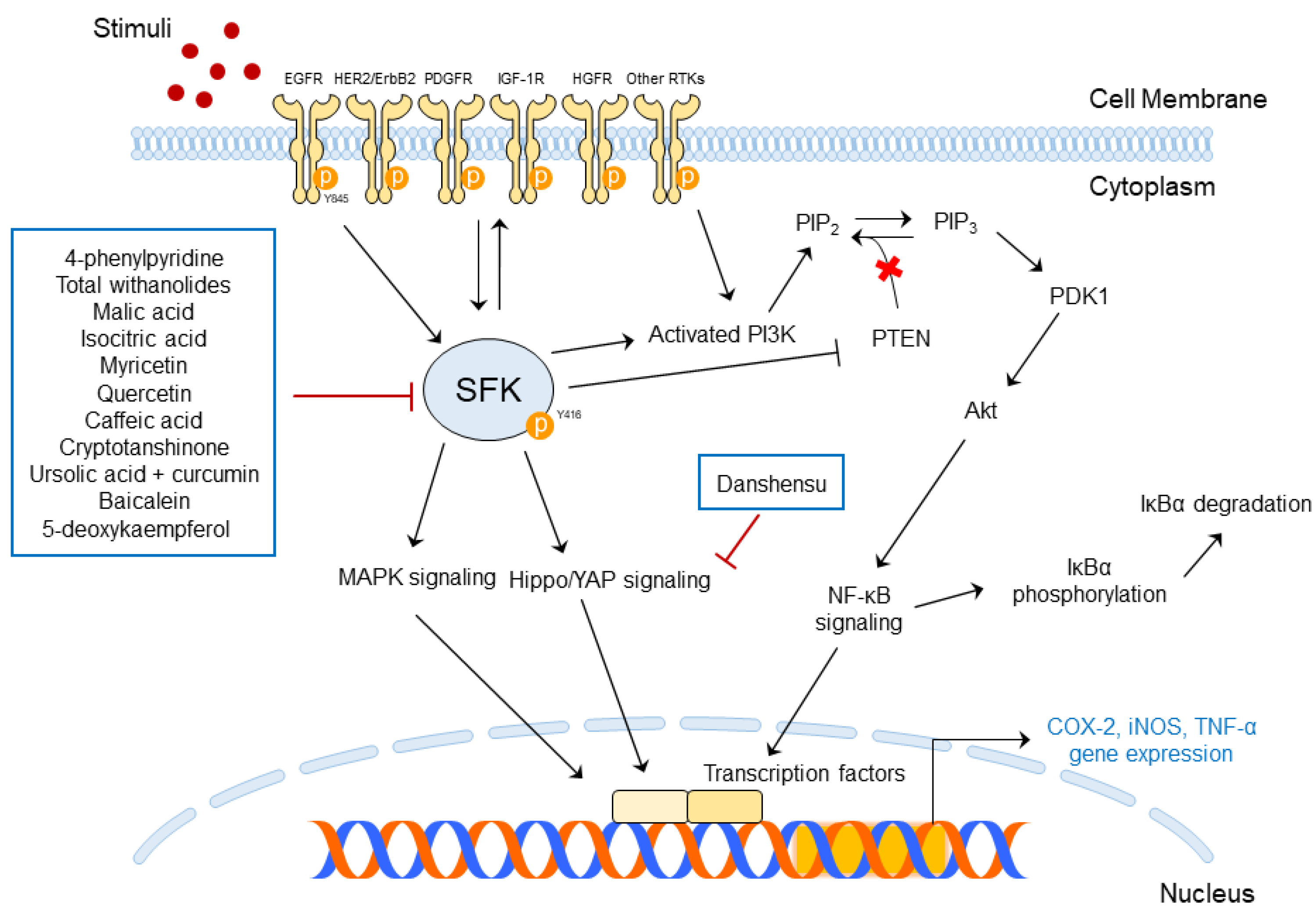
5.1. Functions of SFK-Targeted Phytochemicals in Skin Diseases

The molecular functions of SFK-targeted phytochemicals in skin diseases are summarized in Table 2 and presented schematically in Figure 4.
4-Phenylpyridine (4-PP) suppressed skin inflammation caused by UVB by targeting c-Src in pre-clinical studies. 4-PP decreased inflammation markers, such as COX-2, AP-1, and PGE2 expression, by blocking c-Src-mediated MAPK phosphorylation in human immortalized cell lines. 4-PP reduced epidermal thickness increased by UVB exposure and blocked COX-2 expression, resulting in the phosphorylation of EGFR in UVB-irradiated mice [16].
Datura metel L. contains total withanolides; these phytochemicals can relieve psoriasis-like skin inflammation induced via IMQ application. IMQ is a potent immune activator that stimulates Toll-like receptor (TLR)7/8; thus, it can exaggerate chronic inflammatory skin diseases such as psoriasis. Total withanolides restored IMQ-induced skin damage in male BALB/c mice through various pathways. Furthermore, the phosphorylation of STATs and MAPKs induced by cytokines was decreased, as well as the phosphorylation of Lck, Lyn, and Src [103].
Malic acid and isocitric acid improved wound healing by targeting c-Src. The phosphorylation of Src Y416, EGFR (upstream of Src), and STAT3 Y705 (downstream of Src) was blocked by the treatment of malic acid and isocitric acid [67].
Danshensu inhibited abnormal epidermal proliferation through the yes-associated protein (YAP) pathway, resulting in improved psoriasis. Src has been proven to drive tumor development and metastasis through the activation of the YAP/PD-binding motif (TAZ) axis [104] and lead to the expression of COX-2 [105]. Danshensu was found to suppress M5-induced cell apoptosis and regulate cell cycle-related factor expression in HaCaT cells. Furthermore, it decreased YAP mRNA and protein expression in both HaCaT cells and BALB/c mice.
Myricetin inhibited UVB-induced skin carcinogenesis by targeting Fyn kinase. It regulated Fyn downstream pathways and bound directly to Fyn to block Fyn kinase activity in JB6P+ mouse skin epidermal cells [9,98].
Quercetin exists in many natural plants and is a phyto-compound that downregulates Src expression levels in human keratinocytes. Thus, psoriasis, which frequently occurs along with skin inflammation, can be alleviated by the action of quercetin [99].
Caffeic acid generally found in coffee blocks elevated Fyn activity. Caffeic acid also decreased COX-2 expression and its promoter activity by directly binding with Fyn kinase. Therefore, caffeic acid can prevent UVB-induced skin carcinogenesis [100].
Cryptotanshinone, a component of Salvia miltiorrhiza Bunge, is effective in improving atopic dermatitis. It prevented Lyn and Syk phosphorylation and blocked the MAPK and NF- κB signaling pathways. It also blocked phospholipase C (PLC)γ and PKCδ phosphorylation in cells. The topical treatment of cryptotanshinone improved DNCB-induced atopic dermatitis-like skin lesions in BALB/c mice and decreased immunoglobulin E (IgE) production in mice serum and mice ear [101].
The combination of ursolic acid and curcumin can prevent TPA-induced skin carcinogenesis by targeting c-Src. It is reported that TPA induces skin diseases such as skin cancer [97], oxidative stress, and inflammation [106]. TPA-treated mouse skin showed increased tumor multiplicity and higher incidence and latency of skin cancer; however, topical treatment with a combination of ursolic acid and curcumin reduced these phenomena. It was shown that the combination of ursolic acid and curcumin blocked the phosphorylation of EGFR Y1086 and Src Y416 residues in mice tissues [97].
Baicalein alleviates BaP-induced oxidative stress by regulating Src phosphorylation in human epidermal keratinocytes. BaP is a known environmental pollutant; the compound baicalein is a component of Scutellaria baicalensis root. Treatment with baicalein decreased ROS production and prevented the phosphorylation of Src caused by BaP treatment [102].
5-Deoxykaempferol exerts efficacy on UVB-induced skin cancer by targeting the Src and PI3K pathways. Src was a main target of 5-deoxykaempferol, and the blocking of Src activation decreased the phosphorylation of its downstream factors. A pull-down assay showed the direct binding of 5-deoxykaempferol to Src, PI3K, and RSK in JB5 P+ mouse epidermises [26].
The above literature shows that phytochemicals from natural plants can prevent skin diseases by modulating intracellular signal cascades induced by various stimuli. However, studies on the effect of phytochemicals on air pollutant-induced skin diseases have not been actively investigated except for BaP. Therefore, we propose that it is necessary to prove that phytochemicals can prevent the exacerbation of skin conditions caused by atmospheric pollutants such as PM.

5.2. Phytochemicals Targeting AhR for the Regulation of Skin Diseases

AhR is a double-edged sword because it has two opposite effects on the skin: improving atopic dermatitis and worsening skin inflammation. Atopic dermatitis is closely associated with skin barrier function, and the upregulation of keratinocytes differentiation is important when forming a skin barrier [107]. The activation of AhR leads to the expression of the CYP1A1 gene, and CYP1A1 gene expression also upregulates skin barrier formation [19]. AhR is a positive upstream of skin barrier proteins; thus, activating AhR is important for alleviating atopic dermatitis [108].
Interestingly, AhR is a negative upstream of skin inflammation caused by UVB. Elevated CYP1A1 expression produces ROS and causes skin inflammation by mediating oxidative stress on the skin [27]. Therefore, research on the CYP1A1 gene that affects various functions of the human skin is necessary.
The molecular functions of AhR-targeted phytochemicals in skin diseases are summarized in Table 3 and presented schematically in Figure 5.
Diosmin, a flavone glycoside, belongs to the Rutaceae family. Diosmin activates AhR signaling pathways and alleviates atopic dermatitis. When treated with diosmin, the expression of CYP1A1 gene, a AhR downstream was increased, and the expression of differentiation markers, such as filaggrin (FLG), loricrin (LOR), hornerin (HORN), involucrin (IVL), and ovo-like transcriptional repressor 1 (OVOL1) are also increased. It has been demonstrated that diosmin directly binds with AhR and activates AhR downstream. As a result, diosmin improves atopic dermatitis by restoring skin barrier function via the AhR signaling pathways [19].
Cinnamaldehyde is the main component of Cinnamomum cassia. Cinnamaldehyde reduced BaP-induced oxidative stress through the AhR pathway. BaP activates the AhR pathway and leads to the expression of CYP1A1 gene. In addition, increased CYP1A1 gene expression induces ROS production and oxidative stress on HaCaT cells. However, cinnamaldehyde decreased BaP-induced CYP1A1 expression and its upstream, AhR. Regulated AhR blocked the translocation from the cytosol to the nucleus [20]. Additionally, cinnamaldehyde decreased the oxidative stress production via the NRF2/HO-1 pathway known as an ROS regulator [112]. As a result, cinnamaldehyde reduced ROS production and had an antioxidant effect on the skin [20].
Cynaropicrin is a chemical found in artichoke (Cynara scolymus) that has antioxidant effects. Without UVB irradiation, cynaropicrin translocated AhR from the cytoplasm to the nucleus, leading to the mRNA expression of its downstreams, namely, CYP1A1, NRF2, and Nqo1. Cynaropicrin also reduced oxidative stress induced by UVB irradiation. Cynaropicrin treatment translocated NRF2 to the nucleus in response to UVB-induced oxidative stress. As a result, inflammatory responses such as ROS, IL-6, and TNF- α production has decreased in human keratinocytes. Therefore, it has been demonstrated that cynaropicrin attenuates oxidative stress induced by UVB via the AhR-NRF2-Nqo1 signaling pathway [27].
Baicalein is a potent inhibitor of BaP-induced oxidative stress. BaP phosphorylates Src and translocates AhR to nuclear. AhR located in the nucleus increases the expression of CYP1A1; the activation of AhR-CYP1A1 and the subsequent ROS production are known to induce proinflammatory cytokines secretion. However, baicalein inhibited AhR nuclear translocation and reduced CYP1A1 expression in human keratinocytes. In addition, baicalein activates the NRF2-HMOX1 pathway. Consequently, baicalein reduced oxidative stress through the inhibition of ROS and proinflammatory cytokine production [102].
Kaempferol prevents UVB- and AhR-mediated gene expression. Transcriptomics has proved that kaempferol treatment regulates gene expressions related to AhR, NRF2, and NF-κB. Kaempferol also blocks NF-κB p65 and RelB nuclear translocation in HaCaT and NHEK cells, indicating its anti-inflammatory effects on the skin [109].
Pterostilbene prevents PM-induced skin disease by targeting AhR. PM can aggregate skin inflammation and skin aging as well as reduce the moisturizing ability of the skin by activating AhR. Pterostilbene decreased the nuclear translocation of AhR and downstream gene CYP1A1 expression, as well as COX-2 expression and MAPK phosphorylation. The expressions of matrix metalloproteinase (MMP)-1, MMP-2, and MMP-9 that represent skin aging were also decreased, whereas moisturizing protein aquaporin-3 (AQP-3) was increased in HaCaT cells and CCD-966SK dermal fibroblasts. This indicates that pterostilbene alleviates skin inflammation, aging, and oxidative stress induced by PM by blocking AhR activation [110].
Oleanolic acid has been proven to prevent skin aging caused by PM. It decreases the expressions of CYP1A1, TNF-α, and IL-6, as well as the expressions of MMP-1 and autophagy related proteins p62 and LC-3 I and II in human epidermal keratinocytes and dermal fibroblasts. This indicates the therapeutic effects of oleanolic acid on PM-induced skin diseases by targeting AhR signaling [111].
The evidence in these studies shows that phytochemicals can regulate AhR activity in different directions, depending on the target skin disease. However, phytochemicals have only been shown to be able to prevent skin diseases by regulating AhR-related signaling cascades at the cellular level. Existing research has not proven the preventive effects of phytochemicals in vivo. Therefore, further research is needed to clarify the mechanism of the action of phytochemicals in animal studies.

6. Limitations of Animal Model for AhR Targeting Phytochemicals

It is important to prove that phytochemicals can improve skin health by controlling AhR activity. However, it has not been demonstrated that phytochemicals affect AhR activation in vivo. Therefore, we investigated which in vivo markers could prove that AhR is an essential factor in maintaining skin health. The experimental mouse models of AhR-related skin diseases are summarized in Table 4.
There are several ways to demonstrate that AhR is a key regulator of skin disorders.
The silencing of the AhR gene in mice can be used to prove that AhR is the main factor in maintaining skin health. Yu et al. used AhR-null mice and induced atopic dermatitis in mice skin using MC903. MC903-treated wild-type mice and AhR-null mice had differences in ear thickness and scratching frequency. In AhR-null mine skin, ear thickness and scratching frequency were decreased [113]. Smith et al. proved that tapinarof alleviates skin inflammation through AhR. Tapinarof is a compound that is derived from bacteria. IMQ-treated AhR-sufficient and AhR-knockout mice were topically administered tapinarof, and their clinical scores and cytokine transcription levels were compared. IMQ-induced clinical score increased and elevated IL-17A and IL-17F levels were decreased when treated with tapinarof in AhR+/+ mice; however, there was no difference in AhR−/− mice. This indicates that tapinarof attenuates psoriasis and atopic dermatitis via activation of AhR [114]. To clarify whether AhR is essential for maintaining skin barrier integrity in mice, AhR KO mice also can be used. Increased TEWL, structural changes in epidermis, and microarray results show that AhR is a key factor in skin barrier function [117].
AhR antagonists can be used to demonstrate the effectiveness of substances against skin diseases. Yu et al. used CH223191, an AhR antagonist, to prove the effect of tryptophan metabolites on atopic dermatitis. MC903 was used to induce atopic dermatitis and tryptophan metabolites, and CH223191 was also used to treat wild-type C57BL/6 and BALB/c mice. Tryptophan metabolites attenuated MC903-induced skin inflammation; however, CH223191-treated mice showed increased ear thickness, scratching frequency, TSLP mRNA level, and serum IgE level compared to CH223191+tryptophan metabolites-treated mice. This showed that AhR is required for the action of tryptophan metabolites on atopic dermatitis [113]. In addition, CH223191 can be used to show that esomeprazole inhibits fibro-inflammation in a BLM-induced BALB/c mice model. CH223191-treated mice showed increased dermal thickness and elevated mRNA levels of profibrotic markers such as COL1A1, α-SMA, CTGF, and TGF-β1 [116].
AhR agonists can be used to clarify the effect of AhR activation on skin health. Benvitimod, an AhR agonist, activates AhR, leading to alleviation of rosacea-like eruptions in female BALB/c mice skin. LL-37 was administered to induce rosacea-like eruption in mice; however, topical treatment of benvitimod decreased redness scores, redness areas, the infiltration of dermal inflammatory cells, TLR2 expression, and chemokine expression. Therefore, it is shown that AhR activation can be a positive target for treating rosacea [115].
The removal of AhR ligands from the diet impairs skin barrier integrity in C57BL/6 mice. AhR ligands bind with AhR, leading to AhR pathway activation. Activated AhR is related to skin barrier function; therefore, researchers removed AhR ligands from the mouse diet. Skin deterioration was observed in mice that on a diet without AhR ligands [117].
AhR-CA transgenic mice were used to show that the consecutive activation of the AhR pathway promotes skin inflammation. AhR-CA transgenic mice showed increased skin inflammation-related markers such as increased itching frequency, inflammatory gene expression, and cell-mediated immune response. The results show that the activation of AhR can aggravate skin inflammation in vivo [118].
As the homeostasis of AhR activity is important for maintaining human skin health, it is essential to identify the regulatory molecular mechanism of AhR activity using phytochemicals.

7. Conclusions and Future Research Directions

External environmental factors have recently emerged as major concerns that threaten human health. Since environmental stress can cause skin diseases, it is necessary to develop materials that can prevent diseases caused by external stimuli. This study demonstrated that environmental factors such as UV and/or PM activate various molecular targets including SFK and AhR and cause skin inflammation, photoaging, and skin cancer. Furthermore, it was confirmed that phytochemicals directly or indirectly suppress SFK or AhR activity to prevent the occurrence of skin diseases. Therefore, we suggest that the regulation of SFK and AhR signaling pathways using phytochemicals can help prevent environmental stress-induced skin diseases.
Phytochemical research has been widely described in therapeutic and preventive research; however, several studies have been identified that focus on the effects of a broad range of SFK and AhR-related signaling pathways. For phytochemical materials to be clinically researched and industrialized, it is thought that more in-depth research results on the identification and binding structure of the proteins to which they bind should be provided. Since the skin is the main target organ, most studies have focused on topical treatment; therefore, the degree of application to cosmeceutical is high. However, research on pharmaceutical applicability through oral or intraperitoneal administration is also considered necessary. It is widely described as a skin disease; however, as the environment changes, such as through global warming, the symptoms and causes appear in various ways. Therefore, future research on phytochemicals on various skin diseases such as atopy and psoriasis would also be of interest.

Author Contributions

Conceptualization, D.J.K. and S.K.J.; methodology, S.J.P., D.J.K. and S.K.J.; formal analysis, S.J.P., D.J.K. and S.K.J.; investigation, S.J.P., D.J.K. and S.K.J.; data curation, S.J.P., D.J.K. and S.K.J.; writing—original draft preparation, S.J.P., D.J.K. and S.K.J.; writing—review and editing, D.J.K. and S.K.J.; visualization, S.J.P.; supervision, D.J.K. and S.K.J.; funding acquisition, S.K.J. All authors have read and agreed to the published version of the manuscript.

Funding

This research was funded by Ministry of Education grant number NRF-2022R1A2C1010923, National Research Foundation of Korea grant number NRF-2021M3E5E6022715.

Institutional Review Board Statement

Not applicable.

Informed Consent Statement

Not applicable.

Data Availability Statement

The data presented in this study are available.

Acknowledgments

This project was supported by the Basic Science Research Program through the National Research Foundation of Korea, which is funded by the Ministry of Education (NRF-2022R1A2C1010923) and by the National Research Foundation of Korea Grant funded by the Korean Government (NRF-2021M3E5E6022715).

Conflicts of Interest

The authors declare no conflict of interest.

References

  1. Passeron, T.; Zouboulis, C.C.; Tan, J.; Andersen, M.L.; Katta, R.; Lyu, X.; Aguilar, L.; Kerob, D.; Morita, A.; Krutmann, J.; et al. Adult skin acute stress responses to short-term environmental and internal aggression from exposome factors. J. Eur. Acad. Dermatol. Venereol. 2021, 35, 1963–1975. [Google Scholar] [CrossRef] [PubMed]
  2. Burns, T.; Breathnach, S.M.; Cox, N.; Griffiths, C. Rook’s Textbook of Dermatology, 7th ed.; Blackwell Science: Hoboken, NJ, USA, 2004; Volume 2. [Google Scholar]
  3. Fujiwara, H.; Tsutsui, K.; Morita, R. Multi-tasking epidermal stem cells: Beyond epidermal maintenance. Dev. Growth Differ. 2018, 60, 531–541. [Google Scholar] [CrossRef] [PubMed] [Green Version]
  4. Matsumura, Y.; Ananthaswamy, H.N. Toxic effects of ultraviolet radiation on the skin. Toxicol. Appl. Pharmacol. 2004, 195, 298–308. [Google Scholar] [CrossRef] [PubMed]
  5. So, B.R.; Yeo, H.J.; Lee, J.J.; Jung, Y.H.; Jung, S.K. Cellulose nanocrystal preparation from Gelidium amansii and analysis of its anti-inflammatory effect on the skin in vitro and in vivo. Carbohydr. Polym. 2021, 254, 117315. [Google Scholar] [CrossRef] [PubMed]
  6. Jang, J.H.; So, B.R.; Yeo, H.J.; Kang, H.J.; Kim, M.J.; Lee, J.J.; Jung, S.K.; Jung, Y.H. Preparation of cellulose microfibril (CMF) from Gelidium amansii and feasibility of CMF as a cosmetic ingredient. Carbohydr. Polym. 2021, 257, 117569. [Google Scholar] [CrossRef] [PubMed]
  7. Jung, S.K.; Lee, K.W.; Kim, H.Y.; Oh, M.H.; Byun, S.; Lim, S.H.; Heo, Y.S.; Kang, N.J.; Bode, A.M.; Dong, Z.; et al. Myricetin suppresses UVB-induced wrinkle formation and MMP-9 expression by inhibiting Raf. Biochem. Pharmacol. 2010, 79, 1455–1461. [Google Scholar] [CrossRef] [PubMed] [Green Version]
  8. Ha, S.J.; Park, J.; Lee, J.; Song, K.M.; Um, M.Y.; Cho, S.; Jung, S.K. Rice bran supplement prevents UVB-induced skin photoaging in vivo. Biosci. Biotechnol. Biochem. 2018, 82, 320–328. [Google Scholar] [CrossRef] [Green Version]
  9. Jung, S.K.; Lee, K.W.; Byun, S.; Kang, N.J.; Lim, S.H.; Heo, Y.S.; Bode, A.M.; Bowden, G.T.; Lee, H.J.; Dong, Z. Myricetin suppresses UVB-induced skin cancer by targeting Fyn. Cancer Res. 2008, 68, 6021–6029. [Google Scholar] [CrossRef] [Green Version]
  10. Ha, S.J.; Lee, J.; Park, J.; Kim, Y.H.; Lee, N.H.; Kim, Y.E.; Song, K.M.; Chang, P.S.; Jeong, C.H.; Jung, S.K. Syringic acid prevents skin carcinogenesis via regulation of NoX and EGFR signaling. Biochem. Pharmacol. 2018, 154, 435–445. [Google Scholar] [CrossRef]
  11. Zhu, L.; Jia, W.; Wan, X.; Zhuang, P.; Ma, G.; Jiao, J.; Zhang, Y. Advancing metabolic networks and mapping updated urinary metabolic fingerprints after exposure to typical carcinogenic heterocyclic aromatic amines. Environ. Pollut. 2023, 319, 120936. [Google Scholar] [CrossRef]
  12. Dijkhoff, I.M.; Drasler, B.; Karakocak, B.B.; Petri-Fink, A.; Valacchi, G.; Eeman, M.; Rothen-Rutishauser, B. Impact of airborne particulate matter on skin: A systematic review from epidemiology to in vitro studies. Part Fibre. Toxicol. 2020, 17, 35. [Google Scholar] [CrossRef] [PubMed]
  13. Kim, B.E.; Kim, J.; Goleva, E.; Berdyshev, E.; Lee, J.; Vang, K.A.; Lee, U.H.; Han, S.; Leung, S.; Hall, C.F.; et al. Particulate matter causes skin barrier dysfunction. JCI Insight 2021, 6, e145185. [Google Scholar] [CrossRef] [PubMed]
  14. Yeatman, T.J. A renaissance for SRC. Nat. Rev. Cancer 2004, 4, 470–480. [Google Scholar] [CrossRef] [PubMed]
  15. Neavin, D.R.; Liu, D.; Ray, B.; Weinshilboum, R.M. The role of the aryl hydrocarbon receptor (AHR) in immune and inflammatory diseases. Int. J. Mol. Sci. 2018, 19, 3851. [Google Scholar] [CrossRef] [Green Version]
  16. Kim, J.G.; Kang, H.Y.; Kim, M.J.; Lim, S.; Lee, C.J.; Kim, K.M.; Jung, S.K. 4-phenylpyridine suppresses UVB-induced skin inflammation by targeting c-Src in vitro and in vivo. J. Cell Mol. Med. 2022, 26, 3891–3901. [Google Scholar] [CrossRef]
  17. Su, Y.; Zhang, F.; Wu, L.; Kuang, H.; Wang, Q.; Cheng, G. Total withanolides ameliorates imiquimod-induced psoriasis-like skin inflammation. J. Ethnopharmacol. 2022, 285, 114895. [Google Scholar] [CrossRef]
  18. Jia, J.; Mo, X.; Liu, J.; Yan, F.; Wang, N.; Lin, Y.; Li, H.; Zheng, Y.; Chen, D. Mechanism of danshensu-induced inhibition of abnormal epidermal proliferation in psoriasis. Eur. J. Pharmacol. 2020, 868, 172881. [Google Scholar] [CrossRef]
  19. Lee, J.; Song, K.M.; Jung, C.H. Diosmin restores the skin barrier by targeting the aryl hydrocarbon receptor in atopic dermatitis. Phytomedicine 2021, 81, 153418. [Google Scholar] [CrossRef]
  20. Uchi, H.; Yasumatsu, M.; Morino-Koga, S.; Mitoma, C.; Furue, M. Inhibition of aryl hydrocarbon receptor signaling and induction of NRF2-mediated antioxidant activity by cinnamaldehyde in human keratinocytes. J. Dermatol. Sci. 2017, 85, 36–43. [Google Scholar] [CrossRef]
  21. Shi, Y.; Zeng, Z.; Liu, J.; Pi, Z.; Zou, P.; Deng, Q.; Ma, X.; Qiao, F.; Xiong, W.; Zhou, C.; et al. Particulate matter promotes hyperpigmentation via AhR/MAPK signaling activation and by increasing alpha-MSH paracrine levels in keratinocytes. Environ. Pollut. 2021, 278, 116850. [Google Scholar] [CrossRef]
  22. Byun, S.; Lee, K.W.; Jung, S.K.; Lee, E.J.; Hwang, M.K.; Lim, S.H.; Bode, A.M.; Lee, H.J.; Dong, Z. Luteolin inhibits protein kinase C(epsilon) and c-Src activities and UVB-induced skin cancer. Cancer Res. 2010, 70, 2415–2423. [Google Scholar] [CrossRef] [PubMed] [Green Version]
  23. Vogeley, C.; Rolfes, K.M.; Krutmann, J.; Haarmann-Stemmann, T. The aryl hydrocarbon receptor in the pathogenesis of environmentally-induced squamous cell carcinomas of the skin. Front. Oncol. 2022, 12, 841721. [Google Scholar] [CrossRef]
  24. Kim, M.J.; Jung, S.K. Nutraceuticals for prevention of atherosclerosis: Targeting monocyte infiltration to the vascular endothelium. J. Food Biochem. 2020, 44, e13200. [Google Scholar] [CrossRef] [PubMed]
  25. Lin, Z.; Niu, Y.; Jiang, Y.; Chen, B.; Peng, L.; Mi, T.; Huang, N.; Li, W.; Xu, D.; Chen, R.; et al. Protective effects of dietary fish-oil supplementation on skin inflammatory and oxidative stress biomarkers induced by fine particulate air pollution: A pilot randomized, double-blind, placebo-controlled trial. Br. J. Dermatol. 2021, 184, 261–269. [Google Scholar] [CrossRef]
  26. Lee, K.M.; Lee, K.W.; Byun, S.; Jung, S.K.; Seo, S.K.; Heo, Y.S.; Bode, A.M.; Lee, H.J.; Dong, Z. 5-deoxykaempferol plays a potential therapeutic role by targeting multiple signaling pathways in skin cancer. Cancer Prev. Res. 2010, 3, 454–465. [Google Scholar] [CrossRef] [PubMed] [Green Version]
  27. Takei, K.; Hashimoto-Hachiya, A.; Takahara, M.; Tsuji, G.; Nakahara, T.; Furue, M. Cynaropicrin attenuates UVB-induced oxidative stress via the AhR-Nrf2-Nqo1 pathway. Toxicol. Lett. 2015, 234, 74–80. [Google Scholar] [CrossRef]
  28. Buonanno, M.; Welch, D.; Brenner, D.J. Exposure of human skin models to KrCl excimer lamps: The impact of optical filtering(dagger). Photochem. Photobiol. 2021, 97, 517–523. [Google Scholar] [CrossRef]
  29. Mancebo, S.E.; Wang, S.Q. Skin cancer: Role of ultraviolet radiation in carcinogenesis. Rev. Environ. Health 2014, 29, 265–273. [Google Scholar] [CrossRef]
  30. D’Orazio, J.; Jarrett, S.; Amaro-Ortiz, A.; Scott, T. UV radiation and the skin. Int. J. Mol. Sci. 2013, 14, 12222–12248. [Google Scholar] [CrossRef] [Green Version]
  31. Jung, S.K.; Ha, S.J.; Kim, Y.A.; Lee, J.H.; Lim, T.G.; Kim, Y.T.; Lee, N.H.; Park, J.S.; Yeom, M.H.; Lee, H.J.; et al. MLK3 is a novel target of dehydroglyasperin D for the reduction in UVB-induced COX-2 expression in vitro and in vivo. J. Cell Mol. Med. 2015, 19, 135–142. [Google Scholar] [CrossRef]
  32. Jung, S.K.; Ha, S.J.; Jung, C.H.; Kim, Y.T.; Lee, H.K.; Kim, M.O.; Lee, M.H.; Mottamal, M.; Bode, A.M.; Lee, K.W.; et al. Naringenin targets ERK2 and suppresses UVB-induced photoaging. J. Cell. Mol. Med. 2016, 20, 909–919. [Google Scholar] [CrossRef] [PubMed] [Green Version]
  33. Budden, T.; Bowden, N.A. The role of altered nucleotide excision repair and UVB-induced DNA damage in melanomagenesis. Int. J. Mol. Sci. 2013, 14, 1132–1151. [Google Scholar] [CrossRef] [PubMed] [Green Version]
  34. Khavkin, J.; Ellis, D.A. Aging skin: Histology, physiology, and pathology. Facial Plast. Surg. Clin. N. Am. 2011, 19, 229–234. [Google Scholar] [CrossRef] [PubMed]
  35. Fisher, G.J.; Kang, S.; Varani, J.; Bata-Csorgo, Z.; Wan, Y.; Datta, S.; Voorhees, J.J. Mechanisms of photoaging and chronological skin aging. Arch. Dermatol. 2002, 138, 1462–1470. [Google Scholar] [CrossRef] [PubMed]
  36. Hecker, E. Three stage carcinogenesis in mouse skin--recent results and present status of an advanced model system of chemical carcinogenesis. Toxicol. Pathol. 1987, 15, 245–258. [Google Scholar] [CrossRef] [PubMed] [Green Version]
  37. Zhong, Q.Y.; Lin, B.; Chen, Y.T.; Huang, Y.P.; Feng, W.P.; Wu, Y.; Long, G.H.; Zou, Y.N.; Liu, Y.; Lin, B.Q.; et al. Gender differences in UV-induced skin inflammation, skin carcinogenesis and systemic damage. Environ. Toxicol. Pharmacol. 2021, 81, 103512. [Google Scholar] [CrossRef]
  38. Zhang, W.; Hanks, A.N.; Boucher, K.; Florell, S.R.; Allen, S.M.; Alexander, A.; Brash, D.E.; Grossman, D. UVB-induced apoptosis drives clonal expansion during skin tumor development. Carcinogenesis 2005, 26, 249–257. [Google Scholar] [CrossRef]
  39. Einspahr, J.G.; Bowden, G.T.; Alberts, D.S. Skin cancer chemoprevention: Strategies to save our skin. Recent Results Cancer Res. 2003, 163, 151–164, discussion 264–266. [Google Scholar]
  40. Vierkötter, A.; Schikowski, T.; Ranft, U.; Sugiri, D.; Matsui, M.; Krämer, U.; Krutmann, J. Airborne particle exposure and extrinsic skin aging. J. Investig. Dermatol. 2010, 130, 2719–2726. [Google Scholar] [CrossRef] [Green Version]
  41. Hüls, A.; Vierkötter, A.; Gao, W.; Krämer, U.; Yang, Y.; Ding, A.; Stolz, S.; Matsui, M.; Kan, H.; Wang, S.; et al. Traffic-related air pollution contributes to development of facial lentigines: Further epidemiological evidence from Caucasians and Asians. J. Investig. Dermatol. 2016, 136, 1053–1056. [Google Scholar] [CrossRef] [Green Version]
  42. Fuks, K.B.; Hüls, A.; Sugiri, D.; Altug, H.; Vierkötter, A.; Abramson, M.J.; Goebel, J.; Wagner, G.G.; Demuth, I.; Krutmann, J.; et al. Tropospheric ozone and skin aging: Results from two German cohort studies. Environ. Int. 2019, 124, 139–144. [Google Scholar] [CrossRef] [PubMed]
  43. Diao, P.; He, H.; Tang, J.; Xiong, L.; Li, L. Natural compounds protect the skin from airborne particulate matter by attenuating oxidative stress. Biomed. Pharmacother. 2021, 138, 111534. [Google Scholar] [CrossRef]
  44. Bae, Y.J.; Park, K.Y.; Han, H.S.; Kim, Y.S.; Hong, J.Y.; Han, T.Y.; Seo, S.J. Effects of particulate matter in a mouse model of oxazolone-induced atopic dermatitis. Ann. Dermatol. 2020, 32, 496–507. [Google Scholar] [CrossRef] [PubMed]
  45. Shin, J.W.; Lee, H.S.; Na, J.I.; Huh, C.H.; Park, K.C.; Choi, H.R. Resveratrol inhibits particulate matter-induced inflammatory responses in human keratinocytes. Int. J. Mol. Sci. 2020, 21, 3446. [Google Scholar] [CrossRef] [PubMed]
  46. Kim, M.; Kim, J.H.; Jeong, G.J.; Park, K.Y.; Lee, M.K.; Seo, S.J. Particulate matter induces pro-inflammatory cytokines via phosphorylation of p38 MAPK possibly leading to dermal inflammaging. Exp. Dermatol. 2019, 28, 809–815. [Google Scholar] [CrossRef] [PubMed]
  47. Ryu, Y.S.; Kang, K.A.; Piao, M.J.; Ahn, M.J.; Yi, J.M.; Hyun, Y.M.; Kim, S.H.; Ko, M.K.; Park, C.O.; Hyun, J.W. Particulate matter induces inflammatory cytokine production via activation of NFκB by TLR5-NOX4-ROS signaling in human skin keratinocyte and mouse skin. Redox. Biol. 2019, 21, 101080. [Google Scholar] [CrossRef]
  48. Ahn, K. The role of air pollutants in atopic dermatitis. J. Allergy Clin. Immunol. 2014, 134, 993–999, discussion 1000. [Google Scholar] [CrossRef]
  49. Yi, O.; Kwon, H.J.; Kim, H.; Ha, M.; Hong, S.J.; Hong, Y.C.; Leem, J.H.; Sakong, J.; Lee, C.G.; Kim, S.Y.; et al. Effect of environmental tobacco smoke on atopic dermatitis among children in Korea. Environ. Res. 2012, 113, 40–45. [Google Scholar] [CrossRef]
  50. Kim, L.C.; Song, L.X.; Haura, E.B. Src kinases as therapeutic targets for cancer. Nat. Rev. Clin. Oncol. 2009, 6, 587–595. [Google Scholar] [CrossRef]
  51. Min, J.K.; Park, H.S.; Lee, Y.B.; Kim, J.G.; Kim, J.I.; Park, J.B. Cross-talk between Wnt signaling and Src tyrosine kinase. Biomedicines 2022, 10, 1112. [Google Scholar] [CrossRef]
  52. Linder, M.E.; Deschenes, R.J. Palmitoylation: Policing protein stability and traffic. Nat. Rev. Mol. Cell Biol. 2007, 8, 74–84. [Google Scholar] [CrossRef] [PubMed]
  53. Bagnato, G.; Leopizzi, M.; Urciuoli, E.; Peruzzi, B. Nuclear functions of the tyrosine kinase Src. Int. J. Mol. Sci. 2020, 21, 2675. [Google Scholar] [CrossRef] [PubMed] [Green Version]
  54. Smart, J.E.; Oppermann, H.; Czernilofsky, A.P.; Purchio, A.F.; Erikson, R.L.; Bishop, J.M. Characterization of sites for tyrosine phosphorylation in the transforming protein of Rous sarcoma virus (pp60v-src) and its normal cellular homologue (pp60c-src). Proc. Natl. Acad. Sci. USA 1981, 78, 6013–6017. [Google Scholar] [CrossRef] [PubMed] [Green Version]
  55. Yuan, M.; Song, Z.H.; Ying, M.D.; Zhu, H.; He, Q.J.; Yang, B.; Cao, J. N-myristoylation: From cell biology to translational medicine. Acta Pharmacol. Sin. 2020, 41, 1005–1015. [Google Scholar] [CrossRef] [PubMed]
  56. Mayer, B.J.; Baltimore, D. Signalling through SH2 and SH3 domains. Trends Cell Biol. 1993, 3, 8–13. [Google Scholar] [CrossRef]
  57. Teyra, J.; Huang, H.; Jain, S.; Guan, X.; Dong, A.; Liu, Y.; Tempel, W.; Min, J.; Tong, Y.; Kim, P.M.; et al. Comprehensive analysis of the human SH3 domain family reveals a wide variety of non-canonical specificities. Structure 2017, 25, 1598–1610.e3. [Google Scholar] [CrossRef] [Green Version]
  58. Playford, M.P.; Schaller, M.D. The interplay between Src and integrins in normal and tumor biology. Oncogene 2004, 23, 7928–7946. [Google Scholar] [CrossRef] [Green Version]
  59. Thomas, S.M.; Brugge, J.S. Cellular functions regulated by Src family kinases. Annu. Rev. Cell. Dev. Biol. 1997, 13, 513–609. [Google Scholar] [CrossRef] [Green Version]
  60. Indovina, P.; Forte, I.M.; Pentimalli, F.; Giordano, A. Targeting SRC family kinases in mesothelioma: Time to upgrade. Cancers 2020, 12, 1866. [Google Scholar] [CrossRef]
  61. Ma, Y.C.; Huang, X.Y. Novel regulation and function of Src tyrosine kinase. Cell Mol. Life Sci. 2002, 59, 456–462. [Google Scholar] [CrossRef]
  62. Zhang, S.; Yu, D. Targeting Src family kinases in anti-cancer therapies: Turning promise into triumph. Trends Pharmacol. Sci. 2012, 33, 122–128. [Google Scholar] [CrossRef] [PubMed] [Green Version]
  63. Okada, M. Regulation of the SRC family kinases by CSK. Int. J. Biol. Sci. 2012, 8, 1385–1397. [Google Scholar] [CrossRef] [PubMed] [Green Version]
  64. Matrone, C.; Petrillo, F.; Nasso, R.; Ferretti, G. Fyn tyrosine kinase as harmonizing factor in neuronal functions and dysfunctions. Int. J. Mol. Sci. 2020, 21, 4444. [Google Scholar] [CrossRef] [PubMed]
  65. Matsumoto, T.; Jiang, J.; Kiguchi, K.; Ruffino, L.; Carbajal, S.; Beltrán, L.; Bol, D.K.; Rosenberg, M.P.; DiGiovanni, J. Targeted expression of c-Src in epidermal basal cells leads to enhanced skin tumor promotion, malignant progression, and metastasis. Cancer Res. 2003, 63, 4819–4828. [Google Scholar]
  66. Lee, J.H.; Pyon, J.K.; Kim, D.W.; Lee, S.H.; Nam, H.S.; Kim, C.H.; Kang, S.G.; Lee, Y.J.; Park, M.Y.; Jeong, D.J.; et al. Elevated c-Src and c-yes expression in malignant skin cancers. J. Exp. Clin. Cancer Res. 2010, 29, 116. [Google Scholar] [CrossRef] [Green Version]
  67. Cattaneo, F.; De Marino, S.; Parisi, M.; Festa, C.; Castaldo, M.; Finamore, C.; Duraturo, F.; Zollo, C.; Ammendola, R.; Zollo, F.; et al. Wound healing activity and phytochemical screening of purified fractions of Sempervivum tectorum L. leaves on HCT 116. Phytochem. Anal. 2019, 30, 524–534. [Google Scholar] [CrossRef]
  68. Yagi, R.; Waguri, S.; Sumikawa, Y.; Nada, S.; Oneyama, C.; Itami, S.; Schmedt, C.; Uchiyama, Y.; Okada, M. C-terminal Src kinase controls development and maintenance of mouse squamous epithelia. EMBO J. 2007, 26, 1234–1244. [Google Scholar] [CrossRef] [Green Version]
  69. Coussens, L.M.; Werb, Z. Inflammation and cancer. Nature 2002, 420, 860–867. [Google Scholar] [CrossRef]
  70. Oh, J.; Yu, T.; Choi, S.J.; Yang, Y.; Baek, H.S.; An, S.A.; Kwon, L.K.; Kim, J.; Rho, H.S.; Shin, S.S.; et al. Syk/Src pathway-targeted inhibition of skin inflammatory responses by carnosic acid. Mediat. Inflamm. 2012, 2012, 781375. [Google Scholar] [CrossRef] [Green Version]
  71. Roberts, P.J.; Der, C.J. Targeting the Raf-MEK-ERK mitogen-activated protein kinase cascade for the treatment of cancer. Oncogene 2007, 26, 3291–3310. [Google Scholar] [CrossRef] [Green Version]
  72. Zhu, X.; Jiang, M.; Song, E.; Jiang, X.; Song, Y. Selenium deficiency sensitizes the skin for UVB-induced oxidative damage and inflammation which involved the activation of p38 MAPK signaling. Food Chem. Toxicol. 2015, 75, 139–145. [Google Scholar] [CrossRef] [PubMed]
  73. Arthur, J.S.; Ley, S.C. Mitogen-activated protein kinases in innate immunity. Nat. Rev. Immunol. 2013, 13, 679–692. [Google Scholar] [CrossRef] [PubMed]
  74. Yang, J.; Nie, J.; Ma, X.; Wei, Y.; Peng, Y.; Wei, X. Targeting PI3K in cancer: Mechanisms and advances in clinical trials. Mol. Cancer 2019, 18, 26. [Google Scholar] [CrossRef] [Green Version]
  75. Hennessy, B.T.; Smith, D.L.; Ram, P.T.; Lu, Y.; Mills, G.B. Exploiting the PI3K/AKT pathway for cancer drug discovery. Nat. Rev. Drug Discov. 2005, 4, 988–1004. [Google Scholar] [CrossRef] [PubMed]
  76. Manning, B.D.; Cantley, L.C. AKT/PKB signaling: Navigating downstream. Cell 2007, 129, 1261–1274. [Google Scholar] [CrossRef] [PubMed] [Green Version]
  77. Kim, J.Y.; Lee, Y.G.; Kim, M.Y.; Byeon, S.E.; Rhee, M.H.; Park, J.; Katz, D.R.; Chain, B.M.; Cho, J.Y. Src-mediated regulation of inflammatory responses by actin polymerization. Biochem. Pharmacol. 2010, 79, 431–443. [Google Scholar] [CrossRef] [PubMed]
  78. Xia, L.; Tan, S.; Zhou, Y.; Lin, J.; Wang, H.; Oyang, L.; Tian, Y.; Liu, L.; Su, M.; Wang, H.; et al. Role of the NFκB-signaling pathway in cancer. Oncol. Targets Ther. 2018, 11, 2063–2073. [Google Scholar] [CrossRef] [PubMed] [Green Version]
  79. Moon, H.; White, A.C.; Borowsky, A.D. New insights into the functions of Cox-2 in skin and esophageal malignancies. Exp. Mol. Med. 2020, 52, 538–547. [Google Scholar] [CrossRef] [Green Version]
  80. Dubois, R.N.; Abramson, S.B.; Crofford, L.; Gupta, R.A.; Simon, L.S.; Van De Putte, L.B.; Lipsky, P.E. Cyclooxygenase in biology and disease. FASEB J. 1998, 12, 1063–1073. [Google Scholar] [CrossRef] [Green Version]
  81. Denison, M.S.; Soshilov, A.A.; He, G.; DeGroot, D.E.; Zhao, B. Exactly the same but different: Promiscuity and diversity in the molecular mechanisms of action of the aryl hydrocarbon (dioxin) receptor. Toxicol. Sci. 2011, 124, 1–22. [Google Scholar] [CrossRef] [Green Version]
  82. Wang, Z.Y.; Snyder, M.; Kenison, J.E.; Yang, K.K.; Lara, B.; Lydell, E.; Bennani, K.; Novikov, O.; Federico, A.; Monti, S.; et al. How the AHR became important in cancer: The role of chronically active AHR in cancer aggression. Int. J. Mol. Sci 2020, 22, 387. [Google Scholar] [CrossRef] [PubMed]
  83. Esser, C.; Bargen, I.; Weighardt, H.; Haarmann-Stemmann, T.; Krutmann, J. Functions of the aryl hydrocarbon receptor in the skin. Semin. Immunopathol. 2013, 35, 677–691. [Google Scholar] [CrossRef] [PubMed]
  84. Denison, M.S.; Nagy, S.R. Activation of the aryl hydrocarbon receptor by structurally diverse exogenous and endogenous chemicals. Annu. Rev. Pharmacol. Toxicol. 2003, 43, 309–334. [Google Scholar] [CrossRef] [PubMed]
  85. Nguyen, L.P.; Bradfield, C.A. The search for endogenous activators of the aryl hydrocarbon receptor. Chem. Res. Toxicol. 2008, 21, 102–116. [Google Scholar] [CrossRef] [Green Version]
  86. Ma, Q.; Whitlock, J.P. A novel cytoplasmic protein that interacts with the Ah receptor, contains tetratricopeptide repeat motifs, and augments the transcriptional response to 2,3,7,8-tetrachlorodibenzo-p-dioxin. J. Biol. Chem. 1997, 272, 8878–8884. [Google Scholar] [CrossRef] [Green Version]
  87. Reyes, H.; Reisz-Porszasz, S.; Hankinson, O. Identification of the Ah receptor nuclear translocator protein (Arnt) as a component of the DNA binding form of the Ah receptor. Science 1992, 256, 1193–1195. [Google Scholar] [CrossRef]
  88. Pongratz, I.; Mason, G.G.F.; Poellinger, L. Dual roles of the 90-kDa heat-shock protein hsp90 in modulating functional activities of the dioxin receptor. Evidence that the dioxin receptor functionally belongs to a subclass of nuclear receptors which require hsp90 both for ligand binding activity and repression of intrinsic DNA-binding activity. J. Biol. Chem. 1992, 267, 13728–13734. [Google Scholar]
  89. Kim, H.B.; Um, J.Y.; Chung, B.Y.; Kim, J.C.; Kang, S.Y.; Park, C.W.; Kim, H.O. Aryl hydrocarbon receptors: Evidence of therapeutic targets in chronic inflammatory skin diseases. Biomedicines 2022, 10, 1087. [Google Scholar] [CrossRef]
  90. Tsuji, G.; Takahara, M.; Uchi, H.; Takeuchi, S.; Mitoma, C.; Moroi, Y.; Furue, M. An environmental contaminant, benzo(a)pyrene, induces oxidative stress-mediated interleukin-8 production in human keratinocytes via the aryl hydrocarbon receptor signaling pathway. J. Dermatol. Sci. 2011, 62, 42–49. [Google Scholar] [CrossRef]
  91. Vogel, C.F.A.; Ishihara, Y.; Campbell, C.E.; Kado, S.Y.; Nguyen-Chi, A.; Sweeney, C.; Pollet, M.; Haarmann-Stemmann, T.; Tuscano, J.M. A protective role of aryl hydrocarbon receptor repressor in inflammation and tumor growth. Cancers 2019, 11, 589. [Google Scholar] [CrossRef] [Green Version]
  92. Enan, E.; Matsumura, F. Identification of c-Src as the integral component of the cytosolic Ah receptor complex, transducing the signal of 2,3,7,8-tetrachlorodibenzo-p-dioxin (TCDD) through the protein phosphorylation pathway. Biochem. Pharmacol. 1996, 52, 1599–1612. [Google Scholar] [CrossRef] [PubMed]
  93. Krutmann, J.; Morita, A.; Chung, J.H. Sun exposure: What molecular photodermatology tells us about its good and bad sides. J. Investig. Dermatol. 2012, 132, 976–984. [Google Scholar] [CrossRef] [PubMed] [Green Version]
  94. Rannug, U.; Rannug, A.; Sjoberg, U.; Li, H.; Westerholm, R.; Bergman, J. Structure elucidation of two tryptophan-derived, high affinity Ah receptor ligands. Chem. Biol. 1995, 2, 841–845. [Google Scholar] [CrossRef] [PubMed] [Green Version]
  95. Tigges, J.; Haarmann-Stemmann, T.; Vogel, C.F.A.; Grindel, A.; Hübenthal, U.; Brenden, H.; Grether-Beck, S.; Vielhaber, G.; Johncock, W.; Krutmann, J.; et al. The new aryl hydrocarbon receptor antagonist E/Z-2-benzylindene-5,6-dimethoxy-3,3-dimethylindan-1-one protects against UVB-induced signal transduction. J. Investig. Dermatol. 2014, 134, 556–559. [Google Scholar] [CrossRef] [Green Version]
  96. Castañeda, A.R.; Pinkerton, K.E.; Bein, K.J.; Magaña-Méndez, A.; Yang, H.T.; Ashwood, P.; Vogel, C.F.A. Ambient particulate matter activates the aryl hydrocarbon receptor in dendritic cells and enhances Th17 polarization. Toxicol. Lett. 2018, 292, 85–96. [Google Scholar] [CrossRef]
  97. Tremmel, L.; Rho, O.; Slaga, T.J.; DiGiovanni, J. Inhibition of skin tumor promotion by TPA using a combination of topically applied ursolic acid and curcumin. Mol. Carcinog. 2019, 58, 185–195. [Google Scholar] [CrossRef]
  98. Kang, N.J.; Jung, S.K.; Lee, K.W.; Lee, H.J. Myricetin is a potent chemopreventive phytochemical in skin carcinogenesis. Ann. N. Y. Acad. Sci. 2011, 1229, 124–132. [Google Scholar] [CrossRef]
  99. Sundarrajan, S.; Nandakumar, M.P.; Prabhu, D.; Jeyaraman, J.; Arumugam, M. Conformational insights into the inhibitory mechanism of phyto-compounds against Src kinase family members implicated in psoriasis. J. Biomol. Struct. Dyn. 2020, 38, 1398–1414. [Google Scholar] [CrossRef]
  100. Kang, N.J.; Lee, K.W.; Shin, B.J.; Jung, S.K.; Hwang, M.K.; Bode, A.M.; Heo, Y.S.; Lee, H.J.; Dong, Z. Caffeic acid, a phenolic phytochemical in coffee, directly inhibits Fyn kinase activity and UVB-induced COX-2 expression. Carcinogenesis 2009, 30, 321–330. [Google Scholar] [CrossRef] [Green Version]
  101. Buyanravjikh, S.; Han, S.; Lee, S.; Jeong, A.L.; Ka, H.I.; Park, J.Y.; Boldbaatar, A.; Lim, J.S.; Lee, M.S.; Yang, Y. Cryptotanshinone inhibits IgE mediated degranulation through inhibition of spleen tyrosine kinase and tyrosine-protein kinase phosphorylation in mast cells. Mol. Med. Rep. 2018, 18, 1095–1103. [Google Scholar] [CrossRef] [Green Version]
  102. Tanaka, Y.; Ito, T.; Tsuji, G.; Furue, M. Baicalein inhibits benzo[a]pyrene-induced toxic response by downregulating Src phosphorylation and by upregulating NRF2-HMOX1 system. Antioxidants 2020, 9, 507. [Google Scholar] [CrossRef] [PubMed]
  103. van der Fits, L.; Mourits, S.; Voerman, J.S.A.; Kant, M.; Boon, L.; Laman, J.D.; Cornelissen, F.; Mus, A.M.; Florencia, E.; Prens, E.P.; et al. Imiquimod-induced psoriasis-like skin inflammation in mice is mediated via the IL-23/IL-17 axis. J. Immunol. 2009, 182, 5836–5845. [Google Scholar] [CrossRef] [Green Version]
  104. Lamar, J.M.; Xiao, Y.X.; Norton, E.; Jiang, Z.G.; Gerhard, G.M.; Kooner, S.; Warren, J.S.A.; Hynes, R.O. SRC tyrosine kinase activates the YAP/TAZ axis and thereby drives tumor growth and metastasis. J. Biol. Chem. 2019, 294, 2302–2317. [Google Scholar] [CrossRef] [Green Version]
  105. Li, W.; Cao, Y.; Xu, J.; Wang, Y.; Li, W.; Wang, Q.; Hu, Z.; Hao, Y.; Hu, L.; Sun, Y.; et al. YAP transcriptionally regulates COX-2 expression and GCCSysm-4 (G-4), a dual YAP/COX-2 inhibitor, overcomes drug resistance in colorectal cancer. J. Exp. Clin. Cancer Res. 2017, 36, 144. [Google Scholar] [CrossRef] [PubMed] [Green Version]
  106. Khan, A.Q.; Khan, R.; Qamar, W.; Lateef, A.; Rehman, M.U.; Tahir, M.; Ali, F.; Hamiza, O.O.; Hasan, S.K.; Sultana, S. Geraniol attenuates 12-O-tetradecanoylphorbol-13-acetate (TPA)-induced oxidative stress and inflammation in mouse skin: Possible role of p38 MAP Kinase and NF-κB. Exp. Mol. Pathol. 2013, 94, 419–429. [Google Scholar] [CrossRef]
  107. Proksch, E.; Fölster-Holst, R.; Jensen, J.M. Skin barrier function, epidermal proliferation and differentiation in eczema. J. Dermatol. Sci. 2006, 43, 159–169. [Google Scholar] [CrossRef] [PubMed]
  108. Furue, M.; Tsuji, G.; Mitoma, C.; Nakahara, T.; Chiba, T.; Morino-Koga, S.; Uchi, H. Gene regulation of filaggrin and other skin barrier proteins via aryl hydrocarbon receptor. J. Dermatol. Sci. 2015, 80, 83–88. [Google Scholar] [CrossRef] [PubMed]
  109. Kang, B.Y.; Kim, S.; Lee, K.H.; Lee, Y.S.; Hong, I.; Lee, M.O.; Min, D.; Chang, I.; Hwang, J.S.; Park, J.S.; et al. Transcriptional profiling in human HaCaT keratinocytes in response to kaempferol and identification of potential transcription factors for regulating differential gene expression. Exp. Mol. Med. 2008, 40, 208–219. [Google Scholar] [CrossRef] [PubMed]
  110. Teng, W.L.; Huang, P.H.; Wang, H.C.; Tseng, C.H.; Yen, F.L. Pterostilbene attenuates particulate matter-induced oxidative stress, inflammation and aging in keratinocytes. Antioxidants 2021, 10, 1552. [Google Scholar] [CrossRef] [PubMed]
  111. Kim, Y.J.; Lee, J.E.; Jang, H.S.; Hong, S.Y.; Lee, J.B.; Park, S.Y.; Hwang, J.S. Oleanolic acid protects the skin from particulate matter-induced aging. Biomol. Ther. 2021, 29, 220–226. [Google Scholar] [CrossRef]
  112. Jeong, J.Y.; Cha, H.J.; Choi, E.O.; Kim, C.H.; Kim, G.Y.; Yoo, Y.H.; Hwang, H.J.; Park, H.T.; Yoon, H.M.; Choi, Y.H. Activation of the Nrf2/HO-1 signaling pathway contributes to the protective effects of baicalein against oxidative stress-induced DNA damage and apoptosis in HEI193 Schwann cells. Int. J. Med. Sci. 2019, 16, 145–155. [Google Scholar] [CrossRef] [PubMed] [Green Version]
  113. Yu, J.; Luo, Y.; Zhu, Z.; Zhou, Y.; Sun, L.; Gao, J.; Sun, J.; Wang, G.; Yao, X.; Li, W. A tryptophan metabolite of the skin microbiota attenuates inflammation in patients with atopic dermatitis through the aryl hydrocarbon receptor. J. Allergy Clin. Immunol. 2019, 143, 2108–2119.e12. [Google Scholar] [CrossRef] [PubMed] [Green Version]
  114. Smith, S.H.; Jayawickreme, C.; Rickard, D.J.; Nicodeme, E.; Bui, T.; Simmons, C.; Coquery, C.M.; Neil, J.; Pryor, W.M.; Mayhew, D.; et al. Tapinarof is a natural AhR agonist that resolves skin inflammation in mice and humans. J. Investig. Dermatol. 2017, 137, 2110–2119. [Google Scholar] [CrossRef] [PubMed] [Green Version]
  115. Sun, Y.; Chen, L.; Wang, H.; Zhu, P.; Jiang, S.; Qi, R.; Wu, Y.; Gao, X. Activation of aryl hydrocarbon receptor ameliorates rosacea-like eruptions in mice and suppresses the TLR signaling pathway in LL-37-induced HaCaT cells. Toxicol. Appl. Pharmacol. 2022, 451, 116189. [Google Scholar] [CrossRef]
  116. Liu, J.; Pi, Z.; Xiao, Y.; Zeng, Z.; Yu, J.; Zou, P.; Tang, B.; Qiu, X.; Tang, R.; Shi, Y.; et al. Esomeprazole alleviates fibrosis in systemic sclerosis by modulating AhR/Smad2/3 signaling. Pharmacol. Res. 2022, 176, 106057. [Google Scholar] [CrossRef]
  117. Haas, K.; Weighardt, H.; Deenen, R.; Köhrer, K.; Clausen, B.E.; Zahner, S.; Boukamp, P.; Bloch, W.; Krutmann, J.; Esser, C. Aryl hydrocarbon receptor in keratinocytes is essential for murine skin barrier integrity. J. Investig. Dermatol. 2016, 136, 2260–2269. [Google Scholar] [CrossRef] [Green Version]
  118. Tauchi, M.; Hida, A.; Negishi, T.; Katsuoka, F.; Noda, S.; Mimura, J.; Hosoya, T.; Yanaka, A.; Aburatani, H.; Fujii-Kuriyama, Y.; et al. Constitutive expression of aryl hydrocarbon receptor in keratinocytes causes inflammatory skin lesions. Mol. Cell Biol. 2005, 25, 9360–9368. [Google Scholar] [CrossRef] [Green Version]
Figure 1. Structure of SFKs.
Ijms 24 05953 g001
Figure 2. Src signaling pathways in skin inflammation and skin cancer.
Ijms 24 05953 g002
Figure 3. AhR-mediated signaling pathways in skin inflammation and skin cancer.
Ijms 24 05953 g003
Figure 4. Regulation of SFK signaling pathways by phytochemicals in skin diseases.
Ijms 24 05953 g004
Figure 5. Phytochemicals targeting AhR signaling pathways for skin diseases.
Ijms 24 05953 g005
Table 1. UV-induced skin diseases in mouse models.
UV DoseTermRodent SpeciesSkin DiseasesAbnormal ChangesRef
0.5 J/cm2SingleICR and SKH-1 hairless miceInflammationIncreased epidermal thickness[16,31]
0.045–0.18 J/cm215 weeks
(3 times/week)
SKH-1 hairless micePhotoagingWrinkle formation, increase in epidermal thickness, irregular brown spot[32]
0.18 J/cm215 weeks
(3 times/week)
SKH-1 hairless miceSkin cancerDevelopment of neoplasm, increase in epidermal thickness[22]
Table 2. Molecular functions of SFK-targeted phytochemicals in skin diseases.
NameStimuliTarget
Disease
Target
Signaling
In Vitro
Markers
In Vivo
Markers
Ref
4-PhenylpyridineUVBSkin
inflammation
SrcCOX-2
PGE2
Direct binding with c-Src
Epidermal thickness[16]
Total withanolidesImiquimod (IMQ)Psoriasis-like skin inflammationLyn
Lck
Src
Treg/Th17 axis
Skin damage site pigmentation
PASI score
Dermatological changes
[17]
Malic acid Wound healingSrcEGFR Y845 phosphorylation
Src Y416 phosphorylation
STAT3 Y705 phosphorylation
[67]
Isocitric acid Wound healingSrcEGFR Y845 phosphorylation
Src Y416 phosphorylation
STAT3 Y705 phosphorylation
[67]
DanshensuM5
(in vitro)
IMQ (in vivo)
PsoriasisYAPYAP mRNA expression
YAP expression
Gross morphology
Epidermal thickening
YAP expression
[18]
MyricetinUVBSkin cancerFynCOX-2
AP-1, NF-κB transactivation
MAPK phosphorylation
P90RSK, MSK phosphorylation
MEK, Raf, Fyn phosphorylation
Fyn kinase activity
Direct binding with Fyn
Fyn kinase activity
COX-2
MAPK phosphorylation
Direct binding with Fyn
External appearance of tumors
Number of tumors
Tumor volume
[9,98]
Quercetin Skin inflammation (psoriasis)SrcSrc, Lyn, Fyn expressions [99]
Caffeic acidUVBSkin cancerFynCOX-2
PGE2 generation
Fyn kinase activity
Direct binding with Fyn
COX-2
Fyn kinase activity
Direct binding with Fyn
[100]
CryptotanshinoneIgE, DNCBAtopic dermatitisLyn, Syk, PLCLyn, Syk phosphorylation
PLCγ, PKCδ, IKKβ phosphorylation
Skin pigmentation
Ear thickness
Accumulation of CD11b+ cells
IgE in serum and ear
p65 phosphorylation
[101]
Ursolic acid
+ Curcumin
12-ο-Tetradecanoylphobol-13-acetate (TPA)Skin carcinogenesisSrcEGFR Y1086, Src Y416, p70S6K T389, c-Jun S73 phosphorylation [97]
BaicaleinBenzo[a]pyrene (BAP)Oxidative stressSrcCYP1A1 mRNA
Src phosphorylation
AhR translocation
[102]
5-DeoxykaempferolUVBSkin carcinogenesisSrc, RskCOX-2
Src phosphorylation
Direct binding with Src, PI3K, RSK
Skin pigmentation
Number of tumors
Number of tumor-bearing mice
Tumor volume
Blood vessel formation
[26]
Table 3. Molecular functions of AhR-targeted phytochemicals in skin diseases.
NameStimuliTarget
Disease
Target
Signaling
In Vitro
Markers
In Vivo
Markers
Ref
DiosminAhR ligand (endogenous for kynurenine,
exogenous for TCDD)
Atopic
dermatitis
AhRCYP1A1 transactivity↑
FLG, CYP1A1, NQO1, OVOL1, LOR, HRNR, IVL mRNA expression↑
proFLG, LOR, OVOL1, FLG, HRNR, IVL, NQO1 expression↑
Epidermal thickness↑
(3D-skin model)
STAT3 phosphorylation↓
AhR nuclear translocation↑
Direct binding with AhR
[19]
CinnamaldehydeBaPOxidative stressAhR
NRF2/HO-1 pathway
CYP1A1 mRNA expression↓
AhR nuclear translocation X
AhR expression↓
NRF2 expression↑
NRF2 nuclear translocation↑
HO-1 mRNA expression↑
ROS production↓
[20]
CynaropicrinUVBOxidative stressAhR-NRF2-Nqo1 pathwayWithout UVB radiation
(1) AhR nuclear translocation↑
(2) CYP1A1, NRF2, Nqo1 mRNA expressions↑
(3) NRF2 nuclear translocation↑
With UVB radiation
(1) ROS production↓ (in a NRF2 dose-dependent manner)
(2) IL-6, TNF-α production↓
[27]
BaicaleinBaPOxidative stressAhR
NRF2/HMOX1 axis
CYP1A1 mRNA↓
HMOX1 mRNA↑
NRF2 nuclear translocation↑
ROS production↓
IL-1α, IL-1β↓
Src phosphorylation↓
AhR nuclear translocation↓
[102]
KaempferolUVBXAhRMicroarray
p65, RelB translocation↓
PPARα, PPARβ transcriptional activity↑
[109]
PterostilbenePMSkin inflammation
Skin aging
Decreased moisturizing ability
AhRROS generation↓
AhR translocation↓
CYP1A1 expression↓
MAPK phosphorylation↓
MMP-1, MMP-2, MMP-9↓
COX-2↓
AQP-3↑
[110]
Oleanolic acidPMSkin agingAhRCYP1A1 mRNA↓
TNF-α mRNA↓
IL-6 secretion↓
MMP-1 expression↓
P62, LC-3 I, II↓
[111]
Table 4. Experimental mouse models of AhR-related skin diseases.
StimuliMethodTarget DiseaseIn Vivo MarkersRef
Calcipotriol (MC903)AhR-null miceAtopic dermatitisEar thickness
Scratching frequency
[113]
MC903AhR antagonist (CH223191)Atopic dermatitisGross appearance of mice ears
Ear thickness
Scratching frequency
TSLP mRNA expression
Serum IgE level
[113]
IMQAhR+/+,
AhR −/− mice
Psoriasis
Atopic dermatitis
Clinical score
IL-17A, IL-17F secretion
[114]
LL-37AhR agonist
(benvitimod)
Rosacea-like eruptionsRedness score
Redness area
Dermal inflammatory cell infiltration
TLR2 expression
Chemokine expression
[115]
Bleomycin (BLM)AhR antagonist (CH223191)Skin fibrosisDermal thickness
COL1A1, α-SMA, CTGF, TGF-β1 mRNA level
[116]
AhR-KO miceSkin barrier integrityTrans-epidermal water loss (TEWL)
Structural changes in epidermis
Microarray
[117]
Removal of AhR ligands from dietSkin barrier integrityBarrier integrity[117]
Constitutive active form of AhR (AhR-CA) transgenic miceSkin inflammationTotal duration of grooming behavior
Scratching frequency
Severity of skin disorder
CYP1A1 expression
CYP1B1, NQO1, ADH, AhRR, K1, K6, K16, CCL20, IL-18 gene expression
IgG1, IgE, IL-4, IL-5 secretion
[118]
Disclaimer/Publisher’s Note: The statements, opinions and data contained in all publications are solely those of the individual author(s) and contributor(s) and not of MDPI and/or the editor(s). MDPI and/or the editor(s) disclaim responsibility for any injury to people or property resulting from any ideas, methods, instructions or products referred to in the content.

