Article

The Emerging Importance of Cirsimaritin in Type 2 Diabetes Treatment

1
Department of Clinical Pharmacy and Pharmacy Practice, Faculty of Pharmaceutical Sciences, The Hashemite University, Zarqa 13133, Jordan
2
Department of Medical Laboratory Sciences, Faculty of Allied Science, Zarqa University, Zarqa 13110, Jordan
3
Department of Biology and Biotechnology, Faculty of Science, The Hashemite University, Zarqa 13133, Jordan
4
Department of Clinical Pharmacy and Pharmacy Practice, Faculty of Pharmacy, Yarmouk University, Irbid 21163, Jordan
5
Department of Pharmaceutical Technology, Faculty of Pharmaceutical Sciences, The Hashemite University, Zarqa 13133, Jordan
6
Prince Hamza Hospital, Amman 11123, Jordan
7
Department of Pharmaceutical Chemistry, Faculty of Pharmaceutical Sciences, The Hashemite University, Zarqa 13133, Jordan
8
Physiology Department, School of Medicine and Biomedical Sciences, Arabian Gulf University, Manama 26671, Bahrain
*
Author to whom correspondence should be addressed.
Int. J. Mol. Sci. 2023, 24(6), 5749; https://doi.org/10.3390/ijms24065749
Submission received: 16 February 2023 / Revised: 13 March 2023 / Accepted: 15 March 2023 / Published: 17 March 2023
(This article belongs to the Special Issue Flavonoids and Their Impact on Human Health)

Abstract

:
Cirsimaritin is a dimethoxy flavon that has different biological activities such as antiproliferative, antimicrobial, and antioxidant activities. This study aims to investigate the anti-diabetic effects of cirsimaritin in a high-fat diet and streptozotocin-(HFD/STZ)-induced rat model of type 2 diabetes mellitus (T2D). Rats were fed HFD, followed by a single low dose of STZ (40 mg/kg). HFD/STZ diabetic rats were treated orally with cirsimaritin (50 mg/kg) or metformin (200 mg/kg) for 10 days before terminating the experiment and collecting plasma, soleus muscle, adipose tissue, and liver for further downstream analysis. Cirsimaritin reduced the elevated levels of serum glucose in diabetic rats compared to the vehicle control group (p < 0.001). Cirsimaritin abrogated the increase in serum insulin in the treated diabetic group compared to the vehicle control rats (p < 0.01). The homeostasis model assessment of insulin resistance (HOMA-IR) was decreased in the diabetic rats treated with cirsimaritin compared to the vehicle controls. The skeletal muscle and adipose tissue protein contents of GLUT4 (p < 0.01 and p < 0.05, respectively) and pAMPK-α1 (p < 0.05) were upregulated following treatment with cirsimaritin. Cirsimaritin was able to upregulate GLUT2 and AMPK protein expression in the liver (p < 0.01, <0.05, respectively). LDL, triglyceride, and cholesterol were reduced in diabetic rats treated with cirsimaritin compared to the vehicle controls (p < 0.001). Cirsimaritin reduced MDA, and IL-6 levels (p < 0.001), increased GSH levels (p < 0.001), and reduced GSSG levels (p < 0.001) in diabetic rats compared to the vehicle control. Cirsimaritin could represent a promising therapeutic agent to treat T2D.

1. Introduction

Diabetes is a multifactorial metabolic syndrome characterized by abnormalities in carbohydrates, fat, and protein metabolisms, which lead to hyperglycemia [1]. The most common type of diabetes mellitus is type 2 diabetes (T2D) which is characterized by impaired glucose utilization by skeletal muscle, liver, and adipose tissue resulting from glucose intolerance due to insulin resistance accompanied by insulin deficiency as a result of islet beta-cells injury [2]. Genetic disposition, environmental factors, diet, physical inactivity, and obesity are risk factors that contribute significantly to the progression of insulin resistance and T2D development [3,4,5].
Inflammation and oxidative stress are important biological factors in the pathogenesis of T2D development and its complications [6]. It is not well understood how inflammation contributes to the pathogenesis of T2D; however, it has been observed that pro-inflammatory cytokines such as interleukin-6 (IL-6) are synthesized by adipose tissue, which increases as fat body mass increases, which leads to the development of insulin resistance [7]. Moreover, a large body of evidence has proven that oxidative stress plays a key role in the etiology of T2D [1,8]. The chronic exposure of cells and tissue to hyperglycemia results in the non-enzymatic glycation of proteins, which leads to the production of reactive oxygen species which may cause DNA damage [9]. Furthermore, oxidative stress was found to inhibit the promoter activity and mRNA expression of the insulin gene in pancreatic islet cells, leading to a decrease in insulin gene expression [10]. In addition, oxidative stress is also involved in inducing insulin resistance, thus increasing the incidence of T2D development [11]. Taken together, T2D etiology and its complications are strongly linked to inflammation and oxidative stress.
Glucose disposal is mediated by the effect of insulin on its receptors on the cell surfaces of insulin-sensitive tissues, particularly skeletal muscle, adipose tissues, and the liver [12,13]. Once the insulin receptor is activated, glucose transport over the plasma membrane occurs through different members of the glucose transporter (GLUT) family, such as GLUT 4, which is expressed in skeletal muscle and adipose tissue, and GLUT2, which is expressed in the liver [14]. Insulin resistance develops when insulin signaling is impaired; however, insulin resistance is compensated initially by increasing insulin secretion, but eventually, insulin release from pancreatic β-cells becomes insufficient for maintaining normal blood glucose concentration, which leads to T2D [15]. Several studies have shown that the activation of AMP-activated protein kinase (AMPK) stimulates the translocation of GLUT4 and GLUT2 to the cell surface, which increases glucose uptake through an insulin-independent pathway [16,17,18]. Thus, enhancing insulin sensitivity could be achieved through the activation of the AMPK signaling pathway.
Cirsimaritin is a dimethoxy flavone (Figure 1) that is found in different plants such as Lithocarpus dealbatus, Artemisia Judaica, Microtea debilis, Cirsium japonicum, and Ocimum sanctum [19]. Previous reports show that cirsimaritin has different biological activities such as antimicrobial, antispasmodic, and antiproliferative activities [20,21]. Moreover, extracts from rosemary leaves containing cirsimaritin showed high antioxidant activity [22,23]. Additionally, cirsimaritin also showed anti-inflammatory activity by inhibiting nitric oxide production and the inducible expression of nitric oxide synthase, in addition to blocking different cytokines, including IL-6 and tumor necrosis factor-α (TNF-α) [24]. Furthermore, several studies showed that cirsimaritin has an anti-diabetic effect. One study demonstrated that extracts from T. polium containing cirsimaritin had an insulinotropic effect on a rat insulinoma cell line, INS1E cells. Moreover, the extracts containing cirsimaritin were able to reduce glucose levels significantly in hyperglycemic rats [25]. Another study performed with visual screening revealed that cirsimaritin has a good affinity for blocking dipeptidyl peptidase 4 (DDP-4), which will increase insulin secretion [26]. Additionally, cirsimaritin showed an ability to enhance glucose uptake rate in TNF-α-treated mouse FL83B hepatocytes, suggesting that cirsimaritin might improve insulin resistance in the liver [27]. These results show that cirsimaritin has an antidiabetic effect, but more research is needed to determine the applicability of cirsimaritin in diabetes mellitus treatment. Therefore, this study aims to assess the effect of cirsimaritin on AMPK-GLUT4 and AMPK-GLUT2 pathways in a high-fat diet (HFD)/STZ-induced rat model.

2. Results

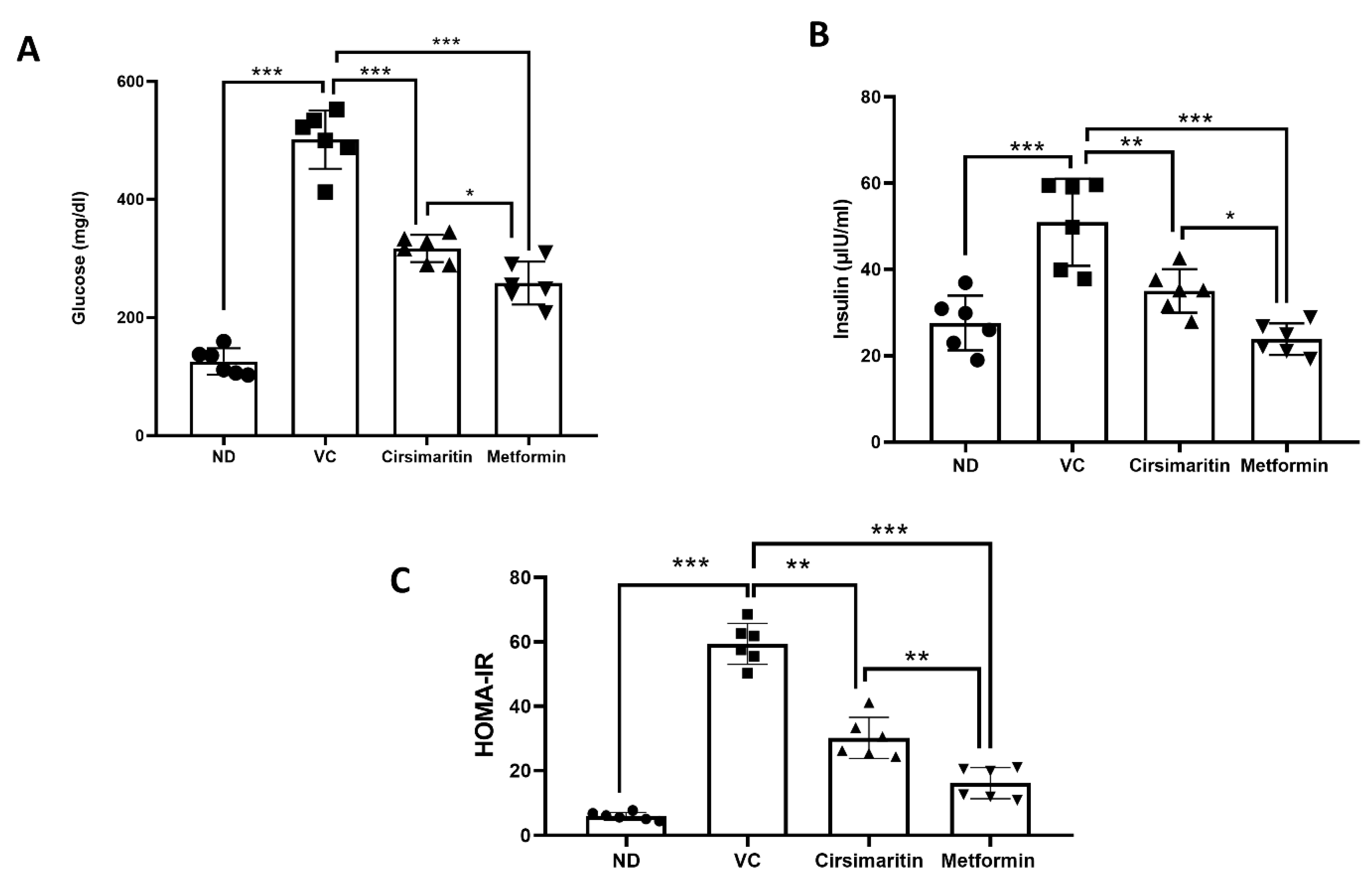
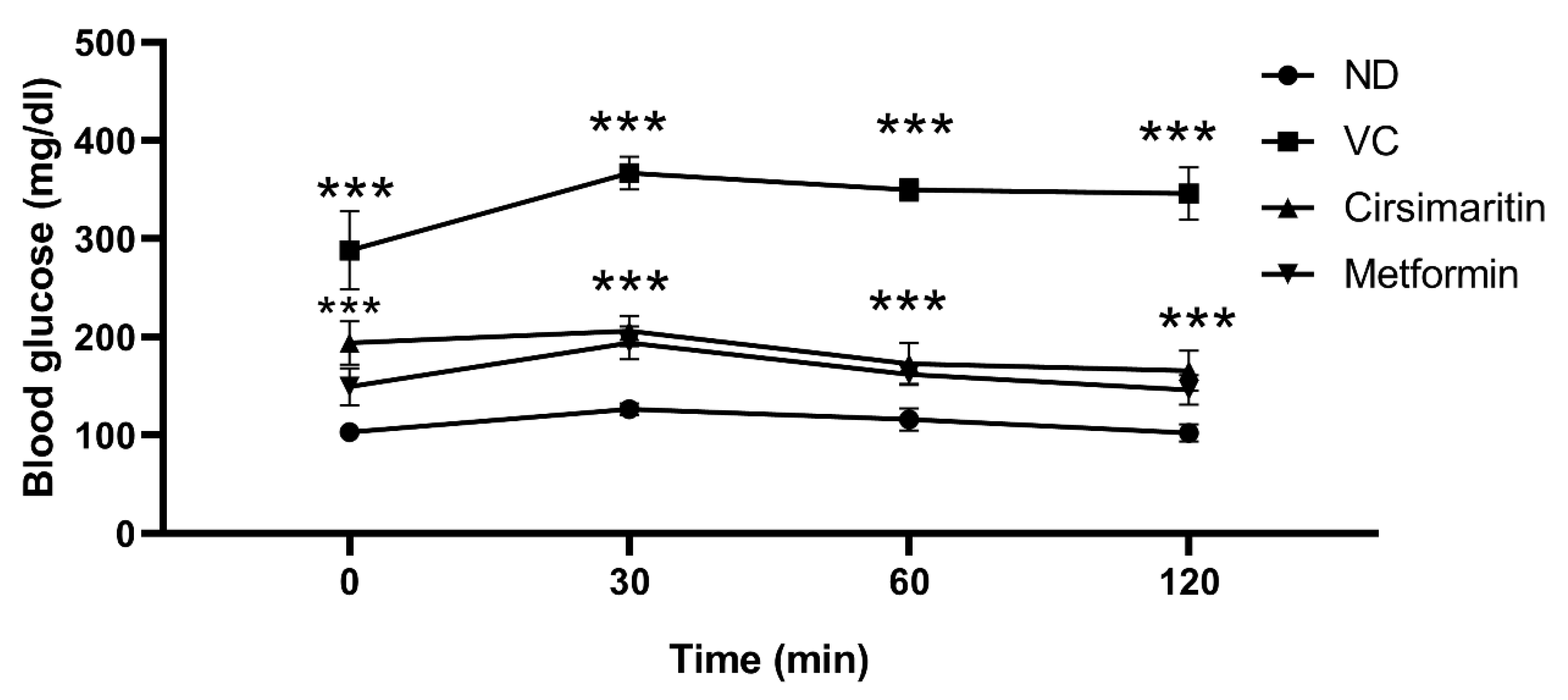
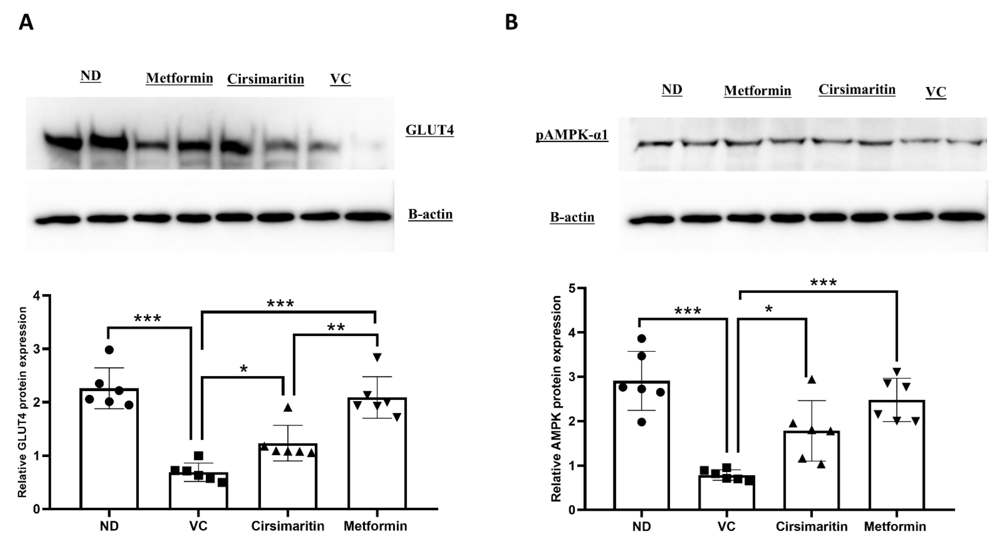
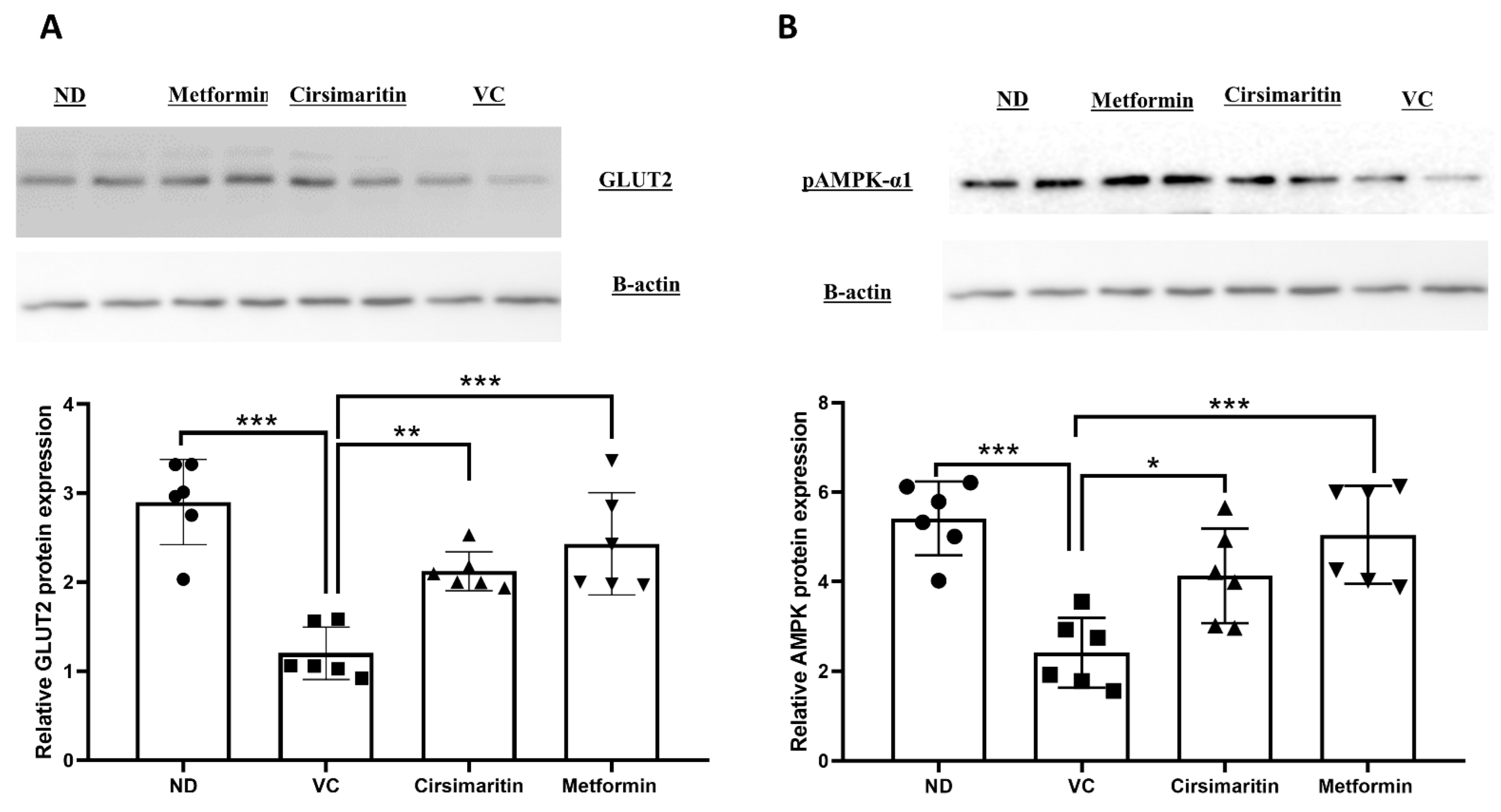
2.1. The Hypoglycemic Effect of Cirsimaritin

Serum glucose was significantly higher in the vehicle control diabetic group compared to the non-diabetic group (Figure 2A, n = 6, p < 0.001). Treatment with cirsimaritin significantly reduced serum glucose concentrations compared to the vehicle controls in the presence of T2D (Figure 2A, n = 6, p < 0.001). Similarly, the mean glucose concentration in the diabetic group was significantly reduced with metformin treatment compared to the vehicle controls (Figure 2A, n = 6, p < 0.01). However, glucose concentration was significantly reduced with metformin treatment compared to cirsimaritin treatment (Figure 2A, n = 6, p < 0.05). Insulin levels were significantly increased in the vehicle control diabetic group compared to the non-diabetic group (Figure 2B, n = 6, p < 0.001); however, treating diabetic rats with cirsimaritin significantly reduced the insulin compared to the vehicle control group (Figure 2B, n = 6, p < 0.01). Like cirsimaritin, treating diabetic rats with metformin significantly reduced insulin levels compared to the vehicle control group (Figure 2B, n = 6, p < 0.001). However, metformin significantly reduced insulin levels compared to the cirsimaritin-treated group (Figure 2B, n = 6, p < 0.05).
To determine the effects of cirsimaritin on insulin resistance, HOMA-IR was measured. The presence of T2D was confirmed via HOMA-IR, which significantly increased in the vehicle control diabetic group compared to the non-diabetic group (Figure 2C, n = 6, p < 0.001). Interestingly, cirsimaritin was able to restore HOMA-IR in the diabetic group, which was comparable to the non-diabetic group. The same effect on HOMA-IR was observed when diabetic rats were treated with metformin. HOMA-IR was significantly reduced with metformin treatment compared to cirsimaritin treatment (Figure 2C, n = 6, p < 0.01). Moreover, the blood glucose level during IPGTT was significantly lower in the cirsimaritin and metformin groups compared to the vehicle control (Figure 3, n = 6, p < 0.001).
To determine the mechanism by which cirsimaritin improves blood glucose and insulin resistance, GLUT4 and phosphorylated AMPK (pAMPK-α1) protein expression were measured in skeletal muscle tissue. GLUT4 protein expression was significantly downregulated in the presence of T2D (Figure 4A, n = 6, p < 0.001); however, treating diabetic rats with cirsimaritin significantly upregulated GLUT4 expression compared to the vehicle control diabetic group (Figure 4A, n = 6, p < 0.01). Metformin was also able to upregulate GLUT4 expression compared to the vehicle control diabetic group (Figure 4A, n = 6, p < 0.001). Metformin treatment was able to upregulate GLUT4 expression significantly higher compared to cirsimaritin treatment (Figure 4A, n = 6, p < 0.05). Similarly, pAMPK-α1 expression was significantly downregulated as a result of T2D (Figure 4B, n = 6, p < 0.001), which was abrogated with cirsimaritin or metformin (Figure 4B, n = 6, p < 0.05, < 0.001, respectively). pAMPK-α1 was significantly higher with metformin treatment compared to cirsimaritin treatment (Figure 4B, n = 6, p < 0.05).
Moreover, GLUT4 and pAMPK-α1 protein expression in adipose tissue were measured. GLUT4 protein expression was significantly downregulated in the presence of T2D (Figure 5A, n = 6, p < 0.001); whereas the administration of cirsimaritin significantly upregulated GLUT4 expression compared to the vehicle controls (Figure 5A, n = 6, p < 0.05). Metformin was also able to upregulate GLUT4 expression compared to the vehicle control diabetic group (Figure 5A, n = 6, p < 0.01). GLUT4 expression in adipose tissue was significantly upregulated with the metformin treatment compared to the cirsimaritin treatment (Figure 5A, n = 6, p < 0.001). Similarly, pAMPK-α1 expression was significantly downregulated as a result of T2D (Figure 5B, n = 6, p < 0.001), and treating diabetic rats with either cirsimaritin or metformin significantly upregulated pAMPK-α1 expression compared to the vehicle control diabetic group (Figure 5B, n = 6, p < 0.05, < 0.001, respectively). No difference was observed in pAMPK-α1 expression between the cirsimaritin and metformin groups.
To further elucidate the mechanism by which cirsimaritin improves insulin sensitivity and reduces glucose levels, hepatic glucose transporter 2 (GLUT2) and pAMPK-α1 protein expression were measured. GLUT2 protein expression was significantly downregulated in the presence of T2D (Figure 6A, n = 6, p < 0.001), whereas cirsimaritin significantly upregulated GLUT2 expression compared to the vehicle control diabetic group (Figure 6A, n = 6, p < 0.01). Metformin was also able to upregulate GLUT2 expression compared to the vehicle controls (Figure 6A, n = 6, p < 0.001). Similarly, pAMPK-α1 expression was significantly downregulated as a result of T2D (Figure 6B, n = 6, p < 0.001), and treating diabetic rats with either cirsimaritin or metformin significantly upregulated pAMPK-α1 expression compared to the vehicle controls (Figure 6B, n = 6, p < 0.05, < 0.001, respectively). No difference was observed in GLUT2 and pAMPK-α1 expression between the cirsimaritin and metformin groups.

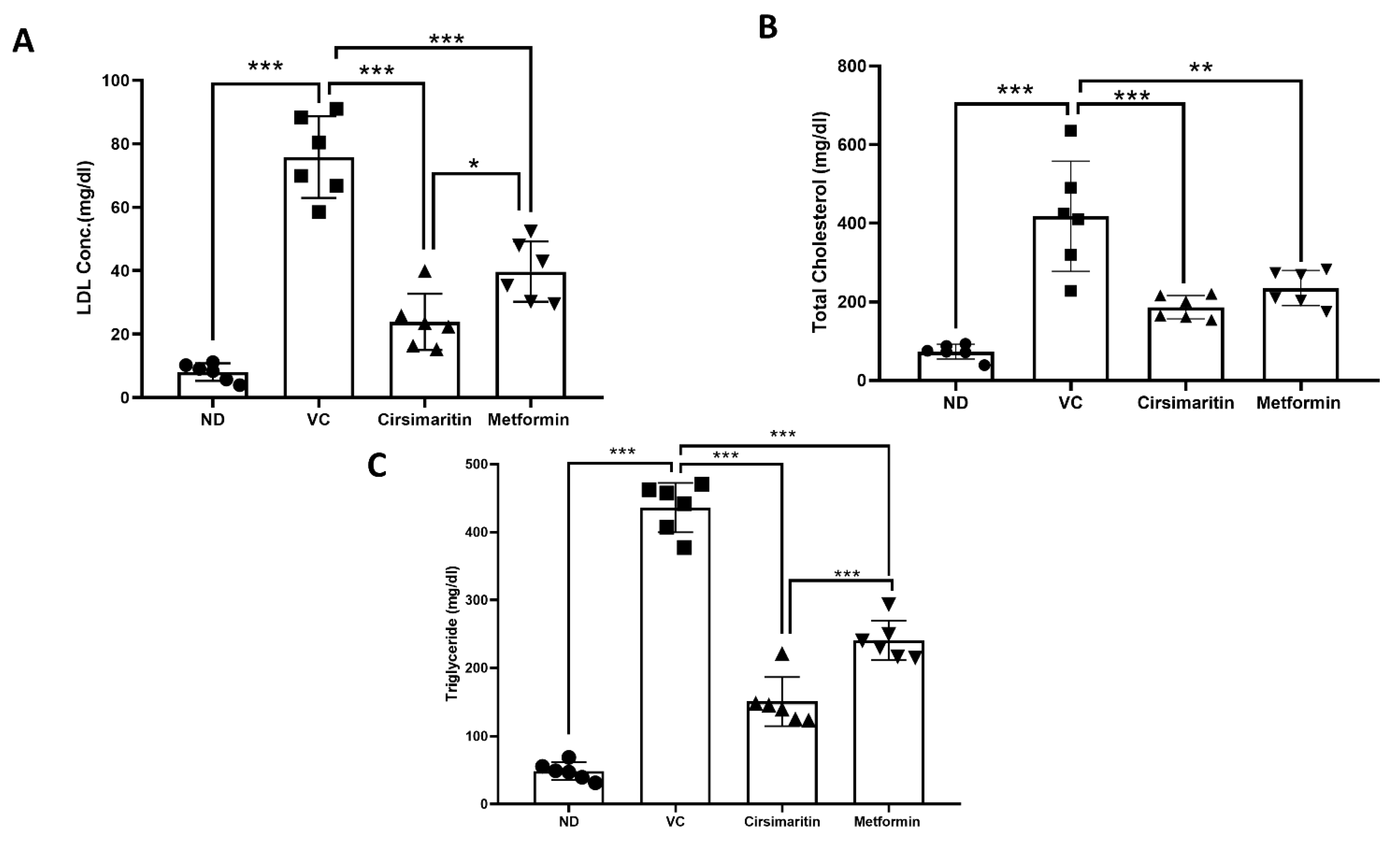
2.2. The Effect of Cirsimaritin on the Lipid Profile

As depicted in Figure 7, dyslipidemia was present in diabetic rats. LDL (Figure 7A, n = 6, p < 0.001), total cholesterol (Figure 7B, n = 6, p < 0.05), and triglycerides (TGs; Figure 7C, n = 6, p < 0.001) were significantly higher in the vehicle control diabetic group, compared to the non-diabetic group. Treating diabetic rats with cirsimaritin significantly reduced serum LDL (Figure 7A, n = 6, p < 0.001), total cholesterol (Figure 7B, n = 6, p < 0.001), and TGs (Figure 7C, n = 6, p < 0.001) systemic concentration, compared to the vehicle control diabetic group. Similarly, metformin was able to significantly reduce LDL (Figure 7A, n = 6, p < 0.001), cholesterol (Figure 7B, n = 6, p < 0.01), and TGs (Figure 7C, n = 6, p < 0.001) levels in diabetic rats, compared to the vehicle controls. Interestingly, LDL and TG systemic concentrations were significantly lower in the cirsimaritin group compared to the metformin group (Figure 7A,C, n = 6, p < 0.05, <0.001, respectively).

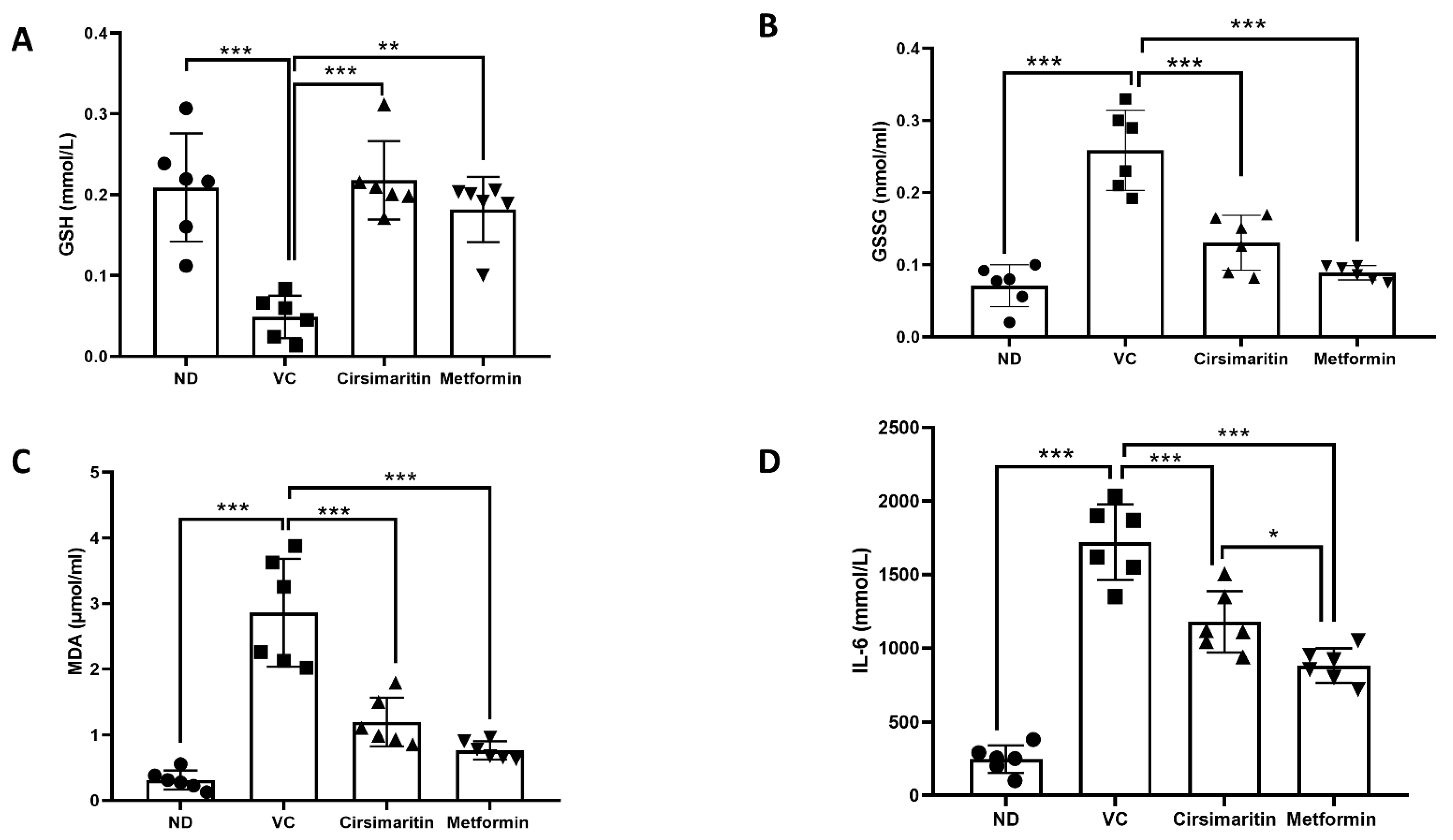
2.3. The Effects of Cirsimaritin on GSH, GSSG, MDA, and IL-6

Serum GSH expression was significantly reduced in vehicle-control diabetic rats, compared to non-diabetic rats (Figure 8A, n = 6, p < 0.001); however, treating diabetic rats with either cirsimaritin or metformin demonstrated a significant increase in GSH compared to the vehicle control diabetic group (Figure 8A, n = 6, p < 0.001, < 0.01, respectively). Moreover, serum GSSG was significantly increased in vehicle-control diabetic rats, compared to non-diabetic rats (Figure 8B, n = 6, p < 0.001); however, cirsimaritin and metformin were able to reduce GSSG levels in diabetic rats, compared to the vehicle control (Figure 8B, n = 6, p < 0.001). On the other hand, serum MDA (Figure 8C, n = 6, p < 0.001) and IL-6 (Figure 8D, n = 6, p < 0.001) concentrations were significantly increased in the presence of T2D. Interestingly, cirsimaritin showed an ability to reduce serum MDA and IL-6 levels significantly in diabetic rats, in comparison to the vehicle control group (Figure 8C,D, n = 6, p < 0.001). The same effect was observed when diabetic rats were treated with metformin (Figure 8C,D, n = 6, p < 0.001). The IL-6 systemic level was significantly lower in the metformin group compared to the cirsimaritin group (Figure 8D, n = 6, p < 0.05).

3. Discussion

Food value is linked to nutritional contents and digestibility, as well as the presence or absence of toxic ingredients [28]. Indeed, the bionutrients that foods offer are essential for life, but also, foods possess other bioactive compounds that are important for health promotion and disease prevention [29]. The consumption of a healthy diet is strongly correlated with a reduction in the risk of several chronic diseases such as cancer, diabetes, cardiovascular diseases and atherosclerosis, neurodegenerative disorders, and inflammation, as well as their complications [30]. These therapeutic properties of food gave rise to medicinal drugs made from certain kinds of food, particularly plants. Numerous medicinal plants have been largely used in the treatment of several diseases, such as cardiovascular diseases [31], cancer [32], diabetes [33], inflammation [34], depression [35,36], and pain management [37]. Therefore, the aim of this study was to assess, for the first time, the antidiabetic efficacy of flavonoid compound, cirsimaritin.
The findings of our study revealed that cirsimaritin significantly reduced glucose, insulin, and HOMA-IR in an HFD/STZ-induced diabetic rat model. In addition, cirsimaritin improved insulin resistance by upregulating GLUT4 and AMPK expression in soleus muscle and adipose tissue. Furthermore, cirsimaritin upregulated GLUT2 and AMPK expression in the liver. Moreover, cirsimaritin exerts antioxidant and anti-inflammatory effects.
Hyperglycemia and insulin resistance are hallmarks of T2D, and they are implicated in T2D complications such as nephropathy, neuropathy, and retinopathy [38]. Our findings demonstrated that cirsimaritin reduced glucose, insulin, and HOMA-IR. This finding is consistent with the previous literature [39] where cirsimaritin enhanced the glucose uptake rate in diabetic mice hepatocytes.
Skeletal muscles and adipose tissue consume a high portion of blood glucose. Several chronic conditions and mechanisms underlie insulin resistance (IR) in these tissues [40,41]. Many studies have linked insulin resistance to impaired GLUT4-mediated glucose uptake [42,43,44]. According to animal models, the reduction in GLUT4 expression was 50% in the skeletal muscles of the hypertriglyceridemia insulin resistance mouse model [43]. Moreover, a 70% reduction in GLUT4 protein expression in the adipose-specific genetic knockout mouse model was associated with insulin resistance [45]. The activation of the AMPK-GLUT4 pathway enhances insulin sensitivity, and it has been shown to improve glucose control in T2D [46,47]. Moreover, GLUT2 is responsible for glucose uptake in the liver, and it is required for the physiological control of glucose-sensitive genes. The inactivation of GLUT2 in the liver leads to impaired glucose-stimulated insulin secretion [13,48]. The findings of the present study demonstrated that cirsimaritin-improved insulin resistance is mediated by the activation of the AMPK-GLUT4 pathway in the skeletal and adipose tissues, and by the activation of the AMPK-GLUT2 in the liver.
Dyslipidemia is tightly associated with T2D, and it is a major risk factor leading to T2D-associated complications [49]. Several studies have linked dyslipidemia to microvascular complications associated with T2D, such as diabetic retinopathy, nephropathy, and neuropathy [50]. The present study showed that cirsimaritin improved the lipid profile in high-fat diabetic rats. Interestingly, cirsimaritin-treated animals demonstrated lower LDL-C and TG levels after 10 days of treatment, compared to the vehicle control group. To our knowledge, this is the first study that shows the lipid-lowering effects of pure cirsimaritin. Previous studies using extracted flavonoids, including cirsimaritin, failed to demonstrate similar findings [25]. Although the exact mechanism is yet to be unrevealed, however, these findings highlights could pave the way to further investigate cirsimaritin in T2D and dyslipidemia.
Oxidative stress is implicated in the early stages of T2D [51]. Oxidative stress is referred to as an overproduction of reactive oxygen species (ROS), and a reduction in the rate of antioxidant defense mechanisms such as GSH (a non-enzymatic antioxidant) [52]. Additionally, ROS induces the release of MDA, a highly reactive compound that interacts with proteins and nucleic acids, and that causes damage to various tissues and cells [53]. MDA has been used as a biomarker of lipid peroxidation and as an indication of free radical damage in the blood [54]. Our findings indicate that similar to metformin, cirsimaritin was able to decrease elevated MDA levels in the high-fat T2D rat model. Furthermore, cirsimaritin was able to restore the GSH/GSSG balance that was disturbed in the high-fat T2D rat model. Taken together, these findings suggest a powerful antioxidant effect of cirsimaritin. Numerous studies have pointed out the antioxidant effects of cirsimaritin in different assays, as revised in [55]. In those studies, cirsimaritin extracted from Teucrium ramosissimum showed an excellent antioxidant activity, using ABTS assay with a Teac value of 2.04 μM. In addition, cirsimaritin extracted from Cirsium japonicum inhibited DPPH free radicals, with a percentage of between 80% to 100% at a dose of 100 μg/mL. Moreover, cirsimaritin extracted from Combretum fragrans showed potent DPPH radicals scavenging activity. It is postulated that as a flavonoid, cirsimaritin has a broad range of biological actions that could be related to the direct scavenging of ROS [55].
Subclinical chronic inflammation has been implicated in the development and progression of insulin resistance, T2D, and its complications [56]. In particular, the increased levels of the multifunctional cytokine, IL-6, have been linked to the pathogenesis of T2D. Our findings demonstrate that cirsimaritin reduced the circulating IL-6 levels with respect to the untreated group. Previous evidence has shown that cirsimaritin inhibited the production of several cytokines such as IL-6 and TNF-α via transcriptional factor-mediated mechanisms that downregulate gene expression [24].
The limitations of this study include the following aspects: (i) cirsimaritin was administered for a short period of time, and (ii) GLUT4 expression was assessed using immunoblotting that was reflective of its total amount; however, immunohistochemistry may be a better technique for assessing its activity and translocation to the cell membrane. Nevertheless, our findings in this study indicate the important role of cirsimaritin in improving the typical features of T2D.

4. Materials and Methods

4.1. The Induction of T2D and Experimental Design

Animal experimental procedures were approved by the animal ethics committee at the Hashemite University (IRB number: 14/4/2021/2022, 14 April 2022), and were in accordance with the guidelines of the US National Institutes of Health on the use and care of laboratory animals, and with the Animal Research: Reporting of in Vivo Experiments (ARRIVE) guidelines (https://arriveguid elines.org, accessed on 2 May 2022).
Thirty-week-old male adult Sprague-Dawley rats (average weight 264 ± 3.5) were maintained under standard conditions, including 12 h light/dark cycles and a 22 ± 2° temperature [57]. T2D was induced by feeding the experimental rats HFD (60% fat) for 3 weeks, followed by one intraperitoneal injection of streptozotocin (STZ; 40 mg/kg) [58]. The average weight after diabetes induction for the mice fed with HFD was 328.4 ± 4.4. One week after STZ injection, plasma glucose was measured, and rats with a plasma glucose concentration over 200 mg/dL were considered to have developed T2D, and were selected for the subsequent experiments.
Rats were randomly divided into four groups (n = 6 each), as follows: (i) normal non-diabetic control group (non-diabetic, ND) receiving normal diet, (ii) vehicle control (VC) diabetic group treated with dimethyl sulfoxide (DMSO, Panreac Quimica SA, Barcelona, Spain) only, (iii) diabetic group treated with 50 mg/kg cirsimaritin (Sigma-Aldrich, Dorset, UK), and (iv) diabetic group treated with 200 mg/kg metformin (MeRCK, Darmstadt, Germany); the dose of metformin was in line with a previous study that used 200 mg/kg metformin for the treatment of diabetic rats [59]. The dose of cirsimaritin used in this study was based on a previous report showing that the doses between 50–200 mg/kg did not produce any noticeable side effects; thus, the lowest dose (50 mg/kg) was chosen for this study [60]. All treatments were given orally once per day. After 10 days of treatment, rats were euthanized as per local standard operating procedures, using carbon dioxide before blood and skeletal muscle (soleus muscle), adipose tissue, and liver were collected for ex-vivo analysis.

4.2. Biochemical Investigations

4.2.1. Measurements of Serum Glucose, Insulin, and Lipids in Rat Serum

Serum glucose and insulin were determined using a commercial kits glucose assay kit (Mybiosource, San Diego, CA, USA), and a rat insulin ELISA kit (Mybiosource, San Diego, CA, USA), respectively, as per the manufacturer’s instructions. Triglyceride (TG, triglyceride assay kit), low-density lipoproteins (LDL, LDL assay kit), and cholesterol (Total Cholesterol assay kit) were also measured using commercially available kits (MybioSource, San Diego, CA, USA) according to the manufacturer’s instructions.

4.2.2. Homeostasis Model Assessment of Insulin Resistance (HOMA-IR)

This model represents the interaction between fasting plasma insulin and fasting plasma glucose, which is a useful tool for determining insulin resistance. In the current study, we used the following formula to compute HOMA-IR [61]:
HOMA-IR = ( F a s t i n g   g l u c o s e   ( m g / d L ) × F a s t i n g   i n s u l i n   ( μ I U / m L ) / 405

4.2.3. Intraperitoneal Glucose Tolerance Test

Rats were given an intraperitoneal injection of glucose (0.5 g/kg) after being fasted for 18 h. Using a glucometer, blood glucose levels were measured from the tail vein at 0, 30, 60, and 120 min (Accu-Check Performa, Roche Diagnostics).

4.2.4. Measurement of Serum Glutathione (GSH), Oxidized Glutathione (GSSG), Malondialdehyde (MDA), and IL-6 Serum Concentrations

Reduced glutathione (GSH, GSH assay kit), oxidized glutathione (GSSG, GSSG assay kit), and IL-6 (IL-6 ELISA kit) levels were measured in the serum using commercially available kits (Mybiosource, San Diego, CA, USA). Plasma MDA level was determined by using a commercially available thiobarbituric acid (TBA) Assay Kit (Mybiosource, San Diego, CA, USA) according to the manufacturer’s instructions.

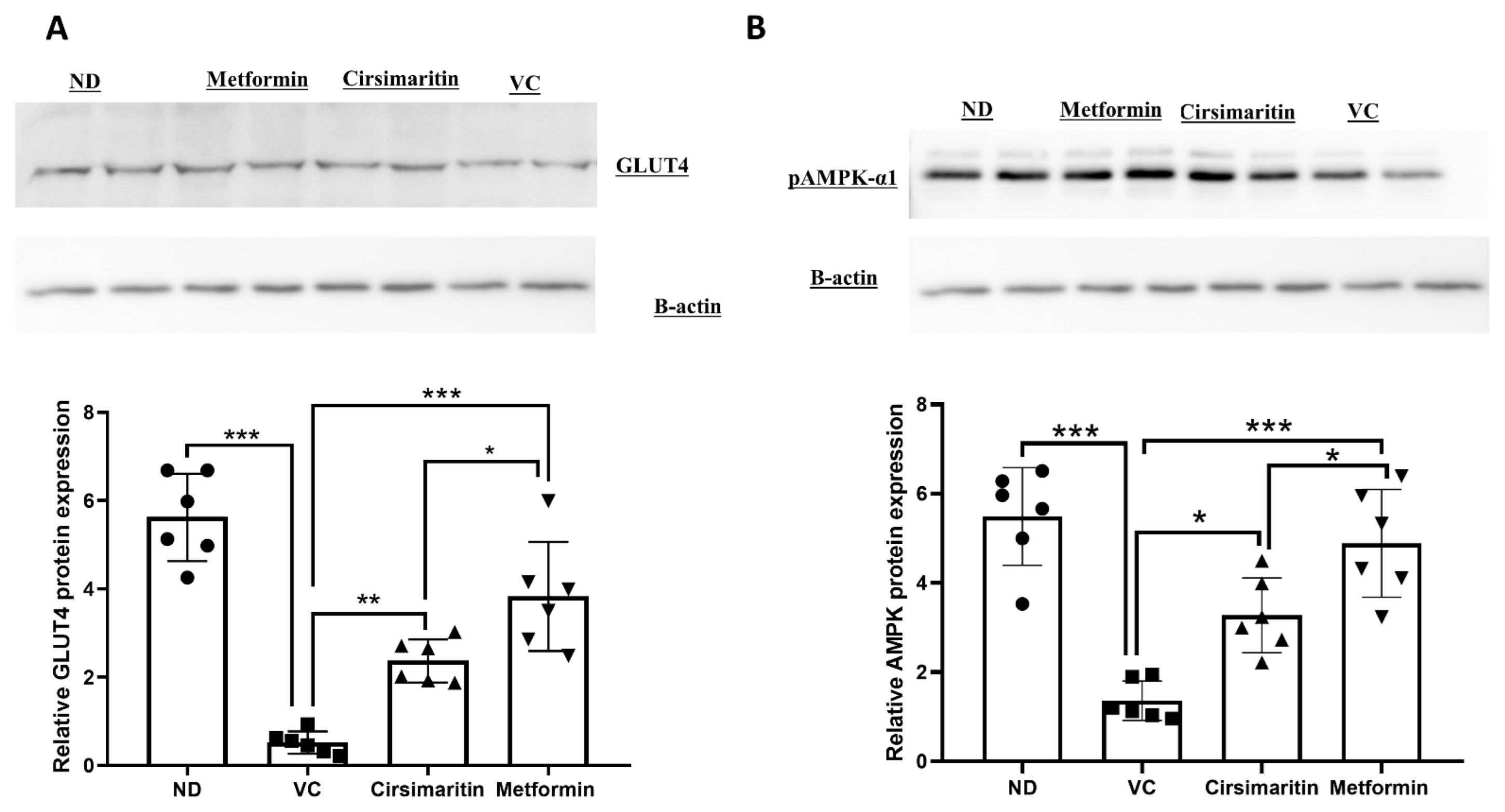
4.3. Western Blotting

Skeletal muscle tissues (soleus muscle), adipose tissue, and liver were homogenized in radioimmunoprecipitation (RIPA)-lysis buffer, containing a protease inhibitor cocktail (Santa Cruz Biotechnology, USA), using a tissue homogenizer. Homogenates were centrifuged at 13,000 rpm for 20 min at 4 °C, and the supernatant was collected. Total protein was quantified using a bicinchoninic acid assay (Bioquochem, Austurias, Spain). Equal amounts of protein were separated using a sodium dodecyl sulfate–polyacrylamide gel, then transferred onto a nitrocellulose membrane (Thermo Fisher Scientific, Carlsbad, CA, USA). The membrane was blocked for 1 h at room temperature using 3% bovine serum albumin (BSA), before incubating overnight with either pAMPK-α1 (Abcam, Cambridge, UK), GLUT2, or GLUT4 (Mybiosource, San Diego, CA, USA) primary antibodies (1:1000 dilution). The membrane was washed three time with a washing buffer (Tween-20/Tris-buffered saline) before incubating it with the goat-anti-rabbit secondary antibody (Mybiosource, San Diego, CA, USA, 1:5000 dilution) for 1 h at room temperature. Following incubation, the membrane was washed three times before submerging it into the ECL substrate (ThermoScientific, Carlsbad, CA, USA) for one minute, followed by imaging with the chemiLITE Chemiluminescence Imaging System (cleaverscientific, Rugby, UK). To ensure equal protein gel loading, β-actin was used as a housekeeping gene (Mybiosource, San Diego, CA, USA, 1:10,000 dilution). The intensities of the bands were measured using Image J software, and adjusted to β-actin.

4.4. Statistical Analysis

All analyzed parameters were tested for the normality of the data using the Kolmogorov-Smirnov test. Data are represented as mean ± SEM. Differences between groups were calculated using a one-way analysis of variance (ANOVA) or two-way ANOVA, followed by a Tukey post hoc test using PGraphPad Prism software version (9.3.1). The significance value of difference was considered when the p-value was less than 0.05.

5. Conclusions

Our findings in this study revealed that cirsimaritin might be a very useful agent for the treatment of T2D due to its ability to reduce insulin resistance, and its activation of the GLUT4-AMPK and GLUT2-AMPK pathways in skeletal muscle, adipose tissue, and liver. In addition, cirsimaritin could be a useful agent for improving lipid profiles and for reducing oxidative stress. Cirsimaritin effects and mechanisms were like metformin, the gold standard for T2D treatment.

Author Contributions

Conceptualization, A.A. and E.Q.; methodology, O.G., M.O. and R.Y.A.; software, H.A.A.; validation, A.A. and E.Q.; formal analysis, O.G.; investigation, N.A.-H.; resources, E.Q.; data curation, M.A.; writing—original draft preparation, A.A.; writing—review and editing, N.A.-H.; visualization, E.Q.; supervision, O.G.; project administration, R.A.; funding acquisition, E.Q. All authors have read and agreed to the published version of the manuscript.

Funding

This research was funded by the Deanship of Scientific Research at The Hashemite University, grant number 16/11/200950.

Institutional Review Board Statement

The study was conducted in accordance with the Declaration of Helsinki, and approved by the Institutional Review Board of the Hashemite University (IRB number: 14/4/2021/2022, 14 April 2022).

Informed Consent Statement

Not applicable.

Data Availability Statement

The datasets generated during and/or analyzed during the current study are available from the corresponding author upon reasonable request.

Acknowledgments

The authors would like to thank the Deanship of Scientific Research at the Hashemite University for sponsoring this research.

Conflicts of Interest

The authors declare that there are no conflict of interest.

References

  1. Rehman, K.; Akash, M.S.H. Mechanism of Generation of Oxidative Stress and Pathophysiology of Type 2 Diabetes Mellitus: How Are They Interlinked? J. Cell. Biochem. 2017, 118, 3577–3585. [Google Scholar] [CrossRef] [PubMed]
  2. Stumvoll, M.; Goldstein, B.J.; Van Haeften, T.W. Type 2 Diabetes: Principles of Pathogenesis and Therapy. Lancet 2005, 365, 1333–1346. [Google Scholar] [CrossRef]
  3. Badawi, A.; Klip, A.; Haddad, P.; Cole, D.E.; Bailo, G.; El-Sohemy, A.; Karmali, M. Type 2 Diabetes Mellitus and Inflammation: Prospects for Biomarkers of Risk and Nutritional Intervention. Diabetes Metab. Syndr. Obes. Targets Ther. 2010, 3, 173–186. [Google Scholar] [CrossRef] [Green Version]
  4. Zimmet, P.; Alberti, K.G.M.M.; Shaw, J. Global and Societal Implications of the Diabetes Epidemic. Nature 2001, 414, 782–787. [Google Scholar] [CrossRef]
  5. Alberti, K.G.M.M. Treating Type 2 Diabetes—Today’s Targets, Tomorrow’s Goals. Diabetes Obes. Metab. 2001, 3, 3–10. [Google Scholar] [CrossRef]
  6. Pickup, J.C. Inflammation and Activated Innate Immunity in the Pathogenesis of Type 2 Diabetes. Diabetes Care 2004, 27, 813–823. [Google Scholar] [CrossRef] [PubMed] [Green Version]
  7. Lemieux, I.; Pascot, A.; Prud’homme, D.; Alméras, N.; Bogaty, P.; Nadeau, A.; Bergeron, J.; Després, J.P. Elevated C-Reactive Protein. Arter. Thromb. Vasc. Biol. 2001, 21, 961–967. [Google Scholar] [CrossRef] [Green Version]
  8. Halliwell, B. The Wanderings of a Free Radical. Free Radic. Biol. Med. 2009, 46, 531–542. [Google Scholar] [CrossRef]
  9. Lee, Y.J.; Suh, K.S.; Choi, M.C.; Chon, S.; Oh, S.; Woo, J.T.; Kim, S.W.; Kim, J.W.; Kim, Y.S. Kaempferol Protects HIT-T15 Pancreatic Beta Cells from 2-Deoxy-D-Ribose-Induced Oxidative Damage. Phytother. Res. 2010, 24, 419–423. [Google Scholar] [CrossRef]
  10. Eriksson, J.W. Metabolic Stress in Insulin’s Target Cells Leads to ROS Accumulation—A Hypothetical Common Pathway Causing Insulin Resistance. FEBS Lett. 2007, 581, 3734–3742. [Google Scholar] [CrossRef] [PubMed] [Green Version]
  11. Kawahito, S.; Kitahata, H.; Oshita, S. Problems Associated with Glucose Toxicity: Role of Hyperglycemia-Induced Oxidative Stress. World J. Gastroenterol. 2009, 15, 4137–4142. [Google Scholar] [CrossRef] [PubMed]
  12. Ji, L.L.; Reid, M.; Sandra Butler-Browne, G.; Llanos, P.; Palomero, J. Reactive Oxygen and Nitrogen Species (RONS) and Cytokines&mdash;Myokines Involved in Glucose Uptake and Insulin Resistance in Skeletal Muscle. Cells 2022, 11, 4008. [Google Scholar] [CrossRef]
  13. Chadt, A.; Al-Hasani, H. Glucose Transporters in Adipose Tissue, Liver, and Skeletal Muscle in Metabolic Health and Disease. Pflug. Arch. Eur. J. Physiol. 2020, 472, 1273–1298. [Google Scholar] [CrossRef] [PubMed]
  14. Alqudah, A.; Qnais, E.Y.; Wedyan, M.A.; Altaber, S.; Abudalo, R.; Gammoh, O.; Alkhateeb, H.; Bataineh, S.; Athamneh, R.Y.; Oqal, M.; et al. New Treatment for Type 2 Diabetes Mellitus Using a Novel Bipyrazole Compound. Cells 2023, 12, 267. [Google Scholar] [CrossRef]
  15. Alqudah, A.; Qnais, E.Y.; Wedyan, M.A.; Altaber, S.; Bseiso, Y.; Oqal, M.; AbuDalo, R.; Alrosan, K.; Alrosan, A.Z.; Bani Melhim, S.; et al. Isorhamnetin Reduces Glucose Level, Inflammation, and Oxidative Stress in High-Fat Diet/Streptozotocin Diabetic Mice Model. Molecules 2023, 28, 502. [Google Scholar] [CrossRef]
  16. Vlavcheski, F.; Den Hartogh, D.J.; Giacca, A.; Tsiani, E. Amelioration of High-Insulin-Induced Skeletal Muscle Cell Insulin Resistance by Resveratrol Is Linked to Activation of AMPK and Restoration of GLUT4 Translocation. Nutrients 2020, 12, 914. [Google Scholar] [CrossRef] [Green Version]
  17. Giacoman-Martínez, A.; Alarcón-Aguilar, F.J.; Zamilpa, A.; Huang, F.; Romero-Nava, R.; Román-Ramos, R.; Almanza-Pérez, J.C. α-Amyrin Induces Glut4 Translocation Mediated by AMPK and PPARδ/γ in C2c12 Myoblasts. Can. J. Physiol. Pharmacol. 2021, 99, 935–942. [Google Scholar] [CrossRef] [PubMed]
  18. Dayarathne, L.A.; Ranaweera, S.S.; Natraj, P.; Rajan, P.; Lee, Y.J.; Han, C.H. The Effects of Naringenin and Naringin on the Glucose Uptake and AMPK Phosphorylation in High Glucose Treated HepG2 Cells. J. Vet. Sci. 2021, 22, e92. [Google Scholar] [CrossRef]
  19. Park, J.Y.; Kim, H.Y.; Shibamoto, T.; Jang, T.S.; Lee, S.C.; Shim, J.S.; Hahm, D.H.; Lee, H.J.; Lee, S.; Kang, K.S. Beneficial Effects of a Medicinal Herb, Cirsium japonicum Var. Maackii, Extract and Its Major Component, Cirsimaritin on Breast Cancer Metastasis in MDA-MB-231 Breast Cancer Cells. Bioorg. Med. Chem. Lett. 2017, 27, 3968–3973. [Google Scholar] [CrossRef]
  20. Pathak, G.; Singh, S.; Kumari, P.; Raza, W.; Hussain, Y.; Meena, A. Cirsimaritin, a Lung Squamous Carcinoma Cells (NCIH-520) Proliferation Inhibitor. J. Biomol. Struct. Dyn. 2020, 39, 3312–3323. [Google Scholar] [CrossRef]
  21. Rijo, P.; Simões, M.F.; Duarte, A.; Rodríguez, B. Isopimarane Diterpenoids from Aeollanthus rydingianus and Their Antimicrobial Activity. Phytochemistry 2009, 70, 1161–1165. [Google Scholar] [CrossRef]
  22. Ibañez, E.; Kubátová, A.; Señoráns, F.J.; Cavero, S.; Reglero, U.; Hawthorne, S.B. Subcritical Water Extraction of Antioxidant Compounds from Rosemary Plants. J. Agric. Food Chem. 2002, 51, 375–382. [Google Scholar] [CrossRef]
  23. Jipa, S.; Zaharescu, T.; Kappel, W.; Dǎneţ, A.F.; Popa, C.V.; Bumbac, M.; Gorghiu, L.M.; Maris, A.M. The Effects of γ-Irradiation on the Antioxidant Activity of Rosemary Extract. Optoelectron. Adv. Mater. Rapid Commun. 2009, 3, 1315–1320. [Google Scholar]
  24. Shin, M.S.; Park, J.Y.; Lee, J.; Yoo, H.H.; Hahm, D.H.; Lee, S.C.; Lee, S.; Hwang, G.S.; Jung, K.; Kang, K.S. Anti-Inflammatory Effects and Corresponding Mechanisms of Cirsimaritin Extracted from Cirsium japonicum Var. Maackii Maxim. Bioorg. Med. Chem. Lett. 2017, 27, 3076–3080. [Google Scholar] [CrossRef]
  25. Stefkov, G.; Kulevanova, S.; Miova, B.; Dinevska-Kjovkarovska, S.; Mølgaard, P.; Jäger, A.K.; Josefsen, K. Effects of Teucrium polium Spp. Capitatum Flavonoids on the Lipid and Carbohydrate Metabolism in Rats. Pharm. Biol. 2011, 49, 885–892. [Google Scholar] [CrossRef] [Green Version]
  26. Bower, A.M.; Real Hernandez, L.M.; Berhow, M.A.; De Mejia, E.G. Bioactive Compounds from Culinary Herbs Inhibit a Molecular Target for Type 2 Diabetes Management, Dipeptidyl Peptidase IV. J. Agric. Food Chem. 2014, 62, 6147–6158. [Google Scholar] [CrossRef] [PubMed]
  27. Xu, J.H.; Lo, Y.M.; Chang, W.C.; Huang, D.W.; Wu, J.S.B.; Jhang, Y.Y.; Huang, W.C.; Ko, C.Y.; Shen, S.C. Identification of Bioactive Components from Ruellia tuberosa L. On Improving Glucose Uptake in TNF-α-Induced Insulin-Resistant Mouse FL83B Hepatocytes. Evid.-Based Complement. Altern. Med. 2020, 2020, 6644253. [Google Scholar] [CrossRef] [PubMed]
  28. Newell-McGloughlin, M. Nutritionally Improved Agricultural Crops. Plant Physiol. 2008, 147, 939–953. [Google Scholar] [CrossRef] [PubMed]
  29. Liu, R.H. Dietary Bioactive Compounds and Their Health Implications. J. Food Sci. 2013, 78, A18–A25. [Google Scholar] [CrossRef]
  30. Pandey, K.B.; Rizvi, S.I. Plant Polyphenols as Dietary Antioxidants in Human Health and Disease. Oxid. Med. Cell. Longev. 2009, 2, 270–278. [Google Scholar] [CrossRef] [Green Version]
  31. Michel, J.; Abd Rani, N.Z.; Husain, K. A Review on the Potential Use of Medicinal Plants from Asteraceae and Lamiaceae Plant Family in Cardiovascular Diseases. Front. Pharmacol. 2020, 11, 852. [Google Scholar] [CrossRef]
  32. Gezici, S.; Şekeroğlu, N. Current Perspectives in the Application of Medicinal Plants against Cancer: Novel Therapeutic Agents. Anticancer Agents Med. Chem. 2019, 19, 101–111. [Google Scholar] [CrossRef] [PubMed]
  33. Nazarian-Samani, Z.; Sewell, R.D.E.; Lorigooini, Z.; Rafieian-Kopaei, M. Medicinal Plants with Multiple Effects on Diabetes Mellitus and Its Complications: A Systematic Review. Curr. Diab. Rep. 2018, 18, 72. [Google Scholar] [CrossRef] [PubMed]
  34. Tasneem, S.; Liu, B.; Li, B.; Choudhary, M.I.; Wang, W. Molecular Pharmacology of Inflammation: Medicinal Plants as Anti-Inflammatory Agents. Pharmacol. Res. 2019, 139, 126–140. [Google Scholar] [CrossRef]
  35. Dalmagro, A.P.; Camargo, A.; Zeni, A.L.B. Morus nigra and Its Major Phenolic, Syringic Acid, Have Antidepressant-like and Neuroprotective Effects in Mice. Metab. Brain Dis. 2017, 32, 1963–1973. [Google Scholar] [CrossRef]
  36. Dantas, D.M.M.; Cahu, T.B.; Oliveira, C.Y.B.; Abadie-Guedes, R.; Roberto, N.A.; Santana, W.M.; Galvez, A.O.; Guedes, R.C.A.; Bezerra, R.S. Chlorella vulgaris Functional Alcoholic Beverage: Effect on Propagation of Cortical Spreading Depression and Functional Properties. PLoS ONE 2021, 16, e0255996. [Google Scholar] [CrossRef]
  37. Dutta, T.; Paul, A.; Majumder, M.; Sultan, R.A.; Emran, T. Bin Pharmacological Evidence for the Use of Cissus Assamica as a Medicinal Plant in the Management of Pain and Pyrexia. Biochem. Biophys. Rep. 2020, 21, 100715. [Google Scholar] [CrossRef]
  38. Fowler, M.J. Microvascular and Macrovascular Complications of Diabetes. Clin. Diabetes 2008, 26, 77–82. [Google Scholar] [CrossRef] [Green Version]
  39. Herman, R.; Kravos, N.A.; Jensterle, M.; Janež, A.; Dolžan, V. Metformin and Insulin Resistance: A Review of the Underlying Mechanisms behind Changes in GLUT4-Mediated Glucose Transport. Int. J. Mol. Sci. 2022, 23, 1264. [Google Scholar] [CrossRef]
  40. Garneau, L.; Aguer, C. Role of Myokines in the Development of Skeletal Muscle Insulin Resistance and Related Metabolic Defects in Type 2 Diabetes. Diabetes Metab. 2019, 45, 505–516. [Google Scholar] [CrossRef] [PubMed]
  41. Ahmed, B.; Sultana, R.; Greene, M.W. Adipose Tissue and Insulin Resistance in Obese. Biomed. Pharmacother. 2021, 137, 111315. [Google Scholar] [CrossRef]
  42. Leguisamo, N.M.; Lehnen, A.M.; Machado, U.F.; Okamoto, M.M.; Markoski, M.M.; Pinto, G.H.; Schaan, B.D. GLUT4 Content Decreases along with Insulin Resistance and High Levels of Inflammatory Markers in Rats with Metabolic Syndrome. Cardiovasc. Diabetol. 2012, 11, 100. [Google Scholar] [CrossRef] [Green Version]
  43. Seböková, E.; Klimes, I.; Moss, R.; Mitková, A.; Wiersma, M.; Bohov, P. Decreased Glucose Transporter Protein (GLUT4) in Skeletal Muscle of Hypertriglyceridaemic Insulin-Resistant Rat. Physiol. Res. 1995, 44, 87–92. [Google Scholar]
  44. Napoli, R.; Hirshman, M.F.; Horton, E.S. Mechanisms and Time Course of Impaired Skeletal Muscle Glucose Transport Activity in Streptozocin Diabetic Rats. J. Clin. Investig. 1995, 96, 427–437. [Google Scholar] [CrossRef] [PubMed]
  45. Abel, E.D.; Peroni, O.; Kim, J.K.; Kim, Y.B.; Boss, O.; Hadro, E.; Minnemann, T.; Shulman, G.I.; Kahn, B.B. Adipose-Selective Targeting of the GLUT4 Gene Impairs Insulin Action in Muscle and Liver. Nature 2001, 409, 729–733. [Google Scholar] [CrossRef] [PubMed]
  46. Viollet, B.; Lantier, L.; Devin-Leclerc, J.; Hebrard, S.; Amouyal, C.; Mounier, R.; Foretz, M.; Andreelli, F. Targeting the AMPK Pathway for the Treatment of Type 2 Diabetes. Front. Biosci. 2009, 9, 3380–3400. [Google Scholar] [CrossRef] [PubMed] [Green Version]
  47. Asrafuzzaman, M.; Rahman, M.M.; Mandal, M.; Marjuque, M.; Bhowmik, A.; Rokeya, B.; Hassan, Z.; Faruque, M.O. Oyster Mushroom Functions as an Anti-Hyperglycaemic through Phosphorylation of AMPK and Increased Expression of GLUT4 in Type 2 Diabetic Model Rats. J. Taibah Univ. Med. Sci. 2018, 13, 465–471. [Google Scholar] [CrossRef] [PubMed]
  48. Thorens, B. GLUT2, Glucose Sensing and Glucose Homeostasis. Diabetologia 2015, 58, 221–232. [Google Scholar] [CrossRef] [Green Version]
  49. Eid, S.; Sas, K.M.; Abcouwer, S.F.; Feldman, E.L.; Gardner, T.W.; Pennathur, S.; Fort, P.E. New Insights into the Mechanisms of Diabetic Complications: Role of Lipids and Lipid Metabolism. Diabetologia 2019, 62, 1539–1549. [Google Scholar] [CrossRef] [Green Version]
  50. Athyros, V.G.; Doumas, M.; Imprialos, K.P.; Stavropoulos, K.; Georgianou, E.; Katsimardou, A.; Karagiannis, A. Diabetes and Lipid Metabolism. Hormones 2018, 17, 61–67. [Google Scholar] [CrossRef] [Green Version]
  51. Oguntibeju, O.O. Type 2 Diabetes Mellitus, Oxidative Stress and Inflammation: Examining the Links. Int. J. Physiol. Pathophysiol. Pharmacol. 2019, 11, 45–63. [Google Scholar]
  52. Chikezie, P.C.; Ojiako, O.A.; Ogbuji, A.C. Oxidative Stress in Diabetes Mellitus. Int. J. Biol. Chem. 2015, 9, 92–109. [Google Scholar] [CrossRef] [Green Version]
  53. Su, L.J.; Zhang, J.H.; Gomez, H.; Murugan, R.; Hong, X.; Xu, D.; Jiang, F.; Peng, Z.Y. Reactive Oxygen Species-Induced Lipid Peroxidation in Apoptosis, Autophagy, and Ferroptosis. Oxid. Med. Cell. Longev. 2019, 2019, 5080843. [Google Scholar] [CrossRef] [Green Version]
  54. Morales, M.; Munné-Bosch, S. Malondialdehyde: Facts and Artifacts. Plant Physiol. 2019, 180, 1246–1250. [Google Scholar] [CrossRef] [Green Version]
  55. Benali, T.; Jaouadi, I.; Ghchime, R.; El Omari, N.; Harboul, K.; Hammani, K.; Rebezov, M.; Shariati, M.A.; Mubarak, M.S.; Simal-Gandara, J.; et al. The Current State of Knowledge in Biological Properties of Cirsimaritin. Antioxidants 2022, 11, 1842. [Google Scholar] [CrossRef] [PubMed]
  56. Esser, N.; Legrand-Poels, S.; Piette, J.; Scheen, A.J.; Paquot, N. Inflammation as a Link between Obesity, Metabolic Syndrome and Type 2 Diabetes. Diabetes Res. Clin. Pract. 2014, 105, 141–150. [Google Scholar] [CrossRef] [PubMed] [Green Version]
  57. Al-Ghaithi, F.; El-Ridi, M.R.; Adeghate, E.; Amiri, M.H. Biochemical Effects of Citrullus colocynthis in Normal and Diabetic Rats. Mol. Cell. Biochem. 2004, 261, 143–149. [Google Scholar] [CrossRef] [PubMed]
  58. De MagalhÃes, D.A.; Kume, W.T.; Correia, F.S.; Queiroz, T.S.; Allebrandt Neto, E.W.; Dos Santos, M.P.; Kawashita, N.H.; De França, S.A. High-Fat Diet and Streptozotocin in the Induction of Type 2 Diabetes Mellitus: A New Proposal. An. Acad. Bras. Cienc. 2019, 91, e20180314. [Google Scholar] [CrossRef] [Green Version]
  59. Al-Trad, B.; Alkhateeb, H.; Alsmadi, W.; Al-Zoubi, M. Eugenol Ameliorates Insulin Resistance, Oxidative Stress and Inflammation in High Fat-Diet/Streptozotocin-Induced Diabetic Rat. Life Sci. 2019, 216, 183–188. [Google Scholar] [CrossRef]
  60. Abdelhalim, A.; Karim, N.; Chebib, M.; Aburjai, T.; Khan, I.; Johnston, G.A.R.; Hanrahan, J.R. Antidepressant, Anxiolytic and Antinociceptive Activities of Constituents from Rosmarinus Officinalis. J. Pharm. Pharm. Sci. 2015, 18, 448–459. [Google Scholar] [CrossRef] [Green Version]
  61. Yoon, H.; Jeon, D.J.; Park, C.E.; You, H.S.; Moon, A.E. Relationship between Homeostasis Model Assessment of Insulin Resistance and Beta Cell Function and Serum 25-Hydroxyvitamin D in Non-Diabetic Korean Adults. J. Clin. Biochem. Nutr. 2016, 59, 139–144. [Google Scholar] [CrossRef] [PubMed] [Green Version]
Figure 1. Chemical structure of cirsimaritin.
Ijms 24 05749 g001
Figure 2. The anti-diabetic effect of cirsimaritin. Cirsimaritin significantly reduced glucose (A) and insulin (B) levels in diabetic rats. HOMA-IR (C) was significantly reduced with cirsimaritin treatment. Rats were fed HFD for 3 weeks, followed by a single dose of STZ injection (40 mg/kg); and once diabetes was confirmed, rats were treated with 50 mg/kg cirsimaritin or 200 mg/kg metformin for 10 days. After the end of the experiment, serum was collected for ELISA analyses. One-way ANOVA was followed by Tukey post hoc multiple comparison test, * p < 0.05, ** p < 0.01, *** p < 0.001. ND; non-diabetic, VC; vehicle control.
Ijms 24 05749 g002
Figure 3. Cirsimaritin reduced glucose levels during the intraperitoneal glucose tolerance test (IPGTT). Rats were fed with HFD for 3 weeks, followed by a single dose of STZ injection (40 mg/kg); once diabetes was confirmed, rats were treated with 50 mg/kg cirsimaritin or 200 mg/kg metformin for 10 days. Rats then fasted overnight before injection with 0.5 g/kg glucose intraperitoneally, and glucose levels were determined at 0, 30, 60, and 120 min. Two-way ANOVA followed by Tukey post hoc multiple comparison test, *** p < 0.001. ND; non-diabetic, VC; vehicle control.
Ijms 24 05749 g003
Figure 4. Cirsimaritin upregulated GLUT4 and pAMPK-α1 expression in the soleus muscle. Cirsimaritin significantly upregulated GLUT4 (A) and pAMPK-α1 (B) expression within soleus muscle in diabetic rats. Rats were fed HFD for 3 weeks, followed by a single dose of STZ injection (40 mg/kg); once diabetes was confirmed, rats were treated with 50 mg/kg cirsimaritin or 200 mg/kg metformin for 10 days. Rats were then sacrificed and after the end of the experiment following euthanasia, soleus muscle was isolated and homogenized for downstream Western blotting. One-way ANOVA was followed by Tukey post hoc multiple comparison test, * p < 0.05, ** p < 0.01, *** p < 0.001. ND; non-diabetic, VC; vehicle control.
Ijms 24 05749 g004
Figure 5. Cirsimaritin upregulated GLUT4 and pAMPK-α1 expression in adipose tissue. Cirsimaritin significantly upregulated adipose tissue GLUT4 (A) and pAMPK-α1 (B) expression in diabetic rats. Rats were fed HFD for 3 weeks, followed by a single dose of STZ injection (40 mg/kg); after diabetes was confirmed, rats were treated with 50 mg/kg cirsimaritin or 200 mg/kg metformin for 10 days. At the end of the experiment following euthanasia, adipose tissue was isolated and homogenized before Western blotting was performed. One-way ANOVA was followed by Tukey post hoc multiple comparison test, * p < 0.05, ** p < 0.01, *** p < 0.001. ND; non-diabetic, VC; vehicle control.
Ijms 24 05749 g005
Figure 6. Cirsimaritin upregulated GLUT2 and pAMPK-α1 expression in the liver. Cirsimaritin significantly upregulated hepatic GLUT2 (A) and pAMPK-α1 (B) expression in diabetic rats. Rats were fed HFD for 3 weeks, followed by a single dose of STZ injection (40 mg/kg); and once diabetes was confirmed, rats were treated with 50 mg/kg cirsimaritin or 200 mg/kg metformin for 10 days. At the end of the experiment following euthanasia, the liver was isolated and homogenized before Western blotting performed. One-way ANOVA was followed by Tukey post hoc multiple comparison test, * p < 0.05, ** p < 0.01, *** p < 0.001. ND; non-diabetic, VC; vehicle control.
Ijms 24 05749 g006
Figure 7. Cirsimaritin improves lipid profile in diabetes. Cirsimaritin significantly reduced LDL (A), total cholesterol (B), and triglyceride (C) systemic concentrations in diabetic rats. Rats were fed HFD for 3 weeks, followed by a single dose of STZ injection (40 mg/kg), and once diabetes was confirmed, rats were treated with 50 mg/kg cirsimaritin or 200 mg/kg metformin for 10 days. At the end of the experiment following euthanasia, serum was collected for ELISA analyses. One-way ANOVA was followed by Tukey post hoc multiple comparison test, * p < 0.05, ** p < 0.01, *** p < 0.001. ND; non-diabetic, VC; vehicle control.
Ijms 24 05749 g007
Figure 8. The antioxidant and anti-inflammatory effect of cirsimaritin. Cirsimaritin significantly increased GSH (A) and reduced GSSG (B), and reduced MDA (C) and IL-6 (D) systemic concentrations in diabetic rats. Rats were fed HFD for 3 weeks, followed by a single dose of STZ injection (40 mg/kg), and once diabetes was confirmed, rats were treated with 50 mg/kg cirsimaritin or 200 mg/kg metformin for 10 days. After the end of the experiment following euthanasia, serum was collected for ELISA analysis. One-way ANOVA was followed by Tukey post hoc multiple comparison test, * p < 0.05, ** p < 0.01, *** p < 0.001. ND; non-diabetic, VC; vehicle control.
Ijms 24 05749 g008
Disclaimer/Publisher’s Note: The statements, opinions and data contained in all publications are solely those of the individual author(s) and contributor(s) and not of MDPI and/or the editor(s). MDPI and/or the editor(s) disclaim responsibility for any injury to people or property resulting from any ideas, methods, instructions or products referred to in the content.

