Review

Hepatic Energy Metabolism under the Local Control of the Thyroid Hormone System

1
Charité-Universitätsmedizin Berlin, Corporate Member of Freie Universität Berlin, Humboldt-Universität zu Berlin, and Berlin Institute of Health, Institut für Experimentelle Endokrinologie, 10115 Berlin, Germany
2
Charité-Universitätsmedizin Berlin, Corporate Member of Freie Universität Berlin, Humboldt-Universität zu Berlin, and Berlin Institute of Health, Department of Endocrinology and Metabolism, 10115 Berlin, Germany
3
Charité-Universitätsmedizin Berlin, Corporate Member of Freie Universität Berlin, Humboldt-Universität zu Berlin, and Berlin Institute of Health, Department of Surgery, 13353 Berlin, Germany
4
NutriAct-Competence Cluster Nutrition Research Berlin-Potsdam, 14558 Nuthetal, Germany
5
Charité-Universitätsmedizin Berlin, Corporate Member of Freie Universität Berlin, Humboldt-Universität zu Berlin, and Berlin Institute of Health, Department of Hepatology & Gastroenterology, Campus Virchow-Klinikum (CVK) and Campus Charité Mitte (CCM), 13353 Berlin, Germany
6
DZHK (German Centre for Cardiovascular Research), Partner Site Berlin, 10115 Berlin, Germany
*
Author to whom correspondence should be addressed.
These authors contributed equally to this work.
Int. J. Mol. Sci. 2023, 24(5), 4861; https://doi.org/10.3390/ijms24054861
Submission received: 7 December 2022 / Revised: 24 February 2023 / Accepted: 25 February 2023 / Published: 2 March 2023
(This article belongs to the Special Issue Local Control of Thyroid Hormone Action)

Abstract

:
The energy homeostasis of the organism is orchestrated by a complex interplay of energy substrate shuttling, breakdown, storage, and distribution. Many of these processes are interconnected via the liver. Thyroid hormones (TH) are well known to provide signals for the regulation of energy homeostasis through direct gene regulation via their nuclear receptors acting as transcription factors. In this comprehensive review, we summarize the effects of nutritional intervention like fasting and diets on the TH system. In parallel, we detail direct effects of TH in liver metabolic pathways with regards to glucose, lipid, and cholesterol metabolism. This overview on hepatic effects of TH provides the basis for understanding the complex regulatory network and its translational potential with regards to currently discussed treatment options of non-alcoholic fatty liver disease (NAFLD) and non-alcoholic steatohepatitis (NASH) involving TH mimetics.

1. Introduction

Thyroid hormones (THs) are well known for their important regulatory functions during development and growth. Further early descriptions of TH actions point to the role of THs supporting homeostasis in metabolic pathways through enhancing and diminishing energy consumption from different dietary sources like carbohydrates and lipids. In addition, effects of THs in mitochondrial biogenesis and activation have been described in various models and species.
TH concentrations, mainly those of thyroxine (T4) and 3,3’,5’-triiodothyronine (T3), are assessed and accounted for mainly in the circulation where they are distributed throughout an organism. Their production and release from the thyroid gland is regulated via feedforward and feedback mechanisms within the hypothalamus-pituitary-thyroid (HPT) axis. Circulating concentrations of THs often reflect the production and release of THs mainly from the thyroid gland. However, TH actions are executed on a local tissue level within cells of the organism through direct binding to TH receptors (TRs), TRα and TRβ, acting as nuclear transcription factors (type 1 signaling) or by type 3 signaling activating signaling cascades (for example, phosphatidylinositol 3-kinase/PI3K) [1]. TR modulated gene expression in hepatocytes is mediated via TRβ, which also mediates TH signals in the HPT axis, while, for example, heart rate and brain development are mainly steered via TRα. The process of pre-receptor control determining the local TH availability within each cell is mediated by the uptake of THs into cells through transmembrane transporters and via local activation and inactivation through deiodinases (Dio).
The eminent role of TH transmembrane transporters became obvious with the discovery of human mutations in the most specific TH transmembrane transporter, the monocarboxylate transporter (MCT) 8. These lead to the Allan-Herndon-Dudley syndrome, which is characterized by psycho-motor retardation along with high T3 and low T4 serum concentrations in patients [2,3]. MCT8 is expressed in a variety of tissues, including the brain, kidneys, and liver. In patients with mutations in MCT8 and mouse models with Mct8 deficiency, despite the loss of Mct8 function, the liver can take up the high circulating T3 concentrations and presents a local hyperthyroid status. This indicates the presence of further TH transmembrane transporters in the liver. Transmembrane transporters that can take up THs are, for example, L-type amino acid transporters, Oatps, Mct10, and the sodium-taurocholate transporter Ntcp, which is highly expressed in the liver (since Ntcp is a major bile acid transporter). Therefore, the liver is obviously exposed to alterations in circulating TH concentrations.
Concentrations of TH in the liver are not only mediated by the uptake of circulating TH but also by local activation and inactivation through Dios. Dios are a group of selenoproteins that are responsible for the local activation and inactivation of TH metabolites via deiodination (removal of one iodide) within cells. The three different Dios are capable of reductively removing iodide from different positions within the TH molecules (for a detailed review, see [4]). While Dio2 activity is functionally relevant in the liver during development [5], the adult liver of rodents and humans displays high Dio1 activity. Dio1 activity is regulated by the presence of T3. However, Dio1 itself is involved in the activation of T4 to T3. On the other hand, it locally inactivates TH, while especially TH sulfates are substrates with high affinity [6]. Injuries to the liver, inflammation, and fasting conditions can lead to the induction of Dio3. While Dio1 is expressed in hepatocytes, up to date, it is still not known in which cells of the liver this induction takes place. While liver deiodinases regulate local availability, they also contribute to changes in circulating TH concentrations with varying contributions between different species. Additionally, the liver has a major impact on transport and stabilization of circulating TH concentrations by secreting the plasma distribution proteins albumin (binding 10–15% of T4 and 10% of T3), transthyretin (TTR, binding 10–15% of T4 and 10% of T3), and thyroxin binding globulin (TBG, binding 70% of both T4 and T3), which steady the equilibrium of free-to-total TH. The majority of THs are carried by TBG due to their much higher affinity, although TTR and albumin are much more abundant in plasma.
While regulation of energy homeostasis in an organism is achieved by the complex interplay between different energy storing and metabolizing organs like brown and white adipose tissue, muscle, and liver, local TH action differentially affects these processes within cells of an organ. Different cell types of the same organ can even have distinct responses to local TH availability, while circulating concentrations of THs are the same throughout the organism. An example can be given through differential local T3 availability in skeletal muscle cells. Upon muscle injury, Dio3 is induced in satellite cells which promote muscle regeneration, while its inactivation leads to satellite cell apoptosis, hampering regeneration [7]. Further evaluation on a single-cell basis led to the identification of specific muscle cell subsets with differential expression and regulation of Dio2 and Dio3 upon injury and regeneration [8]. Therefore, the evaluation of local TH availability and action within an organ remains crucial. With regards to different liver cell types, the expression of Dio1, as well as TRβ, has been described many times. However, expressions of transmembrane transporters for TH, Dios, and TRs remain elusive for Kupffer and hepatic stellate cells, which also play major roles in the development of NAFLD and NASH.
In the adult organism, THs systemically regulate the energy metabolism in oxidative tissues most noticeably, so more energy-rich compounds must be supplied or mobilized from storage sites for energy production. Therefore, it is not surprising that the liver is a target organ of endocrine signaling of the TH axis, complementing the peripheral TH action. On the other hand, the liver itself is an active player in modulating TH concentrations locally and systemically by providing or removing active TH from circulation. In particular, local hepatic T3 availability orchestrates carbohydrate, lipid, cholesterol, and bile acid biogenesis at the level of gene expression, translation, and enzyme function, together with other hormones, signaling molecules, and transcription factors of nutritional regulation.
In this review, we will evaluate and summarize data from animal models and cell culture systems with regards to the impact of THs exerted on liver physiology and metabolism. To delineate between data generated in animals vs. human cell culture systems, genes and proteins of animal origin are written with only the first letter capitalized, while human genes and proteins are written in all uppercase letters. Genes are depicted in italics. Nomenclature of human and mouse genes is in accordance with HGNC and MGI. In some cases, we included data derived from human studies to either illustrate known similarities or differences between the different systems needed for the mechanistic understanding of treatment possibilities in humans. Consequences of supplying or restricting different nutrients on regulation of TH system-related genes and proteins in the liver will be discussed. In addition, we will also focus on the regulation of genes and enzymes by THs that are involved in carbohydrate, lipid, and bile acid metabolism in the liver.

2. Diet-Induced Alterations of the TH System

2.1. Fasting and Energy Restriction

Fasting influences the regulation of the hypothalamus-pituitary-thyroid (HPT) axis in various species. These physiological alterations in TH synthesis, metabolism, and action might be needed to preserve energy due to a low nutrient supply shutting down stimulatory effects of THs on energy consumption in various tissues. During fasting, a gradual decrease of serum T3 and T4 concentrations is observed, while thyroid-stimulating hormone (TSH) concentrations remain unaltered. These alterations are linked to rapidly decreased activity of hepatic Dio already observed in rat models undergoing fasting and refeeding before the individual contributions of the three Dio isoenzymes to systemic and local TH provision are distinguished [9,10] (for a recent review, see Russo et al. [11]). The data are summarized in Table 1 and Table 2.

2.1.1. Systemic Alterations

According to van der Wal et al. [15], fasting resulted in a decrease of serum T3 concentrations after 12 h and serum T4 concentrations after 48 h in rats, whilst serum TSH remained constant. Lower serum concentrations of T3 and T4 (some even undetectable) were also reported upon 48-h fasting in mice [13,14] and in rats [18]. Similarly, 36-h fasting also led to decreased serum T3 and T4 concentrations in male rats [16,17] (Table 1), while three weeks of food restriction (50% of their individual baseline 24 h intake) led to a decrease only in T4 but not in T3 [17] (Table 2). Additionally, Visser et al. [21] also described that three-day fasting caused a decrease in serum T3 and T4 concentrations in both male and female rats, while TSH concentrations only decreased in male rats. Three-week food restriction (one-third of normal food intake) led to significantly lower serum T4, T3, and TSH concentrations in both male and female rats. Similar data on T4 have also been reported by Giacco et al. [20]. In a more recent study from 2020 [14], in accordance with previously reported data, 24-h fasting caused a remarkable decline in serum T4 and T3 concentrations in 12-week-old male mice.
Moreover, fasting also affects binding of TH to plasma distribution proteins. Young et al. reported that T4 bound to Tbg and albumin increased during four-day and seven-day fasting in lean rats, while T4 bound to Ttr (designated as thyroxine-binding prealbumin, TBPA) was reduced. In obese rats, Tbg-bound T4 constantly increased up to 28 days already from day 4 upon fasting, whilst Ttr bound T4 steadily dropped and albumin bound T4 remained unaffected [22]. The fasting-induced generation of serum Tbg may account for these alterations. Moreover, serum Ttr decreased significantly during fasting, proportionally to the duration (one-, two-, and three-day fasting) in rats, accompanied by a decreased T4-bound fraction [23].

2.1.2. Local Alterations in Liver

Fasting for 48 h caused a lower T3 generation rate from T4 in rat liver homogenate, indicating reduced Dio1 activity [18]. In the rat liver, T3 was lower in both 36-h fasting and three-week food restriction, whilst T4 diminished only with three-week food restriction (50% of their individual baseline 24-h intake) [17]. In mice, hepatic T4 decreased during 16- and 36-h fasting and T3 decreased upon 28- and 36-h fasting [12]. In agreement with these results, upon 24-h fasting, mice showed a decrease in hepatic T4 and T3 concentrations [14]. In male rats, hepatic TH contents were unaffected after 36-h fasting according to de Vries et al. [16]. The expression of hepatic TRs was not affected during fasting and food restriction, while the expression of TH responsive genes fatty acid synthase (Fasn) and Spot14 was lower during fasting [17]. In addition, van der Wal et al. [15] showed that decreased serum T3 led to increased low-density lipoprotein (Ldl) cholesterol from 24 h onwards, associated with a lower liver Ldl receptor mRNA (Ldlr). Serum triglyceride (TG) content decreased, while serum free fatty acid (FFA) concentrations increased. Timing of fasting-induced alterations in TH availability therefore differs between species.
The clearance of THs plays a major role in regulating the energy metabolism in a state of hunger, which is possibly responsible for the drop in TH concentrations. There are three major pathways of TH metabolism in the liver that are involved in this clearance of TH upon fasting: deiodination, sulfation, and glucuronidation.
Deiodination contributes to the modification of TH bioactivity and availability locally via deiodinases [24,25,26]. There are two different isoforms, Dio1 and Dio3, which are capable of inactivating TH. Although Dio1 is believed to be the major source of circulating T3 in humans, the enzyme displays high affinity towards reverse T3 (rT3; 3,3′,5′-triiodo-L-thyronine) and sulfate conjugates of TH [19]. It is evident that hepatic Dio1 activity is significantly lower in both male and female rats after three-day fasting, as well as after three weeks of food restriction (one-third of normal food intake) [21]. In mice, a fasted liver showed a decrease in Dio1 activity compared with a liver in a fed state [12]. In contrast, Dio3 mRNA expression and activity was higher during 36-h fasting and three-week food restriction (50% of their individual baseline 24-h intake), while Dio1 activity remained unchanged despite a lower mRNA expression in mice [16,17]. Thus, the fasting duration and the extent of food restriction play a role in the regulation of local TH concentrations in the liver.
To elucidate how and which deiodinases have effects on the TH concentration in both circulation and in the liver, different knockout mice of deiodinases were examined. Despite the alterations in Dio3 expression and Dio1 activity upon fasting described by de Vries et al. [16,17], Galton et al. [12] pointed out that hepatic Dio3 activity was undetectable upon 30-h fasting in WT mice but showed minimal activity in Dio1/Dio2 double KO (D1/D2KO) mice. In accordance with above-mentioned data, serum T4 and T3 also decreased upon 30-h fasting in WT, D1KO, D2KO, and D1/D2KO mice, while TSH remained unchanged. Although D3KO showed a lower baseline of T4 and T3 than WT, 30-h fasting still caused a decline in serum T4 and T3 for D3KO as well as for WT mice. Upon fasting, rT3 is increased in D1KO and D1/D2KO mice, likely resulting from elevated Dio3 activity. Furthermore, [125I] labeled TH were used as Dio substrates in vivo to sensitively monitor their function during fasting. Mice were injected with either [125I] T4, [125I] T3, or [125I] rT3 on the same day of fasting. As a result, fasted WT mice showed a higher distribution of [125I] T3 in liver (and other tissues) than fed mice. In a fasted state, hepatic [125I] T3, [125I] rT3, and [125I] T4 were generally higher than in a fed state in WT and D1/D2KO mice, which also had increased Dio3 activity indicating alterations in uptake, efflux, and/or metabolism of THs. Taken together, fasting-induced systemic TH changes were not dependent on Dio1 or Dio2 but rather on Dio3 and sequestration of T4 and T3 in tissues but not excretion.
Sulfation and glucuronidation are responsible for marking TH for degradation and enterohepatic recycling, and remarkable substrate preferences for the individual TH metabolites were observed for various members of the enzyme families catalyzing these conjugation reactions [19,27,28]. Sulfate conjugation may lead to the inactivation of TH by Dio1 [29] or reversible inactivation during fetal development or when TH sulfates are cleaved and liberate unconjugated TH during microbiota-dependent enterohepatic recycling [30,31,32] or cleavage by sulfatases expressed in many tissues [33,34,35]. Glucuronidation of iodothyronine facilitates its biliary and urinary excretion (reversible) [36]. Sulfotransferases (Sults) and UDP- glucuronyltransferases (Ugts) are enzymes performing sulfation and glucuronidation. Phenol sulfotransferases, which can make sulfate conjugates out of TH, belong to the Sult1 family, including Sult1a1, 1a2, 1a3, 1b1, and 1c2 [33,34,37,38]. Most UGTs involved in TH degradation are members of the Ugt1a and Ugt2b families [36,39]. It has been reported that hepatic expression of Sultn, Sult1a1, Sult2a1, and Ugt1a1 increased during fasting in mice [40,41]. Reported as bilirubin UGT [21], Ugt1a1 showed higher activity in rats upon both three-day fasting and three weeks of food restriction to one-third of normal food intake. Sult1b1 expression upon fasting and Sult1c1 expression with food restriction were lower in rats [17]. Fasting for 36 h induced gene expression of Ugt1a1, Sult1a1, and Sult1d1 in mice, associated with an upregulation of the constitutive androstane receptor (Car) mRNA expression [16], and 24-h fasting induced similar effects [14].
Car is an upstream regulator of Sult and Ugt expression and a key target of nutrients, nutritional xenobiotics, and drugs interfering with hepatic metabolism both during regular nutrition and steatosis-induced alterations [42]. Nr1l3 (Car) expression was upregulated during 36 h of fasting, whose serval target genes showed an upregulation during fasting to increase TH metabolism [14]. Similar induction of conjugating enzymes was achieved by Car agonist TCPOBOP, while these induced changes were absent in Car-KO mice, which showed similar TH concentrations in fed states compared with WT mice [40]. Car-/- female mice resembled WT mice upon 24-h fasting in TH concentration with lower basal and fasting-induced hepatic T3 concentrations, and they showed an attenuated induction of Ugt1a1, Sult1a1, and Sult1d1. Interestingly, Car-/- mice showed an elevated Dio3 expression and activity compared with WT mice, but the fasting-induced upregulation of Dio3 was absent [14]. Apart from direct effects on hepatic genes related to TH metabolism and conjugation, fasting and altered leptin secretion from white adipocytes also impacted on hypothalamic neuronal circuits regulated by neuropeptide Y (Npy) and melanocortin 4 receptor (Mc4r). Such central hypothalamic inputs are also required for adaptive hepatic responses of T4 metabolizing pathways during fasting [41].
Apart from the local hepatic changes of TH, the uptake into and efflux of TH from the liver may play an important role in regulating systemic and local TH concentrations [43]. Evidently, 48 h of fasting led to decreased uptake of T3 and T4 into the liver, presumably due to depletion of ATP in a perfused rat liver, accompanied by diminished T3 glucuronidation [33,43]. Notably, Mct10 expression was higher during fasting in male rats, while expression of Mct8 remained unchanged during fasting and food restriction [17]. In mice, mRNA expression of hepatic TH transporter Mct10 was enhanced, whilst Mct8 expression decreased [14], similar to data obtained from rats fasting for three days [20]. In addition, 48-h fasting increased hepatic Ntcp mRNA expression, and 72-h fasting significantly enhanced Ntcp protein expression in the liver of rats [44]. Whether fasting-related altered expression of hepatic TH transporter transcripts is similarly reflected in changes of protein content and function, subsequently resulting in variation of the net import or export of TH, remains to be clarified.
Sirt1 is a nuclear deacetylase that is activated upon binding with ligand-bound TRß1, leading to downstream modulation of activities of various gluconeogenic transcription factors/modulators. Cordeiro et al. [13] reported that decreased TH concentrations during fasting resulted in the upregulation of Sirt1 protein and its activity via TRβ, which has beneficial effects upon 48-h fasting, for instance, on life-span extension. Glucose metabolism also plays an important role during nutrient restriction. Studies pointed out that hepatocytes regulated glucose metabolism via the hepatic cAMP/PKA/CREB pathway possibly involving Tsh receptors (Tshrs) [45,46,47]. Studies with cell-type specific knockouts of Tshr in hepatocytes and white adipocytes have been conducted [48,49]. So far, observations reported on the role of TSH and Tshr activation for hepatocyte metabolic function are mainly based on the hepatocyte-specific TshrKO mouse model of one research group [47] and need to be independently confirmed.

2.2. Dietary Interventions

Diet-induced obesity has become increasingly prevalent worldwide in association with additional comorbidities such as diabetes, hyperlipidemia, and cardiovascular disease. Obesity is recognized as a major risk factor for non-alcoholic fatty liver disease (NAFLD) and non-alcoholic steatohepatitis (NASH). The imbalance between energy intake, storage, and expenditure plays a pivotal role in diet-induced obesity. TH are key modulators on energy metabolism, regulating both glucose and lipid metabolism. However, there is little data on the systemic and local regulation of TH during different diets (Table 3).
Gonzalez-Ramos et al. [50] reported that six weeks of a high-fat diet (HFD) (10.2% hydrogenated coconut fat and 0.75% cholesterol) did not alter serum T3 and T4 concentrations in WT and Nod1-/- mice. However, three and six months of HFD with excessive iodine intake (15% lard, 10% yolk powder, and 79% standard laboratory powder chow; with 1200 μg/L iodine in the form of potassium iodate (KIO3) in the drinking water) led to increases of serum T3 and T4 concentrations and a decrease in TSH concentration [51].
In the LoCoTAct consortium and in our lab, we found that HFD induced Dio1 mRNA expression and activity already after four weeks, and this remained elevated for up to 18 weeks induced by HFD [52]. In accordance with these findings, induction of Dio1 mRNA expression and activity was observed in mice fed with a western diet (D12079B; Research Diets), supplemented with 15% weight/volume fructose in drinking water for eight or 16 weeks [53]. However, Han et al. [51] pointed out that only six months of HFD caused elevated hepatic Dio1 activity, while one month of HFD did not show any influence on TH concentrations or Dio1 activity. According to Gonzalez-Ramos et al. [50], although hepatic Dio1 activity was unaffected by HFD, Nod1-/- mice showed significantly lower Dio1 activity independent of diet.
Based on a liver transcriptome analysis [54], only 106 hepatic genes were differently regulated in male mice on a HFD by treatment with the thyromimetic TH metabolite 3,5-T2 (2.5µg/g bw; HFD: 60 kJ% fat; 9% soybean oil, 90% lard, four weeks), while 221 genes responded in mice fed normal chow (ND: 10 kJ% fat, 55% soybean oil, 44% lard). Among these, 56 genes were differently regulated in HFD mice. Strikingly, 12 genes (Cyp1a2, Cyp39a1, Cyp46a1, Cyp51, Cyp2d9, Ces1(f,g) and 2a, Sult1b1, Slc13a3, Slc39a4, Gpx6) involved in xenobiotic metabolism and detoxification were differentially expressed only in HFD mice. Elevated Cyp39a1 expression and reduced Cyp46a1 expression exclusively in HFD mice indicated that 3,5-T2 affected genes involved in bile acid synthesis in obese mice. Treatment with 3,5-T2 altered TH responsive gene expression such as upregulation of Dio1 and downregulation of Serpina7 (Tbg) for both HFD and a regular diet.
In addition to factors that regulate the TH concentrations, expression of genes involved in lipid metabolism is altered in HFD mice. Wu et al. reported that C57BL/6 male mice fed with HFD (45% fat, 35% carbohydrate, 20% protein; 12 weeks) and db/db male mice showed upregulation of thyroid hormone-inducible hepatic protein (Thrsp) expression [55]. Thrsp, similar to the initially discovered rat S14 protein expressed in liver and adipocytes, has been used as a lipogenesis marker and endpoint of T3 action, as it rapidly responds to nutritional changes and is regulated by TH but also by steroids and other hormonal factors [56]. In db/db mice, silencing of the hepatic Thrsp gene led to reduced hepatic TG content and attenuated liver steatosis, while hepatic Thrsp overexpression in C57Bl/6 mice led to increased hepatic TG and cholesterol content, as well as upregulation of lipogenesis genes such as sterol regulatory binding proteins (Srebp) Srebf1, Fasn and Acc, associated with elevated enzyme activity. Fatty acid uptake decreased, and fatty acid oxidation increased, due to enhanced expression of peroxisome proliferator-activated receptor α (Ppara), acyl-CoA oxidase, and peroxisomal ketothiolase. Remarkably, treatment with TO901317 (5 mg/kg/day), a LXR agonist, led to upregulation of hepatic Thrsp expression, mediated by LXRα but not LXRβ [55]. According to Jornayvaz et al., TRα-0/0 mice fed with HFD (three weeks, 54.8% fat, 24% carbohydrate, 21.2% protein, energy density 4.8 Kcal/g) showed a reduction in hepatic lipid intermediates, triglyceride and DAG, as well as a decrease in expression of hepatic lipogenic genes, Srebf1, and its downstream targets, Acc1 and Fasn. Expression of genes involved in lipid oxidation like Fgf21, carnitine palmitoyltransferase 1, acyl-CoA oxidase, and 70-kDa peroxisomal membrane protein mRNA levels was similar to WT [57].
It Is also worth mentioning that the liver is a sexually dimorphic organ [58]. Smati et al. pointed out that male mice are more susceptible to NAFLD. Male mice fed with a high-fat diet for 15 weeks (D12492, Research Diets) showed the highest lipid accumulation, and male mice fed with a western diet for 15 weeks (WD, TD.88137, Envigo) displayed the most severe inflammation/fibrosis. It is possible that the development of the disease may take much longer in females. Nevertheless, transcriptome analysis for different dietary challenges (HFD, Choline deficient-HFD (CDHFD, D05010402, Research Diets), WD and WD with glucose (18.9 g/L) and fructose (23.1 g/L) in drinking water) revealed that hepatocyte Ppara serves as a sexually dimorphic factor in mouse liver [59]. Dio1, which is altered by dietary interventions, underlies together with other selenoproteins sexual dimorphism in mouse kidneys and livers [60].

3. Effects of THs on Metabolic Pathways in the Liver

Circulating as well as local TH concentrations in an organism can be regulated by nutritional conditions. However, local TH also directly affect the regulation of metabolic pathways in the liver. To further elucidate mechanisms of direct canonical and non-canonical TR-mediated TH signaling in energy metabolism, a comprehensive overview of major energy pathways is presented. Since TH signaling is not always an effector in gene regulation, but a master regulator, we also introduce other selected cascades with their signal molecules and transcription factor activation in detail.

3.1. Carbohydrate Metabolism

Carbohydrate metabolism in the liver comprises the major energy generating pathways in the body. In this context, TH regulate basal global energy turnover and reciprocally glucose supply from stores or by recycling of energy-rich compounds in the liver.

3.1.1. T3 Is the Pacemaker for Global Energy Demand

Especially during hyperthyroidism, high T3 concentrations affect glucose utilization in the whole body. One reason for the basally increased cellular energy consumption is the T3-dependent increased membrane potential, built up via Na/K-ATPases, as demonstrated in rat and hepatic rat liver cell lines [61], and by Ca2+ resequestration via Serca in mouse myocytes [62].
The glucose transporter family (Glut) supplies the raised demand for intracellular glucose in a T3-regulated manner. Weinstein et al. reported that induced hyperthyroidism in rats increased the Glut2 transcript and protein levels in the liver [63].

3.1.2. Local T3 Signal Modulation in Oxidative Tissue Affects Glucose Demand by the Liver

The liver stores glycogen as an energy reserve for extrahepatic tissues. It accumulates glucose after food intake in excess nutrient situations and releases it when blood glucose levels threaten to drop due to peripheral utilization [64]. Glucose consumption in muscle and adipose tissue is controlled locally by factors like T3 availability. Therefore, this part focuses on the component of the regulated tissue-specific T3 concentration that largely influences carbohydrate metabolism in the liver by providing energy substrates.
Intracellular deiodination of T4 to T3 mediated by Dio2 is a noteworthy element of local TH availability. While Dio2 expression and activity in the liver is only found during early development with Dio1 being the main deiodinase for local conversion of TH, in muscle and adipose tissue, this conversion is achieved by Dio2.
The rapid ubiquitination of Dio2 indicates how dynamically T3 concentration can be regulated in a tissue-specific manner and at the cellular level [65]. Salvatore et al. demonstrated by northern blotting and enzyme activity assays of human skeletal muscle biopsies that DIO2 is not only expressed but also active in skeletal muscle [66]. T3 can control glucose uptake via Glut4 expression in muscle since the rat Glut4 promoter shows binding affinity for TRs, as shown in the electrophoretic mobility shift assay. Glut4 gene expression is increased in vivo after T3 administration in rats, as well as functional Glut4 transporter expression, so basal and insulin-stimulated glucose uptake are increased [67,68,69]. Thus, local Dio2 activity has a non-negligible role in insulin-stimulated glucose disposal in the muscle. Dio2 possesses a cAMP-inducible promoter, leading to a Dio2 expression increment after dibutyryl-cAMP treatment in rat astrocytes [70]. Moreover, bile acids can bind the G-protein coupled receptor Tgr5 that produces cAMP, which increases Dio2 expression in mice in brown adipocytes and skeletal myocytes; thus, more available T3 increases cellular energy expenditure and protects against insulin resistance in BAT and muscle [71]. The ß3-adrenergic signaling due to cAMP signaling is crucial for Dio2 expression during thermogenesis in brown adipose tissue [71].

3.1.3. Gluconeogenesis Responds to Various Tissue-Specific Effects of T3 Signaling and via Direct T3 Target Genes

T3 stimulates hepatic gluconeogenesis in rats already at the level of the hypothalamic paraventricular nucleus. T3-induced sympathetic excitation influences the liver’s endogenous glucose production independently of circulating glucoregulatory hormone concentrations. This was demonstrated by subjecting rats to bilateral T3 microdialysis in the PVN, and as a result, endogenous glucose production and plasma glucose levels increased. The effect was absent when the rats underwent selective hepatic sympathectomy [72], indicating the mediating role of the sympathetic nervous system.
Local thyrotoxicosis in muscles during hyperthyroidism leads to proteolysis of muscle protein [73], with hepatic nitrogen excretion via the Cahill cycle. In this process, free ammonium is stored in alanine and transported to the liver where it undergoes deamination by alanine-aminotransferase. The resulting pyruvate is subsequently available for gluconeogenesis [74].
Gluconeogenesis is TH regulated by three rate-limiting enzymes, which provide phosphoenolpyruvate (via PCK1), control pyruvate formation (via PFK4), and the formation of D-glucose (via G6PC). T3 induces the activation of various gluconeogenic transcription factors/modulators such as FOXO1, PGC1α, ERRα, and PPAR via Sirt1 [75]. These transcription factors amplify the transcription of TH target genes, demonstrated for PCK1, pyruvate dehydrogenase kinase isoform 4 (PDK4), and G6pc [76,77]. It has been shown with Sirt1 knockdown in rat livers that absent signal amplification decreases hepatic glucose production [78]. Although insulin sensitivity and disposal in skeletal muscle increases during hyperthyroidism, glucose intolerance and elevated plasma glucose concentrations have been reported in patients with Graves’ disease or non-insulin-dependent diabetes during experimental hyperthyroidism [79,80,81]. T3 signaling may trigger preferential anaerobic glycolysis, causing tissues to produce lactate rather than oxidatively degrade glucose. Pyruvate and lactate thus serve as substrates for endogenous glucose production in the liver and prevent glycogen stores from being excessively depleted by increased cellular energy expenditure [82]. However, an excessive availability of T3 overrides this protective mechanism. Battarbee et al. treated rats with 100 µg/g b.w. L-T4 and reported reduced hepatic glycogen stores, caused by T3-driven increased G6Pase activity in the hyperthyroid state [83]. The thyrotoxic enhanced enzyme activity might explain the observation by Burton et al. in 1957 when livers of rats fed 0.1% L-T4 depleted glycogen despite perfusion with high glucose concentrations in contrast to the euthyroid control group, although they had fewer glycogen stores at the start of the perfusion [84].

3.2. Lipid Metabolism

The thyroid state significantly affects various stages of lipid metabolism, including hepatic de novo lipogenesis. Energy consumption in the hyperthyroid organism outweighs anabolic processes over time and clears FA from the liver. Lipid metabolism in the body begins with uptake of FA from nutrition or its release from adipose tissue by lipases and circulation in the blood [85]. This chapter outlines TH regulation at the levels of FA uptake, the precursor supply from other energy metabolic pathways such as glycolysis for de novo biogenesis, and the coordination of mitochondrial FA breakdown.

3.2.1. T3 Orchestrates the Fatty Acid Uptake in a Tissue-Specific Manner

The expression of FA transporters in hepatocytes, such as FA translocase (Fat/Cd36), FA binding proteins (Fabpl), and FA transporter proteins (Fatp), regulate the influx of free FA into the liver. PPARs cooperate with TH-responsive genes for mobilization, degradation, and oxidation of lipids [86]. Wierzbicki et al. demonstrated the regulatory role of Ppara in the expression of Fat/Cd36 by determining the transporter expression levels and the FA saturation status of lipids in rat liver [87].
Knock-down of FA transport protein Fatp5 in mice after diet-induced non-alcoholic fatty liver disease (NAFLD) by HFD remarkably reduced FA uptake in the liver and reversed the NAFLD status in terms of TH content and lipid droplet formation [88]. There is certainly an influence of THs on FA uptake, while the exact mechanism needs to be further elucidated. Klieverik et al. showed that TG-derived radio-labeled FA uptake in oxidative tissue in rats increases during hyperthyroidism and decreases in BAT, whereas hypothyroidism elevates FA uptake in WAT [89]. Fabpl expression in the liver increases in hypothyroid rats upon T3 administration at the transcriptional and functional levels [90,91].

3.2.2. TH Tightly Regulate Various Stages of de novo Lipogenesis via Canonical and Non-Canonical Action

In addition to uptake of FA from serum, the liver can also build FA itself for energy storage or synthesis of complex lipids. Malic enzyme (Me) forms the bridge to add energy-rich carbon compounds from glycolysis to the assembly of FA, catalyzing pyruvate to acetyl-CoA metabolism with simultaneous regeneration of one NADH equivalent. Petty et al. demonstrated in promoter studies with COS-7 cells that T3 induces Me1 expression via a TRE [92].
As another source for de novo lipogenesis (DNL), acetyl-CoA carboxylases (ACC) provide the conversion of acetyl-CoA from the citrate cycle to malonyl-CoA, the substrate for the assembly of long-chain amino acids. The Acc1 isoform is present primarily in liver and adipose tissue of rats to form the substrate reservoir for the synthesis of longer-chain FA, whereas the expression of Acc2 in oxidative tissue such as heart and skeletal muscle has a regulatory function in rats [93]. Interestingly, human RNA pools from skeletal muscle, heart, and liver, as well as skeletal muscle biopsies revealed that ACC2 expression predominates in both lipogenic and oxidative tissues [94,95]. In chick embryo hepatocytes, the promoter of Acc1 can bind different transcription factor complexes depending on T3 availability and thus contributes to basal expression via LXR-RXR control, whereas administration of TH could increase Acc1 expression 7-fold [96] (Figure 1, part 11). Blennemann et al. presented, in northern blot analysis, hepatic Acc upregulation in hyperthyroid rats compared to hypothyroid rats [97], while data from hyperthyroid mice in comparison to euthyroid mice revealed less Acca, as well as its phosphorylated form, on the protein level [98].
Spot14 facilitates gene regulation in de novo lipogenesis. Spot14 is located in a chromosomal region associated with obesity, which phenotypically reflects the abnormal lipogenesis of Spot14-null mice [99]. The distance from TRE to transcription start (−2700) is enlarged compared to the usual T3-regulated genes. Studies of Campbell et al. using primary rat hepatocytes revealed that both TH and carbohydrate signaling enhanced Spot14 expression. They proposed that the coregulating carbohydrate response element might be causal for the unexpected distance [100].
Contrary to general upregulation of the lipogenic pathway, TH negatively regulates steroyl-CoA desaturase 1 (Scd1), which is positively coregulated by Srebf1, whereas TH signaling plays the dominant role. Scd1 transforms saturated to monounsaturated fatty acids critical for complex lipid assembly of phospholipids, TG, cholesterol esters, and alkyldiacylglycerols. Experiments in HepG2 cells with co-transfection of TRß1 and RXR revealed a negative TRE in human SCD1 [101] accounting for the downregulation via THs.
Fasn generates the assembly of malonyl-CoA and acetyl-CoA to longer-chained FA such as palmitate or stearate in the presence of NADPH in the liver. Radenne et al. reported, in HepG2 cells, an increase in FASN protein expression after T3 treatment between 10 nM and 1.6 µM, which could be further increased by the additional administration of 100-nM insulin. The synergistic effect of 1.6-µM T3 and 100-nM insulin was confirmed in chicken embryo hepatocytes by a much stronger Fasn enzyme activity compared to the administration of the individual hormones. Using CAT-reporter and electrophoretic mobility shift assay, a TRE was confirmed in the goose Fasn 5’ UTR [102]. Furthermore, there is evidence for a non-canonical T3 action targeting the TRE by activating a PI3-kinase-ERK1/2-MAPK-dependent pathway [102]. Hönes et al. focused on the non-canonical influence of TRß on fatty acid synthesis in vivo using mouse models in which either the DNA-binding domain (TRßGS) or the domain involved in activation of PI3K were mutated (TRß147F), as well as in TRß KO (TRß-/-) mice. After a single administration of T3 at 7 ng/g BW, comparable TG levels were reported in the WT and TRßGS, whereas the TRß-/- and TRß147F exhibited similarly increased hepatic TG. This observation was explained by increased protein levels of Fasn in the livers of animals without non-canonical TRß signaling (TRß-/- and TRß147F), whereas FASN activity in HepG2 cells was strongly reduced after treatment with T3 and PI3-kinase inhibitor LY-290042 or MEK1/2 inhibitor PD-98059 [102,103].

3.2.3. TH Signaling Is the Master Regulator of Transcription Factors Controlling Hepatic Fatty Acid Metabolism

Besides direct enzymatic activity regulation to assemble FA, TH also activate lipogenic transcription factors. CHREBP, SREBF1, and LXL are noteworthy key regulators that further mediate the action of TH in the liver.
Mendoza et al. demonstrated in wild-type mice that the Chrebpa protein levels were positively influenced during hyperthyroidism while downregulated in a hypothyroid state. In NCoR1-KO and NCoR1/TRß1 double knock-out mice, which provide models for altered TH action through KO of the nuclear receptor corepressor 1 (Ncor1) that directly interacts with TRs [104,105], there was no change in Chrebpa according to TH status. The positive regulation of T3-regulated genes of DNL such as Acaca, Acacb, Fasn, and Me1 was absent in liver-specific Chrebp-KOs compared with wild types. Concentrations of two endpoint markers reflecting thyroid-state-dependent regulation (acetate for DNL and palmitate for FAO) were unaffected in the Chrebp-KO model, further highlighting the necessity of Chrebp for hepatic lipid metabolism. However, Chrebp-KO showed decreased hepatic TG concentrations during hyperthyroidism and increased concentrations in a hypothyroid situation, compared to WT. The authors proposed as an underlying mechanism that the increased hepatic TG content in the Chrebp-KO model in hypothyroidism is related to decreased export of VLDL from the liver, whereas in the hyperthyroid animals, the decreased FAO results from lowered availability of substrates and cofactors from DNL [106].
Downstream of the previously described TRß1-specific activation of Sirt1, the master regulator of lipogenesis, Pgc1a is activated. Sirt1 coregulates TH-signaling specific TRß1-regulated gene sets and thus mainly influences FAO, e.g., via carnitine-palmitoyltransferase-1a (Cpt1a) and Pdk4 expression [75,77]. Based on a ChIP assay in rat hepatocytes, Thakran et al. proposed that via TR mediation, Sirt1 associates with the Cpt1a promoter to activate Pgc1a, which further activates Ppara (Figure 1, part 11). A PPAR response element (PPRE) in the first intron of Cpt1a is known to form the completion in the cascade from TH-mediated signal transduction to gene expression [107]. T3-induced Ppara signaling further leads to the expression of Fgf21, another transcription factor with essential effects on energy provision in the liver and adipose tissue in mice [108]. Fgf21 contributes to the enhancement of the mitochondrial oxidative function by activating the Ampk-Sirt1-Pgc1a-dependent pathway in adipocytes of the 3T3-L1 murine cell line and increasing total energy expenditure while decreasing hepatic TG content via downregulation of lipogenic gene expression in diet-induced obese mice [109,110].

3.2.4. TH Regulate the Release of Fatty Acids from Intracellular Stores during Lipophagy

Autophagy is a self-digestion process that primarily recycles cellular fuel stores in lysosomes to generate amino acids, glucose, and FA [111]. Sinha et al. demonstrated that activated TR-mediated TH signaling increases autophagic flux in HepG2 cells after transfection with TRA1. Qualitative and quantitative protein-level analysis of autophagosome marker LC3-II indicated an increased autophagy activation. Likewise, they observed enhanced autophagy in hepatic cell lines AML-12, Hep3B, and Huh7 cells after administration of 1 µM T3 for 72 h. Furthermore, they elucidated the coupling of substrate-providing lipophagy to ß-oxidation. Phagophore membrane elongation in autophagic vesicles depends on ATG5, and siRNA-induced ATG5 knock-down prevented a T3-driven increase in lipophagy in HepG2 and in mice to decreased ß-oxidation interpreted by reduced endpoint marker ß-hydroxybutyrate [112].
In the lysosome, lysosomal acid lipase (Lal) hydrolyzes TG and cholesterol. Coates et al. were the first to report that TH status controls Lal/cholesteryl ester hydrolase activity in rats. T3 administration of either 2 µg/g BW for four days or high single administration of 10 µg/g BW in euthyroid animals increased Lal enzyme activity compared to control animals. Accordingly, thyroidectomized animals exhibited a decreased Lal activity after four weeks [113]. Furthermore, direct TH signaling or transduction through transcription factor Foxo1 activated by deacetylation of Sirt1 affected the expression of protein determinants for autophagic processes [114], such as ULK1, Pink1, DAPK2, betatrophin, and LC3 [112,115,116,117,118]. TH-dependent modulation of the master transcription factor EB activity for lysosomal biogenesis and autophagy or the signaling cascade via PGC1A-CAMKK2-AMPK to inhibit mTOR signaling and activate autophagy by ULK1 phosphorylation expanded the picture of complex TH-induced lipophagy regulation [111,115].

3.2.5. TH Regulate the Conversion of Fatty Acids into Building Blocks for Energy Production

The released FA are metabolized mainly in the mitochondrion to be supplied to the citric acid cycle for energy production or thermogenesis in BAT [119]. The peroxisome activates long-chain FA, where they are converted to membrane-permeable acyl-CoA [120]. Undoubtedly, there is an influence of TH on peroxisome activity, without an elucidated mechanism yet [85].
The transport of activated FA into the mitochondrial matrix is processed by CPT1α whereas shorter-chain FAs pass freely through the membrane [121]. Malonyl-CoA as the precursor of DNL downregulates FAO with a substrate-sensing mechanism by sterical hindrance of CPT1α [85]. Hyperthyroidism decreases intrahepatic malonyl-CoA levels. An electrophoretic mobility shift assay using hepatic rat nuclear extract and promoter analysis via luciferase readout led to the identification of a TRE and further transcription factor recognition sequences in the CPT1A promoter, consistent with the complex CPT1A regulation described above [122].

3.2.6. TH Regulate Targets in Fatty Acid Catabolism either Directly or through Prolonged Mitochondrial Signaling Axis

The aforementioned PGC1α controls a mitochondrial signal axis and resembles the connection between TH signaling and mitochondrial homeostasis. In parallel to a number of directly regulated targets, the physiological response is mainly directed by transcription factors such as NRF1, NRF2, and coactivators [123].
Direct TH-regulated target genes necessary for ß-oxidation of FA in the mitochondrion include medium-chain acyl-CoA dehydrogenase (Mcad, identified in vivo in rats) [124], Pdk4 (promotor analysis of rat gene) [76], and mitochondrial uncoupling protein 2 (Ucp2) (T3 treatment of thyroidectomized mice) [125] and hyperthyroid patients (gene expression analysis on fat tissue biopsies) [126].
There is evidence that the two TR isoforms preferentially regulate different lipid metabolic pathways. Fozzatti et al. observed in TRßPV mice that lipid turnover decreased, whereas the lipid content of the liver increased. In contrast, they described a decrease in lipogenic gene expression and changes in liver mass in TRαPV mice [127].

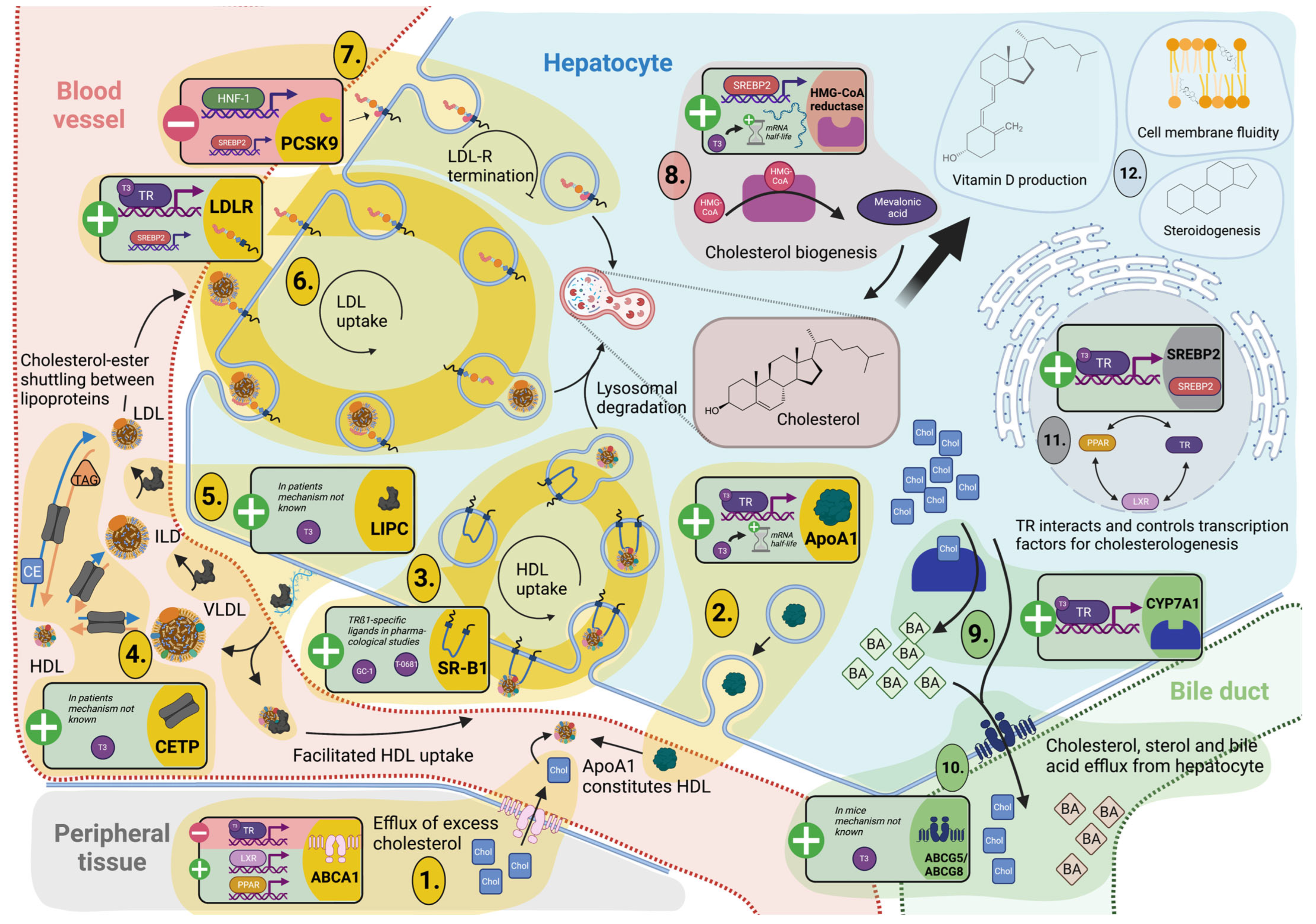
3.3. Cholesterol Metabolism and Turnover

Lipid metabolism takes place in the liver, which also synthesizes and recycles cholesterol. The sterol cholesterol is crucial for cell integrity and provides the precursor of steroid hormones, bile acids, and vitamin D (Figure 1, part 12).
Srebps represent an interface between TH signaling, on the one hand, and cholesterol/lipid homeostasis, as well as intracellular cholesterol sensing, on the other. These transcription factors are directly dependent on TH signaling and enhance or extend the effects of T3, which could be demonstrated by Shin et al. [128]. The isoform mainly involved in cholesterologenesis, SREPB2, was shown to be positively regulated by T3 in human hepatic cell lines [129].

3.3.1. TH Tightly Regulate Hepatic Cholesterol Formation and Peripheral Secretion

The β-hydroxy-β-methylglutaryl-CoA (HMG-CoA) reductase marks the rate-limiting step in the formation of cholesterol in the liver (Figure 1, part 8). THs primarily regulate HMG-CoA reductase, besides estrogen, glucagon, insulin, and glucocorticoids [130,131]. In hypophysectomized rats, T3 steers the HMG-CoA reductase activity positively [131]. While a study using kidney hamster cell cultures (BHK) showed no direct effect of T3 on expression levels, T3 does stabilize HMG-CoA reductase mRNA [132]. Nevertheless, T3 controls Srebp2 expression and thus influences HMG-CoA reductase gene regulation via their upstream regulatory elements [130].
After synthesis, loading onto lipoproteins solubilizes cholesterol so that it can be secreted from the liver to circulate in the periphery. One way to eliminate excess cholesterol from the circulation is the T3-dependent LDL endocytosis with LDLR (Figure 1, part 6). In thyroidectomized mice, oral T3 administration (10 to 50 nmol/kg/day) for seven days could recover the Ldl serum concentrations compared to sham-operated controls. With higher T3 dosing (up to 330 nmol/kg/day), overcompensation of Ldl decrease was observed [133]. The Ldlr promoter in the rat hepatoma cell line H4IIE contains two functional independent TREs, of which the US-TRE at −612 exhibits increased TRß1 binding affinity. Therefore, regulation via T3 is the determinant independent of the described regulation via the sterol response element [134,135] and indicates liver-specific TR isoform signaling.
The serine protease PCSK9 impairs receptor recycling and thus the uptake of LDL-bound cholesterol into the liver by facilitating lysosomal degradation. PCSK9 antibodies have already been successfully used to treat sequelae of hypercholesterolemia [136] (Figure 1, part 7). A comparative study between hyperthyroid patients versus euthyroid patients treated with liver-selective TH-analog KB2115 emphasized the effect of TH on cholesterol metabolism. Bonde et al. reported that hyperthyroidism and treatment with KB2115 reduced plasma concentrations of PCSK9, lipoprotein cholesterol, apolipoproteins B and AI, and lipoprotein(a) [137]. In line with this, two human studies showed a positive correlation between PCSK9 and TSH and a negative correlation with free T3/free T4 [138,139]. SREPBs form the direct link between TH action and PCSK9 expression, although there is no evidence for a TRE, yet. The identified regulatory sequences of PCSK9 in HepG2 cells consist of a sterol response element (SRE) and, with higher regulatory capacity, a binding site for hepatocyte nuclear factor 1 (HNF1) [140]. Worth noting is that the effect of TSH (mainly mediated via SREPB2) might have a steering capacity on PCSK9 expression in humans and HepG2 cells [141].

3.3.2. TH-Mediated Processing of Circulating Cholesterol Protects against Hypercholesterolemia

Cholesterol 7a-hydroxylation is the rate-limiting step to convert LDL-bound cholesterol to bile acids. Thus, it protects against hypercholesterolemia and its sequela, such as atherosclerosis, and bile acids provide a vehicle for dietary absorption in the intestine and serve as signaling molecules [142]. The role of TH in cholesterol degradation is already evident in subclinical hypothyroid patients, who exhibit significantly elevated serum total cholesterol and decreased bile acid concentrations [143,144]. Ldlr knock-out mice treated with TRß-specific thyromimetics GC-1 and KB2115 demonstrated a dramatic decrease in total cholesterol, particularly LDL bound, after ten days. Lindemann et al. also reported a remarkable increase in Cyp7a1 transcripts and serum levels of C4, a marker of bile acid synthesis and a physiological indicator of Cyp7a1 activity [145,146]. TH steer the CYP7A1 expression in HepG2 cells with physiological TR levels via the regulatory element (N1) [147] (Figure 1, part 9).
A heterodimer structure consisting of the two semi-transporter ATP-binding cassette subfamily G, member 5 (ABCG5) and ABCG8, is responsible for the transport of formed bile acid and sterols across the canalicular membrane of hepatocytes into the gallbladder [148] (Figure 1, part 10). However, ABCG5/8 are also present in the apical membrane of enterocytes, where they mediate transintestinal cholesterol excretion (TICE) [149]. In the study of intestinal absorption of cholesterol in hypophysectomized mice, administration of TH did not normalize Abcg5/8 expression in the small intestine but strongly upregulated it in the liver [150].

3.4. Reverse Cholesterol Transport

Excess cholesterol after conversion to cholesteryl ester can be transported from the periphery back to the liver by high-density lipoproteins (HDL). This process contributes to the natural cholesterol cycle and prevents manifestation of hypercholesterolemia provoked by excessive fat intake from food and cholesterol circulation in the body via VLDL and LDL. The formation of atheromatous plaques in the arteries would be promoted without reverse cholesterol transport (RCT) back to the liver and would have fatal effects on the cardio- and cerebrovascular systems.

3.4.1. Cholesterol Shuttling from Peripheral Cells Is TH Dependent

RCT initially requires a cholesterol efflux pump in peripheral cells. ABCA1 encodes the cholesterol efflux regulatory protein (CERP) and therefore resembles the shuttle for excess cholesterol. The importance of CERP in reverse cholesterol transport is reflected by the various regulators that control its expression and activity (Figure 1, part 1). Among these regulators are metabolites and signal molecules such as fatty acids, glucose, bilirubin, and adiponectin, which act via the nuclear receptors LXRs, TRs, RXRs, and PPARs [151,152,153,154,155]. Comparative promoter analysis in HEK293 cells overexpressing LXR and TR showed that both receptors bind a classical TH response element (TRE) in the ABCA1 promoter, with LXR leading to upregulation and TR to downregulation of ABCA1 expression [154] (Figure 1, part 11).

3.4.2. TH Control Hepatic Lipoprotein Secretion and Uptake for Reverse Cholesterol Transport and Modulation of Lipoprotein Fractions

ApoAI forms the major component of HDL secreted by the liver. Studies in rats showed that administration of TH has a positive rapid regulatory effect on Apoa1 transcription and increases mRNA stability [156] (Figure 1, part 2). When studied in human cell lines, promoter analysis in Huh7 cells demonstrated positive regulation [157], and the increased transcripts in HepG2 cells were presumably mainly related to increased mRNA stability [158], in a similar way to the aforementioned T3 regulation on HMG-CoA reductase. In humans, however, the TH-dependent regulation of HDL is less clear than in previous animal or cell culture models of mechanistic elucidation. In patients, HDL regulation, as well as HDL particle size and the ability to bind cholesterol esters, appear to depend on the severity of TH dysregulation [159]. Trends are toward normal to high HDL in overt, normal to low in subclinical hypothyroidism, and normal to low HDL in both subclinical and overt hyperthyroidism [160,161,162]. There are additional Apoa1 expression regulators described that act via transcription factors such as Foxa3 and epigenetic locus control with long non-coding RNA, which may explain the indistinct correlation between TH status and lipoprotein composition [163,164].
Hepatic lipase (HL) catalyzes the conversion of lipoprotein fractions from HDL via very-low-density lipoproteins (VLDL) and intermediate-low-density proteins (ILP) to low-density lipoproteins (LDL). Cholesteryl ester transport proteins (CETP) are pore forming, and they shuttle the neutral lipids cholesteryl esters and TG between lipoprotein fractions into the hydrophobic core [165] (Figure 1, part 4). HL and CETP are positively TH regulated and influence the lipoprotein composition in plasma [166]. These direct regulations via THs might be the underlying cause for increases in TG concentrations in hypothyroid individuals having low HL activity (Figure 1, part 5). Treatments with L-T4 can increase HL activity and lower TG concentrations in the circulation [167,168,169].
The uptake of the HDL-bound cholesterol ester into the liver is supported by the transporter scavenger receptor beta 1 (Srb1) (Figure 1, part 3). To date, only data on regulation are available from pharmacological studies in mice. After administration with thyromimetics GC-1 or T-0681, an increase of Srb1 at the protein level could be detected [170,171].

4. Future Challenges

Nutritional influences like diets and fasting impact both the systemic and the local hepatic TH system, resulting in alterations of local TH concentrations and subsequent modulation of gene expression in all pathways. With the wealth of data presented in this comprehensive overview, species- and sex-specific differences with regards to these regulations are obvious and reveal open questions. Much data has been compiled from rat models, which differ from those in mice, e.g., with regards to the extent of reduction in circulating TH concentrations upon fasting time or details of the regulation of the TH axis. Underlying molecular mechanisms fully describing the (re)distribution and enzymatic cascades involved in the clearly present reduction of circulating TH concentrations in all species remain partially elusive. Future research needs to document alterations in the liver TH system by associating different disease stages with cell-type-specific regulations of TH action, for example, for NAFLD, NASH, and diabetes models with or without local inflammation. This would provide underlying evidence for further translational insights for the possible usage and mechanism of TH mimetics that are currently in clinical trials [172,173]. While TH mimetics have been under evaluation as drugs for altering energy metabolism for many years, early substances that activated both TRα and TRß failed clinical trials due to adverse reactions. Recently, TRß-isoform-selective drugs (eliminating TRα-related adverse effects) like Resmetirom have been evaluated for the treatment of NAFLD/NASH in phase 3 clinical trials with effective reductions of hepatic and serum lipids and triglycerides. Details on thyromimetics in the context of NAFLD/NASH can be found in our recent review [172].
Nonetheless, based on the central role of local TH actions for intermediary, energy, and structural metabolism in various tissues, it is promising to therapeutically leverage the beneficial actions of THs in a tissue- and cell-type-specific manner for the treatment of metabolic disorders, including NAFLD, dyslipidemia, and obesity, as well as type 2 diabetes.

Author Contributions

All authors (J.S. (Joshua Seifert), Y.C., W.S., K.M., F.T., J.S. (Joachim Spranger), J.K., E.K.W.) contributed to the conceptualization, writing, and visualization of the article. All authors have read and agreed to the published version of the manuscript.

Funding

This work was supported by the German Research Foundation (DFG SFB/TRR296, TP15 and TP16).

Data Availability Statement

Not applicable.

Acknowledgments

Figures were created using BioRender.com.

Conflicts of Interest

J.S. (Joshua Seifert), Y.C., W.S., K.M., J.S. (Joachim Spranger), J.K. and E.K.W. declare no conflict of interest. F.T. received research funding from Allergan, Bristol-Myers Squibb, Gilead, and Inventiva. The funders had no role in the design of the study; in collection, analysis, or interpretation of the data; in the writing of the manuscript; or the decision to publish the results.

References

  1. Hönes, G.S.; Geist, D.; Moeller, L.C. Noncanonical Action of Thyroid Hormone Receptors α and β. Exp. Clin. Endocrinol. Diabetes 2020, 128, 383–387. [Google Scholar] [CrossRef] [PubMed]
  2. Friesema, E.C.H.; Ganguly, S.; Abdalla, A.; Manning Fox, J.E.; Halestrap, A.P.; Visser, T.J. Identification of Monocarboxylate Transporter 8 as a Specific Thyroid Hormone Transporter. J. Biol. Chem. 2003, 278, 40128–40135. [Google Scholar] [CrossRef] [PubMed] [Green Version]
  3. Dumitrescu, A.M.; Liao, X.-H.; Best, T.B.; Brockmann, K.; Refetoff, S. A Novel Syndrome Combining Thyroid and Neurological Abnormalities Is Associated with Mutations in a Monocarboxylate Transporter Gene. Am. J. Hum. Genet. 2004, 74, 168–175. [Google Scholar] [CrossRef] [PubMed] [Green Version]
  4. Köhrle, J.; Frädrich, C. Deiodinases control local cellular and systemic thyroid hormone availability. Free Radic. Biol. Med. 2022, 193, 59–79. [Google Scholar] [CrossRef]
  5. Fonseca, T.L.; Fernandes, G.W.; McAninch, E.A.; Bocco, B.M.L.C.; Abdalla, S.M.; Ribeiro, M.O.; Mohácsik, P.; Fekete, C.; Li, D.; Xing, X.; et al. Perinatal deiodinase 2 expression in hepatocytes defines epigenetic susceptibility to liver steatosis and obesity. Proc. Natl. Acad. Sci. USA 2015, 112, 14018–14023. [Google Scholar] [CrossRef] [Green Version]
  6. Visser, T.J.; van Buuren, J.C.; Rutgers, M.; Rooda, S.J.E.; de Herder, W.W. The role of sulfation in thyroid hormone metabolism. Trends Endocrinol. Metab. 1990, 1, 211–218. [Google Scholar] [CrossRef] [Green Version]
  7. Dentice, M.; Ambrosio, R.; Damiano, V.; Sibilio, A.; Luongo, C.; Guardiola, O.; Yennek, S.; Zordan, P.; Minchiotti, G.; Colao, A.; et al. Intracellular Inactivation of Thyroid Hormone Is a Survival Mechanism for Muscle Stem Cell Proliferation and Lineage Progression. Cell Metab. 2014, 20, 1038–1048. [Google Scholar] [CrossRef] [Green Version]
  8. Ogawa-Wong, A.; Carmody, C.; Le, K.; Marschner, R.A.; Larsen, P.R.; Zavacki, A.M.; Wajner, S.M. Modulation of Deiodinase Types 2 and 3 during Skeletal Muscle Regeneration. Metabolites 2022, 12, 612. [Google Scholar] [CrossRef]
  9. Kaplan, M.M.; Utiger, R.D. Iodothyronine Metabolism in Rat Liver Homogenates. J. Clin. Investig. 1978, 61, 459–471. [Google Scholar] [CrossRef]
  10. Balsam, A.; Ingbar, S.H.; Sexton, F. The Influence of Fasting, Diabetes, and Several Pharmacological Agents on the Pathways of Thyroxine Metabolism in Rat Liver. J. Clin. Investig. 1978, 62, 415–424. [Google Scholar] [CrossRef]
  11. Russo, S.C.; Salas-Lucia, F.; Bianco, A.C. Deiodinases and the Metabolic Code for Thyroid Hormone Action. Endocrinology 2021, 162, bqab059. [Google Scholar] [CrossRef]
  12. Galton, V.A.; Hernandez, A.; Germain, D.L.S. The 5′-Deiodinases Are Not Essential for the Fasting-Induced Decrease in Circulating Thyroid Hormone Levels in Male Mice: Possible Roles for the Type 3 Deiodinase and Tissue Sequestration of Hormone. Endocrinology 2014, 155, 3172–3181. [Google Scholar] [CrossRef] [Green Version]
  13. Cordeiro, A.; Souza, L.; Oliveira, L.S.; Faustino, L.C.; Santiago, L.A.; Bloise, F.F.; Ortiga-Carvalho, T.M.; Almeida, N.A.D.S.; Pazos-Moura, C.C. Thyroid hormone regulation of Sirtuin 1 expression and implications to integrated responses in fasted mice. J. Endocrinol. 2012, 216, 181–193. [Google Scholar] [CrossRef] [Green Version]
  14. De Vries, E.M.; van Beeren, H.C.; van Wijk, A.C.W.A.; Kalsbeek, A.; Romijn, J.A.; Fliers, E.; Boelen, A. Regulation of type 3 deiodinase in rodent liver and adipose tissue during fasting. Endocr. Connect. 2020, 9, 552–562. [Google Scholar] [CrossRef]
  15. Van der Wal, A.M.; Bakker, O.; Wiersinga, W.M. The decrease of liver LDL receptor mRNA during fasting is related to the decrease in serum T3. Int. J. Biochem. Cell Biol. 1998, 30, 209–215. [Google Scholar] [CrossRef]
  16. De Vries, E.M.; Eggels, L.; Van Beeren, H.C.; Ackermans, M.T.; Kalsbeek, A.; Fliers, E.; Boelen, A. Fasting-Induced Changes in Hepatic Thyroid Hormone Metabolism in Male Rats Are Independent of Autonomic Nervous Input to the Liver. Endocrinology 2014, 155, 5033–5041. [Google Scholar] [CrossRef] [Green Version]
  17. De Vries, E.M.; Van Beeren, H.C.; Ackermans, M.T.; Kalsbeek, A.; Fliers, E.; Boelen, A. Differential effects of fasting vs food restriction on liver thyroid hormone metabolism in male rats. J. Endocrinol. 2014, 224, 25–35. [Google Scholar] [CrossRef] [Green Version]
  18. Naito, K.; Inada, M.; Mashio, Y.; Tanaka, K.; Ishii, H.; Ishikawa, M.; Imura, H. Modulation of T4 5’-monodeiodination in rat anterior pituitary and liver homogenates by thyroid states and fasting. Endocrinol. Jpn. 1981, 28, 793–798. [Google Scholar] [CrossRef] [Green Version]
  19. Visser, T.J. Pathways of thyroid hormone metabolism. Acta Med. Austriaca 1996, 23, 10–16. [Google Scholar]
  20. Giacco, A.; Paoli, G.D.; Simiele, R.; Caterino, M.; Ruoppolo, M.; Bloch, W.; Kraaij, R.; Uitterlinden, A.G.; Santillo, A.; Senese, R.; et al. Exercise with food withdrawal at thermoneutrality impacts fuel use, the microbiome, AMPK phosphorylation, muscle fibers, and thyroid hormone levels in rats. Physiol. Rep. 2020, 8, e14354. [Google Scholar] [CrossRef] [Green Version]
  21. Visser, T.J.; van Haasteren, G.A.; Linkels, E.; Kaptein, E.; van Toor, H.; de Greef, W.J. Gender-specific changes in thyroid hormone-glucuronidating enzymes in rat liver during short-term fasting and long-term food restriction. Eur. J. Endocrinol. 1996, 135, 489–497. [Google Scholar] [CrossRef] [PubMed] [Green Version]
  22. Young, R.A.; Rajatanavin, R.; Moring, A.F.; Braverman, L.E. Fasting Induces the Generation of Serum Thyronine-Binding Globulin in Zucker Rats*. Endocrinology 1985, 116, 1248–1252. [Google Scholar] [CrossRef] [PubMed]
  23. Wade, S.; Bleiberg-Daniel, F.; Le Moullac, B. Rat Transthyretin: Effects of Acute Short-Term Food Deprivation and Refeeding on Serum and Cerebrospinal Fluid Concentration and on Hepatic mRNA Level. J. Nutr. 1988, 118, 199–205. [Google Scholar] [CrossRef]
  24. Gereben, B.; Zavacki, A.M.; Ribich, S.; Kim, B.W.; Huang, S.A.; Simonides, W.S.; Zeöld, A.; Bianco, A.C. Cellular and Molecular Basis of Deiodinase-Regulated Thyroid Hormone Signaling1. Endocr. Rev. 2008, 29, 898–938. [Google Scholar] [CrossRef] [PubMed] [Green Version]
  25. Bianco, A.C.; Kim, B.W. Deiodinases: Implications of the local control of thyroid hormone action. J. Clin. Investig. 2006, 116, 2571–2579. [Google Scholar] [CrossRef] [Green Version]
  26. Köhrle, J. Local activation and inactivation of thyroid hormones: The deiodinase family. Mol. Cell. Endocrinol. 1999, 151, 103–119. [Google Scholar] [CrossRef]
  27. Kester, M.H.A.; Van Dijk, C.H.; Tibboel, D.; Hood, A.M.; Rose, N.J.M.; Meinl, W.; Pabel, U.; Glatt, H.; Falany, C.N.; Coughtrie, M.; et al. Sulfation of Thyroid Hormone by Estrogen Sulfotransferase. J. Clin. Endocrinol. Metab. 1999, 84, 2577–2580. [Google Scholar] [CrossRef]
  28. Wu, S.-Y.; Green, W.L.; Huang, W.-S.; Hays, M.T.; Chopra, I.J. Alternate Pathways of Thyroid Hormone Metabolism. Thyroid 2005, 15, 943–958. [Google Scholar] [CrossRef]
  29. Visser, T.J. Role of sulfation in thyroid hormone metabolism. Chem. Interact. 1994, 92, 293–303. [Google Scholar] [CrossRef] [Green Version]
  30. Distefano, J.J.; Sternlicht, M.; Harris, D.R. Rat Enterohepatic Circulation and Intestinal Distribution of Enterally Infused Thyroid Hormones*. Endocrinology 1988, 123, 2526–2539. [Google Scholar] [CrossRef]
  31. De Herder, W.W.; Hazenberg, M.P.; Pennock-Schröder, A.M.; Hennemann, G.; Visser, T.J. Rapid and bacteria-dependent in vitro hydrolysis of iodothyronine-conjugates by intestinal contents of humans and rats. Med. Biol. 1986, 64, 31–35. [Google Scholar]
  32. Virili, C.; Centanni, M. “With a little help from my friends”—The role of microbiota in thyroid hormone metabolism and enterohepatic recycling. Mol. Cell. Endocrinol. 2017, 458, 39–43. [Google Scholar] [CrossRef]
  33. De Jong, M.; Docter, R.; Van Der Hoek, H.J.; Vos, R.A.; Krenning, E.P.; Hennemann, G. Transport of 3,5,3’-triiodothyronine into the perfused rat liver and subsequent metabolism are inhibited by fasting. Endocrinology 1992, 131, 463–470. [Google Scholar] [CrossRef]
  34. Kester, M.H.A.; Kaptein, E.; Roest, T.J.; van Dijk, C.H.; Tibboel, D.; Meinl, W.; Glatt, H.; Coughtrie, M.W.H.; Visser, T.J. Characterization of rat iodothyronine sulfotransferases. Am. J. Physiol. Metab. 2003, 285, E592–E598. [Google Scholar] [CrossRef] [Green Version]
  35. Darras, V.M.; Hume, R.; Visser, T.J. Regulation of thyroid hormone metabolism during fetal development. Mol. Cell. Endocrinol. 1999, 151, 37–47. [Google Scholar] [CrossRef]
  36. Visser, T.J.; Kaptein, E.; Gijzel, A.L.; De Herder, W.W.; Ebner, T.; Burchell, B. Glucuronidation of thyroid hormone by human bilirubin and phenol UDP-glucuronyltransferase isoenzymes. FEBS Lett. 1993, 324, 358–360. [Google Scholar] [CrossRef] [Green Version]
  37. Fujita, K.-I.; Nagata, K.; Ozawa, S.; Sasano, H.; Yamazoe, Y. Molecular Cloning and Characterization of Rat ST1B1 and Human ST1B2 cDNAs, Encoding Thyroid Hormone Sulfotransferases. J. Biochem. 1997, 122, 1052–1061. [Google Scholar] [CrossRef]
  38. Blanchard, R.L.; Freimuth, R.; Buck, J.; Weinshilboum, R.M.; Coughtrie, M. A proposed nomenclature system for the cytosolic sulfotransferase (SULT) superfamily. Pharmacogenetics 2004, 14, 199–211. [Google Scholar] [CrossRef]
  39. Mackenzie, P.I.; Bock, K.W.; Burchell, B.; Guillemette, C.; Ikushiro, S.-I.; Iyanagi, T.; Miners, J.O.; Owens, I.S.; Nebert, D.W. Nomenclature update for the mammalian UDP glycosyltransferase (UGT) gene superfamily. Pharmacogenet. Genom. 2005, 15, 677–685. [Google Scholar] [CrossRef]
  40. Maglich, J.M.; Watson, J.; McMillen, P.J.; Goodwin, B.; Willson, T.M.; Moore, J.T. The Nuclear Receptor CAR Is a Regulator of Thyroid Hormone Metabolism during Caloric Restriction. J. Biol. Chem. 2004, 279, 19832–19838. [Google Scholar] [CrossRef] [Green Version]
  41. Vella, K.R.; Ramadoss, P.; Lam, F.S.; Harris, J.C.; Ye, F.D.; Same, P.D.; O’Neill, N.F.; Maratos-Flier, E.; Hollenberg, A.N. NPY and MC4R Signaling Regulate Thyroid Hormone Levels during Fasting through Both Central and Peripheral Pathways. Cell Metab. 2011, 14, 780–790. [Google Scholar] [CrossRef] [PubMed] [Green Version]
  42. Küblbeck, J.; Niskanen, J.; Honkakoski, P. Metabolism-Disrupting Chemicals and the Constitutive Androstane Receptor CAR. Cells 2020, 9, 2306. [Google Scholar] [CrossRef] [PubMed]
  43. Everts, M.E.; de Jong, M.; Lim, C.-F.; Docter, R.; Krenning, E.P.; Visser, T.J.; Hennemann, G. Different Regulation of Thyroid Hormone Transport in Liver and Pituitary: Its Possible Role in the Maintenance of Low T3 Production during Nonthyroidal Illness and Fasting in Man. Thyroid 1996, 6, 359–368. [Google Scholar] [CrossRef] [PubMed]
  44. Dietrich, C.G.; Martin, I.V.; Porn, A.C.; Voigt, S.; Gartung, C.; Trautwein, C.; Geier, A. Fasting induces basolateral uptake transporters of the SLC family in the liver via HNF4α and PGC1α. Am. J. Physiol. Liver Physiol. 2007, 293, G585–G590. [Google Scholar] [CrossRef] [Green Version]
  45. Tian, L.; Song, Y.; Xing, M.; Zhang, W.; Ning, G.; Li, X.; Yu, C.; Qin, C.; Liu, J.; Tian, X.; et al. A novel role for thyroid-stimulating hormone: Up-regulation of hepatic 3-hydroxy-3-methyl-glutaryl-coenzyme a reductase expression through the cyclic adenosine monophosphate/protein kinase A/cyclic adenosine monophosphate-responsive element binding protei. Hepatology 2010, 52, 1401–1409. [Google Scholar] [CrossRef]
  46. Zhang, W.; Tian, L.-M.; Han, Y.; Ma, H.-Y.; Wang, L.-C.; Guo, J.; Gao, L.; Zhao, J.-J. Presence of thyrotropin receptor in hepatocytes: Not a case of illegitimate transcription. J. Cell. Mol. Med. 2009, 13, 4636–4642. [Google Scholar] [CrossRef] [Green Version]
  47. Wang, T.; Xu, J.; Bo, T.; Zhou, X.; Jiang, X.; Gao, L.; Zhao, J. Decreased fasting blood glucose is associated with impaired hepatic glucose production in thyroid-stimulating hormone receptor knockout mice. Endocr. J. 2013, 60, 941–950. [Google Scholar] [CrossRef] [Green Version]
  48. Elgadi, A.; Zemack, H.; Marcus, C.; Norgren, S. Tissue-specific knockout of TSHr in white adipose tissue increases adipocyte size and decreases TSH-induced lipolysis. Biochem. Biophys. Res. Commun. 2010, 393, 526–530. [Google Scholar] [CrossRef]
  49. Zhou, L.; Wu, K.; Zhang, L.; Gao, L.; Chen, S. Liver-specific deletion of TSHR inhibits hepatic lipid accumulation in mice. Biochem. Biophys. Res. Commun. 2018, 497, 39–45. [Google Scholar] [CrossRef]
  50. González-Ramos, S.; Paz-García, M.; Fernández-García, V.; Portune, K.J.; Acosta-Medina, E.F.; Sanz, Y.; Castrillo, A.; Martín-Sanz, P.; Obregon, M.J.; Boscá, L. NOD1 deficiency promotes an imbalance of thyroid hormones and microbiota homeostasis in mice fed high fat diet. Sci. Rep. 2020, 10, 1–17. [Google Scholar] [CrossRef]
  51. Han, H.; Xin, P.; Zhao, L.; Xu, J.; Xia, Y.; Yang, X.; Sun, X.; Hao, L. Excess Iodine and High-Fat Diet Combination Modulates Lipid Profile, Thyroid Hormone, and Hepatic LDLr Expression Values in Mice. Biol. Trace Element Res. 2012, 147, 233–239. [Google Scholar] [CrossRef]
  52. Lopez, A.N.; Geissler, C.; Naujack, A.-M.; Chen, Y.; Taege, N.; Britsemmer, J.H.; Vinicius, M.D.A.L.; Oster, H.; Spranger, J.; Katrin, W.E.; et al. Deiodinase type I (DIO1) regulation in non-alcoholic fatty liver disease (NAFLD). Endocr. Abstr. 2022, 84, PS2-09-80. [Google Scholar] [CrossRef]
  53. Bruinstroop, E.; Zhou, J.; Tripathi, M.; Yau, W.W.; Boelen, A.; Singh, B.K.; Yen, P.M. Early induction of hepatic deiodinase type 1 inhibits hepatosteatosis during NAFLD progression. Mol. Metab. 2021, 53, 101266. [Google Scholar] [CrossRef]
  54. Lietzow, J.; Golchert, J.; Homuth, G.; Völker, U.; Jonas, W.; Köhrle, J. 3,5-T2 alters murine genes relevant for xenobiotic, steroid, and thyroid hormone metabolism. J. Mol. Endocrinol. 2016, 56, 311–323. [Google Scholar] [CrossRef] [Green Version]
  55. Wu, J.; Wang, C.; Li, S.; Li, S.; Wang, W.; Li, J.; Chi, Y.; Yang, H.; Kong, X.; Zhou, Y.; et al. Thyroid hormone-responsive SPOT 14 homolog promotes hepatic lipogenesis, and its expression is regulated by Liver X receptor α through a sterol regulatory element-binding protein 1c-dependent mechanism in mice. Hepatology 2013, 58, 617–628. [Google Scholar] [CrossRef]
  56. LaFave, L.T.; Augustin, L.B.; Mariash, C.N. S14: Insights from Knockout Mice. Endocrinology 2006, 147, 4044–4047. [Google Scholar] [CrossRef] [Green Version]
  57. Jornayvaz, F.; Lee, H.-Y.; Jurczak, M.; Alves, T.C.; Guebre-Egziabher, F.; Guigni, B.; Zhang, D.; Samuel, V.T.; Silva, J.E.; Shulman, G.I. Thyroid Hormone Receptor-α Gene Knockout Mice Are Protected from Diet-Induced Hepatic Insulin Resistance. Endocrinology 2012, 153, 583–591. [Google Scholar] [CrossRef] [Green Version]
  58. Goldfarb, C.N.; Karri, K.; Pyatkov, M.; Waxman, D.J. Interplay Between GH-regulated, Sex-biased Liver Transcriptome and Hepatic Zonation Revealed by Single-Nucleus RNA Sequencing. Endocrinology 2022, 163, bqac059. [Google Scholar] [CrossRef]
  59. Smati, S.; Polizzi, A.; Fougerat, A.; Ellero-Simatos, S.; Blum, Y.; Lippi, Y.; Régnier, M.; Laroyenne, A.; Huillet, M.; Arif, M.; et al. Integrative study of diet-induced mouse models of NAFLD identifies PPARα as a sexually dimorphic drug target. Gut 2021, 71, 807–821. [Google Scholar] [CrossRef]
  60. Riese, C.; Michaelis, M.; Mentrup, B.; Götz, F.; Köhrle, J.; Schweizer, U.; Schomburg, L. Selenium-Dependent Pre- and Posttranscriptional Mechanisms Are Responsible for Sexual Dimorphic Expression of Selenoproteins in Murine Tissues. Endocrinology 2006, 147, 5883–5892. [Google Scholar] [CrossRef] [Green Version]
  61. Desai-Yajnik, V.; Zeng, J.; Omori, K.; Sherman, J.; Morimoto, T. The effect of thyroid hormone treatment on the gene expression and enzyme activity of rat liver sodium-potassium dependent adenosine triphosphatase. Endocrinology 1995, 136, 629–639. [Google Scholar] [CrossRef] [PubMed]
  62. Kiss, E.; Jakab, G.; Kranias, E.G.; Edes, I. Thyroid hormone-induced alterations in phospholamban protein expression. Regulatory effects on sarcoplasmic reticulum Ca2+ transport and myocardial relaxation. Circ. Res. 1994, 75, 245–251. [Google Scholar] [CrossRef] [PubMed] [Green Version]
  63. Weinstein, S.P.; O’Boyle, E.; Fisher, M.; Haber, R.S. Regulation of GLUT2 glucose transporter expression in liver by thyroid hormone: Evidence for hormonal regulation of the hepatic glucose transport system. Endocrinology 1994, 135, 649–654. [Google Scholar] [CrossRef] [PubMed]
  64. Stalmans, W. The Role of the Liver in the Homeostasis of Blood Glucose. Curr. Top. Cell. Regul. 1976, 11, 51–97. [Google Scholar] [CrossRef]
  65. Kim, B.W.; Zavacki, A.M.; Curcio-Morelli, C.; Dentice, M.; Harney, J.W.; Larsen, P.R.; Bianco, A.C. Endoplasmic Reticulum-Associated Degradation of the Human Type 2 Iodothyronine Deiodinase (D2) is Mediated via an Association between Mammalian UBC7 and the Carboxyl Region of D2. Mol. Endocrinol. 2003, 17, 2603–2612. [Google Scholar] [CrossRef] [Green Version]
  66. Salvatore, D.; Bartha, T.; Harney, J.W.; Larsen, P.R. Molecular biological and biochemical characterization of the human type 2 selenodeiodinase. Endocrinology 1996, 137, 3308–3315. [Google Scholar] [CrossRef] [Green Version]
  67. Torrance, C.J.; Devente, J.E.; Jones, J.P.; Dohm, G.L. Effects of Thyroid Hormone on GLUT4 Glucose Transporter Gene Expression and NIDDM in Rats. Endocrinology 1997, 138, 1204–1214. [Google Scholar] [CrossRef]
  68. Torrance, C.J.; Usala, S.J.; Pessin, J.E.; Dohm, G.L. Characterization of a Low Affinity Thyroid Hormone Receptor Binding Site within the Rat GLUT4 Gene Promoter. Endocrinology 1997, 138, 1215–1223. [Google Scholar] [CrossRef]
  69. Weinstein, S.P.; O’Boyle, E.; Haber, R.S. Thyroid Hormone Increases Basal and Insulin-Stimulated Glucose Transport in Skeletal Muscle: The Role of GLUT4 Glucose Transporter Expression. Diabetes 1994, 43, 1185–1189. [Google Scholar] [CrossRef]
  70. Leonard, J.L. Dibutyryl cAMP induction of type II 5′deiodinase activity in rat brain astrocytes in culture. Biochem. Biophys. Res. Commun. 1988, 151, 1164–1172. [Google Scholar] [CrossRef]
  71. Watanabe, M.; Houten, S.M.; Mataki, C.; Christoffolete, M.A.; Kim, B.W.; Sato, H.; Messaddeq, N.; Harney, J.W.; Ezaki, O.; Kodama, T.; et al. Bile acids induce energy expenditure by promoting intracellular thyroid hormone activation. Nature 2006, 439, 484–489. [Google Scholar] [CrossRef]
  72. Klieverik, L.P.; Janssen, S.F.; van Riel, A.; Foppen, E.; Bisschop, P.H.; Serlie, M.J.; Boelen, A.; Ackermans, M.T.; Sauerwein, H.P.; Fliers, E.; et al. Thyroid hormone modulates glucose production via a sympathetic pathway from the hypothalamic paraventricular nucleus to the liver. Proc. Natl. Acad. Sci. USA 2009, 106, 5966–5971. [Google Scholar] [CrossRef] [Green Version]
  73. Carter, W.J.; Benjamin, W.S.V.D.W.; Faas, F.H. Effect of experimental hyperthyroidism on skeletal-muscle proteolysis. Biochem. J. 1981, 194, 685–690. [Google Scholar] [CrossRef] [Green Version]
  74. Felig, P. Amino Acid Metabolism in Man. Annu. Rev. Biochem. 1975, 44, 933–955. [Google Scholar] [CrossRef]
  75. Suh, J.H.; Sieglaff, U.H.; Zhang, A.; Xia, X.; Cvoro, A.; Winnier, G.E.; Webb, P. SIRT1 is a Direct Coactivator of Thyroid Hormone Receptor β1 with Gene-Specific Actions. PLoS ONE 2013, 8, e70097. [Google Scholar] [CrossRef]
  76. Attia, R.R.; Connnaughton, S.; Boone, L.R.; Wang, F.; Elam, M.B.; Ness, G.C.; Cook, G.A.; Park, E.A. Regulation of Pyruvate Dehydrogenase Kinase 4 (PDK4) by Thyroid Hormone: Role of the peroxisome proliferator-activated receptor gamma coactivator (PGC-1 alpha). J. Biol. Chem. 2010, 285, 2375–2385. [Google Scholar] [CrossRef] [Green Version]
  77. Thakran, S.; Sharma, P.; Attia, R.R.; Hori, R.T.; Deng, X.; Elam, M.B.; Park, E.A. Role of Sirtuin 1 in the Regulation of Hepatic Gene Expression by Thyroid Hormone. J. Biol. Chem. 2013, 288, 807–818. [Google Scholar] [CrossRef] [Green Version]
  78. Erion, D.M.; Yonemitsu, S.; Nie, Y.; Nagai, Y.; Gillum, M.P.; Hsiao, J.J.; Iwasaki, T.; Stark, R.; Weismann, D.; Yu, X.X.; et al. SirT1 knockdown in liver decreases basal hepatic glucose production and increases hepatic insulin responsiveness in diabetic rats. Proc. Natl. Acad. Sci. USA 2009, 106, 11288–11293. [Google Scholar] [CrossRef] [Green Version]
  79. Bratusch-Marrain, P.R.; Komjati, M.; Waldhäusl, W.K. Glucose Metabolism in Noninsulin-Dependent Diabetic Patients with Experimental Hyperthyroidism*. J. Clin. Endocrinol. Metab. 1985, 60, 1063–1068. [Google Scholar] [CrossRef]
  80. Moon, S.W.; Hahm, J.R.; Lee, G.-W.; Kang, M.Y.; Jung, J.H.; Jung, T.S.; Lee, K.W.; Jung, K.A.; Ahn, Y.J.; Kim, S.; et al. A Case of Hyperglycemic Hyperosmolar State Associated with Graves’ Hyperthyroidism: A Case Report. J. Korean Med. Sci. 2006, 21, 765–767. [Google Scholar] [CrossRef]
  81. Wintergerst, K.A.; Rogers, E.S.; Foster, M.B. Hyperthyroidism presenting with hyperglycemia in an adolescent female. J. Pediatr. Endocrinol. Metab. 2011, 24, 385–387. [Google Scholar] [CrossRef] [PubMed]
  82. Dimitriadis, G.D.; Raptis, S.A. Thyroid hormone excess and glucose intolerance. Exp. Clin. Endocrinol. Diabetes 2001, 109 (Suppl. S2), S225–S239. [Google Scholar] [CrossRef] [PubMed]
  83. Battarbee, H.D. The Effects of Thyroid State on Rat Liver Glucose-6-Phosphatase Activity and Glycogen Content. Exp. Biol. Med. 1974, 147, 337–343. [Google Scholar] [CrossRef] [PubMed]
  84. Burton, S.D.; Robbins, E.; Byers, S.O. Effect of Hyperthyroidism on Glycogen Content of the Isolated Rat Liver. Am. J. Physiol. Content 1957, 188, 509–513. [Google Scholar] [CrossRef]
  85. Sinha, R.A.; Singh, B.K.; Yen, P.M. Direct effects of thyroid hormones on hepatic lipid metabolism. Nat. Rev. Endocrinol. 2018, 14, 259–269. [Google Scholar] [CrossRef]
  86. Lu, C.; Cheng, S.-Y. Thyroid hormone receptors regulate adipogenesis and carcinogenesis via crosstalk signaling with peroxisome proliferator-activated receptors. J. Mol. Endocrinol. 2009, 44, 143–154. [Google Scholar] [CrossRef]
  87. Wierzbicki, M.; Chabowski, A.; Zendzian-Piotrowska, M.; Górski, J. Differential effects of in vivo PPAR alpha and gamma activation on fatty acid transport proteins expression and lipid content in rat liver. J. Physiol. Pharmacol. 2009, 60, 99–106. [Google Scholar]
  88. Doege, H.; Grimm, D.; Falcon, A.; Tsang, B.; Storm, T.A.; Xu, H.; Ortegon, A.M.; Kazantzis, M.; Kay, M.A.; Stahl, A. Silencing of Hepatic Fatty Acid Transporter Protein 5 in Vivo Reverses Diet-induced Non-alcoholic Fatty Liver Disease and Improves Hyperglycemia. J. Biol. Chem. 2008, 283, 22186–22192. [Google Scholar] [CrossRef] [Green Version]
  89. Klieverik, L.P.; Coomans, C.P.; Endert, E.; Sauerwein, H.P.; Havekes, L.M.; Voshol, P.J.; Rensen, P.C.N.; Romijn, J.A.; Kalsbeek, A.; Fliers, E. Thyroid Hormone Effects on Whole-Body Energy Homeostasis and Tissue-Specific Fatty Acid Uptake in Vivo. Endocrinology 2009, 150, 5639–5648. [Google Scholar] [CrossRef] [Green Version]
  90. Nakagawa, S.; Kawashima, Y.; Hirose, A.; Kozuka, H. Regulation of hepatic level of fatty-acid-binding protein by hormones and clofibric acid in the rat. Biochem. J. 1994, 297, 581–584. [Google Scholar] [CrossRef] [Green Version]
  91. Santana-Farré, R.; Mirecki-Garrido, M.; Bocos, C.; Henríquez-Hernández, L.A.; Kahlon, N.; Herrera, E.; Norstedt, G.; Parini, P.; Flores-Morales, A.; Fernández-Pérez, L. Influence of Neonatal Hypothyroidism on Hepatic Gene Expression and Lipid Metabolism in Adulthood. PLoS ONE 2012, 7, e37386. [Google Scholar] [CrossRef] [Green Version]
  92. Petty, K.J.; Desvergne, B.; Mitsuhashi, T.; Nikodem, V.M. Identification of a thyroid hormone response element in the malic enzyme gene. J. Biol. Chem. 1990, 265, 7395–7400. [Google Scholar] [CrossRef]
  93. López, M.; Lelliott, C.J.; Vidal-Puig, A. Hypothalamic fatty acid metabolism: A housekeeping pathway that regulates food intake. Bioessays 2007, 29, 248–261. [Google Scholar] [CrossRef]
  94. Pender, C.; Trentadue, A.R.; Pories, W.J.; Dohm, G.L.; Houmard, J.A.; Youngren, J.F. Expression of genes regulating Malonyl-CoA in human skeletal muscle. J. Cell. Biochem. 2006, 99, 860–867. [Google Scholar] [CrossRef]
  95. Kreuz, S.; Schoelch, C.; Thomas, L.; Rist, W.; Rippmann, J.F.; Neubauer, H. Acetyl-CoA carboxylases 1 and 2 show distinct expression patterns in rats and humans and alterations in obesity and diabetes. Diabetes/Metab. Res. Rev. 2009, 25, 577–586. [Google Scholar] [CrossRef]
  96. Zhang, Y.; Yin, L.; Hillgartner, F.B. Thyroid Hormone Stimulates Acetyl-CoA Carboxylase-α Transcription in Hepatocytes by Modulating the Composition of Nuclear Receptor Complexes Bound to a Thyroid Hormone Response Element. J. Biol. Chem. 2001, 276, 974–983. [Google Scholar] [CrossRef] [Green Version]
  97. Blennemann, B.; Leahy, P.; Kim, T.-S.; Freake, H.C. Tissue-specific regulation of lipogenic mRNAs by thyroid hormone. Mol. Cell. Endocrinol. 1995, 110, 1–8. [Google Scholar] [CrossRef]
  98. López, M.; Varela, L.; Vázquez, M.J.; Rodríguez-Cuenca, S.; González, C.R.; Velagapudi, V.R.; Morgan, D.A.; Schoenmakers, E.; Agassandian, K.; Lage, R.; et al. Hypothalamic AMPK and fatty acid metabolism mediate thyroid regulation of energy balance. Nat. Med. 2010, 16, 1001–1008. [Google Scholar] [CrossRef]
  99. Zhu, Q.; Mariash, A.; Margosian, M.R.; Gopinath, S.; Fareed, M.T.; Anderson, G.W.; Mariash, C.N. Spot 14 Gene Deletion Increases Hepatic de Novo Lipogenesis. Endocrinology 2001, 142, 4363–4370. [Google Scholar] [CrossRef]
  100. Campbell, M.C.; Anderson, G.W.; Mariash, C.N. Human Spot 14 Glucose and Thyroid Hormone Response: Characterization and Thyroid Hormone Response Element Identification. Endocrinology 2003, 144, 5242–5248. [Google Scholar] [CrossRef]
  101. Paton, C.M.; Ntambi, J.M. Biochemical and physiological function of stearoyl-CoA desaturase. Am. J. Physiol. Metab. 2009, 297, E28–E37. [Google Scholar] [CrossRef] [PubMed] [Green Version]
  102. Radenne, A.; Akpa, M.; Martel, C.; Sawadogo, S.; Mauvoisin, D.; Mounier, C. Hepatic regulation of fatty acid synthase by insulin and T3: Evidence for T3 genomic and nongenomic actions. Am. J. Physiol. Metab. 2008, 295, E884–E894. [Google Scholar] [CrossRef] [PubMed] [Green Version]
  103. Hönes, G.S.; Rakov, H.; Logan, J.; Liao, X.-H.; Werbenko, E.; Pollard, A.S.; Præstholm, S.M.; Siersbæk, M.S.; Rijntjes, E.; Gassen, J.; et al. Noncanonical thyroid hormone signaling mediates cardiometabolic effects in vivo. Proc. Natl. Acad. Sci. USA 2017, 114, E11323–E11332. [Google Scholar] [CrossRef] [PubMed] [Green Version]
  104. Shabtai, Y.; Nagaraj, N.K.; Batmanov, K.; Cho, Y.-W.; Guan, Y.; Jiang, C.; Remsberg, J.; Forrest, D.; Lazar, M.A. A coregulator shift, rather than the canonical switch, underlies thyroid hormone action in the liver. Genes Dev. 2021, 35, 367–378. [Google Scholar] [CrossRef]
  105. Ritter, M.J.; Amano, I.; Imai, N.; De Oliveira, L.S.; Vella, K.R.; Hollenberg, A.N. Nuclear Receptor CoRepressors, NCOR1 and SMRT, are required for maintaining systemic metabolic homeostasis. Mol. Metab. 2021, 53, 101315. [Google Scholar] [CrossRef]
  106. Mendoza, A.; Tang, C.; Choi, J.; Acuña, M.; Logan, M.; Martin, A.G.; Al-Sowaimel, L.; Desai, B.N.; Tenen, D.E.; Jacobs, C.; et al. Thyroid hormone signaling promotes hepatic lipogenesis through the transcription factor ChREBP. Sci. Signal. 2021, 14, eabh3839. [Google Scholar] [CrossRef]
  107. Song, S.; Attia, R.R.; Connaughton, S.; Niesen, M.I.; Ness, G.C.; Elam, M.B.; Hori, R.T.; Cook, G.A.; Park, E.A. Peroxisome proliferator activated receptor α (PPARα) and PPAR gamma coactivator (PGC-1α) induce carnitine palmitoyltransferase IA (CPT-1A) via independent gene elements. Mol. Cell. Endocrinol. 2010, 325, 54–63. [Google Scholar] [CrossRef] [Green Version]
  108. Adams, A.C.; Astapova, I.; Fisher, F.M.; Badman, M.K.; Kurgansky, K.E.; Flier, J.S.; Hollenberg, A.N.; Maratos-Flier, E. Thyroid Hormone Regulates Hepatic Expression of Fibroblast Growth Factor 21 in a PPARα-dependent Manner. J. Biol. Chem. 2010, 285, 14078–14082. [Google Scholar] [CrossRef] [Green Version]
  109. Chau, M.D.L.; Gao, J.; Yang, Q.; Wu, Z.; Gromada, J. Fibroblast growth factor 21 regulates energy metabolism by activating the AMPK–SIRT1–PGC-1α pathway. Proc. Natl. Acad. Sci. USA 2010, 107, 12553–12558. [Google Scholar] [CrossRef] [Green Version]
  110. Xu, J.; Lloyd, D.J.; Hale, C.; Stanislaus, S.; Chen, M.; Sivits, G.; Vonderfecht, S.; Hecht, R.; Li, Y.-S.; Lindberg, R.A.; et al. Fibroblast Growth Factor 21 Reverses Hepatic Steatosis, Increases Energy Expenditure, and Improves Insulin Sensitivity in Diet-Induced Obese Mice. Diabetes 2009, 58, 250–259. [Google Scholar] [CrossRef] [Green Version]
  111. Chi, H.-C.; Tsai, C.-Y.; Tsai, M.-M.; Yeh, C.-T.; Lin, K.-H. Molecular functions and clinical impact of thyroid hormone-triggered autophagy in liver-related diseases. J. Biomed. Sci. 2019, 26, 24. [Google Scholar] [CrossRef] [Green Version]
  112. Sinha, R.A.; You, S.-H.; Zhou, J.; Siddique, M.M.; Bay, B.-H.; Zhu, X.; Privalsky, M.L.; Cheng, S.-Y.; Stevens, R.D.; Summers, S.A.; et al. Thyroid hormone stimulates hepatic lipid catabolism via activation of autophagy. J. Clin. Investig. 2012, 122, 2428–2438. [Google Scholar] [CrossRef] [Green Version]
  113. Coates, P.M.; Brown, S.A.; Lau, H.; Krulich, L.; Koldovský, O. Effect of thyroxine on acid lipase activity of adult rat liver. FEBS Lett. 1978, 86, 45–48. [Google Scholar] [CrossRef] [Green Version]
  114. Singh, B.K.; Sinha, R.A.; Zhou, J.; Tripathi, M.; Ohba, K.; Wang, M.-E.; Astapova, I.; Ghosh, S.; Hollenberg, A.N.; Gauthier, K.; et al. Hepatic FOXO1 Target Genes Are Co-regulated by Thyroid Hormone via RICTOR Protein Deacetylation and MTORC2-AKT Protein Inhibition. J. Biol. Chem. 2016, 291, 198–214. [Google Scholar] [CrossRef] [Green Version]
  115. Sinha, R.A.; Singh, B.K.; Zhou, J.; Wu, Y.; Farah, B.L.; Ohba, K.; Lesmana, R.; Gooding, J.; Bay, B.-H.; Yen, P.M. Thyroid hormone induction of mitochondrial activity is coupled to mitophagy via ROS-AMPK-ULK1 signaling. Autophagy 2015, 11, 1341–1357. [Google Scholar] [CrossRef] [Green Version]
  116. Chi, H.-C.; Chen, S.-L.; Lin, S.-L.; Tsai, C.-Y.; Chuang, W.-Y.; Lin, Y.-H.; Huang, Y.-H.; Tsai, M.-M.; Yeh, C.-T.; Lin, K.-H. Thyroid hormone protects hepatocytes from HBx-induced carcinogenesis by enhancing mitochondrial turnover. Oncogene 2017, 36, 5274–5284. [Google Scholar] [CrossRef]
  117. Chi, H.-C.; Chen, S.-L.; Tsai, C.-Y.; Chuang, W.-Y.; Huang, Y.-H.; Tsai, M.-M.; Wu, S.-M.; Sun, C.-P.; Yeh, C.-T.; Lin, K.-H. Thyroid hormone suppresses hepatocarcinogenesis via DAPK2 and SQSTM1-dependent selective autophagy. Autophagy 2016, 12, 2271–2285. [Google Scholar] [CrossRef] [Green Version]
  118. Tseng, Y.-H.; Ke, P.-Y.; Liao, C.-J.; Wu, S.-M.; Chi, H.-C.; Tsai, C.-Y.; Chen, C.-Y.; Lin, Y.-H.; Lin, K.-H. Chromosome 19 open reading frame 80 is upregulated by thyroid hormone and modulates autophagy and lipid metabolism. Autophagy 2013, 10, 20–31. [Google Scholar] [CrossRef] [PubMed] [Green Version]
  119. Lee, J.; Ellis, J.M.; Wolfgang, M.J. Adipose Fatty Acid Oxidation Is Required for Thermogenesis and Potentiates Oxidative Stress-Induced Inflammation. Cell Rep. 2015, 10, 266–279. [Google Scholar] [CrossRef] [PubMed] [Green Version]
  120. Watkins, P.A.; Ellis, J.M. Peroxisomal acyl-CoA synthetases. Biochim. Biophys. Acta (BBA) Mol. Basis Dis. 2012, 1822, 1411–1420. [Google Scholar] [CrossRef] [PubMed] [Green Version]
  121. Longo, N.; Frigeni, M.; Pasquali, M. Carnitine transport and fatty acid oxidation. Biochim. Biophys. Acta 2016, 1863, 2422–2435. [Google Scholar] [CrossRef]
  122. Jackson-Hayes, L.; Song, S.; Lavrentyev, E.N.; Jansen, M.S.; Hillgartner, F.B.; Tian, L.; Wood, P.A.; Cook, G.A.; Park, E.A. A Thyroid Hormone Response Unit Formed between the Promoter and First Intron of the Carnitine Palmitoyltransferase-Iα Gene Mediates the Liver-specific Induction by Thyroid Hormone. J. Biol. Chem. 2003, 278, 7964–7972. [Google Scholar] [CrossRef] [Green Version]
  123. Weitzel, J.M.; Iwen, K.A. Coordination of mitochondrial biogenesis by thyroid hormone. Mol. Cell. Endocrinol. 2011, 342, 1–7. [Google Scholar] [CrossRef] [Green Version]
  124. Djouadi, F.; Riveau, B.; Merlet-Benichou, C.; Bastin, J. Tissue-specific regulation of medium-chain acyl-CoA dehydrogenase gene by thyroid hormones in the developing rat. Biochem. J. 1997, 324, 289–294. [Google Scholar] [CrossRef] [Green Version]
  125. Jekabsons, M.B.; Gregoire, F.M.; Schonfeld-Warden, N.A.; Warden, C.H.; Horwitz, B.A. T3 stimulates resting metabolism and UCP-2 and UCP-3 mRNA but not nonphosphorylating mitochondrial respiration in mice. Am. J. Physiol. Metab. 1999, 277, E380–E389. [Google Scholar] [CrossRef]
  126. Hoffstedt, J.; Folkesson, R.; Wahrenberg, H.; Wennlund, A.; van Harmelen, V.; Arner, P. A Marked Upregulation of Uncoupling Protein 2 Gene Expression in Adipose Tissue of Hyperthyroid Subjects. Horm. Metab. Res. 2000, 32, 475–479. [Google Scholar] [CrossRef]
  127. Fozzatti, L.; Lu, C.; Kim, D.-W.; Cheng, S.-Y. Differential Recruitment of Nuclear Coregulators Directs the Isoform-Dependent Action of Mutant Thyroid Hormone Receptors. Mol. Endocrinol. 2011, 25, 908–921. [Google Scholar] [CrossRef] [Green Version]
  128. Shin, D.-J.; Osborne, T.F. Thyroid Hormone Regulation and Cholesterol Metabolism Are Connected through Sterol Regulatory Element-binding Protein-2 (SREBP-2). J. Biol. Chem. 2003, 278, 34114–34118. [Google Scholar] [CrossRef] [Green Version]
  129. Horton, J.D.; Goldstein, J.L.; Brown, M.S. SREBPs: Activators of the complete program of cholesterol and fatty acid synthesis in the liver. J. Clin. Investig. 2002, 109, 1125–1131. [Google Scholar] [CrossRef] [PubMed]
  130. Ness, G.C.; Chambers, C.M. Feedback and Hormonal Regulation of Hepatic 3-Hydroxy-3-Methylglutaryl Coenzyme A Reductase: The Concept of Cholesterol Buffering Capacity. Proc. Soc. Exp. Boil. Med. 2008, 224, 8–19. [Google Scholar] [CrossRef]
  131. Ness, G.C.; Dugan, R.E.; Lakshmanan, M.R.; Nepokroeff, C.M.; Porter, J.W. Stimulation of Hepatic β-Hydroxy-β-methylglutaryl Coenzyme A Reductase Activity in Hypophysectomized Rats by L-Triiodothyronine. Proc. Natl. Acad. Sci. USA 1973, 70, 3839–3842. [Google Scholar] [CrossRef] [PubMed] [Green Version]
  132. Choi, J.W.; Choi, H.S. The Regulatory Effects of Thyroid Hormone on The Activity Of 3-Hydroxy-3-Methylglutaryl Coenzyme A Reductase. Endocr. Res. 2000, 26, 1–21. [Google Scholar] [CrossRef] [PubMed]
  133. Ness, G.C.; Lopez, D.; Chambers, C.M.; Newsome, W.P.; Cornelius, P.; Long, C.A.; Harwood, H. Effects of l-Triiodothyronine and the Thyromimetic L-94901 on Serum Lipoprotein Levels and Hepatic Low-Density Lipoprotein Receptor, 3-Hydroxy-3-methylglutaryl Coenzyme A Reductase, and Apo A-I Gene Expression. Biochem. Pharmacol. 1998, 56, 121–129. [Google Scholar] [CrossRef]
  134. Lopez, D.; Socarrás, J.F.A.; Bedi, M.; Ness, G.C. Activation of the hepatic LDL receptor promoter by thyroid hormone. Biochim. Biophys. Acta (BBA) Mol. Cell Biol. Lipids 2007, 1771, 1216–1225. [Google Scholar] [CrossRef]
  135. Smith, J.R.; Osborne, T.F.; Goldstein, J.L.; Brown, M.S. Identification of nucleotides responsible for enhancer activity of sterol regulatory element in low density lipoprotein receptor gene. J. Biol. Chem. 1990, 265, 2306–2310. [Google Scholar] [CrossRef]
  136. Ogura, M. PCSK9 inhibition in the management of familial hypercholesterolemia. J. Cardiol. 2018, 71, 1–7. [Google Scholar] [CrossRef] [Green Version]
  137. Bonde, Y.; Breuer, O.; Lütjohann, D.; Sjöberg, S.; Angelin, B.; Rudling, M. Thyroid hormone reduces PCSK9 and stimulates bile acid synthesis in humans. J. Lipid Res. 2014, 55, 2408–2415. [Google Scholar] [CrossRef] [Green Version]
  138. Yildirim, A.M.; Koca, A.O.; Beyan, E.; Dogan, O.; Karakaya, S.; Aksoz, Z.; Ertugrul, D.T. Association of serum proprotein convertase Subtilisin/Kexin Type 9 (PCSK9) level with thyroid function disorders. Eur. Rev. Med. Pharmacol. Sci. 2021, 25, 5511–5517. [Google Scholar] [CrossRef]
  139. Sadik, N.A.; Rashed, L.A.; El-Sawy, S.S. The Relationship of Circulating Proprotein Convertase Subtilisin/Kexin Type 9 With TSH and Lipid Profile in Newly Diagnosed Patients with Subclinical and Overt Hypothyroidism. Clin. Med. Insights Endocrinol. Diabetes 2022, 15, 1177271918765137. [Google Scholar] [CrossRef]
  140. Li, H.; Dong, B.; Park, S.W.; Lee, H.-S.; Chen, W.; Liu, J. Hepatocyte Nuclear Factor 1α Plays a Critical Role in PCSK9 Gene Transcription and Regulation by the Natural Hypocholesterolemic Compound Berberine. J. Biol. Chem. 2009, 284, 28885–28895. [Google Scholar] [CrossRef] [Green Version]
  141. Gong, Y.; Ma, Y.; Ye, Z.; Fu, Z.; Yang, P.; Gao, B.; Guo, W.; Hu, D.; Ye, J.; Ma, S.; et al. Thyroid stimulating hormone exhibits the impact on LDLR/LDL-c via up-regulating hepatic PCSK9 expression. Metabolism 2017, 76, 32–41. [Google Scholar] [CrossRef] [PubMed]
  142. Li, T.; Matozel, M.; Boehme, S.; Kong, B.; Nilsson, L.-M.; Guo, G.; Ellis, E.; Chiang, J.Y.L. Overexpression of cholesterol 7α-hydroxylase promotes hepatic bile acid synthesis and secretion and maintains cholesterol homeostasis. Hepatology 2011, 53, 996–1006. [Google Scholar] [CrossRef] [PubMed] [Green Version]
  143. Song, Y.; Zhao, M.; Zhang, H.; Zhang, X.; Zhao, J.; Xu, J.; Gao, L. Thyroid-stimulating Hormone Levels Are Inversely Associated With Serum Total Bile Acid Levels: A Cross-Sectional Study. Endocr. Pract. 2016, 22, 420–426. [Google Scholar] [CrossRef] [PubMed]
  144. Mason, R.L.; Hunt, H.M.; Hurxthal, L. Blood Cholesterol Values in Hyperthyroidism and Hypothyroidism—Their Significance. N. Engl. J. Med. 1930, 203, 1273–1278. [Google Scholar] [CrossRef]
  145. Honda, A.; Yamashita, K.; Numazawa, M.; Ikegami, T.; Doy, M.; Matsuzaki, Y.; Miyazaki, H. Highly sensitive quantification of 7α-hydroxy-4-cholesten-3-one in human serum by LC-ESI-MS/MS. J. Lipid Res. 2007, 48, 458–464. [Google Scholar] [CrossRef] [Green Version]
  146. Lin, J.Z.; Martagón, A.J.; Hsueh, W.A.; Baxter, J.D.; Gustafsson, J.; Webb, P.; Phillips, K.J. Thyroid Hormone Receptor Agonists Reduce Serum Cholesterol Independent of the LDL Receptor. Endocrinology 2012, 153, 6136–6144. [Google Scholar] [CrossRef] [Green Version]
  147. Lindemann, J.A.L.; Angajala, A.; Engler, D.A.; Webb, P.; Ayers, S.D. Thyroid hormone induction of human cholesterol 7 alpha-hydroxylase (Cyp7a1) in vitro. Mol. Cell. Endocrinol. 2014, 388, 32–40. [Google Scholar] [CrossRef] [Green Version]
  148. Berge, K.E.; Tian, H.; Graf, G.A.; Yu, L.; Grishin, N.V.; Schultz, J.; Kwiterovich, P.; Shan, B.; Barnes, R.; Hobbs, H.H. Accumulation of Dietary Cholesterol in Sitosterolemia Caused by Mutations in Adjacent ABC Transporters. Science 2000, 290, 1771–1775. [Google Scholar] [CrossRef] [Green Version]
  149. Grefhorst, A.; Verkade, H.J.; Groen, A.K. The TICE Pathway: Mechanisms and Lipid-Lowering Therapies. Methodist DeBakey Cardiovasc. J. 2019, 15, 70–76. [Google Scholar] [CrossRef]
  150. Gälman, C.; Bonde, Y.; Matasconi, M.; Angelin, B.; Rudling, M. Dramatically Increased Intestinal Absorption of Cholesterol Following Hypophysectomy Is Normalized by Thyroid Hormone. Gastroenterology 2008, 134, 1127–1136. [Google Scholar] [CrossRef]
  151. Wang, D.; Tosevska, A.; Heiß, E.H.; Ladurner, A.; Mölzer, C.; Wallner, M.; Bulmer, A.; Wagner, K.; Dirsch, V.M.; Atanasov, A.G. Bilirubin Decreases Macrophage Cholesterol Efflux and ATP-Binding Cassette Transporter A1 Protein Expression. J. Am. Heart Assoc. 2017, 6, e005520. [Google Scholar] [CrossRef]
  152. Hafiane, A.; Gasbarrino, K.; Daskalopoulou, S.S. The role of adiponectin in cholesterol efflux and HDL biogenesis and metabolism. Metabolism 2019, 100, 153953. [Google Scholar] [CrossRef]
  153. Schmitz, G.; Langmann, T. Transcriptional regulatory networks in lipid metabolism control ABCA1 expression. Biochim. Biophys. Acta (BBA) Mol. Cell Biol. Lipids 2005, 1735, 1–19. [Google Scholar] [CrossRef]
  154. Huuskonen, J.; Vishnu, M.; Pullinger, C.R.; Fielding, P.E.; Fielding, C.J. Regulation of ATP-Binding Cassette Transporter A1 Transcription by Thyroid Hormone Receptor. Biochemistry 2004, 43, 1626–1632. [Google Scholar] [CrossRef]
  155. Mauerer, R.; Ebert, S.; Langmann, T. High glucose, unsaturated and saturated fatty acids differentially regulate expression of ATP-binding cassette transporters ABCA1 and ABCG1 in human macrophages. Exp. Mol. Med. 2009, 41, 126–132. [Google Scholar] [CrossRef] [Green Version]
  156. Strobl, W.; Gorder, N.L.; Lin-Lee, Y.C.; Gotto, A.M.; Patsch, W. Role of thyroid hormones in apolipoprotein A-I gene expression in rat liver. J. Clin. Investig. 1990, 85, 659–667. [Google Scholar] [CrossRef] [Green Version]
  157. Romney, J.S.; Chan, J.; Carr, F.E.; Mooradian, A.D.; Wong, N.C. Identification of the thyroid hormone-responsive messenger RNA spot 11 as apolipoprotein-A1 messenger RNA and effects of the hormone on the promoter. Mol. Endocrinol. 1992, 6, 943–950. [Google Scholar] [CrossRef]
  158. Vandenbrouck, Y.; Janvier, B.; Loriette, C.; Bereziat, G.; Mangeney-Andreani, M. Thyroid Hormone Modulates Apolipoprotein-AI Gene Expression at the Post-Transcriptional Level in Hep G2 Cells. JBIC J. Biol. Inorg. Chem. 1995, 231, 126–132. [Google Scholar] [CrossRef]
  159. Post, A.; Garcia, E.; Gruppen, E.G.; Kremer, D.; Connelly, M.A.; Bakker, S.J.L.; Dullaart, R.P.F. Higher Free Triiodothyronine Is Associated With Higher HDL Particle Concentration and Smaller HDL Particle Size. J. Clin. Endocrinol. Metab. 2022, 107, e1807–e1815. [Google Scholar] [CrossRef]
  160. Peppa, M.; Betsi, G.; Dimitriadis, G. Lipid Abnormalities and Cardiometabolic Risk in Patients with Overt and Subclinical Thyroid Disease. J. Lipids 2011, 2011, 1–9. [Google Scholar] [CrossRef] [Green Version]
  161. Duntas, L.H. Thyroid Disease and Lipids. Thyroid 2002, 12, 287–293. [Google Scholar] [CrossRef] [PubMed]
  162. Duntas, L.H.; Mantzou, E.; Koutras, D.A. Circulating Levels of Oxidized Low-Density Lipoprotein in Overt and Mild Hypothyroidism. Thyroid 2002, 12, 1003–1007. [Google Scholar] [CrossRef] [PubMed]
  163. Halley, P.; Kadakkuzha, B.M.; Faghihi, M.A.; Magistri, M.; Zeier, Z.; Khorkova, O.; Coito, C.; Hsiao, J.; Lawrence, M.; Wahlestedt, C. Regulation of the Apolipoprotein Gene Cluster by a Long Noncoding RNA. Cell Rep. 2014, 6, 222–230. [Google Scholar] [CrossRef] [PubMed] [Green Version]
  164. Li, Y.; Xu, Y.; Jadhav, K.; Zhu, Y.; Yin, L.; Zhang, Y. Hepatic Forkhead Box Protein A3 Regulates ApoA-I (Apolipoprotein A-I) Expression, Cholesterol Efflux, and Atherogenesis. Arter. Thromb. Vasc. Biol. 2019, 39, 1574–1587. [Google Scholar] [CrossRef] [PubMed] [Green Version]
  165. Goedeke, L.; Fernández-Hernando, C. Regulation of cholesterol homeostasis. Cell. Mol. Life Sci. 2011, 69, 915–930. [Google Scholar] [CrossRef]
  166. Tan, K.C.B.; Shiu, S.W.M.; Kung, A.W.C. Plasma Cholesteryl Ester Transfer Protein Activity in Hyper- and Hypothyroidism 1. J. Clin. Endocrinol. Metab. 1998, 83, 140–143. [Google Scholar] [CrossRef]
  167. Ito, M.; Arishima, T.; Kudo, T.; Nishihara, E.; Ohye, H.; Kubota, S.; Fukata, S.; Amino, N.; Kuma, K.; Sasaki, I.; et al. Effect of Levo-Thyroxine Replacement on Non-High-Density Lipoprotein Cholesterol in Hypothyroid Patients. J. Clin. Endocrinol. Metab. 2006, 92, 608–611. [Google Scholar] [CrossRef] [Green Version]
  168. Brenta, G.; Berg, G.; Arias, P.; Zago, V.; Schnitman, M.; Muzzio, M.L.; Sinay, I.; Schreier, L. Lipoprotein Alterations, Hepatic Lipase Activity, and Insulin Sensitivity in Subclinical Hypothyroidism: Response to L-T4 Treatment. Thyroid 2007, 17, 453–460. [Google Scholar] [CrossRef]
  169. Brenta, G.; Berg, G.; Miksztowicz, V.; Lopez, G.; Lucero, D.; Faingold, C.; Murakami, M.; Machima, T.; Nakajima, K.; Schreier, L. Atherogenic Lipoproteins in Subclinical Hypothyroidism and Their Relationship with Hepatic Lipase Activity: Response to Replacement Treatment with Levothyroxine. Thyroid 2016, 26, 365–372. [Google Scholar] [CrossRef]
  170. Johansson, L.; Rudling, M.; Scanlan, T.S.; Lundåsen, T.; Webb, P.; Baxter, J.; Angelin, B.; Parini, P. Selective thyroid receptor modulation by GC-1 reduces serum lipids and stimulates steps of reverse cholesterol transport in euthyroid mice. Proc. Natl. Acad. Sci. USA 2005, 102, 10297–10302. [Google Scholar] [CrossRef] [Green Version]
  171. Tancevski, I.; Wehinger, A.; Demetz, E.; Hoefer, J.; Eller, P.; Huber, E.; Stanzl, U.; Duwensee, K.; Auer, K.; Schgoer, W.; et al. The thyromimetic T-0681 protects from atherosclerosis. J. Lipid Res. 2009, 50, 938–944. [Google Scholar] [CrossRef] [Green Version]
  172. Wirth, E.K.; Puengel, T.; Spranger, J.; Tacke, F. Thyroid hormones as a disease modifier and therapeutic target in nonalcoholic steatohepatitis. Expert Rev. Endocrinol. Metab. 2022, 17, 425–434. [Google Scholar] [CrossRef]
  173. Sane, R.; Wirth, E.K.; Köhrle, J. 3,5-T2-an Endogenous Thyroid Hormone Metabolite as Promising Lead Substance in Anti-Steatotic Drug Development? Metabolites 2022, 12, 582. [Google Scholar] [CrossRef]
Figure 1. Thyroid hormone signaling controls the reverse cholesterol transport from peripheral tissues to the liver (orange) as well as the cholesterol uptake, buildup (red), and turnover (green) in the liver on various rate-limiting stages. Besides the direct gene regulation, T3 signaling steers other transcription factors in the liver that mediate or amplify the T3 action (gray). A profile with a schematic protein icon is given for each known regulated gene, with (+) for T3-associated upregulation and (−) for T3-associated downregulation. The mechanism, if known, is depicted on that profile. Orchestration of transcription factors (gray) 11. T3 signaling not only regulates cholesterol metabolism in the liver via TR mediation but also positively influences SREBP2 expression via type 1 regulation and the activity of other involved transcription factors, such as PPARs and LXR, via type 3 regulation. Reverse cholesterol transport (orange) 1. Excess cholesterol from cells of the peripheral tissues is shuttled by cholesterol efflux regulatory protein (CERP). ABCA1 encodes this efflux pump and is regulated negatively by T3 but positively via nuclear receptors LXRs and PPARs. Cholesterol is transported from extrahepatic tissues via plasma back to the liver by binding to high-density lipoprotein (HDL) in blood vessels. 2. ApoAI forms the major component of HDL and is secreted by the liver. T3 upregulates Apoa1 transcription and increases mRNA stability. 3. HDL cholesterol can then either be taken up directly via SRB1, which has been increased in pharmacological studies with GC-1 and T-0681, or redistributed to other lipoprotein fractions. 4. The antiport mediated by the pore-forming CETP of cholesterol esters against triglycerides between HDL, on the one hand, and VLDL, intermediate density lipoprotein (ILD), and LDL, on the other hand, describes a pathway of indirect reverse cholesterol transport. 5. It is complemented by hepatic triglyceride lipase (encoded by LIPC) through the formation of the respective lipoprotein fractions. Both modulators are increased in serum in hyperthyroid patients, without known mechanisms. 6. LDL cholesterol can be endocytosed at the end of this cascade by a hepatic LDL receptor. Its gene is coregulated by T3 and SREPB2, whereas the regulation via T3 is the determinant independent of regulation via the sterol response element. 7. The serine protease PCSK9 circulating in serum can mediate its proteasomal termination upon binding to the LDL receptor and thus inhibit LDL uptake from serum. A negative regulation mainly via HNF1, but to a lesser extent also SREPB2, is described. Cholesterol biogenesis (red). The liver is the site of cholesterol biogenesis, which serves for 12 further downstream applications such as vitamin D or steroid hormone production or secretion via lipoproteins to stabilize global cellular membrane fluidity. 8. HMG-CoA reductase provides mevalonic acid as a rate-limiting step in cholesterol buildup; it is gene regulated by SREBP2 and undergoes mRNA stabilization by T3 signaling. Further cholesterol processing and secretion (green) 9. Excess cholesterol is converted to bile acid via CYP7A1, whose gene regulation is directly positively controlled by T3. 10. The ABCG5/G8 heterodimer complex mediates secretion of cholesterol and bile acids into the bile duct. Its activity is increased by T3 in the liver without any known mechanism.
Ijms 24 04861 g001
Table 1. Regulation of the TH system upon fasting. N/A: not available, ↔: no change, ↓: lower than control, ↑: higher than control.
PublicationSpeciesSexAgeFastingSerum TSHSerum T4Serum T3Liver T4Liver T3Dio1 mRNADio1 ActivityDio3 mRNADio3 Activity
Galton et al., 2014, [12]mousemale10–16 weeks30 hN/AN/AN/AN/AN/AN/A
Galton et al., 2014, [12]mousemale10–16 weeks36 hN/AN/AN/AN/AN/AN/AN/A
Cordeiro et al., 2013, [13]mousemale3 months48 hN/Anot detectableN/AN/AN/AN/AN/AN/A
de Vries et al., 2020, [14]mousemale12 weeks48 hN/AN/AN/A
van der Wal et al., 1998, [15]ratN/AN/A12 & 24 hN/AN/AN/AN/A
de Vries et al., 2014, [16]ratmaleN/A36 hN/A
de Vries et al., 2015, [17]ratmale8–12 weeks36 hN/A
Naito et al., 1981, [18]ratmaleN/A48 hlower T3 generation from T4N/AN/AN/AN/A
van der Wal et al., 1998, [15]ratN/AN/A48 hN/AN/AN/AN/AN/A
Visser et al., 1996, [19]ratmaleN/A3 daysN/AN/AN/AN/AN/A
Visser et al., 1996, [19]ratfemaleN/A3 daysN/AN/AN/AN/AN/A
Giacco et al., 2020, [20]ratmale3 months66 hN/AN/AN/AN/AN/AN/AN/A
Table 2. Regulation of the TH system upon food restriction. N/A: not available, ↔: no change, ↓: lower than control, ↑: higher than control.
PublicationSpeciesSexAgeFood RestrictionDuration of RestrictionSerum TSHSerum T4Serum T3Liver T4Liver T3Dio1 mRNADio1 ActivityDio3 mRNADio3 Activity
Visser et al., 1996, [19]ratmale and femaleN/Aone-third of normal food intake3 weeksN/AN/AN/AN/AN/A
de Vries et al., 2015, [17]ratmale8–12 weeks50% of their individual baseline 24 h intake21 daysN/A
Table 3. Regulation of the TH system through dietary interventions. N/A: not available, ↔: no change, ↓: lower than control, ↑: higher than control.
PublicationSpeciesSexAgeGenotypeDiet CompositionDuration of Dietary InterventionSerum TSHSerum
T4
Serum T3Dio1 mRNADio1
Activity
Other TH-Related Genes
Gonzalez-Ramos et al., 2020, [50]micemale12 weeksNod1-/-HFD (10.2% hydrogenated coconut fat and 0.75% cholesterol)6 weeksN/AN/A↓ (independent of diet)Glut4
Gonzalez-Ramos et al., 2020, [50]micemale12 weeksWTHFD (10.2% hydrogenated coconut fat and 0.75% cholesterol)6 weeksN/AN/A
Han et al., 2012, [51]micefemaleN/A (10–13 g)WTHFD (15% lard, 10% yolk powder, and 79% standard laboratory powder chow; with 1200 μg/L iodine in the form of potassium iodate (KIO3) in drinking water)6 monthsN/A
Lopez et al., 2022, [52]micemale5 weeksWTHFD (D12492; research diets)4–18 weeksN/A↓ (12 weeks)
Bruinstroop et al., 2021, [53]micemale10 weeksWTWE supplemented with 15% weight/volume fructose in drinking water (D12079B; Research Diets)8 or 16 weeksN/AN/AN/A
Lietzow et al., 2016, [54]mice 20 weeksWT2.5 µg/g bw; HFD: 60 kJ% fat; 9% soybean oil, 90% lard, D12492, Research Diets4 weeksN/AN/AN/AN/ACyp1a2, Cyp39a1, Cyp46a1, Cyp51, Cyp2d9, Ces1(f,g) and 2a, Sult1b1, Slc13a3, Slc39a4, Gpx6, Cyp39a1 ↑,Cyp46a1 ↓
Disclaimer/Publisher’s Note: The statements, opinions and data contained in all publications are solely those of the individual author(s) and contributor(s) and not of MDPI and/or the editor(s). MDPI and/or the editor(s) disclaim responsibility for any injury to people or property resulting from any ideas, methods, instructions or products referred to in the content.

