Abstract
Flagella are vital bacterial organs that allow microorganisms to move to favorable environments. However, their construction and operation consume a large amount of energy. The master regulator FlhDC mediates all flagellum-forming genes in E. coli through a transcriptional regulatory cascade, the details of which remain elusive. In this study, we attempted to uncover a direct set of target genes in vitro using gSELEX-chip screening to re-examine the role of FlhDC in the entire E. coli genome regulatory network. We identified novel target genes involved in the sugar utilization phosphotransferase system, sugar catabolic pathway of glycolysis, and other carbon source metabolic pathways in addition to the known flagella formation target genes. Examining FlhDC transcriptional regulation in vitro and in vivo and its effects on sugar consumption and cell growth suggested that FlhDC activates these new targets. Based on these results, we proposed that the flagella master transcriptional regulator FlhDC acts in the activation of a set of flagella-forming genes, sugar utilization, and carbon source catabolic pathways to provide coordinated regulation between flagella formation, operation and energy production.
1. Introduction
Flagella motility helps bacteria reach favorable environments and is vital in substrate adhesion, biofilm formation, and virulence processes [1]. Bacterial flagellum synthesis genes form an ordered cascade in which the expression of one gene at a given level requires the transcription of another gene at a higher level [2,3]. This regulatory cascade includes three gene classes. In Escherichia coli, a model bacterium, class I genes form the flhDC master operon at the top of the hierarchy, encoding an FlhDC (more specifically, FlhD4C2) transcriptional activator of class II gene expression [4]. Most class II genes encode flagella export system and basal body components. The fliA gene at this second level encodes a sigma factor, σ28 (or RpoF), specific for flagella genes [5,6]. σ28 and the anti-sigma factor FlgM positively and negatively regulate class III operons, respectively [7]. The cell retains the anti-sigma factor until the flagella basal body and hook are completed [8].
The flhDC operon is at the top of the flagella regulatory cascade and encodes the FlhDC activator required for the expression of all other genes in the flagella regulon. FlhD and FlhC form a heterohexamer (D4C2) that binds upstream of class II promoters and stimulates class II gene transcription [9]. The FlhD and FlhC proteins are class I transcription factors (TFs). The C-terminal domain of the RNA polymerase α-subunit is essential for transcriptional activation [10]. The DNA fragment bound by FlhDC, called the FlhDC-box, contains two 16 bp inverted repeats, with a 10–12 bp spacer between them [11,12,13,14]. The flhDC gene is under the control of multiple TFs that integrate various environmental cues (listed in the RegulonDB database (regulondb.ccg.unam.mx). Additionally, see the review in Ishihama et al., 2016 [15]). CRP (TF for secondary carbon source catabolism), H-NS (for nucleoid-associated multifunction), and QseB (for quorum-sensing) are known activators. AcrR (for acriflavine resistance), Fur (for iron homeostasis), FliZ (for curli and motility), HdfR (for flagella master operon flhDC), IHF (for nucleoid-associated multifunction), LrhA (for fimbriae synthesis), MatA (for flagellum biosynthesis), OmpR (for outer membrane proteins), RcsAB (for capsule synthesis), and YjjQ (for flagella synthesis) are known flhDC repressors.
Researchers have attempted to identify the FlhDC regulon using ChIP-seq to identify FlhDC-binding sites across the E. coli genome [14] and microarray analysis to determine the effect of flhDC deletion and overexpression [12,16] on global gene expression in vivo. These genome-wide approaches have revealed novel FlhDC targets. However, it is difficult to distinguish between the direct and indirect effects of FlhDC. For instance, the direct targets of TFs generally represent only minor fractions of the genes detected by microarray and RNA-seq analyses. Especially for FlhDC, genes encoding other transcription regulators are organized together to form a complex regulatory network downstream of a specific regulator [17,18]. In vivo analyses can also be difficult because a set of regulatory proteins involved in single promoter regulation often compete for binding at overlapping DNA sites [15].
In this study, we performed genomic SELEX (gSELEX) to screen FlhDC-recognized genomic DNA sequences to avoid the problems associated with in vivo experiments describe above. The original SELEX screening method uses randomized synthetic oligonucleotides to identify the target DNA sequence of a tested protein. However, after an in silico computer-based homology search for consensus sequences, it is difficult to uncover the whole set of target genes from the entire genome because one must distinguish between true and false positives. On the other hand, the gSELEX screening system uses genome fragments with all FlhDC target DNA sequences. It was developed to directly identify DNA sequences recognized in vitro by DNA-binding TFs [19,20] and target promoters, genes, and operons under the direct control of each TF. We have successfully applied the gSELEX screening system to identify the regulatory targets of more than 150 TFs from a single species: E. coli K-12 W3350 [15,21]. Here, we used a gSELEX-chip system to identify FlhDC-binding sites in the E. coli K-12 genome. The results indicated that FlhDC regulates genes involved in flagella formation, sugar utilization, and carbon source metabolism.
2. Results
2.1. Identification of FlhDC Regulation Targets per gSELEX-Chip Screening
We performed gSELEX screening using an improved method to identify a set of FlhDC-binding sequences [19,20]. Purified His-tagged FlhDC was mixed with a mixture of 200–300 bp E. coli genomic fragments, and FlhDC-bound DNA fragments were affinity-isolated. The original substrate mixture of original genomic DNA fragments showed smear PAGE bands. However, after six cycles of gSELEX, DNA fragments with a high FlhDC affinity were enriched, forming sharper bands on the PAGE gels (Figure S1).
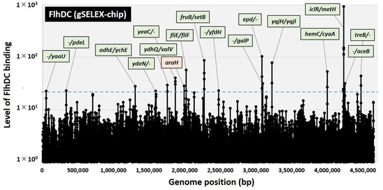
gSELEX fragments and the original DNA library were labeled with Cy3 and Cy5, respectively, to uncover a comprehensive set of FlhDC-controlled targets. A mixture of fluorescently labeled samples was hybridized onto an E. coli DNA tiling microarray [22]. The ratio of the fluorescence intensity between the FlhDC sample and the original DNA library of each probe was measured and plotted against the corresponding position along the E. coli K-12 genome. The extent of FlhDC binding correlated with its FlhDC affinity. After setting the cutoff level to twenty, 17 peaks were identified in the gSELEX-chip pattern (Figure 1), and 38 additional peaks were identified by decreasing the cutoff level to ten (Table 1). Of these 55 high-level binding peaks, 18 FlhDC-binding sites were located within spacers of bidirectional transcription units (Table 1, type-A), and 26 FlhDC-binding sites were located within spacers upstream of one ORF but downstream of another ORF (Table 1, type-B). Eleven FlhDC-binding sites were located within the genes or downstream of both transcription units (Table 1, type-C). Hence, we predicted that the total number of FlhDC regulatory targets was between at least 44 (18 type-A and 26 type-B) and 62 (18 × 2 type-A and 26 × 1 type-B).

Figure 1.
Identification of FlhDC-binding sites in the E. coli K-12 genome using gSELEX-chip. gSELEX screening of DNA-binding sequences was performed for FlhDC, a flagella master regulator TF of E. coli, using purified His-tagged FlhDC and a library of DNA segments from the E. coli K-12 W3110 genome. Following gSELEX, a collection of DNA fragments was subjected to gSELEX-chip analysis using the tiling array of the E. coli K-12 genome. The blue dotted line represents the cutoff level of twenty, and Table 1 lists the FlhDC-binding sites from cutoff level ten. Peaks shown in green represent the FlhDC-binding sites inside spacer regions, whereas peaks shown in orange represent the FlhDC-binding sites inside ORFs.

Table 1.
FlhDC-binding sites on the E. coli genome.
Currently, the RegulonDB database lists 20 genes or operons as FlhDC targets ([23,24]. regulondb.ccg.unam.mx (accessed on 1 December 2022)). gSELEX screening detected seven of these targets above the cutoff level of ten (Table 1), with the highest peak intensity at 54 for the fliE gene, which encodes a component of the flagellum hook-basal body subunit. Notably, at this cutoff setting, many known FlhDC targets involved with the flagella genes, including the flgAMN, flgBCDEFGHIJ, fliAZ-tcyJ, and fliLMNOPQR operons, were detected (Table 1). However, the known target, the fliDST operon, which is critical in flagellin biosynthesis and assembly, showed a peak intensity of six.
2.2. Verification of FlhDC Consensus Recognition Sequence
Using the DNA-binding sequence information from a set of FlhDC target sequences, a 42–44 bp palindromic sequence consisting of AAYGSSNNAAATAGCG-N10-12-CGCTATTTNNSSCRTT was proposed as the consensus recognition sequence for FlhDC [11,12,13,14]. As we obtained 49 new FlhDC-binding sites, the FlhDC consensus sequence was analyzed using the MEME program [25] to identify the FlhDC-box sequence in the newly targeted regions. We identified the same 42–44 bp sequence in 44 of the 55 FlhDC target regions (Table 1), and the high conservation of the A1, A2, G4, A9, A10, A11, T12, and A13 bases was also consistent (Figure 2) [12]. These results suggest that the novel target genes of FlhDC identified by the gSELEX screening was valid.

Figure 2.
Consensus sequence of the FlhDC-box. Sequences of all the probes with high levels of FlhDC binding were analyzed using MEME (https://meme-suite.org/meme/ (accessed on 1 December 2022)) (see Table 1). WEBLOGO (http://weblogo.berkeley.edu/logo.cgi (accessed on 1 December 2022)) was used to perform matrix construction.
2.3. FlhDC Binding In Vitro to the New Target Genes Involved in Sugar Utilization and Carbon Source Metabolism
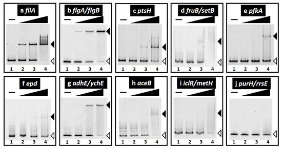
FlhDC (formerly FlbBl) was originally identified as a flagella operon regulator [26]. Subsequently, several research groups have performed comprehensive in vivo analyses, including transcriptome and ChIP-seq analyses [11,12,14]. FlhDC is the principal regulator of bacterial flagellum biogenesis and swarming migration, and a microarray showed that it regulates several non-flagella genes. However, there was no evidence that these genes were directly regulated by FlhDC. gSELEX screening identified FlhDC-binding sites in several promoter regions of genes involved in sugar utilization and carbon source metabolism (Figure S2c–i), in addition to known FlhDC target genes involved in flagella formation (Figure S2a,b). Genes involved in sugar utilization, such as the ptsHI-crr operon (phosphoenolpyruvate: sugar phosphotransferase system (PTS) for PTS sugar utilization), fruBKA operon (fructose-specific PTS system and fructose utilization), and setB (sugar transporter), and genes involved in carbon source metabolism, such as pfkA (6-phosphofructokinase (I), the epd-pgk-fbaA operon (D-erythrose-4-phosphate dehydrogenase, phosphoglycerate kinase, fructose-bisphosphate aldolase (II), adhE (acetaldehyde-CoA dehydrogenase/alcohol dehydrogenase), the aceBAK operon (malate synthase, isocitrate lyase, isocitrate dehydrogenase kinase/phosphatase), and iclR (isocitrate lyase TF), were included in the list of predicted FlhDC targets (Table 1, Figure S2).
We focused on a set of genes involved in sugar utilization and carbon source metabolism to verify this novel role of FlhDC and analyzed the effects of FlhDC on gene regulation. We examined the in vitro binding of purified FlhDC complexes to sequences isolated by gSELEX screening using PAGE to confirm FlhDC–DNA complex formation. Upon increasing FlhDC supplementation, DNA fragments from the probes of known FlhDC targets detected in the gSELEX-chip formed FlhDC–DNA complexes in an FlhDC concentration-dependent manner (Figure 3a,b). The new targets were tested under the same conditions. FlhDC–DNA complex formation was observed for all tested probes depending on the FlhDC concentration (Figure 3c–i). In addition, FlhDC consensus sequences were identified in all the binding regions (Table 1). In contrast, the purH/rrsE intergenic region, the reference DNA added as a negative control (Figure S2j), did not form FlhDC–DNA complexes under the same conditions (Figure 3j). These results indicated specific binding of all seven FlhDC target sequences.

Figure 3.
Gel shift assay of FlhDC–DNA complex formation. (a–j) Purified FlhDC was mixed with 0.1 pmol of each target DNA probe of the FlhDC-binding regions shown in Table 1 (also see Figure S2). FlhDC added: lane 1, 0 nM; lane 2, 10 nM; lane 3, 30 nM; lane 4, 100 nM. The filled triangles indicate the FlhDC–DNA probe complex, whereas open triangles indicate free probes.
2.4. In Vivo Transcription Regulation of the Set of FlhDC Target Genes Involved in Sugar Utilization and Carbon Source Metabolism
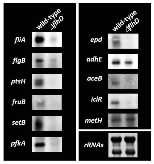
We performed a Northern blotting analysis to examine the possible influence of FlhDC on target promoters detected in vitro based on FlhDC-binding sites and determined mRNA levels in vivo for each of the predicted FlhDC target genes in the presence or absence of FlhDC. Wild-type E. coli K-12 and the flhD-deleted mutant grown in M9-glucose (0.2%) medium and total RNA was extracted in the exponential phase. The mRNA levels of individual FlhDC target genes were analyzed by Northern blotting analysis. The mRNA levels of fliAZY and flgBCDEFGHIJ, known targets for which FlhDC acts as an activator, were detected using fliA and flgB probes, respectively. Both transcripts decreased markedly in the flhD knockout mutant (Figure 4). ptsHI-crr, fruBKA, setB, pfkA, epd-pgk-fbaA, aceBAK, and iclR mRNA levels also significantly decreased in the flhD mutant (Figure 4), implying that FlhDC has an activating role in these transcription units. adhE mRNA levels slightly decreased, indicating that FlhDC also acts as an adhE activator. metH, located in the divergence from iclR, was not affected, suggesting that FlhDC binding between the iclR/metH intergenic region activates iclR. pfkA encodes 6-phosphofructokinase I, a key rate-limiting enzyme in the glycolysis pathway [27,28]. These results suggest that FlhDC directly activates genes involved in flagella formation and carbon source metabolism, mainly the PTS sugar uptake and glycolysis pathways.

Figure 4.
Northern blotting analysis of mRNAs from FlhDC target genes. Wild-type E. coli K-12 BW25113 and its flhD mutant JW1881 were grown in M9 0.2% glucose media. Total RNA was prepared during the exponential phase and subjected to Northern blotting analysis. DIG-labeled hybridization probes are shown on the left side of each panel. The amounts of total RNA analyzed were examined by measuring the intensity of ribosomal RNAs.
2.5. The Effects of FlhDC on Sugar Utilization and Cell Growth
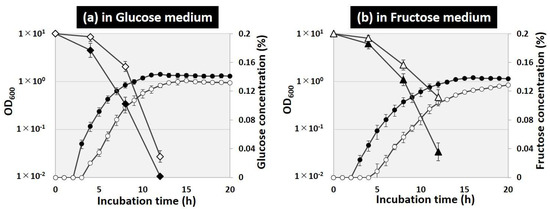
Transcriptional regulation analyses suggested that FlhDC activates a set of carbon source metabolism genes involved in glucose uptake (ptsHI-crr), fructose uptake and utilization (fruBKA), and glycolysis (pfkA, pgk, fbaA) (Figure 4). Cell growth and sugar consumption of wild-type and flhD-deficient strains in glucose and fructose sole carbon source media were observed to evaluate the effects of FlhDC on glucose and fructose uptake and catabolism. After 8 h of incubation in the glucose sole carbon source medium, the cell density of the wild-type strain reached OD600 = 0.85, and the glucose concentration decreased to 0.1% (Figure 5a). The OD600 was less than 0.3 in the flhD-deficient strain, and approximately 0.15% of the glucose remained. After 12 h of incubation, the wild-type strain entered the stationary phase, and the glucose in the medium was depleted, whereas the flhD-deficient strain continued to grow and left 0.03% of the glucose remaining. The effect of flhD deficiency on the fructose medium was similar to that on the glucose medium, with slower growth and more fructose remaining in the medium in the flhD-deficient strain than in the wild-type strain (Figure 5b). Note that there was no difference in cell growth between the wild-type and flhD deficient strains on LB medium (data not shown). These results suggest that flhDC stimulates energy production by accelerating the uptake and catabolism of PTS sugars, such as glucose and fructose.

Figure 5.
Effect of flhD deficiency on the growth curve and sugar consumption. E. coli BW25113 (closed symbols) and its flhD mutant JW1881 (open symbols) were grown in M9 media with 0.2% glucose (a) or 0.2% fructose (b). Cell density (circles in a,b) was monitored every hour and plotted along the time course. The concentrations of glucose (rhomboids in a) and fructose (rhomboids in b) were monitored every 4 h for up to 12 h.
3. Discussion
Motility is necessary for microorganisms to adapt to environmental changes, and the precise control of flagella formation is critical. Flagella formation in E. coli has been well-described [2,3]. The class I flagella master regulator FlhDC initiates the signal cascade mechanism for flagella formation. FlhDC-activated class II genes include those involved in flagella basal body formation and flagella sigma factors, RpoF (σ28). RpoF induction stimulates class III genes to assemble the hook and flagella filaments and complete flagella formation. Comprehensive in vivo analysis has been used to uncover the direct regulation of a set of genes involved in flagella formation [12,14]. Hitherto, microarray analysis showed that FlhDC regulates several non-flagella genes and/or operons involved in metabolism [11,16], such as the glpABC operon for glycerol degradation, the gltI-sroC-gltJKL operon (glutamate/aspartate transporter), mdh (malate dehydrogenase), the mglBAC operon (D-galactose transporter), the napFDAGHBC-ccmABCDEFGH operon (nitrate reductase), and the nrfABCDEFG operon (nitrite reductase) (listed in the RegulonDB database as FlhDC targets). However, the direct effect on these genes has not yet been determined, and only the nrfABCDEFG operon was detected in our gSELEX screening among the FlhDC low-intensity peaks (intensity level of eight; data not shown). In this study, we used a gSELEX analysis, which can identify direct target DNA sequences in vitro, to determine the genomic regulatory mechanisms of FlhDC. The results showed that previously unknown targets of class II flagellum-forming genes are involved in carbon source metabolism (Figure 1, Table 1), and FlhDC activates these genes (Figure 3 and Figure 4). As for the effects of these regulations on the phenotype, FlhDC stimulated the consumption of glucose and fructose and accelerated cell growth (Figure 5).
Novel genes regulated by FlhDC affect sugar utilization and metabolism of carbohydrate sources. The ptsHI-crr operon imports PTS sugars, such as glucose and fructose, and the fruBKA operon controls fructose import and utilization [29]. After uptake, sugars are catabolized in the glycolysis pathway and used for energy production such as ATP and NADH [28]. In the glycolysis pathway, the conversion of D-fructose 6-P to D-fructose 1,6-P2 via 6-phosphofructokinase is crucial for regulating the activity of the glycolysis pathway because this reaction is irreversible. E. coli has two 6-phosphofructokinase genes: pfkA encoding 6-phosphofructokinase 1 (PFK I), and pfkB encoding 6-phosphofructokinase 2 (PFK II). More than 90% of the phosphofructokinase activity in wild-type E. coli can be attributed to PFK I [27], suggesting that pfkA transcriptional activation via FlhDC has a marked effect on glycolysis activation. The epd-pgk-fbaB operon, activated by FlhDC, also contains pgk-encoded phosphoglycerate kinase and fbaB-encoded fructose-bisphosphate aldolase, enzymes for reversible reactions, suggesting that an increased carbon flux in the glycolysis pathway also occurs. Two pathways that metabolize the glycolysis product acetyl coenzyme A (acetyl-CoA) from sugars, the ethanol fermentation and glyoxylate pathways, were also identified as targets of FlhDC. adhE encodes fused acetaldehyde-CoA dehydrogenase and alcohol dehydrogenase. Thus, this enzyme catalyzes sequential two-step reactions, converting acetyl-CoA + NADH → acetaldehyde + coenzyme A + NAD+ and acetaldehyde + NADH → ethanol + NAD+. This multifunctional enzyme (AdhE) acts under anaerobic conditions to re-oxidize NADH to NAD+ because the glycolysis pathway requires NAD+ for sugar catabolism. FlhDC may activate adhE to maintain glycolysis activity under anaerobic conditions and energy production for flagella. In contrast, under aerobic conditions, the TCA cycle consumes an acetyl-CoA-derived carbon source to produce NADH for ATP production via an oxidative electron transfer system. The glyoxylate pathway is an anaplerotic reaction that enables E. coli to use acetyl-CoA as a carbon substrate. It consists of two enzymes, isocitrate lyase encoded by aceA, and malate synthase encoded by aceB. Isocitrate dehydrogenase kinase/phosphatase controls the activity of icd-encoded isocitrate dehydrogenase, a key enzyme for switching shifts between the TCA and glyoxylate pathways. FlhDC may activate the aceABK operon to maintain the activity of the TCA cycle under aerobic conditions to produce NADH for ATP production. On the other hand, this study focused on a group of genes in which FlhDC bound to the promoter region, and several binding regions were identified on the ORF (Table 1). It has been reported that transcription factors bind on ORFs [30] and examining these effects of these bindings may reveal new regulatory mechanisms.
Flagella synthesis and function are costly for cells [2,31]. Recently, it was estimated that all the flagella on a cell consume 10.2% of the intercellular ATP produced by E. coli (5% for construction and 5.2% for operating costs) [32]. Hence, we propose that cooperative transcription regulation of flagella and energy production (sugar utilization and carbon source metabolism) occur via the flagella master regulator FlhDC. Although this study examined the effects of FlhDC on a set of genes involved in central carbon metabolism with high FlhDC-binding strength using gSELEX screening, various other genes involved in carbon source metabolism and other biological functions were identified as novel FlhDC targets. Further studies could validate the effects of these genes to accelerate our understanding of the role of FlhDC in the entire E. coli genome.
4. Materials and Methods
4.1. Bacterial Strains and Plasmids
E. coli K-12 W3110 type-A [33] were used as the DNA source to construct the FlhD and FlhC expression plasmids. The E. coli K-12 W3110 type-A genome was also used to construct the DNA library required for gSELEX screening. E. coli DH5α cells were used for plasmid amplification, and E. coli BL21 (DE3) cells were used for FlhD and FlhC expression. E. coli BW25113 [34] and its flhD single-gene knockout mutant JW1881 [35] were obtained from the E. coli Stock Center (National Bio-Resource Center, Chiba, Japan). The pET21a(+) plasmid was used to construct the FlhD and FlhC co-expression plasmids. Cells were grown in an LB medium at 37 °C with constant shaking at 150 rpm. When necessary, 20 μg mL−1 of kanamycin or 50 μg mL−1 of ampicillin were added to the medium. Cell growth was monitored by measuring the turbidity at 600 nm.
4.2. Purifying the FlhDC Protein
A pFlhDC plasmid for expressing and purifying FlhDC was constructed according to the standard procedure, with slight modifications [19]. Briefly, FlhDC-coding sequences were amplified via PCR with a pair of primers (GTGGGACATATGCATCATCATCATCATCATCATACCTCCGAGTTGCTGAA and TACCGCTGCGCGGCCGCTCAGTGGTGGTGGTGGTGGTGAACAGCCTGTACTCTCTGTT) using E. coli K-12 W3110 genomic DNA as a template. They were inserted into the pET21a (+) vector (Novagen, Vadodara, India) between the NdeI and NotI sites, with the addition of a His x6 tag to the C-terminus of flhC, leading to the construction of the pFlhDC. The pFlhDC expression plasmid was transformed into E. coli BL21 (DE3) cells. Transformants were grown in an LB medium, and FlhD and FlhC were co-expressed by adding IPTG during the exponential phase. The FlhDC protein complex was purified by affinity purification using a NTA agarose column. The affinity-purified FlhDC protein complex was frozen in the storage buffer at −80 °C until use. Protein purity was greater than 90%, as determined by SDS-PAGE.
4.3. gSELEX Screening of FlhDC-Binding Sequences
gSELEX screening was performed as previously described [19,20]. Briefly, a mixture of DNA fragments from the E. coli K-12 W3110 genome was prepared by sonicating purified genomic DNA and cloned into a multi-copy plasmid, pBR322. For each gSELEX screening, the DNA mixture was regenerated by PCR. For gSELEX screening, 5 pmol of the DNA fragment mixture and 200 pmol FlhDC were mixed in a binding buffer (10 mM Tris-HCl, pH 7.8 at 4 °C, 3 mM magnesium acetate, 150 mM NaCl, and 1.25 mg/mL bovine serum albumin). The SELEX cycle was repeated six times to enrich the FlhDC-binding sequences. SELEX fragments were mapped along the E. coli genome using a gSELEX-chip system with a 43,450-feature DNA microarray [30]. The gSELEX sample obtained using FlhDC and the original genomic DNA library were labeled with Cy3 and Cy5, respectively. Following sample hybridization to the DNA tilling array (Agilent Technologies, Santa Clara, CA, USA), the Cy3/Cy5 ratio was measured, and the peaks of the scanned patterns were plotted against the positions of the DNA probes along the E. coli K-12 genome.
4.4. Gel Shift Assay
A gel shift assay was performed according to the standard procedure [36]. Probes for FlhDC-binding target sequences were generated via PCR amplification using a pair of primers (Table S1a) and Ex Taq DNA polymerase (TaKaRa). A mixture of each probe and FlhDC was incubated at 37 °C for 30 min in a gel shift buffer. After adding the DNA-loading solution, the mixture was subjected to PAGE. The gel was stained with GelRed (Biotium, Fremont, CA, USA) and detected using a LuminoGraph I (Atto, Amherst, MA, USA).
4.5. Consensus Sequence Analysis
An FlhDC-binding sequence set identified by the gSELEX-chip was analyzed using the MEME program [25] to analyze the FlhDC-binding sequence. Sequences were aligned, and a consensus sequence logo was created using WEBLOGO (http://weblogo.berkeley.edu/logo.cgi (accessed on 1 December 2022)).
4.6. Northern Blotting Analysis
Total RNA was extracted from the exponential phase of E. coli cells (OD600 = 0.4) in an M9 minimal medium supplemented with 0.2% glucose using an ISOGEN solution (Nippon Gene, Tokyo, Japan). RNA purity was verified by electrophoresis on a 1.5% agarose gel with formaldehyde, followed by staining with GelRed. Northern blotting analysis was performed as previously described [37]. DIG-labeled probes were prepared by PCR amplification using W3110 genomic DNA (50 ng) as a template with a pair of primers (Table S1b), DIG-11-dUTP (Roche, Basel, Switzerland), dNTP, gene-specific forward and reverse primers, and Ex Taq DNA polymerase. Total RNA (3 μg) was incubated in a formaldehyde-MOPS gel-loading buffer for 10 min at 65 °C for denaturation, subjected to electrophoresis on a formaldehyde-supplemented 1.5% agarose gel, and transferred onto a nylon membrane (Roche). Hybridization was performed using a DIG easy Hyb system (Roche) at 50 °C overnight with a DIG-labeled probe. The membrane was treated with anti-DIG-AP Fab fragments and CDP-Star (Roche) to detect the DIG-labeled probe, and the image was scanned using LuminoGraph I (Atto).
4.7. Measuring Glucose and Fructose Concentrations in the Culture Medium
E. coli cells were grown in an M9 minimal medium supplemented with 0.2% glucose or 0.2% fructose. A 0.5 mL aliquot of the bacterial culture was collected in a 1.5 mL tube and centrifuged at 20,400× g for 2 min. The supernatant was transferred to a new 1.5 mL tube for glucose and fructose measurements. The concentrations of glucose and fructose in the samples were measured using the ENZYTEC Liquid for D-Glucose/D-Fructose assay (R-Biopharm, Darmstadt, Germany).
Supplementary Materials
The following supporting information can be downloaded at: https://www.mdpi.com/article/10.3390/ijms24043696/s1.
Author Contributions
Conceptualization, T.S. and A.I. (Akira Ishihama); methodology, T.S., A.I. (Akira Ishiguro), H.T. and K.K.; formal analysis, H.T., T.S., K.K. and A.I. (Akira Ishiguro); investigation, H.T., T.S. and K.K.; resources, A.I. (Akira Ishihama) and T.S.; writing—original draft preparation, T.S. and H.T.; writing—review and editing, A.I. (Akira Ishiguro), T.S., H.T. and K.K.; funding acquisition, T.S., A.I. (Akira Ishihama) and H.T. All authors have read and agreed to the published version of the manuscript.
Funding
This research was funded by the MEXT Grants-in-Aid for Scientific Research (C) (22K06184) to T.S., (B) (18310133) and (C) (25430173) to A.I., and (Young Scientists B) (15K18676) to H.T.
Data Availability Statement
gSELEX data for FlhDC have been deposited in the ‘Transcription factor profiling of Escherichia coli’ (TEC) database at the National Institute of Genetics (https://shigen.nig.ac.jp/ecoli/tec/ (accessed on 13 December 2022)).
Acknowledgments
We thank the National BioResource Project, National Institute of Genetics, Japan, for providing the E. coli K-12 BW25113 and its single-gene deletion mutants.
Conflicts of Interest
The authors declare no conflict of interest.
Abbreviations
SELEX (systematic evolution of ligands by exponential enrichment), TF (transcription factor), PAGE (polyacrylamide gel electrophoresis), SDS-PAGE (sodium dodecyl-sulphate PAGE), LB (Luria-Bertani), NTA (Ni-nitrilotriacetic acid), MOPS (morpholine propanesulfonic acid), DIG (digoxigenin), PTS (phosphotransferase system), ATP (adenosine triphosphate), PCR (polymerase chain reaction), LB (lysogeny broth), ORF (open reading frame), NADH (nicotinamide adenine dinucleotide), PFK I (6-phosphofructokinase 1), PFK II (6-phosphofructokinase 2), Acetyl-CoA (acetyl coenzyme A), RNA-seq (RNA sequencing), ChIP-seq (chromatin immunoprecipitation sequencing), CRP (cAMP receptor protein), H-NS (histone-like nucleoid structuring protein), QseB (Quorum sensing Escherichia coli regulators B), IHF (integration host factor), TCA (tricarboxylic acid), IPTG (isopropyl ß-D-1-thiogalactopyranoside), dNTP (deoxynucleoside triphosphate).
References
- Macnab, R.M. Flagella and motility. In Escherichia coli and Salmonella typhimurium: Cellular and Molecular Biology; Neidhardt, F.C., Ed.; American Society for Microbiology: Washington, DC, USA, 1996; pp. 123–145. [Google Scholar]
- Soutourina, O.A.; Bertin, P.N. Regulation cascade of flagellar expression in Gram-negative bacteria. FEMS Microbiol. Rev. 2003, 27, 505–523. [Google Scholar] [CrossRef] [PubMed]
- Apel, D.; Surette, M.G. Bringing order to a complex molecular machine: The assembly of the bacterial flagella. Biochim. Biophys. Acta 2008, 1778, 1851–1858. [Google Scholar] [CrossRef] [PubMed]
- Komeda, Y. Transcriptional control of flagellar genes in Escherichia coli K-12. J. Bacteriol. 1986, 168, 1315–1318. [Google Scholar] [CrossRef]
- Ohnishi, I.; Kutsukake, K.; Suzuki, H.; Iino, T. Gene fliA encodes an alternative sigma factor specific for flagellar operons in Salmonella typhimurium. Mol. Gen. Genet. 1990, 221, 1139–1147. [Google Scholar] [CrossRef] [PubMed]
- Shimada, T.; Tanaka, K.; Ishihama, A. The whole set of the constitutive promoters recognized by four minor sigma subunits of Escherichia coli RNA polymerase. PLoS ONE 2017, 12, e0179181. [Google Scholar] [CrossRef] [PubMed]
- Kutsukake, K.; Iyoda, S.; Ohnishi, K.; Iino, T. Genetic and molecular analyses of the interaction between the flagellum-specific sigma and anti-sigma factors in Salmonella typhimurium. EMBO J. 1994, 13, 4568–4576. [Google Scholar] [CrossRef] [PubMed]
- Hughes, K.T.; Gillen, K.L.; Semon, M.J.; Karlinsey, J.E. Sensing structural intermediates in bacterial flagellar assembly by export of a negative regulator. Science 1993, 262, 277–280. [Google Scholar] [CrossRef] [PubMed]
- Liu, X.; Matsumura, P. The FlhD/FlhC complex, a transcriptional activator of the Escherichia coli flagellar class II operons. J. Bacteriol. 1994, 176, 7345–7351. [Google Scholar] [CrossRef] [PubMed]
- Liu, X.; Fujita, N.; Ishihama, A.; Matsumura, P. The C-terminal region of the α subunit of Escherichia coli RNA polymerase is required for transcriptional activation of the flagellar level II operons by the FlhD/FlhC complex. J. Bacteriol. 1995, 177, 5186–5188. [Google Scholar] [CrossRef]
- Stafford, G.P.; Ogi, T.; Hughes, C. Binding and transcriptional activation of non-flagellar genes by the Escherichia coli flagellar master regulator FlhD2C2. Microbiology 2005, 151, 1779–1788. [Google Scholar] [CrossRef]
- Zhao, K.; Liu, M.; Burgess, R.R. Adaptation in bacterial flagellar and motility systems: From regulon members to ‘foraging’-like behavior in E. coli. Nucleic Acids Res. 2007, 35, 4441–4452. [Google Scholar] [CrossRef] [PubMed]
- Lee, Y.Y.; Barker, C.S.; Matsumura, P.; Belas, R. Refining the binding of the Escherichia coli flagellar master regulator, FlhD4C2, on a base-specific level. J. Bacteriol. 2011, 193, 4057–4068. [Google Scholar] [CrossRef] [PubMed]
- Fitzgerald, D.M.; Bonocora, R.P.; Wade, J.T. Comprehensive mapping of the Escherichia coli flagellar regulatory network. PLoS Genet. 2014, 11, e1005456. [Google Scholar] [CrossRef] [PubMed]
- Ishihama, A.; Shimada, T.; Yamazaki, Y. Transcription profile of Escherichia coli: Genomic SELEX search for regulatory targets of transcription factors. Nucleic Acids Res. 2016, 44, 2058–2074. [Google Scholar] [CrossRef] [PubMed]
- Pruss, B.M.; Liu, X.; Hendrickson, W.; Matsumura, P. FlhD/FlhC-regulated promoters analyzed by gene array and lacZ gene fusions. FEMS Microbiol. Lett. 2001, 197, 91–97. [Google Scholar] [CrossRef]
- Ishihama, A. Prokaryotic genome regulation: A revolutionary paradigm. Proc. Jpn. Acad. Ser. B Phys. Biol. Sci. 2012, 88, 485–508. [Google Scholar] [CrossRef]
- Ishihama, A. Prokaryotic genome regulation: Multifactor promoters, multitarget regulators and hierarchic networks. FEMS Microbiol. Rev. 2010, 34, 628–645. [Google Scholar] [CrossRef]
- Shimada, T.; Fujita, N.; Maeda, M.; Ishihama, A. Systematic search for the Cra-binding promoters using genomic SELEX system. Genes Cells 2005, 10, 907–918. [Google Scholar] [CrossRef]
- Shimada, T.; Ogasawara, H.; Ishihama, A. Genomic SELEX screening of regulatory targets of Escherichia coli transcription factors. Methods Mol. Biol. 2018, 1837, 49–69. [Google Scholar]
- Shimada, T.; Ogasawara, H.; Ishihama, A. Single-target regulators form a minor group of transcription factors in Escherichia coli K-12. Nucleic Acids Res. 2018, 46, 3921–3936. [Google Scholar] [CrossRef]
- Shimada, T.; Bridier, A.; Briandet, R.; Ishihama, A. Novel roles of LeuO in transcription regulation of E. coli genome: Antagonistic interplay with the universal silencer H-NS. Mol. Microbiol. 2011, 82, 378–397. [Google Scholar] [CrossRef] [PubMed]
- Salgado, H.; Gama-Castro, S.; Martínez-Antonio, A.; Diaz-Peredo, E.; Sánchez-Solano, F.; Peralta-Gil, M.; García-Alonso, D.; Jimenez-Jacinto, V.; Sánchez-Zavaleta, A.; Bonavides-Martínez, C.; et al. RegulonDB (version 4.0): Transcriptional regulation, operon organization and growth conditions in Escherichia coli K-12. Nucleic Acids Res. 2004, 32, 303D–306D. [Google Scholar] [CrossRef] [PubMed]
- Salgado, H.; Peralta-Gil, M.; Gama-Castro, S.; Santos-Zavaleta, A.; Muñiz-Rascado, L.; García-Sotelo, J.S.; Weiss, V.; Solano-Lira, H.; Martínez-Flores, I.; Medina-Rivera, A.; et al. RegulonDB v8.0: Omics data sets, evolutionary conservation, regulatory phrases, cross-validated gold standards and more. Nucleic Acids Res. 2013, 41, D203–D213. [Google Scholar] [CrossRef]
- Bailey, T.L.; Boden, M.; Buske, F.A.; Frith, M.; Grant, C.E.; Clementi, L.; Ren, J.; Li, W.W.; Noble, W.S. MEME SUITE: Tools for motif discovery and searching. Nucleic Acids Res. 2009, 37, W202–W208. [Google Scholar] [CrossRef] [PubMed]
- Bartlett, D.H.; Frantz, B.B.; Matsumura, P. Flagellar transcriptional activators FlbB and FlaI: Gene sequences and 5’ consensus sequences of operons under FlbB and FlaI control. J. Bacteriol. 1988, 170, 1575–1581. [Google Scholar] [CrossRef]
- Kotlarz, D.; Garreau, H.; Buc, H. Regulation of the amount and of the activity of phosphofructokinases and pyruvate kinases in Escherichia coli. Biochim. Biophys. Acta 1975, 381, 257–268. [Google Scholar] [CrossRef] [PubMed]
- Berg, J.M.; Tymoczko, J.L.; Stryer, L. Biochemistry, 7th ed.; W.H.Freeman: New York, NY, USA, 2010. [Google Scholar]
- Deutscher, J. The mechanisms of carbon catabolite repression in bacteria. Curr. Opin. Microbiol. 2006, 11, 87–93. [Google Scholar] [CrossRef]
- Shimada, T.; Ishihama, A.; Busby, S.J.W.; Grainger, D.C. The Escherichia coli RutR transcription factor binds at targets within genes as well as intergenic regions. Nucleic Acids Res. 2008, 36, 3950–3955. [Google Scholar] [CrossRef] [PubMed]
- Toft, C.; Fares, M.A. The evolution of the flagellar assembly pathway in endosymbiotic bacterial genomes. Mol. Biol. Evol. 2008, 22, 2069–2076. [Google Scholar] [CrossRef] [PubMed]
- Schavemaker, P.E.; Lynch, M. Flagellar energy costs across the tree of life. eLife 2022, 11, e77266. [Google Scholar] [CrossRef]
- Jishage, M.; Ishihama, A. Variation in RNA polymerase sigma subunit composition within different stocks of Escherichia coli W3110. J. Bacteriol. 1997, 179, 959–963. [Google Scholar] [CrossRef]
- Datsenko, K.A.; Wanner, B.L. One-step inactivation of chromosomal genes in Escherichia coli K-12 using PCR products. Proc. Natl. Acad. Sci. USA 2000, 97, 6640–6645. [Google Scholar] [CrossRef]
- Baba, T.; Ara, T.; Hasegawa, M.; Takai, Y.; Okumura, Y.; Baba, M.; Datsenko, K.A.; Tomita, M.; Wanner, B.L.; Mori, H. Construction of Escherichia coli K-12 in-frame, single-gene knockout mutants: The Keio collection. Mol. Syst. Biol. 2006, 2, 2006-0008. [Google Scholar] [CrossRef] [PubMed]
- Shimada, T.; Tanaka, K.; Ishihama, A. Transcription factor DecR (YbaO) controls detoxification of L-cysteine in Escherichia coli. Microbiology 2016, 162, 1698–1707. [Google Scholar] [CrossRef]
- Shimada, T.; Saito, N.; Maeda, M.; Tanaka, K.; Ishihama, A. Expanded roles of leucine-responsive regulatory protein in transcription regulation of the Escherichia coli genome: Genomic SELEX screening of the regulation targets. Microb. Genom. 2015, 1, e000001. [Google Scholar] [CrossRef]
Disclaimer/Publisher’s Note: The statements, opinions and data contained in all publications are solely those of the individual author(s) and contributor(s) and not of MDPI and/or the editor(s). MDPI and/or the editor(s) disclaim responsibility for any injury to people or property resulting from any ideas, methods, instructions or products referred to in the content. |
© 2023 by the authors. Licensee MDPI, Basel, Switzerland. This article is an open access article distributed under the terms and conditions of the Creative Commons Attribution (CC BY) license (https://creativecommons.org/licenses/by/4.0/).