Article

CRISPR/Cas9-Targeted Disruption of Two Highly Homologous Arabidopsis thaliana DSS1 Genes with Roles in Development and the Oxidative Stress Response

by
Ivana Nikolić
,
Jelena Samardžić
,
Strahinja Stevanović
,
Jovanka Miljuš-Đukić
,
Mira Milisavljević
and
Gordana Timotijević
*
Laboratory for Plant Molecular Biology, Institute of Molecular Genetics and Genetic Engineering, University of Belgrade, Vojvode Stepe 444a, 11042 Belgrade, Serbia
*
Author to whom correspondence should be addressed.
Deceased.
Int. J. Mol. Sci. 2023, 24(3), 2442; https://doi.org/10.3390/ijms24032442
Submission received: 23 December 2022 / Revised: 13 January 2023 / Accepted: 21 January 2023 / Published: 26 January 2023
(This article belongs to the Section Molecular Plant Sciences)

Abstract

:
Global climate change has a detrimental effect on plant growth and health, causing serious losses in agriculture. Investigation of the molecular mechanisms of plant responses to various environmental pressures and the generation of plants tolerant to abiotic stress are imperative to modern plant science. In this paper, we focus on the application of the well-established technology CRISPR/Cas9 genome editing to better understand the functioning of the intrinsically disordered protein DSS1 in plant response to oxidative stress. The Arabidopsis genome contains two highly homologous DSS1 genes, AtDSS1(I) and AtDSS1(V). This study was designed to identify the functional differences between AtDSS1s, focusing on their potential roles in oxidative stress. We generated single dss1(I) and dss1(V) mutant lines of both Arabidopsis DSS1 genes using CRISPR/Cas9 technology. The homozygous mutant lines with large indels (dss1(I)del25 and dss1(V)ins18) were phenotypically characterized during plant development and their sensitivity to oxidative stress was analyzed. The characterization of mutant lines revealed differences in root and stem lengths, and rosette area size. Plants with a disrupted AtDSS1(V) gene exhibited lower survival rates and increased levels of oxidized proteins in comparison to WT plants exposed to oxidative stress induced by hydrogen peroxide. In this work, the dss1 double mutant was not obtained due to embryonic lethality. These results suggest that the DSS1(V) protein could be an important molecular component in plant abiotic stress response.

1. Introduction

DSS1 (deletion of split hand/split foot 1) is a highly conserved acidic eukaryotic protein consisting of a short chain ranging from 70–90 amino acids and a mass of 7–9 kDa, depending on the species. The DSS1 protein or SHFM1 (split hand/split foot malformation type 1) was primarily linked to the human developmental syndrome characterized by the absence and/or fusion of certain fingers [1]. As an intrinsically disordered protein, DSS1 interacts with components of multiple protein complexes and participates in diverse biological functions due to its flexible and non-defined three-dimensional structure [2,3,4,5,6]. DSS1 from different species has an alpha helix at the C-terminus end and two conserved acidic regions with a poorly conserved linker between them [3]. DSS1 protein without a helix complements the Schizosaccharomyces pombe dss1 mutant phenotype; accordingly, most bindings to DSS1 occur in the disordered region [7]. Its wide range of functions is manifested in its participation in the ubiquitin-26S proteasome system [8,9,10], the breast cancer 2 (BRCA2)-DNA repair complex [11,12,13], the pre-mRNA splicing complex [14] and the transcription-export-2 (TREX-2) complex [15]. In budding yeast, the suppressor of exocyst mutations 1 (Sem1), the ortholog of human DSS1, establishes a greater number of interactions with proteins that contain the proteasome, COP9 signalosome, eukaryotic translation initiation factor 3 (PCI) domain, such as proteins of 26S proteasome, constitutive photomorphogenesis 9 (COP9) signalosome and the eukaryotic translation initiation factor 3 (eIF3) [14,15]. Importantly, PCI-domain-containing proteins are necessary for post-transcriptional gene and protein regulation [16].
In the light of proteostasis, Sem1 is the smallest regulatory and structural subunit of the 26S proteasome where it participates in the proteasome assembly. It contributes to the stability of the 19S proteasome lid by the successive recruitment of the regulatory particle non-ATPase 3 (Rpn3) and Rpn7 lid subunits [8,17,18]. While proteasome assembly is possible, it is not fully functional in the absence of Sem1 because Sem1 is necessary for Rpn7 modulation for efficient ATP-dependent substrate unfolding during proteolysis [19].
In the homologous recombination (HR) repair pathway, the most accurate mechanism in double-strand break repair, mammalian DSS1 interacts with the predominant mediator, the tumor suppressor protein BRCA2. DSS1 blocks BRCA2 multimerization and stabilizes the BRCA2 monomer through major structural rearrangements [13]. Overall, DSS1 is necessary for the proper activity of this recombination mediator protein [20,21,22]. In addition, the small acidic DSS1 protein has been shown to mimic DNA and to reduce the affinity of the replication protein A (RPA) for single-stranded DNA in exchange for RAD51 loading to single-strand DNA during repair [23]. In the HR machinery, DSS1 is likely to form protein complexes that do not contain PCI domains [7].
Sem1 is part of a large system that is important for transcription and pre-mRNA splicing during nuclear export. It was suggested that Sem1 as a subunit is involved in two structurally related complexes with a distinctive function in mRNA processing. The first complex is the ternary Thp3-Csn12-Sem1 complex, which mediates pre-mRNA splicing. Thp3 tethers to the Csn2-Sem1 binary complex where Sem1 strongly connects only with the Cop9 signalosome complex subunit 12 (Csn12) subunit [24,25]. The second complex is the nuclear mRNA-export complex that is built by Sac3-Thp1-Sem1. Although Sem1, as a single chain, binds primarily to Thp1, it establishes a weak contact with Sac3. Tethering between the human homologs Sem1 (DSS1) and Thp1 (PCID2) was also confirmed. It appears that Sem1 has a stabilizing role on Scn12 and Thp1 that facilitates complex assembly [25]. Once again, it is proved that Sem1 stabilizes the PCI domain-containing proteins and promotes complex assembly.
Interestingly, besides the well-known role of the DSS1 protein in 26S proteasome constitution, where it serves as a linkage between proteasome building blocks, it has been revealed that this molecule also interacts with the oxidized proteins [26]. Namely, in the context of a high reactive oxygen species (ROS) level and molecular damage, DSS1 plays a role in damaged protein labelling, thus leading to their degradation through the ubiquitin-proteasome system. This novel post-translational modification that leads to recognition and removal of oxidized protein was named DSSylation [26]. However, no data on the role of DSS1 and DSSylation in plant response to oxidative stress are currently available. Thus, our aim was to investigate the contribution of two Arabidopsis DSS1 proteins in maintaining protein homeostasis and to elucidate their potential role in prevention of the accumulation of toxic protein aggregates during oxidative stress.
Noteworthy, the Arabidopsis genome contains two highly homologous genes, DSS1(I) (At1g64750) and DSS1(V) (At5g45010), located in chromosomes I and V, respectively. The best-studied role of plant DSS1 proteins is their involvement in DNA repair mediated by BRCA2-driven HR [27]. There are also two isoforms of BRCA2 in Arabidopsis that can bind two DSS1 proteins, DSS1(V) interacts only with the AtBRCA2 (V) protein, whereas DSS1(I) interacts with both BRCA2(V) and BRCA2(IV) [27]. Studies have shown that Arabidopsis DSS1 proteins are independently associated with both the TREX-2 complex and the 26S proteasome. It is assumed that AtDSS1 proteins potentially link these two complexes when it is necessary to bring them into physical proximity [28]. As was shown in other species, one of the two Arabidopsis homologs, DSS1(V), has been identified as an mRNA nuclear export interaction partner and thus as a TREX-2 component [28].
In the functional analysis of DSS1 genes, several dss1 mutants from different species have been generated and described phenotypically. While sem1 mutants of yeast [29], dss1 mutants of Ustilago maydis [12] and dss-1 mutants of Caenorhabditis elegans [30], have been characterized, information about plant dss1 mutants is insubstantial. Only the T-DNA Atdss1(V) mutant with intron insertion has been characterized so far [31].
Understanding the possible functional dissimilarity of highly homologous genes in the genome of certain organisms is a challenge. A widely used efficient and precise gene editing strategy based on the clustered regularly interspaced short palindromic repeats-associated protein 9 system (CRISPR/Cas9) is routinely applied in experimental models from bacteria to mammalian cell lines and is also easily adapted for plant genome modification [32]. The CRISPR/Cas9 strategy has several advantages in plant technology, from the examination of gene functions to the improvement of crop varieties [33,34,35]. Carefully designed single guide RNA (sgRNA) ensures specific targeting, even when the genome contains closely related paralogous genes [36].
In this paper, we analyzed the structural differences between DSS1(I) and DSS1(V) as well as their interactions with putative protein partners in silico. The approach for the creation and selection of mutations in these two highly similar genes via the Agrobacterium-mediated CRISPR/Cas9 genome editing system is also reported. The generated mutant lines were phenotypically characterized and compared to wild-type (WT) plants during plant development. In addition, the present study shows the results of the examination of the susceptibility of both Arabidopsis dss1 mutants to oxidative stress and of different levels of oxidized proteins between them.

2. Results

2.1. In Silico Comparative Analysis of DSS1(I) and DSS1(V) Proteins and Prediction of Their Specific Interactomes

We analyzed and compared the primary amino acid sequences of DSS1(I) and DSS1(V) using the PSIPRED algorithm, and the alignment is shown in Figure 1A, where blue and red are the hydrophilic and hydrophobic parts, respectively. A remarkable similarity was noted between these two isoforms: DSS1(V) is only one amino acid shorter and contains seven different amino acids in comparison to DSS1(I). Out of these seven alterations, only one represents a significant change in amino acid polarity—from glutamine to leucine at position 44 (Q44L) (Figure 1A, yellow square). Potential differences in folding between DSS1(I) and DSS1(V) isoforms due to amino acid changes are shown in Figure 1B. The mutation annotated as Q44L could also distinguish the polarity properties between DSS1 types, which could contribute to different protein folding.
Due to the intrinsically disordered nature of DSS1 proteins, the docking results were insufficient to accurately explain the differences between DSS1(I) and DSS1(V) in interactions with their protein partners. However, a comparison of the protein–protein interaction between DSS1s and their potentially different partners may be useful in elucidating the evolutionary nature of the effect of a single amino acid change. Figure 1C shows the energy requirements of DSS1s to bind to respective potential partners. The length of the bar correlates with the energy needed for the interaction. Interactions of DSS1(I) are shown in blue and of DSS1(V) in red. According to the binding energies, DSS1(I) protein binds more easily to RPNs and EMBRYO DEFECTIVE 2719 (EMB2719, AT1G20200) but with a lower affinity to CHLOROPLAST HEAT SHOCK PROTEIN 70-1 (AT4G24280) and the PCI/PINT associated module (PAM) domain protein (AT1G75990). The binding of both DSS1 types to BRCA2B and non-ATPase subunit 9 (ATS9) appears to be energetically similar (Figure S1). In addition, in terms of binding energies, only enhanced ethylene response 5 protein (EERH5, AT2G19560) could bind DSS1(V), whereas it was predicted that the AAA-type ATPase family protein (AT5G2000) could be an exclusive DSS1(I) partner (Figure 1C).

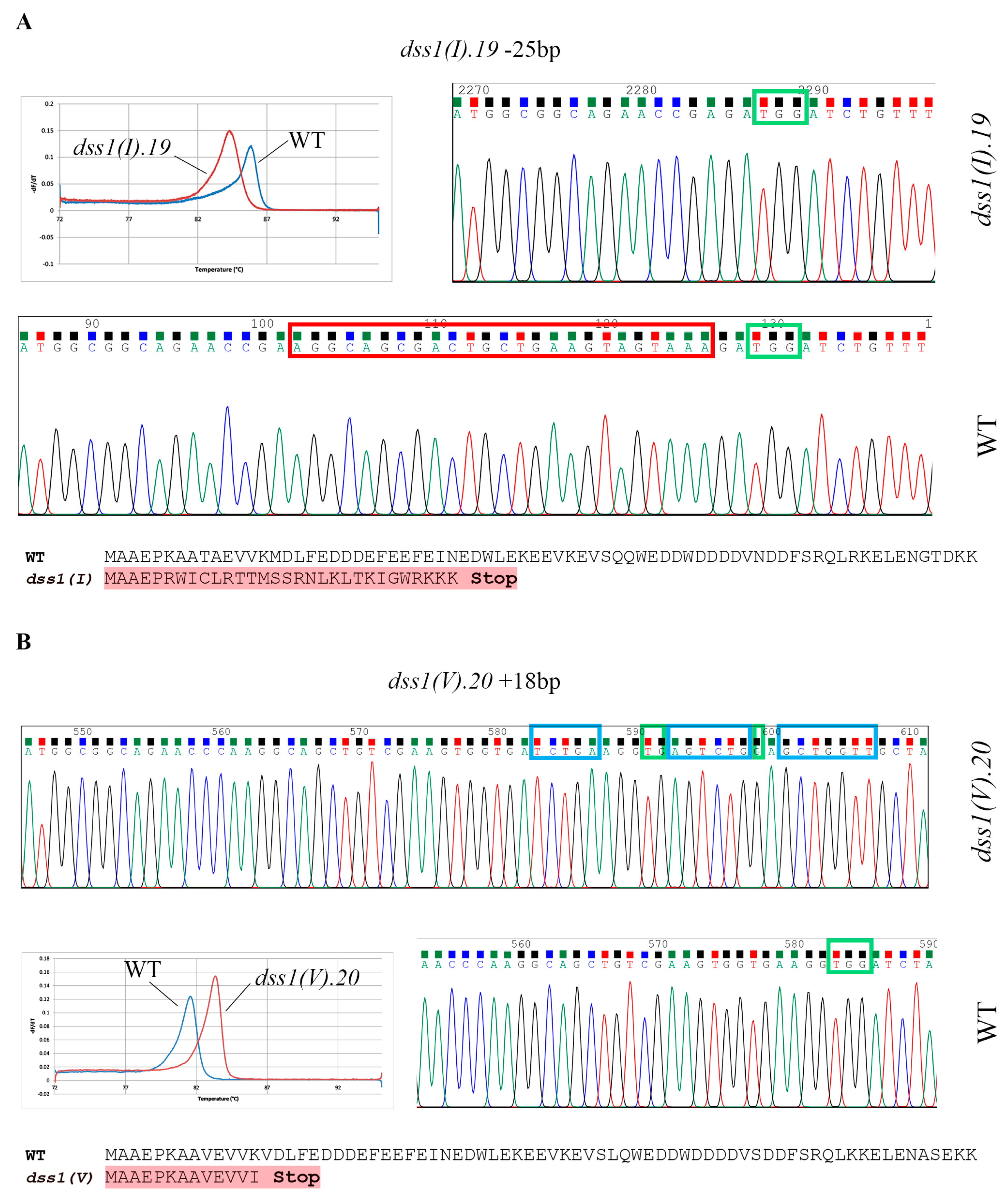
2.2. CRISPR/Cas9 Based Mutagenesis of Two Highly Homologous DSS1 Genes

To date, only Arabidopsis dss1(V) was subjected to mutant analysis and partial characterization. Homozygous T-DNA insertional dss1(V) mutant plants were obtained and they showed a 75% reduction in the level of gene expression. Physiologically, the mutants displayed a significant reduction in rosette size and shoot length during growth, as well as a higher sensitivity to oxidative stress [31]. However, since two gene copies, DSS1(I) and DSS1(V), are present in the Arabidopsis genome, single genetic mutants need to be generated for appropriate functional analyses.
We designed two separate sgRNA oligonucleotide inserts targeting the Arabidopsis DSS1(I) and DSS1(V) genes and subsequently constructed vectors for CRISPR/Cas9 gene-editing. Mutagenesis of duplicated genes with almost identical and extremely short sequences and functional redundancy was a challenge. To predict suitable sgRNA sequences, we had to meet important requirements to obtain high-quality target guide sequences. Namely, gDNA had to be located in the coding region near the 5’-end of the genes, to contain a suitable protospacer adjacent motif (PAM) sequence for Cas9 recognition, to be highly specific to avoid off-targeting, and to contain specific restriction sites at the position of the expected mutation for rapid screening by restriction analysis. Two separate sgRNAs (sgRNA-DSS1(I) and sgRNA-DSS1(V)) were created to target the first exon of both DSS1(I) and DSS1(V) genes that span the sequence (Figure 2A and Figure 3A). The sgRNAs were separately assembled with CRISPR/Cas9 constructs using the binary pHEE401E vectors containing a Zea mays codon-optimized zCas9 gene under the control of an Arabidopsis egg-cell-specific promoter fused with an egg-cell-specific enhancer (Figure 2B and Figure 3B). Progeny seeds (T1) of transformed plants (T0) were collected and transferred to agar plates containing hygromycin as a selective marker. A transformation frequency of at least 0.4–0.8% was obtained with seeds collected from only two pots of the infiltrated plants. The first generation of transformed plants (Figure 2C and Figure 3C) was grown and individual plants were self-pollinated to produce the second generation. As we could not find a suitable restriction site that would overlap with the site of a potential indel, the search for potential mutant plants required the analysis of a larger number of potential mutants, as well as several screening approaches. Genotyping of T2 was conducted using three different strategies: PCR fragment length analysis, restriction analysis of PCR products using Bsp143I enzyme (data not shown) and high melting resolution (HRM) PCR. During the search for CRISPR/Cas9-DSS1(I) mutant plants, we conducted the genotyping of tens of young plants by PCR analysis. Amplicons encompassing the CRISPR/Cas9 target site were analyzed and only one PCR product deviating from the expected WT length was detected (Figure 2D). The plant labelled as dss1(I).19 had a fragment that was about 20 nt shorter, which was selected for further sequencing. The heterozygosity of dss1(I).19 was confirmed by HRM analysis (Figure 2E), while Sanger sequencing revealed that plant lost 25 nucleotides in mutated allele (Figure 2F). The gene editing event resulted in the generation of a mutant line with the introduction of a premature stop codon.
The progeny of the hygromycin-resistant CRISPR/Cas9-DSS1(V) plant was subjected to PCR fragment length analysis (Figure 3D). PCR products encompassing the potential target site of mutagenesis in plant dss1(V).20 contained an additional band, as a result of mutagenesis. Sequence analysis confirmed that this plant is a heterozygous mutant line containing an 18 nt insertion in the DSS1(V) gene (Figure 3E). Although the 18 nt long insertion represents an in-frame mutation, a premature stop codon was introduced within the mutated transcript.
Mutant lines with larger indels (del25 nt and ins18 nt) were selected for further characterization because these mutations were easy to trace in further generations. Plants homozygous for dss1(I) and dss1(V) were selected in the T3 generation by HRM, and their homozygosity was confirmed by sequencing (Figure 4). To confirm disruption of the DSS1 genes, i.e., the absence of WT transcripts, forward primers were designed to overlap the site of the putative indels. Using conventional PCR, we confirmed the complete absence of specific DSS1 transcripts in the obtained mutant lines (Figure S2). In order to show that mutant lines are T-DNA free, i.e., that they do not contain CRISPR/Cas9 cassette between T-DNA right/left border sequence, we performed PCR on gDNA isolated from dss1(I).19 and dss1(V).20 and primers that amplify 423 bp of U6-26p promoter sequence. The corresponding gel electrophoresis is presented as Figure S3 in the Supplementary material.
After backcrossing pure homozygous single mutants with WT plants and recovering the desired mutations to the homozygous stage, we proceeded to traditional crossing between dss1(I)−/− and dss1(V)−/− to generate double dss1(I)/dss1(V)/ mutants. Seeds obtained from pollination of dss1(V)/ pistils by dss1(I)/ pollen and vice versa were subjected to genotyping. Plants that were dss1(I)/dss1(V)/+ and dss1(I)/+dss1(V)/ were selected by PCR and this genotype was allowed to self-pollinate. More than hundreds of the produced seedlings were analyzed by HRM and none of the dss1(I)/dss1(V)/ genotypes were detected (data not shown). The inability to generate double mutant strains suggests that the functions of these highly homologous genes may be essential to the plant cell.
To confirm that certain sgRNAs do not introduce off-target mutations, HRM analysis of potential off-target sites was carried out using gDNA from WT and mutant plants as templates. The best ranked potential off-target sites that contained 3 bp or more mismatches were selected for validation (Table S1). The genomic DNA sequences surrounding the potential off-target sites were amplified by PCR using gene-specific primers (Table S2). PCR products were analyzed by Sanger sequencing, and no changes in potential off-targets were detected.

2.3. Phenotypic Characterization of Arabidopsis dss1(I) and dss1(V) Mutant Lines

Plant phenotyping experiments aimed to reveal the correlation between the plant’s genome and its morphological traits. Monitoring was carried out throughout the different developmental stages (Figure 5). Plants were initially grown in MS medium for 2 weeks and then in soil until the end of the life cycle. In the early stage, slower germination of the mutant seedlings was observed compared to the rapid germination of WT controls (Figure 5A). In addition, WT seedlings had fully opened cotyledons, while mutant cotyledons were still closed at this developmental stage. Analysis of 12-day-old seedlings showed that the length of dss1(V) mutants was almost identical to that of the WT, but a significant difference in root length was observed between dss1(I) mutants and WT plants; the dss1(I) plants were 1.6-fold shorter (Figure 5B and Figure S4). At the post-sowing stage, a similar, but statistically non-significant trend continued among the three seedling genotypes on day 25 (Figure 5C). Subsequently, 7-week-old dss1(V) exhibited accelerated development and demonstrated a significant 1.7-fold increase when compared with WT rosettes. In contrast, dss1(I) plants showed a significant 1.9-fold reduction in growth compared with the WT (Figure 5D). The rapid growth of dss1(V) led to faster ripening and splitting open of most siliques and earlier seed release in comparison to WT plants. Compared to the WT, dss1(I) siliques were shorter and produced fewer seeds per silique (Figure 5E). Further measurements showed that dss1(I) stems were on average shorter than dss1(V) and WT (Figure 5F). Overall, the growth of dss1(I) was delayed compared to the WT, whereas dss1(V) developed faster than the WT.

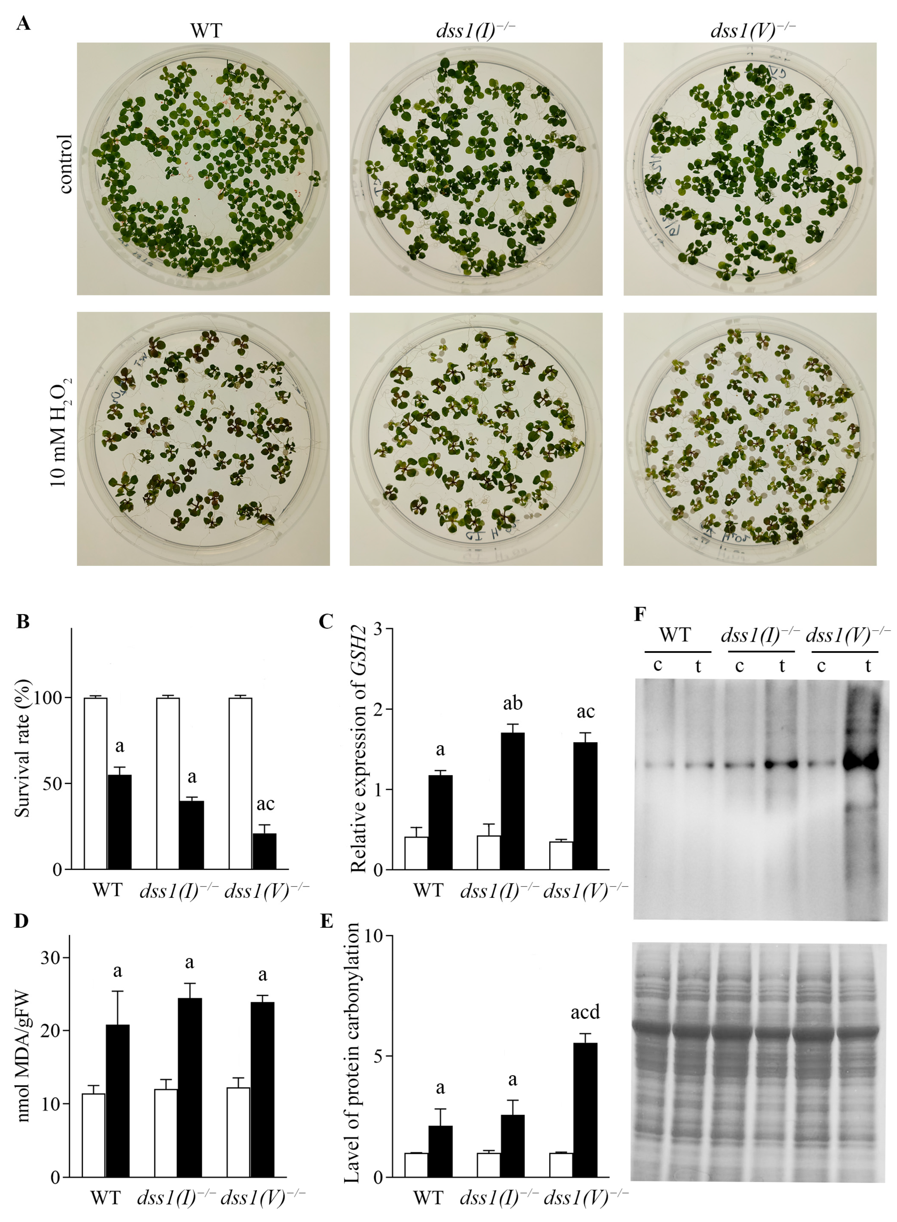
2.4. Sensitivity of Atdss1(I) and Atdss1(V) Lines to Oxidative Stress

Seeds of WT and mutated dss1(I) and dss1(V) lines were germinated in culture media containing 10 mM hydrogen peroxide (H2O2) (Figure 6). Chlorosis of young leaves in mutant lines treated with the stressors at the early stage of stress was more conspicuous than in WT plants (Figure 6A). To quantify the effects of the applied stressor, we determined the survival rate of the seedlings as the percentage of seedlings that germinated after the stress treatment compared to the control seedlings (Figure 6B). In the presence of H2O2, all three lines showed a significantly lower survival rate compared to the control seedlings. Interestingly, under H2O2 stress, dss1(V) plants had a 61% lower survival rate in comparison to WT plants under the same treatment, while the survival rate of dss1(I) seedlings was only 27% lower than the WT. In contrast to dss1(I) mutants, dss1(V) were clearly more sensitive to oxidative stress (Figure 6B).
In addition, the expression profiles of some stress responsive genes were analyzed, such as genes encoding the antioxidant enzymes glutathione synthetase (GSH) and catalase 1 (CAT1). The most significant differences were observed in the expression of the GSH gene (Figure 6C). According to our results, oxidative stress upregulated the expression of GSH in both dss1(I) and dss1(V) plants, as they had significantly elevated levels of GSH transcripts after H2O2 treatment in comparison to WT plants. Specifically, the level of the GSH transcript in the dss1(I) and dss1(V) mutant lines was 31% and 26% higher in comparison to the WT level of expression, respectively (Figure 6C). We found no statistically significant differences in the expression levels of the CAT1 gene among dss1(I), dss1(V) and WT plants exposed to oxidative stress (data not shown).
We also quantified the malondialdehyde (MDA) content as a measure of cellular damage caused by oxidative stress. All three lines exhibited a 20–24% increase in MDA content under oxidative stress when compared with the WT, while under control conditions they showed no significant difference; however, no significant elevation of lipid peroxidation was detected in the mutant lines (Figure 6D). Finally, protein carbonylation, an irreversible oxidative protein modification, was measured by immunoblotting to determine the effects of oxidative stress. The amount of carbonylated products that accumulated in the mutant lines was greater in dss1(I) and dss1(V) plants than in WT plants. The most intensive oxidation was detected in the dss1(V) mutant line, where the amount of carbonylated proteins was 60% higher than in WT plants (Figure 6E,F). In the dss1(I) mutant line, only 18% more oxidatively damaged proteins were recorded than in WT plants. Our results suggest that dss1(V) mutants were more sensitive to oxidative stress than dss1(I) mutants as significantly more carbonylated products were detected in dss1(V) plants.

3. Discussion

It is well known that plants as sessile organisms are constantly exposed to different stimuli from the natural environment. Even small variations in environmental conditions, such as temperature fluctuations or lack of water, high salinity, exposure to heavy metals or radiation, produce an abiotic stress response in plants [37]. Environmental stressors trigger an exaggerated production and accumulation of ROS, which induce comprehensive plant stress response [38]. Molecules containing activated oxygen cause major cellular damage and dysfunctionality of all cellular compartments. Under conditions of oxidative stress, nucleic acids, lipids and proteins are oxidized and must be neutralized immediately to prevent their deleterious effects on cells. Diverse cellular antioxidant defense mechanisms have evolved to reduce oxidative damage in plant cells [39]. One of the most important goals of modern plant science is to identify new factors involved in the efforts of plants to maintain cellular homeostasis and overcome the effects of stress [40]. The results of intensive research on this topic can be used to improve agricultural productivity by sophisticated genetic engineering [41,42].
Our previous study on the role of plant DSS1 proteins indicated that protein and gene expression of plant DSS1 has an altered profile under the influence of different oxidative stressors [31]. Analysis of the Arabidopsis dss1(V)−/− T-DNA insertion mutant line showed increased sensitivity to oxidative stress, suggesting that it may be an important player in the molecular mechanisms of a plant’s abiotic stress defense [31]. The Arabidopsis genome contains two copies of the DSS1 gene that differ slightly in length. DSS1(I) contains 222 bp whereas DSS1(V) contains 219 bp, encoding 74 and 73 amino acid-long protein isoforms, respectively [27]. Herein, we analyzed the primary amino acid sequences of DSS1(I) and DSS1(V) in silico and noted a remarkable similarity. DSS1(V) is shorter than DSS1(I) and differs in seven amino acids but with only one fundamentally significant change, from glutamine to leucine at position 44, which can affect protein structure and function. Considering that the primary amino-acid sequences of DSS1 orthologs in S. pombe, S. cerevisiae [8] and C. elegans [30] contain the polar amino acids threonine or asparagine at the same position in AtDSS1(I), it can be assumed that AtDSS1(V) is the result of gene duplication. About 65% of annotated genes in plant genomes are duplicated, and these genes are often resistant to the selective pressure to which they are normally exposed [43]. This can lead to an accumulation of multiple mutations in the sequences of the duplicated gene, potentially resulting in new functions [44]. The high percentage of homology between two Arabidopsis DSS1 proteins indicates that they may overlap in their functions and partner interactions. Since DSS1s are multifunctional proteins involved in diverse essential biological functions, plant DSS1s are likely to be functionally complemental in most of these functions. However, we hypothesized that minor, but significant changes in the primary structure of these extremely disordered proteins underlie the emergence of new functions.
Additionally, the comparison of protein–protein interactions between AtDSS1 proteins may also be useful in explaining the nature of the interactions and functional differences. According to our prediction, both isoforms generally have the potential to bind the same protein partners. However, we predicted that the distinctive predicted partner of DSS1(I) could be a protein encoded by locus AT5G2000, the 26S proteasome regulatory subunit 8 homologue B. The exclusive partner of DSS1(V) is the EER5 protein, which is involved in promoting a reduced ethylene response upstream or within the ethylene-activated signaling pathway. Accordingly, promoter analyses revealed the presence of ethylene-responsive elements in the DSS1(V) promoter sequence but not in the DSS1(I) promoter [31].
To ascertain other functional relationships between DSS1 genes in cellular protein homeostasis during oxidative stress, we used the CRISPR/Cas9 approach for precise gene mutagenesis. The advantage of the Cas9 system is its capacity to simultaneously edit multiple loci in the same plant, enabling synchronized mutagenesis of multiple functionally related genes that belong to the same gene family [45]. We used CRISPR targeted mutagenesis for separate editing of two highly homologous DSS1 genes. Although multiplexing the CRISPR/Cas9 gene editing approach is a strategy of choice for simultaneous and rapid mutagenesis, we used the traditional crossing method to generate double mutants. Our aim was to ensure the segregation of unwanted mutated loci by dissolving potential off-target mutations through traditional backcrossing with WT plants over several generations and restoration of the desired mutation to a homozygous stage.
Besides several mutants with small indels in each of the DSS1 genes, we managed to select two lines with larger mutations that could be easily confirmed by genotyping. Two CRISPR constructs targeting different DSS1 gene copies were generated and two homozygous mutant lines were selected for further characterization—a mutant line with a deletion of 25 nt in DSS1(I) and a line with an insertion of 18 nt in the DSS1(V) gene. CRISPR/Cas9-induced mutations occurred in both mutant lines and resulted in the generation of premature stop codons and protein truncation.
Several online prediction tools found some off-target loci in addition to the beneficial mutation(s) in DSS1(I) and DSS1(V) genes. However, CRISPR/Cas9 precision and off-target issues are not as problematic in plants as in animals. This is mainly because off-target mutations in plants can be successfully segregated and removed by backcrossing [46]. To prevent the undesired influence of random mutations on dss1 mutant line phenotypes, backcrossing and further selection were conducted. Therefore, potential unwanted mutations were diluted and segregated over several generations. In addition, potential off-targets were screened by HRM PCR and Sanger sequencing.
Further characterization of Arabidopsis mutant lines with disruptions in each of the DSS1 genes (dss1(I)/ and dss1(V)/) revealed visible phenotype dissimilarities in comparison to WT plants. Based on the phenotypic description, the differences between dss1 CRISPR/Cas9 mutants suggest that they most likely have different cellular roles in addition to their overlapping functions, which we have already mentioned. The mature dss1(I) plants produced abnormal siliques and a very tiny habitus with only a few shoots at the fruit-ripening stage. The appearance of abortive seeds indicates a possible disruption in the homologous recombination that takes place in meiosis during sexual reproduction in Arabidopsis. For instance, the absence of AtBRCA2 is known to impair female gametophyte development [47]. Moreover, development of the C. elegans dss-1 mutant germ line was slightly delayed and numerous oocytes were not produced [30]. Owing to the multifunctionality of Atdss1(I), this aberrant regulation may not be exactly in meiosis, but in another DSS1 dependent mechanism required for oogenesis, combined with errors in HR. This suggests that the AtDSS1(I) transcript could be involved in the process of embryogenesis.
It is well known that the ethylene signaling pathway leads to cotyledon expansion when the seedling reaches the soil surface, as well as to silique maturation in the ripening developmental stage [48,49,50]. The presence of ethylene cis-responsive elements in the promotor region of the DSS1(V) gene indicates that this gene is likely an important player in the process of maturation. We assume that plants with a disrupted DSS1(V) gene have closed cotyledons, shorter primary roots in the early stage of development and faster maturing of siliques in comparison to WT plants, possibly because of the defective response of dss1(V) to ethylene.
Interestingly, while the dss1(V) mutant lines generated using CRISPR/Cas9 technology showed similar sensitivity to oxidative stress as the T-DNA insertional dss1(V) mutant plants from our previous work, significant and unforeseen phenotypical differences were noted. For example, mature plants with a T-DNA disruption of the DSS1(V) gene showed stunted growth, while CRISPR/Cas9 dss1(V) mutant plants on the contrary had a very bushy habitus with many shoots. This highly branched phenotype is consistent with the phenotype of the brca2 mutant of Arabidopsis [51] and is not surprising, considering that both DSS1 and BRCA2 are from a complex that plays a key role in initiating this process [27,52,53]. On the other hand, the reason for the diversity of mutants in relation to different mutagenesis approaches could be explained by the partial cleavage of T-DNA together with the intron during the splicing process. Some expression regulators could be embedded within the introns; furthermore, some introns also contain promoter sequences for alternative transcripts [54,55,56]. Recent studies have also revealed that a large number of microRNAs are located within the introns of protein-coding genes [57,58,59,60]. Intron-derived microRNA mirtrons are alternative precursors for microRNA biogenesis and arise from the processing of gene introns [58]. Thus, the altered phenotype of the intron insertion mutant dss1(V) can be attributed to the disruption of both the DSS1 gene and a potential regulatory molecule within the intron. This explains the importance and predictability of precise gene editing, which is made possible thanks to CRISPR/Cas9 technology.
We found that both single dss1(I) and dss1(V) mutants are viable and fertile, although the dss1(I) mutant line had reduced fertility. Our experiments showed that two gene mutations in DSS1 genes are likely to be lethal when combined in a double mutant. When the pistils of dss1(I) mutant lines were hand-crossed with pollen donor mutant lines of dss1(V) and vice versa, analysis of the next generations revealed no double mutant genotypes among hundreds of analyzed young plants. This finding points to the importance of DSS1 gene function as disruption of both gene copies clearly does not compromise viability. In addition, our results indicate that these two genes complement each other in essential biological functions.
Our findings indicated that dss1(V) mutant seedlings were more sensitive to oxidative stress induced by H2O2 in comparison to dss1(I) mutant seedlings. Although an increase in lipid peroxidation was not significantly different among analyzed genotypes, the survival rate of the dss1(V) line, but not the dss1(I) line, was markedly lower in comparison to WT counterparts under the stress condition. This implies that dss1(V) has an important role in plant stress defence. In addition, we noticed significantly increased GSH2 gene expression in both the dss1(I) and dss1(V) lines in comparison to the WT line. Glutathione synthase 2 could be considered as one of the oxidative stress markers [61]. It regulates glutathione biosynthesis, especially in the catabolism pathways induced by endoplasmic reticulum stress [62]. We assume that DSS1 proteins could be involved in the organization of gene-regulatory machinery and, thus, in regulation of GSH2 gene expression. Our assumption is supported by the fact that BRCA2-RAD51 complex, known for its function in HR, also plays a direct role in the regulation of defense gene transcription during plant immune responses [47]. On the other hand, DSS1-BRCA2 complex promotes RAD51 interaction with single stranded DNA, and therefore initiation of DNA-repair by homologous recombination [63]. Therefore, we assume DSS1 could be molecular glue that contributes to the functional integrity of multiple protein complexes involved in gene transcription. In addition, the most interesting finding was a dramatic increase in the level of oxidized proteins in dss1(V) mutant seedlings exposed to H2O2 in comparison to WT, but also to dss1(I) counterparts. This again implies that DSS1(V), in distinction to DSS1(I), has a role in oxidative stress response in Arabidopsis. Excessively accumulated oxidized proteins in dss1(V) mutant plants exposed to H2O2 stress indicate that DSS1(V) gene is one of the vital players in cellular detoxification from oxidatively damaged proteins and maintenance of protein homeostasis. Our results are in line with a novel DSS1-dependent post-translational protein modification reported in human cells, i.e., DSSylation [26].
In addition, we assume that Q44L substitution could contribute to the observed phenotypic differences between two DSS1 isoforms. One of the possibilities is that these two proteins differently contribute to the constitution of 26S proteasome and, consequently, to elimination of damaged proteins. The other is that these two proteins might have different affinity to bind oxidatively damaged proteins in the process of DSSylation. Indeed, our results corroborate such assumption, as exclusion of DSS1(V) led to inefficient elimination of oxidized proteins, unlike the disruption of DSS1(I) protein that had a limited effect on the process.
Our study was the first to demonstrate functional differences between AtDSS1 genes and their influence on the plant phenotype and developmental dynamics. Two DSS1 protein isoforms have different effects on the maintenance of protein homeostasis, which could be attributed to the different sensitivity of mutant plants to oxidative stress in comparison to WT plants. These findings also emphasize the importance of developing efficient strategies for mutagenesis and are highly relevant for understanding the molecular mechanism of the plant response to oxidative stress.

4. Materials and Methods

4.1. Plant Material and Treatment

Arabidopsis plants were cultured in sterile Murashige and Skoog (MS) agar medium containing 1% sucrose (Suc) [64]. For sterile cultivation in MS + Suc plates adjusted to pH 5.7, seeds were surface-sterilized with 70% (v/v) ethanol for 5 min, followed by 20 min incubation in a sterilizing solution (10% commercial bleach, 0.05% (v/v) Tween 20 (Sigma-Aldrich, St.Loius, MO, USA). The seeds were washed 3 times with sterile distilled water (dH2O), resuspended in 0.1% (w/v) agarose and seeded on MS + Suc plates. After 5 days of dormancy at +4 °C, seedlings were grown on the plates for 7 days. For genotyping and phenotyping, Arabidopsis plants were grown in soil (Flora substrate mixed with sand at a 4:1 ratio, fertilized by watering with liquid fertilizer substrate 0.5% (v/v)) using the Arasystem (Ghent, Belgium). Plants were grown in a growth chamber under standard conditions as follows: a long-day regime (16 h day), light intensity of 150 μmol m−2s−1, relative humidity of 70%, day/night temperatures of 21 °C/19 °C. ImageJ open-source software was used to measure the root length, total leaf area and stem length [65]. Root lengths were determined both in vertical and horizontal plates. Measuring of the root lengths was performed by tracing the main root using “freehand lines” tool in the ImageJ software. To measure rosette area, a white square was added to the images for scaling. To analyze the stress response, Arabidopsis was grown on agar plates containing 10 mM H2O2. Seedlings were collected and immediately frozen in liquid nitrogen 3 days after the start of treatment. All samples were stored at −80 °C prior to use.

4.2. In Silico Analysis of Protein Structure

Protein–protein interactions were predicted using the STRING functional protein association network [66]. Coordinate files for homology models were determined using sequence identity criteria (average identity 37%). The DSS1 model structures were created using the Phyre2 protein folding recognition server by uploading the following sequences: AtDSS1(I) (Uniprot:AT1G64750) and AtDSS1(V) (Uniprot:AT5G45010) [67]. The best template structure, human 26S proteasome bound to ubiquitin carboxyl-terminal hydrolase 14 (USP14)-ubiquitin-like modifier-activating enzyme 1 (UbAl) (pdb:5gjg) has 66% identity with a query sequence [68]. After preparing the protein data bank (PDB) model, the docking protocol was applied using the PatchDock algorithm with a cluster root mean square deviation (RMSD) of 4 Å [69]. The top 20 cluster structures were subjected to FireDock refinement based on transformations [70]. The scoring function is based on geometric fitting and atomic desolvation energy [71]. The parameters used to calculate the solution global binding energy (GBE) are the atomic contact energy or desolvation energy for the two proteins at transit from the unbound state to the complex (ACE), hydrogen and disulfide bonds (HB) and aliphatic interactions (ALIPH), attractive and repulsive van der Walls forces, short- and long-range Coulomb forces, cation-π and π-π stacking interactions.

4.3. sgRNA Design

Appropriate sgRNA sequences were selected using two in silico tools: CHOPCHOP v2 [72] and CRISPR-P v2.0 [34] to target the DSS1(I) (acc. no. AT1G64750) and DSS1(V) (acc. no. AT5G45010) genes of Arabidopsis thaliana.

4.4. Construction of the CRISPR/Cas9 Binary Vector and Plant Transformation

According to the selected sgRNA for site specific editing of AtDSS1(I) and AtDSS1(V) genes, two pairs of oligonucleotides complementary to each other and containing tails composed of 4 nt from the BsaI restriction recognition site were synthetized by Metabion (Planegg, Germany): sgDSS1(I)f: 5’-ATTGGACTGCTGAAGTAGTAAAGA-3’; sgDSS1(I)r: 3’-CTGACGACTTCATCATTTCTCAAA-5’ and sgDSS1(V)f: 5’-ATTGAGCTGTCGAAGTGGTGAAGG-3’; sgDSS1(V)r: 3’-TCGACAGCTTCACCACTTCCCAAA-5’. For CRISPR/Cas9 editing we used pHEE401E binary vector. Plasmid pHEE401E (No. 71287) enable egg cell-specific promoter-controlled expression of 3 × FLAG-NLS-zCas9-NLS and it contains gRNA scaffold for insertion of target sequence controlled by U6-26 promoter, as well as hygromycin gene resistance. To clone guide oligonucleotides into the binary vector pHEE401E, the Golden Gate reactions containing 20 µM oligonucleotides (sgDSS1(I)f and sgDSS1(I)r or sgDSS1(V)f and sgDSS1(V)r, 200 ng of pHEE401E vector, 1 × bovine serum albumin and 5 U of BsaI (Restriction Endonuclease Products, New England Biolabs Inc. (NEB), (Ipswich, MA, USA) in 1 × T4 ligase buffer (NEB) were carried out. The reactions were incubated at 37 °C for 5 h, at 50 °C for 5 min and 80 °C for 10 min on a Biometra T1 thermocycler (Göttingen, Germany). The obtained vector was used for transformation of E. coli by heat-shock transformation [73]. The isolated plasmids (kit obtained from Qiagen, Hilden, Germany) were sequenced with the following primers: U6-26p-F 5’-TGTCCCAGGATTAGAATGATTAGGC-3’ and U6-26t-R: 5’-CCCCAGAAATTGAACGCCGAAGAAC-3’ by Macrogen Europe BV (Amsterdam, The Netherlands). The obtained pHEEK401E-DSS1(I) and pHEEK401E-DSS1(V) vectors were introduced into the C58C1 strain of Agrobacterium by electroporation. After electroporation, the Agrobacterium suspension was transferred to agar plates containing 50 μg·mL−1 kanamycin and incubated overnight at 28 °C. Arabidopsis plants were transformed by the floral dip infiltration method [74] using Agrobacterium C58C1 carrying the vectors pHEEK401E-DSS1(I) or pHEEK401E-DSS1(V). Seedlings of Arabidopsis thaliana Col-0 seeds obtained from plants subjected to floral dip transformation were plated on 1% agar containing MS medium and hygromycin at a concentration of 50 μg·mL−1.

4.5. Genomic DNA Isolation

For genotyping, individual young leaves of Arabidopsis seedlings were placed in plastic tubes containing 400 µL of Extraction buffer (200 mM Tris-pH 7.5–8, 250 mM NaCl, 25 mM ethylenediaminetetraacetic acid (EDTA), 0.5% sodium dodecyl sulfate (SDS)). Leaf tissue disruption was carried out using stainless steel beads in the Tissue Lyser II (Qiagen, Hilden, Germany) 2× for 45 s at 30 Hz. After pelleting, the cellular debris by centrifugation at >10,000× g for 5 min, 300 µL of the supernatant was transferred to tubes containing 300 µL of isopropanol. The solution was mixed by inverting the tube several times and left at room temperature for 5 min. Samples were then centrifuged at full speed for 10 min and dried pellets were dissolved in 100 µL of sterile 10 mM Tris-HCl, pH 8, 1mM EDTA (TE buffer). Genomic DNA for analysis by sequencing was isolated using the DNeasy® Plant Kit (Qiagen, Hilden, Germany).

4.6. Workflow for Screening Gene-CRISPR/Cas9-Modified Plants

To evaluate the mutations introduced into transgenic plants, the flanking sequence around the CRISPR/Cas9 target sites was amplified by PCR with gene-specific primers: ScsI f: 5’-TCTCGATCTGGTTGGTTCCT-3’/ScsIr: 5’-CCTATCTCACACCTGAAATTGACA-3’ and ScsVf: 5’-TCTCGATCTGGTTGATTTGCT-3’/ScsVr: 5’-ATCCGTTTTCGCATCAGAAC-3’, which were designed to amplify 477 bp and 374 bp of DSS1(I) and DSS1(V), respectively. PCR was performed under the following cycling program: 5 min at 95 °C, 30 cycles at 95 °C for 45 s, at 61 °C for 30 s, 72 °C for 1 min and for 5 min at 72 °C. For PCR-restriction enzyme analysis, PCR-amplicons were used directly for digestion with the restriction enzyme Bsp143I (Sau3AI) (Thermo Fisher Scientific™, Waltham, MA, USA).
Prior to sequencing, HRM analysis was performed on diluted (2000×) PCR products of undigested Bsp143I DNA. Melting curves were analyzed with the MicPCR software Version 2.12.2 (Bio Molecular Systems, Brisbane, QLD, Australia), using a 5 × HOT FIREPol® EvaGreen® HRM Mix (Solis BioDyne, Tartu, Estonia). HRM was performed under the following cycling program: 15 min at 95 °C, 40 cycles at 95 °C for 15 s, at 60 °C for 20 s, at 72 °C for 20 s; a standard melting curve was generated with small temperature increments (0.1 °C/s) using a Mic Real Time PCR Cycler (Bio Molecular Systems, Brisbane, QLD, Australia). For fine HRM determination, insert-specific nested primers were designed to amplify smaller fragments: 105 bp of DSS1(I) and 103 bp of DSS1(V), (ScaIf: 5’-TCTCGATCTGGTTGGTTCCT-3’/ScaIr: 5’-CTCATCGTCGTCCTCAAACA-3’ and ScaVf: 5’-TCTCGATCTGGTTGATTTGCT-3’/ScaVr: 5’-ACTCATCGTCGTCCTCGAAT-3’. PCR amplicons with melting curves different from WT melting curves were selected and sequenced by Macrogen Europe BV (Amsterdam, The Netherlands). The sequencing chromatograms were compared using Tide software (accessed on 13 Jun 2021) [75].
To show that mutant lines are T-DNA-free, the following primers were used to amplify U6-26p promoter: U6-26p-F: 5’-TGT CCC AGG ATT AGA ATG ATT AGG C-3’, and U6-26p-R: 5’-CCC CAG AAA TTG AAC GCC GAA GAA C-3’.

4.7. Off-Target Site Analysis

Potential off-target sites were searched using CHOPCHOP v2 and CRISPR-P v2.0 software, and low potential off-targets were detected with up to 2 mismatches. Primers designed to overlap the nominated potential off-target sites are listed in Table S2. Further, amplicon sequencing was performed to identify off-target cleavage events in selected regions.

4.8. RNA Isolation and cDNA Synthesis

Plant tissue was frozen in liquid nitrogen and ground using a mortar and pestle. RNA was isolated from 30 mg of powdered sample by the GeneJET RNA Purification kit (Thermo Fisher Scientific, Waltham, MA, USA) according to the manufacturer’s protocol. To remove any DNA remains prior to cDNA synthesis, total RNA samples were treated with the DNA-free™ DNase Treatment and Removal DNA Kit (Ambion®, Austin, TX, USA). cDNA synthesis was performed according to the Thermo Fisher Scientific protocol using a random hexamer primer and the RevertAid™ (Thermo Fisher Scientific, Waltham, MA, USA) reverse transcriptase.

4.9. PCR and Gene Expression

The following primer pairs were designed using Primer Express software (accessed on 14 December 2020) for the detection of DSS1 transcripts: tdIf: 5’-CTGAAGTAGTAAAGATGGATCTGTTT-3’/tdIr: 5’-TGGCTAACTTCCTTCACTTCT-3’, and tdVf: 5’-AAGTGGTGAAGGTGGATCTATTC-3’/tdVr: 5’-CATTTCTTCTCACTAGCATTCTCAAG-3’. The primers are complementary to the first exon of the WT sequences of DSS1 genes. PCR was performed under the following cycling program: 5 min at 95 °C, 35 cycles at 95 °C for 30 s, at 62 °C for 30 s, at 72 °C for 30 s and for 5 min at 72 °C.
For gene expression analysis, prior to the SYBR Green assay, total cDNAs were diluted 1:5 with nuclease-free water. Reactions were performed in 25 µL containing 300 nM of each primer and 2 × SYBR Green PCR Master Mix (Thermo Fisher Scientific, Waltham, MA, USA). Real-time PCR was conducted on the ABI Prism 7500 Sequence Detection System (Applied Biosystem, Waltham, MA, USA) under the following cycles: for 2 min at 50 °C, 10 min at 95 °C and 40 cycles at 95 °C for 15 s and 60 °C for 1 min. Each PCR reaction was performed in triplicate and no-template controls were included. Amplification of PCR products was detected in real time and the results were analyzed with 7500 System Software (Applied Biosystem) and presented as 2−ΔCt. For gene expression analysis of selected genes related to the oxidative stress response (AT5G27380 glutathione synthase 2 and AT1G20630—catalase 1), the following primers were used: GSH2F: 5’-ATTGGCTAAAGCTTGGTTGGAGTA-3’/GSH2R: 5’-CGTTCTTCTGGCTGTACAATTACCA-3’ and CAT1F: 5’ -AGGAGCCAATCACAGCC-3’/CAT1R 5’-TCAAGACCAAGCGACCA-3’. Actin was used as endogenous control and amplified by ActF: 5’-CTTGCACCAAGCAGCATGAA-3’ and ActR: 5’-CCGATCCAGACACTGTACTTCCTT-3’.

4.10. Lipid Peroxidation Measurement

The lipid peroxidation level in the samples was measured by determining the peroxidation product MDA [76]. One hundred mg of powdered plant tissue was homogenized in 800 µL of 20% trichloroacetic acid (TCA). After centrifugation at 14,000× g for 20 min, 600 µL of the supernatant was added to 600 µL of 20% TCA containing 0.5% thiobarbituric acid (TBA). After heating at 95 °C for 30 min, the mixture was cooled on ice for 5 min and centrifuged at 14,000× g for 10 min. Specific absorbance was measured at 532 nm and non-specific absorbance at 600 nm. MDA concentration was calculated using the extinction coefficient of 155 mM−1·cm−1 and expressed as MDA nmol per 1 g of fresh weight (FW).

4.11. Protein Isolation and Immunoblot Analysis

Pulverized samples (100 mg) were resuspended in 0.2 mL of buffer containing 25 mM Tris-HCl, 2 mM EDTA and 0.1 mM phenylmethylsulfonyl fluoride (PMSF). Samples were stored on ice for 60 min with periodical vigorous vortexing. Samples were centrifuged at 10,000× g for 15 min at 4 °C and the supernatants were transferred to new tubes. To verify the quality of the protein extracts, samples were analyzed by SDS-polyacrylamide-gel electrophoresis (PAGE) in 12% acrylamide. The gels were stained with 0.2% Coomassie Brilliant Blue (CBB).

4.12. OxyBlot Analysis

For protein gel blot analysis, equal amounts of protein (25 μg) were separated from total protein extracts isolated from Arabidopsis seedlings by 12% SDS-PAGE and transferred to a polyvinylidene difluoride membrane (PVDF) membrane (Millipore, Burlington, MA, USA) using the Fastblot B43 transfer system (Biometra, Analytik Jena, Germany) according to the manufacturer’s instructions. For the detection of carbonyl groups introduced into the protein by oxidation, the OxyBlot Protein Oxidation Detection Kit (Millipore, Burlington, MA, USA) was used. To inhibit protein oxidation during the protein extraction (as described above), 50 mM dithiothreitol (DTT) was added to the samples. About 25 µg of protein in 5 µL of each sample was denatured with an equal volume of 12% SDS. The carbonyl groups in the protein side chains were derivatized to 2,4-dinitrophenilhydrazone (DNP hydrazone) by 2,4-dinitrophenilhydrazine at room temperature for 15 min. DNP-derivatized proteins were separated by 12% polyacrylamide gel electrophoresis. DNP protein modifications were detected by rabbit anti-DNP antibodies according to the manufacturer’s protocol. Chemiluminescent detection was done by the Immobilon Western Chemiluminescent Horseradish Peroxidase (HRP) Substrate (Millipore) according to the manufacturer’s recommendations. The membrane was covered with a mixture of HRP Substrate Peroxide Solution and HRP Luminol Reagent (1:1) for 5 min at room temperature. Signals were visualized using the ChemiDoc Imaging System (Bio Rad, Hercules, CA, USA). Protein quantification was performed by densitometry using ImageJ software.

4.13. Statistical Analysis

Data are presented as the mean ± SD of values obtained in repeated experiments. For statistical analysis, one-way or two-way ANOVA followed by Tukey’s multiple comparison test was performed using the GrafPad Prism 8.0. Differences that reached a p value of less than 0.05 were considered statistically significant.

Supplementary Materials

The following supporting information can be downloaded at: https://www.mdpi.com/article/10.3390/ijms24032442/s1, Figure S1: Protein-Energy Interactions; Figure S2: Detection of DSS1(I) and DSS1(V) transcripts; Figure S3: Detection of the U6-26p promoter sequence from the CRISPR/Cas9 cassette in the T1 and T3 generation of the dss1(I), dss1(V) mutants, and WT plants; Figure S4: Images of 12-day-old plants grown in MS vertical plates; Table S1: List of potential off-target site positions; Table S2: List of primers used to amplify the potential off-target sequences.

Author Contributions

G.T. and I.N. contributed to the idea and experimental design and carried out experimental work. J.S. designed the sgRNA molecules and constructed the CRISPR/Cas9 binary vectors for DSS1s genes. J.M.-Đ. participated in the plant transformation process. In silico comparative analysis of proteins was done by S.S. Data analysis was performed and the first draft of the manuscript was written by G.T. and I.N. J.S. and M.M. contributed to experiments and provided critical feedback and analysis of the manuscript. All authors have read and agreed to the published version of the manuscript.

Funding

This work was supported by the Science Fund of the Republic of Serbia, Grant No. 7730230, “Factors of the BRCA2-mediated Homologous Recombination: Uncovering New Players, their Interplay, and Contribution to Genome Integrity and Stress Response”—GENOVA, and by the Fund of the Ministry of Education, Science and Technological Development of the Republic of Serbia, registration number: 451-03-68/2022-14/200042 agreement on the implementation and financing of research in 2022.

Institutional Review Board Statement

Not applicable.

Informed Consent Statement

Not applicable.

Data Availability Statement

The data and material that support the findings of this study are available from the corresponding author upon reasonable request.

Acknowledgments

The authors are grateful to Vesna Maksimović and Milorad Kojić for providing us with an idea and immense motivation to conduct this investigation. We also thank Goran and Myra (Macpherson) Poznanovic for proofreading the manuscript, and Jelena Brkljačić for technical advice regarding mutagenesis.

Conflicts of Interest

The authors declare no conflict of interest.

References

  1. Crackower, M. Characterization of the Split Hand/Split Foot Malformation Locus SHFM1 at 7q21.3-Q22.1 and Analysis of a Candidate Gene for Its Expression during Limb Development. Hum. Mol. Genet. 1996, 5, 571–579. [Google Scholar] [CrossRef] [PubMed] [Green Version]
  2. Dunker, A.K.; Garner, E.; Guilliot, S.; Romero, P.; Albrecht, K.; Hart, J.; Obradovic, Z.; Kissinger, C.; Villafranca, J.E. Protein Disorder and the Evolution of Molecular Recognition: Theory, Predictions and Observations. In Proceedings of the Pac Symp Biocomput, Maui, Hawaii, 4–9 January 1998; Volume 3, pp. 473–484. [Google Scholar]
  3. Kragelund, B.B.; Schenstrøm, S.M.; Rebula, C.A.; Panse, V.G.; Hartmann-Petersen, R. DSS1/Sem1, a Multifunctional and Intrinsically Disordered Protein. Trends Biochem. Sci. 2016, 41, 446–459. [Google Scholar] [CrossRef] [PubMed]
  4. Tompa, P. Intrinsically Unstructured Proteins. Trends Biochem. Sci. 2002, 27, 527–533. [Google Scholar] [CrossRef]
  5. Uversky, V.N. Natively Unfolded Proteins: A Point Where Biology Waits for Physics. Protein Sci. 2002, 11, 739–756. [Google Scholar] [CrossRef] [Green Version]
  6. Dyson, H.J.; Wright, P.E. Intrinsically Unstructured Proteins and Their Functions. Nat. Rev. Mol. Cell Biol. 2005, 6, 197–208. [Google Scholar] [CrossRef]
  7. Ruidiaz, S.F.; Dreier, J.E.; Hartmann-Petersen, R.; Kragelund, B.B. The Disordered PCI -Binding Human Proteins CSNAP and DSS1 Have Diverged in Structure and Function. Protein Sci. 2021, 30, 2069–2082. [Google Scholar] [CrossRef] [PubMed]
  8. Funakoshi, M.; Li, X.; Velichutina, I.; Hochstrasser, M.; Kobayashi, H. Sem1, the Yeast Ortholog of a Human BRCA2-Binding Protein, is a Component of the Proteasome Regulatory Particle That Enhances Proteasome Stability. J. Cell Sci. 2004, 117, 6447–6454. [Google Scholar] [CrossRef] [Green Version]
  9. Krogan, N.J.; Lam, M.H.Y.; Fillingham, J.; Keogh, M.-C.; Gebbia, M.; Li, J.; Datta, N.; Cagney, G.; Buratowski, S.; Emili, A.; et al. Proteasome Involvement in the Repair of DNA Double-Strand Breaks. Mol. Cell 2004, 16, 1027–1034. [Google Scholar] [CrossRef]
  10. Sone, T.; Saeki, Y.; Toh-e, A.; Yokosawa, H. Sem1p Is a Novel Subunit of the 26 S Proteasome from Saccharomyces Cerevisiae. J. Biol. Chem. 2004, 279, 28807–28816. [Google Scholar] [CrossRef] [Green Version]
  11. Yang, H. BRCA2 Function in DNA Binding and Recombination from a BRCA2-DSS1-SsDNA Structure. Science 2002, 297, 1837–1848. [Google Scholar] [CrossRef]
  12. Kojic, M.; Yang, H.; Kostrub, C.F.; Pavletich, N.P.; Holloman, W.K. The BRCA2-Interacting Protein DSS1 Is Vital for DNA Repair, Recombination, and Genome Stability in Ustilago Maydis. Mol. Cell 2003, 12, 1043–1049. [Google Scholar] [CrossRef] [PubMed]
  13. Le, H.P.; Ma, X.; Vaquero, J.; Brinkmeyer, M.; Guo, F.; Heyer, W.-D.; Liu, J. DSS1 and SsDNA Regulate Oligomerization of BRCA2. Nucleic Acids Res. 2020, 48, 7818–7833. [Google Scholar] [CrossRef] [PubMed]
  14. Wilmes, G.M.; Bergkessel, M.; Bandyopadhyay, S.; Shales, M.; Braberg, H.; Cagney, G.; Collins, S.R.; Whitworth, G.B.; Kress, T.L.; Weissman, J.S.; et al. A Genetic Interaction Map of RNA-Processing Factors Reveals Links between Sem1/Dss1-Containing Complexes and MRNA Export and Splicing. Mol. Cell 2008, 32, 735–746. [Google Scholar] [CrossRef] [Green Version]
  15. Faza, M.B.; Kemmler, S.; Jimeno, S.; González-Aguilera, C.; Aguilera, A.; Hurt, E.; Panse, V.G. Sem1 Is a Functional Component of the Nuclear Pore Complex–Associated Messenger RNA Export Machinery. J. Cell Biol. 2009, 184, 833–846. [Google Scholar] [CrossRef] [Green Version]
  16. Pick, E.; Hofmann, K.; Glickman, M.H. PCI Complexes: Beyond the Proteasome, CSN, and EIF3 Troika. Mol. Cell 2009, 35, 260–264. [Google Scholar] [CrossRef] [PubMed]
  17. Kolog Gulko, M.; Heinrich, G.; Gross, C.; Popova, B.; Valerius, O.; Neumann, P.; Ficner, R.; Braus, G.H. Sem1 Links Proteasome Stability and Specificity to Multicellular Development. PLoS Genet. 2018, 14, e1007141. [Google Scholar] [CrossRef] [Green Version]
  18. Tomko, R.J.; Hochstrasser, M. The Intrinsically Disordered Sem1 Protein Functions as a Molecular Tether during Proteasome Lid Biogenesis. Mol. Cell 2014, 53, 433–443. [Google Scholar] [CrossRef] [Green Version]
  19. Reed, R.G.; Jobin, G.W.; Tomko, R.J. Sem1/DSS1 Accelerates ATP-Dependent Substrate Unfolding by the Proteasome through a Conformation-Dependent Intercomplex Contact. bioRxiv 2022. [Google Scholar] [CrossRef]
  20. Liu, J.; Doty, T.; Gibson, B.; Heyer, W.-D. Human BRCA2 Protein Promotes RAD51 Filament Formation on RPA-Covered Single-Stranded DNA. Nat. Struct. Mol. Biol. 2010, 17, 1260–1262. [Google Scholar] [CrossRef]
  21. Jeyasekharan, A.D.; Liu, Y.; Hattori, H.; Pisupati, V.; Jonsdottir, A.B.; Rajendra, E.; Lee, M.; Sundaramoorthy, E.; Schlachter, S.; Kaminski, C.F.; et al. A Cancer-Associated BRCA2 Mutation Reveals Masked Nuclear Export Signals Controlling Localization. Nat. Struct. Mol. Biol. 2013, 20, 1191–1198. [Google Scholar] [CrossRef] [PubMed]
  22. Kristensen, C.N.; Bystol, K.M.; Li, B.; Serrano, L.; Brenneman, M.A. Depletion of DSS1 Protein Disables Homologous Recombinational Repair in Human Cells. Mutat. Res. Fundam. Mol. Mech. Mutagen. 2010, 694, 60–64. [Google Scholar] [CrossRef] [PubMed]
  23. Zhao, W.; Vaithiyalingam, S.; San Filippo, J.; Maranon, D.G.; Jimenez-Sainz, J.; Fontenay, G.V.; Kwon, Y.; Leung, S.G.; Lu, L.; Jensen, R.B.; et al. Promotion of BRCA2-Dependent Homologous Recombination by DSS1 via RPA Targeting and DNA Mimicry. Mol. Cell 2015, 59, 176–187. [Google Scholar] [CrossRef] [PubMed] [Green Version]
  24. Kuang, Z.; Ke, J.; Hong, J.; Zhu, Z.; Niu, L. Structural Assembly of the Nucleic-Acid-Binding Thp3–Csn12–Sem1 Complex Functioning in MRNA Splicing. Nucleic Acids Res. 2022, 50, 8882–8897. [Google Scholar] [CrossRef] [PubMed]
  25. Ellisdon, A.M.; Dimitrova, L.; Hurt, E.; Stewart, M. Structural Basis for the Assembly and Nucleic Acid Binding of the TREX-2 Transcription-Export Complex. Nat. Struct. Mol. Biol. 2012, 19, 328–336. [Google Scholar] [CrossRef] [Green Version]
  26. Zhang, Y.; Chang, F.-M.; Huang, J.; Junco, J.J.; Maffi, S.K.; Pridgen, H.I.; Catano, G.; Dang, H.; Ding, X.; Yang, F.; et al. DSSylation, a Novel Protein Modification Targets Proteins Induced by Oxidative Stress, and Facilitates Their Degradation in Cells. Protein Cell 2014, 5, 124–140. [Google Scholar] [CrossRef] [Green Version]
  27. Dray, E.; Siaud, N.; Dubois, E.; Doutriaux, M.-P. Interaction between Arabidopsis Brca2 and Its Partners Rad51, Dmc1, and Dss1. Plant Physiol. 2006, 140, 1059–1069. [Google Scholar] [CrossRef] [Green Version]
  28. Lu, Q.; Tang, X.; Tian, G.; Wang, F.; Liu, K.; Nguyen, V.; Kohalmi, S.E.; Keller, W.A.; Tsang, E.W.T.; Harada, J.J.; et al. Arabidopsis Homolog of the Yeast TREX-2 MRNA Export Complex: Components and Anchoring Nucleoporin. Plant J. 2009, 61, 259–270. [Google Scholar] [CrossRef]
  29. Jäntti, J.; Lahdenranta, J.; Olkkonen, V.M.; Söderlund, H.; Keränen, S. SEM1, a Homologue of the Split Hand/Split Foot Malformation Candidate Gene Dss1, Regulates Exocytosis and Pseudohyphal Differentiation in Yeast. Proc. Natl. Acad. Sci. USA 1999, 96, 909–914. [Google Scholar] [CrossRef] [Green Version]
  30. Pispa, J.; Palmén, S.; Holmberg, C.I.; Jäntti, J.C. Elegans Dss-1 Is Functionally Conserved and Required for Oogenesis and Larval Growth. BMC Dev. Biol. 2008, 8, 51. [Google Scholar] [CrossRef] [Green Version]
  31. Nikolić, I.P.; Nešić, S.B.; Samardžić, J.T.; Timotijević, G.S. Intrinsically Disordered Protein AtDSS1(v) Participates in Plant Defense Response to Oxidative Stress. Protoplasma 2021, 258, 779–792. [Google Scholar] [CrossRef]
  32. Jang, G.; Joung, Y.H. CRISPR/Cas-Mediated Genome Editing for Crop Improvement: Current Applications and Future Prospects. Plant Biotechnol. Rep. 2018, 13, 1–10. [Google Scholar] [CrossRef]
  33. Liu, W.; Zhu, X.; Lei, M.; Xia, Q.; Botella, J.R.; Zhu, J.-K.; Mao, Y. A Detailed Procedure for CRISPR/Cas9-Mediated Gene Editing in Arabidopsis Thaliana. Sci. Bull. 2015, 60, 1332–1347. [Google Scholar] [CrossRef] [Green Version]
  34. Liu, H.; Ding, Y.; Zhou, Y.; Jin, W.; Xie, K.; Chen, L.-L. CRISPR-P 2.0: An Improved CRISPR-Cas9 Tool for Genome Editing in Plants. Mol. Plant 2017, 10, 530–532. [Google Scholar] [CrossRef] [PubMed] [Green Version]
  35. Jaganathan, D.; Ramasamy, K.; Sellamuthu, G.; Jayabalan, S.; Venkataraman, G. CRISPR for Crop Improvement: An Update Review. Front. Plant Sci. 2018, 9, 985. [Google Scholar] [CrossRef] [PubMed]
  36. Baysal, C.; Bortesi, L.; Zhu, C.; Farré, G.; Schillberg, S.; Christou, P. CRISPR/Cas9 Activity in the Rice OsBEIIb Gene Does Not Induce Off-Target Effects in the Closely Related Paralog OsBEIIa. Mol. Breeding 2016, 36, 108. [Google Scholar] [CrossRef]
  37. Zhang, H.; Zhu, J.; Gong, Z.; Zhu, J.-K. Abiotic Stress Responses in Plants. Nat. Rev. Genet. 2022, 23, 104–119. [Google Scholar] [CrossRef] [PubMed]
  38. Hasanuzzaman, M.; Bhuyan, M.H.M.B.; Parvin, K.; Bhuiyan, T.F.; Anee, T.I.; Nahar, K.; Hossen, M.S.; Zulfiqar, F.; Alam, M.M.; Fujita, M. Regulation of ROS Metabolism in Plants under Environmental Stress: A Review of Recent Experimental Evidence. Int. J. Mol. Sci. 2020, 21, 8695. [Google Scholar] [CrossRef] [PubMed]
  39. Chmielowska-BÄk, J.; IzbiaÅska, K.; Deckert, J. Products of Lipid, Protein and RNA Oxidation as Signals and Regulators of Gene Expression in Plants. Front. Plant Sci. 2015, 6, 405. [Google Scholar] [CrossRef] [PubMed]
  40. Kmiecik, P.; Leonardelli, M.; Teige, M. Novel Connections in Plant Organellar Signalling Link Different Stress Responses and Signalling Pathways. J. Exp. Bot. 2016, 67, 3793–3807. [Google Scholar] [CrossRef] [Green Version]
  41. Roy, B.; Noren, S.K.; Mandal, A.B.; Basu, A.K. Genetic Engineering for Abiotic Stress Tolerance in Agricultural Crops. Biotechnology 2011, 10, 1–22. [Google Scholar] [CrossRef]
  42. Esmaeili, N.; Shen, G.; Zhang, H. Genetic Manipulation for Abiotic Stress Resistance Traits in Crops. Front. Plant Sci. 2022, 13, 1011985. [Google Scholar] [CrossRef] [PubMed]
  43. Bozorgmehr, J.E.H. The Effect of Functional Compensation among Duplicate Genes Can Constrain Their Evolutionary Divergence. J. Genet. 2012, 91, 1–8. [Google Scholar] [CrossRef] [PubMed]
  44. Panchy, N.; Lehti-Shiu, M.; Shiu, S.-H. Evolution of Gene Duplication in Plants. Plant Physiol. 2016, 171, 2294–2316. [Google Scholar] [CrossRef] [PubMed] [Green Version]
  45. Yang, N.; Yan, L.; Zheng, Z.; Zhang, Y.; Zhan, H.; Tian, Y.; Zhang, T.; Li, R.; Gong, X.; Xu, M.; et al. Editing Gene Families by CRISPR/Cas9: Accelerating the Isolation of Multiple Transgene-Free Null Mutant Combinations with Much Reduced Labor-Intensive Analysis. Plant Biotechnol. J. 2021, 20, 241–243. [Google Scholar] [CrossRef]
  46. Hahn, F.; Nekrasov, V. CRISPR/Cas Precision: Do We Need to Worry about Off-Targeting in Plants? Plant Cell Rep. 2018, 38, 437–441. [Google Scholar] [CrossRef] [PubMed] [Green Version]
  47. Wang, S.; Durrant, W.E.; Song, J.; Spivey, N.W.; Dong, X. Arabidopsis BRCA2 and RAD51 Proteins Are Specifically Involved in Defense Gene Transcription during Plant Immune Responses. Proc. Natl. Acad. Sci. USA 2010, 107, 22716–22721. [Google Scholar] [CrossRef] [Green Version]
  48. Zdarska, M.; Dobisová, T.; Gelová, Z.; Pernisová, M.; Dabravolski, S.; Hejátko, J. Illuminating Light, Cytokinin, and Ethylene Signalling Crosstalk in Plant Development. J. Exp. Bot. 2015, 66, 4913–4931. [Google Scholar] [CrossRef] [Green Version]
  49. Vaseva, I.I.; Vandenbussche, F.; Simon, D.; Vissenberg, K.; Van Der Straeten, D. Cell Type Specificity of Plant Hormonal Signals: Case Studies and Reflections on Ethylene. Russ. J. Plant Physiol. 2016, 63, 577–586. [Google Scholar] [CrossRef]
  50. Shi, H.; Shen, X.; Liu, R.; Xue, C.; Wei, N.; Deng, X.; Zhong, S. The Red Light Receptor Phytochrome B Directly Enhances Substrate-E3 Ligase Interactions to Attenuate Ethylene Responses. Dev. Cell 2016, 39, 597–610. [Google Scholar] [CrossRef] [Green Version]
  51. Seeliger, K.; Dukowic-Schulze, S.; Wurz-Wildersinn, R.; Pacher, M.; Puchta, H. BRCA2 Is a Mediator of RAD51- and DMC1-Facilitated Homologous Recombination in Arabidopsis Thaliana. New Phytol. 2011, 193, 364–375. [Google Scholar] [CrossRef]
  52. Yang, H.; Li, Q.; Fan, J.; Holloman, W.K.; Pavletich, N.P. The BRCA2 Homologue Brh2 Nucleates RAD51 Filament Formation at a DsDNA–SsDNA Junction. Nature 2005, 433, 653–657. [Google Scholar] [CrossRef] [PubMed]
  53. Kojic, M.; Zhou, Q.; Lisby, M.; Holloman, W.K. Brh2-Dss1 Interplay Enables Properly Controlled Recombination in Ustilago Maydis. Mol. Cell Biol. 2005, 25, 2547–2557. [Google Scholar] [CrossRef] [Green Version]
  54. Furger, A. Promoter Proximal Splice Sites Enhance Transcription. Genes. Dev. 2002, 16, 2792–2799. [Google Scholar] [CrossRef] [PubMed] [Green Version]
  55. Heyn, P.; Kalinka, A.T.; Tomancak, P.; Neugebauer, K.M. Introns and Gene Expression: Cellular Constraints, Transcriptional Regulation, and Evolutionary Consequences. BioEssays 2014, 37, 148–154. [Google Scholar] [CrossRef] [PubMed]
  56. Gallegos, J.E.; Rose, A.B. Intron DNA Sequences Can Be More Important than the Proximal Promoter in Determining the Site of Transcript Initiation. Plant Cell 2017, 29, 843–853. [Google Scholar] [CrossRef] [PubMed] [Green Version]
  57. Ambros, V.; Lee, R.C.; Lavanway, A.; Williams, P.T.; Jewell, D. MicroRNAs and Other Tiny Endogenous RNAs in C. Elegans. Curr. Biol. 2003, 13, 807–818. [Google Scholar] [CrossRef] [PubMed] [Green Version]
  58. Lin, S.-L.; Chang, D.; Wu, D.-Y.; Ying, S.-Y. A Novel RNA Splicing-Mediated Gene Silencing Mechanism Potential for Genome Evolution. Biochem. Biophys. Res. Commun. 2003, 310, 754–760. [Google Scholar] [CrossRef] [PubMed]
  59. Rodriguez, A. Identification of Mammalian MicroRNA Host Genes and Transcription Units. Genome Res. 2004, 14, 1902–1910. [Google Scholar] [CrossRef] [PubMed] [Green Version]
  60. Meng, Y.; Shao, C. Large-Scale Identification of Mirtrons in Arabidopsis and Rice. PLoS ONE 2012, 7, e31163. [Google Scholar] [CrossRef]
  61. Zhu, F.; Zhang, Q.; Che, Y.; Zhu, P.; Zhang, Q.; Ji, Z. Glutathione Contributes to Resistance Responses to TMV through a Differential Modulation of Salicylic Acid and Reactive Oxygen Species. Mol. Plant Pathol. 2021, 22, 1668–1687. [Google Scholar] [CrossRef]
  62. Uzilday, B.; Ozgur, R.; Sekmen, A.H.; Turkan, I. Endoplasmic Reticulum Stress Regulates Glutathione Metabolism and Activities of Glutathione Related Enzymes in Arabidopsis. Funct. Plant Biol. 2018, 45, 284. [Google Scholar] [CrossRef] [PubMed]
  63. Marston, N.J.; Richards, W.J.; Hughes, D.; Bertwistle, D.; Marshall, C.J.; Ashworth, A. Interaction between the product of the breast cancer susceptibility gene BRCA2 and DSS1, a protein functionally conserved from yeast to mammals. Mol. Cell Biol. 1999, 19, 4633–4642. [Google Scholar] [CrossRef] [PubMed] [Green Version]
  64. Murashige, T.; Skoog, F. A Revised Medium for Rapid Growth and Bio Assays with Tobacco Tissue Cultures. Physiol. Plant 1962, 15, 473–497. [Google Scholar] [CrossRef]
  65. Schneider, C.A.; Rasband, W.S.; Eliceiri, K.W. NIH Image to ImageJ: 25 Years of Image Analysis. Nat. Methods 2012, 9, 671–675. [Google Scholar] [CrossRef] [PubMed]
  66. Jensen, L.J.; Kuhn, M.; Stark, M.; Chaffron, S.; Creevey, C.; Muller, J.; Doerks, T.; Julien, P.; Roth, A.; Simonovic, M.; et al. STRING 8--a Global View on Proteins and Their Functional Interactions in 630 Organisms. Nucleic Acids Res. 2009, 37 (Suppl. S1), D412–D416. [Google Scholar] [CrossRef] [Green Version]
  67. Kelley, L.A.; Mezulis, S.; Yates, C.M.; Wass, M.N.; Sternberg, M.J.E. The Phyre2 Web Portal for Protein Modeling, Prediction and Analysis. Nat. Protoc. 2015, 10, 845–858. [Google Scholar] [CrossRef] [PubMed] [Green Version]
  68. Huang, X.; Luan, B.; Wu, J.; Shi, Y. An Atomic Structure of the Human 26S Proteasome. Nat. Struct. Mol. Biol. 2016, 23, 778–785. [Google Scholar] [CrossRef]
  69. Duhovny, D.; Nussinov, R.; Wolfson, H.J. Efficient Unbound Docking of Rigid Molecules. In Algorithms in Bioinformatics; Guigó, R., Gusfield, D., Eds.; Springer: Berlin/Heidelberg, Germany, 2002; Volume 2452, pp. 185–200. [Google Scholar] [CrossRef] [Green Version]
  70. Andrusier, N.; Nussinov, R.; Wolfson, H.J. FireDock: Fast Interaction Refinement in Molecular Docking. Proteins. Struct. Funct. Genet. 2007, 69, 139–159. [Google Scholar] [CrossRef]
  71. Zhang, C.; Vasmatzis, G.; Cornette, J.L.; DeLisi, C. Determination of Atomic Desolvation Energies from the Structures of Crystallized Proteins. J. Mol. Biol. 1997, 267, 707–726. [Google Scholar] [CrossRef]
  72. Labun, K.; Montague, T.G.; Gagnon, J.A.; Thyme, S.B.; Valen, E. CHOPCHOP V2: A Web Tool for the next Generation of CRISPR Genome Engineering. Nucleic Acids Res. 2016, 44, W272–W276. [Google Scholar] [CrossRef] [Green Version]
  73. Inoue, H.; Nojima, H.; Okayama, H. High Efficiency Transformation of Escherichia Coli with Plasmids. Gene 1990, 96, 23–28. [Google Scholar] [CrossRef]
  74. Clough, S.J.; Bent, A.F. Floral Dip: A Simplified Method For Agrobacterium-Mediated Transformation of Arabidopsis Thaliana. Plant J. 1998, 16, 735–743. [Google Scholar] [CrossRef] [PubMed] [Green Version]
  75. Brinkman, E.K.; Chen, T.; Amendola, M.; van Steensel, B. Easy Quantitative Assessment of Genome Editing by Sequence Trace Decomposition. Nucleic Acids Res. Spec. Publ. 2014, 42, e168. [Google Scholar] [CrossRef] [PubMed]
  76. Hodges, D.M.; DeLong, J.M.; Forney, C.F.; Prange, R.K. Improving the Thiobarbituric Acid-Reactive-Substances Assay for Estimating Lipid Peroxidation in Plant Tissues Containing Anthocyanin and Other Interfering Compounds. Planta 1999, 207, 604–611. [Google Scholar] [CrossRef]
Figure 1. In silico DSS1 protein analysis. (A) DSS1(I) and DSS1(V) primary amino acid sequence alignment colored by hydrophobicity: hydrophobic—red, hydrophilic—blue; (B) visual comparison of DSS1(I) and DSS1(V) proteins using superimposed 3D protein models obtained using PHYRE V 2.0; black squares depict Q44L; (C) Binding energy of the solution in kcal/mol, given for specific partners. Blue and red represent the potential interaction of DSS1(I) and DSS1(V), respectively. The PPI (protein-protein interaction) properties were calculated using PatchDock and FireDock.
Ijms 24 02442 g001
Figure 2. Construction strategy for AtDSS1(I) gene editing by CRISPR/Cas9 via Agrobacterium-mediated transformation of Arabidopsis and mutant screening approaches. (A) Schematic diagram of the AtDSS1(I) gene indicating the sgRNA target site (tgDSS1(I)) (red line), its sequence with PAM (green square), and Bsp143I restriction site; 5’ and 3’ UTRs are shown as white boxes; exons are shown as black boxes; the line indicates intron position; ATG is start codon and TAA is stop codon. (B) Structure of the CRISPR/Cas9 binary vectors pHEE401E for Arabidopsis transformation: tgDSS1(I) was controlled by the U6-26p–U6 promoter for PolIII; Sc-scaffold; the Cas9 cassette was driven by the EC1.2en and EC1.1p—egg-specific enhancer-promoter combination for zCas9 (Zea mays codon-optimized Cas9) expression in egg and zygote; rbcS-E9t—terminator of zCas9; Hyg—hygromycin-resistance gene; RB/LB are T-DNA right/left borders. (C) The photo of the selection of hygromycin-resistant Arabidopsis seedlings in the T1 generation. (D) The electropherogram of DNA fragments amplified by the primers designed to encompass the potential indel site (plant I.19 with large deletion (del); I.17, I.18—examples of plants with same PCR fragment length as WT plant). (E) HRM analysis of dss1(I).19 genotype. (F) Sanger sequencing chromatogram of the generated heterozygous Atdss1(I).19 plants in T2 generation, followed by the alignment of WT and mutant sequence. Bold letters represent PAM sequence.
Ijms 24 02442 g002
Figure 3. Construction strategy for AtDSS1(V) gene editing by CRISPR/Cas9 via Agrobacterium-mediated transformation of Arabidopsis and mutant screening approaches. (A) Schematic diagram of the AtDSS1(V) gene indicating the sgRNA target site (tgDSS1(V)) (red line), its sequence with PAM (green square), and Bsp143I restriction site; 5’ and 3’ UTRs are shown as white boxes; exons are shown as black boxes; the line indicates intron position; ATG is start codon and TGA is stop codon. (B) Structure of the CRISPR/Cas9 binary vectors pHEE401E for Arabidopsis transformation: tgDSS1(V) was controlled by the U6-26p–U6 promoter for PolIII; Sc-scaffold; the Cas9 cassette was driven by the EC1.2en and EC1.1p—egg-specific enhancer-promoter combination for zCas9 (Zea mays codon-optimized Cas9) expression in egg and zygote; rbcS-E9t—terminator of zCas9; Hyg—hygromycin-resistance gene; RB/LB are T-DNA right/left borders. (C) The photo of the selection of hygromycin-resistant Arabidopsis seedlings in the T1 generation. (D) The electropherogram of DNA fragments amplified by the primers designed to encompass the potential indel sites (plant V.20 with large insertion (ins); V.21—example of plant with same PCR fragment length as WT plant). (E) Sanger sequencing chromatogram of the generated heterozygous Atdss1(V).20 plants in T2 generation, followed by the alignment of WT and mutant sequence. Bold letters represent PAM sequence.
Ijms 24 02442 g003
Figure 4. Mutation analysis of T3 generation. (A) HRM profiles, DNA-sequence chromatograms and amino acid sequences of the homozygous dss1(I) mutant and WT plant. (B) DNA-sequence chromatograms, HRM profiles, and amino acid sequences of the homozygous dss1(V) mutant and WT plant. Green squares represent PAM sequence, red square marks 25 nt of WT sequence that are deleted in mutant line named dss1(I).19; blue boxes mark 18 nt of insertion in dss1(V).20 mutant line. The derived protein sequences were obtained by using ExPASy Translate tool (http://web.expasy.org/translate/; accessed on 28 July 2022)).
Ijms 24 02442 g004
Figure 5. Phenotypic differences between Arabidopsis WT, dss1(I) and dss1(V) mutant lines during development. Seeds were cultured on the Murashige and Skoog (MS) solid medium under long-day conditions. After 2 weeks of growth in MS, all plants were transplanted into soil. (A) Images of 2-day-old WT, dss1(I), and dss1(V) mutant plants grown on MS and histogram of radicle length in cm. (B) Images of 12-day-old plants grown in MS and histogram of seedling length in cm. (C) Images of 25-day-old plants grown in soil and histogram of rosette area in cm2. (D) Images of 7-week-old plants in soil and histogram of the rosette area in cm2. (E) Representative image of WT (left), dss1(I) (center) and dss1(V) (right) siliques. (F) Stems of 7-weeks-old WT, dss1(I), and dss1(V) mutant plants in soil and histogram of stem length in cm. White bars correspond to quantifications of various parameters for WT, grey bars correspond to dss1(I) mutants, black bars correspond to dss1(V) mutants. Photographs show representative seedlings and plants. Data are presented as the mean ± SD of values obtained from three experiments (n = 30 per experiment). Significance of differences in histograms is labeled with lowercase letters; a, WT vs. dss1(I) (p < 0.05); b, WT vs. dss1(V) (p < 0.05); c, dss1(I) vs. dss1(V) (p < 0.05).
Ijms 24 02442 g005
Figure 6. Effects of H2O2 stress on WT and CRISPR/Cas9 dss1(I)−/− and dss1(V)−/− mutant seedlings. 14-day-old MS-grown WT and mutant seedlings were transferred to control MS medium containing 10 mM H2O2. (A) Seedlings were grown for three days under stress condition before the images were taken. (B) Survival rates of Arabidopsis WT and dss1(I) and dss1(V) mutant seedlings grown under 10 mM H2O2. (C) Relative expression of GSH2. (D) Level of peroxidation (nmol/g FW) in WT and dss1(I) and dss1(V) mutant seedlings exposed to 10 mM H2O2 for one day. (E) Level of protein carbotilation (total band densities were determined by ImageJ software v1.53n for densitometry). (F) Determination of protein carbonylation: OxyBlot- (top) and Coomassie-Brilliant Blue (CBB) stained SDS-PAGE (bottom) of control (c) or 10 mM H2O2-treated (t) Arabidopsis WT or dss1(I) and dss1(V) mutant seedlings. Data shown are representative of three experiments; relative band density is presented as the total band density of carbonylated proteins detected by anti-DNP antibodies/band density of total proteins stained with CBB. In histograms white bars show control (c) and black bars show 10 mM H2O2-treated (t) Arabidopsis WT or dss1(I) and dss1(V) mutant seedlings. Experiments were performed in biological triplicates and data correspond to the mean ± SD. Significance of differences in histograms is labeled with lowercase letters; a, c vs. t (p < 0.05); b, WT vs. dss1(I) (p < 0.05); c, WT vs. dss1(V) (p < 0.05) and d, dss1(I) vs. dss1(V).
Ijms 24 02442 g006
Disclaimer/Publisher’s Note: The statements, opinions and data contained in all publications are solely those of the individual author(s) and contributor(s) and not of MDPI and/or the editor(s). MDPI and/or the editor(s) disclaim responsibility for any injury to people or property resulting from any ideas, methods, instructions or products referred to in the content.

