Review

Methyl Donors, Epigenetic Alterations, and Brain Health: Understanding the Connection

Department of Biological Sciences, Rutgers University, Newark, NJ 07102, USA
Int. J. Mol. Sci. 2023, 24(3), 2346; https://doi.org/10.3390/ijms24032346
Submission received: 23 December 2022 / Revised: 18 January 2023 / Accepted: 21 January 2023 / Published: 25 January 2023
(This article belongs to the Special Issue Latest Review Papers in Molecular and Cellular Biology 2023)

Abstract

:
Methyl donors such as choline, betaine, folic acid, methionine, and vitamins B6 and B12 are critical players in the one-carbon metabolism and have neuroprotective functions. The one-carbon metabolism comprises a series of interconnected chemical pathways that are important for normal cellular functions. Among these pathways are those of the methionine and folate cycles, which contribute to the formation of S-adenosylmethionine (SAM). SAM is the universal methyl donor of methylation reactions such as histone and DNA methylation, two epigenetic mechanisms that regulate gene expression and play roles in human health and disease. Epigenetic mechanisms have been considered a bridge between the effects of environmental factors, such as nutrition, and phenotype. Studies in human and animal models have indicated the importance of the optimal levels of methyl donors on brain health and behavior across the lifespan. Imbalances in the levels of these micronutrients during critical periods of brain development have been linked to epigenetic alterations in the expression of genes that regulate normal brain function. We present studies that support the link between imbalances in the levels of methyl donors, epigenetic alterations, and stress-related disorders. Appropriate levels of these micronutrients should then be monitored at all stages of development for a healthier brain.

1. Introduction

Environmental factors such as diet and stressors have substantial effects on brain health [1]. The impact of these factors could be long-lasting if we are exposed to them during early life. Methyl-donor micronutrients play an important role in normal brain development and function [2,3,4,5]. Micronutrients such as choline, betaine, folic acid, methionine, and vitamins B6 and B12 have been shown to modulate the epigenome [5]. They are critical players in the one-carbon metabolism which consists of chemical reactions and some of these reactions lead to the formation of S-adenosylmethionine (SAM). SAM is a universal methyl donor for key epigenetic mechanisms such as DNA methylation and histone methylation [6]. These mechanisms regulate gene expression and function without altering the gene sequence and play a key role in human health and disease [7].
Studies have shown that methyl-donor micronutrients can act as neuroprotectants in the developing brain by causing epigenetic alterations in key neuronal genes [5,8,9]. For example, changes in DNA methylation and changes in histone marks have been reported in stress-related disorders [10,11,12,13]. Exposure to stressors during early development has been linked to epigenetic changes in different brain regions that play a role in cognitive function or regulation of the stress response and behavior [14,15,16]. Early life stressors could cause epigenetic programming of stress-related genes with long-term effects on the functionality of the stress or the hypothalamic–pituitary–adrenal (HPA) axis and other neuronal networks [17,18]. Research studies conducted in human and animal models show a link between early supplementation of dietary components with methyl donors and changes in cognitive functions and behavior [9,19,20,21,22].
In this review, we will focus on the role of methyl-donor micronutrients in the one-carbon metabolism and how they impact gene expression regulation by epigenetic mechanisms. We will summarize those studies that show the effects of early life stressors on brain function and report whether the supplementation of these micronutrients can cause epigenetic alterations of key genes related to cognitive functions and behavior and hence influence disease progression or prevention.

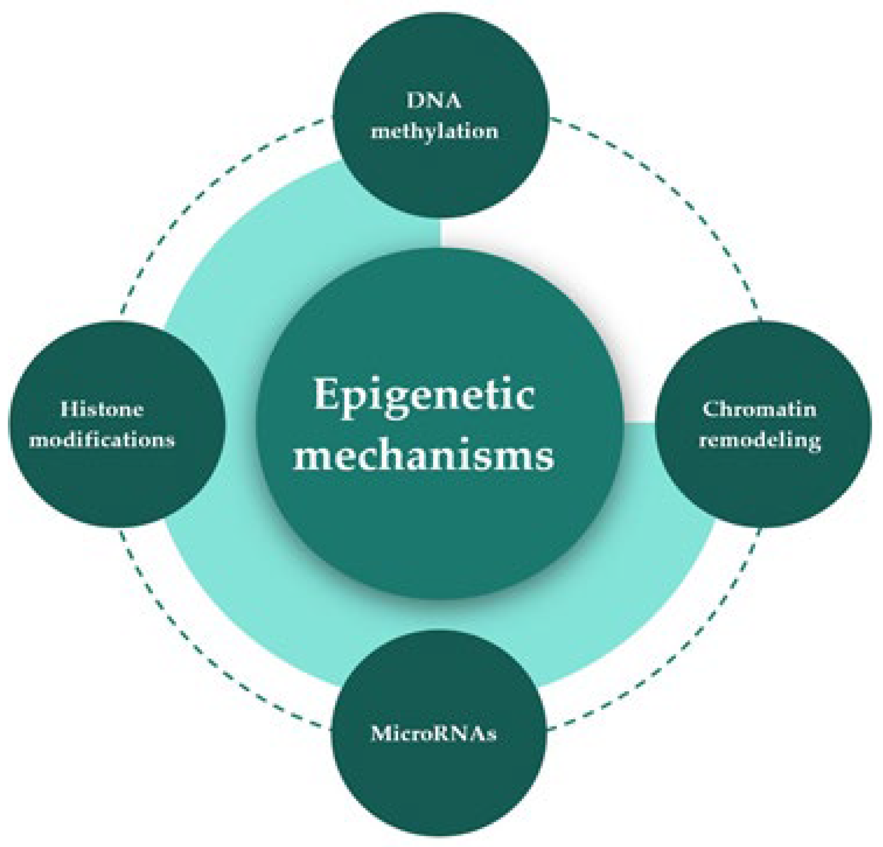
2. Epigenetic Mechanisms

Epigenetic mechanisms regulate gene expression and function by altering the chromatin structure without altering the base sequence of DNA. These mechanisms are interrelated and include DNA methylation, posttranslational modifications of histones or histone modifications, chromatin remodeling, and the role of microRNAs [23,24,25,26,27] (Figure 1). These mechanisms can be induced by environmental factors, could be reversible, and have been linked to the etiology of many diseases or disorders such as cancers, cardiovascular diseases, metabolic disorders, and neurological disorders [7]. It is, therefore, critical to understand the role of nutrients, as environmental factors, in altering gene expression by their epigenetic mechanisms and how these alterations that happen at critical stages of development are linked to human health and disease.
DNA methylation plays essential roles in a range of biological functions in mammals such as the silencing of transposable elements, regulation of gene expression, DNA replication, parental imprinting, X-chromosome inactivation, control of cellular differentiation, normal embryonic development, normal brain development, and brain plasticity [28,29]. DNA methylation is a covalent inert modification that chemically modifies the DNA without altering its charge. It does that by adding the methyl group CH3 to the carbon 5 on cytosine which is located next to guanine in the CpG dinucleotides, which leads to the formation of 5-methylcytosine (5-mC) as a nitrogenous base [25,30,31]. This covalent modification is catalyzed by the activity of DNA methyltransferases (Dnmt1, Dnmt3a, Dnmt3b, and Dnmt3L) that utilize S-adenosylmethionine (SAM) as a universal methyl-donor. Structurally, these mammalian enzymes share a conserved C-terminal catalytic domain important for their enzymatic activity except for Dnmt3L. They also contain an N-terminal regulatory domain except for Dnmt2. The N-terminal domain is essential for protein–protein interactions such as interactions of Dnmts with proteins or effectors that are involved in the modulation of chromatin structure and function [32]. These enzymes are expressed in a spatial and temporal manner and have been shown to be abundantly expressed in the brain. Although DNA methylation is considered one of the most stable and inert epigenetic marks in the mammalian genome, it can be reversible [33,34]. This was demonstrated with the discovery of DNA demethylases such as the ten-eleven-translocation (TET) family of proteins. The activity of these enzymes led to the discovery of the 5-hydroxymethylcytosine (5-hmC) as a modified nitrogenous base with a role in cellular differentiation, aging, and cancer [35,36,37]. The majority of CpGs are concentrated in the gene promoter and in the first exon of a gene they are known as CpG islands and are often unmethylated. The abnormal methylation of these CpGs is linked to gene repression [30]. Studies have shown that the supplementation of specific micronutrients that act as methyl donors can cause global or gene-specific changes in methylation in the developing and mature brain, thus altering gene expression and susceptibility to diseases [3,5,38].
Histone modification is another epigenetic mechanism that chemically modifies the N-terminal tail of histones. In this way, it alters the interaction of DNA with histone proteins, around which the DNA is wrapping itself resulting in changes in gene expression. The switch between chromatin compaction and chromatin relaxation is regulated by the ability of the N-terminal tail to perform malleable posttranslational modifications (PTMs) at specific amino acid residues along this tail. These modifications are not random but occur at specific residues such as lysine (K), arginine (R), serine (S), or threonine (T) and accordingly, can alter the accessibility of transcription factors to their binding sites [39,40]. The best-understood and well-characterized histone modifications include methylation, acetylation, phosphorylation, ubiquitination, and sumoylation. These modifications are written by “writers”, erased by “erasers”, and read by effector proteins. Most histone methylation and acetylation happen at the lysine and arginine residues of histone H3 and H4; lysine methylation is catalyzed by histone methyltransferases (HMTs/KMTs) and lysine demethylation by histone demethylases (HDMs/KDMs). Similarly, histone acetylation is regulated by histone acetyltransferases (HATs/KATs) and deacetylation by histone deacetylases (HDACs) [40,41,42,43,44]. The epigenetic factors of microRNAs represent another layer of gene expression regulation. They are described as short RNAs that do not code for proteins but regulate the expression of many protein-coding genes. They require the RNA-induced silencing complex (RISC) to guide them to their target which is the 3′-untranslated region (3′UTR) of a gene. Once they reach their target, they cause gene silencing by either degrading the mRNA or halting its translation [26,45,46]. These epigenetic factors are abundantly expressed in the developing and mature brain and can modulate gene expression at different stages of neuronal development in diverse organisms [47,48]. The dysregulation in microRNA expression has been associated with several diseases or disorders [49], including neurological disorders [48,50,51,52]. These epigenetic mechanisms described above are now considered plausible mechanisms in the etiology of many diseases that are environmentally induced and not necessarily caused by genetic factors.

3. One-Carbon Metabolism

One-carbon metabolism consists of chemical reactions that are catalyzed by several enzymes with the contribution or presence of methyl-donor micronutrients. These reactions support several pathways such as nucleotide metabolism, redox state, neurotransmitters synthesis such as acetylcholine, and regulation of epigenetic mechanisms via the formation of SAM [53,54,55,56]. Folate and methionine cycles are two main components of the one-carbon metabolism [54], among which the methyl-donor micronutrients such as folate, methionine, choline, betaine, and vitamins B6 and B12 are critical players. There is a link between the changes in the levels of these micronutrients and epigenetic alterations (Figure 2). Table 1 provides a list of methyl-donor micronutrients that contribute to the one-carbon metabolism.
The main outcome of the methionine cycle is the formation of SAM that powers methylation reactions such as DNA methylation and histone methylation. SAM donates the methyl group to the epigenetic machinery such as DNA methyltransferases (DNMTs) that methylate the DNA or to histone methyltransferases (HMTS/KMTs) that methylate histones [6,38,53] (Figure 2). After the transfer of the methyl group to the substrate, SAM is converted to S-adenosylhomocysteine (SAH). Under normal conditions, SAH is hydrolyzed to yield adenine and homocysteine by S-adenosylhomocysteine hydrolase (SAHH) (Figure 2). On the other hand, the elevation in SAH levels compared to SAM has an inhibitory effect on DNA methyltransferases with the potential to alter DNA methylation [74,75]. Via the catalytic activity of cystathionine β-synthase (CBS), homocysteine with serine can form cystathionine, which is further catalyzed to form products with antioxidant properties. Methionine can be regenerated by the activity of two enzymes, betaine-homocysteine methyltransferase (BHMT) and methionine synthase (MS). Betaine-homocysteine methyltransferase can transfer a methyl group from betaine, as a choline precursor or derived from the diet, and generate methionine and dimethylglycine (DMG). The folate metabolism leads to the formation of purine and 5-methyltetrahydrofolate (5-MTHF) using 5-methyltetrahydrofolate reductase (5-MTHFR). It is worth noting that methyltetrahydrofolate reductase (MTHFR) gene polymorphism alters its enzymatic activity and has been linked to altered cognitive functions [76]. Another enzyme, 5-methyltetrahydrofolate-homocysteine methyltransferase or methionine synthase (MS), a VitB12-dependent enzyme, can transfer a methyl group from 5-methyltetrahydrofolate (5-MTHF) to homocysteine and in this manner produces tetrahydrofolate (THF) and methionine (Figure 2). Methionine is adenylated by methionine adenosyltransferase (MAT) to generate SAM [53]. Dietary methyl donors such as choline, betaine, folic acid, methionine, and the B vitamins are interconnected in this one-carbon metabolism. Changes in their levels can alter gene expression and regulation by altering the levels of SAM, linking changes in the intake of dietary methyl donors to alterations in cellular functions.

4. Dietary Methyl Donors, Epigenetic Alterations, and Stress-Related Disorders

Studies have shown that early life experiences cause a developmental programming of the hypothalamic–pituitary–adrenal (HPA) axis or stress axis and behavioral responses to stressors [17,77,78]. This developmental programming of the stress axis induced by early life experiences could be explained by changes in the expression of stress-related genes by epigenetic mechanisms such as DNA methylation with long-term neurobehavioral outcomes [79,80,81,82]. The supplementation or deficiency in the levels of methyl-donor micronutrients during early life can have an impact on offspring brain development with long-term effects on behavior [83,84,85,86]. In this section, we will describe the programming of the stress axis through early life experiences and the role of methylation, then we will summarize research studies that explain this intricate link between micronutrients, changes in methylation, and stress-related or neurodevelopmental disorders.

4.1. HPA Axis Programming by Early Life Stress and the Role of Methylation

The hypothalamic–pituitary–adrenal (HPA) axis represents the organism’s neuroendocrine response to stressors. Upon HPA axis activation, the immune system and the nervous system are also activated. This results in the release of stress mediators such as endocrine hormones, cytokines, and neurotransmitters. These mediators enable the organism to react and respond to stress leading to an adaptive or maladaptive response [87,88,89]. The main mediator that is released upon HPA axis activation is glucocorticoid GC (or cortisol), which exerts a negative feedback mechanism at the levels of the anterior pituitary, hypothalamus, and hippocampus. GC can cross the blood–brain barrier and bind to its glucocorticoid receptor (GR), with higher affinity to the mineralocorticoid receptor (MR). Glucocorticoid receptors and mineralocorticoid receptors are widely expressed in limbic regions such as the prefrontal cortex, hippocampus, amygdala, and hypothalamus (Figure 3). The binding of GC to its receptor induces signaling mechanisms that regulate the stress response and behavior via the activation or repression of stress-related genes [90]. Aberrant release of GC or a blunted HPA axis response is harmful and has been shown to contribute to psychopathology [89,90,91].
The dysregulation of the HPA axis and imbalances in the levels of GCs and the expression of its receptors in limbic regions have been linked to psychiatric disorders [92,93,94]. In particular, exposure to stress during early life is considered one of the main early life experiences that can result in long-term consequences such as neurobehavioral changes that may develop into major psychosis [95,96,97,98]. One plausible explanation that mediates this link between early life experiences and long-term neurobehavioral outcomes are epigenetic mechanisms that chemically modify the expression of stress-related genes via methylation [1,12,81,99,100]. Genome-wide changes in methylation, hypo- and hypermethylated sites, were reported in the brain tissues of suicidal individuals or individuals with a history of early life adversity [101,102].
The brain is plastic, especially early in development where exposure to adaptive or maladaptive environmental factors can have positive or negative long-term effects on health that could pass to the next generations [103,104]. We evaluate here evidence of the role of DNA methylation in the embedding of early life experiences and the role of nutrition. One of the most studied brain regions in humans and rodents that demonstrates the long-term effects of early life experiences on behavior is the hippocampus. For example, the effects of maternal care in the form of licking and grooming during postnatal life impact the behavior and the stress response of offspring during adulthood. The alterations in the offspring stress response later in life were associated with a change in the methylation status of the CpG islands of the glucocorticoid receptor (GR) (NR3C1) gene promoter that altered the binding of the nerve growth factor-induced protein A (NGF1-A) transcription factor to its binding site in the GR promoter and altered GR gene expression in the rat hippocampus. This study demonstrated the epigenetic effects of maternal care on the offspring’s stress response in relation to changes in the methylation of the GR gene promoter [105]. Similarly, human postmortem hippocampal tissues of suicidal victims with a history of childhood abuse or maltreatment showed an increase in the methylation of GR with a decrease in its expression [106]. The effects of early life experiences impact other stress-related genes besides GR. Exposing infant rats during early postnatal life to stress altered the methylation status and the expression of the brain-derived neurotrophic factor Bdnf gene in the adult prefrontal cortex. This alteration persisted in the next generation of infant rats demonstrating the transgenerational transmission of the effect of early stress on brain-derived neurotrophic factor Bdnf and behavior [107]. Another study conducted in mice showed the effects of early life stress (ELS) on neuroendocrine function in the hypothalamic paraventricular nucleus (PVN). Early life stress resulted in hypomethylation of the promoter of the Vasopressin gene (Avp) with an increase in its expression. These epigenetic changes were associated with an elevation in blood corticosterone levels, an impaired ability of offspring to cope with stress, and an impaired memory [108]. An interesting study conducted in Wistar Kyoto rats, a highly stress-susceptible strain with anxiety/depression-like phenotypes and a hyperactive HPA axis, showed a positive effect of ELS on offspring neurodevelopment during adulthood. Maternal separation for 180 min from postnatal days 1 to 14 caused global hypermethylation in the rat hippocampus but not in other limbic regions with reduced methylation in the insulin receptor and its downstream targets, known to have a role in memory and neuronal survival. Several other genes that are linked to cell proliferation, tyrosine kinase signaling, axonal guidance, synaptogenesis, and transmission were differentially methylated. Interestingly, the GR (Nr3c1) gene methylation was not altered in this study. These methylation changes were associated with diminished depressive or anxiety-like behavior, increased exploratory behavior, and increased sociability in offspring suggesting stress resilience in adulthood as an adaptive response of offspring to persistent ELS. Dietary methyl-donor supplementation for four weeks during adulthood had anxiolytic and antidepressant effects in these rats with improved cardiovascular responses to stress [109].
Methyl-CpG-binding protein (MeCP2) has been implicated in the etiology of the developmental disorder, Rett syndrome (RTT). Rett syndrome mouse models show altered corticosterone response to stress, dysregulated levels of the stress hormone corticotropin-releasing hormone or factor (Crh/Crf), and dysregulation of the stress axis [110,111,112]. In this context, one study investigated the effects of ELS such as maternal separation (MS) from postnatal days 3 to 21 in MeCP2 heterozygote female mice (MeCP2-het-MS) and wild-type (WT-MS) mice on anxious behavior during adolescence using behavioral tests such as open field, the forced swim test, and elevated plus maze. At six weeks of age, MeCP2-het-MS mice show less anxiety and less depressive-like behaviors compared to WT-MS mice, with reduced neuronal activation in the PVN, as depicted by the immunoreactivity of c-fos/Avp and c-fos/Crh. These findings indicate the role of MeCP2 functionality on stress axis regulation and its impact on emotional behavior and neuronal activity later in life [113].
Interestingly, one study reported the hypermethylation of a distal cytosine guanine island (CGI) shore of the GR (Nr3c1) in Crh-producing neurons in the PVN of the hypothalamus, a brain region that is involved in stress regulation, leading to upregulation of GR and thus preventing the elevation of Crh in response to stress in adulthood [114]. Other stress-related genes that are found to be altered epigenetically in response to early life stress are vasopressin (Avp) and Crh/Crf in the hypothalamic PVN. For example, maternal separation in mice resulted in the hypomethylation of CpG sites along the enhancer of Avp leading to an increase in its expression with an increase in stress responsiveness due to an elevation in corticosterone. This elevation led to altered feedback inhibition of the HPA axis response leading to a hyperactive stress response [108]. Interestingly, this hypomethylation of Avp was linked to the phosphorylation of MeCP2 and an altered ability of phosphorylated MeCP2 to bind to the Avp enhancer and recruit DNA methyltransferases to cause gene repression [115]. Early prenatal stress caused hypomethylation of CpG sites of the Crf promoter in the hypothalamus and central amygdala of mice with elevated levels of corticotropin-releasing factor (CRF), suggesting dysregulation of their stress axis during adulthood [116].
What about Bdnf in the context of ELS and epigenetic alterations? Epigenetic regulation of Bdnf expression is affected in response to ELS. For example, the release of MeCP2 repressor complex from the Bdnf promoter results in its demethylation with an increase in Bdnf expression [117], and the epigenetic mediator microRNA-132 has been shown to downregulate MeCP2 expression and indirectly reduce Bdnf levels in the hippocampus of a rat model of chronic stress-induced depression [118]. Epigenetic regulation of Bdnf has been linked to neuroplasticity and neuronal activity in mature hippocampal neurons [119]. Studies have demonstrated that Bdnf expression is critical for normal dendritic branching in the hippocampus and amygdala implicating the role of Bdnf in major behavioral correlates of stress disorders such as anxiety and depressive-like behaviors [120,121]. The type, intensity, and duration of the stressor, the developmental period studied, the brain region involved, and the rodent strain used in studies may have resulted in different interpretations and results in the scientific field. For example, postnatal stress resulted in Bdnf hypermethylation and reduced Bdnf expression in the rat adult prefrontal cortex that persisted in the next generation [107], whereas prenatal stress resulted in decreased Bdnf expression and hypermethylation at Bdnf exon IV in the rat amygdala and hippocampal regions during adulthood with an increased expression of Dnmt1 and Dnmt3a [122].

4.2. Potential Neuroprotective Effects of Dietary Methyl Donors: More or Less?

The etiology of many diseases is now believed to be caused by not only genetic factors but also environmental factors via epigenetic mechanisms that do not follow mendelian inheritance patterns [24,123]. Dietary methyl-donor micronutrients that contribute to the one-carbon metabolism have been shown to be critical during development as they contribute to the formation of SAM which is involved in methylation reactions that are essential for brain health across the lifespan [5]. In this section, we will present evidence from findings that demonstrate the effects of the optimal levels of these micronutrients during specific stages of development on human health and disease.
Folate maternal intake is quite important to decrease the incidence of neural tube defects (NTDs) in children and its deficiency has been linked to many diseases including anemia, atherosclerosis, psychiatric disorders, and cancer [93,124]. Consistent results in clinical trials showed the beneficial effects of using folate in conjunction with other pharmacological interventions in mitigating the effects of psychiatric disorders such as schizophrenia, bipolar disease, autism, and attention-deficit hyperactivity disorder [125]. Several human studies demonstrated the correlation between maternal inadequate intake of micronutrients and altered neurodevelopment such as brain defects, altered behavior, altered cognition, and potential contribution to psychiatric disorders [10,126,127]. A large study conducted in humans showed that maternal intake of folate and VitB12 during the first trimester of pregnancy correlated with a higher score in cognitive measures in children at age 3 years [128]; in addition, a Norwegian study demonstrated a reduced risk of language delay in children at 3 years of age with folate maternal intake during early pregnancy [129]. The neuroprotective effects of micronutrients are not only evident during early life. Several studies linked the intake of specific micronutrients to cognitive performance during adolescence. For example, higher dietary folate intake positively correlated with academic performance in adolescents [127,128,129,130]. Animal studies showed comparable findings. For example, prenatal folate deficiency during the gestational days GD11-GD17 in rats is linked to structural changes in the brain such as a reduction in the number of progenitor cells in the fetal neocortex [131]. VitB deficiency correlated with the elevation of homocysteine in neurons and astrocytes in specific brain regions such as the striatum, hippocampus, and cerebellum. Homocysteine-positive cells showed markers of death. These rats had altered motor function and altered cognitive functions such as learning and memory deficits during adulthood [132].
In an early life induced model of depression in rats, maternal separation for 180 min from postnatal days PD2 to PD21 altered the levels of total high-density lipoprotein-cholesterol (HDL-cholesterol) levels and increased depressive-like behaviors in offspring, as measured by an increased immobility time in the forced swimming test displayed by these rats. An eighteen-week supplementation of choline, betaine, folate, and VitB12 at PD60 reduced the depressive-like behavior in offspring and increased total DNA methylation in the hippocampus, as measured by the levels of 5-methylcytosine [21]. The anti-depressive action of methyl donors was demonstrated in chronically high-fructose-treated rats, an animal model of anxiety and mood disorders. Eight weeks of methyl-donor supplementation at 4 weeks of age in female rats reduced oxidative stress, as measured by the nitrite content in the rat hippocampus, reduced anxiety-like behavior in the elevated plus maze test, and depressive-like behavior in the forced swimming test, reinforcing the notion that methyl donors could act as nutri-therapeutic agents in mitigating the effects of stress-related disorders such as anxiety and depression [133].
The neuroprotective effects of methyl donors of the one-carbon metabolism were also reported in humans with and in animal models of neurodegenerative disorders such as Alzheimer’s disease (AD). AD is one of the most common types of cognitive impairment with aging and is associated with altered one-carbon metabolism. Its neuropathology is characterized by the accumulation of amyloid beta peptide plaques and the hyperphosphorylation of the microtubule-associated protein tau, causing the formation of neurofibrillary tangles [134]. Recent studies support the notion that diets rich in micronutrients that play a role in the one-carbon metabolism have promising effects in mitigating AD pathogenesis [67,135,136,137,138,139]. In the context of methyl donors, an increase in plasma homocysteine, and hence SAH as a methyltransferase inhibitor, was reported in AD brain samples and correlated with cognitive impairment [140]. The association between VitB and AD is still debatable. Presenilin (PSEN1) plays a role in increasing secretase activity that cleaves the amyloid precursor protein (APP) into amyloid beta peptides. Studies have linked VitB deficiency to hypomethylation of the PSEN1 promoter and an increase in its expression in an AD TgCRND8 mouse model. Supplementation of SAM to these mice reversed the effects on PSEN1 hypomethylation and expression, tau phosphorylation, and reduced amyloid beta peptide production [141,142]. On the other hand, folic acid supplementation in an amyloid precursor protein/presenilin (APP/PSNE1) transgenic mouse model of AD reduced the levels of amyloid beta proteins as this correlated with increased activity of Dnmt1 and the hypermethylation of PSEN1 and APP promoters which correlated with a decrease in their expression [143]. Deficiency or disruption in glutathione (GSH) levels, an antioxidant that is produced by homocysteine in the transsulfuration pathway (Figure 2), has been linked to cognitive decline in AD patients and contributes to AD-related oxidative stress [144,145]. In the context of betaine, another methyl-donor, human studies showed that betaine supplementation in AD patients results in: decreased levels of homocysteine, phosphorylated tau, and amyloid beta accumulation; a reduction in blood inflammatory markers such as interleukin-1 beta (IL-1β) and tumor necrotic factor-alpha (TNF-α); an increase in the levels of memory-related proteins such as NR1, NR2A, and NR2B (NMDA receptors); an increase in the levels of synaptic proteins such as synaptophysin, synaptotagmin, and phosphorylated synapsin I [146].
What about choline? Choline is the main precursor for betaine and serves as a methyl donor for the formation of SAM via the methionine cycle. Choline is neuroprotective, can be derived from food, has essential functions related to the production of neurotransmitters such as acetylcholine that regulates cholinergic signaling, and maintains the integrity of cellular membranes via the formation of phosphatidylcholine [147] (Figure 2). Dietary supplementation of choline in the amyloid precursor protein/presenilin1 (APP/PS1) mouse model of AD improved spatial memory in the Morris water maze and reduced the processing of APP to amyloid beta peptides with a reduction in microglia activity, which is known to play a role in neuroinflammation, seen in AD, suggesting the potential benefits of diets rich with choline on brain function [148]. Since acetylcholine is quite essential for cholinergic signaling in the brain, acetylcholine esterase inhibitor, the enzyme that degrades acetylcholine, is currently used as a drug for AD treatment, although its use showed side effects and did not prevent AD progression due to low efficacy [149]. Choline perinatal supplementation (fetal and early postnatal) in AD mouse models showed positive improvements at several levels. These mice showed in the hippocampus: reduced levels of amyloid beta proteins; reduction in amyloid plaque accumulation; elevation of choline acetyltransferase (ChAT), the enzyme that forms acetylcholine from choline; a decrease in the levels of glial fibrillary acidic protein (GFAP) proteins, a marker of gliosis, suggesting the importance of choline intake during pregnancy and shortly after birth [150]. Another study demonstrated the beneficial effects of choline supplementation in an AD mouse model (APP/PS1 mice) during adulthood. Supplementing these mice from 2 to 11 months of age with choline resulted in reduced anxiety and improved spatial and learning memory in several behavioral tests. This correlated with reducing amyloid beta accumulation in the cortex and the hippocampus with a decrease in the levels of microglia activation markers, restoration of choline and acetylcholine levels in the cerebral cortex and the hippocampus, and increased ChAT-positive cholinergic neurons in the basal forebrain and amygdala. Moreover, choline effects on synapses were also determined. Choline supplementation increased the levels of synaptophysin and the postsynaptic density protein (PSD95) in the hippocampus of this AD mouse model [151].

5. Conclusions

Early life stress can have adverse long-term effects on health with an increased possibility of developing neuropsychiatric disorders later in life. The ability to respond and react to stress involves the regulation of the HPA axis and the involvement of brain regions and neuronal networks that belong to the limbic system. Our mental health such as our adaptation to stress and our resilience in the face of adversity is dependent on the time of exposure to stressors during development, the duration, intensity, and the type of stressor. It also includes the genetic make-up of an individual and other environmental factors. These factors greatly impact brain health across the lifespan. Epigenetic mechanisms are considered plausible mechanisms that explain this intricate relationship between our genes and our environment that may impact behavior and brain health. Studies have shown that supplementation of methyl donors during early life is essential for normal brain growth and development and has long-lasting effects on mental health. The regulated consumption of these nutrients later in life has been shown to improve cognitive functions and mitigate the symptoms of neurodegenerative disorders when used with other pharmaceutical interventions. The micronutrients of methyl donors in the one-carbon metabolism have been reported to be neuroprotective, can cause global or gene-specific changes by epigenetic mechanisms, and could be used in a regulated manner to mitigate the symptoms of neurodegenerative diseases and stress-related disorders at early stages of prognosis. The dosage and the timing of intake of these micronutrients in healthy individuals should be monitored by a health professional to prevent unwanted side effects or overdosage, as these micronutrients play crucial roles in many physiological processes and may have an impact on brain health.

Funding

This research received no external funding.

Institutional Review Board Statement

Not applicable.

Informed Consent Statement

Not applicable.

Data Availability Statement

Not applicable.

Conflicts of Interest

The author declares no conflict of interest.

References

  1. Abdul, Q.A.; Yu, B.P.; Chung, H.Y.; Jung, H.A.; Choi, J.S. Epigenetic Modifications of Gene Expression by Lifestyle and Environment. Arch. Pharm. Res. 2017, 40, 1219–1237. [Google Scholar] [CrossRef]
  2. Zeisel, S.H. Choline: Needed for Normal Development of Memory. J. Am. Coll. Nutr. 2000, 19 (Suppl. S5), 528S–531S. [Google Scholar] [CrossRef]
  3. Niculescu, M.D.; Zeisel, S.H. Diet, Methyl Donors and DNA Methylation: Interactions between Dietary Folate, Methionine and Choline. J. Nutr. 2002, 132 (Suppl. S8), 2333S–2335S. [Google Scholar] [CrossRef] [PubMed] [Green Version]
  4. Zeisel, S.H. Nutritional Importance of Choline for Brain Development. J. Am. Coll. Nutr. 2004, 23 (Suppl. S6), 621S–626S. [Google Scholar] [CrossRef]
  5. Bekdash, R.A. Neuroprotective Effects of Choline and Other Methyl Donors. Nutrients 2019, 11, 2995. [Google Scholar] [CrossRef] [PubMed] [Green Version]
  6. Mentch, S.J.; Locasale, J.W. One-Carbon Metabolism and Epigenetics: Understanding the Specificity. Ann. N. Y. Acad. Sci. 2016, 1363, 91–98. [Google Scholar] [CrossRef] [Green Version]
  7. Zhang, L.; Lu, Q.; Chang, C. Epigenetics in Health and Disease. Adv. Exp. Med. Biol. 2020, 1253, 3–55. [Google Scholar] [CrossRef] [PubMed]
  8. Arumugam, M.K.; Paal, M.C.; Donohue, T.M.; Ganesan, M.; Osna, N.A.; Kharbanda, K.K. Beneficial Effects of Betaine: A Comprehensive Review. Biology 2021, 10, 456. [Google Scholar] [CrossRef]
  9. Irvine, N.; England-Mason, G.; Field, C.J.; Dewey, D.; Aghajafari, F. Prenatal Folate and Choline Levels and Brain and Cognitive Development in Children: A Critical Narrative Review. Nutrients 2022, 14, 364. [Google Scholar] [CrossRef]
  10. Chmielewska, N.; Szyndler, J.; Maciejak, P.; Płaźnik, A. Epigenetic Mechanisms of Stress and Depression. Psychiatr. Pol. 2019, 53, 1413–1428. [Google Scholar] [CrossRef]
  11. Penner-Goeke, S.; Binder, E.B. Epigenetics and Depression. Dialogues Clin. Neurosci. 2019, 21, 397–405. [Google Scholar] [CrossRef]
  12. Klengel, T.; Pape, J.; Binder, E.B.; Mehta, D. The Role of DNA Methylation in Stress-Related Psychiatric Disorders. Neuropharmacology 2014, 80, 115–132. [Google Scholar] [CrossRef]
  13. Torres-Berrío, A.; Issler, O.; Parise, E.M.; Nestler, E.J. Unraveling the Epigenetic Landscape of Depression: Focus on Early Life Stress. Dialogues Clin. Neurosci. 2019, 21, 341–357. [Google Scholar] [CrossRef]
  14. Panariello, F.; Fanelli, G.; Fabbri, C.; Atti, A.R.; De Ronchi, D.; Serretti, A. Epigenetic Basis of Psychiatric Disorders: A Narrative Review. CNS Neurol. Disord. Drug Targets 2022, 21, 302–315. [Google Scholar] [CrossRef]
  15. Matrisciano, F.; Tueting, P.; Dalal, I.; Kadriu, B.; Grayson, D.R.; Davis, J.M.; Nicoletti, F.; Guidotti, A. Epigenetic Modifications of GABAergic Interneurons Are Associated with the Schizophrenia-like Phenotype Induced by Prenatal Stress in Mice. Neuropharmacology 2013, 68, 184–194. [Google Scholar] [CrossRef] [PubMed] [Green Version]
  16. Anier, K.; Malinovskaja, K.; Pruus, K.; Aonurm-Helm, A.; Zharkovsky, A.; Kalda, A. Maternal Separation Is Associated with DNA Methylation and Behavioural Changes in Adult Rats. Eur. Neuropsychopharmacol. 2014, 24, 459–468. [Google Scholar] [CrossRef]
  17. Lux, V. Epigenetic Programming Effects of Early Life Stress: A Dual-Activation Hypothesis. Curr. Genom. 2018, 19, 638–652. [Google Scholar] [CrossRef]
  18. van Bodegom, M.; Homberg, J.R.; Henckens, M.J.A.G. Modulation of the Hypothalamic-Pituitary-Adrenal Axis by Early Life Stress Exposure. Front. Cell. Neurosci. 2017, 11, 87. [Google Scholar] [CrossRef] [PubMed] [Green Version]
  19. Ryan, D.P.; Henzel, K.S.; Pearson, B.L.; Siwek, M.E.; Papazoglou, A.; Guo, L.; Paesler, K.; Yu, M.; Müller, R.; Xie, K.; et al. A Paternal Methyl Donor-Rich Diet Altered Cognitive and Neural Functions in Offspring Mice. Mol. Psychiatry 2018, 23, 1345–1355. [Google Scholar] [CrossRef] [PubMed] [Green Version]
  20. Ishii, D.; Matsuzawa, D.; Matsuda, S.; Tomizawa, H.; Sutoh, C.; Shimizu, E. Methyl Donor-Deficient Diet during Development Can Affect Fear and Anxiety in Adulthood in C57BL/6J Mice. PLoS ONE 2014, 9, e105750. [Google Scholar] [CrossRef] [Green Version]
  21. Paternain, L.; Martisova, E.; Campión, J.; Martínez, J.A.; Ramírez, M.J.; Milagro, F.I. Methyl Donor Supplementation in Rats Reverses the Deleterious Effect of Maternal Separation on Depression-like Behaviour. Behav. Brain Res. 2016, 299, 51–58. [Google Scholar] [CrossRef]
  22. Sahara, Y.; Matsuzawa, D.; Ishii, D.; Fuchida, T.; Goto, T.; Sutoh, C.; Shimizu, E. Paternal Methyl Donor Deficient Diets during Development Affect Male Offspring Behavior and Memory-Related Gene Expression in Mice. Dev. Psychobiol. 2019, 61, 17–28. [Google Scholar] [CrossRef] [PubMed] [Green Version]
  23. Kouzarides, T. Chromatin Modifications and Their Function. Cell 2007, 128, 693–705. [Google Scholar] [CrossRef] [Green Version]
  24. Robertson, K.D.; Wolffe, A.P. DNA Methylation in Health and Disease. Nat. Rev. Genet. 2000, 1, 11–19. [Google Scholar] [CrossRef] [PubMed]
  25. Bird, A.P.; Wolffe, A.P. Methylation-Induced Repression—Belts, Braces, and Chromatin. Cell 1999, 99, 451–454. [Google Scholar] [CrossRef] [PubMed] [Green Version]
  26. Bartel, D.P. MicroRNAs: Genomics, Biogenesis, Mechanism, and Function. Cell 2004, 116, 281–297. [Google Scholar] [CrossRef] [PubMed] [Green Version]
  27. Guil, S.; Esteller, M. DNA Methylomes, Histone Codes and MiRNAs: Tying It All Together. Int. J. Biochem. Cell Biol. 2009, 41, 87–95. [Google Scholar] [CrossRef]
  28. Li, E.; Zhang, Y. DNA Methylation in Mammals. Cold Spring Harb. Perspect. Biol. 2014, 6, a019133. [Google Scholar] [CrossRef] [Green Version]
  29. Moore, L.D.; Le, T.; Fan, G. DNA Methylation and Its Basic Function. Neuropsychopharmacology 2013, 38, 23–38. [Google Scholar] [CrossRef] [Green Version]
  30. Deaton, A.M.; Bird, A. CpG Islands and the Regulation of Transcription. Genes Dev. 2011, 25, 1010–1022. [Google Scholar] [CrossRef] [Green Version]
  31. Klose, R.J.; Bird, A.P. Genomic DNA Methylation: The Mark and Its Mediators. Trends Biochem. Sci. 2006, 31, 89–97. [Google Scholar] [CrossRef]
  32. Chen, Z.; Riggs, A.D. DNA Methylation and Demethylation in Mammals. J. Biol. Chem. 2011, 286, 18347–18353. [Google Scholar] [CrossRef] [Green Version]
  33. Jeltsch, A. Beyond Watson and Crick: DNA Methylation and Molecular Enzymology of DNA Methyltransferases. Chembiochem 2002, 3, 274–293. [Google Scholar] [CrossRef] [PubMed]
  34. Hermann, A.; Gowher, H.; Jeltsch, A. Biochemistry and Biology of Mammalian DNA Methyltransferases. Cell. Mol. Life Sci. 2004, 61, 2571–2587. [Google Scholar] [CrossRef]
  35. Tahiliani, M.; Koh, K.P.; Shen, Y.; Pastor, W.A.; Bandukwala, H.; Brudno, Y.; Agarwal, S.; Iyer, L.M.; Liu, D.R.; Aravind, L.; et al. Conversion of 5-Methylcytosine to 5-Hydroxymethylcytosine in Mammalian DNA by MLL Partner TET1. Science 2009, 324, 930–935. [Google Scholar] [CrossRef] [PubMed] [Green Version]
  36. Williams, K.; Christensen, J.; Helin, K. DNA Methylation: TET Proteins—Guardians of CpG Islands? EMBO Rep. 2012, 13, 28–35. [Google Scholar] [CrossRef] [Green Version]
  37. Zhang, H.; Zhang, X.; Clark, E.; Mulcahey, M.; Huang, S.; Shi, Y.G. TET1 Is a DNA-Binding Protein That Modulates DNA Methylation and Gene Transcription via Hydroxylation of 5-Methylcytosine. Cell Res. 2010, 20, 1390–1393. [Google Scholar] [CrossRef] [Green Version]
  38. Anderson, O.S.; Sant, K.E.; Dolinoy, D.C. Nutrition and Epigenetics: An Interplay of Dietary Methyl Donors, One-Carbon Metabolism and DNA Methylation. J. Nutr. Biochem. 2012, 23, 853–859. [Google Scholar] [CrossRef] [PubMed] [Green Version]
  39. Strahl, B.D.; Allis, C.D. The Language of Covalent Histone Modifications. Nature 2000, 403, 41–45. [Google Scholar] [CrossRef]
  40. Jenuwein, T.; Allis, C.D. Translating the Histone Code. Science 2001, 293, 1074–1080. [Google Scholar] [CrossRef] [Green Version]
  41. Zhang, Y.; Reinberg, D. Transcription Regulation by Histone Methylation: Interplay between Different Covalent Modifications of the Core Histone Tails. Genes Dev. 2001, 15, 2343–2360. [Google Scholar] [CrossRef] [PubMed] [Green Version]
  42. Lachner, M.; Jenuwein, T. The Many Faces of Histone Lysine Methylation. Curr. Opin. Cell Biol. 2002, 14, 286–298. [Google Scholar] [CrossRef] [PubMed]
  43. Bannister, A.J.; Kouzarides, T. Regulation of Chromatin by Histone Modifications. Cell Res. 2011, 21, 381–395. [Google Scholar] [CrossRef]
  44. Gregory, P.D.; Wagner, K.; Hörz, W. Histone Acetylation and Chromatin Remodeling. Exp. Cell Res. 2001, 265, 195–202. [Google Scholar] [CrossRef] [PubMed]
  45. Schratt, G. Fine-Tuning Neural Gene Expression with MicroRNAs. Curr. Opin. Neurobiol. 2009, 19, 213–219. [Google Scholar] [CrossRef]
  46. Baek, D.; Villén, J.; Shin, C.; Camargo, F.D.; Gygi, S.P.; Bartel, D.P. The Impact of MicroRNAs on Protein Output. Nature 2008, 455, 64–71. [Google Scholar] [CrossRef] [Green Version]
  47. Kapsimali, M.; Kloosterman, W.P.; de Bruijn, E.; Rosa, F.; Plasterk, R.H.A.; Wilson, S.W. MicroRNAs Show a Wide Diversity of Expression Profiles in the Developing and Mature Central Nervous System. Genome Biol. 2007, 8, R173. [Google Scholar] [CrossRef] [PubMed] [Green Version]
  48. Barbato, C.; Giorgi, C.; Catalanotto, C.; Cogoni, C. Thinking about RNA? MicroRNAs in the Brain. Mamm. Genome 2008, 19, 541–551. [Google Scholar] [CrossRef]
  49. Wahid, F.; Khan, T.; Kim, Y.Y. MicroRNA and Diseases: Therapeutic Potential as New Generation of Drugs. Biochimie 2014, 104, 12–26. [Google Scholar] [CrossRef]
  50. Juźwik, C.A.; Drake, S.S.; Zhang, Y.; Paradis-Isler, N.; Sylvester, A.; Amar-Zifkin, A.; Douglas, C.; Morquette, B.; Moore, C.S.; Fournier, A.E. MicroRNA Dysregulation in Neurodegenerative Diseases: A Systematic Review. Prog. Neurobiol. 2019, 182, 101664. [Google Scholar] [CrossRef]
  51. Wang, W.; Kwon, E.J.; Tsai, L.-H. MicroRNAs in Learning, Memory, and Neurological Diseases. Learn. Mem. 2012, 19, 359–368. [Google Scholar] [CrossRef] [Green Version]
  52. Mehta, S.L.; Chokkalla, A.K.; Vemuganti, R. Noncoding RNA Crosstalk in Brain Health and Diseases. Neurochem. Int. 2021, 149, 105139. [Google Scholar] [CrossRef] [PubMed]
  53. Friso, S.; Udali, S.; De Santis, D.; Choi, S.-W. One-Carbon Metabolism and Epigenetics. Mol. Asp. Med. 2017, 54, 28–36. [Google Scholar] [CrossRef]
  54. Ducker, G.S.; Rabinowitz, J.D. One-Carbon Metabolism in Health and Disease. Cell Metab. 2017, 25, 27–42. [Google Scholar] [CrossRef] [PubMed] [Green Version]
  55. Shuvalov, O.; Petukhov, A.; Daks, A.; Fedorova, O.; Vasileva, E.; Barlev, N.A. One-Carbon Metabolism and Nucleotide Biosynthesis as Attractive Targets for Anticancer Therapy. Oncotarget 2017, 8, 23955–23977. [Google Scholar] [CrossRef] [PubMed] [Green Version]
  56. Coppedè, F. One-Carbon Epigenetics and Redox Biology of Neurodegeneration. Free Radic. Biol. Med. 2021, 170, 19–33. [Google Scholar] [CrossRef] [PubMed]
  57. Kalhan, S.C.; Marczewski, S.E. Methionine, Homocysteine, One Carbon Metabolism and Fetal Growth. Rev. Endocr. Metab. Disord. 2012, 13, 109–119. [Google Scholar] [CrossRef]
  58. Zeisel, S.H. Choline: Essential for Brain Development and Function. Adv. Pediatr. 1997, 44, 263–295. [Google Scholar]
  59. Fisher, M.C.; Zeisel, S.H.; Mar, M.-H.; Sadler, T.W. Perturbations in Choline Metabolism Cause Neural Tube Defects in Mouse Embryos in Vitro. FASEB J. 2002, 16, 619–621. [Google Scholar] [CrossRef] [Green Version]
  60. Zeisel, S.H. A Brief History of Choline. Ann. Nutr. Metab. 2012, 61, 254–258. [Google Scholar] [CrossRef] [Green Version]
  61. Tayebati, S.K.; Marucci, G.; Santinelli, C.; Buccioni, M.; Amenta, F. Choline-Containing Phospholipids: Structure-Activity Relationships Versus Therapeutic Applications. Curr. Med. Chem. 2015, 22, 4328–4340. [Google Scholar] [CrossRef]
  62. Ueland, P.M. Choline and Betaine in Health and Disease. J. Inherit. Metab. Dis. 2011, 34, 3–15. [Google Scholar] [CrossRef] [PubMed]
  63. Zhao, G.; He, F.; Wu, C.; Li, P.; Li, N.; Deng, J.; Zhu, G.; Ren, W.; Peng, Y. Betaine in Inflammation: Mechanistic Aspects and Applications. Front. Immunol. 2018, 9, 1070. [Google Scholar] [CrossRef] [PubMed] [Green Version]
  64. Slow, S.; Elmslie, J.; Lever, M. Dietary Betaine and Inflammation. Am. J. Clin. Nutr. 2008, 88, 247–248, author reply 248. [Google Scholar] [CrossRef] [Green Version]
  65. van Gool, J.D.; Hirche, H.; Lax, H.; De Schaepdrijver, L. Folic Acid and Primary Prevention of Neural Tube Defects: A Review. Reprod. Toxicol. 2018, 80, 73–84. [Google Scholar] [CrossRef]
  66. Bottiglieri, T. Folate, Vitamin B₁₂, and S-Adenosylmethionine. Psychiatr. Clin. N. Am. 2013, 36, 1–13. [Google Scholar] [CrossRef]
  67. Mattson, M.P.; Shea, T.B. Folate and Homocysteine Metabolism in Neural Plasticity and Neurodegenerative Disorders. Trends Neurosci. 2003, 26, 137–146. [Google Scholar] [CrossRef]
  68. Froese, D.S.; Fowler, B.; Baumgartner, M.R. Vitamin B12, Folate, and the Methionine Remethylation Cycle-Biochemistry, Pathways, and Regulation. J. Inherit. Metab. Dis. 2019, 42, 673–685. [Google Scholar] [CrossRef] [Green Version]
  69. Smith, A.D.; Warren, M.J.; Refsum, H. Vitamin B12. Adv. Food Nutr. Res. 2018, 83, 215–279. [Google Scholar] [CrossRef] [PubMed]
  70. van de Lagemaat, E.E.; de Groot, L.C.P.G.M.; van den Heuvel, E.G.H.M. Vitamin B12 in Relation to Oxidative Stress: A Systematic Review. Nutrients 2019, 11, 482. [Google Scholar] [CrossRef] [Green Version]
  71. di Salvo, M.L.; Contestabile, R.; Safo, M.K. Vitamin B(6) Salvage Enzymes: Mechanism, Structure and Regulation. Biochim. Biophys. Acta 2011, 1814, 1597–1608. [Google Scholar] [CrossRef] [PubMed]
  72. Spinneker, A.; Sola, R.; Lemmen, V.; Castillo, M.J.; Pietrzik, K.; González-Gross, M. Vitamin B6 Status, Deficiency and Its Consequences—An Overview. Nutr. Hosp. 2007, 22, 7–24. [Google Scholar] [PubMed]
  73. Hellmann, H.; Mooney, S. Vitamin B6: A Molecule for Human Health? Molecules 2010, 15, 442–459. [Google Scholar] [CrossRef] [Green Version]
  74. Mandaviya, P.R.; Stolk, L.; Heil, S.G. Homocysteine and DNA Methylation: A Review of Animal and Human Literature. Mol. Genet. Metab. 2014, 113, 243–252. [Google Scholar] [CrossRef] [PubMed]
  75. Finkelstein, J.D. Metabolic Regulatory Properties of S-Adenosylmethionine and S-Adenosylhomocysteine. Clin. Chem. Lab. Med. 2007, 45, 1694–1699. [Google Scholar] [CrossRef] [PubMed]
  76. You, M.; Zhou, X.; Yin, W.; Wan, K.; Zhang, W.; Li, C.; Li, M.; Zhu, W.; Zhu, X.; Sun, Z. The Influence of MTHFR Polymorphism on Gray Matter Volume in Patients with Amnestic Mild Cognitive Impairment. Front. Neurosci. 2021, 15, 778123. [Google Scholar] [CrossRef]
  77. Faravelli, C.; Lo Sauro, C.; Lelli, L.; Pietrini, F.; Lazzeretti, L.; Godini, L.; Benni, L.; Fioravanti, G.; Talamba, G.A.; Castellini, G.; et al. The Role of Life Events and HPA Axis in Anxiety Disorders: A Review. Curr. Pharm. Des. 2012, 18, 5663–5674. [Google Scholar] [CrossRef]
  78. Koe, A.S.; Salzberg, M.R.; Morris, M.J.; O’Brien, T.J.; Jones, N.C. Early Life Maternal Separation Stress Augmentation of Limbic Epileptogenesis: The Role of Corticosterone and HPA Axis Programming. Psychoneuroendocrinology 2014, 42, 124–133. [Google Scholar] [CrossRef]
  79. Weaver, I.C.G.; Korgan, A.C.; Lee, K.; Wheeler, R.V.; Hundert, A.S.; Goguen, D. Stress and the Emerging Roles of Chromatin Remodeling in Signal Integration and Stable Transmission of Reversible Phenotypes. Front. Behav. Neurosci. 2017, 11, 41. [Google Scholar] [CrossRef] [Green Version]
  80. Saunderson, E.A.; Spiers, H.; Mifsud, K.R.; Gutierrez-Mecinas, M.; Trollope, A.F.; Shaikh, A.; Mill, J.; Reul, J.M.H.M. Stress-Induced Gene Expression and Behavior Are Controlled by DNA Methylation and Methyl Donor Availability in the Dentate Gyrus. Proc. Natl. Acad. Sci. USA 2016, 113, 4830–4835. [Google Scholar] [CrossRef] [Green Version]
  81. Szyf, M. The Early Life Social Environment and DNA Methylation: DNA Methylation Mediating the Long-Term Impact of Social Environments Early in Life. Epigenetics 2011, 6, 971–978. [Google Scholar] [CrossRef] [PubMed] [Green Version]
  82. Babenko, O.; Kovalchuk, I.; Metz, G.A.S. Stress-Induced Perinatal and Transgenerational Epigenetic Programming of Brain Development and Mental Health. Neurosci. Biobehav. Rev. 2015, 48, 70–91. [Google Scholar] [CrossRef] [PubMed]
  83. McKee, S.E.; Reyes, T.M. Effect of Supplementation with Methyl-Donor Nutrients on Neurodevelopment and Cognition: Considerations for Future Research. Nutr. Rev. 2018, 76, 497–511. [Google Scholar] [CrossRef] [Green Version]
  84. Dauncey, M.J. Nutrition, the Brain and Cognitive Decline: Insights from Epigenetics. Eur. J. Clin. Nutr. 2014, 68, 1179–1185. [Google Scholar] [CrossRef]
  85. Vanhees, K.; Vonhögen, I.G.C.; van Schooten, F.J.; Godschalk, R.W.L. You Are What You Eat, and so Are Your Children: The Impact of Micronutrients on the Epigenetic Programming of Offspring. Cell. Mol. Life Sci. 2014, 71, 271–285. [Google Scholar] [CrossRef]
  86. Lucassen, P.J.; Naninck, E.F.G.; van Goudoever, J.B.; Fitzsimons, C.; Joels, M.; Korosi, A. Perinatal Programming of Adult Hippocampal Structure and Function; Emerging Roles of Stress, Nutrition and Epigenetics. Trends Neurosci. 2013, 36, 621–631. [Google Scholar] [CrossRef] [PubMed]
  87. McEwen, B.S. Physiology and Neurobiology of Stress and Adaptation: Central Role of the Brain. Physiol. Rev. 2007, 87, 873–904. [Google Scholar] [CrossRef] [Green Version]
  88. McEwen, B.S. Protective and Damaging Effects of Stress Mediators: Central Role of the Brain. Dialogues Clin. Neurosci. 2006, 8, 367–381. [Google Scholar] [CrossRef]
  89. de Kloet, E.R.; Joëls, M.; Holsboer, F. Stress and the Brain: From Adaptation to Disease. Nat. Rev. Neurosci. 2005, 6, 463–475. [Google Scholar] [CrossRef]
  90. Shirazi, S.N.; Friedman, A.R.; Kaufer, D.; Sakhai, S.A. Glucocorticoids and the Brain: Neural Mechanisms Regulating the Stress Response. Adv. Exp. Med. Biol. 2015, 872, 235–252. [Google Scholar] [CrossRef]
  91. Finsterwald, C.; Alberini, C.M. Stress and Glucocorticoid Receptor-Dependent Mechanisms in Long-Term Memory: From Adaptive Responses to Psychopathologies. Neurobiol. Learn. Mem. 2014, 112, 17–29. [Google Scholar] [CrossRef] [PubMed] [Green Version]
  92. Schatzberg, A.F.; Keller, J.; Tennakoon, L.; Lembke, A.; Williams, G.; Kraemer, F.B.; Sarginson, J.E.; Lazzeroni, L.C.; Murphy, G.M. HPA Axis Genetic Variation, Cortisol and Psychosis in Major Depression. Mol. Psychiatry 2014, 19, 220–227. [Google Scholar] [CrossRef] [Green Version]
  93. Porter, R.J.; Gallagher, P. Abnormalities of the HPA Axis in Affective Disorders: Clinical Subtypes and Potential Treatments. Acta Neuropsychiatr. 2006, 18, 193–209. [Google Scholar] [CrossRef]
  94. Shah, J.L.; Malla, A.K. Much Ado about Much: Stress, Dynamic Biomarkers and HPA Axis Dysregulation along the Trajectory to Psychosis. Schizophr. Res. 2015, 162, 253–260. [Google Scholar] [CrossRef] [PubMed]
  95. Gluckman, P.D.; Hanson, M.A.; Beedle, A.S. Early Life Events and Their Consequences for Later Disease: A Life History and Evolutionary Perspective. Am. J. Hum. Biol. 2007, 19, 1–19. [Google Scholar] [CrossRef] [PubMed]
  96. McEwen, B.S. Mood Disorders and Allostatic Load. Biol. Psychiatry 2003, 54, 200–207. [Google Scholar] [CrossRef] [PubMed]
  97. Tyborowska, A.; Volman, I.; Niermann, H.C.M.; Pouwels, J.L.; Smeekens, S.; Cillessen, A.H.N.; Toni, I.; Roelofs, K. Early-Life and Pubertal Stress Differentially Modulate Grey Matter Development in Human Adolescents. Sci. Rep. 2018, 8, 9201. [Google Scholar] [CrossRef]
  98. Saleh, A.; Potter, G.G.; McQuoid, D.R.; Boyd, B.; Turner, R.; MacFall, J.R.; Taylor, W.D. Effects of Early Life Stress on Depression, Cognitive Performance and Brain Morphology. Psychol. Med. 2017, 47, 171–181. [Google Scholar] [CrossRef] [Green Version]
  99. Vaiserman, A.M.; Koliada, A.K. Early-Life Adversity and Long-Term Neurobehavioral Outcomes: Epigenome as a Bridge? Hum. Genom. 2017, 11, 34. [Google Scholar] [CrossRef] [PubMed] [Green Version]
  100. Perez-Polo, J.R. Epigenetics: Stress and Disease. Int. J. Dev. Neurosci. 2017, 62, 54–55. [Google Scholar] [CrossRef]
  101. Labonté, B.; Suderman, M.; Maussion, G.; Lopez, J.P.; Navarro-Sánchez, L.; Yerko, V.; Mechawar, N.; Szyf, M.; Meaney, M.J.; Turecki, G. Genome-Wide Methylation Changes in the Brains of Suicide Completers. Am. J. Psychiatry 2013, 170, 511–520. [Google Scholar] [CrossRef]
  102. Bick, J.; Naumova, O.; Hunter, S.; Barbot, B.; Lee, M.; Luthar, S.S.; Raefski, A.; Grigorenko, E.L. Childhood Adversity and DNA Methylation of Genes Involved in the Hypothalamus-Pituitary-Adrenal Axis and Immune System: Whole-Genome and Candidate-Gene Associations. Dev. Psychopathol. 2012, 24, 1417–1425. [Google Scholar] [CrossRef] [PubMed] [Green Version]
  103. Blanco Rodríguez, J.; Camprubí Sánchez, C. Epigenetic Transgenerational Inheritance. Adv. Exp. Med. Biol. 2019, 1166, 57–74. [Google Scholar] [CrossRef] [PubMed]
  104. Weaver, I.C.G. Integrating Early Life Experience, Gene Expression, Brain Development, and Emergent Phenotypes: Unraveling the Thread of Nature via Nurture. Adv. Genet. 2014, 86, 277–307. [Google Scholar] [CrossRef] [PubMed]
  105. Weaver, I.C.G.; Cervoni, N.; Champagne, F.A.; D’Alessio, A.C.; Sharma, S.; Seckl, J.R.; Dymov, S.; Szyf, M.; Meaney, M.J. Epigenetic Programming by Maternal Behavior. Nat. Neurosci. 2004, 7, 847–854. [Google Scholar] [CrossRef]
  106. McGowan, P.O.; Sasaki, A.; D’Alessio, A.C.; Dymov, S.; Labonté, B.; Szyf, M.; Turecki, G.; Meaney, M.J. Epigenetic Regulation of the Glucocorticoid Receptor in Human Brain Associates with Childhood Abuse. Nat. Neurosci. 2009, 12, 342–348. [Google Scholar] [CrossRef] [PubMed] [Green Version]
  107. Roth, T.L.; Lubin, F.D.; Funk, A.J.; Sweatt, J.D. Lasting Epigenetic Influence of Early-Life Adversity on the BDNF Gene. Biol. Psychiatry 2009, 65, 760–769. [Google Scholar] [CrossRef] [Green Version]
  108. Murgatroyd, C.; Patchev, A.V.; Wu, Y.; Micale, V.; Bockmühl, Y.; Fischer, D.; Holsboer, F.; Wotjak, C.T.; Almeida, O.F.X.; Spengler, D. Dynamic DNA Methylation Programs Persistent Adverse Effects of Early-Life Stress. Nat. Neurosci. 2009, 12, 1559–1566. [Google Scholar] [CrossRef] [PubMed]
  109. McCoy, C.R.; Rana, S.; Stringfellow, S.A.; Day, J.J.; Wyss, J.M.; Clinton, S.M.; Kerman, I.A. Neonatal Maternal Separation Stress Elicits Lasting DNA Methylation Changes in the Hippocampus of Stress-Reactive Wistar Kyoto Rats. Eur. J. Neurosci. 2016, 44, 2829–2845. [Google Scholar] [CrossRef] [PubMed] [Green Version]
  110. McGill, B.E.; Bundle, S.F.; Yaylaoglu, M.B.; Carson, J.P.; Thaller, C.; Zoghbi, H.Y. Enhanced Anxiety and Stress-Induced Corticosterone Release Are Associated with Increased Crh Expression in a Mouse Model of Rett Syndrome. Proc. Natl. Acad. Sci. USA 2006, 103, 18267–18272. [Google Scholar] [CrossRef] [Green Version]
  111. Fyffe, S.L.; Neul, J.L.; Samaco, R.C.; Chao, H.-T.; Ben-Shachar, S.; Moretti, P.; McGill, B.E.; Goulding, E.H.; Sullivan, E.; Tecott, L.H.; et al. Deletion of Mecp2 in Sim1-Expressing Neurons Reveals a Critical Role for MeCP2 in Feeding Behavior, Aggression, and the Response to Stress. Neuron 2008, 59, 947–958. [Google Scholar] [CrossRef] [PubMed] [Green Version]
  112. Cosentino, L.; Bellia, F.; Pavoncello, N.; Vigli, D.; D’Addario, C.; De Filippis, B. Methyl-CpG Binding Protein 2 Dysfunction Provides Stress Vulnerability with Sex- and Zygosity-Dependent Outcomes. Eur. J. Neurosci. 2022, 55, 2766–2776. [Google Scholar] [CrossRef] [PubMed]
  113. Abellán-Álvaro, M.; Stork, O.; Agustín-Pavón, C.; Santos, M. MeCP2 Haplodeficiency and Early-Life Stress Interaction on Anxiety-like Behavior in Adolescent Female Mice. J. Neurodev. Disord. 2021, 13, 59. [Google Scholar] [CrossRef]
  114. Bockmühl, Y.; Patchev, A.V.; Madejska, A.; Hoffmann, A.; Sousa, J.C.; Sousa, N.; Holsboer, F.; Almeida, O.F.X.; Spengler, D. Methylation at the CpG Island Shore Region Upregulates Nr3c1 Promoter Activity after Early-Life Stress. Epigenetics 2015, 10, 247–257. [Google Scholar] [CrossRef] [PubMed] [Green Version]
  115. Murgatroyd, C.; Wu, Y.; Bockmühl, Y.; Spengler, D. Genes Learn from Stress: How Infantile Trauma Programs Us for Depression. Epigenetics 2010, 5, 194–199. [Google Scholar] [CrossRef] [Green Version]
  116. Mueller, B.R.; Bale, T.L. Sex-Specific Programming of Offspring Emotionality after Stress Early in Pregnancy. J. Neurosci. 2008, 28, 9055–9065. [Google Scholar] [CrossRef] [Green Version]
  117. Martinowich, K.; Hattori, D.; Wu, H.; Fouse, S.; He, F.; Hu, Y.; Fan, G.; Sun, Y.E. DNA Methylation-Related Chromatin Remodeling in Activity-Dependent BDNF Gene Regulation. Science 2003, 302, 890–893. [Google Scholar] [CrossRef] [Green Version]
  118. Su, M.; Hong, J.; Zhao, Y.; Liu, S.; Xue, X. MeCP2 Controls Hippocampal Brain-Derived Neurotrophic Factor Expression via Homeostatic Interactions with MicroRNA-132 in Rats with Depression. Mol. Med. Rep. 2015, 12, 5399–5406. [Google Scholar] [CrossRef] [Green Version]
  119. Lubin, F.D.; Roth, T.L.; Sweatt, J.D. Epigenetic Regulation of BDNF Gene Transcription in the Consolidation of Fear Memory. J. Neurosci. 2008, 28, 10576–10586. [Google Scholar] [CrossRef] [PubMed]
  120. Palomer, E.; Carretero, J.; Benvegnù, S.; Dotti, C.G.; Martin, M.G. Neuronal Activity Controls Bdnf Expression via Polycomb De-Repression and CREB/CBP/JMJD3 Activation in Mature Neurons. Nat. Commun. 2016, 7, 11081. [Google Scholar] [CrossRef] [PubMed] [Green Version]
  121. Govindarajan, A.; Rao, B.S.S.; Nair, D.; Trinh, M.; Mawjee, N.; Tonegawa, S.; Chattarji, S. Transgenic Brain-Derived Neurotrophic Factor Expression Causes Both Anxiogenic and Antidepressant Effects. Proc. Natl. Acad. Sci. USA 2006, 103, 13208–13213. [Google Scholar] [CrossRef] [PubMed] [Green Version]
  122. Boersma, G.J.; Lee, R.S.; Cordner, Z.A.; Ewald, E.R.; Purcell, R.H.; Moghadam, A.A.; Tamashiro, K.L. Prenatal Stress Decreases Bdnf Expression and Increases Methylation of Bdnf Exon IV in Rats. Epigenetics 2014, 9, 437–447. [Google Scholar] [CrossRef] [Green Version]
  123. Portela, A.; Esteller, M. Epigenetic Modifications and Human Disease. Nat. Biotechnol. 2010, 28, 1057–1068. [Google Scholar] [CrossRef]
  124. Bailey, L.B.; Rampersaud, G.C.; Kauwell, G.P.A. Folic Acid Supplements and Fortification Affect the Risk for Neural Tube Defects, Vascular Disease and Cancer: Evolving Science. J. Nutr. 2003, 133, 1961S–1968S. [Google Scholar] [CrossRef] [Green Version]
  125. Lam, N.S.K.; Long, X.X.; Li, X.; Saad, M.; Lim, F.; Doery, J.C.; Griffin, R.C.; Galletly, C. The Potential Use of Folate and Its Derivatives in Treating Psychiatric Disorders: A Systematic Review. Biomed. Pharmacother. 2022, 146, 112541. [Google Scholar] [CrossRef]
  126. Cortés-Albornoz, M.C.; García-Guáqueta, D.P.; Velez-van-Meerbeke, A.; Talero-Gutiérrez, C. Maternal Nutrition and Neurodevelopment: A Scoping Review. Nutrients 2021, 13, 3530. [Google Scholar] [CrossRef] [PubMed]
  127. Li, Q.; Yang, T.; Chen, L.; Dai, Y.; Wu, L.-J.; Jia, F.-Y.; Hao, Y.; Li, L.; Zhang, J.; Ke, X.-Y.; et al. Serum Folate Status Is Primarily Associated with Neurodevelopment in Children with Autism Spectrum Disorders Aged Three and Under-A Multi-Center Study in China. Front. Nutr. 2021, 8, 661223. [Google Scholar] [CrossRef] [PubMed]
  128. Villamor, E.; Rifas-Shiman, S.L.; Gillman, M.W.; Oken, E. Maternal Intake of Methyl-Donor Nutrients and Child Cognition at 3 Years of Age. Paediatr. Perinat. Epidemiol. 2012, 26, 328–335. [Google Scholar] [CrossRef] [Green Version]
  129. Roth, C.; Magnus, P.; Schjølberg, S.; Stoltenberg, C.; Surén, P.; McKeague, I.W.; Davey Smith, G.; Reichborn-Kjennerud, T.; Susser, E. Folic Acid Supplements in Pregnancy and Severe Language Delay in Children. JAMA 2011, 306, 1566–1573. [Google Scholar] [CrossRef] [Green Version]
  130. Nilsson, T.K.; Yngve, A.; Böttiger, A.K.; Hurtig-Wennlöf, A.; Sjöström, M. High Folate Intake Is Related to Better Academic Achievement in Swedish Adolescents. Pediatrics 2011, 128, e358–e365. [Google Scholar] [CrossRef]
  131. Craciunescu, C.N.; Brown, E.C.; Mar, M.-H.; Albright, C.D.; Nadeau, M.R.; Zeisel, S.H. Folic Acid Deficiency during Late Gestation Decreases Progenitor Cell Proliferation and Increases Apoptosis in Fetal Mouse Brain. J. Nutr. 2004, 134, 162–166. [Google Scholar] [CrossRef] [PubMed] [Green Version]
  132. Blaise, S.A.; Nédélec, E.; Schroeder, H.; Alberto, J.-M.; Bossenmeyer-Pourié, C.; Guéant, J.-L.; Daval, J.-L. Gestational Vitamin B Deficiency Leads to Homocysteine-Associated Brain Apoptosis and Alters Neurobehavioral Development in Rats. Am. J. Pathol. 2007, 170, 667–679. [Google Scholar] [CrossRef] [PubMed] [Green Version]
  133. Coulibaly, S.M.; Mesfioui, A.; Berkiks, I.; Ennaciri, A.; Chahirou, Y.; Diagana, Y.; Ouichou, A.; El Midaoui, A.; El Hessni, A. Effects of the Methyl Donors Supplementation on Hippocampal Oxidative Stress, Depression and Anxiety in Chronically High Fructose-Treated Rats. Neuroscience 2021, 476, 1–11. [Google Scholar] [CrossRef]
  134. Knopman, D.S.; Amieva, H.; Petersen, R.C.; Chételat, G.; Holtzman, D.M.; Hyman, B.T.; Nixon, R.A.; Jones, D.T. Alzheimer Disease. Nat. Rev. Dis. Prim. 2021, 7, 33. [Google Scholar] [CrossRef] [PubMed]
  135. Otaegui-Arrazola, A.; Amiano, P.; Elbusto, A.; Urdaneta, E.; Martínez-Lage, P. Diet, Cognition, and Alzheimer’s Disease: Food for Thought. Eur. J. Nutr. 2014, 53, 1–23. [Google Scholar] [CrossRef]
  136. Ravaglia, G.; Forti, P.; Maioli, F.; Martelli, M.; Servadei, L.; Brunetti, N.; Porcellini, E.; Licastro, F. Homocysteine and Folate as Risk Factors for Dementia and Alzheimer Disease. Am. J. Clin. Nutr. 2005, 82, 636–643. [Google Scholar] [CrossRef] [Green Version]
  137. Gröber, U.; Kisters, K.; Schmidt, J. Neuroenhancement with Vitamin B12-Underestimated Neurological Significance. Nutrients 2013, 5, 5031–5045. [Google Scholar] [CrossRef] [Green Version]
  138. Dauncey, M.J. Recent Advances in Nutrition, Genes and Brain Health. Proc. Nutr. Soc. 2012, 71, 581–591. [Google Scholar] [CrossRef] [Green Version]
  139. Luchsinger, J.A.; Tang, M.-X.; Miller, J.; Green, R.; Mayeux, R. Relation of Higher Folate Intake to Lower Risk of Alzheimer Disease in the Elderly. Arch. Neurol. 2007, 64, 86–92. [Google Scholar] [CrossRef] [Green Version]
  140. Kennedy, B.P.; Bottiglieri, T.; Arning, E.; Ziegler, M.G.; Hansen, L.A.; Masliah, E. Elevated S-Adenosylhomocysteine in Alzheimer Brain: Influence on Methyltransferases and Cognitive Function. J. Neural Transm. 2004, 111, 547–567. [Google Scholar] [CrossRef]
  141. Fuso, A.; Nicolia, V.; Pasqualato, A.; Fiorenza, M.T.; Cavallaro, R.A.; Scarpa, S. Changes in Presenilin 1 Gene Methylation Pattern in Diet-Induced B Vitamin Deficiency. Neurobiol. Aging 2011, 32, 187–199. [Google Scholar] [CrossRef] [PubMed]
  142. Fuso, A.; Nicolia, V.; Ricceri, L.; Cavallaro, R.A.; Isopi, E.; Mangia, F.; Fiorenza, M.T.; Scarpa, S. S-Adenosylmethionine Reduces the Progress of the Alzheimer-like Features Induced by B-Vitamin Deficiency in Mice. Neurobiol. Aging 2012, 33, e1–e1482. [Google Scholar] [CrossRef] [PubMed]
  143. Li, W.; Liu, H.; Yu, M.; Zhang, X.; Zhang, M.; Wilson, J.X.; Huang, G. Folic Acid Administration Inhibits Amyloid β-Peptide Accumulation in APP/PS1 Transgenic Mice. J. Nutr. Biochem. 2015, 26, 883–891. [Google Scholar] [CrossRef] [PubMed]
  144. Mandal, P.K.; Saharan, S.; Tripathi, M.; Murari, G. Brain Glutathione Levels—A Novel Biomarker for Mild Cognitive Impairment and Alzheimer’s Disease. Biol. Psychiatry 2015, 78, 702–710. [Google Scholar] [CrossRef]
  145. Saharan, S.; Mandal, P.K. The Emerging Role of Glutathione in Alzheimer’s Disease. J. Alzheimer’s Dis. 2014, 40, 519–529. [Google Scholar] [CrossRef]
  146. Sun, J.; Wen, S.; Zhou, J.; Ding, S. Association between Malnutrition and Hyperhomocysteine in Alzheimer’s Disease Patients and Diet Intervention of Betaine. J. Clin. Lab. Anal. 2016, 31, e22090. [Google Scholar] [CrossRef]
  147. Blusztajn, J.K.; Slack, B.E.; Mellott, T.J. Neuroprotective Actions of Dietary Choline. Nutrients 2017, 9, 815. [Google Scholar] [CrossRef] [Green Version]
  148. Velazquez, R.; Ferreira, E.; Knowles, S.; Fux, C.; Rodin, A.; Winslow, W.; Oddo, S. Lifelong Choline Supplementation Ameliorates Alzheimer’s Disease Pathology and Associated Cognitive Deficits by Attenuating Microglia Activation. Aging Cell 2019, 18, e13037. [Google Scholar] [CrossRef] [Green Version]
  149. Sharma, K. Cholinesterase Inhibitors as Alzheimer’s Therapeutics (Review). Mol. Med. Rep. 2019, 20, 1479–1487. [Google Scholar] [CrossRef] [Green Version]
  150. Mellott, T.J.; Huleatt, O.M.; Shade, B.N.; Pender, S.M.; Liu, Y.B.; Slack, B.E.; Blusztajn, J.K. Perinatal Choline Supplementation Reduces Amyloidosis and Increases Choline Acetyltransferase Expression in the Hippocampus of the APPswePS1dE9 Alzheimer’s Disease Model Mice. PLoS ONE 2017, 12, e0170450. [Google Scholar] [CrossRef] [Green Version]
  151. Wang, Y.; Guan, X.; Chen, X.; Cai, Y.; Ma, Y.; Ma, J.; Zhang, Q.; Dai, L.; Fan, X.; Bai, Y. Choline Supplementation Ameliorates Behavioral Deficits and Alzheimer’s Disease-Like Pathology in Transgenic APP/PS1 Mice. Mol. Nutr. Food Res. 2019, 63, 1801407. [Google Scholar] [CrossRef] [PubMed]
Figure 1. Epigenetics mechanisms. This is a schematic view of the epigenetic mechanisms that regulate gene expression. They are interrelated and include DNA methylation, histone modifications, chromatin remodeling, and the role of microRNAs.
Ijms 24 02346 g001
Figure 2. One-carbon metabolism. This figure shows the main components of the one-carbon metabolism, the folate cycle, the methionine cycle, and the contribution of micronutrients such as choline, betaine, folate, methionine, VitB6, and VitB12 in several biological processes. Enzymes and vitamins are written in red and biological processes are highlighted in green. Micronutrients are highlighted in blue. THF: tetrahydrofolate, SAM: S-adenosylmethionine, SHA: S-adenosylhomocysteine, SAHH: SAH hydrolase, BHMT: betaine-homocysteine methyltransferase, MAT: methionine acetyltransferase, DNMTs: DNA methyltransferases, HMTs: histone methyltransferases, MS: methionine synthase, MTHFR: methyltetrahydrofolate reductase, ChAT: choline acetyltransferase, AchE: acetylcholine esterase, GSH: glutathione. Adapted from Ref. [5].
Ijms 24 02346 g002
Figure 3. HPA axis. This figure represents the HPA axis or the stress axis where glucocorticoids (GCs) are the outcome of its activation. GC exerts feedback at the levels of the anterior pituitary, hypothalamus, and hippocampus to regulate the stress response. GC receptors are expressed in the hippocampus, hypothalamus, prefrontal cortex, and amygdala, which are considered part of the limbic system. CRF/CRH: corticotropin-releasing hormone or factor, ACTH: adrenocorticotropin hormone, GC: glucocorticoid. Red arrows on the right side mean negative feedback. Green arrow means positive feedback.
Ijms 24 02346 g003
Table 1. This table summarizes the functions of methyl donors that contribute to the one-carbon metabolism.
Methyl DonorsFunctionReferences
MethioninePrecursor for SAM formation, maintenance of the redox state, and brain health. [57]
CholineRegulation of cholinergic signaling, maintaining cellular membrane integrity, and contributing to the formation of SAM.[58,59,60,61]
BetaineCholine precursor, a methyl-donor in the BHMT pathway, and anti-inflammatory functions.[62,63,64]
Folic acidNormal brain development, nucleotide synthesis, and prevention of neural tube defects. [65,66,67]
Vitamin B12Nucleotide synthesis, antioxidant properties, and maintaining brain health.[68,69,70]
Vitamin B6Maintenance of the redox state and brain health. Role in transamination and decarboxylation reactions required for the metabolism of several neurotransmitters. Nucleotide synthesis and protein/lipid metabolism.[71,72,73]
Disclaimer/Publisher’s Note: The statements, opinions and data contained in all publications are solely those of the individual author(s) and contributor(s) and not of MDPI and/or the editor(s). MDPI and/or the editor(s) disclaim responsibility for any injury to people or property resulting from any ideas, methods, instructions or products referred to in the content.

