Article

Gut Microbiota Composition of Insectivorous Synanthropic and Fructivorous Zoo Bats: A Direct Metagenomic Comparison

by
Igor V. Popov
1,2,3,*,
Ilia V. Popov
1,
Anastasya A. Krikunova
1,
Tatyana A. Lipilkina
1,
Tatyana N. Derezina
1,
Michael L. Chikindas
1,4,5,
Koen Venema
3,† and
Alexey M. Ermakov
1,†
1
Faculty of Bioengineering and Veterinary Medicine and Center for Agrobiotechnology, Don State Technical University, 344000 Rostov-on-Don, Russia
2
Division of Immunobiology and Biomedicine, Center of Genetics and Life Sciences, Sirius University of Science and Technology, 354340 Federal Territory Sirius, Russia
3
Centre for Healthy Eating & Food Innovation (HEFI), Maastricht University Campus Venlo, 5928 SZ Venlo, The Netherlands
4
Health Promoting Naturals Laboratory, School of Environmental and Biological Sciences, Rutgers State University, New Brunswick, NJ 08901, USA
5
Department of General Hygiene, I.M. Sechenov First Moscow State Medical University, 119435 Moscow, Russia
*
Author to whom correspondence should be addressed.
These authors contributed equally to this work.
Int. J. Mol. Sci. 2023, 24(24), 17301; https://doi.org/10.3390/ijms242417301
Submission received: 28 October 2023 / Revised: 6 December 2023 / Accepted: 7 December 2023 / Published: 9 December 2023
(This article belongs to the Special Issue Animal Gut Microbial Metagenome)

Abstract

:
Bats are natural reservoirs for many emerging viral diseases. That is why their virome is widely studied. But at the same time, studies of their bacterial gut microbiota are limited, creating a degree of uncertainty about the role of bats in global microbial ecology. In this study, we analyzed gut microbiota of insectivorous Nyctalus noctula and Vespertilio murinus from rehabilitation centers from Rostov-on-Don and Moscow, respectively, and fructivorous Carollia perspicillata from the Moscow Zoo based on V3–V4 16S rRNA metagenomic sequencing. We revealed that microbial diversity significantly differs between the insectivorous and fructivorous species studied, while the differences between N. noctula and V. murinus are less pronounced, which shows that bats’ gut microbiota is not strictly species-specific and depends more on diet type. In the gut microbiota of synanthropic bats, we observed bacteria that are important for public health and animal welfare such as Bacteroides, Enterobacter, Clostridiaceae, Enterococcus, Ureaplasma, Faecalibacterium, and Helicobacter, as well as some lactic acid bacteria such as Pediococcus, Lactobacillus, Lactococcus, and Weisella. All these bacteria, except for Bacteroides and Weisella, were significantly less abundant in C. perspicillata. This study provides a direct metagenomic comparison of synanthropic insectivorous and zoo fructivorous bats, suggesting future directions for studying these animals’ role in microbial ecology.

1. Introduction

Chiroptera is one of the most diverse orders of mammals, which includes more than 1400 species and represents approximately 20% of the world’s mammal population [1,2]. A relatively long lifespan, the ability to fly, unique immune responses, and their microbiome distinguish bats from other mammals [3,4,5].
In recent years, bats have received increasing attention from scientists as they are recognized as a potential natural reservoir for many emerging diseases. Nipah and Hendra viruses, Ebola-related viruses, SARS-CoV, MERS-CoV, and SARS-CoV-2 are some of the most notorious viruses that wreaked havoc on public health. Several studies show that these viruses have a bat origin, or some bat species could be at least an intermediate reservoir of transmissible infections caused by these viruses [6,7,8,9,10,11].
As most of the bat-derived emerging pathogens have a viral origin, the virome of bats is studied more than their bacterial or fungal microbiota [10]. Nevertheless, studies show that bats are hosts to fungi and bacteria, which are also critical for public health and animal welfare [12,13]. The wide diversity of species in the Chiroptera order makes it difficult to identify bat species-specific microbiomes. Considering that some studies show that the gut microbiota composition of bats also depends on their habitat location and diet type [14], this task becomes nearly impossible. However, the close relationship between the habitat of bats and their microbiota further demonstrates that these animals are an integral part of the global microbial ecology.
The strong dependence of the bat gut microbiota on the environment could be related to their ability to fly, which led to the overall shortening of their gastrointestinal tract (GIT) during their evolution. Some bat species lack a caeca [15,16] and an ascending and transverse colon [16], resulting in a shorter GIT compared to animals of the same size [17]. A short GIT and the absence of some of its parts result in relatively shorter food digestion time and, most importantly, in lower anaerobic volumes, which in turn provide unfavorable conditions for anaerobic bacteria and increase the proportion of transient environmental microbes [18]. All the above-mentioned characteristics, combined with the ability to fly long distances and a long lifespan, make bats unpredictable carriers of various microbes.
Our study aimed to characterize the gut microbiota composition of synanthropic insectivorous bat species Nyctalus noctula and Vespertilio murinus, for which samples were obtained from the bat rehabilitation centers of Rostov-on-Don and Moscow, respectively, and zoo fructivorous species Carollia perspicillata and make a direct statistical comparison of their features, such as richness, diversity, taxa abundance, and the abundance of the predicted functional pathways.

2. Results

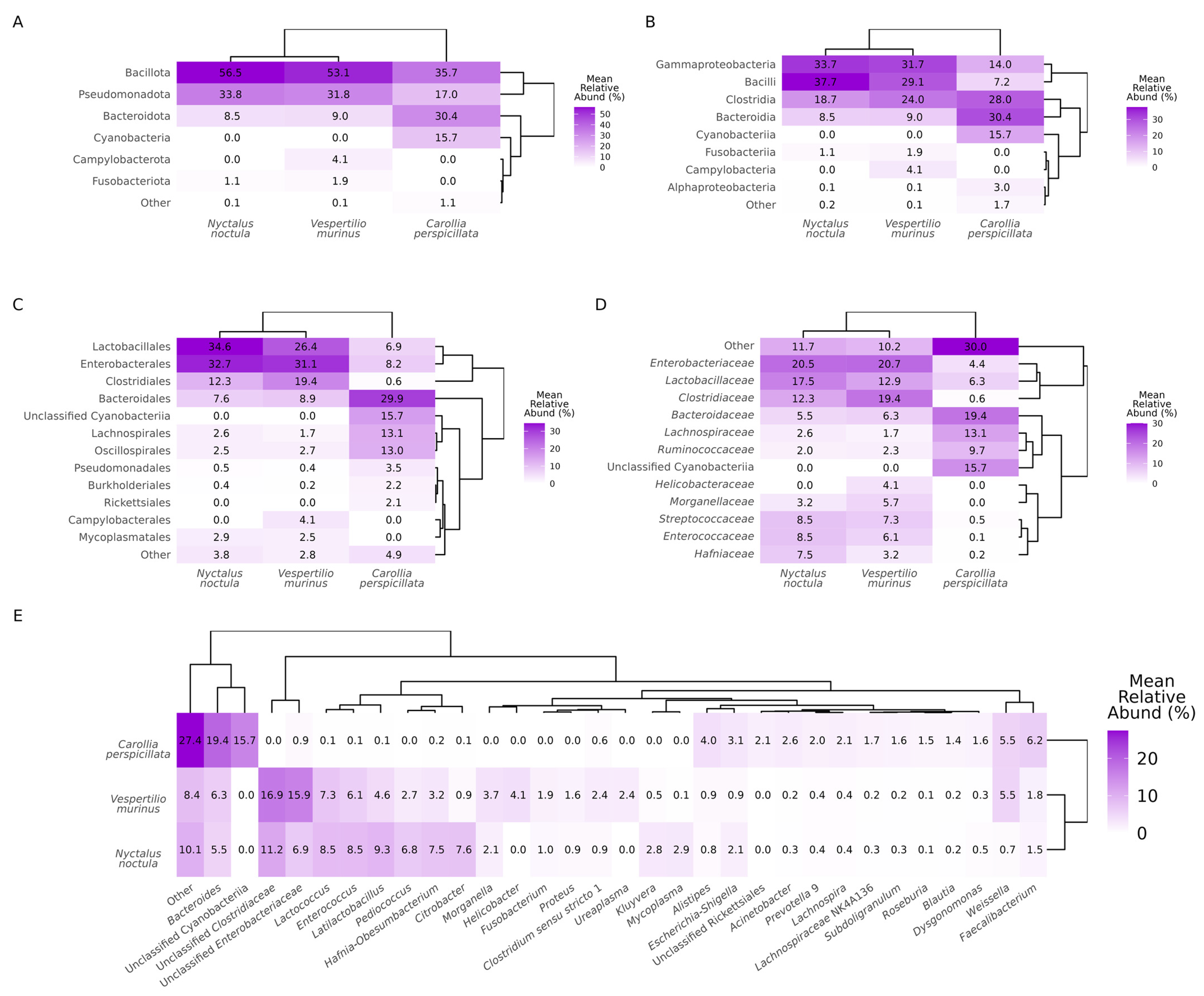
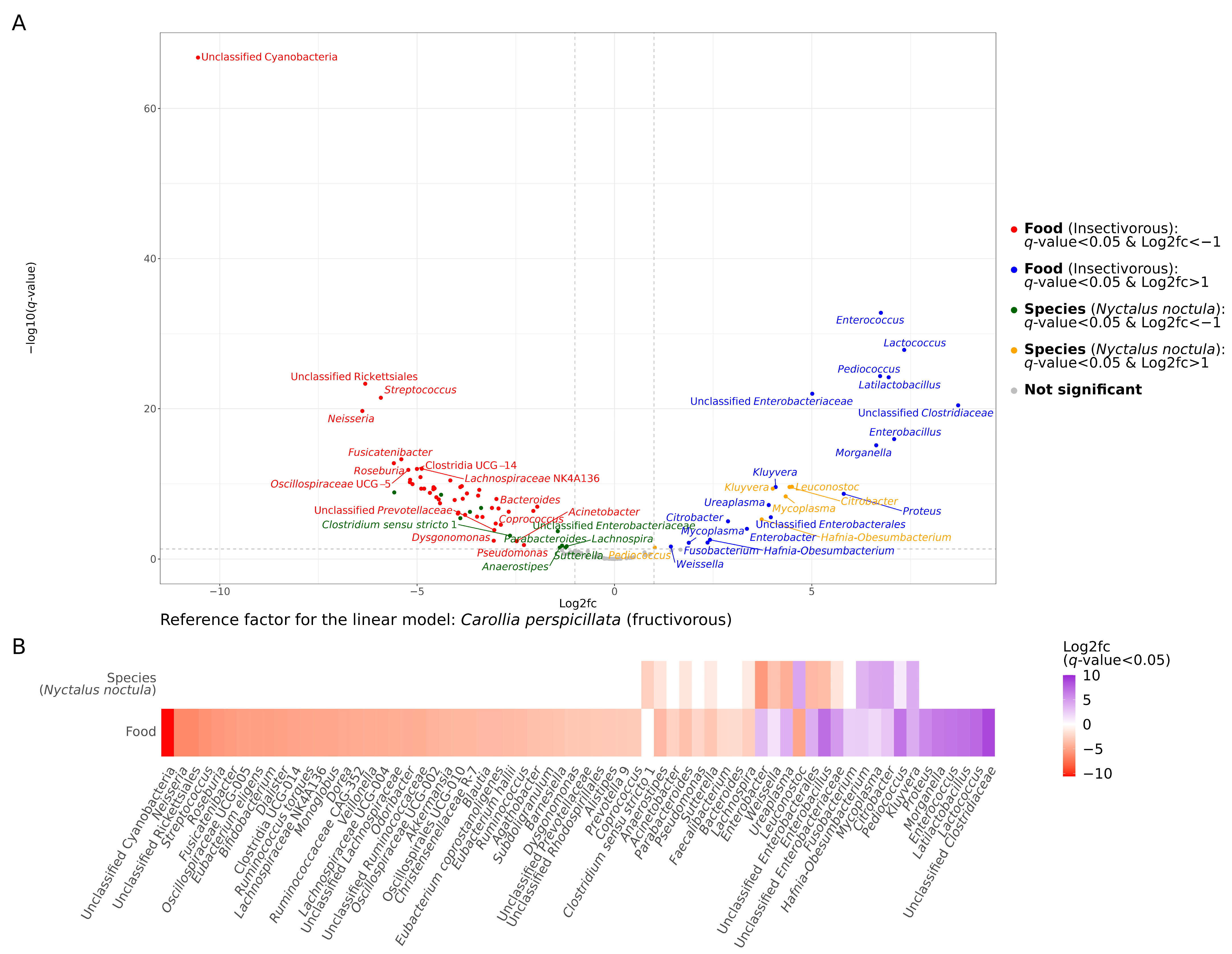
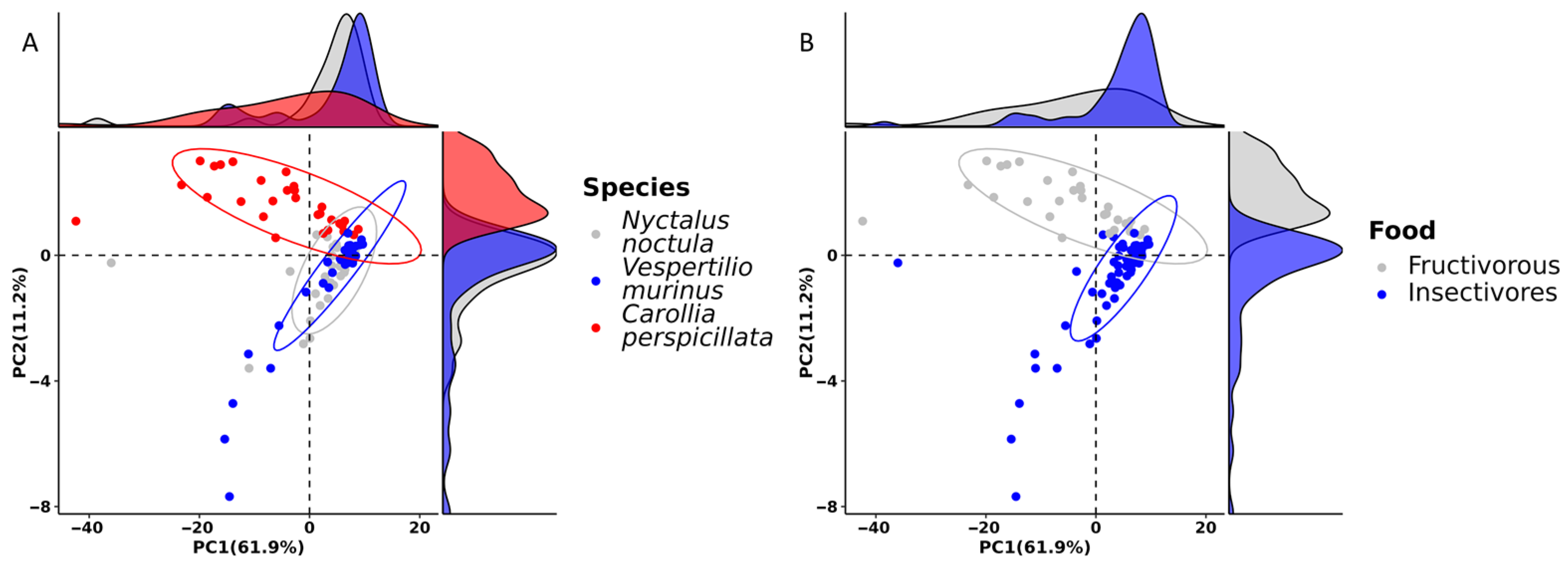
2.1. Gut Microbiota Composition

To investigate gut microbiota composition in N. noctula, V. murinus, and C. perspicillata, we sequenced amplicons of the V3–V4 regions of the 16S rRNA gene from metagenomic DNA isolated from fecal samples. Then, the classification of the sequences into amplicon sequence variants with further taxonomical identification on phylum, class, order, family, and genus levels and calculation of relative abundances (RAs) were performed. As a result, on the phylum level, we observed Bacillota, Pseudomonadota, and Bacterioidota in all three studied bat species. In samples from C. perspicillata, 15.7% of the observed sequences were identified as unclassified Cyanobacteria at all taxonomic levels. Additionally, on the phylum level, Fusobacteriota were observed in N. noctula and V. murinus, while Campilobacteriota was observed exclusively in V. murinus. Other phyla were observed in less than 1% of RA (Figure 1A). On the class level, Gammaproteobacteria, Bacilli, Clostridia, and Bacteroidia were observed in all three species studied, while Campylobacteria was identified only in V. murinus. Alphaproteobacteria was also detected in all three bat species studied, although its RA in insectivorous bats was less than 1%. Fusobacteria, as well as the phylum Fusobacteriota, were observed in N. noctula and V. murinus (Figure 1B). On the order level, the major abundant taxa that were observed in all studied bats were Lactobacillales, Enterobacterales, Clostridiales, Bacteroidales, Lachnospirales, Oscillospirales, Pseudomonadales, and Burkholderiales. Rickettsiales were observed only in C. perspicillata gut microbiota, Campylobacteriales in samples from V. murinus, and Mycoplasmatales only in studied insectivorous bats (Figure 1C). Enterobacteriaceae, Lactobacillaceae, Clostridiaceae, Bacteroidaceae, Lachnopiraceae, Ruminococceae, Streptococcaceae, and Enterococcaceae were among the most abundant families observed in all bat species studied, although the RA of some families varied several folds between species (Figure 1D). It is worth mentioning that the family Helicobacteraceae, as well as the genus Helicobacter, were observed exclusively in V. murinus. Genera with an RA greater than 1% in the gut microbiota of insectivorous bats were Bacteroides, unclassified Clostridiaceae, unclassified Enterobacteriaceae, Lactococcus, Enterococcus, Latilactobacillus, Pediococcus, Hafnia-Obesumbacterium, Morganella, Fusobacterium, and Faecalibacterium. Additionally, among genera with more than 1% RA, there were Helicobacter, Proteus, Clostridium sensu stricto 1, Ureaplasma, and Weisella in the gut microbiota of V. murinus and Citrobacter, Kluyvera, Mycoplasma, and Escherichia-Shigella in N. noctula. In samples from C. perspicillata, the following genera with an RA greater than 1% were present: Bacteroides, Alistipes, Escherichia-Shigella, Acinetobacter, Prevotella 9, Lachnospira, Lachnospiraceae NK4A136, Subdoligranulum, Roseburia, Blautia, Dysgonomonas, Weisella, and Faecalibacterium (Figure 1E).
To compare the gut microbiota composition of the studied bats, we performed a differential abundance analysis with the MaAsLin2 package [19] using linear models and fructivorous C. perspicillata as the reference factor. As a result, we found more pronounced differences in the RA of bacterial taxa between bats with different food types than between insectivorous N. noctula and V. murinus species (Figure 2 for genera, Figures S1–S4 for phylum, class, order, and family, respectively). There were 47 genera with an RA higher in fructivorous C. perspicillata and lower or even absent in insectivorous bats, while, on the other hand, there were 18 genera that were significantly more abundant in insectivorous bats compared to fructivorous species (Figure 2). Nonetheless, it is worth noting that there were also significant differences in the RA of some taxes between N. noctula and V. murinus. The most prominent differences were in Kluyvera, Citrobacter, Mycoplasma, Hafnia-Obesumbacterium, and Leuconostoc, which are significantly more abundant in N. noctula than in V. murinus and C. perspicillata, while Enterobacter, Ureaplasma, unclassified Enterobacterales, Enterobacillus, and Weisella were less abundant or absent in N. noctula gut microbiota (Figure 2).

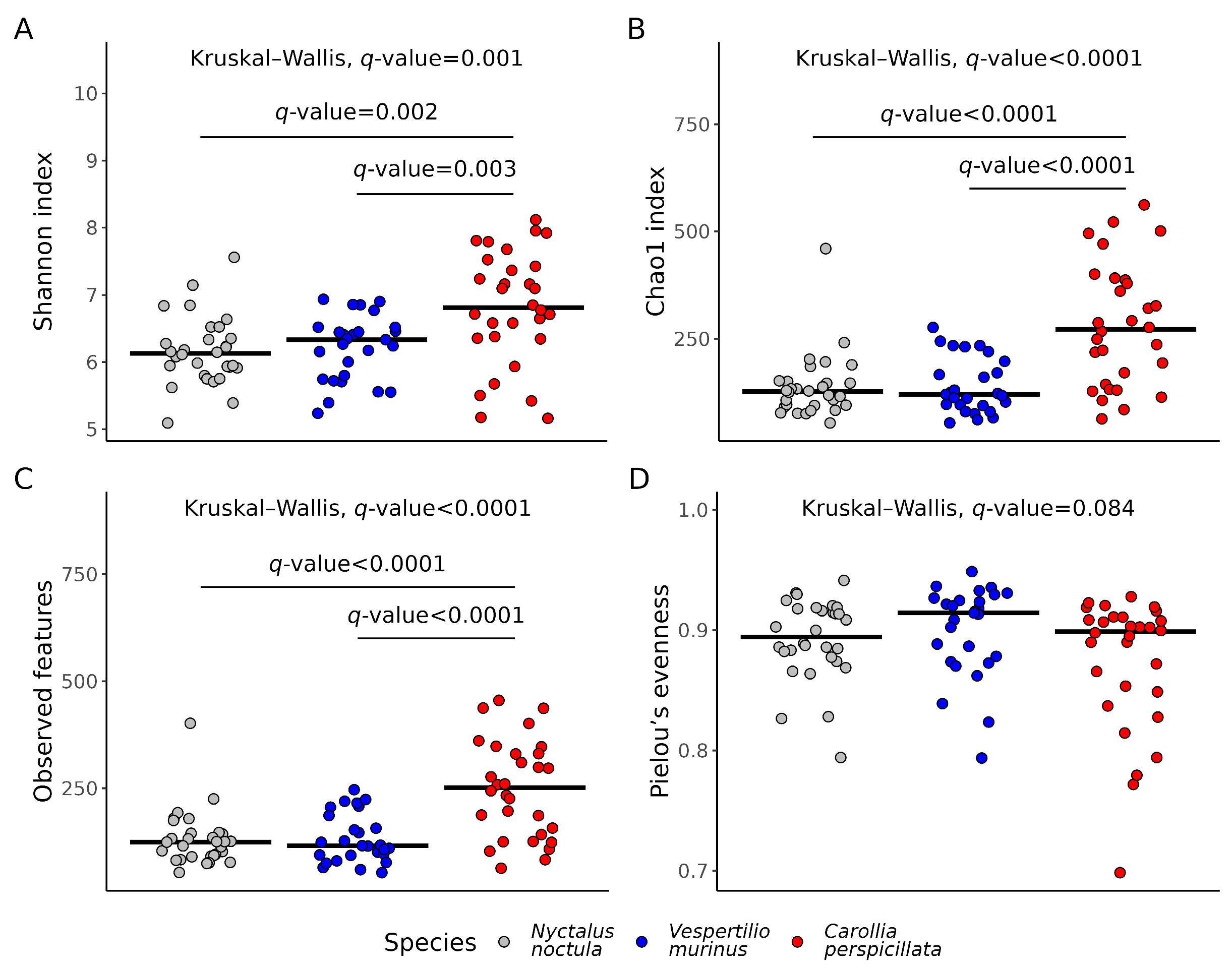
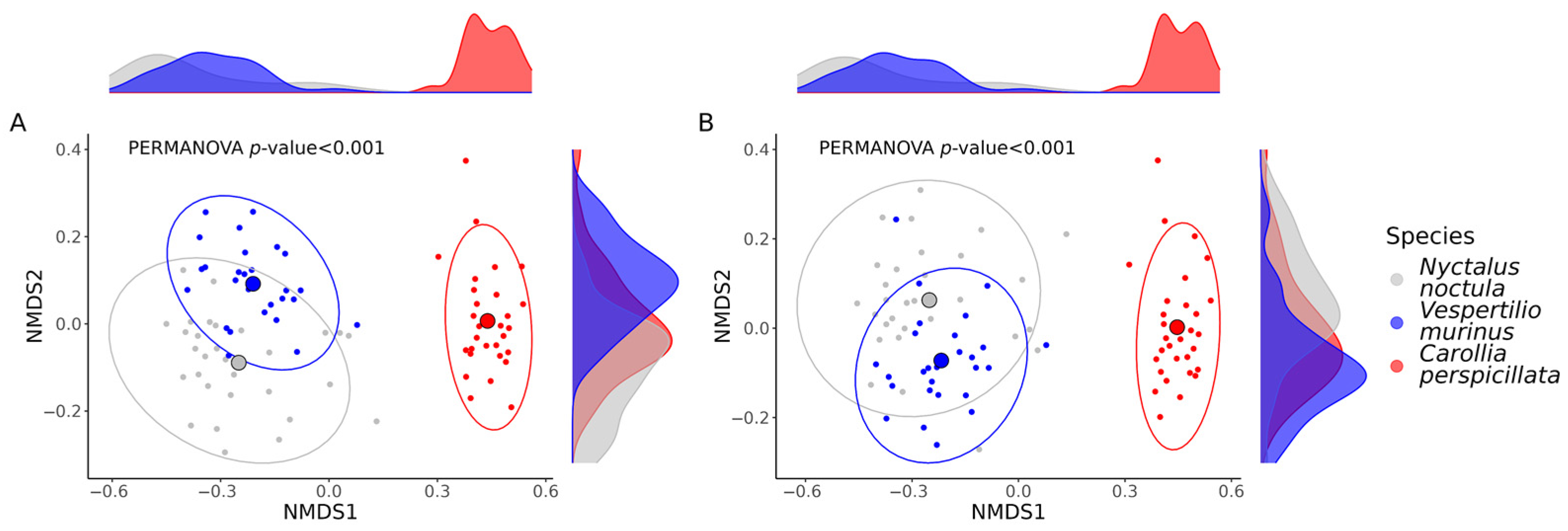
2.2. Gut Microbiota Richness and Diversity

To compare microbial alpha diversity we used the Shannon, Chao1, and Pielou’s evenness indexes, as well as the total number of features in the acquired samples. According to the multiple comparison testing, there were statistically significant differences between the fructivorous C. perspicillata and the two insectivores N. noctula and V. murinus in all used alpha diversity metrics, except for Pielou’s evenness, while there were no statistical differences between the alpha diversity indexes in the gut microbiota between the two studied insectivore species (Figure 3).
To compare microbial beta diversity between studied bat species we used Bray–Curtis dissimilarity and Jaccard similarity indexes. As for the alpha diversity, the most significant differences were observed in the beta diversity between bats with different food types, while differences between the two insectivorous bats were less pronounced, as seen in the non-metric multidimensional scaling ordination plots (Figure 4).

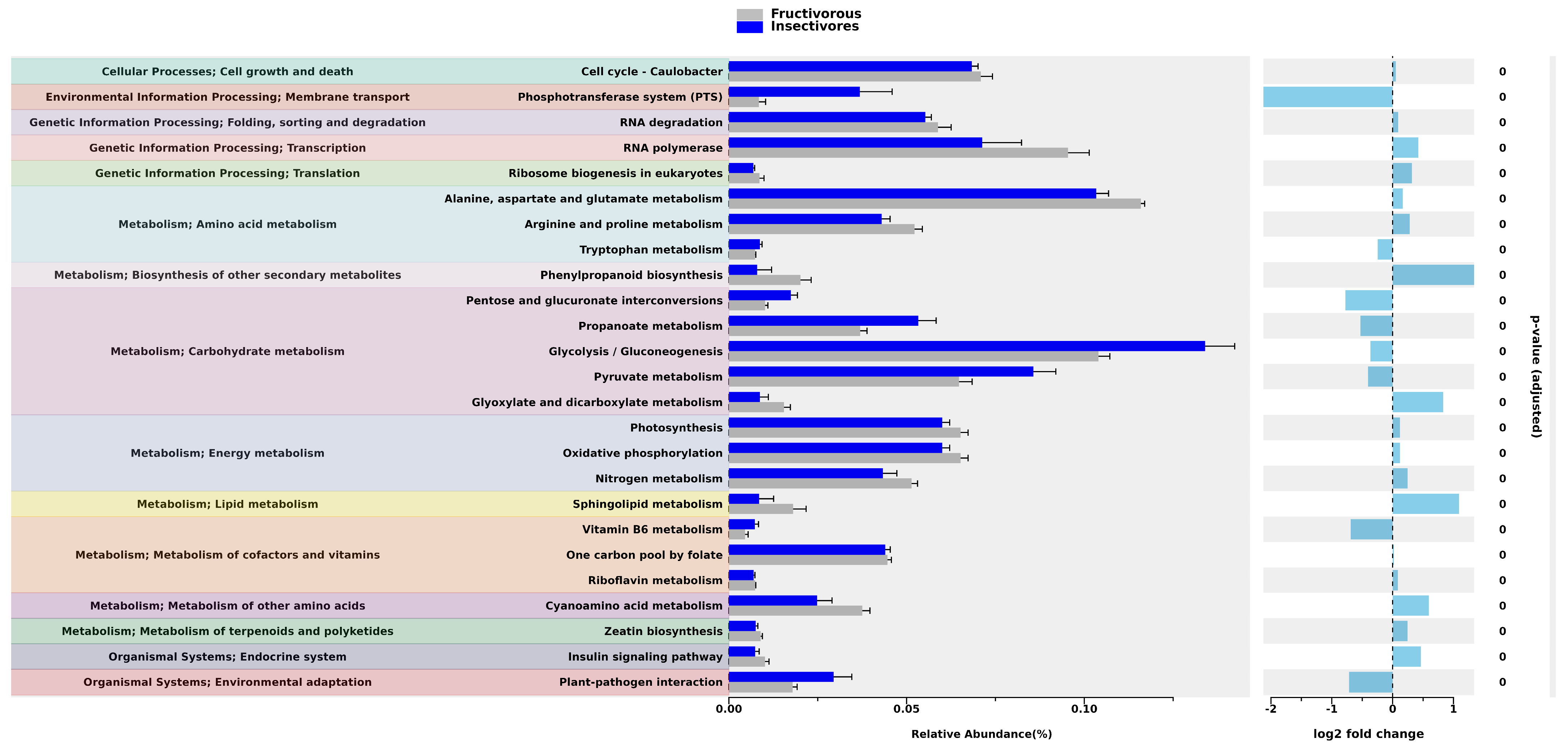
2.3. Prediction of the Functional Pathways

To predict functional pathways based on the 16S rRNA metagenomic sequencing data, we used the PICRUSt2 pipeline [20], and the “ggpicrust2” R package [21] for the statistical analysis and the visualization of acquired results. First, we performed principal component analysis to investigate the overall differences in the distance of functional pathways. As a result, we observed that there were almost no differences in pathway distances between N. noctula and V. murinus, while the distances between C. perspicillata and insectivorous bats were more prominent (Figure 5).
Based on the results of the differential abundance analysis of annotated functional pathways, there were 104 pathways for which differential abundance was closely tied with the food type. Most of the observed functional pathways were associated with microbial metabolisms, such as amino acids, carbohydrates, lipids, vitamins, and energy metabolism, although there were pathways associated with environmental interaction and genetic information processing. Figure 6 shows the top 25 functional pathways that have the most significant differential abundance between bats of different food types. Tables S1 and S2 contain full information about differentially abundant functional pathways in bats of different food niches and studied species.

3. Discussion

In this study, we investigated the gut microbiota composition and alpha/beta diversity of N. noctula, V. murinus, and C. perspicillata, predicted the microbiota functions, and made a statistical comparison of these features. Samples from N. noctula and V. murinus were obtained from bat rehabilitation centers in Rostov-on-Don and Moscow, respectively. The work of these centers is aimed at protecting the population of synanthropic bats, especially during fall and winter. Synanthropic insectivorous bats play a crucial role in the ecosystems of urban areas as they are natural controllers of agricultural pests [22,23]. In a recent study, Nsengimana et al. determined that 60% to 78% of the diet of insectivorous bats consists of agricultural pests [24]. Aguiar et al. calculated the value of pest suppression by synanthropic insectivorous bats and concluded that bats are saving USD 94 per hectare of cornfields and overall USD 390.6 million per harvest in Brazil [25]. These studies prove the beneficial role of bats in the human ecosystem; however, the process of urbanization may be detrimental to the bat population [26]. Due to their small size and ability to fly, bats are often found in households, as there are few places in cities where bats can safely roost and hibernate. When people find a bat or an entire colony, they tend to destroy them, putting the lives of these animals at risk. In some cases, bats are unable to survive hibernation, as the temperature conditions in some households could be extremely low. To save the population of synanthropic bats and the urban ecosystems, volunteers bring animals to bat rehabilitation centers, where the animals are fed insects, receive qualified veterinary care, and are placed in controlled hibernation at temperatures of 4–10 °C for the winter period before they are released in spring. In addition to the primary purpose of bat rehabilitation centers, they provide unique opportunities to study bats [27,28].
Samples from C. perspicillata were obtained from the Moscow Zoo. The natural habitat of this bat species is Central America, South America, and the Antilles Islands, and it is one of the most common bat species in zoos [29,30]. C. perspicillata bats were included in our study as the reference for the comparison with the gut microbiota of synanthropic bats, as the microbiota of this bat species was studied before [31,32]. Carrillo-Araujo et al. studied the gut microbiota in intestinal biopsies obtained from three C. perspicillata bats and found that the most abundant phyla in their gut microbiota were Pseudomonadota, Mycoplasmatota, Bacillota, and Cyanobacteria, which is generally consistent with our study, except for Mycoplasmatota, as we did not identify this phylum in the studied bats of the same species. Also, Bacteroidota were several times more abundant in our study. Interestingly, and entirely in line with our own observations, the authors found that Cyanobacteria were observed only in C. perspicillata among all other bat species [31]. The presence of Cyanobacteria in C. perspicillata is obviously linked to their food preferences; however, it is unknown why this phylum was observed only in this fructivorous bat species in previous studies. Lemieux-Labonté et al. studied the skin microbiota of C. perspicillata, which on the phylum level also corresponds to the gut microbiota of this species, as they observed Pseudomonadota, Bacillota, Bacteroidota, and Cyanobacteria [32], although their RA could differ among different body sites. Nevertheless, there should be a connection between gut and skin microbiota in bats, as feces could sometimes stick to the bats’ skin and affect skin microbiota composition, especially in the captive environment.
While there are some data on the microbiota of C. perspicillata, the data for N. noctula and V. murinus are very limited. The only study we found was conducted by Corduneanu et al., who reported the heart microbiome of insectivorous bats from Central and Southeastern Europe, including N. noctula [33]. In our study, we found that the RAs of at least 10 bacterial genera in the gut microbiota of N. noctula and V. murinus are significantly different between the two insectivores, while there were a minimum of 60 genera for which the RAs were different in C. perspicillata compared to the studied insectivorous bats. These results reveal much higher similarities in gut microbiota composition between synanthropic N. noctula and V. murinus from Rostov-on-Don and Moscow than between zoo C. perspicillata and these two bat species, although V. murinus was also collected in Moscow. This was confirmed by the results of alpha and beta diversity analyses, in which the differences between fructivorous and insectivorous bats appeared more prominent and significant than between the two insectivores. In our study, the food type appeared to be much more closely associated with the differences in the gut microbiota of studied bats than their species, supporting the hypothesis that the gut microbiota in bats is not only host phylogeny-specific [31]. Phillips et al. also observed that the gut microbiota community in fructivorous bats from Guatemala and Arizona was characterized as significantly more diverse than in insectivorous bats [14], which fully correlated with our results. This dependence of gut microbiota alpha and beta diversity on the food type is typical of mammals. Ley et al. demonstrated that the gut microbial diversity of herbivore mammals is significantly greater than that of carnivores [34]. However, it is worth mentioning that due to the anatomical and physiological features of the bat GIT, as well as the dependence of its microbiota composition on the environment, the relationship between food type and gut microbial diversity could be more prominent in bats.
Another important feature includes a markedly lower RA of facultative or obligatory anaerobic bacteria in the gut microbiota of N. noctula and V. murinus compared to C. perspicillata, as shown by the differential abundance analysis on the phylum and class levels. For example, the higher RA of Bacteroidota phylum and Bacteroides genus was significantly more associated with the gut microbiota of C. perspicillata compared to insectivorous N. noctula and V. murinus. Bacteroidota is a key taxon for human gut microbiota, and this phylum makes up the majority of the bacterial taxa present in the human intestinal microbiota, which is provided by the favorable anaerobic conditions in the human colon [35]. In comparison, bats have lower anaerobic volumes in the intestines, resulting in less favorable conditions for the strict anaerobic microbiota community found in other animal species. We did not find detailed data on anaerobic volumes of GIT in N. noctula and V. murinus, although the food transition time and maximum retention time in the GIT of these bat species are relatively short compared to other mammals, which may also indicate less favorable conditions for gut anaerobic bacteria [36]. The unique properties of the microbiota may also stem from the ability to fly, leading to increased energy metabolism and distinctive immune responses, as well as the GIT features [11,37,38]. In this study, we also identified lactic acid bacteria in bats such as Pedicoccus, Latilactobacillus, and Lactococcus, which were more abundant in N. noctula and V. murinus and less common or even absent in C. perspicillata. Lactic acid bacteria play a significant role in animal welfare, as they have immunomodulatory properties [39]. However, the immune system of bats significantly differs from other animals, which allows them to co-exist with dangerous pathogens due to limited inflammatory response and also provides them with tolerance to malignant tumors [40,41]. Some bat species, such as N. noctula and V. murinus, hibernate during winter, which also contributes to their immunity-associated features, such as longevity [42,43]. The role of lactic acid bacteria in all of these features is not yet investigated, but we speculate that revealing new mechanisms of interaction between lactic acid bacteria and the bat immune system could provide new insights into understanding complex relationships between bacteria, host organisms, and even viruses [44].
Notably, in synanthropic bats, we identified bacterial genera that could have an important role in public health and animal welfare, such as Bacteroides, Enterobacter, Clostridiaceae, Enterococcus, Ureaplasma, Faecalibacterium, Helicobacter, Mycoplasma, Esherichia-Shigella, and Proteus. Some of these bacteria are considered to be potentially pathogenic to humans and animals [45,46,47], although their pathogenic potential [47,48,49] can only be uncovered by additional studies. Most importantly, as these genera are common for farm and domestic animals, and also for humans, they could be involved in spreading antibiotic resistance genes in urban areas. For example, in recent studies of Enterobacter spp. isolated from bat fecal samples, the resistance of these bacteria to ampicillin and amoxicillin was reported in bats from Brazil [50], and the resistance to amoxicillin, ampicillin, amoxicillin-clavulanic acid, aztreonam, cefotaxime, cefepime, ceftazidime, and many other antibiotics was reported in bats from Gabon [51]. In another example, Enterococcus spp. isolated from rectal samples of bats in Spain showed resistance to erythromycin, quinupristin-dalfopristin, and ciprofloxacin [52], while Nowakiewicz et al. reported that Enterococcus faecalis isolated from guano samples and rectal swabs from bats in Poland displayed resistance to chloramphenicol, ciprofloxacin, erythromycin, gentamycin, kanamycin, rifampin, streptomycin, and tetracycline [53]. Moreover, the presence of some common antibiotic-resistance genes, such as blaCTX-M15, in bats from different populations in Nigeria [54], Gabon [51], Poland [53], and Peru [55] marks the global role of bats in spreading antibiotic-resistant genes. However, a scoping review identified E. coli as the most significant taxon involved in bat-derived antimicrobial resistance spread. However, in our study, the Escherichia genus was among the least abundant taxa in all three studied bat species. Nonetheless, this does not necessarily imply that the studied bats are unable to contribute to the spread of antibiotic resistance. This aspect requires further investigation.
According to the principal component analysis, the diversity of predicted functional pathways in the gut microbiota of N. noctula and V. murinus showed more similarities compared to C. perspicillata. As expected, these results demonstrate that gut microbiota features, including microbial metabolism, strongly depend on bat diet [56,57]. The RA of predicted functional pathways in the gut microbiota of studied bats is in agreement with previous studies. Ingala et al. conducted a comparison of microbial functional pathways based on the 16S rRNA sequenced gut microbiota data from 545 bats, which were obtained from multiple studies. In those studies, 360 and 141 of the samples were from insectivorous and fructivorous bats, respectively, while the remaining 44 samples were from bats of different food niches (sanguivorous, omnivorous, and carnivorous) [58]. The researchers found that the RA of at least 29 predicted functional pathways were enriched and significantly different between bats with these two food niches. Interestingly, in fructivorous bats, pathways were observed that could be associated with the production of essential amino acids, such as methionine, valine, isoleucine, and tryptophan. The authors suggested that the gut microbiota of fructivorous bats could compensate for the lack of essential amino acids for the host that could not be obtained through the food, which marks host-microbiota relationships in bats [58]. In our study, predicted pathways for amino acids in the gut microbiota of C. perspicillata were also significantly enriched in this bat species compared to insectivorous bats, although they were not essential (alanine, aspartate, glutamate, arginine, proline) [59]. The only differentially abundant essential amino acid synthesis pathway that could be possibly due to the gut microbiota was tryptophan synthesis, which was more abundant in insectivorous bats. The other major groups of functional pathways, for which the RA significantly differed among bats with different diet types in our study, were associated with carbohydrate and energy metabolism. The most prominent differences were in glycolysis/gluconeogenesis, which is significantly enriched in insectivorous bats compared to fructivorous. Given that the microbiota could be involved in the host metabolism, it is obvious that the gut microbiota of insectivorous bats could provide the host with the necessary carbohydrates during the microbial metabolism, which are already delivered in high concentrations in fruits consumed by fructivorous bats [60,61]. We should also note that the gut microbiota of the studied bats could be involved in the synthesis of some essential vitamins, such as vitamin B5 and riboflavin. Functional microbial pathways associated with vitamin synthesis were also previously observed in the gut microbiota of bats [58]. One of the interesting features that we noted in the predicted pathways was the potential impact of gut microbiota on genetic information processing, particularly, on transcription, which may be related to the pro-mutagenic properties of the lactic acid gut microbiota of bats that we observed previously [44]. However, this hypothesis requires further research.
We performed a differential abundance analysis of microbiota composition and functional pathway data with the MaAsLin2 package [19] after filtering the least abundant taxa/pathways. According to the results of the bioinformatic benchmarking study of Nearing et al., MaAsLin2 showed the most consistent results in avoiding false positives, but only with filtered data [62]. Some other studies of the bat gut microbiota relied on other methods of differential abundance analysis, so this should be taken into account when comparing the results of the published studies. Although it is clear that the gut microbiota of bats is very diverse and varies with the environment, the differences are evident with different analysis methods [18,63,64].
The sampling from synanthropic bats was conducted after hibernation in bat rehabilitation centers. Thus, we speculate that we observed bacteria that survived low temperatures within the bats’ bodies. Our recent study shows that hibernation results in the decrease of the gut microbiota diversity in bats, as food deprivation and low body temperature create unfavorable conditions for gut microbes [65]. Therefore, it is important to mention that the diversity of gut microbiota of studied insectivorous bats could have been decreased, as they awoke from hibernation at the moment of the study. Also, we should mention that the gut microbiota composition of the studied fructivorous and insectivorous bats was very diverse, although all of these bats were captive or semi-captive when sampled. This could mean that the gut microbiota of the studied bats was not grossly altered by humans, as all three bat species were in close contact with volunteers, veterinarians, biologists, and rehabilitation specialists, especially for the C. perspicillata colony in the zoo. However, further studies may reveal how close human contact impacts the gut microbiota of bats, which would require sampling from bats on their arrival [27,66].

4. Materials and Methods

4.1. Sampling

Sampling was conducted in February–March 2022 at the Bat Rehabilitation Center of Don State Technical University (Rostov-on-Don, Russia), the Bat Rehabilitation Center of the Moscow Zoo (Moscow, Russia), and the Experimental Department of Small Mammals of the Moscow Zoo (Moscow, Russia). In the study, 3 bat species were included, with 89 bats in total (Table 1).
All of the studied insectivorous bats were in an active state and recently arrived at the bat rehabilitation center facilities. Sampling of fecal samples from insectivorous species was performed before they were put into hibernation. A fructivorous bat colony was kept collectively at the zoo facility and consisted of captive-born individuals. A minimum of 0.5 g of fecal samples were collected from each bat and placed in sterile tubes with DNA/RNA Shield (Zymo Research, Irvine, CA, USA) and transported to the laboratory at room temperature.

4.2. Gut Microbiota Composition and Metagenomes Functions Prediction

Amplicons of the V3–V4 region of the 16S rRNA gene were sequenced to determine microbiota composition. In short, from the fecal samples, DNA was isolated with QIAamp Fast DNA Stool Mini Kit (Qiagen, Hilden, Germany), amplified with barcoding, pooled, and subsequently sequenced with the Illumina MiSeq sequencing system according to the manufacturer’s instructions (Illumina, San Diego, CA, USA). The Binary Base Call text-based format for storing the biological sequence and corresponding quality scores pipeline (BCL2FASTQ, version 1.8.3, Illumina, San Diego, CA, USA) was used to convert the sequences into a text-based format for storing biological sequence and corresponding quality score (FASTQ) files after quality checking. The results were then analyzed using the Quantitative Insights Into Microbial Ecology 2 (QIIME-2, version 2023.5) software [67]. For the data analysis, we performed denoising with DADA2 [68] and used the Silva database (version 138 (available online: https://www.arb-silva.de/documentation/release-138/, accessed on 11 July 2023)) as a reference 16S rRNA database for the classification of amplicon sequence variants [69,70]. A prediction of metagenome functions was conducted using the PICRUSt2 (version 2.5.2.) [20]. Annotations of functional pathways were performed with the KEGG database [71].

4.3. Statistical Analysis

The programming language R v4.2.3 (R Foundation for Statistical Computing, Vienna, Austria) was used for statistical analyses. MaAsLin2 package was used for the differential abundance analysis [19]. The C. perspicillata species was used as a reference factor for the linear model analysis. Before differential abundance analysis, we performed filtering by applying a minimal 10% threshold for the prevalence of the taxa to obtain more robust results [62]. Based on the acquired counts of data, the RA of taxa was calculated. Taxa with an RA higher than 1% were used for the differential abundance analysis. The rarefaction threshold for the alpha and beta diversity analyses was 2500 sequences/sample. The Kruskal–Wallis test was performed to determine differences in the alpha diversity matrix. PERMANOVA with the adonis function from the “vegan” package was used to determine differences in beta diversity distances (the number of permutations was set to 1000). We used the “ggplot2” package for the visualization of the differential abundance analysis results as volcano plots and heatmaps, alpha diversity analysis results as jitter plots, and beta diversity analysis as non-metric multidimensional scaling ordination plots [72]. The PICRUSt2 outcome was analyzed and visualized with the”ggpicrust2” package [21]. Differential abundance analysis of predicted functional pathways was carried out in the same way as with the taxa RA described above. Multiple comparisons were performed with the Mann–Whitney test, and the results were adjusted with the Benjamini–Hochberg false discovery rate. Adjusted p-values (q-values) were considered significant at q < 0.05.

5. Conclusions

In conclusion, in this study, we provide the first data on gut microbiota composition and potential functional activity of two synanthropic species, N. noctula and V. murinus, from Rostov-on-Don and Moscow, respectively, and performed a direct comparison of these features with C. perspicillata bats from a zoo colony. The observed features point to possible host-microbiota interactions and the significance of studied bat gut microbiota for public health and animal welfare. The overall results are consistent with the previous studies of bat gut microbiota, although all bat species studied here showed some unique microbiota features compared to previous reports, which is reasonable given their different geographical location and climatic environments.

Supplementary Materials

The following supporting information can be downloaded at: https://www.mdpi.com/article/10.3390/ijms242417301/s1.

Author Contributions

Conceptualization, I.V.P. (Igor V. Popov), K.V. and A.M.E.; methodology, I.V.P. (Igor V. Popov); software, I.V.P. (Ilia V. Popov); validation, K.V.; formal analysis, I.V.P. (Igor V. Popov) and I.V.P. (Ilia V. Popov); investigation, A.A.K. and T.A.L.; resources, T.N.D. and M.L.C.; data curation, I.V.P. (Ilia V. Popov); writing—original draft preparation, I.V.P. (Igor V. Popov); writing—review and editing, K.V. and M.L.C.; visualization, I.V.P. (Igor V. Popov) and I.V.P. (Ilia V. Popov); supervision, K.V. and A.M.E.; project administration, A.M.E.; funding acquisition, I.V.P. (Igor V. Popov), K.V. and A.M.E. All authors have read and agreed to the published version of the manuscript.

Funding

Sampling and metagenomic sequencing were funded by the Russian Science Foundation (project 23-14-00316). Bioinformatical analysis was funded by the Ministry of Science and Higher Education of the Russian Federation, (Agreement 075-10-2021-093, Project [IMB-2102]). Statistical analysis and APC were funded by the Dutch Province of Limburg with a grant to the Centre for Healthy Eating & Food Innovation (HEFI) of Maastricht University Campus Venlo.

Institutional Review Board Statement

The reported study does not involve the results of any in vivo intervention experiments. The handling of animals at the bat rehabilitation centers was approved by the local ethics committee of Don State Technical University (Protocol No. 5 2022).

Data Availability Statement

All sequences associated with this study have been deposited int the National Center for Biotechnological Information’s Short Read Archive and are available under BioProject ID PRJNA1012124.

Acknowledgments

We acknowledge the support of Olga Ilchenko and the team of the bat rehabilitation center and the experimental department of small mammals of the Moscow Zoo. We also acknowledge the team of the bat rehabilitation center of Don State Technical University. Igor V. Popov expresses his deep gratitude to Sergey A. Nedospasov for his guidance and editing of the manuscript.

Conflicts of Interest

The authors declare no conflict of interest. The funders had no role in the design of the study; in the collection, analyses, or interpretation of data; in the writing of the manuscript; or in the decision to publish the results.

References

  1. Arnaout, Y.; Djelouadji, Z.; Robardet, E.; Cappelle, J.; Cliquet, F.; Touzalin, F.; Jimenez, G.; Hurstel, S.; Borel, C.; Picard-Meyer, E. Genetic identification of bat species for pathogen surveillance across France. PLoS ONE 2022, 17, e0261344. [Google Scholar] [CrossRef] [PubMed]
  2. Pena, S.A.; Alencastre-Santos, A.B.; da Silva, J.B.; Correia, L.L.; Urbieta, G.L.; Graciolli, G.; Palheta, L.R.; Vieira, T.B. Bats (Mammalia, Chiroptera) and bat flies (Diptera, Streblidae) from the Cazumba-Iracema and Chico Mendes Reserve, Western Brazilian Amazon. Parasitol. Res. 2023, 122, 451–459. [Google Scholar] [CrossRef] [PubMed]
  3. Weinberg, M.; Yovel, Y. Revising the paradigm: Are bats really pathogen reservoirs or do they possess an efficient immune system? iScience 2022, 25, 104782. [Google Scholar] [CrossRef] [PubMed]
  4. Banerjee, A.; Baker, M.L.; Kulcsar, K.; Misra, V.; Plowright, R.; Mossman, K. Novel Insights Into Immune Systems of Bats. Front. Immunol. 2020, 11, 26. [Google Scholar] [CrossRef] [PubMed]
  5. Irving, A.T.; Ahn, M.; Goh, G.; Anderson, D.E.; Wang, L.F. Lessons from the host defences of bats, a unique viral reservoir. Nature 2021, 589, 363–370. [Google Scholar] [CrossRef] [PubMed]
  6. Islam, M.S.; Sazzad, H.M.; Satter, S.M.; Sultana, S.; Hossain, M.J.; Hasan, M.; Rahman, M.; Campbell, S.; Cannon, D.L.; Stroher, U.; et al. Nipah Virus Transmission from Bats to Humans Associated with Drinking Traditional Liquor Made from Date Palm Sap, Bangladesh, 2011–2014. Emerg. Infect. Dis. 2016, 22, 664–670. [Google Scholar] [CrossRef] [PubMed]
  7. Edson, D.; Field, H.; McMichael, L.; Vidgen, M.; Goldspink, L.; Broos, A.; Melville, D.; Kristoffersen, J.; de Jong, C.; McLaughlin, A.; et al. Routes of Hendra Virus Excretion in Naturally-Infected Flying-Foxes: Implications for Viral Transmission and Spillover Risk. PLoS ONE 2015, 10, e0140670. [Google Scholar] [CrossRef] [PubMed]
  8. Goldstein, T.; Anthony, S.J.; Gbakima, A.; Bird, B.H.; Bangura, J.; Tremeau-Bravard, A.; Belaganahalli, M.N.; Wells, H.L.; Dhanota, J.K.; Liang, E.; et al. The discovery of Bombali virus adds further support for bats as hosts of ebolaviruses. Nat. Microbiol. 2018, 3, 1084–1089. [Google Scholar] [CrossRef]
  9. Al-Salihi, K.A.; Khalaf, J.M. The emerging SARS-CoV, MERS-CoV, and SARS-CoV-2: An insight into the viruses zoonotic aspects. Vet. World 2021, 14, 190–199. [Google Scholar] [CrossRef]
  10. Popov, I.V.; Ohlopkova, O.V.; Donnik, I.M.; Zolotukhin, P.V.; Umanets, A.; Golovin, S.N.; Malinovkin, A.V.; Belanova, A.A.; Lipilkin, P.V.; Lipilkina, T.A.; et al. Detection of coronaviruses in insectivorous bats of Fore-Caucasus, 2021. Sci. Rep. 2023, 13, 2306. [Google Scholar] [CrossRef]
  11. Donnik, I.M.; Popov, I.V.; Sereda, S.V.; Popov, I.V.; Chikindas, M.L.; Ermakov, A.M. Coronavirus Infections of Animals: Future Risks to Humans. Biol. Bull. Russ. Acad. Sci. 2021, 48, 26–37. [Google Scholar] [CrossRef] [PubMed]
  12. Becker, P.; van den Eynde, C.; Baert, F.; D’Hooge, E.; De Pauw, R.; Normand, A.C.; Piarroux, R.; Stubbe, D. Remarkable fungal biodiversity on northern Belgium bats and hibernacula. Mycologia 2023, 115, 484–498. [Google Scholar] [CrossRef] [PubMed]
  13. Devnath, P.; Karah, N.; Graham, J.P.; Rose, E.S.; Asaduzzaman, M. Evidence of Antimicrobial Resistance in Bats and Its Planetary Health Impact for Surveillance of Zoonotic Spillover Events: A Scoping Review. Int. J. Environ. Res. Public Health 2022, 20, 243. [Google Scholar] [CrossRef] [PubMed]
  14. Phillips, C.D.; Phelan, G.; Dowd, S.E.; McDonough, M.M.; Ferguson, A.W.; Delton Hanson, J.; Siles, L.; Ordonez-Garza, N.; San Francisco, M.; Baker, R.J. Microbiome analysis among bats describes influences of host phylogeny, life history, physiology and geography. Mol. Ecol. 2012, 21, 2617–2627. [Google Scholar] [CrossRef] [PubMed]
  15. Donaldson, G.P.; Lee, S.M.; Mazmanian, S.K. Gut biogeography of the bacterial microbiota. Nat. Rev. Microbiol. 2016, 14, 20–32. [Google Scholar] [CrossRef]
  16. Gadelha-Alves, R.; Rozensztranch, A.M.D.; Rocha-Barbosa, O. Comparative Intestinal Histomorphology of Five Species of Phyllostomid Bats (Phyllostomidae, Microchiroptera): Ecomorphological Relations with Alimentary Habits. Int. J. Morphol. 2008, 26, 591–602. [Google Scholar] [CrossRef]
  17. Caviedes-Vidal, E.; McWhorter, T.J.; Lavin, S.R.; Chediack, J.G.; Tracy, C.R.; Karasov, W.H. The digestive adaptation of flying vertebrates: High intestinal paracellular absorption compensates for smaller guts. Proc. Natl. Acad. Sci. USA 2007, 104, 19132–19137. [Google Scholar] [CrossRef]
  18. Song, S.J.; Sanders, J.G.; Delsuc, F.; Metcalf, J.; Amato, K.; Taylor, M.W.; Mazel, F.; Lutz, H.L.; Winker, K.; Graves, G.R.; et al. Comparative Analyses of Vertebrate Gut Microbiomes Reveal Convergence between Birds and Bats. mBio 2020, 11, 02901-19. [Google Scholar] [CrossRef]
  19. Mallick, H.; Rahnavard, A.; McIver, L.J.; Ma, S.; Zhang, Y.; Nguyen, L.H.; Tickle, T.L.; Weingart, G.; Ren, B.; Schwager, E.H.; et al. Multivariable association discovery in population-scale meta-omics studies. PLoS Comput. Biol. 2021, 17, e1009442. [Google Scholar] [CrossRef]
  20. Douglas, G.M.; Maffei, V.J.; Zaneveld, J.R.; Yurgel, S.N.; Brown, J.R.; Taylor, C.M.; Huttenhower, C.; Langille, M.G.I. PICRUSt2 for prediction of metagenome functions. Nat. Biotechnol. 2020, 38, 685–688. [Google Scholar] [CrossRef]
  21. Yang, C.; Mai, J.; Cao, X.; Burberry, A.; Cominelli, F.; Zhang, L. ggpicrust2: An R package for PICRUSt2 predicted functional profile analysis and visualization. Bioinformatics 2023, 39, btad470. [Google Scholar] [CrossRef] [PubMed]
  22. Tuneu-Corral, C.; Puig-Montserrat, X.; Riba-Bertolin, D.; Russo, D.; Rebelo, H.; Cabeza, M.; Lopez-Baucells, A. Pest suppression by bats and management strategies to favour it: A global review. Biol. Rev. Camb. Philos. Soc. 2023, 98, 1564–1582. [Google Scholar] [CrossRef] [PubMed]
  23. Wolf, J.M.; Jeschke, J.M.; Voigt, C.C.; Itescu, Y. Urban affinity and its associated traits: A global analysis of bats. Glob. Chang. Biol. 2022, 28, 5667–5682. [Google Scholar] [CrossRef] [PubMed]
  24. Nsengimana, O.; Walker, F.M.; Webala, P.W.; Twizeyimana, I.; Dusabe, M.C.; Sanchez, D.E.; Sobek, C.J.; Ruhagazi, D.; Iribagiza, P.; Muvunyi, R.; et al. Our good neighbors: Understanding ecosystem services provided by insectivorous bats in Rwanda. PLoS ONE 2023, 18, e0287536. [Google Scholar] [CrossRef]
  25. Aguiar, L.M.S.; Bueno-Rocha, I.D.; Oliveira, G.; Pires, E.S.; Vasconcelos, S.; Nunes, G.L.; Frizzas, M.R.; Togni, P.H.B. Going out for dinner-The consumption of agriculture pests by bats in urban areas. PLoS ONE 2021, 16, e0258066. [Google Scholar] [CrossRef]
  26. Lewanzik, D.; Straka, T.M.; Lorenz, J.; Marggraf, L.; Voigt-Heucke, S.; Schumann, A.; Brandt, M.; Voigt, C.C. Evaluating the potential of urban areas for bat conservation with citizen science data. Environ. Pollut. 2022, 297, 118785. [Google Scholar] [CrossRef]
  27. Jones, S.; Bell, T.; Coleman, C.M.; Harris, D.; Woodward, G.; Worledge, L.; Roberts, H.; McElhinney, L.; Aegerter, J.; Ransome, E.; et al. Testing bats in rehabilitation for SARS-CoV-2 before release into the wild. Conserv. Sci. Pract. 2022, 4, e12707. [Google Scholar] [CrossRef]
  28. Minor, R.L.; Dopfer, D.; Lemley, E.M.; Thurber, M.I. Factors Impacting Successful Rehabilitation of Big Brown Bats (Eptesicus Fuscus) in a Wisconsin Wildlife Rehabilitation Center: A 5-Year Retrospective. J. Zoo Wildl. Med. 2023, 54, 32–39. [Google Scholar] [CrossRef]
  29. Lim, B.K.; Lee, T.E. Community Ecology and Phylogeography of Bats in the Guianan Savannas of Northern South America. Diversity 2018, 10, 129. [Google Scholar] [CrossRef]
  30. Hahn, K.; Veiga, I.B.; Schediwy, M.; Wiederkehr, D.; Meniri, M.; Schneeberger, M.; den Broek, P.R.; Gurtner, C.; Fasel, N.J.; Kittl, S.; et al. Yersinia pseudotuberculosis serotype O:1 infection in a captive Seba’s short tailed-fruit bat (Carollia perspicillata) colony in Switzerland. BMC Vet. Res. 2021, 17, 92. [Google Scholar] [CrossRef]
  31. Carrillo-Araujo, M.; Tas, N.; Alcantara-Hernandez, R.J.; Gaona, O.; Schondube, J.E.; Medellin, R.A.; Jansson, J.K.; Falcon, L.I. Phyllostomid bat microbiome composition is associated to host phylogeny and feeding strategies. Front. Microbiol. 2015, 6, 447. [Google Scholar] [CrossRef] [PubMed]
  32. Lemieux-Labonte, V.; Tromas, N.; Shapiro, B.J.; Lapointe, F.J. Environment and host species shape the skin microbiome of captive neotropical bats. PeerJ 2016, 4, e2430. [Google Scholar] [CrossRef] [PubMed]
  33. Corduneanu, A.; Mihalca, A.D.; Sandor, A.D.; Hornok, S.; Malmberg, M.; Viso, N.P.; Bongcam-Rudloff, E. The heart microbiome of insectivorous bats from Central and South Eastern Europe. Comp. Immunol. Microbiol. Infect. Dis. 2021, 75, 101605. [Google Scholar] [CrossRef] [PubMed]
  34. Ley, R.E.; Hamady, M.; Lozupone, C.; Turnbaugh, P.J.; Ramey, R.R.; Bircher, J.S.; Schlegel, M.L.; Tucker, T.A.; Schrenzel, M.D.; Knight, R.; et al. Evolution of mammals and their gut microbes. Science 2008, 320, 1647–1651. [Google Scholar] [CrossRef]
  35. King, C.H.; Desai, H.; Sylvetsky, A.C.; LoTempio, J.; Ayanyan, S.; Carrie, J.; Crandall, K.A.; Fochtman, B.C.; Gasparyan, L.; Gulzar, N.; et al. Baseline human gut microbiota profile in healthy people and standard reporting template. PLoS ONE 2019, 14, e0206484. [Google Scholar] [CrossRef]
  36. Roswag, A.; Becker, N.I.; Encarnacao, J.A. Inter- and intraspecific comparisons of retention time in insectivorous bat species (Vespertilionidae). J. Zool. 2012, 288, 85–92. [Google Scholar] [CrossRef]
  37. Subudhi, S.; Rapin, N.; Misra, V. Immune System Modulation and Viral Persistence in Bats: Understanding Viral Spillover. Viruses 2019, 11, 192. [Google Scholar] [CrossRef]
  38. Luo, J.; Liang, S.; Jin, F. Gut microbiota in antiviral strategy from bats to humans: A missing link in COVID-19. Sci. China Life Sci. 2021, 64, 942–956. [Google Scholar] [CrossRef]
  39. Zoghi, A.; Massoud, R.; Todorov, S.D.; Chikindas, M.L.; Popov, I.; Smith, S.; Khosravi-Darani, K. Role of the lactobacilli in food bio-decontamination: Friends with benefits. Enzym. Microb. Technol. 2021, 150, 109861. [Google Scholar] [CrossRef]
  40. Jebb, D.; Huang, Z.; Pippel, M.; Hughes, G.M.; Lavrichenko, K.; Devanna, P.; Winkler, S.; Jermiin, L.S.; Skirmuntt, E.C.; Katzourakis, A.; et al. Six reference-quality genomes reveal evolution of bat adaptations. Nature 2020, 583, 578–584. [Google Scholar] [CrossRef]
  41. Scheben, A.; Mendivil Ramos, O.; Kramer, M.; Goodwin, S.; Oppenheim, S.; Becker, D.J.; Schatz, M.C.; Simmons, N.B.; Siepel, A.; McCombie, W.R. Long-Read Sequencing Reveals Rapid Evolution of Immunity- and Cancer-Related Genes in Bats. Genome Biol. Evol. 2023, 15, evad148. [Google Scholar] [CrossRef] [PubMed]
  42. Sullivan, I.R.; Adams, D.M.; Greville, L.J.S.; Faure, P.A.; Wilkinson, G.S. Big brown bats experience slower epigenetic ageing during hibernation. Proc. Biol. Sci. 2022, 289, 20220635. [Google Scholar] [CrossRef] [PubMed]
  43. Wilkinson, G.S.; South, J.M. Life history, ecology and longevity in bats. Aging Cell 2002, 1, 124–131. [Google Scholar] [CrossRef] [PubMed]
  44. Popov, I.V.; Mazanko, M.S.; Kulaeva, E.D.; Golovin, S.N.; Malinovkin, A.V.; Aleshukina, I.S.; Aleshukina, A.V.; Prazdnova, E.V.; Tverdokhlebova, T.I.; Chikindas, M.L.; et al. Gut microbiota of bats: Pro-mutagenic properties and possible frontiers in preventing emerging disease. Sci. Rep. 2021, 11, 21075. [Google Scholar] [CrossRef] [PubMed]
  45. Ramirez, D.; Giron, M. Enterobacter Infections; StatPearls: Treasure Island, FL, USA, 2023. [Google Scholar]
  46. Bandelj, P.; Knapic, T.; Rousseau, J.; Podgorelec, M.; Presetnik, P.; Vengust, M.; Scott Weese, J. Clostridioides difficile in bat guano. Comp. Immunol. Microbiol. Infect. Dis. 2019, 65, 144–147. [Google Scholar] [CrossRef] [PubMed]
  47. Huang, Y.; Sun, Y.; Huang, Q.; Lv, X.; Pu, J.; Zhu, W.; Lu, S.; Jin, D.; Liu, L.; Shi, Z.; et al. The Threat of Potentially Pathogenic Bacteria in the Feces of Bats. Microbiol. Spectr. 2022, 10, e0180222. [Google Scholar] [CrossRef]
  48. Chakravorty, S.; Helb, D.; Burday, M.; Connell, N.; Alland, D. A detailed analysis of 16S ribosomal RNA gene segments for the diagnosis of pathogenic bacteria. J. Microbiol. Methods 2007, 69, 330–339. [Google Scholar] [CrossRef]
  49. Lopez-Aladid, R.; Fernandez-Barat, L.; Alcaraz-Serrano, V.; Bueno-Freire, L.; Vazquez, N.; Pastor-Ibanez, R.; Palomeque, A.; Oscanoa, P.; Torres, A. Determining the most accurate 16S rRNA hypervariable region for taxonomic identification from respiratory samples. Sci. Rep. 2023, 13, 3974. [Google Scholar] [CrossRef]
  50. Sens, H.; Trindade, W.A.; Oliveira, A.F.; Zaniolo, M.M.; Serenini, G.F.; Araujo-Ceranto, J.B.; Goncalves, D.D.; Germano, R.M. Bacterial resistance in bats from the Phyllostomidae family and its relationship with unique health. Pesqui. Vet. Bras. 2018, 38, 1207–1216. [Google Scholar] [CrossRef]
  51. Mbehang Nguema, P.P.; Onanga, R.; Ndong Atome, G.R.; Obague Mbeang, J.C.; Mabika Mabika, A.; Yaro, M.; Lounnas, M.; Dumont, Y.; Zohra, Z.F.; Godreuil, S.; et al. Characterization of ESBL-Producing Enterobacteria from Fruit Bats in an Unprotected Area of Makokou, Gabon. Microorganisms 2020, 8, 138. [Google Scholar] [CrossRef]
  52. Garcia, L.A.; Torres, C.; Lopez, A.R.; Rodriguez, C.O.; Valencia, C.S. Antimicrobial Resistance of Enterococcus Species Isolated from Wild Mammals in Aragon, Spain. J. Vet. Res. 2022, 66, 151–159. [Google Scholar] [CrossRef] [PubMed]
  53. Nowakiewicz, A.; Zieba, P.; Gnat, S.; Osinska, M.; Lagowski, D.; Kosior-Korzecka, U.; Puzio, I.; Krol, J. Analysis of the occurrence and molecular characteristics of drug-resistant strains of Enterococcus faecalis isolated from the gastrointestinal tract of insectivorous bat species in Poland: A possible essential impact on the spread of drug resistance? Environ. Pollut. 2021, 269, 116099. [Google Scholar] [CrossRef]
  54. Obodoechi, L.O.; Carvalho, I.; Chenouf, N.S.; Martinez-Alvarez, S.; Sadi, M.; Nwanta, J.A.; Chah, K.F.; Torres, C. Antimicrobial resistance in Escherichia coli isolates from frugivorous (Eidolon helvum) and insectivorous (Nycteris hispida) bats in Southeast Nigeria, with detection of CTX-M-15 producing isolates. Comp. Immunol. Microbiol. Infect. Dis. 2021, 75, 101613. [Google Scholar] [CrossRef] [PubMed]
  55. Benavides, J.A.; Shiva, C.; Virhuez, M.; Tello, C.; Appelgren, A.; Vendrell, J.; Solassol, J.; Godreuil, S.; Streicker, D.G. Extended-spectrum beta-lactamase-producing Escherichia coli in common vampire bats Desmodus rotundus and livestock in Peru. Zoonoses Public Health 2018, 65, 454–458. [Google Scholar] [CrossRef] [PubMed]
  56. Corduneanu, A.; Wu-Chuang, A.; Maitre, A.; Obregon, D.; Sandor, A.D.; Cabezas-Cruz, A. Structural differences in the gut microbiome of bats using terrestrial vs. aquatic feeding resources. BMC Microbiol. 2023, 23, 93. [Google Scholar] [CrossRef] [PubMed]
  57. Gong, L.; Liu, B.; Wu, H.; Feng, J.; Jiang, T. Seasonal Dietary Shifts Alter the Gut Microbiota of Avivorous Bats: Implication for Adaptation to Energy Harvest and Nutritional Utilization. mSphere 2021, 6, e0046721. [Google Scholar] [CrossRef]
  58. Ingala, M.R.; Simmons, N.B.; Dunbar, M.; Wultsch, C.; Krampis, K.; Perkins, S.L. You are more than what you eat: Potentially adaptive enrichment of microbiome functions across bat dietary niches. Anim. Microbiome 2021, 3, 82. [Google Scholar] [CrossRef]
  59. Lopez, M.J.; Mohiuddin, S.S. Biochemistry, Essential Amino Acids; StatPearls: Treasure Island, FL, USA, 2023. [Google Scholar]
  60. Batista, C.B.; Reis, N.R.; Rezende, M.I. Nutritional content of bat-consumed fruits in a forest fragment in Southern Brazil. Braz. J. Biol. 2017, 77, 244–250. [Google Scholar] [CrossRef]
  61. Amitai, O.; Holtze, S.; Barkan, S.; Amichai, E.; Korine, C.; Pinshow, B.; Voigt, C.C. Fruit bats (Pteropodidae) fuel their metabolism rapidly and directly with exogenous sugars. J. Exp. Biol. 2010, 213, 2693–2699. [Google Scholar] [CrossRef]
  62. Nearing, J.T.; Douglas, G.M.; Hayes, M.G.; MacDonald, J.; Desai, D.K.; Allward, N.; Jones, C.M.A.; Wright, R.J.; Dhanani, A.S.; Comeau, A.M.; et al. Microbiome differential abundance methods produce different results across 38 datasets. Nat. Commun. 2022, 13, 342. [Google Scholar] [CrossRef]
  63. Lutz, H.L.; Jackson, E.W.; Webala, P.W.; Babyesiza, W.S.; Kerbis Peterhans, J.C.; Demos, T.C.; Patterson, B.D.; Gilbert, J.A. Ecology and Host Identity Outweigh Evolutionary History in Shaping the Bat Microbiome. mSystems 2019, 4, 511–519. [Google Scholar] [CrossRef] [PubMed]
  64. Ingala, M.R.; Simmons, N.B.; Wultsch, C.; Krampis, K.; Speer, K.A.; Perkins, S.L. Comparing Microbiome Sampling Methods in a Wild Mammal: Fecal and Intestinal Samples Record Different Signals of Host Ecology, Evolution. Front. Microbiol. 2018, 9, 803. [Google Scholar] [CrossRef] [PubMed]
  65. Popov, I.V.; Berezinskaia, I.S.; Popov, I.V.; Martiusheva, I.B.; Tkacheva, E.V.; Gorobets, V.E.; Tikhmeneva, I.A.; Aleshukina, A.V.; Tverdokhlebova, T.I.; Chikindas, M.L. Cultivable Gut Microbiota in Synanthropic Bats: Shifts of Its Composition and Diversity Associated with Hibernation. Animals 2023, 13, 3658. [Google Scholar] [CrossRef]
  66. Salinas-Ramos, V.B.; Tomassini, A.; Ferrari, F.; Boga, R.; Russo, D. Admittance to Wildlife Rehabilitation Centres Points to Adverse Effects of Climate Change on Insectivorous Bats. Biology 2023, 12, 543. [Google Scholar] [CrossRef] [PubMed]
  67. Bolyen, E.; Rideout, J.R.; Dillon, M.R.; Bokulich, N.A.; Abnet, C.C.; Al-Ghalith, G.A.; Alexander, H.; Alm, E.J.; Arumugam, M.; Asnicar, F.; et al. Reproducible, interactive, scalable and extensible microbiome data science using QIIME 2. Nat. Biotechnol. 2019, 37, 852–857. [Google Scholar] [CrossRef] [PubMed]
  68. Callahan, B.J.; McMurdie, P.J.; Rosen, M.J.; Han, A.W.; Johnson, A.J.; Holmes, S.P. DADA2: High-resolution sample inference from Illumina amplicon data. Nat. Methods 2016, 13, 581–583. [Google Scholar] [CrossRef]
  69. Quast, C.; Pruesse, E.; Yilmaz, P.; Gerken, J.; Schweer, T.; Yarza, P.; Peplies, J.; Glockner, F.O. The SILVA ribosomal RNA gene database project: Improved data processing and web-based tools. Nucleic Acids Res. 2013, 41, D590–D596. [Google Scholar] [CrossRef]
  70. Yilmaz, P.; Parfrey, L.W.; Yarza, P.; Gerken, J.; Pruesse, E.; Quast, C.; Schweer, T.; Peplies, J.; Ludwig, W.; Glockner, F.O. The SILVA and “All-species Living Tree Project (LTP)” taxonomic frameworks. Nucleic Acids Res. 2014, 42, D643–D648. [Google Scholar] [CrossRef]
  71. Kanehisa, M.; Goto, S. KEGG: Kyoto encyclopedia of genes and genomes. Nucleic Acids Res. 2000, 28, 27–30. [Google Scholar] [CrossRef]
  72. Wickham, H. Ggplot2: Elegant Graphics for Data Analysis; Springer: New York, NY, USA, 2016. [Google Scholar]
Figure 1. Heatmaps representing gut microbiota composition (as relative abundance (RA) of bacterial taxa) in samples from the Nyctalus noctula, Vespertilio murinus, and Carollia perspicillata species. RA at (A) phylum, (B) class, (C) order, (D) family, and (E) genus levels. Dendrograms represent clustering analysis based on the taxa RA similarities.
Ijms 24 17301 g001
Figure 2. Results of the differential abundance analysis of bacterial genera identified in fecal samples of insectivorous (Nyctalus noctula and Vespertilio murinus) and fructivorous (Carollia perspicillata) bats. The fructivorous species Carollia perspicillata was used as a reference factor for the linear model analysis. (A) Volcano plots with genera, the relative abundance (RA) of which had significant associations with food type and studied bat species. Blue dots represent genera for which the RA was strongly associated with the insectivorous diet of bats. Red dots represent genera that were significantly less abundant or even absent in insectivorous bats compared to fructivorous ones. Yellow dots show genera that had a greater RA in Nyctalus noctula compared to Carollia perspicillata and Vespertilio murinus, while green dots represent genera, which were less abundant in Nyctalus noctula compared to Carollia perspicillata and Vespertilio murinus. (B) Heatmap with genera, the RA of which had significant associations with food type and studied bat species. In the top row, the purple cells in the heatmap represent genera for which the RA was significantly higher in Nyctalus noctula compared to Carollia perspicillata and Vespertilio murinus, while the orange cells show the opposite. In the second line, the purple cells represent genera with an RA significantly higher in the insectivorous bats studied compared to the fructivorous bats, and the orange cells show genera, which are significantly less abundant or absent in insectivorous bats compared to fructivorous bats.
Ijms 24 17301 g002
Figure 3. Alpha diversity metrics in samples from the Nyctalus noctula, Vespertilio murinus, and Carollia perspicillata. (A) Shannon index, (B) Chao1 index, (C) number of observed features, and (D) Pielou’s evenness. The results of the Kruskal–Wallis tests and multiple comparisons with the Mann–Whitney test were adjusted with the Benjamini–Hochberg procedure.
Ijms 24 17301 g003
Figure 4. Non-metric multidimensional scaling (NMDS) ordination plot illustrating differences in beta diversity among samples from the Nyctalus noctula, Vespertilio murinus, and Carollia perspicillata. (A) NMDS plot showing a Bray–Curtis distance matrix. (B) NMDS plot showing a Jaccard distance matrix. p-values were calculated with the PERMANOVA test; the number of permutations was set to 1000.
Ijms 24 17301 g004
Figure 5. Principal component analysis (PCA) plot showing distances of predicted functional pathway abundances (A) between Nyctalus noctula, Vespertilio murinus, and Carollia perspicillata and (B) between bats with different food types.
Ijms 24 17301 g005
Figure 6. Results of the differential abundance analysis of predicted functional pathways in bats with different food types representing the top 25 pathways clustered into groups of functional categories. Error bar plots show a direct comparison of pathway abundance. Bar plots on the right represent log2 fold changes in the abundance of the pathways in bats with the different food types.
Ijms 24 17301 g006
Table 1. Bat species, sampling location site, and number of animals included in the study.
Bat SpeciesFood TypeSampling Location SiteNumber
Nyctalus noctulaInsectivorousBat Rehabilitation Center of Don State Technical University, Rostov-on-Don30
Vespertilio murinusInsectivorousBat Rehabilitation Center of Moscow Zoo, Moscow29
Carollia perspicillataFructivorousExperimental Department of Small Mammals of Moscow Zoo, Moscow30
Disclaimer/Publisher’s Note: The statements, opinions and data contained in all publications are solely those of the individual author(s) and contributor(s) and not of MDPI and/or the editor(s). MDPI and/or the editor(s) disclaim responsibility for any injury to people or property resulting from any ideas, methods, instructions or products referred to in the content.

