Communication

Knockouts of TNFRSF1A and TNFRSF1B Genes in K562 Cell Line Lead to Diverse Long-Lasting Responses to TNF-α

by
Olga Perik-Zavodskaia
1,
Saleh Alrhmoun
1,2,
Roman Perik-Zavodskii
1,2,
Julia Zhukova
1,2,
Julia Lopatnikova
1,2,
Marina Volynets
1,
Alina Alshevskaya
2 and
Sergey Sennikov
1,2,*
1
Laboratory of Molecular Immunology, Research Institute of Fundamental and Clinical Immunology, 630099 Novosibirsk, Russia
2
Laboratory of Immune Engineering, Federal State Autonomous Educational Institution of Higher Education I.M. Sechenov First Moscow State Medical University of the Ministry of Health of the Russian Federation (Sechenov University), 119991 Moscow, Russia
*
Author to whom correspondence should be addressed.
Int. J. Mol. Sci. 2023, 24(24), 17169; https://doi.org/10.3390/ijms242417169
Submission received: 26 October 2023 / Revised: 17 November 2023 / Accepted: 2 December 2023 / Published: 6 December 2023
(This article belongs to the Special Issue Signaling Pathways in Acute and Chronic Inflammation)

Abstract

:
This research delves into the intricate landscape of tumor necrosis factor-alpha (TNF-α) signaling, a multi-functional cytokine known for its diverse cellular effects. Specifically, we investigate the roles of two TNF receptors, TNFR1 and TNFR2, in mediating TNF-α-induced transcriptional responses. Using human K562 cell lines with TNFR1 and TNFR2 knockouts, we explore changes in gene expression patterns following TNF-α stimulation. Our findings reveal distinct transcriptional profiles in TNFR1 and TNFR2 knockout cells, shedding light on the unique contributions of these receptors to TNF-α signaling. Notably, several key pathways associated with inflammation, apoptosis, and cell proliferation exhibit altered regulation in the absence of TNFR1 or TNFR2. This study provides valuable insights into the intricate mechanisms governing TNF-α signaling and its diverse cellular effects, with potential implications for targeted therapeutic strategies.

1. Introduction

Tumor necrosis factor alpha (TNF-α) is a multi-functional pro-inflammatory cytokine that plays a key role in physiological and pathological processes [1]. This cytokine not only controls inflammatory processes but also regulates various cellular processes, including proliferation and cell differentiation, as well as apoptosis and necroptosis [2,3]. The wide range of biological effects of TNF-α is possible due to the presence of two types of typical competing receptors: TNFR1 and TNFR2 (encoded by the TNFRSF1A and TNFRSF1B genes, respectively), which activate different intracellular signaling pathways [4].
Despite many hypotheses, there is still no sophisticated understanding of how TNF-α mediates its diverse biological effects [2,5]. One of the key hypotheses is the assumption of differential interactions between different forms of cytokines and receptor complexes, including soluble and membrane-bound forms [6]. Another hypothesis suggests that the type of regulation and efficiency of interaction may be possible by changing the ratio between ligands and receptors. The study of cytokine levels on the realized effects has been proven both in experimental models and in clinical studies [7], while the number and ratio of different types of receptors are being actively investigated [8,9].
An experimental model on human cell lines with a knockout of one type of receptor allows us to evaluate the contribution of each type of receptor to the activation of certain signaling pathways and the possibility of isolated interaction of cytokines with each receptor. The triggering of signaling pathways in the body is a complex and multi-step process intricately dependent on specific activation mechanisms, including the characterization of the interaction between various forms of cytokines and receptor complexes [10,11], identification of different types of receptors [12], and the ligand–receptor ratio [7].
A recent murine TNFR1 and TNFR2 knockout study on iTreg [13] showed that TNFR2 deficiency “hampered iTreg differentiation, proliferation, and function”, while TNFR1 deficiency “decreased the differentiation of inflammatory T cells such as Th1 and Th17 cells”, thus highlighting the possible differences of the TNF signaling.
Thus, the purpose of this study was to investigate the impact of TNF-α stimulation on the transcriptomic activity of human K562 cells with gene knockouts of TNF receptor types 1 and 2.

2. Results

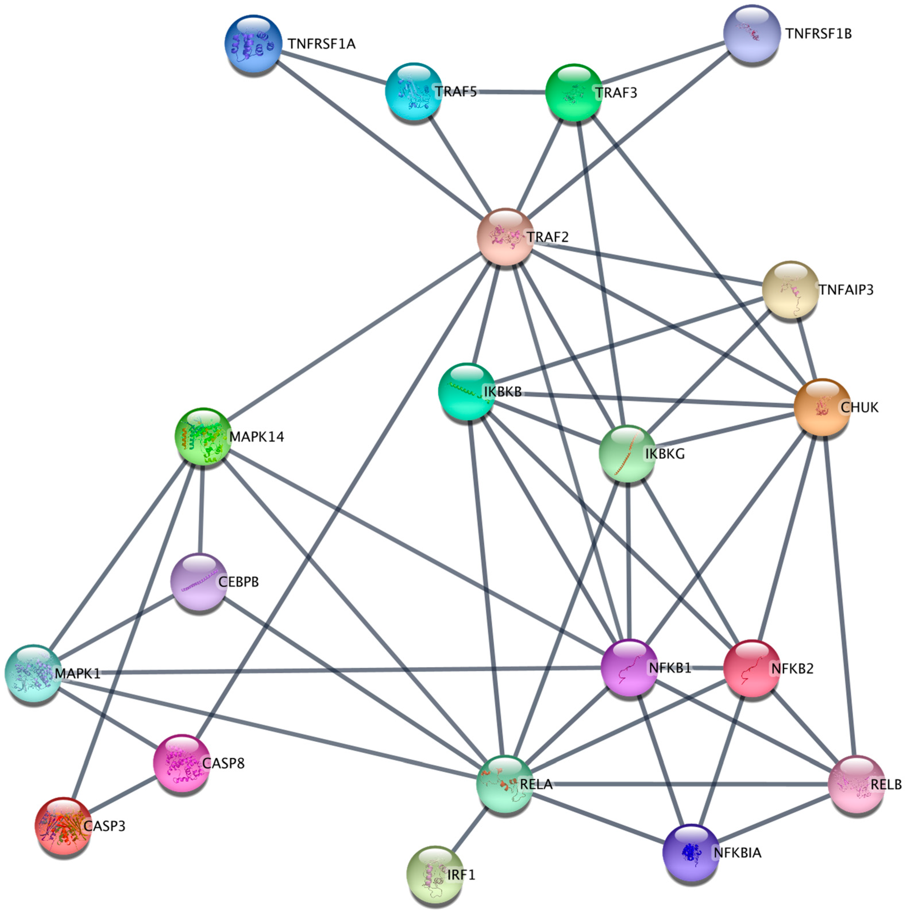
2.1. Transcriptomic Analysis of TNFR1 and TNFR2 Signaling Pathways in the K562 Cell Line

In this study, we conducted a comprehensive transcriptomic analysis of the K562 cell line, focusing on gene expression. The gene expression data, presented in descending order of copy numbers detected, included: TFRC, PSMB7, B2M, C1QBP, IFITM1, ILF3, PSMD7, CTNNB1, PSMC2, C14orf166, MCL1, CD81, MAPK1, CTSC, CD164, PSMB5, FN1, UBE2L3, CD46, GP1BB, STAT5A, FKBP5, ITGB1, MIF, STAT3, TGFBR1, NFKBIA, MAP4K4, ATG5, BCAP31, CD99, NFATC3, PSMB8, CD3D, STAT5B, RAF1, CD59, CFH, ARHGDIB, CHUK, CD44, PTK2, CCND3, CD53, MAPKAPK2, BCL10, TGFB1, CTSS, CD58, FCGR2A, CASP2, TAL1, BAX, ITGA5, IL2RG, GPI, SMAD5, FYN, SKI, TMEM173, ABL1, IFNGR1, IL13RA1, TRAF4, STAT2, MAPK14, CD97, MYD88, IKBKAP, LCP2, TRAF5, ICAM3, TYK2, PSMB9, AHR, IRAK1, XBP1, TBK1, FCGRT, JAK1, BST2, CD45R0, KIT, ATG16L1, SYK, NFIL3, EGR1, SMAD3, JAK2, SLC2A1, IL6ST, LTBR, TRAF2, IFNAR2, CFD, TAPBP, RELA, IL8, MALT1, ITGAE, ADA, CD9, PLAU, BTK, ICAM4, ATG10, LEF1, NFATC2, PTPN2, PDCD2, BCL2L11, ICOSLG, CD3EAP, LGALS3, LITAF, CASP3, CD276, CRADD, IKBKB, PML, TNFAIP3, RUNX1, TCF4, CD24, TNFRSF1B, CUL9, IGF2R, ARG2, KLRC3, TNFRSF8, STAT1, ITGA2B, TRAF3, MUC1, IKZF2, IKZF1, TICAM1, CISH, ICAM1, IRF1, TGFBR2, IRAK4, ATG7, NFKB2, ICAM2, TAP1, HRAS, STAT6, TAP2, IKBKG, CEBPB, PTPN22, CCBP2, CASP8, TCF7, TRAF6, IRF3, MAP4K2, IKBKE, EDNRB, CDKN1A, NFATC1, TOLLIP, ATG12, IL18R1, NFKB1, ZBTB16, and NOTCH2.
Among the detected genes, NFKB2, RELB, CASP8, NFKB1, CEBPB, NFKBIA, TNFRSF1B, TRAF5, MAPK1, IKBKB, RELA, TRAF3, IKBKG, MAPK14, IRF1, CHUK, TRAF2, and CASP3 were identified as components of the TNF-α signaling pathway from TNFR1 and TNFR2.
We subsequently performed a gene enrichment analysis and constructed a small gene network of the TNF-α signaling pathway for TNFR1 and TNFR2 in K562 cell line in Cytoscape incorporating the above genes, enriched in “KEGG TNF signaling pathway” term, and imputing the TNFRSF1A gene using the STRING database (Figure 1).
An analysis of the signaling pathways, starting from the TNF-α receptors revealed the potential activation of the NF-κB signaling pathway via both the canonical (RELA and NFKB1) [14] and the alternative pathways (RELB and NFKB2) [15], which contribute to cell survival and the synthesis of pro-inflammatory cytokines [16]. As well, the data suggested possible activation of apoptosis through TNFR1 (involving CASP8 and CASP3) [17].

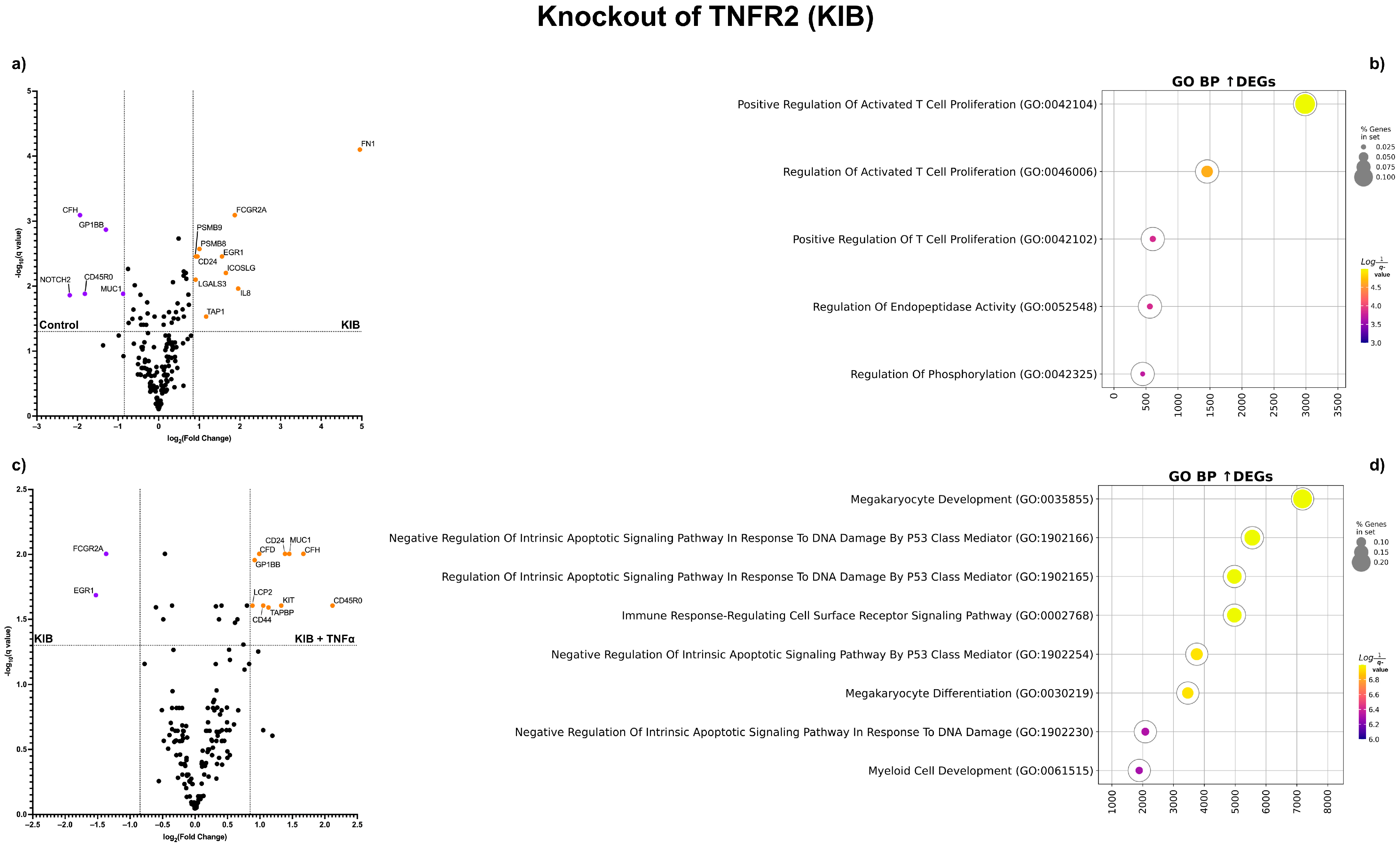
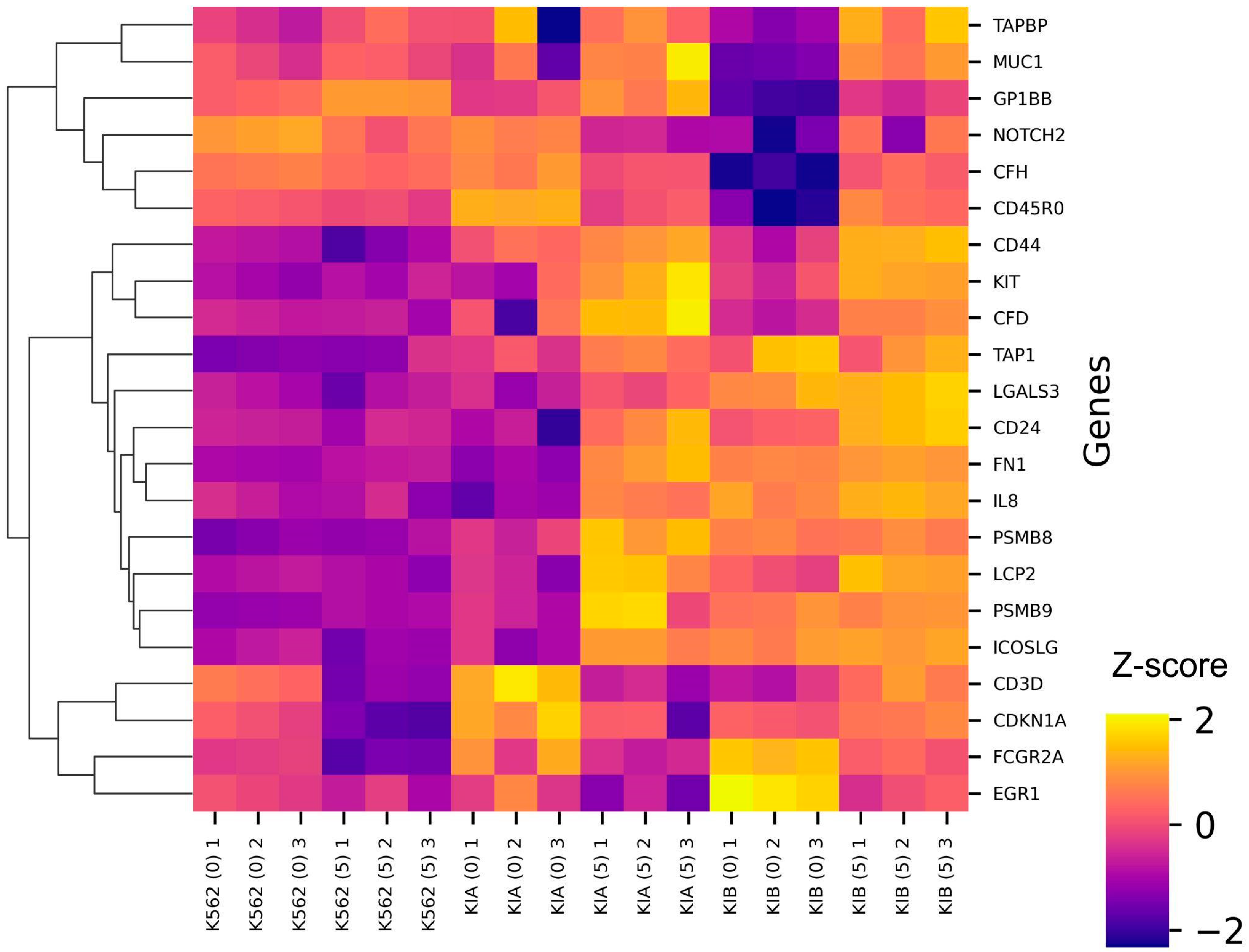
2.2. Transcriptomic Analysis of K562 with TNFR1 and TNFR2 Knockouts and TNF-α Addition

The exposure of K562 cells to TNF-α itself resulted in a notable decreased expression of the FCGR2A, CD3D, and CDKN1A genes after 72 h (Figure 2a). Gene set enrichment analysis of these down-regulated genes showed an enrichment in cell cycle transition-related Gene Ontology “Biological Process” terms (Figure 2b).
The knockout of TNFR1 (KIA) did not lead to significant changes in gene expression, except for an increase in CD45RO gene expression (Figure 3a). However, the knockout of TNFR2 (KIB) revealed distinct alterations; after 72 h, the expression of CD24, FCGR2A, FN1, IL8, TAP1, LGALS3, ICOSLG, EGR1, PSMB8, and PSMB9 increased, while the expression of MUC1, NOTCH2, CD45RO, CFH, and GP1BB decreased (Figure 3b). After the addition of TNF-α to the TNFR1 knockout K562 cell culture, a similar pattern of gene expression was observed as with the TNFR2 knockout, but without the addition of TNF-α—the expression of the FN1 and IL8 genes increased, and the expression of the NOTCH2, CD45RO, and CD3D genes decreased (Figure 4a). A gene set enrichment analysis of these up-regulated genes showed an enrichment in cell proliferation-related Gene Ontology “Biological Process” terms (Figure 4b).
Remarkably, the knockout of TNFR2 with the addition of TNF-α after 72 h resulted in a unique pattern of gene expression changes. Following this intervention, the expression of the genes CD45RO, MUC1, CD24, CD44, GP1BB, TAPBP, KIT, CGH, CFD, and LCP2 increased, while the expression of FCGR2A and EGR1 decreased (Figure 4c). A gene set enrichment analysis of these down-regulated genes showed an enrichment in negative regulation of apoptosis-related Gene Ontology “Biological Process” terms (Figure 4d).
To summarize these findings, we created a heatmap illustrating the differential gene expression data (Figure 5).

3. Discussion

In this study, we conducted an investigation into gene expression patterns following TNFR1 and TNFR2 knockouts within the K562 cell culture.
A reconstruction of a small gene network of signaling from TNFR1 and TNFR2 suggested the presence of multi-directional responses to TNF-α—proliferative, anti-proliferative—and associated with the synthesis of proinflammatory cytokines.
Upon the addition of TNF-α to K562 cells without knockouts, the expression of the CDKN1A gene, an inhibitor of cyclin/CDK complexes, decreased, suggesting a proliferative response due to a free cell cycle progression in reaction to TNF-α.
The knockout of TNFR2 in K562 produced a similar transcriptomic effect as the knockout of TNFR1 with the addition of TNF-α, in which signaling occurs through TNFR2. This suggests that knocking out TNFR2 potentially locks it into the activated position for cultured K562 cells.
All detected differentially expressed genes were not directly affiliated with the signaling pathways from TNFR1 and TNFR2 and, apparently, reflect long-term changes in the transcriptome in response to TNF-α; the TNFR1 knockout with the addition of TNF-α and the TNFR2 knockout led to a pro-inflammatory response type with an increased expression of the FN1 and IL8 genes [16,18,19], while the knockout of TNFR2 with the addition of TNF-α led to an increased expression of the KIT proto-oncogene, suggesting a proliferative response [20].
Overall, the knockouts of TNFR1 and TNFR2 induced noticeable alterations in the K562 cell transcriptome, and the addition of TNF-α further modulated such changes, ranging from pro-proliferative to pro-inflammatory.

4. Materials and Methods

4.1. Cell Culture

The K562 cell line was provided by the Federal State Budgetary Institution of Science, Institute of Cytology of the Russian Academy of Sciences (FGBI INC RAS, St. Petersburg, Russia). The ampoules with cells were thawed in a water bath at a temperature of +37 °C for 1–2 min. Then, the cells were washed once and cultured in RPMI-1640 medium containing 10% fetal bovine serum (FBS) (HyClone, Logan, UT, USA), 2 mM L-glutamine (BioloT LLC, Saint-Petersburg, Russia), 5 × 10⁻⁴ M2-mercaptoethanol (Sigma-Aldrich, St. Louis, MO, USA), 80 µg/mL gentamicin (KRKA, Novo mesto, Slovenia), 10 mM HEPES buffer (Sigma-Aldrich, USA), and 100 µg/mL benzylpenicillin (JSC Biosintez, Penza, Russia) in an incubator in a humid atmosphere at 37 °C and a CO2 concentration of 5%. Cells were collected by washing away the cells with a liquid stream using a pipette. After washing, the cells were collected and centrifuged for 10 min at 1500 rpm, followed by re-suspension in complete medium and transferred into vials (TPP, Trasadingen, Switzerland) at the optimal density for each cell line.

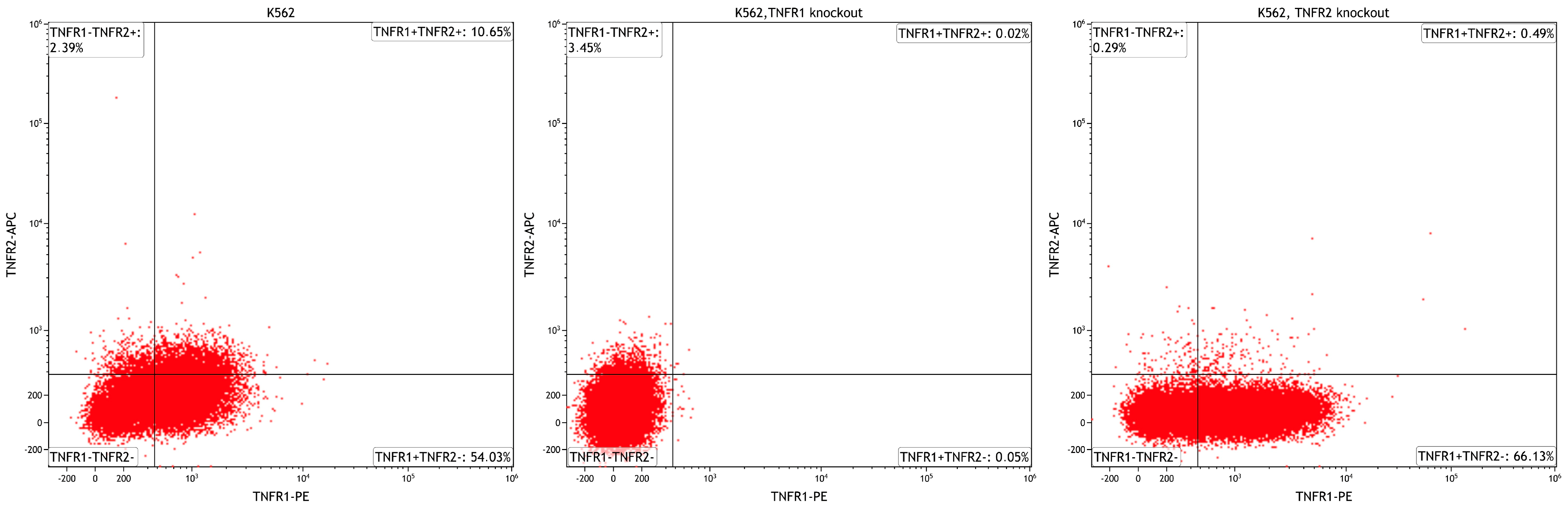
4.2. TNFRSF1A and TNFRSF1B Gene Knockouts

For each target gene, two single-guide RNAs (sgRNAs) were designed using bioinformatics tools, including Crispor (available at crispor.org, accessed on 23 October 2023), Benchling (benchling.com, accessed on 23 October 2023), and CHOPCHOP (chopchop.cbu.uib.no, accessed on 23 October 2023). These sgRNAs were strategically designed to specifically target the genomic loci of TNFRSF1A and TNFRSF1B.
To create the sgRNA expression vectors, oligo-nucleotides encoding these sgRNAs were synthesized and further processed to generate the necessary compatible sticky ends for ligation. Each sgRNA-coding oligonucleotide was designed to match the respective target gene, ensuring precise and selective gene knockout.
The next step involved ligating these sgRNA-coding oligo-nucleotides into a suitable and well-characterized expression vector, pSpCas9(BB)-2A-GFP (Addgene, #48138, Watertown, MA, USA). The ligation process was carried out using restriction enzymes and their corresponding recognition sites within the plasmid. Following ligation, the recombinant plasmids were introduced into E. coli NEB stable cells, where the cells were transformed with the ligated constructs. This process enabled the creation of a library of colonies that contained the assembled plasmids targeting both TNFRSF1A and TNFRSF1B genes.
To confirm the successful assembly and integrity of the plasmids, colonies were subjected to thorough screening. PCR analysis was employed to confirm the presence of the sgRNAs within the plasmids. Additionally, the structure and fidelity of the final plasmids were confirmed through Sanger sequencing, ensuring that the knockout strategy was both accurate and specific. The effectiveness and stability of knockouts were confirmed using flow cytometry, and the TNFR1 and TNFR2 were absent on cell membranes during subsequent passages. The expression of TNFR1 and TNFR2 was assessed by using flow cytometry (AttuneNxT cytofluorimeter (ThermoFisher, Waltham, MA, USA)) using monoclonal antibodies: anti-human CD120a-PE and anti-human CD120b-APC (R&D Systems, Minneapolis, MN, USA). Data processing and calculation of fluorescence intensity indicators were carried out using Attune™NxT Software version 3.2.1 (ThermoFisher, Waltham, MA, USA) (Scheme 1).

4.3. TNF-α Co-Culturing

Cell lines were cultured either in the presence of recombinant TNF (R&D Systems, USA) at a concentration of 5 ng/mL or simply without anything for 72 h.

4.4. Total RNA Extraction

We isolated total RNA from 250,000 cells with the Total RNA Purification Plus Kit (Norgen Biotek, Thorold, ON, Canada). We then measured the concentration and assessed the quality of the total RNA in each sample on a NanoDrop 2000c (Thermo Fisher Scientific, USA). The total RNA samples were frozen at −80 °C until the gene expression analysis.

4.5. NanoString RNA Profiling

We performed gene expression profiling with the help of the Nanostring nCounter SPRINT Profiler analytical system using 100 ng of total RNA from each sample. We used nCounter Human Immunology v2 panel to analyze the total RNA samples. nCounter Human Immunology v2 panel consisted of 579 immune- and inflammation-associated genes, 15 housekeeping genes and eight negative and six positive controls. The samples (n = 3) were subjected to a 20 h hybridization reaction at 65 °C, where 5–14 μL of total RNA was combined with 3 μL of nCounter Reporter probes, 0–7 μL of DEPC-treated water, 11 μL of hybridization buffer, and 5 μL of nCounter capture probes (total reaction volume = 33 μL). After the hybridization of the probes to targets of interest in the samples, the number of target molecules was determined on the nCounter digital analyzer. We performed normalization and QC in nSolver version 4.0 using the positive controls and the 15 housekeeping genes included in the panel. We then performed background thresholding on the normalized data to remove non-expressing genes. The background level was determined as the mean of the POS_E controls. We then log2-transformed the data.

4.6. TNFR1 and TNFR2 Signaling Gene Network Reconstruction for K562

We used Cytoscape version 17.0 [21] for the TNF signaling gene network reconstruction. We imported a new network from the STRING database [22] with a query containing every gene with detected expression. We then performed STRING functional enrichment of the network in KEGG pathway terms and created a sub-network of genes enriched in the “KEGG TNF signaling pathway” term. We used 0.95 protein–protein interaction confidence for the edge creation.

4.7. Differential Gene Expression Testing

We performed differential gene expression using multiple t-tests (with Q < 0.05) in GraphPad Prism version 9.4. The Volcano plots were created in GraphPad Prism 9.4. The heatmap of the differentially expressed genes across all conditions was created using bioinfokit (https://github.com/reneshbedre/bioinfokit, accessed on 23 October 2023). We then performed gene ontology biological process enrichment analysis (GOEA) of the detected genes via GSEApy version 1.0.0 [23].

Author Contributions

Conceptualization, S.S., A.A., O.P.-Z., R.P.-Z. and J.L.; methodology, O.P.-Z., J.Z. and R.P.-Z.; software, O.P.-Z., M.V., S.A. and R.P.-Z.; validation, O.P.-Z., R.P.-Z. and S.S.; investigation, O.P.-Z., J.Z. and R.P.-Z.; resources, O.P.-Z. and J.Z.; data curation, O.P.-Z., R.P.-Z., J.L., A.A. and S.S.; writing—original draft preparation, O.P.-Z., J.Z. and J.L.; writing—review and editing, O.P.-Z., R.P.-Z. and S.S.; visualization, O.P.-Z. and R.P.-Z.; supervision, S.S.; project administration, J.L., A.A. and S.S.; funding acquisition, A.A., J.Z., J.L. and S.S. All authors have read and agreed to the published version of the manuscript.

Funding

The work on evaluation of the profile of expressed genes was performed within a state task «The study of expression indicators of cytokine receptors and their ligands in the formation of functional properties and the type of response of cell populations of various genesis in normal and pathological conditions» registration number 122011800353-4 in the Unified State Accounting Information System research, development, and technological work for civil purposes. The work on developing protocols for obtaining cell lines with knockout of TNF receptor genes and obtaining cell cultures with TNF stimulation was supported by the Russian Science Foundation, project number No 20-75-10051, (https://rscf.ru/en/project/20-75-10051, accessed on 26 October 2023).

Institutional Review Board Statement

Not applicable.

Data Availability Statement

The datasets generated and analyzed during the current research are accessible from the corresponding author upon an email request.

Acknowledgments

This research work was supported by the academic leadership program Priority 2030 proposed by the Federal State Autonomous Educational Institution of Higher Education I.M. Sechenov First Moscow State Medical University of the Ministry of Health of the Russian Federation (Sechenov University).

Conflicts of Interest

The authors declare no conflict of interest. The funders had no role in the design of the study; in the collection, analyses, or interpretation of data; in the writing of the manuscript; or in the decision to publish the results.

References

  1. Salomon, B.L. Insights into the biology and therapeutic implications of TNF and regulatory T cells. Nat. Rev. Rheumatol. 2021, 17, 487–504. [Google Scholar] [CrossRef] [PubMed]
  2. Wajant, H.; Siegmund, D. TNFR1 and TNFR2 in the Control of the Life and Death Balance of Macrophages. Front. Cell Dev. Biol. 2019, 7, 91. [Google Scholar] [CrossRef] [PubMed]
  3. Yang, S.; Wang, J.; Brand, D.D.; Zheng, S.G. Role of TNF–TNF receptor 2 signal in regulatory T cells and its therapeutic implications. Front. Immunol. 2018, 9, 784. [Google Scholar] [CrossRef] [PubMed]
  4. Fischer, R.; Kontermann, R.E.; Pfizenmaier, K. Selective targeting of TNF receptors as a novel therapeutic approach. Front. Cell Dev. Biol. 2020, 8, 401. [Google Scholar] [CrossRef] [PubMed]
  5. Leone, G.M.; Mangano, K.; Petralia, M.C.; Nicoletti, F.; Fagone, P. Past, Present and (Foreseeable) Future of Biological Anti-TNF Alpha Therapy. J. Clin. Med. 2023, 12, 1630. [Google Scholar] [CrossRef]
  6. Medler, J.; Kucka, K.; Wajant, H. Tumor necrosis factor receptor 2 (TNFR2): An emerging target in cancer therapy. Cancers 2022, 14, 2603. [Google Scholar] [CrossRef]
  7. Sennikov, S.V.; Alshevskaya, A.A.; Zhukova, J.; Belomestnova, I.; Karaulov, A.V.; Lopatnikova, J.A. Expression density of receptors as a potent regulator of cell function and property in health and pathology. Int. Arch. Allergy Immunol. 2019, 178, 182–191. [Google Scholar] [CrossRef]
  8. Alshevskaya, A.; Koneva, O.; Belomestnova, I.; Lopatnikova, J.; Evsegneeva, I.; Zhukova, J.; Kireev, F.; Karaulov, A.; Sennikov, S. Ligand-regulated expression of TNF receptors 1 and 2 determines receptor-mediated functional responses. Int. Arch. Allergy Immunol. 2021, 182, 1077–1088. [Google Scholar] [CrossRef]
  9. Sander, C.A.; Rush, E.A.; Shi, J.; Arantes, L.M.; Tesi, R.J.; Ross, M.A.; Calderon, M.J.; Watkins, S.C.; Kirkwood, J.M.; Ferris, R.L.; et al. Co-expression of TNF receptors 1 and 2 on melanomas facilitates soluble TNF-induced resistance to MAPK pathway inhibitors. J. Transl. Med. 2022, 20, 331. [Google Scholar] [CrossRef]
  10. Gough, P.; Myles, I.A. Tumor necrosis factor receptors: Pleiotropic signaling complexes and their differential effects. Front. Immunol. 2020, 11, 585880. [Google Scholar] [CrossRef]
  11. Jang, D.I.; Lee, A.H.; Shin, H.Y.; Song, H.R.; Park, J.H.; Kang, T.B.; Lee, S.R.; Yang, S.H. The role of tumor necrosis factor alpha (TNF-α) in autoimmune disease and current TNF-α inhibitors in therapeutics. Int. J. Mol. Sci. 2021, 22, 2719. [Google Scholar] [CrossRef] [PubMed]
  12. Lo, C.H.; Huber, E.C.; Sachs, J.N. Conformational states of TNFR1 as a molecular switch for receptor function. Protein Sci. 2020, 29, 1401–1415. [Google Scholar] [CrossRef] [PubMed]
  13. Yang, S.; Xie, C.; Chen, Y.; Wang, J.; Chen, X.; Lu, Z.; June, R.R.; Zheng, S.G. Differential roles of TNFα-TNFR1 and TNFα-TNFR2 in the differentiation and function of CD4+ Foxp3+ induced Treg cells in vitro and in vivo periphery in autoimmune diseases. Cell Death Dis. 2019, 10, 27. [Google Scholar] [CrossRef] [PubMed]
  14. Cartwright, T.; Perkins, N.D.; Wilson, C.L. NFKB1: A suppressor of inflammation, ageing and cancer. FEBS J. 2016, 283, 1812–1822. [Google Scholar] [CrossRef] [PubMed]
  15. Roy, P.; Mukherjee, T.; Chatterjee, B.; Vijayaragavan, B.; Banoth, B.; Basak, S. Non-canonical NFκB mutations reinforce pro-survival TNF response in multiple myeloma through an autoregulatory RelB: p50 NFκB pathway. Oncogene 2017, 36, 1417–1429. [Google Scholar] [CrossRef]
  16. Kunsch, C.; Rosen, C.A. NF-kappa B subunit-specific regulation of the interleukin-8 promoter. Mol. Cell. Biol. 1993, 13, 6137–6146. [Google Scholar] [CrossRef] [PubMed]
  17. Hopkins-Donaldson, S.; Bodmer, J.L.; Bourloud, K.B.; Brognara, C.B.; Tschopp, J.; Gross, N. Loss of caspase-8 expression in highly malignant human neuroblastoma cells correlates with resistance to tumor necrosis factor-related apoptosis-inducing ligand-induced apoptosis. Cancer Res. 2000, 60, 4315–4319. [Google Scholar]
  18. Osawa, Y.; Nagaki, M.; Banno, Y.; Brenner, D.A.; Asano, T.; Nozawa, Y.; Moriwaki, H.; Nakashima, S. Tumor necrosis factor alpha-induced interleukin-8 production via NF-κB and phosphatidylinositol 3-kinase/Akt pathways inhibits cell apoptosis in human hepatocytes. Infect. Immun. 2002, 70, 6294–6301. [Google Scholar] [CrossRef]
  19. Gil, A.; Aguilera, C.M.; Gil-Campos, M.; Canete, R. Altered signalling and gene expression associated with the immune system and the inflammatory response in obesity. Br. J. Nutr. 2007, 98, S121–S126. [Google Scholar] [CrossRef]
  20. Zhang, Y.; Harada, A.; Bluethmann, H.; Wang, J.B.; Nakao, S.; Mukaida, N.; Matsushima, K. Tumor necrosis factor (TNF) is a physiologic regulator of hematopoietic progenitor cells: Increase of early hematopoietic progenitor cells in TNF receptor p55-deficient mice in vivo and potent inhibition of progenitor cell proliferation by TNF alpha in vitro. Blood 1995, 86, 2930–2937. [Google Scholar] [CrossRef]
  21. Shannon, P.; Markiel, A.; Ozier, O.; Baliga, N.S.; Wang, J.T.; Ramage, D.; Amin, N.; Schwikowski, B.; Ideker, T. Cytoscape: A software environment for integrated models of biomolecular interaction networks. Genome Res. 2003, 13, 2498–2504. [Google Scholar] [CrossRef] [PubMed]
  22. Doncheva, N.T.; Morris, J.H.; Gorodkin, J.; Jensen, L.J. Cytoscape StringApp: Network analysis and visualization of proteomics data. J. Proteome Res. 2018, 18, 623–632. [Google Scholar] [CrossRef] [PubMed]
  23. Fang, Z.; Liu, X.; Peltz, G. GSEApy: A comprehensive package for performing gene set enrichment analysis in Python. Bioinformatics 2023, 39, btac757. [Google Scholar] [CrossRef] [PubMed]
Figure 1. Reconstruction of the small gene network of the TNF-α signaling pathway from TNFR1 and TNFR2 in K562.
Ijms 24 17169 g001
Figure 2. Control K562 cell line—differential gene expression of K562 + TNF-α vs. K562: (a) Volcano plots of the differentially expressed genes: (b) Gene set enrichment analysis of the down-regulated genes.
Ijms 24 17169 g002
Figure 3. K562 cell line with the TNFR1 gene knockout (KIA)—Volcano plots of the differentially expressed genes: (a) KIA vs. control K562 and (b) KIA + TNF-α vs. KIA.
Ijms 24 17169 g003
Figure 4. K562 cell line with the TNFR2 gene knockout (KIB)—Volcano plots of the differentially expressed genes: (a) KIB vs. control K562, (b) KIB + TNF-α vs. KIB; Gene set enrichment analysis of the up-regulated genes: (c) KIB vs. control K562, and (d) KIB + TNF-α vs. KIB.
Ijms 24 17169 g004
Figure 5. Heatmap of the differentially expressed genes across all conditions, KIA—TNFR1 KO K562, KIB—TNFR2 KO K562, (0)—control, and (5)—addition of 5 ng/mL of TNF-α for 72 h.
Ijms 24 17169 g005
Scheme 1. Flow cytometry scatter plot confirming the effectiveness of knockouts.
Ijms 24 17169 sch001
Disclaimer/Publisher’s Note: The statements, opinions and data contained in all publications are solely those of the individual author(s) and contributor(s) and not of MDPI and/or the editor(s). MDPI and/or the editor(s) disclaim responsibility for any injury to people or property resulting from any ideas, methods, instructions or products referred to in the content.

