Communication

A Transcriptome Array-Based Approach to Link SGLT-2 and Intrarenal Complement C5 Synthesis in Diabetic Nephropathy

Department of Nephrology and Rheumatology, University Medical Center Göttingen, 37075 Göttingen, Germany
*
Author to whom correspondence should be addressed.
Int. J. Mol. Sci. 2023, 24(23), 17066; https://doi.org/10.3390/ijms242317066
Submission received: 15 September 2023 / Revised: 26 November 2023 / Accepted: 1 December 2023 / Published: 2 December 2023

Abstract

:
Diabetic nephropathy is a common microvascular complication of diabetes mellitus. It is characterized by progressive chronic kidney disease (CKD) with decline of kidney function by hyperfiltration. On a mechanistic level, activation of the complement system has been implicated in the pathogenesis of diabetic nephropathy. Therefore, here we pursued a transcriptome array-based approach to link intrarenal SGLT-2 and the synthesis of distinct complement components in diabetic nephropathy. Publicly available datasets for SLC5A2 (encoding SGLT-2) and complement system components were extracted specifically from microdissected tubulointerstitial (healthy controls: n = 31, diabetic nephropathy: n = 17) and glomerular compartments (healthy controls: n = 21, diabetic nephropathy: n = 12). First, we compared tubulointerstitial and glomerular log2 SLC5A2 mRNA expression levels and confirmed a predominant synthesis within the tubulointerstitial compartment. Among various complement components and receptors, the only significant finding was a positive association between SLC5A2 and the tubulointerstitial synthesis of the complement component C5 in diabetic nephropathy (p = 0.0109). Finally, intrarenal expression of SLC5A2 was associated predominantly with pathways involved in metabolic processes. Interestingly, intrarenal complement C5 synthesis was also associated with enrichment of metabolic signaling pathways, overlapping with SLC5A2 for “metabolism” and “biological oxidations”. These observations could be of relevance in the pathogenesis of diabetic nephropathy and implicate a mechanistic link between SGLT-2 and intrarenal complement synthesis.

1. Introduction

Diabetic nephropathy is a common microvascular complication of diabetes mellitus. It is characterized by progressive chronic kidney disease (CKD) with the decline of kidney function by hyperfiltration. Diabetic nephropathy is the leading cause of chronic and end-stage kidney disease (ESKD) worldwide [1,2]. Importantly, diabetic nephropathy is associated with increased cardiovascular morbidity and mortality [2]. For treatment of diabetic nephropathy to slow down CKD progression, angiotensin-converting enzyme inhibitors (ACEi), angiotensin II receptor antagonists (ARB), and the sodium-glucose cotransporter 2 inhibitors (SGLT-2is) are approved. On a mechanistic level, activation of the complement system has been implicated in the pathogenesis of diabetic nephropathy. Proteomic analysis in human kidney biopsies showed increased complement deposition in patients with diabetic nephropathy [3]. Hyperglycemia has been shown to activate the lectin pathway by glycation of pattern recognition molecules and complement regulatory proteins, resulting in intrarenal activation of the complement system and tubulointerstitial injury in diabetic nephropathy [4,5]. Compared to other interventions, no prior trial of any therapeutic agent in diabetic nephropathy has demonstrated an effect of the magnitude observed with SGLT-2i treatment [6]. Because complement is increasingly recognized in the pathogenesis of diabetic nephropathy, SGLT-2is may also exert nephroprotection by affecting complement system activation and signaling in diabetic nephropathy to achieve such beneficial effects [1].
In general, systemic or locally produced complement activation has also been implicated in the disease pathogenesis of antibody-mediated glomerular diseases [1]. We have already described that various autoimmune diseases are characterized by intrarenal complement depositions, at least in part driven by local synthesis of complement components, serving as biomarkers and direct mediators of injury [7,8,9,10,11]. Due to complement activity in many tissues, the consequences of its dysregulation are extensive, and increasing evidence has suggested the potential clinical relevance of complement pathway intervention. In diabetic nephropathy, systemic consumption of complement components and associated hypocomplementemia have not been described, suggesting a local synthesis of complement components [12]. Therefore, here we pursued a transcriptome array-based approach to link intrarenal SGLT-2 and the synthesis of distinct complement components in diabetic nephropathy. Particularly, we have included a dataset on diabetic nephropathy where a genome-wide transcriptome analysis already shows upregulation of the complement pathway [13].

2. Results

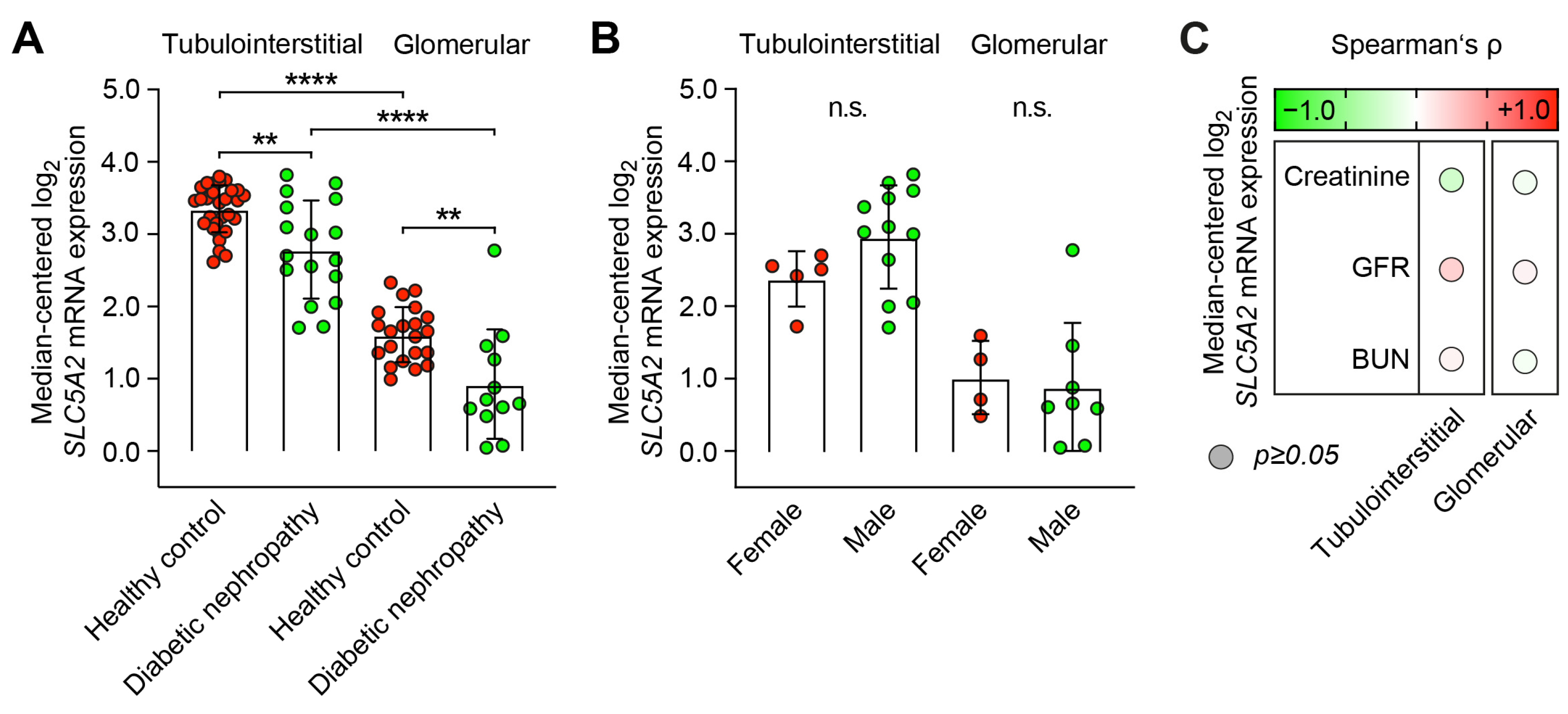
2.1. Predominant Tubulointerstitial SLC5A2 Expression in Healthy Controls and Diabetic Nephropathy

First, we compared tubulointerstitial and glomerular log2 SLC5A2 mRNA expression levels and confirmed a predominant synthesis within the tubulointerstitial compartment in healthy controls and diabetic nephropathy (Figure 1A and Tables S1 and S2) [14]. Interestingly, we identified decreased SLC5A2 mRNA expression in diabetic nephropathy as compared to healthy controls (Figure 1A). Specifically, in diabetic nephropathy, there were no differences between female and male patients (Figure 1B). Furthermore, there was an association with kidney function impairment as reflected by serum creatinine, glomerular filtration rate (GFR), or blood urea nitrogen (BUN, Figure 1C).

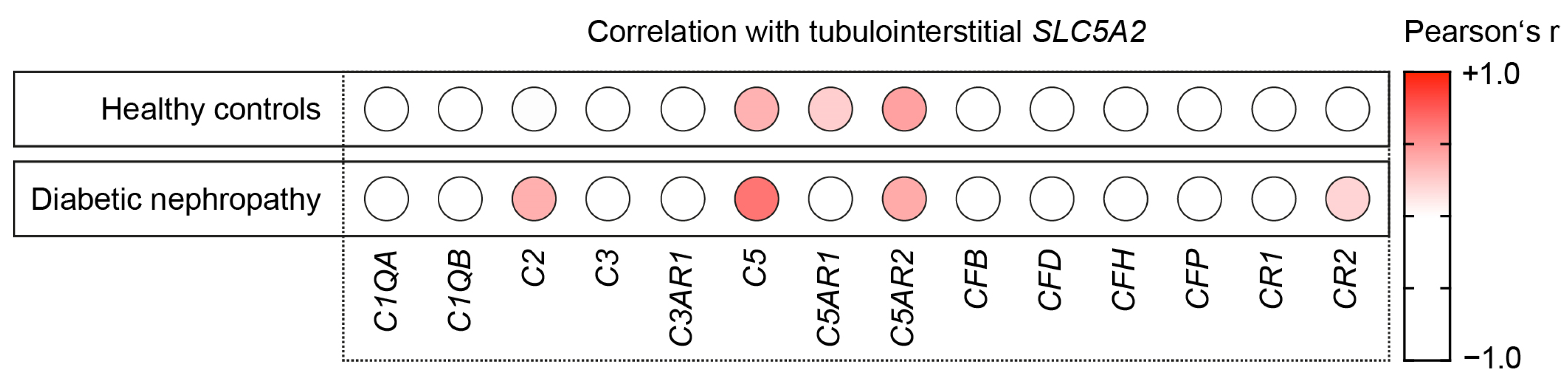
2.2. Intrarenal SLC5A2 Expression Is Associated with Tubulointerstitial Synthesis of Distinct Complement Components

Because we found a predominant tubulointerstitial expression of SLC5A2 in both healthy controls and diabetic nephropathy, we next analyzed the association between tubulointerstitial SLC5A2 mRNA expression levels, focusing on various complement components and receptors. Among them, there were positive correlations with complement components C5 and receptors C5AR1 and C5AR2 in healthy controls (Spearman’s p > 0.2, Figure 2). In diabetic nephropathy, we identified positive correlations specifically with complement components C2 and C5 and receptors C5AR2 and CR2 (Figure 2).

2.3. Intrarenal SLC5A2 Expression Is Associated with Tubulointerstitial Synthesis of Complement C5 in Diabetic Nephropathy

We next analyzed the relevance of tubulointerstitial SLC5A2 mRNA expression in association with the identified synthesis of distinct complement components, specifically in diabetic nephropathy. As assessed by stepwise logistic regression, the only significant finding was a positive association between SLC5A2 and the tubulointerstitial synthesis of the complement component C5 (p = 0.0109, Table 1).

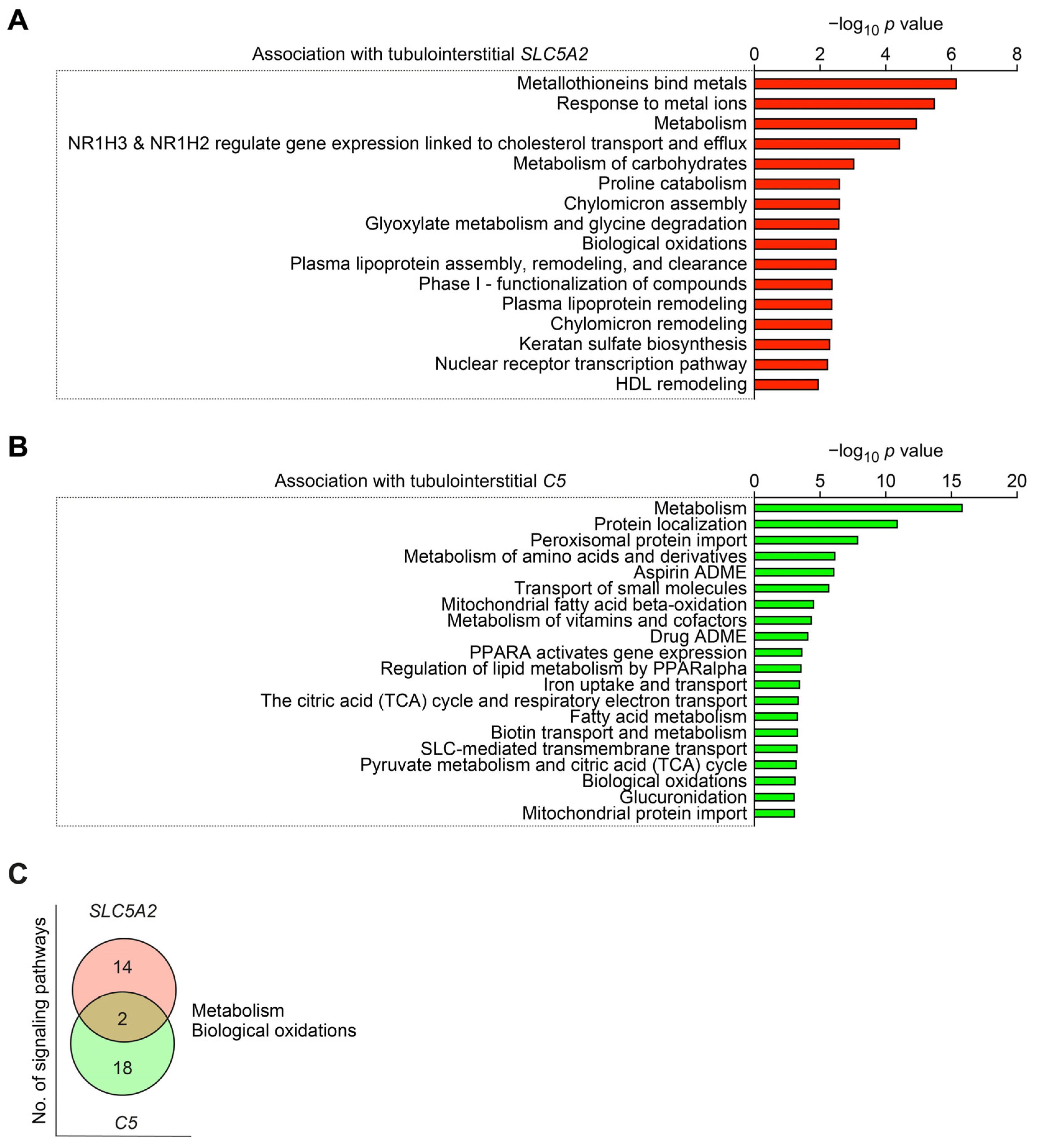
2.4. Intrarenal SLC5A2 Expression and Complement C5 Synthesis Associated with Distinct Molecular Signatures in Diabetic Nephropathy

Finally, we aimed to identify molecular signatures associated with SLC5A2 and intrarenal synthesis of complement component C5 [13]. Intrarenal expression of SLC5A2 was associated predominantly with pathways involved in metabolic processes (Figure 3A and Table S3). Interestingly, intrarenal complement C5 synthesis was also associated with enrichment of metabolic signaling pathways (Figure 3B and Table S4), overlapping with SLC5A2 for “metabolism” and “biological oxidations” (Figure 3C). In summary, we confirm a predominant tubulointerstitial expression of SGLT-2 in diabetic nephropathy. Furthermore, tubulointerstitial SGLT-2 correlated explicitly with the tubulointerstitial synthesis of complement component C5 in diabetic nephropathy.

3. Discussion

SGLT-2i exerts unequivocal nephroprotective effects by reducing glomerular hyperfiltration and albuminuria, tubular injury, loss of kidney function, and incidence of acute kidney injury. Although the mechanisms by which SGLT-2i can protect kidney function are not fully understood, it has previously been proposed that other pathways, including suppression of inflammation and fibrosis, are involved [15,16]. This is also relevant to autoimmune kidney diseases, including ANCA-associated renal vasculitis and lupus nephritis [17,18,19]. Interestingly, it has recently been reported that SGLT-2i exerts nephroprotection by attenuating autoimmunity, at least in part attributed to complement system modulation [20]. In the present study, we expand our current knowledge and report novel findings linking SGLT-2 and complement synthesis in diabetic nephropathy. Particularly, this comparative analysis revealed a predominant tubulointerstitial expression of SGLT-2 in both healthy controls and diabetic nephropathy. This is in line with previous observations confirming the abundance of SGLT-2 predominantly in the proximal tubular epithelium but also in glomerular podocytes [18,21,22]. Furthermore, we identified a positive correlation between SGLT-2 and tubulointerstitial synthesis of distinct complement components. Specifically in diabetic nephropathy, we here identified a positive association between SGLT-2 expression and the tubulointerstitial synthesis of the complement component C5. Evidence of glycated complement components was first described in patients with diabetic kidney disease more than three decades ago [23]. In this context, glycated complement components C3 and C4 are observed in the early stages of diabetic nephropathy [24]. Regarding complement component C5, tubular depositions have already been shown to correlate with the severity of diabetic nephropathy in humans [25]. Moreover, there is evidence for a pivotal role of the complement component C5 and its anaphylatoxin C5a signaling axis in contributing to kidney injury in experimental diabetic nephropathy [26,27]. Complement C5a and its receptor C5aR1 are upregulated early in the disease process and prior to manifesting kidney injury in several experimental models of diabetic nephropathy [26]. Particularly, C5aR1 has been shown to be expressed on proximal tubular cells, podocytes, fibroblasts, mesangial cells, and vascular endothelial and smooth muscle cells [28,29,30,31]. Finally, blocking C5a/C5a receptor 1 signaling has been shown to ameliorate interstitial fibrosis in experimental diabetic nephropathy [26]. Regarding molecular mechanisms, we here identified that intrarenal complement C5 synthesis was associated with the enrichment of metabolic signaling pathways, overlapping with SGLT-2 for “metabolism” and “biological oxidations”. This is in line with previous findings in experimental diabetic nephropathy confirming that C5 signaling is predominantly involved in immunometabolic signaling pathways [26]. Furthermore, several signaling pathways have already been implicated in linking increased oxidative stress and activation of inflammatory pathways to progressive kidney function decline in diabetic nephropathy [32,33]. This could be of relevance in the pathogenesis of diabetic nephropathy and could implicate a mechanistic link between SGLT-2 and intrarenal complement synthesis. Because the dataset included in our study was generated from an ethnically diverse population with various histopathological lesions, our findings could be of relevance for a large patient population, as it has been observed for nephroprotection by SGLT-2is in diabetic nephropathy [13,14,34].
Since new therapeutics targeting the complement system are emerging, these agents could be potentially beneficial in diabetic nephropathy. The recombinant humanized monoclonal antibody eculizumab is selective against C5 and inhibits its cleavage, reducing the release of anaphylatoxin C5a. Beneficial effects, such as temporary stabilization of renal function or reduction in albuminuria, have already been observed with eculizumab in progressive IgA nephropathy [35]. The first long-acting complement inhibitor, ravulizumab, has similar effects by C5 antagonism and is currently under preclinical evaluation in IgA nephropathy [36]. Additionally, a randomized phase II study is currently evaluating the efficacy and safety of ravulizumab in IgA nephropathy patients (NCT04564339). Furthermore, cemdisiram (ALN-CC5) as synthetic RNAi that blocks hepatic C5 production is also being tested in a phase II study in patients at high risk of progressive IgA nephropathy (NCT03841448). Regarding C5a receptor 1 (C5aR1) inhibition, avacopan (CCX168) is an orally active small molecule that selectively antagonizes C5aR1. Avacopan is currently approved for the treatment of ANCA-associated systemic vasculitis and evaluated for the treatment of IgA nephropathy (NCT02384317) [37]. Although we are aware that these observations require further validation with regard to protein expression and experimental models, here we link SGLT-2 and intrarenal synthesis of complement C5 in diabetic nephropathy, as it has also been recently described in experimental lupus nephritis [20].

4. Materials and Methods

4.1. Analyses of Publicly Available Array Datasets

Datasets provided publicly were analyzed according to general recommendations [38,39]. Publicly available datasets for SLC5A2 (encoding SGLT-2) and complement system components of mRNA expression were extracted from Nephroseq (www.nephroseq.org, accessed on 8 April 2023, University of Michigan, Ann Arbor, MI, USA). Particularly, median-centered log2 SLC5A2 mRNA expression levels (reporter ID: 6524, platform: Affymetrix Human Genome U133 Plus 2.0 Array, altCDF v10), C1QA (reporter ID: 712), C1QB (713), C2 (717), C3 (718), C3AR1 (719), C5 (727), C5AR1 (728), GPR77 (encoding C5AR2, 27202), CFB (629), CFD (1675), CFH (3075), CFP (5199), CR1 (1378), and CR2 (1380) were extracted specifically from microdissected tubulointerstitial (healthy controls: n = 31, diabetic nephropathy: n = 17) and glomerular compartments (healthy controls: n = 21, diabetic nephropathy: n = 12, Tables S1 and S2) [14]. From the same dataset, patient demographics and laboratory parameters for kidney function were also available and included in the analysis [14]. For multivariate analysis, mean values of Spearman’s ρ > 0.2 were included. Pathway analysis was performed separately for gene enrichment associated with tubulointerstitial SLC5A2 and C5 mRNA expression (healthy controls: n = 12, diabetic nephropathy: n = 10) [13]. Associated genes with a correlation threshold of ≥0.5 were included for pathway analysis by using reactome (http://reactome.org, accessed on 20 April 2023), and pathways with a predefined entity value of p ≤ 0.001 were included and shown in Tables S3 and S4 [40].

4.2. Statistical Methods

Spearman’s correlation was used to assess the correlation between continuous variables; multivariate analysis was performed by stepwise linear regression presenting beta coefficients (ß) and values of p. Data analyses were performed using GraphPad Prism (version 9.3.1 for macOS, GraphPad Software, San Diego, CA, USA) and IBM SPSS Statistics (version 29.0.0.0 for MacOS, IBM Corporation, Armonk, NY, USA); a value of p < 0.05 was considered statistically significant.

5. Conclusions

Here, we show a positive association between SLC5A2 and the tubulointerstitial synthesis of the complement component C5. Intrarenal expression of SLC5A2 was associated predominantly with pathways involved in metabolic processes. These observations could be of relevance in the pathogenesis of diabetic nephropathy and implicate a mechanistic link between SGLT-2 and intrarenal complement synthesis.

Supplementary Materials

The supporting information can be downloaded at: https://www.mdpi.com/article/10.3390/ijms242317066/s1.

Author Contributions

Conceptualization, B.T.; methodology, B.T.; software, B.T.; validation, B.T.; formal analysis, B.T.; investigation, B.T.; resources, B.T.; data curation, B.T.; writing—original draft preparation, B.T.; writing—review and editing, P.K.; visualization, B.T.; supervision, B.T.; project administration, B.T.; funding acquisition, P.K. and B.T. All authors have read and agreed to the published version of the manuscript.

Funding

We acknowledge support by the Open Access Publication Funds of the Göttingen University.

Institutional Review Board Statement

Data were extracted from Nephroseq Research Edition (Ann Arbor, MI, USA: University of Michigan; available from: www.nephroseq.org accessed on 8 April 2023).

Informed Consent Statement

Patient consent for participation was not required for this study because data were extracted from Nephroseq Research Edition (Ann Arbor, MI, USA: University of Michigan; available from: www.nephroseq.org, accessed on 8 April 2023, University of Michigan, Ann Arbor, MI, USA).

Data Availability Statement

The original contributions presented in the study are included in the article/Supplementary Materials; further data and material are available from the corresponding author upon reasonable request.

Conflicts of Interest

P.K. has received research grants from GlaxoSmithKline and Diamed Medizintechnik GmbH, all unrelated to this paper. P.K. has received honoraria or travel support from Abbvie, Amgen, AstraZeneca, Boehringer Ingelheim, Bristol-Myers-Squibb, Chugai, Galapagos, GlaxoSmithKline, Janssen-Cilag, Lilly, Novartis, and Pfizer, all unrelated to this paper. B.T. has received research grants from Evotec SE and CSL Vifor, all unrelated to this paper. B.T. has received honoraria or travel support from CSL Vifor, all unrelated to this paper.

References

  1. Budge, K.; Dellepiane, S.; Yu, S.M.; Cravedi, P. Complement, a Therapeutic Target in Diabetic Kidney Disease. Front. Med. 2020, 7, 599236. [Google Scholar] [CrossRef]
  2. Afkarian, M.; Zelnick, L.R.; Hall, Y.N.; Heagerty, P.J.; Tuttle, K.; Weiss, N.S.; de Boer, I.H. Clinical Manifestations of Kidney Disease among US Adults with Diabetes, 1988–2014. JAMA 2016, 316, 602–610. [Google Scholar] [CrossRef] [PubMed]
  3. Satoskar, A.A.; Shapiro, J.P.; Bott, C.N.; Song, H.; Nadasdy, G.M.; Brodsky, S.V.; Hebert, L.A.; Birmingham, D.J.; Nadasdy, T.; Freitas, M.A.; et al. Characterization of Glomerular Diseases Using Proteomic Analysis of Laser Capture Microdissected Glomeruli. Mod. Pathol. 2012, 25, 709–721. [Google Scholar] [CrossRef] [PubMed]
  4. Zheng, J.M.; Ren, X.G.; Jiang, Z.H.; Chen, D.J.; Zhao, W.J.; Li, L.J. Lectin-Induced Renal Local Complement Activation Is Involved In Tubular Interstitial Injury In Diabetic Nephropathy. Clin. Chim. Acta 2018, 482, 65–73. [Google Scholar] [CrossRef] [PubMed]
  5. Qin, X.; Goldfine, A.; Krumrei, N.; Grubissich, L.; Acosta, J.; Chorev, M.; Hays, A.P.; Halperin, J.A. Glycation Inactivation of The Complement Regulatory Protein Cd59: A Possible Role in the Pathogenesis of the Vascular Complications of Human Diabetes. Diabetes 2004, 53, 2653–2661. [Google Scholar] [CrossRef]
  6. Heerspink, H.J.L.; Stefansson, B.V.; Correa-Rotter, R.; Chertow, G.M.; Greene, T.; Hou, F.F.; Mann, J.F.E.; McMurray, J.J.V.; Lindberg, M.; Rossing, P.; et al. Dapagliflozin in Patients with Chronic Kidney Disease. N. Engl. J. Med. 2020, 383, 1436–1446. [Google Scholar] [CrossRef]
  7. Hakroush, S.; Tampe, D.; Korsten, P.; Strobel, P.; Tampe, B. Complement Components C3 and C4 Indicate Vasculitis Manifestations to Distinct Renal Compartments in Anca-Associated Glomerulonephritis. Int. J. Mol. Sci. 2021, 22, 6588. [Google Scholar] [CrossRef]
  8. Hakroush, S.; Tampe, D.; Baier, E.; Kluge, I.A.; Strobel, P.; Tampe, B. Intrarenal Synthesis Of Complement C3 Localized to Distinct Vascular Compartments in Anca-Associated Renal Vasculitis. J. Autoimmun. 2022, 133, 102924. [Google Scholar] [CrossRef]
  9. Hakroush, S.; Kluge, I.A.; Baier, E.; Tampe, D.; Tampe, B. Relevance of Complement C4 Deposits Localized to Distinct Vascular Compartments in Anca-Associated Renal Vasculitis. Int. J. Mol. Sci. 2022, 23, 14325. [Google Scholar] [CrossRef]
  10. Baier, E.; Tampe, D.; Kluge, I.A.; Hakroush, S.; Tampe, B. Implication of Platelets and Complement C3 as Link between Innate Immunity and Tubulointerstitial Injury in Renal Vasculitis with Mpo-Anca Seropositivity. Front. Immunol. 2022, 13, 1054457. [Google Scholar] [CrossRef]
  11. Korsten, P.; Baier, E.; Hakroush, S.; Tampe, B. C-Reactive Protein Levels Are Associated with Complement C4 Deposits and Interstitial Arteritis in ANCA-Associated Renal Vasculitis. Int. J. Mol. Sci. 2023, 24, 3075. [Google Scholar] [CrossRef] [PubMed]
  12. Hebert, L.A.; Cosio, F.G.; Neff, J.C. Diagnostic Significance of Hypocomplementemia. Kidney Int. 1991, 39, 811–821. [Google Scholar] [CrossRef] [PubMed]
  13. Woroniecka, K.I.; Park, A.S.; Mohtat, D.; Thomas, D.B.; Pullman, J.M.; Susztak, K. Transcriptome Analysis Of Human Diabetic Kidney Disease. Diabetes 2011, 60, 2354–2369. [Google Scholar] [CrossRef] [PubMed]
  14. Ju, W.; Nair, V.; Smith, S.; Zhu, L.; Shedden, K.; Song, P.X.K.; Mariani, L.H.; Eichinger, F.H.; Berthier, C.C.; Randolph, A.; et al. Tissue Transcriptome-Driven Identification of Epidermal Growth Factor as a Chronic Kidney Disease Biomarker. Sci. Transl. Med. 2015, 7, 316ra193. [Google Scholar] [CrossRef] [PubMed]
  15. Woods, T.C.; Satou, R.; Miyata, K.; Katsurada, A.; Dugas, C.M.; Klingenberg, N.C.; Fonseca, V.A.; Navar, L.G. Canagliflozin Prevents Intrarenal Angiotensinogen Augmentation and Mitigates Kidney Injury and Hypertension in Mouse Model of Type 2 Diabetes Mellitus. Am. J. Nephrol. 2019, 49, 331–342. [Google Scholar] [CrossRef]
  16. Sen, T.; Heerspink, H.J.L. A Kidney Perspective on the Mechanism of Action of Sodium Glucose Co-Transporter 2 Inhibitors. Cell Metab. 2021, 33, 732–739. [Google Scholar] [CrossRef] [PubMed]
  17. Saemann, M.; Kronbichler, A. Call for action in ANCA-Associated Vasculitis and Lupus Nephritis: Promises and Challenges of Sglt-2 Inhibitors. Ann. Rheum. Dis. 2022, 81, 614–617. [Google Scholar] [CrossRef]
  18. Hakroush, S.; Tampe, D.; Kluge, I.A.; Baier, E.; Korsten, P.; Tampe, B. Comparative Analysis of Sglt-2 Expression in Renal Vasculitis and Lupus Nephritis. Ann. Rheum. Dis. 2022, 81, 1048–1050. [Google Scholar] [CrossRef]
  19. Saemann, M.; Kronbichler, A. Response to: Correspondence on ‘Call for Action in ANCA-Associated Vasculitis and Lupus Nephritis: Promises and Challenges of SGLT-2 Inhibitors’ by Saemann and Kronbichler. Ann. Rheum. Dis. 2022, 8. [Google Scholar] [CrossRef]
  20. Zhao, X.Y.; Li, S.S.; He, Y.X.; Yan, L.J.; Lv, F.; Liang, Q.M.; Gan, Y.H.; Han, L.P.; Xu, H.D.; Li, Y.C.; et al. SGLT2 Inhibitors Alleviated Podocyte Damage in Lupus Nephritis by Decreasing Inflammation and Enhancing Autophagy. Ann. Rheum. Dis. 2023, 82, 1328–1340. [Google Scholar] [CrossRef]
  21. Cassis, P.; Locatelli, M.; Cerullo, D.; Corna, D.; Buelli, S.; Zanchi, C.; Villa, S.; Morigi, M.; Remuzzi, G.; Benigni, A.; et al. Sglt2 Inhibitor Dapagliflozin Limits Podocyte Damage in Proteinuric Nondiabetic Nephropathy. JCI Insight 2018, 3, e98720. [Google Scholar] [CrossRef]
  22. Wang, X.X.; Levi, J.; Luo, Y.; Myakala, K.; Herman-Edelstein, M.; Qiu, L.; Wang, D.; Peng, Y.; Grenz, A.; Lucia, S.; et al. SGLT2 Protein Expression Is Increased in Human Diabetic Nephropathy: Sglt2 Protein Inhibition Decreases Renal Lipid Accumulation, Inflammation, and the Development of Nephropathy in Diabetic Mice. J. Biol. Chem. 2017, 292, 5335–5348. [Google Scholar] [CrossRef] [PubMed]
  23. Austin, G.E.; Mullins, R.H.; Morin, L.G. Non-Enzymic Glycation of Individual Plasma Proteins in Normoglycemic and Hyperglycemic Patients. Clin. Chem. 1987, 33, 2220–2224. [Google Scholar] [CrossRef] [PubMed]
  24. Peake, P.W.; Charlesworth, J.A.; Timmermans, V.; Gavrilovic, L.; Pussell, B. Does Non-Enzymatic Glycosylation Affect Complement Function In Diabetes? Diabetes Res. 1989, 11, 109–114. [Google Scholar] [PubMed]
  25. Li, L.; Chen, L.; Zang, J.; Tang, X.; Liu, Y.; Zhang, J.; Bai, L.; Yin, Q.; Lu, Y.; Cheng, J.; et al. C3a and C5a Receptor Antagonists Ameliorate Endothelial-Myofibroblast Transition Via the Wnt/Beta-Catenin Signaling Pathway In Diabetic Kidney Disease. Metabolism 2015, 64, 597–610. [Google Scholar] [CrossRef]
  26. Tan, S.M.; Ziemann, M.; Thallas-Bonke, V.; Snelson, M.; Kumar, V.; Laskowski, A.; Nguyen, T.V.; Huynh, K.; Clarke, M.V.; Libianto, R.; et al. Complement C5a Induces Renal Injury in Diabetic Kidney Disease by Disrupting Mitochondrial Metabolic Agility. Diabetes 2020, 69, 83–98. [Google Scholar] [CrossRef] [PubMed]
  27. Li, L.; Wei, T.; Liu, S.; Wang, C.; Zhao, M.; Feng, Y.; Ma, L.; Lu, Y.; Fu, P.; Liu, J. Complement C5 Activation Promotes Type 2 Diabetic Kidney Disease Via Activating Stat3 Pathway And Disrupting The Gut-Kidney Axis. J. Cell Mol. Med. 2021, 25, 960–974. [Google Scholar] [CrossRef]
  28. Zahedi, R.; Braun, M.; Wetsel, R.A.; Ault, B.H.; Khan, A.; Welch, T.R.; Frenzke, M.; Davis, A.E. The C5a Receptor is Expressed by Human Renal Proximal Tubular Epithelial Cells. Clin. Exp. Immunol. 2000, 121, 226–233. [Google Scholar] [CrossRef]
  29. Weiss, S.; Rosendahl, A.; Czesla, D.; Meyer-Schwesinger, C.; Stahl, R.A.; Ehmke, H.; Kurts, C.; Zipfel, P.F.; Kohl, J.; Wenzel, U.O. The Complement Receptor C5ar1 Contributes to Renal Damage But Protects the Heart in Angiotensin Ii-Induced Hypertension. Am. J. Physiol. Renal Physiol. 2016, 310, F1356–F1365. [Google Scholar] [CrossRef]
  30. Mathern, D.R.; Heeger, P.S. Molecules Great and Small: The Complement System. Clin. J. Am. Soc. Nephrol. 2015, 10, 1636–1650. [Google Scholar] [CrossRef]
  31. Abe, K.; Miyazaki, M.; Koji, T.; Furusu, A.; Nakamura-Kurashige, T.; Nishino, T.; Ozono, Y.; Harada, T.; Sakai, H.; Kohno, S. Enhanced Expression of Complement C5a Receptor Mrna in Human Diseased Kidney Assessed by In Situ Hybridization. Kidney Int. 2001, 60, 137–146. [Google Scholar] [CrossRef] [PubMed]
  32. Mohandes, S.; Doke, T.; Hu, H.; Mukhi, D.; Dhillon, P.; Susztak, K. Molecular Pathways that Drive Diabetic Kidney Disease. J. Clin. Investig. 2023, 133. [Google Scholar] [CrossRef] [PubMed]
  33. Hong, Q.; Zhang, L.; Fu, J.; Verghese, D.A.; Chauhan, K.; Nadkarni, G.N.; Li, Z.; Ju, W.; Kretzler, M.; Cai, G.Y.; et al. LRG1 Promotes Diabetic Kidney Disease Progression by Enhancing TGF-beta-Induced Angiogenesis. J. Am. Soc. Nephrol. 2019, 30, 546–562. [Google Scholar] [CrossRef]
  34. Kidney Disease: Improving Global Outcomes (KDIGO) Diabetes Work Group. KDIGO 2022 Clinical Practice Guideline for Diabetes Management in Chronic Kidney Disease. Kidney Int 2022, 102, S1–S127. [Google Scholar] [CrossRef] [PubMed]
  35. Ring, T.; Pedersen, B.B.; Salkus, G.; Goodship, T.H. Use of Eculizumab in Crescentic IgA Nephropathy: Proof of Principle and Conundrum? Clin. Kidney J. 2015, 8, 489–491. [Google Scholar] [CrossRef] [PubMed]
  36. McKeage, K.; Duggan, S. Risankizumab: First Global Approval. Drugs 2019, 79, 893–900. [Google Scholar] [CrossRef]
  37. Jayne, D.R.W.; Merkel, P.A.; Schall, T.J.; Bekker, P.; Group, A.S. Avacopan for the Treatment of ANCA-Associated Vasculitis. N. Engl. J. Med. 2021, 384, 599–609. [Google Scholar] [CrossRef] [PubMed]
  38. Rung, J.; Brazma, A. Reuse of Public Genome-Wide Gene Expression Data. Nat. Rev. Genet. 2013, 14, 89–99. [Google Scholar] [CrossRef]
  39. Berthier, C.C.; Bethunaickan, R.; Gonzalez-Rivera, T.; Nair, V.; Ramanujam, M.; Zhang, W.; Bottinger, E.P.; Segerer, S.; Lindenmeyer, M.; Cohen, C.D.; et al. Cross-Species Transcriptional Network Analysis Defines Shared Inflammatory Responses in Murine and Human Lupus Nephritis. J. Immunol. 2012, 189, 988–1001. [Google Scholar] [CrossRef]
  40. Fabregat, A.; Sidiropoulos, K.; Viteri, G.; Forner, O.; Marin-Garcia, P.; Arnau, V.; D’Eustachio, P.; Stein, L.; Hermjakob, H. Reactome Pathway Analysis: A High-Performance In-Memory Approach. BMC Bioinform. 2017, 18, 142. [Google Scholar] [CrossRef]
Figure 1. Predominant tubulointerstitial SLC5A2 expression in healthy controls and diabetic nephropathy. (A) Median-centered log2 SLC5A2 expression levels in microdissected tubulointerstitial (healthy controls: n = 31, diabetic nephropathy: n = 17) and glomerular compartments (healthy controls: n = 21, diabetic nephropathy: n = 12) are shown by scatter dot plots including mean ± SD. Comparisons of groups were performed using the Mann–Whitney U-test to determine differences in medians (** p < 0.01, **** p < 0.0001). (B) Median-centered log2 SLC5A2 mRNA expression levels in diabetic nephropathy are shown by scatter dot plots including mean ± SD with group separation for female and male sex. Comparisons of groups were performed using the Mann–Whitney U-test to determine differences in medians (n.s: not significant). (C) Correlations between tubulointerstitial and glomerular SLC5A2 mRNA expression levels and laboratory markers of kidney function in diabetic nephropathy are shown by heatmap reflecting mean values of Spearman’s p; circle size represents significance level.
Ijms 24 17066 g001
Figure 2. Intrarenal SLC5A2 expression is associated with tubulointerstitial synthesis of distinct complement components. Correlations between tubulointerstitial SLC5A2 mRNA expression levels and various complement components in healthy controls (n = 31) and diabetic nephropathy (n = 17) are shown by heatmap reflecting mean values of Spearman’s p; circle size represents significance level.
Ijms 24 17066 g002
Figure 3. Intrarenal SLC5A2 expression and complement C5 synthesis associated with distinct molecular signatures in diabetic nephropathy. (A) Entities −log10 p values of signaling pathways associated with tubulointerstitial SLC5A2 mRNA expression in diabetic nephropathy are shown. (B) Entities −log10 p values of signaling pathways associated with tubulointerstitial C5 mRNA expression in diabetic nephropathy are shown. (C) Number of signaling pathways associated with either tubulointerstitial SLC5A2, complement C5 mRNA expression, or both in diabetic nephropathy are shown.
Ijms 24 17066 g003
Table 1. Multiple comparisons between SLC5A2 and the tubulointerstitial synthesis of the complement components in diabetic nephropathy.
Variableßp Value
C2—median-centered log2 mRNA expression0.29810.1633
C5—median-centered log2 mRNA expression0.59970.0109
C5AR2—median-centered log2 mRNA expression0.39180.0572
CR2—median-centered log2 mRNA expression0.09760.6626
Disclaimer/Publisher’s Note: The statements, opinions and data contained in all publications are solely those of the individual author(s) and contributor(s) and not of MDPI and/or the editor(s). MDPI and/or the editor(s) disclaim responsibility for any injury to people or property resulting from any ideas, methods, instructions or products referred to in the content.

