
Advancements in 2D and 3D In Vitro Models for Studying Neuromuscular Diseases


Abstract
:1. Introduction
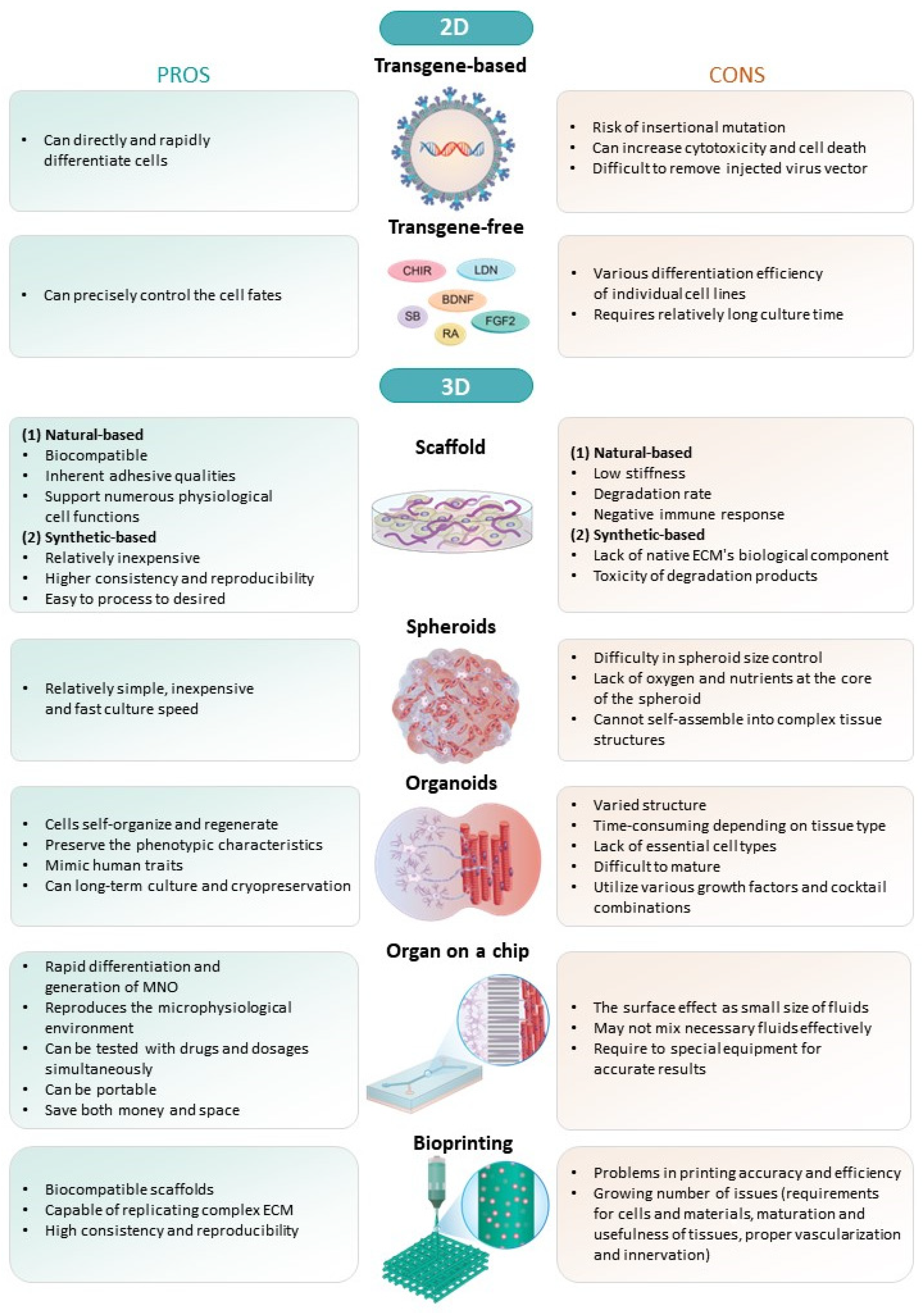
2. 2D Culture In Vitro
2.1. Transgene-Based Method
2.2. Transgene-Free Small Molecule-Based Cocktail Method
3. 3D Culture In Vitro
3.1. Scaffold-Based
3.2. Scaffold-Free
3.2.1. Spheroids
3.2.2. Organoids
4. Hybrid
4.1. Microfluidics and Organ-on-a-Chip
4.2. Bioprinting
5. NMD Models
5.1. Amyotrophic Lateral Sclerosis (ALS)
5.2. Myasthenia Gravis (MG)
5.3. Others
6. Conclusions
Author Contributions
Funding
Institutional Review Board Statement
Informed Consent Statement
Data Availability Statement
Conflicts of Interest
Abbreviations
References
- Biehl, J.K.; Russell, B. Introduction to stem cell therapy. J. Cardiovasc. Nurs. 2009, 24, 98–103; quiz 104–105. [Google Scholar] [CrossRef] [PubMed]
- Matsui, T.K.; Tsuru, Y.; Hasegawa, K.; Kuwako, K.I. Vascularization of human brain organoids. Stem Cells 2021, 39, 1017–1024. [Google Scholar] [CrossRef] [PubMed]
- Meran, L.; Massie, I.; Campinoti, S.; Weston, A.E.; Gaifulina, R.; Tullie, L.; Faull, P.; Orford, M.; Kucharska, A.; Baulies, A.; et al. Engineering transplantable jejunal mucosal grafts using patient-derived organoids from children with intestinal failure. Nat. Med. 2020, 26, 1593–1601. [Google Scholar] [CrossRef] [PubMed]
- Nishinakamura, R. Human kidney organoids: Progress and remaining challenges. Nat. Rev. Nephrol. 2019, 15, 613–624. [Google Scholar] [CrossRef] [PubMed]
- Lam, D.T.U.H.; Dan, Y.Y.; Chan, Y.S.; Ng, H.H. Emerging liver organoid platforms and technologies. Cell Regen. 2021, 10, 27. [Google Scholar] [CrossRef] [PubMed]
- Onyak, J.R.; Vergara, M.N.; Renna, J.M. Retinal organoid light responsivity: Current status and future opportunities. Transl. Res. 2022, 250, 98–111. [Google Scholar] [CrossRef] [PubMed]
- Seo, K.; Cho, S.; Shin, H.; Shin, A.; Lee, J.H.; Kim, J.H.; Lee, B.; Jang, H.; Kim, Y.; Cho, H.M.; et al. Symmetry Breaking of Human Pluripotent Stem Cells (hPSCs) in Micropattern Generates a Polarized Spinal Cord-Like Organoid (pSCO) with Dorsoventral Organization. Adv. Sci. 2023, 10, e2301787. [Google Scholar] [CrossRef] [PubMed]
- Grapin-Botton, A.; Kim, Y.H. Pancreas organoid models of development and regeneration. Development 2022, 149, dev201004. [Google Scholar] [CrossRef]
- Kim, S.K.; Kim, Y.H.; Park, S.; Cho, S.W. Organoid engineering with microfluidics and biomaterials for liver, lung disease, and cancer modeling. Acta Biomater. 2021, 132, 37–51. [Google Scholar] [CrossRef]
- Iolascon, G.; Paoletta, M.; Liguori, S.; Curci, C.; Moretti, A. Neuromuscular Diseases and Bone. Front. Endocrinol. 2019, 10, 794. [Google Scholar] [CrossRef]
- Sanes, J.R.; Lichtman, J.W. Development of the vertebrate neuromuscular junction. Annu. Rev. Neurosci. 1999, 22, 389–442. [Google Scholar] [CrossRef] [PubMed]
- Van der Plas, M.C.; Pilgram, G.S.; Plomp, J.J.; de Jong, A.; Fradkin, L.G.; Noordermeer, J.N. Dystrophin is required for appropriate retrograde control of neurotransmitter release at the Drosophila neuromuscular junction. J. Neurosci. 2006, 26, 333–344. [Google Scholar] [CrossRef] [PubMed]
- Baskoylu, S.N.; Yersak, J.; O’Hern, P.; Grosser, S.; Simon, J.; Kim, S.; Schuch, K.; Dimitriadi, M.; Yanagi, K.S.; Lins, J.; et al. Single copy/knock-in models of ALS SOD1 in C. elegans suggest loss and gain of function have different contributions to cholinergic and glutamatergic neurodegeneration. PLoS Genet. 2018, 14, e1007682. [Google Scholar] [CrossRef] [PubMed]
- Singh, J.; Patten, S.A. Modeling neuromuscular diseases in zebrafish. Front. Mol. Neurosci. 2022, 15, 1054573. [Google Scholar] [CrossRef] [PubMed]
- Cahalan, S.D.; Boehm, I.; Jones, R.A.; Piercy, R.J. Recognising the potential of large animals for modelling neuromuscular junction physiology and disease. J. Anat. 2022, 241, 1120–1132. [Google Scholar] [CrossRef] [PubMed]
- Jones, R.A.; Harrison, C.; Eaton, S.L.; Llavero Hurtado, M.; Graham, L.C.; Alkhammash, L.; Oladiran, O.A.; Gale, A.; Lamont, D.J.; Simpson, H.; et al. Cellular and molecular anatomy of the human neuromuscular junction. Cell Rep. 2017, 21, 2348–2356. [Google Scholar] [CrossRef]
- Castellanos-Montiel, M.J.; Velasco, I.; Escobedo-Avila, I. Modeling the neuromuscular junction in vitro: An approach to study neuromuscular junction disorders. Ann. N. Y. Acad. Sci. 2021, 1488, 3–15. [Google Scholar] [CrossRef]
- Fralish, Z.; Lotz, E.M.; Chavez, T.; Khodabukus, A.; Bursac, N. Neuromuscular development and disease: Learning from in vitro and in vivo Models. Front. Cell Dev. Biol. 2021, 9, 764732. [Google Scholar] [CrossRef]
- Steinbeck, J.A.; Jaiswal, M.K.; Calder, E.L.; Kishinevsky, S.; Weishaupt, A.; Toyka, K.V.; Goldstein, P.A.; Studer, L. Functional connectivity under optogenetic control allows modeling of human neuromuscular disease. Cell Stem Cell 2016, 18, 134–143. [Google Scholar] [CrossRef]
- Jensen, C.; Teng, Y. Is it time to start transitioning from 2D to 3D cell culture? Front. Mol. Biosci. 2020, 7, 33. [Google Scholar] [CrossRef]
- Faustino Martins, J.M.; Fischer, C.; Urzi, A.; Vidal, R.; Kunz, S.; Ruffault, P.L.; Kabuss, L.; Hube, I.; Gazzerro, E.; Birchmeier, C.; et al. Self-organizing 3D human trunk neuromuscular organoids. Cell Stem Cell 2020, 27, 498. [Google Scholar] [CrossRef]
- Osaki, T.; Uzel, S.G.M.; Kamm, R.D. On-Chip 3D neuromuscular model for drug screening and precision medicine in neuromuscular disease. Nat. Protoc. 2020, 15, 421–449. [Google Scholar] [CrossRef]
- Afshar Bakooshli, M.; Lippmann, E.S.; Mulcahy, B.; Iyer, N.; Nguyen, C.T.; Tung, K.; Stewart, B.A.; van den Dorpel, H.; Fuehrmann, T.; Shoichet, M.; et al. A 3D culture model of innervated human skeletal muscle enables studies of the adult neuromuscular junction. eLife 2019, 8, e44530. [Google Scholar] [CrossRef] [PubMed]
- Barbeau, S.; Tahraoui-Bories, J.; Legay, C.; Martinat, C. Building neuromuscular junctions in vitro. Development 2020, 147, dev193920. [Google Scholar] [CrossRef] [PubMed]
- Filogamo, G.; Gabella, G. The development of neuro-muscular correlations, in vertebrates. Arch. Biol. 1967, 78, 9–60. [Google Scholar]
- Chen, M.; Wang, X.; Li, C.; Lan, T.; Wei, Y.; Tang, C.; Zhou, X.; Zhou, R.; Rosa, A.; Zheng, X.; et al. Inducible motor neuron differentiation of human induced pluripotent stem cells in vivo. Cell Prolif. 2022, 55, e13319. [Google Scholar] [CrossRef] [PubMed]
- Fernandopulle, M.S.; Prestil, R.; Grunseich, C.; Wang, C.; Gan, L.; Ward, M.E. Transcription factor-mediated differentiation of human iPSCs into neurons. Curr. Protoc. Cell Biol. 2018, 79, e51. [Google Scholar] [CrossRef] [PubMed]
- De Santis, R.; Garone, M.G.; Pagani, F.; de Turris, V.; Di Angelantonio, S.; Rosa, A. Direct conversion of human pluripotent stem cells into cranial motor neurons using a piggyBac vector. Stem Cell Res. 2018, 29, 189–196. [Google Scholar] [CrossRef]
- Shi, Y.; Lin, S.; Staats, K.A.; Li, Y.; Chang, W.H.; Hung, S.T.; Hendricks, E.; Linares, G.R.; Wang, Y.; Son, E.Y.; et al. Haploinsufficiency leads to neurodegeneration in C9ORF72 ALS/FTD human induced motor neurons. Nat. Med. 2018, 24, 313–325. [Google Scholar] [CrossRef]
- Mateos-Aierdi, A.J.; Dehesa-Etxebeste, M.; Goicoechea, M.; Aiastui, A.; Richaud-Patin, Y.; Jiménez-Delgado, S.; Raya, A.; Naldaiz-Gastesi, N.; López de Munain, A. Patient-specific iPSC-derived cellular models of LGMDR1. Stem Cell Res. 2021, 53, 102333. [Google Scholar] [CrossRef]
- Tan, G.W.; Kondo, T.; Imamura, K.; Suga, M.; Enami, T.; Nagahashi, A.; Tsukita, K.; Inoue, I.; Kawaguchi, J.; Shu, T.; et al. Simple derivation of skeletal muscle from human pluripotent stem cells using temperature-sensitive Sendai virus vector. J. Cell. Mol. Med. 2021, 25, 9586–9596. [Google Scholar] [CrossRef] [PubMed]
- Caputo, L.; Granados, A.; Lenzi, J.; Rosa, A.; Ait-Si-Ali, S.; Puri, P.L.; Albini, S. Acute conversion of patient-derived Duchenne muscular dystrophy iPSC into myotubes reveals constitutive and inducible over-activation of TGFbeta-dependent pro-fibrotic signaling. Skelet. Muscle 2020, 10, 13. [Google Scholar] [CrossRef] [PubMed]
- Sasaki-Honda, M.; Jonouchi, T.; Arai, M.; Hotta, A.; Mitsuhashi, S.; Nishino, I.; Matsuda, R.; Sakurai, H. A patient-derived iPSC model revealed oxidative stress increases facioscapulohumeral muscular dystrophy-causative DUX4. Hum. Mol. Genet. 2018, 27, 4024–4035. [Google Scholar] [CrossRef]
- Onda-Ohto, A.; Hasegawa-Ogawa, M.; Matsuno, H.; Shiraishi, T.; Bono, K.; Hiraki, H.; Kanegae, Y.; Iguchi, Y.; Okano, H.J. Specific vulnerability of iPSC-derived motor neurons with TDP-43 gene mutation to oxidative stress. Mol. Brain 2023, 16, 62. [Google Scholar] [CrossRef] [PubMed]
- Cutarelli, A.; Martínez-Rojas, V.A.; Tata, A.; Battistella, I.; Rossi, D.; Arosio, D.; Musio, C.; Conti, L. A monolayer system for the efficient generation of motor neuron progenitors and functional motor neurons from human pluripotent stem cells. Cells 2021, 10, 1127. [Google Scholar] [CrossRef] [PubMed]
- Sato, T.; Imaizumi, K.; Watanabe, H.; Ishikawa, M.; Okano, H. Generation of region-specific and high-purity neurons from human feeder-free iPSCs. Neurosci. Lett. 2021, 746, 135676. [Google Scholar] [CrossRef]
- Garcia-Diaz, A.; Efe, G.; Kabra, K.; Patel, A.; Lowry, E.R.; Shneider, N.A.; Corneo, B.; Wichterle, H. Standardized reporter systems for purification and imaging of human pluripotent stem cell-derived motor neurons and other cholinergic cells. Neuroscience 2020, 450, 48–56. [Google Scholar] [CrossRef]
- Faye, P.A.; Vedrenne, N.; Miressi, F.; Rassat, M.; Romanenko, S.; Richard, L.; Bourthoumieu, S.; Funalot, B.; Sturtz, F.; Favreau, F.; et al. Optimized protocol to generate spinal motor neuron cells from induced pluripotent stem cells from Charcot Marie Tooth patients. Brain Sci. 2020, 10, 407. [Google Scholar] [CrossRef]
- Bianchi, F.; Malboubi, M.; Li, Y.; George, J.H.; Jerusalem, A.; Szele, F.; Thompson, M.S.; Ye, H. Rapid and efficient differentiation of functional motor neurons from human iPSC for neural injury modelling. Stem Cell Res. 2018, 32, 126–134. [Google Scholar] [CrossRef]
- Zhao, C.; Devlin, A.C.; Chouhan, A.K.; Selvaraj, B.T.; Stavrou, M.; Burr, K.; Brivio, V.; He, X.; Mehta, A.R.; Story, D.; et al. Mutant C9orf72 human iPSC-derived astrocytes cause non-cell autonomous motor neuron pathophysiology. Glia 2020, 68, 1046–1064. [Google Scholar] [CrossRef]
- Choi, I.Y.; Lim, H.T.; Che, Y.H.; Lee, G.; Kim, Y.J. Inhibition of the combinatorial signaling of transforming growth factor-β and NOTCH promotes myotube formation of human pluripotent stem cell-derived skeletal muscle progenitor cells. Cells 2021, 10, 1649. [Google Scholar] [CrossRef] [PubMed]
- Kim, E.; Wu, F.; Wu, X.; Choo, H.J. Generation of craniofacial myogenic progenitor cells from human induced pluripotent stem cells for skeletal muscle tissue regeneration. Biomaterials 2020, 248, 119995. [Google Scholar] [CrossRef]
- Lynch, E.; Semrad, T.; Belsito, V.S.; FitzGibbons, C.; Reilly, M.; Hayakawa, K.; Suzuki, M. C9ORF72-related cellular pathology in skeletal myocytes derived from ALS-patient induced pluripotent stem cells. Dis. Model. Mech. 2019, 12, dmm039552. [Google Scholar] [CrossRef] [PubMed]
- Sakai-Takemura, F.; Narita, A.; Masuda, S.; Wakamatsu, T.; Watanabe, N.; Nishiyama, T.; Nogami, K.; Blanc, M.; Takeda, S.; Miyagoe-Suzuki, Y. Premyogenic progenitors derived from human pluripotent stem cells expand in floating culture and differentiate into transplantable myogenic progenitors. Sci. Rep. 2018, 8, 6555. [Google Scholar] [CrossRef] [PubMed]
- van der Wal, E.; Herrero-Hernandez, P.; Wan, R.; Broeders, M.; In ’t Groen, S.L.M.; van Gestel, T.J.M.; van IJcken, W.F.J.; Cheung, T.H.; van der Ploeg, A.T.; Schaaf, G.J.; et al. Large-scale expansion of human iPSC-derived skeletal muscle cells for disease modeling and cell-based therapeutic strategies. Stem Cell Rep. 2018, 10, 1975–1990. [Google Scholar] [CrossRef] [PubMed]
- Mazaleyrat, K.; Badja, C.; Broucqsault, N.; Chevalier, R.; Laberthonniere, C.; Dion, C.; Baldasseroni, L.; El-Yazidi, C.; Thomas, M.; Bachelier, R.; et al. Multilineage differentiation for formation of innervated skeletal muscle fibers from healthy and diseased human pluripotent stem cells. Cells 2020, 9, 1531. [Google Scholar] [CrossRef]
- Lee, S.; Cuvillier, J.M.; Lee, B.; Shen, R.; Lee, J.W.; Lee, S.K. Fusion protein Isl1-Lhx3 specifies motor neuron fate by inducing motor neuron genes and concomitantly suppressing the interneuron programs. Proc. Natl. Acad. Sci. USA 2012, 109, 3383–3388. [Google Scholar] [CrossRef]
- Goto, K.; Imamura, K.; Komatsu, K.; Mitani, K.; Aiba, K.; Nakatsuji, N.; Inoue, M.; Kawata, A.; Yamashita, H.; Takahashi, R.; et al. Simple derivation of spinal motor neurons from ESCs/iPSCs using Sendai virus vectors. Mol. Ther. Methods Clin. Dev. 2017, 4, 115–125. [Google Scholar] [CrossRef]
- Akiyama, T.; Wakabayashi, S.; Soma, A.; Sato, S.; Nakatake, Y.; Oda, M.; Murakami, M.; Sakota, M.; Chikazawa-Nohtomi, N.; Ko, S.B.; et al. Transient ectopic expression of the histone demethylase JMJD3 accelerates the differentiation of human pluripotent stem cells. Development 2016, 143, 3674–3685. [Google Scholar] [CrossRef]
- Warren, L.; Lin, C. mRNA-based genetic reprogramming. Mol. Ther. 2019, 27, 729–734. [Google Scholar] [CrossRef]
- Qi, Y.; Zhang, X.J.; Renier, N.; Wu, Z.; Atkin, T.; Sun, Z.; Ozair, M.Z.; Tchieu, J.; Zimmer, B.; Fattahi, F.; et al. Combined small-molecule inhibition accelerates the derivation of functional cortical neurons from human pluripotent stem cells. Nat. Biotechnol. 2017, 35, 154–163. [Google Scholar] [CrossRef] [PubMed]
- Volpato, V.; Smith, J.; Sandor, C.; Ried, J.S.; Baud, A.; Handel, A.; Newey, S.E.; Wessely, F.; Attar, M.; Whiteley, E.; et al. Reproducibility of molecular phenotypes after long-term differentiation to human iPSC-derived neurons: A multi-site omics study. Stem Cell Rep. 2018, 11, 897–911. [Google Scholar] [CrossRef] [PubMed]
- De Leeuw, S.M.; Davaz, S.; Wanner, D.; Milleret, V.; Ehrbar, M.; Gietl, A.; Tackenberg, C. Increased maturation of iPSC-derived neurons in a hydrogel-based 3D culture. J. Neurosci. Methods 2021, 360, 109254. [Google Scholar] [CrossRef]
- Kang, S.M.; Kim, D.; Lee, J.H.; Takayama, S.; Park, J.Y. Engineered microsystems for spheroid and organoid studies. Adv. Healthc. Mater. 2021, 10, e2001284. [Google Scholar] [CrossRef] [PubMed]
- Castellanos-Montiel, M.J.; Chaineau, M.; Franco-Flores, A.K.; Haghi, G.; Carrillo-Valenzuela, D.; Reintsch, W.E.; Chen, C.X.; Durcan, T.M. An optimized workflow to generate and characterize iPSC-derived motor neuron (MN) spheroids. Cells 2023, 12, 545. [Google Scholar] [CrossRef]
- Osaki, T.; Chow, S.Y.A.; Nakanishi, Y.; Hernández, J.; Kawada, J.; Fujii, T.; Ikeuchi, Y. Three-dimensional motor nerve organoid generation. J. Vis. Exp. 2020, 163, e61544. [Google Scholar] [CrossRef]
- Wang, J.; Zhou, C.J.; Khodabukus, A.; Tran, S.; Han, S.O.; Carlson, A.L.; Madden, L.; Kishnani, P.S.; Koeberl, D.D.; Bursac, N. Three-dimensional tissue-engineered human skeletal muscle model of Pompe disease. Commun. Biol. 2021, 4, 524. [Google Scholar] [CrossRef]
- Rajabian, N.; Shahini, A.; Asmani, M.; Vydiam, K.; Choudhury, D.; Nguyen, T.; Ikhapoh, I.; Zhao, R.; Lei, P.; Andreadis, S.T. Bioengineered skeletal muscle as a model of muscle aging and regeneration. Tissue Eng. Part A 2021, 27, 74–86. [Google Scholar] [CrossRef]
- Fleming, J.W.; Capel, A.J.; Rimington, R.P.; Wheeler, P.; Leonard, A.N.; Bishop, N.C.; Davies, O.G.; Lewis, M.P. Bioengineered human skeletal muscle capable of functional regeneration. BMC Biol. 2020, 18, 145. [Google Scholar] [CrossRef]
- Capel, A.J.; Rimington, R.P.; Fleming, J.W.; Player, D.J.; Baker, L.A.; Turner, M.C.; Jones, J.M.; Martin, N.R.W.; Ferguson, R.A.; Mudera, V.C.; et al. Scalable 3D printed molds for human tissue engineered skeletal muscle. Front. Bioeng. Biotechnol. 2019, 7, 20. [Google Scholar] [CrossRef]
- Massih, B.; Veh, A.; Schenke, M.; Mungwa, S.; Seeger, B.; Selvaraj, B.T.; Chandran, S.; Reinhardt, P.; Sterneckert, J.; Hermann, A.; et al. A 3D cell culture system for bioengineering human neuromuscular junctions to model ALS. Front. Cell Dev. Biol. 2023, 11, 996952. [Google Scholar] [CrossRef] [PubMed]
- Rimington, R.P.; Fleming, J.W.; Capel, A.J.; Wheeler, P.C.; Lewis, M.P. Bioengineered model of the human motor unit with physiologically functional neuromuscular junctions. Sci. Rep. 2021, 11, 11695. [Google Scholar] [CrossRef]
- Stoklund Dittlau, K.S.; Krasnow, E.N.; Fumagalli, L.; Vandoorne, T.; Baatsen, P.; Kerstens, A.; Giacomazzi, G.; Pavie, B.; Rossaert, E.; Beckers, J.; et al. Human motor units in microfluidic devices are impaired by FUS mutations and improved by HDAC6 inhibition. Stem Cell Rep. 2021, 16, 2213–2227. [Google Scholar] [CrossRef] [PubMed]
- Yamamoto, K.; Yamaoka, N.; Imaizumi, Y.; Nagashima, T.; Furutani, T.; Ito, T.; Okada, Y.; Honda, H.; Shimizu, K. Development of a human neuromuscular tissue-on-a-chip model on a 24-well-plate-format compartmentalized microfluidic device. Lab Chip 2021, 21, 1897–1907. [Google Scholar] [CrossRef] [PubMed]
- Pereira, J.D.; DuBreuil, D.M.; Devlin, A.C.; Held, A.; Sapir, Y.; Berezovski, E.; Hawrot, J.; Dorfman, K.; Chander, V.; Wainger, B.J. Human sensorimotor organoids derived from healthy and amyotrophic lateral sclerosis stem cells form neuromuscular junctions. Nat. Commun. 2021, 12, 4744. [Google Scholar] [CrossRef] [PubMed]
- Andersen, J.; Revah, O.; Miura, Y.; Thom, N.; Amin, N.D.; Kelley, K.W.; Singh, M.; Chen, X.; Thete, M.V.; Walczak, E.M.; et al. Generation of functional human 3D cortico-motor assembloids. Cell 2020, 183, 1913–1929.e26. [Google Scholar] [CrossRef]
- Osaki, T.; Uzel, S.G.M.; Kamm, R.D. Microphysiological 3D model of amyotrophic lateral sclerosis (ALS) from human iPS-derived muscle cells and optogenetic motor neurons. Sci. Adv. 2018, 4, eaat5847. [Google Scholar] [CrossRef]
- Utech, S.; Boccaccini, A.R. A review of hydrogel-based composites for biomedical applications: Enhancement of hydrogel properties by addition of rigid inorganic fillers. J. Mater. Sci. 2016, 51, 271–310. [Google Scholar] [CrossRef]
- Fang, Y.; Eglen, R.M. Three-dimensional cell cultures in drug discovery and development. SLAS Discov. 2017, 22, 456–472. [Google Scholar] [CrossRef]
- Sun, G.; Liu, W.; Fan, Z.; Zhang, D.; Han, Y.; Xu, L.; Qi, J.; Zhang, S.; Gao, B.T.; Bai, X.; et al. The three-dimensional culture system with matrigel and neurotrophic factors preserves the structure and function of spiral ganglion neuron in vitro. Neural Plast. 2016, 2016, 4280407. [Google Scholar] [CrossRef]
- Nikolova, M.P.; Chavali, M.S. Recent advances in biomaterials for 3D scaffolds: A review. Bioact. Mater. 2019, 4, 271–292. [Google Scholar] [CrossRef] [PubMed]
- Li, G.N.; Livi, L.L.; Gourd, C.M.; Deweerd, E.S.; Hoffman-Kim, D. Genomic and morphological changes of neuroblastoma cells in response to three-dimensional matrices. Tissue Eng. 2007, 13, 1035–1047. [Google Scholar] [CrossRef] [PubMed]
- Hinds, S.; Bian, W.; Dennis, R.G.; Bursac, N. The role of extracellular matrix composition in structure and function of bioengineered skeletal muscle. Biomaterials 2011, 32, 3575–3583. [Google Scholar] [CrossRef] [PubMed]
- Wolfe, P.S.; Sell, S.A.; Bowlin, G.L. Natural and synthetic scaffolds. In Tissue Engineering: From Lab to Clinic; Springer: Berlin/Heidelberg, Germany, 2010; pp. 41–67. [Google Scholar]
- Salimath, A.S.; García, A.J. Biofunctional hydrogels for skeletal muscle constructs. J. Tissue Eng. Regen. Med. 2016, 10, 967–976. [Google Scholar] [CrossRef] [PubMed]
- Molyneaux, K.; Wnek, M.D.; Craig, S.E.L.; Vincent, J.; Rucker, I.; Wnek, G.E.; Brady-Kalnay, S.M. Physically-cross-linked poly(vinyl alcohol) cell culture plate coatings facilitate preservation of cell-cell interactions, spheroid formation, and stemness. J. Biomed. Mater. Res. B Appl. Biomater. 2021, 109, 1744–1753. [Google Scholar] [CrossRef] [PubMed]
- Lee, S.Y.; Koo, I.S.; Hwang, H.J.; Lee, D.W. In vitro three-dimensional (3D) cell culture tools for spheroid and organoid models. SLAS Discov. 2023, 28, 119–137. [Google Scholar] [CrossRef] [PubMed]
- Gunti, S.; Hoke, A.T.K.; Vu, K.P.; London, N.R., Jr. Organoid and spheroid tumor models: Techniques and applications. Cancers 2021, 13, 874. [Google Scholar] [CrossRef]
- Li, L.K.; Huang, W.C.; Hsueh, Y.Y.; Yamauchi, K.; Olivares, N.; Davila, R.; Fang, J.; Ding, X.; Zhao, W.; Soto, J.; et al. Intramuscular delivery of neural crest stem cell spheroids enhances neuromuscular regeneration after denervation injury. Stem Cell Res. Ther. 2022, 13, 205. [Google Scholar] [CrossRef]
- Osaki, T.; Sivathanu, V.; Kamm, R.D. Engineered 3D vascular and neuronal networks in a microfluidic platform. Sci. Rep. 2018, 8, 5168. [Google Scholar] [CrossRef]
- Lee, N.H.; Bayaraa, O.; Zechu, Z.; Kim, H.S. Biomaterials-assisted spheroid engineering for regenerative therapy. BMB Rep. 2021, 54, 356–367. [Google Scholar] [CrossRef]
- Rockel, A.F.; Wagner, N.; Spenger, P.; Ergün, S.; Wörsdörfer, P. Neuro-mesodermal assembloids recapitulate aspects of peripheral nervous system development in vitro. Stem Cell Rep. 2023, 18, 1155–1165. [Google Scholar] [CrossRef] [PubMed]
- Fatehullah, A.; Tan, S.H.; Barker, N. Organoids as an in vitro model of human development and disease. Nat. Cell Biol. 2016, 18, 246–254. [Google Scholar] [CrossRef] [PubMed]
- Wu, Q.; Liu, J.; Wang, X.; Feng, L.; Wu, J.; Zhu, X.; Wen, W.; Gong, X. Organ-on-a-chip: Recent breakthroughs and future prospects. Biomed. Eng. OnLine 2020, 19, 9. [Google Scholar] [CrossRef] [PubMed]
- Huh, D.; Matthews, B.D.; Mammoto, A.; Montoya-Zavala, M.; Hsin, H.Y.; Ingber, D.E. Reconstituting organ-level lung functions on a chip. Science 2010, 328, 1662–1668. [Google Scholar] [CrossRef]
- Danku, A.E.; Dulf, E.H.; Braicu, C.; Jurj, A.; Berindan-Neagoe, I. Organ-on-a-chip: A survey of technical results and problems. Front. Bioeng. Biotechnol. 2022, 10, 840674. [Google Scholar] [CrossRef] [PubMed]
- Kim, J.H.; Kim, I.; Seol, Y.J.; Ko, I.K.; Yoo, J.J.; Atala, A.; Lee, S.J. Neural cell integration into 3D bioprinted skeletal muscle constructs accelerates restoration of muscle function. Nat. Commun. 2020, 11, 1025. [Google Scholar] [CrossRef]
- Sun, M.; Liu, A.; Yang, X.; Gong, J.; Yu, M.; Yao, X.; Wang, H.; He, Y. 3D cell culture—Can it be as popular as 2d cell culture? Adv. NanoBiomed Res. 2021, 1, 2000066. [Google Scholar] [CrossRef]
- Prasad, A.; Bharathi, V.; Sivalingam, V.; Girdhar, A.; Patel, B.K. Molecular mechanisms of TDP-43 misfolding and pathology in amyotrophic lateral sclerosis. Front. Mol. Neurosci. 2019, 12, 25. [Google Scholar] [CrossRef]
- Smith, V.M.; Nguyen, H.; Rumsey, J.W.; Long, C.J.; Shuler, M.L.; Hickman, J.J. A functional human-on-a-chip autoimmune disease model of myasthenia gravis for development of therapeutics. Front. Cell Dev. Biol. 2021, 9, 745897. [Google Scholar] [CrossRef]
- Iwasa, K.; Furukawa, Y.; Yoshikawa, H.; Yamada, M.; Ono, K. CD59 Expression in skeletal muscles and its role in myasthenia gravis. Neurol. Neuroimmunol. Neuroinflamm. 2023, 10, e200057. [Google Scholar] [CrossRef]
- Cheng, Y.S.; Yang, S.; Hong, J.; Li, R.; Beers, J.; Zou, J.; Huang, W.; Zheng, W. Modeling CNS involvement in Pompe disease using neural stem cells generated from patient-derived induced pluripotent stem cells. Cells 2020, 10, 8. [Google Scholar] [CrossRef] [PubMed]
- Fornetti, E.; Testa, S.; De Paolis, F.; Fuoco, C.; Bernardini, S.; Pozo Devoto, V.P.; Stokin, G.B.; Giannitelli, S.M.; Rainer, A.; Bigot, A.; et al. Dystrophic muscle affects motoneuron axon outgrowth and NMJ assembly. Adv. Mater. Technol. 2022, 7, 2101216. [Google Scholar] [CrossRef]
- Schmitt, R.E.; Smith, D.Y., IV; Cho, D.S.; Kirkeby, L.A.; Resch, Z.T.; Liewluck, T.; Niu, Z.; Milone, M.; Doles, J.D. Myogenesis defects in a patient-derived iPSC model of hereditary GNE myopathy. NPJ Regen. Med. 2022, 7, 48. [Google Scholar] [CrossRef] [PubMed]
- Park, J.-C.; Kim, J.; Jang, H.-K.; Lee, S.-Y.; Kim, K.-T.; Kwon, E.-J.; Park, S.; Lee, H.S.; Choi, H.; Park, S.-Y. Multiple isogenic GNE-myopathy modeling with mutation specific phenotypes from human pluripotent stem cells by base editors. Biomaterials 2022, 282, 121419. [Google Scholar] [CrossRef]

| A. Transgene-Based | ||||||
|---|---|---|---|---|---|---|
| Cell Type | Cell Source | Transgene | Function | Disease Modeling | Ref. | |
| Stimulation | Readout | |||||
| MN | hiPSC | NGN2, ISL-1, LHX3 (Tet-on system, PiggyBac) | — | patch-clamp recordings | — | [26] |
| hiPSC | NGN2, ISL-1, LHX3 (Lentiviral) | — | — | — | [27] | |
| hiPSC | NGN2, ISL-1, LHX3, NGN2 + ISL1 + PHOX2A (PiggyBac) | — | patch-clamp recordings | — | [28] | |
| hiPSC | NGN2, LHX3, ISL1, NeuroD1, ASCL1, MYT1L, BRN2 (Lentiviral) | Glutamate | patch-clamp recordings | Amyotrophic lateral sclerosis/ frontotemporal dementia (ALS/FTD) | [29] | |
| SkM | hiPSC (CAPN3 pathogenic variants) | Pax7 (lentiviral) | — | — | Limb-girdle muscular dystrophy 2A (LGMD2A) | [30] |
| hESC, hiPSC | MyoD (SeV) | EFS | kinetic fluorometric plate reader | — | [31] | |
| hiPSC | MyoD, BAF60C (piggyBac) | Electrical pacing | xCELLigence® RTCA CardioECR System | Duchenne muscular dystrophy (DMD) | [32] | |
| hiPSC | MyoD (PiggyBac) SMCHD1 (CRISPR/Cas9 system) | — | — | Facioscapulohumeral muscular dystrophy (FSHD) | [33] | |
| B. Transgene-Free | ||||||
| Cell Type | Cell Source | Small Molecule | Function | Disease Modeling | Ref. | |
| Stimulation | Readout | |||||
| MN | hiPSC | CHIR, SB, Dorsomorphin, RA, PMA, bFGF, hLIF, BDNF, GDNF, dbcAMP NGN2, ISL-1, LHX3 (AdV-Transgenes) | — | — | ALS (TDP-43_CRISPR/Cas-9) | [34] |
| hiPSC | RA, PMA, BDNF, GDNF, IGF-1, c-AMP | — | patch-clamp recordings, Ca2+ imaging | — | [35] | |
| hiPSC | LDN, SB, IWR1e, CHIR, RA, PMA, dbcAMP, DAPT | — | MEA, Ca2+ imaging | — | [36] | |
| hiPSC | GDNF, BDNF, IGF-1, CNTF | — | Live imaging | — | [37] | |
| hiPSC | SB, Dorsomorphin, FGF2, Noggin, RA, SHH, BDNF, GDNF, IGF-1 | — | patch-clamp recordings | Charcot–Marie–Tooth disease (CMT) Type II | [38] | |
| hiPSC | SB, CHIR, Dorsomorphin, Compound E, bFGF, EGF, RA, SHH, PMA, SAG, CNTF, BDNF, NT-3, GNDF | — | Calcium Activity patch-clamp recordings | — | [39] | |
| MN and astrocytes | hiPSC | MN: LDN, FGF, RA, BDNF, GDNF, PMA (sphere culture) Astrocytes: FGF-2, EGF, CNTF | — | Ca2+ imaging | ALS (C9orf72_CRISPR/Cas-9) | [40] |
| SkM | hiPSC | FGF2, CHIR, DAPT, FGF-8, PD, LDN, SB, PMP, XAV, BMP4, RA, TGFβ2, TGFβ3, PMA | — | — | DMD | [41] |
| hiPSC | CHIR, BMP4, DAPT, recombinant bFGF, LDN, recombinant IGF-1, recombinant HGF | — | — | DMD | [42] | |
| hiPSC | CHIR, LDN, hEGF, hFGF-2 | spontaneous or 10 mM caffeine | — | ALS | [43] | |
| hiPSC | CHIR, LDN, SB, FGF-2, EGF, HGF, IGF-1, DAPT | — | — | DMD | [44] | |
| hiPSC | CHIR, FGF2 | — | — | classic infantile Pompe disease | [45] | |
| MN and SkM | hiPSC | ITS-A, LDN, CHIR, IGF-1, HGF, DAPT | spontaneous contraction | DMD DM1 FSHD2 LGMD2A | [46] | |
| Cell Type | Cell Source | Platform | Function | Disease Modeling | Ref. | |
|---|---|---|---|---|---|---|
| Stimulation | Readout | |||||
| MN | hiPSC | Ultra-low attachment plate | — | MEA | — | [55] |
| hiPSC | Organoids in microfluidic devices (PDMS) | — | Calcium imaging | — | [56] | |
| SkM | Human biopsy | Cell/hydrogel in PDMS molds | EFS | Contraction | Pompe disease | [57] |
| Human biopsy | Cell/hydrogel in PDMS molds | Electrical stimulation | Ca2+ transients Contraction | Atrophy, lower contractility and differentiation ability in senescent muscles | [58] | |
| Human biopsy | 3D bioprint (FDM parts- PLA, Collagen/Matrigel® hydrogels) | EFS | Contraction | Regenerate function observed after barium chloride injury | [59] | |
| Human biopsy | 3D bioprint (FDM parts- PLA, LS parts in polyamide-12). Collagen hydrogel, Collagen/Matrigel® | Electrical stimulation | Contraction | — | [60] | |
| SkM + MNs | Human biopsy, hiPSC | Microfabrication of the 3D culture dish (PDMS, hydrogel) | Glutamate | Calcium imaging | ALS | [61] |
| Human biopsy, hiPSC | 3D bioprint (type I collagen/hydrogel) | EFS, spontaneous contraction | — | — | [62] | |
| hiPSC | Microfluidic devices | — | Calcium fluorescent imaging | ALS (FUS mutation) | [63] | |
| hiPSC | Human neuromuscular tissue-on-a-chip | Glutamate, injected Cell Brite™ membrane dyes | — | — | [64] | |
| hESC, hiPSC, | Cell/Hydrogel | Electrical and optogenetic stimulation | Ca2+ transients contraction | Myasthenia gravis (MG) | [23] | |
| NMJ complex | hiPSC | Non-adherent culture | Optogenetic stimulation | Calcium imaging, patch-clamp recordings | ALS | [65] |
| hiPSC | Organoids on low adhesion plates | Optogenetic stimulation | Ca2+ transients contraction | — | [66] | |
| hiPSC | Organoids on low adhesion plates | — | Calcium imaging | MG patient antibodies reduce NMJ function | [21] | |
| Human biopsy, ESCs | Three compartment microfluidic device | Electrical stimulation | Contraction | ALS | [67] | |
Disclaimer/Publisher’s Note: The statements, opinions and data contained in all publications are solely those of the individual author(s) and contributor(s) and not of MDPI and/or the editor(s). MDPI and/or the editor(s) disclaim responsibility for any injury to people or property resulting from any ideas, methods, instructions or products referred to in the content. |
© 2023 by the authors. Licensee MDPI, Basel, Switzerland. This article is an open access article distributed under the terms and conditions of the Creative Commons Attribution (CC BY) license (https://creativecommons.org/licenses/by/4.0/).
Share and Cite
Kim, H.; Kim, G.S.; Hyun, S.-H.; Kim, E. Advancements in 2D and 3D In Vitro Models for Studying Neuromuscular Diseases. Int. J. Mol. Sci. 2023, 24, 17006. https://doi.org/10.3390/ijms242317006
Kim H, Kim GS, Hyun S-H, Kim E. Advancements in 2D and 3D In Vitro Models for Studying Neuromuscular Diseases. International Journal of Molecular Sciences. 2023; 24(23):17006. https://doi.org/10.3390/ijms242317006
Chicago/Turabian StyleKim, Haneul, Gon Sup Kim, Sang-Hwan Hyun, and Eunhye Kim. 2023. "Advancements in 2D and 3D In Vitro Models for Studying Neuromuscular Diseases" International Journal of Molecular Sciences 24, no. 23: 17006. https://doi.org/10.3390/ijms242317006
APA StyleKim, H., Kim, G. S., Hyun, S.-H., & Kim, E. (2023). Advancements in 2D and 3D In Vitro Models for Studying Neuromuscular Diseases. International Journal of Molecular Sciences, 24(23), 17006. https://doi.org/10.3390/ijms242317006