
Environmental Enrichment in Cancer as a Possible Tool to Combat Tumor Development: A Systematic Review

 , , , ,
  ,
,  , and
, and
Abstract
:1. Introduction
2. Methods
2.1. Study Selection and Eligibility Criteria
2.2. Information Sources and Search Strategy
2.3. Selection and Data Collection Process
2.4. Items
2.5. Methodological Quality Assessment
3. Results
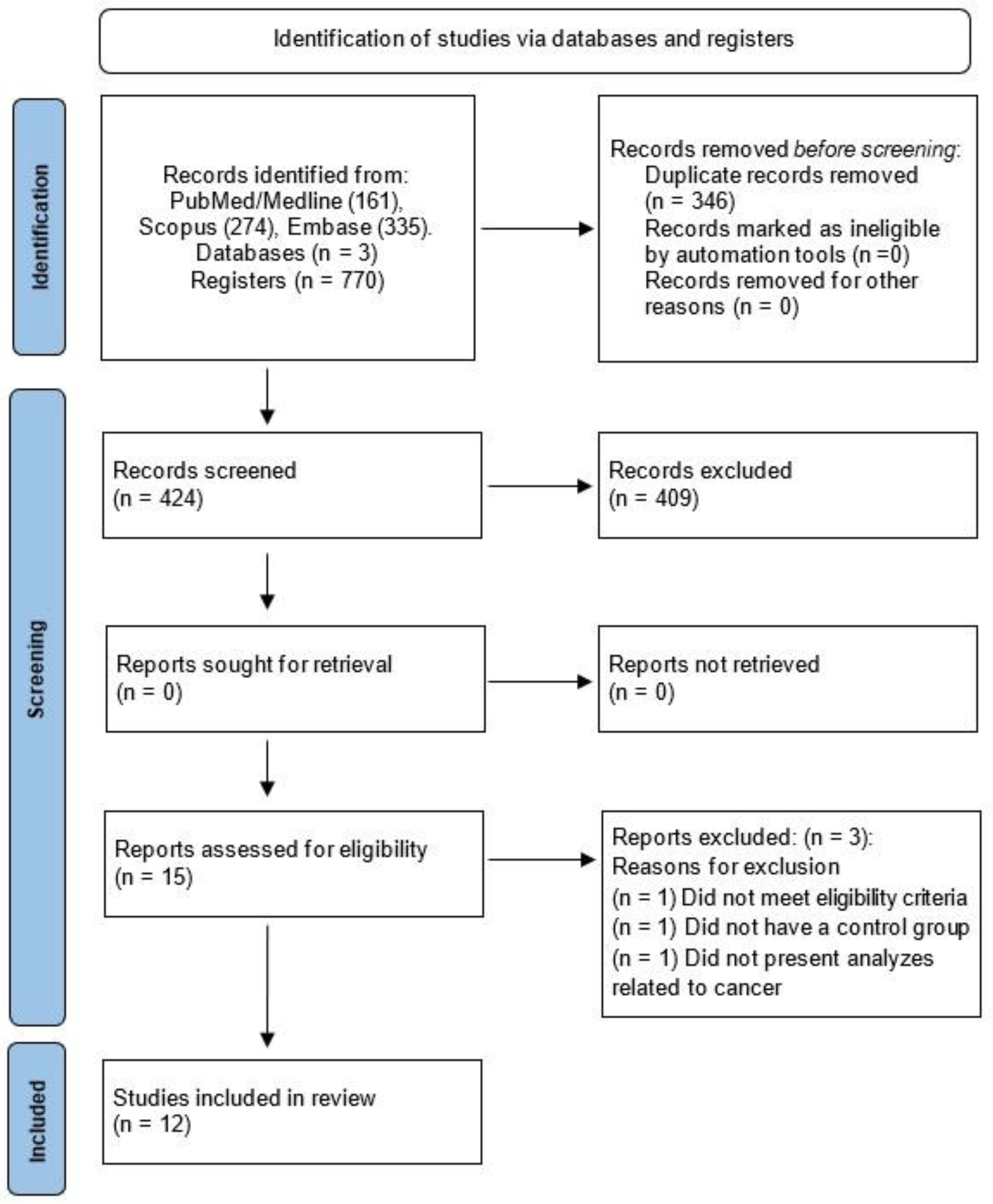
3.1. Search Results
3.2. Methodological Quality Assessment
3.3. Study Characteristics
3.4. Types and Cancer Models
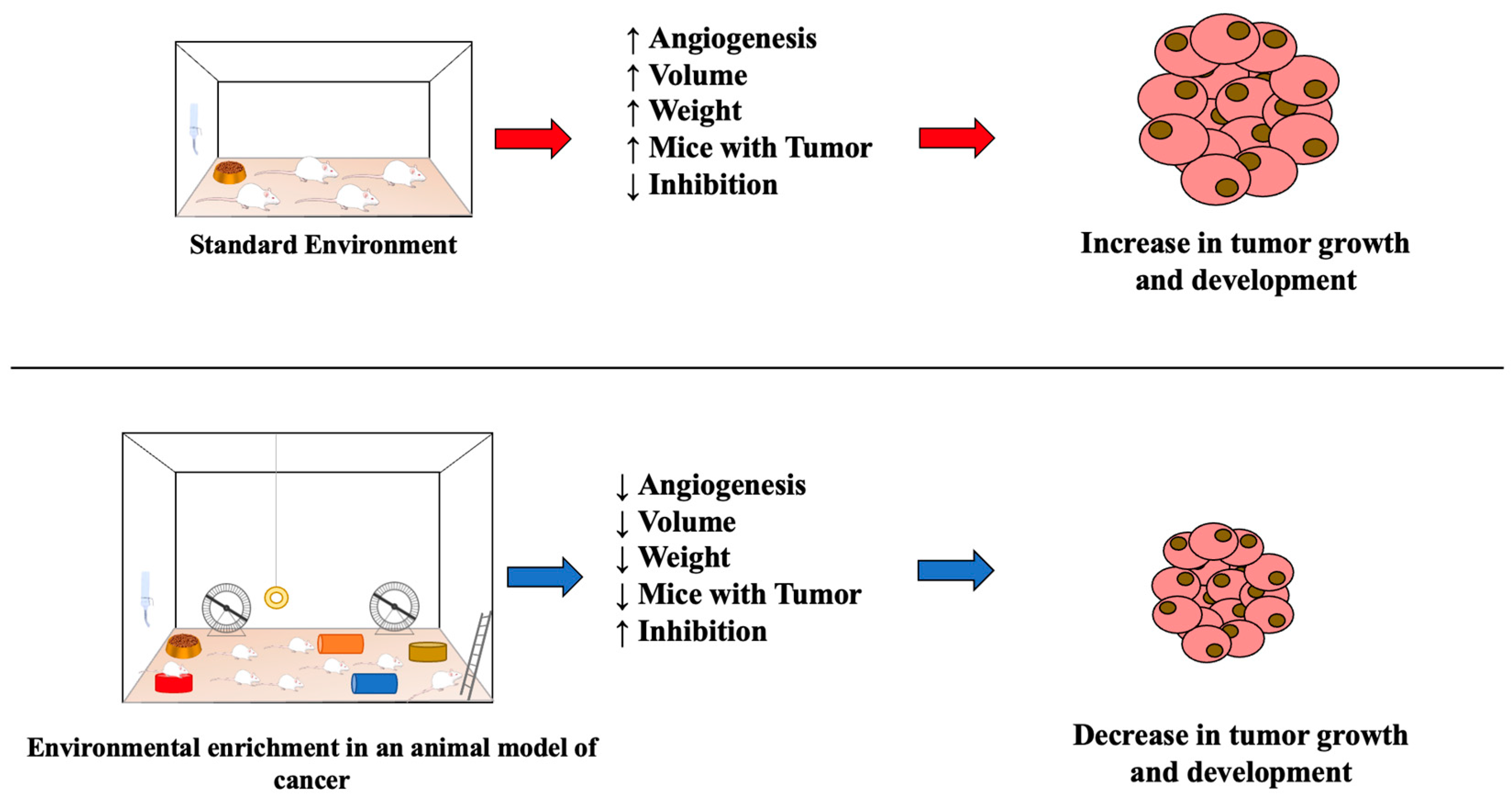
3.5. Tumor Volume, Weight, Size, and Angiogenesis after Environmental Enrichment
3.6. Tumor Number, Occurrence, Inhibition, and Mice with Tumor after Different Protocols of Environmental Enrichment
4. Discussion
Strengths and Limitations
5. Conclusions
Funding
Institutional Review Board Statement
Informed Consent Statement
Data Availability Statement
Acknowledgments
Conflicts of Interest
References
- Crofton, E.J.; Zhang, Y.; Green, T.A. Inoculation stress hypothesis of environmental enrichment. Neurosci. Biobehav. Rev. 2015, 49, 19–31. [Google Scholar] [CrossRef] [PubMed]
- Ismail, T.R.; Yap, C.G.; Naidu, R.; Pamidi, N. Enrichment Protocol for Rat Models. Curr. Protoc. 2021, 1, e152. [Google Scholar] [CrossRef] [PubMed]
- Cho, H.; Kang, K. Effects of Environmental Enrichment on Neurotrophins in an MPTP-Induced Parkinson’s Disease Animal Model: A Randomized Trial. Biol. Res. Nurs. 2020, 22, 506–513. [Google Scholar] [CrossRef]
- Liew, A.K.Y.; Teo, C.H.; Soga, T. The Molecular Effects of Environmental Enrichment on Alzheimer’s Disease. Mol. Neurobiol. 2022, 59, 7095–7118. [Google Scholar] [CrossRef]
- Wei, F.; Xian, D.; He, Y.; Yan, Z.; Deng, X.; Chen, Y.; Zhao, L.; Zhang, Y.; Li, W.; Ma, B.; et al. Effects of maternal deprivation and environmental enrichment on anxiety-like and depression-like behaviors correlate with oxytocin system and CRH level in the medial-lateral habenula. Peptides 2022, 158, 170882. [Google Scholar] [CrossRef]
- De Sousa Fernandes, M.S.; Santos, G.C.J.; Filgueira, T.O.; Gomes, D.A.; Barbosa, E.A.S.; Dos Santos, T.M.; Câmara, N.O.S.; Castoldi, A.; Souto, F.O. Cytokines and Immune Cells Profile in Different Tissues of Rodents Induced by Environmental Enrichment: Systematic Review. Int. J. Mol. Sci. 2022, 23, 11986. [Google Scholar] [CrossRef]
- Greten, F.R.; Grivennikov, S.I. Inflammation and Cancer: Triggers, Mechanisms, and Consequences. Immunity 2019, 51, 27–41. [Google Scholar] [CrossRef]
- Kuninaka, S.; Yano, T.; Yokoyama, H.; Fukuyama, Y.; Terazaki, Y.; Uehara, T.; Kanematsu, T.; Asoh, H.; Ichinose, Y. Direct influences of pro-inflammatory cytokines (IL-1beta, TNF-alpha, IL-6) on the proliferation and cell-surface antigen expression of cancer cells. Cytokine 2000, 12, 8–11. [Google Scholar] [CrossRef]
- Zhang, X.; Qiu, H.; Li, C.; Cai, P.; Qi, F. The positive role of traditional Chinese medicine as an adjunctive therapy for cancer. Biosci. Trends 2021, 15, 283–298. [Google Scholar] [CrossRef]
- Cao, L.; Liu, X.; Lin, E.J.; Wang, C.; Choi, E.Y.; Riban, V.; Lin, B.; During, M.J. Environmental and genetic activation of a brain-adipocyte BDNF/leptin axis causes cancer remission and inhibition. Cell 2010, 142, 52–64. [Google Scholar] [CrossRef]
- Hassan, Q.N., 2nd; Queen, N.J.; Cao, L. Regulation of aging and cancer by enhanced environmental activation of a hypothalamic-sympathoneural-adipocyte axis. Transl. Cancer Res. 2020, 9, 5687–5699. [Google Scholar] [CrossRef] [PubMed]
- Queen, N.J.; Deng, H.; Huang, W.; Mo, X.; Wilkins, R.K.; Zhu, T.; Wu, X.; Cao, L. Environmental Enrichment Mitigates Age-Related Metabolic Decline and Lewis Lung Carcinoma Growth in Aged Female Mice. Cancer Prev. Res. 2021, 14, 1075–1088. [Google Scholar] [CrossRef] [PubMed]
- Bice, B.D.; Stephens, M.R.; Georges, S.J.; Venancio, A.R.; Bermant, P.C.; Warncke, A.V.; Affolter, K.E.; Hidalgo, J.R.; Angus-Hill, M.L. Environmental Enrichment Induces Pericyte and IgA-Dependent Wound Repair and Lifespan Extension in a Colon Tumor Model. Cell Rep. 2017, 19, 760–773. [Google Scholar] [CrossRef] [PubMed]
- Foglesong, G.D.; Queen, N.J.; Huang, W.; Widstrom, K.J.; Cao, L. Enriched environment inhibits breast cancer progression in obese models with intact leptin signaling. Endocr. Relat. Cancer 2019, 26, 483–495. [Google Scholar] [CrossRef]
- Garofalo, S.; D’Alessandro, G.; Chece, G.; Brau, F.; Maggi, L.; Rosa, A.; Porzia, A.; Mainiero, F.; Esposito, V.; Lauro, C.; et al. Enriched environment reduces glioma growth through immune and non-immune mechanisms in mice. Nat. Commun. 2015, 6, 6623. [Google Scholar] [CrossRef] [PubMed]
- Nachat-Kappes, R.; Pinel, A.; Combe, K.; Lamas, B.; Farges, M.C.; Rossary, A.; Goncalves-Mendes, N.; Caldefie-Chezet, F.; Vasson, M.P.; Basu, S. Effects of enriched environment on COX-2, leptin and eicosanoids in a mouse model of breast cancer. PLoS ONE 2012, 7, e51525. [Google Scholar] [CrossRef]
- Li, G.; Gan, Y.; Fan, Y.; Wu, Y.; Lin, H.; Song, Y.; Cai, X.; Yu, X.; Pan, W.; Yao, M.; et al. Enriched environment inhibits mouse pancreatic cancer growth and down-regulates the expression of mitochondria-related genes in cancer cells. Sci. Rep. 2015, 5, 7856. [Google Scholar] [CrossRef]
- Liu, C.; Yang, Y.; Chen, C.; Li, L.; Li, J.; Wang, X.; Chu, Q.; Qiu, L.; Ba, Q.; Li, X.; et al. Environmental eustress modulates β-ARs/CCL2 axis to induce anti-tumor immunity and sensitize immunotherapy against liver cancer in mice. Nat. Commun. 2021, 12, 5725. [Google Scholar] [CrossRef]
- Takai, D.; Abe, A.; Miura, H.; Tanaka, S.; Komura, J.I. Minimum environmental enrichment is effective in activating antitumor immunity to transplanted tumor cells in mice. Exp. Anim. 2019, 68, 569–576. [Google Scholar] [CrossRef]
- Watanabe, J.; Kagami, N.; Kawazoe, M.; Arata, S. A simplified enriched environment increases body temperature and suppresses cancer progression in mice. Exp. Anim. 2020, 69, 207–218. [Google Scholar] [CrossRef]
- Westwood, J.A.; Darcy, P.K.; Kershaw, M.H. Environmental enrichment does not impact on tumor growth in mice. F1000Res 2013, 2, 140. [Google Scholar] [CrossRef] [PubMed]
- Wu, Y.; Gan, Y.; Yuan, H.; Wang, Q.; Fan, Y.; Li, G.; Zhang, J.; Yao, M.; Gu, J.; Tu, H. Enriched environment housing enhances the sensitivity of mouse pancreatic cancer to chemotherapeutic agents. Biochem. Biophys. Res. Commun. 2016, 473, 593–599. [Google Scholar] [CrossRef] [PubMed]
- Hosey, G.; Melfi, V.; Pankhurst, S. Zoo Animals: Behaviour, Management, and Welfare; Oxford University Press: Oxford, UK, 2013. [Google Scholar]
- Kamiya, A.; Hiyama, T.; Fujimura, A.; Yoshikawa, S. Sympathetic and parasympathetic innervation in cancer: Therapeutic implications. Clin. Auton. Res. 2021, 31, 165–178. [Google Scholar] [CrossRef] [PubMed]
- Mészáros Crow, E.; López-Gigosos, R.; Mariscal-López, E.; Agredano-Sanchez, M.; García-Casares, N.; Mariscal, A.; Gutiérrez-Bedmar, M. Psychosocial interventions reduce cortisol in breast cancer patients: Systematic review and meta-analysis. Front. Psychol. 2023, 14, 1148805. [Google Scholar] [CrossRef] [PubMed]
- Magnon, C.; Hall, S.J.; Lin, J.; Xue, X.; Gerber, L.; Freedland, S.J.; Frenette, P.S. Autonomic nerve development contributes to prostate cancer progression. Science 2013, 341, 1236361. [Google Scholar] [CrossRef] [PubMed]
- Dubben, H.H.; Thames, H.D.; Beck-Bornholdt, H.P. Tumor volume: A basic and specific response predictor in radiotherapy. Radiother. Oncol. 1998, 47, 167–174. [Google Scholar] [CrossRef]
- Detmar, M. Tumor angiogenesis. J. Investig. Dermatol. Symp. Proc. 2000, 5, 20–23. [Google Scholar] [CrossRef]
- Kretschmer, M.; Rüdiger, D.; Zahler, S. Mechanical Aspects of Angiogenesis. Cancers 2021, 13, 4987. [Google Scholar] [CrossRef]
- Kiselev, S.M.; Lutsenko, S.V.; Severin, S.E.; Severin, E.S. Tumor angiogenesis inhibitors. Biochemistry 2003, 68, 497–513. [Google Scholar]
- Lugano, R.; Ramachandran, M.; Dimberg, A. Tumor angiogenesis: Causes, consequences, challenges and opportunities. Cell Mol. Life Sci. 2020, 77, 1745–1770. [Google Scholar] [CrossRef]
- Xiao, R.; Ali, S.; Caligiuri, M.A.; Cao, L. Enhancing Effects of Environmental Enrichment on the Functions of Natural Killer Cells in Mice. Front. Immunol. 2021, 12, 695859. [Google Scholar] [CrossRef] [PubMed]
- Bloor, C.M. Angiogenesis during exercise and training. Angiogenesis 2005, 8, 263–271. [Google Scholar] [CrossRef] [PubMed]
- Rampino, A.; Annese, T.; Margari, A.; Tamma, R.; Ribatti, D. Nutraceuticals and their role in tumor angiogenesis. Exp. Cell Res. 2021, 408, 112859. [Google Scholar] [CrossRef]
- Di Castro, M.A.; Garofalo, S.; De Felice, E.; Meneghetti, N.; Di Pietro, E.; Mormino, A.; Mazzoni, A.; Caleo, M.; Maggi, L.; Limatola, C. Environmental enrichment counteracts the effects of glioma in primary visual cortex. Neurobiol. Dis. 2022, 174, 105894. [Google Scholar] [CrossRef] [PubMed]
- Zhang, B.; Vogelzang, A.; Miyajima, M.; Sugiura, Y.; Wu, Y.; Chamoto, K.; Nakano, R.; Hatae, R.; Menzies, R.J.; Sonomura, K.; et al. B cell-derived GABA elicits IL-10(+) macrophages to limit anti-tumour immunity. Nature 2021, 599, 471–476. [Google Scholar] [CrossRef] [PubMed]
- Mansour, A.G.; Xiao, R.; Bergin, S.M.; Huang, W.; Chrislip, L.A.; Zhang, J.; Ali, S.; Queen, N.J.; Caligiuri, M.A.; Cao, L. Enriched environment enhances NK cell maturation through hypothalamic BDNF in male mice. Eur. J. Immunol. 2021, 51, 557–566. [Google Scholar] [CrossRef]


| Inclusion Criteria | Exclusion Criteria | |
|---|---|---|
| Population | Rodents | Non-rodents |
| Intervention | Environmental enrichment | Non-environmental enrichment |
| Control | Non-environmental enrichment | Any other comparison group |
| Outcomes | Type of cancer; cancer model description; angiogenesis; tumor occurrence; tumor volume (%, cubic millimeters); tumor weight (milligrams, grams); mice with tumor and inhibition (%); tumor size (square millimeters) | No tumor parameters |
| Study design | Animal studies | Reviews; case reports; letters to the editor; comments, etc. |
| Author, Year | Q1 | Q2 | Q3 | Q4 | Q5 | Q6 | Q7 | Q8 | Q9 | Q10 |
|---|---|---|---|---|---|---|---|---|---|---|
| Bice et al., 2017 [13] | Y | Y | Y | Y | N | Y | N | Y | Y | Y |
| Cao et al., 2010 [10] | Y | Y | Y | Y | N | Y | N | Y | Y | Y |
| Foglesong et al., 2019 [14] | Y | Y | Y | Y | N | Y | N | Y | Y | Y |
| Garofalo et al., 2014 [15] | Y | U | Y | Y | N | Y | N | Y | Y | Y |
| Nachat-Kappes et al., 2012 [16] | Y | Y | Y | Y | N | Y | N | Y | Y | Y |
| Li et al., 2015 [17] | Y | Y | Y | Y | N | Y | N | Y | Y | Y |
| Liu et al., 2021 [18] | Y | Y | Y | Y | N | Y | N | Y | Y | Y |
| Queen et al., 2021 [12] | Y | Y | Y | Y | N | Y | N | Y | Y | Y |
| Takai et al., 2016 [19] | Y | Y | Y | Y | N | Y | N | Y | Y | Y |
| Watanabe et al., 2019 [20] | Y | Y | Y | Y | N | Y | N | Y | Y | Y |
| Westwood et al., 2013 [21] | Y | U | Y | Y | N | Y | N | Y | Y | Y |
| Wu et al., 2016 [22] | Y | Y | Y | Y | N | Y | N | Y | Y | Y |
| Author, Year | Species, Sex, and Age | Animals per Cage | Environmental Enrichment Protocol and Housing Dimensions (Type of Enrichment, Length, Width, and Depth or Height) | Exposure Time to Environmental Enrichment |
|---|---|---|---|---|
| Bice et al., 2017 [13] | C57BL6 mice; female and male; 16 wks old | 15–20 | Physical and Social Enrichment; Huts; Mouse Igloos; Rafters; Running Wheels; and Tunnel; 15 cm × 20 cm × 29 cm | Short and long term (16 wks) |
| Cao et al., 2010 [10] | C57BL6 mice; male; 3 wks old | 18–20 | Physical and Social Enrichment; Igloos; Huts; Maze; Nesting Material; Retreats; Running Wheels; Tunnels; and Wood Toys; 1.5 m × 1.5 m × 1.0 m | 3–6 wks |
| Foglesong et al., 2019 [14] | C57BL6 transgenic mice; female; 6 wks old | 5 | Physical and Social Enrichment; Igloos; Huts; Maze; Nesting Material; Retreats; Running Wheels; Tunnels; and Wood Toys; 63 cm × 49 cm × 44 cm | 4 wks |
| Garofalo et al., 2014 [15] | C57BL6 mice; male; 3 wks−2 months old | 10 | Physical and Social Enrichment; Climbing Ladders; Seesaws; Running Wheel; Balls; Plastic; Wood; Cardboard Boxes; and Nesting Material; 36 cm × 54 cm × 19 cm | 5 wks |
| Nachat-Kappes et al., 2012 [16] | C57BL6 mice; female; 3 wks old | 10 | Physical and Social Enrichment; Running Wheel; Tunnels; Igloos; Nesting Material; and Wooden Toys; 60 cm × 38 cm × 20 cm | 16 wks |
| Li et al., 2015 [17] | C57BL6 mice; male; 3 wks old | 12 | Physical and Social Enrichment; Running Wheel; Small Huts; Tunnels; Wood Toys; and Nesting Material; 61 cm × 43 cm × 21 cm | 3–5 wks |
| Liu et al., 2021 [18] | C57BL/6 mice; male; 2–3 wks old and BALB/c mice; male; 3 wks old | 8–25 | Physical and Social Enrichment; Running Wheels; Tunnels; Huts; Retreats; and Wood Toys; 40 cm × 30 cm × 20 cm | 3–10 wks |
| Queen et al., 2021 [12] | C57BL6 mice; female; 3 and 14 months old | 10 | Physical and Social Enrichment; Running Wheels; Huts; Shelters; Toys; Tunnels; Maze; and Nesting Material; 120 cm × 90 cm × 76 cm | 11 wks |
| Takai et al., 2016 [19] | B6C3F1 mice; female; 6 wks old | 12–24 | Physical and Social Enrichment; and Mouse Igloos; 218 mm × 320 mm × 133 mm | 6 wks–100 days |
| Watanabe et al., 2019 [20] | C57BL/6 mice; male; 24–35 wks old | 4–14 | Physical and Social Enrichment; Mouse Igloos; and Fast -Track; 21.8 cm × 32 cm × 13.3 cm | 10–14 wks |
| Westwood et al., 2013 [21] | C57BL/6 mice; male; 3 wks old | 20 | Physical and Social Enrichment; Exercise Wheels; Cardboard Boxes; PVC Tubes; and Plumbing T Piece; 81 cm × 57 cm × 34 cm | 6 wks |
| Wu et al., 2016 [22] | C57BL/6 mice; male; 3 wks old | 12 | Physical and Social Enrichment; Exercise Wheels; Tunnels; Wood Toys; and Plastic Tubes; 61 cm × 43 cm × 21 cm | 3 wks |
| Author, Year | Type of Cancer | Cancer Model | Tumor Outcomes |
|---|---|---|---|
| Bice et al., 2017 [13] | Colon Tumor | Genetically induced by mutant’s phenotypes (ApcMin and ApcMin Tcf4Het) | ↓ Tumor angiogenesis; Volume (mm3);  tumor number and Weight (mg) tumor number and Weight (mg) |
| Cao et al., 2010 [10] | Melanoma | Implanted subcutaneously in the flank (B16F10 syngeneic melanoma cell line/1 × 105 per mouse). | ↓ Tumor volume (% and mm3); tumor weight (%);  rumor occurrence (%); mice with tumor (%) rumor occurrence (%); mice with tumor (%) |
| Foglesong et al., 2019 [14] | Breast Tumor | Inoculation of 50.000 breast cancer cells derived from MMTV-PyMT mice in 100 μL serum-free in the right mammary | ↓ Tumor occurrence (%); volume (mm3) |
| Garofalo et al., 2014 [15] | Glioma | Were injected intracranially GL261 or CD133+-GL261 (7.5 × 10⁴), and U87MG (5 × 10⁴) glioma cells | ↓ Tumor volume (% and mm3);  mice with tumor mice with tumor |
| Nachat-Kappes et al., 2012 [16] | Breast Tumor | Mammary cell line EO771 (56,105 cells in 100 mL) were transplanted subcutaneously into the fourth right mammary fat pad | ↓ Tumor volume (mm3); weight (mg) |
| Li et al., 2015 [17] | Pancreatic | Subcutaneous tumors were prepared and implanted using Panc02 cells (6 × 10⁵ per mouse) in their right flank | ↓ Tumor volume (mm3); weight (g); ↑ tumor inhibition (%) |
| Liu et al., 2021 [18] | Liver | Murine HCC cells (Hepa1-6, H22, and LPC-H12) were transplanted subcutaneously and injected into the right flanks of mice (~5 × 10⁵–1 × 10⁶) cells | ↓ Tumor occurrence; volume (mm3); weight (g) |
| Queen et al., 2021 [12] | Lung | LLC cells (2.5 × 10⁵) were implanted in mouse subcutaneous tissue with 100 μL of serum-free | ↓ Tumor volume (mm3); weight (g) |
| Takai et al., 2016 [19] | Ovarian | OV3121 cells, derived from an ovarian granulosa cell tumor (5 × 10⁵ cells) were injected subcutaneously onto the back of the mice | ↓ Mice with tumor (%) |
| Watanabe et al., 2019 [20] | Lung | 3LL tumor cells (5 × 10⁴) were injected subcutaneously in the right flanks to develop solid intra-abdominal tumors. Alternatively, 3LL cells (1 × 10⁵) were injected into the tail vein to form colonies of metastatic cells | ↓ Tumor weight (g); occurrence |
| Westwood et al., 2013 [21] | Melanoma | Were inoculated subcutaneously with 100 μL of a single-cell suspension of 1 × 10⁵ B16F10 melanoma cells |  Tumor size (mm2) Tumor size (mm2) |
| Wu et al., 2016 [22] | Pancreatic | Panc02, Panc02-VC, or Panc02-ABCA8b cells (6 × 10⁵ per mouse) were implanted subcutaneously in the right flank | ↓ Tumor weight (g), ↑ tumor inhibition (%) |
 : No significant difference (p > 0.05). ↓ Significant decrease; ↑ Significant increase.
Disclaimer/Publisher’s Note: The statements, opinions and data contained in all publications are solely those of the individual author(s) and contributor(s) and not of MDPI and/or the editor(s). MDPI and/or the editor(s) disclaim responsibility for any injury to people or property resulting from any ideas, methods, instructions or products referred to in the content. |
© 2023 by the authors. Licensee MDPI, Basel, Switzerland. This article is an open access article distributed under the terms and conditions of the Creative Commons Attribution (CC BY) license (https://creativecommons.org/licenses/by/4.0/).
Share and Cite
Fernandes, M.S.d.S.; Lacerda, T.R.; Fidélis, D.E.d.S.; Santos, G.C.J.; Filgueira, T.O.; de Souza, R.F.; Lagranha, C.J.; Lira, F.S.; Castoldi, A.; Souto, F.O. Environmental Enrichment in Cancer as a Possible Tool to Combat Tumor Development: A Systematic Review. Int. J. Mol. Sci. 2023, 24, 16516. https://doi.org/10.3390/ijms242216516
Fernandes MSdS, Lacerda TR, Fidélis DEdS, Santos GCJ, Filgueira TO, de Souza RF, Lagranha CJ, Lira FS, Castoldi A, Souto FO. Environmental Enrichment in Cancer as a Possible Tool to Combat Tumor Development: A Systematic Review. International Journal of Molecular Sciences. 2023; 24(22):16516. https://doi.org/10.3390/ijms242216516
Chicago/Turabian StyleFernandes, Matheus Santos de Sousa, Tiago Ramos Lacerda, Débora Eduarda da Silva Fidélis, Gabriela Carvalho Jurema Santos, Tayrine Ordonio Filgueira, Raphael Fabrício de Souza, Claúdia Jacques Lagranha, Fábio S. Lira, Angela Castoldi, and Fabrício Oliveira Souto. 2023. "Environmental Enrichment in Cancer as a Possible Tool to Combat Tumor Development: A Systematic Review" International Journal of Molecular Sciences 24, no. 22: 16516. https://doi.org/10.3390/ijms242216516
APA StyleFernandes, M. S. d. S., Lacerda, T. R., Fidélis, D. E. d. S., Santos, G. C. J., Filgueira, T. O., de Souza, R. F., Lagranha, C. J., Lira, F. S., Castoldi, A., & Souto, F. O. (2023). Environmental Enrichment in Cancer as a Possible Tool to Combat Tumor Development: A Systematic Review. International Journal of Molecular Sciences, 24(22), 16516. https://doi.org/10.3390/ijms242216516