Article

Suppressors of lapC Mutation Identify New Regulators of LpxC, Which Mediates the First Committed Step in Lipopolysaccharide Biosynthesis

Laboratory of Bacterial Genetics, Gdansk University of Technology, 80-233 Gdansk, Poland
*
Authors to whom correspondence should be addressed.
These authors contributed equally to this work.
Int. J. Mol. Sci. 2023, 24(20), 15174; https://doi.org/10.3390/ijms242015174
Submission received: 5 September 2023 / Revised: 10 October 2023 / Accepted: 12 October 2023 / Published: 14 October 2023
(This article belongs to the Special Issue Lipopolysaccharides 2.0)

Abstract

:
Gram-negative bacteria, such as Escherichia coli, are characterized by an asymmetric outer membrane (OM) with lipopolysaccharide (LPS) located in the outer leaflet and phospholipids facing the inner leaflet. E. coli recruits LPS assembly proteins LapB, LapC and LapD in concert with FtsH protease to ensure a balanced biosynthesis of LPS and phospholipids. We recently reported that bacteria either lacking the periplasmic domain of the essential LapC protein (lapC190) or in the absence of LapD exhibit an elevated degradation of LpxC, which catalyzes the first committed step in LPS biosynthesis. To further understand the functions of LapC and LapD in regulating LPS biosynthesis, we show that the overproduction of the intact LapD suppresses the temperature sensitivity (Ts) of lapC190, but not when either its N-terminal transmembrane anchor or specific conserved amino acids in the C-terminal domain are mutated. Moreover, overexpression of srrA, marA, yceJ and yfgM genes can rescue the Ts phenotype of lapC190 bacteria by restoring LpxC amounts. We further show that MarA-mediated suppression requires the expression of mla genes, whose products participate in the maintenance of OM asymmetry, and the SrrA-mediated suppression requires the presence of cardiolipin synthase A.

1. Introduction

The most distinguishing feature of Gram-negative bacteria, such as Escherichia coli, is the presence of an asymmetric outer membrane (OM) [1]. This asymmetry is due to the characteristic location of lipopolysaccharide (LPS) in the outer leaflet of the cell envelope, with phospholipids facing its inner leaflet [1,2]. The maintenance of this OM asymmetry is essential to impart a permeability barrier, which prevents the entry of bulky toxic molecules, such as antibiotics and bile salts, inside the cells. Thus, with few exceptions, LPS is essential for bacterial viability and is one of the major virulence factors in pathogenic Gram-negative bacteria. LPS is a complex glycolipid and, in general, they share a common architecture and can be, for convenience, divided into three parts. The most conserved component is the hydrophobic membrane-anchored lipid A part, which constitutes the endotoxin principal. To the lipid A part is attached a core oligosaccharide, to which an oligosaccharide of variable length called the O-antigen is attached in bacteria with smooth LPS [3]. The biosynthesis of LPS at the genetic and biochemical levels is well established, although how LPS amounts are regulated and its final assembly in the OM is not well understood. This is particularly important because bacteria must maintain a strict balance of 1:0.15 between phospholipids and LPS, the two essential components of the cell envelope [4]. In E. coli, this is achieved by regulating the first committed step in LPS biosynthesis, catalyzed by the essential enzyme LpxC, because LPS and phospholipids use the same (R)-3-hydroxymyristate as the common metabolic precursor [5]. However, the regulation of LpxC as we understand now turns out to be more complex than it was thought a few years ago, as it responds to multiple physiological cues and a regulated turnover by FtsH and HslVU proteases (Figure 1) [5,6].
In E. coli, the biosynthesis and transport of LPS require products of more than thirty genes and many of them are essential for bacterial viability [2]. Briefly, LPS biosynthesis occurs on the inner leaflet of the inner membrane and begins with the acylation of UDP-GlcNAc, catalyzed by LpxA. In E. coli, LpxA is selective for β-hydroxymyristoyl acyl carrier protein [7]. This is followed by deacetylation of UDP-3-O-(acyl)-GlcNAc by the Zn2+-dependent LpxC enzyme [8]. As the equilibrium constant for LpxA-mediated acylation is unfavorable, LpxC-catalyzed deacetylation becomes the first committed step in lipid A biosynthesis [9]. Following the deacetylation step, a second β-hydroxymyristoyl chain is incorporated by E. coli LpxD, leading to the synthesis of UDP-2,3-diacyl-GlcN [10]. Three additional enzymes, LpxH, LpxB and LpxK, act successively to generate the intermediate lipid IVA [11]. This intermediate serves as an acceptor for the incorporation of two 3-deoxy-α-D-manno-oct-2-ulosonic acid (Kdo) residues by WaaA, with CMP-Kdo as the donor, generating Kdo2-lipid IVA [12]. In E. coli, up to the step of Kdo2-lipid IVA biosynthesis, all enzymes are essential for bacterial viability, although strains synthesizing only a lipid IVA precursor or Kdo2-lipid IVA can be constructed [13]. The incorporation of Kdo residues serves as a key intermediate to generate hexaacylated lipid A via sequential acylation by lauroyl (LpxL) and myristoyl (LpxM) transferases and further incorporation of sugar residues using specific glycosyltransferases to produce a core oligosaccharide linked to the lipid A part [13,14]. Strains synthesizing tetraacylated lipid A can only grow on minimal medium and exhibit an elevated cell envelope stress response [13,14]. Interestingly, suppressors mapping to the msbA gene encoding LPS flippase can allow the growth of strains devoid of LpxL, LpxM and LpxP on rich medium and at elevated temperatures. These observations are in agreement with the known higher selectivity exerted by MsbA for hexaacylated lipid A as compared to the less favored underacylated species. Individually, all three late acyltransferases are nonessential, although ΔlpxL exhibits a temperature-sensitive growth phenotype. However, LpxL and LpxM become essential when cardiolipin synthase A, encoded by the clsA gene, is absent, suggesting that MsbA and ClsA could cooperate in LPS trafficking, although the precise mechanism of their interaction remains unknown [15,16]. Thus, besides the regulatory control exerted by the regulation of LpxC amounts, the preferential selection of hexaacylated lipid A species by MsbA constitutes early checkpoints in controlling the proper amounts of LPS and its translocation across the inner membrane [17].
Initial studies on the regulation of LpxC deacetylase revealed that its levels are controlled in a 20-fold range in relation to the lipid A content without any increase in the corresponding mRNA levels of the encoded gene [18]. Subsequently, it was shown that LpxC is a substrate of FtsH protease and this proteolytic activity might be affected by alterations in acyl-ACP pools [5]. Such a model of FtsH-mediated proteolysis of LpxC could, in turn, provide a balance between biosynthesis of phospholipids and lipid A since both derive their fatty acyl chains from the same (R)-3-hydroxyacyl-ACP pool (Figure 1) [5]. In an important breakthrough, a new essential inner membrane protein was identified and shown to co-purify with FtsH and several proteins involved in either LPS and phospholipid biosynthesis or LPS translocation, and hence designated the LPS assembly protein LapB [19]. The absence of LapB could be tolerated either in the presence of a hyperactive fabZ variant that allowed an ftsH deletion or when the biosynthesis of LPS was reduced [19,20]. The co-purification of LapB with FtsH, FabZ, WaaC and Lpt proteins led to the proposal that LapB could act as a hub for LPS assembly in the IM and further cooperate with FtsH to regulate LpxC turnover [19]. More recent studies have validated these earlier observations and indeed, LapB was found to interact with LpxA, LpxC, LpxD and FabZ [21]. Similarly, recent structural and biochemical studies have shown that LpxC degradation by FtsH requires LapB [22]. However, how such LpxC degradation by FtsH-LapB is adjusted to the cellular demand for LPS remained unknown till the more recent discovery of LapC [2]. We and other independent research groups have shown that LapC acts as an antagonist of the proteolytic pathway of LpxC mediated by FtsH-LapB [6,22,23,24,25,26]. Our results were further supported by an examination of the properties of strains with chromosomal mutations that cause truncations in the periplasmic domain of LapC [6]. Such mutant bacteria exhibited a temperature-sensitive (Ts) phenotype, hypersensitivity to the LpxC inhibitor CHIR090, hyperdegradation of LpxC and consequently reduced levels of LPS (Figure 1). Thus, not only our group, but parallel independent studies, identified extragenic suppressors of lapC mutant bacteria mapping to the lapA/B operon, lpxC and ftsH genes [6,23,24,26,27]. Furthermore, an additional partner, LapD, which also co-purifies with LapB, was identified [28]. ΔlapD bacteria were found to exhibit a Ts phenotype and membrane permeability defects with a synthetic lethality in the absence of either cardiolipin synthase A, or when LpxL or LpxM acyltransferase was absent or when bacteria synthesize LPS composed of only Kdo2-lipid A due to the absence of WaaC heptosyltransferase [28,29]. Moreover, extragenic suppressors that overcome defects of lapC mutant bacteria were also found to rescue the Ts phenotype or vancomycin sensitivity of ΔlapD bacteria [28].
However, how LapB, LapC and LapD sense LPS biosynthetic demand and whether there are additional factors that either inhibit or enhance LpxC degradation remain unknown. Adding to this complexity, LpxC can be degraded in the absence of LapB by HslVU, and levels of LpxC can also be regulated by regulatory small RNAs, although the precise mechanism remains unknown (Figure 1) [6]. LPS synthesis can also be stimulated by additional signals that may involve systems that respond to lipid asymmetry, including the activation of outer membrane phospholipase PldA, the Mla system that facilitates retrograde lipid trafficking, and PagP palmitoyltransferase [30]. LpxC stability can further respond to changes in levels of acyl-CoA, ppGpp and alterations in the levels of saturated fatty acids as compared to unsaturated [17,31]. Thus, the regulation of LpxC and, in turn, that of LPS involves several factors. Hence, in this study, we identified additional genes whose products might regulate LpxC or LPS levels by selecting for suppressors whose overexpression can overcome the Ts phenotype of lapC mutant bacteria (Figure 2). We show that overexpression of the lapD gene and some other potential new regulatory factors (MarA, SrrA, DksA and YceJ) can suppress the Ts phenotype of lapC190 mutant bacteria and, among these, some act by increasing the amount of LpxC. MarA is a well-characterized transcription factor that regulates the expression of several genes involved in regulating antibiotic resistance [32]. Using transposon mutagenesis, gene(s) whose products are required for MarA- and SrrA-mediated suppression were identified to gain a better understanding of that factors that limit bacterial growth when LapC is dysfunctional and additional players that regulate LpxC amounts (Figure 2).

2. Results

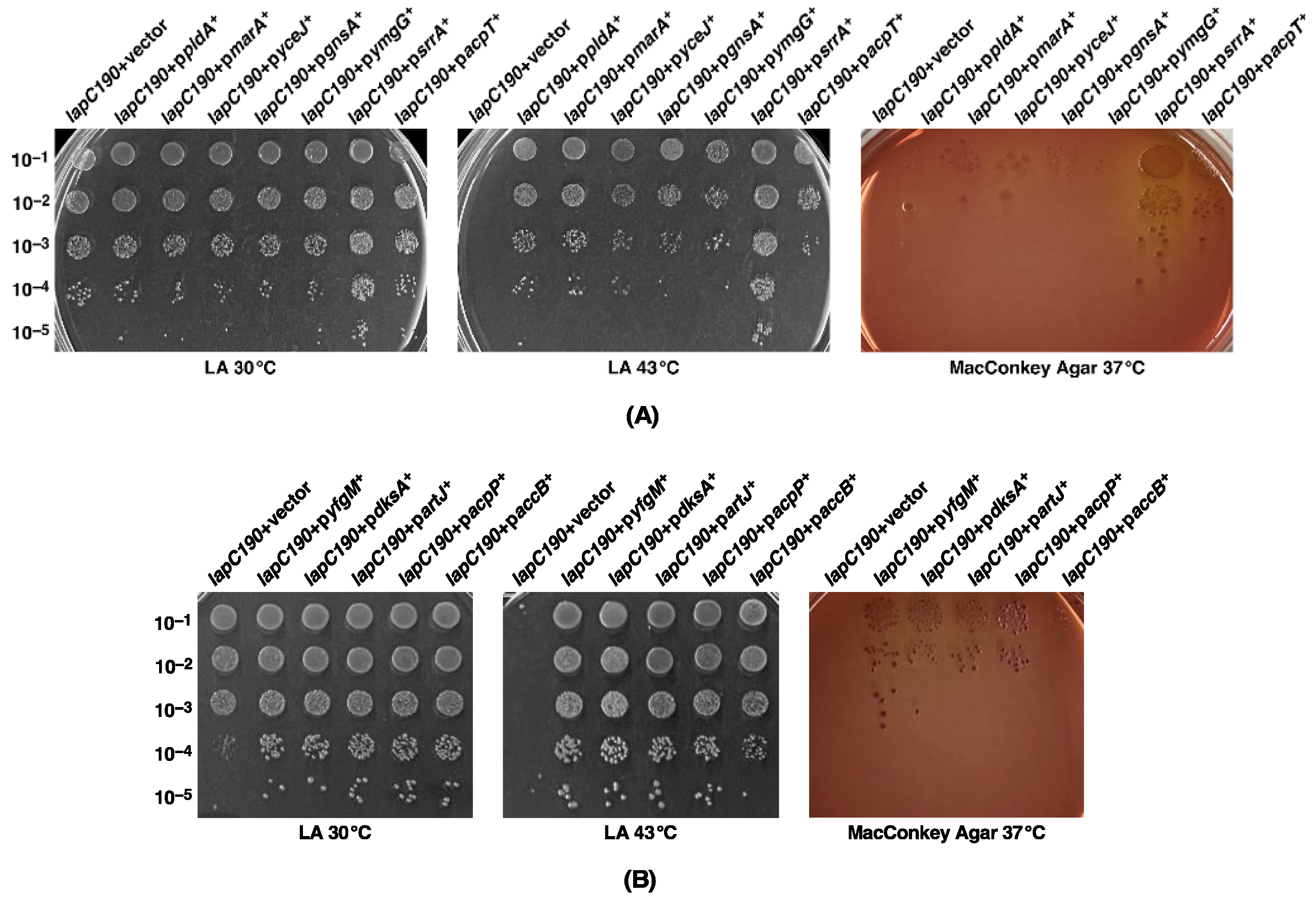
2.1. Genes Whose Overexpression Overcomes the Temperature Sensitivity (Ts) of lapC Mutant Bacteria with a Truncation of LapC Periplasmic Domain Identify New Players That Can Regulate LpxC Amounts

We previously reported the isolation of mutations in the essential gene encoding the LapC protein. Such mutations cause truncation in the periplasmic domain of LapC and confer a Ts phenotype, permeability defects and a reduced amount of LPS due to the hyperdegradation of LpxC [6]. Some of these phenotypic defects could be suppressed by chromosomal single-copy mutations mapping to lpxC, ftsH and the lapA/B operon [6]. This suppression was attributed to the stabilization of LpxC. However, since several factors can contribute towards the regulation of LpxC, here we asked if the overexpression of any gene can also suppress growth defects of lapC mutant bacteria. Thus, we used lapC190 mutant bacteria, which lack the periplasmic domain of LapC on the chromosome, to isolate multicopy suppressors, using a complete genomic library. To achieve this goal, plasmid DNA pools from the ASKA collection of single ORFs expressed from the tightly regulated IPTG-inducible PT5-lac promoter in the vector pCA24N [33] were used to transform competent cells of lapC190 mutant bacteria. Transformants were selected for the restoration of growth on LA medium supplemented with 75 μM IPTG at 43.5 °C, a condition that is non-permissive for lapC190 mutant bacteria. This concentration of inducer of gene expression has been optimized as described earlier [19]. Cultures from identified Ts+ colonies were used to extract plasmid DNA to retransform lapC190 bacteria. Plasmids that bred true were retained and their DNA was sequenced, which identified 13 unique genes, whose mild overexpression can rescue the Ts phenotype of lapC190 mutant bacteria (Table 1, Figure 3). Based on their known or predicted function, these genes can be grouped into various classes: those with a role in LPS, phospholipid/fatty acid biosynthesis or sensing (lapD, acpP, acpT, accB, pldA) (Figure 3 and Figure 4), transcription factors (marA, dksA, srrA), an IM-anchored heat shock protein-encoding gene with an unknown function (yceJ) and others whose products could either titrate the FtsH protease responsible for LpxC degradation (yfgM) or modulate saturated versus unsaturated fatty acid abundance (gnsA) or related with the stress response (ymgG), whose product is the part of toxin–antitoxin system.
Next, we examined the suppression of permeability and growth defects. For such studies, we investigated the degree of restoration of bacterial growth/viability. Thus, spot dilution assays were performed on LA medium and on MacConkey agar supplemented with 75 μM IPTG or when supplemented with 15 μg/mL of erythromycin. Data are presented for growth on MacConkey agar as a similar pattern was observed with resistance to erythromycin. These experiments revealed that although most of the multicopy suppressors restored bacterial growth on LA agar at 43.5 °C, the permeability defect was suppressed to a variable extent (Figure 3). Even on LA medium, the colony-forming ability and the colony size indicated the degree of suppression of Ts phenotype was variable. For example, the overexpression of the acpT gene conferred only a partial suppression (Figure 3). Thus, on MacConkey agar, only overexpression of srrA and yfgM genes restored growth to a near-wild-type level. On this medium, which reflects a permeability phenotype, a modest suppression is also conferred by the overexpression of dksA, acpP and acpT genes (Figure 3). Noteworthy of these suppressor-encoding genes is the isolation of a set of genes (srrA, dksA, acpP, yfgM, ymgG, artJ and accB) whose overexpression was earlier shown to suppress the Ts phenotype of ΔlapD bacteria [28]. Such results suggest that some common defects in bacteria that are either lacking the periplasmic domain of LapC or when LapD is absent, can be rescued by overexpression of this set of genes (Table 1). However, the identification of marA and pldA genes is interesting, and their overexpression only suppresses the Ts phenotype of lapC190 bacteria but not of ΔlapD (Table 1). MarA (multiple antibiotic resistance) is a well-characterized transcriptional regulator, known to regulate the expression of several genes whose products are required for antibiotic resistance, oxidative stress and heavy metals tolerance [32,34,35]. Similarly, overexpression of the acpT gene only suppresses the Ts phenotype of lapC190 bacteria but not of the ΔlapD bacteria. Thus, although some suppressors identified in this work are common to ΔlapD and lapC190, the overexpression of some genes such as marA, pldA and acpT is unique for overcoming the Ts phenotype of only lapC190 bacteria, suggesting a partial overlap in the function of LapC and LapD and not all interacting partners could be the same. These conclusions draw further support from results showing that while in multicopy the lapD gene can suppress the Ts phenotype of lapC190 bacteria (Figure 4), but in converse, overexpression of the lapC gene does not rescue the Ts phenotype of ΔlapD bacteria.

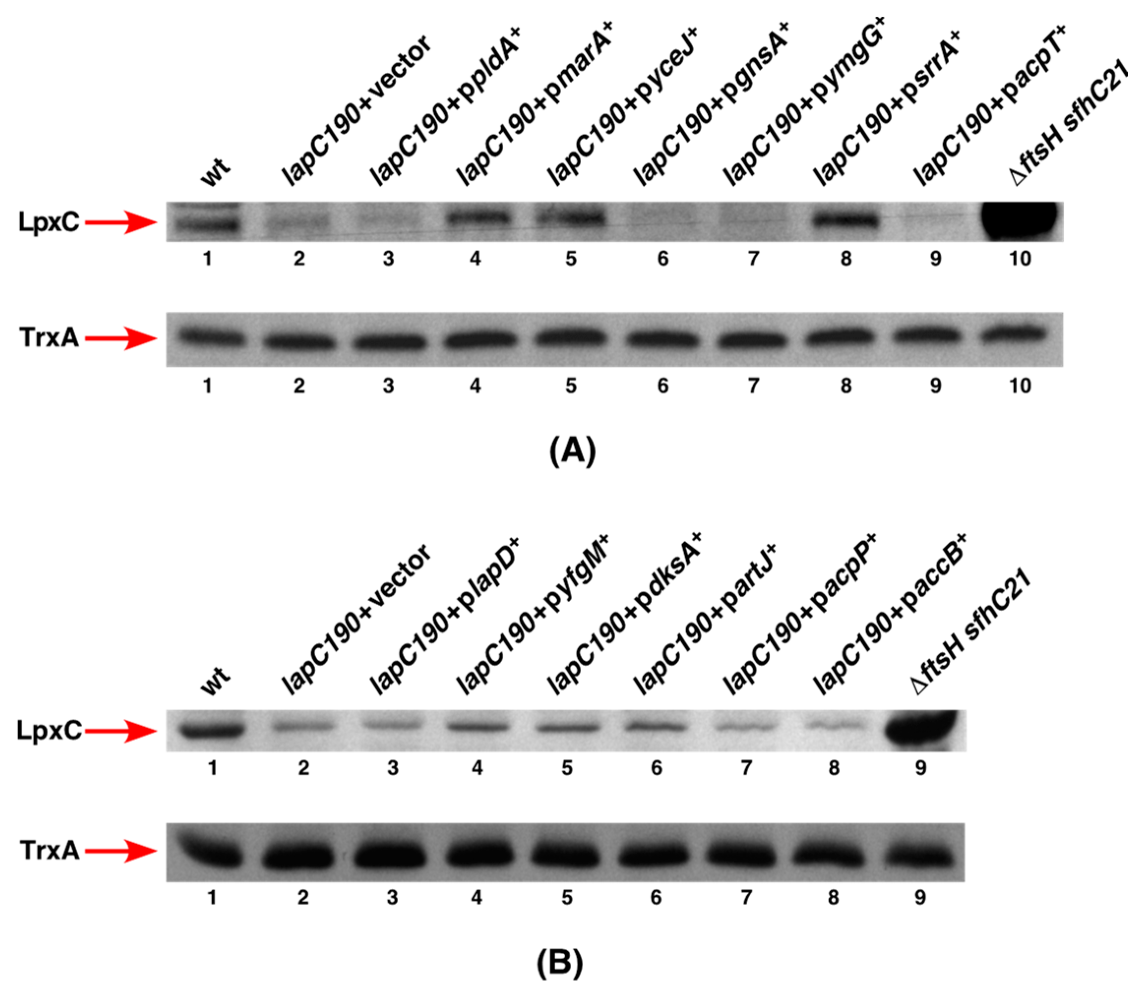
2.2. Overexpression of srrA, marA, yceJ and yfgM Restore LpxC Amounts in lapC Mutant Bacteria

To address the molecular basis of the suppression of growth defects at elevated temperatures by overexpression of the above-mentioned multicopy suppressor-encoding genes, experiments were undertaken to examine the amount of LpxC upon their mild induction. The rationale for these studies is to verify if the major defect reflected by the reduction in LpxC amounts associated with the absence of the periplasmic domain of LapC can be overcome by overexpression of any of these genes. Thus, total cell extracts were obtained from the wild type and panels of isogenic Δlap190 derivatives either with an empty vector or when the overexpressing plasmid was present under conditions described in the legend to Figure 5. The equivalent amount of proteins was resolved on a 12% SDS-PAGE, and LpxC was detected by immunoblotting with LpxC-specific antibodies. The estimation of relative amounts of LpxC in such experiments prominently shows that induction of expression of marA, yceJ and srrA restore LpxC to near wild-type levels (Figure 5A, lanes 4, 5 and 8). We applied the same amounts of total proteins from the ΔftsH sfhC21 strain since, in the absence of FtsH, LpxC amounts are increased to serve as a marker for LpxC and as a control. In parallel, the same samples were immunoblotted with anti-TrxA antibodies serving as an additional loading control. Among the remaining genes, whose overexpression also suppresses the Ts phenotype of lapC190 bacteria, a modest increase in LpxC abundance can be observed in strains with induced expression of either yfgM or dksA or artJ genes in the lapC190 background (Figure 5B, lanes 4, 5 and 6). These results suggest that the induction of marA, yceJ and srrA genes and, to a certain extent, of yfgM, dksA and artJ can suppress the growth defect of lapC190 bacteria by restoring LpxC amounts. Surprisingly, the overexpression of pldA, gnsA, ymgG and acpT genes (Figure 5A) as well as lapD, acpP and accB (Figure 5B) did not cause any noticeable increase in LpxC amounts and remained similar to when extracts from lapC190 with the vector alone were applied. It is possible that in the case of expression of such multicopy-encoding suppressor genes, a mild induction of their expression may not be sufficient to significantly increase LpxC amounts, although Ts of lapC190 bacteria can be suppressed. Overall, we can conclude that a mild overproduction of SrrA, MarA, YceJ and YfgM suppress the Ts phenotype and also lead to the restoration of LpxC amounts, explaining the molecular basis of their contribution in the regulation of LpxC.

2.3. Overexpression of srrA, marA, yceJ and dksA also Restore LPS Amounts in lapC190 Bacteria

We previously showed that lapC190 bacteria have reduced amounts of LPS due to the enhanced degradation of LpxC as compared to the wild type. Thus, to address the mechanism of suppression by overexpression of specific genes that provide a robust restoration of growth at elevated temperatures, we estimated levels of LPS from total cell extracts. For such experiments, we measured levels of LPS from cellular extracts obtained from exponentially grown cultures of the isogenic wild type, its lapC190 derivative with the vector alone and lapC190 bacteria carrying plasmid that expresses a specific suppressor encoding gene under an inducible PT5-lac promoter. Bacterial cultures were grown under permissive growth conditions at 30 °C in LB medium, the gene expression was induced with the addition of 75 μM IPTG at OD595 0.1 and samples obtained under the same conditions when LpxC amounts were measured as described in Figure 5. Total cell extracts were also applied from isogenic ΔftsH sfhC21 derivative as a control. Equivalent amount of whole cell lysates was treated with Proteinase K. Samples were resolved on an SDS-Tricine gel and LPS was transferred by Western blotting. Amounts of LPS were revealed using an LPS-specific WN1 222-5 monoclonal antibody as described in the Methods section.
The estimation of relative amounts of LPS in such experiments show that lapC190 bacteria have highly diminished amounts of LPS (Figure 6 lane 9) as compared to the wild type and the ΔftsH sfhC21 derivative contains elevated amounts of LPS. Most importantly, results from such immunoblots reveal that extracts from the lapC190 derivative overexpressing either marA, or yceJ, or srrA or the dksA gene show the restoration of LPS amounts to near wild-type levels explaining the mechanism of their suppression (Figure 6). As with the estimation of LpxC amounts, overexpression of the acpP gene did not cause any increase in the amounts of LPS and remained low as observed with lapC190 bacteria. Similarly, overexpression of the lapD gene only marginally enhances LPS amounts. These results allow us to conclude that some of the multicopy suppressors such as marA, dksA, srrA and yceJ contribute via increasing LpxC and LPS amounts, providing a reasonable explanation for their identification. However, the mode of restoration of growth of lapC mutant bacteria by an overexpression of the acpP gene could involve the LpxC independent mode.

2.4. srrA, marA, lapD and pldA Are Indispensable in the lapC190 Mutant Background

To further understand the mechanism of suppression by various genes whose increased expression mitigates the Ts phenotype of lapC190 bacteria, a systematic series of bacteriophage-mediated transduction was executed. This was to ascertain if there are any genetic interactions and if any of such genes are essential for the growth and viability of lapC190 bacteria. We focused on those genes, whose overexpression also restores LpxC levels. Thus, null alleles of such candidates were introduced in lapC190 bacteria. In parallel, converse reverse transductions were also carried out by attempting to bring in a lapC190 mutation in deletion derivatives of genes whose overexpression results in the restoration of growth. Such transductions revealed that the presence of SrrA, MarA, LapD and PldA are absolutely required for the viability of lapC190 bacteria even at 30 °C but not in the wild-type parental strain (Table 2). Thus, no viable transductional ΔsrrA lapC190 and ΔlapD lapC190 combinations could be obtained. However, when the wild-type copy of the srrA or lapD gene was provided on a plasmid, viable chromosomal ΔsrrA lapC190 and ΔlapD lapC190 mutational combinations were obtained at the same frequency when the wild-type parental strain was used as a recipient to bring in either srrA or lapD null mutations (Table 2 and Table 3). In contrast, the ΔdksA lapC190 mutational combination was found to be viable up to 37 °C, although the colony size was heterogeneous. A deletion of either the marA gene or the pldA gene could be introduced in lapC190 bacteria, albeit at a highly reduced frequency, and the majority of such transductants were found to be not viable (Table 2). These genetic interactions allow us to conclude that SrrA, MarA, LapD and PldA are essential for the viability of strains lacking the chromosome of the DNA sequence encoding the periplasmic domain of LapC.

2.5. Lauroyl and Myristoyl Transferases Are Required for the Viability of lapC190 Mutant Bacteria

Next, we examined if acyltransferases that mediate the transfer of lauroyl (LpxL) and myristoyl (LpxM) acyl chains to Kdo2-lipid IVA are essential for the growth of lapC190 mutant bacteria. Bacteriophage P1-mediated transfer of lapC190 or an introduction of ΔlpxL or ΔlpxM revealed that ΔlpxL lapC190 and ΔlpxM lapC190 mutational combinations are only possible in the ectopic presence of covering plasmid containing the wild-type gene (Table 2 and Table 3). However, at a very low frequency, viable suppressors could be obtained after an incubation of more than 48 h when ΔlpxL lapC190 and ΔlpxM lapC190 transductants were plated. Further characterization of two suppressors of each such mutational combinations identified a single amino acid exchange, L412P, in the msbA gene. Interestingly, the same msbA suppressor mutation was found to overcome the lethality of Δ(lpxL lapD) and Δ(lpxM lapD) combinations [28]. Thus, we can conclude that truncation of the lapC gene, resulting in the expression of only the transmembrane anchor, requires complete acylation of the lipid A. Since underacylated LPS (lack of LpxL or LpxM) is poorly translocated by MsbA as compared to hexaacylated lipid A, this could lead to a further reduction in the amount of LPS in the OM in lapC190 bacteria and hence the lethality. Taken together, lapC190 derivatives synthesizing either tetraacylated or pentaacylated lipid A, cannot be constructed unless a suppressor mutation in the msbA gene, which encodes lipid A flippase, is present.

2.6. MarA-Mediated Suppression Requires the Presence of mla Genes

As the induction of the marA gene expression restored the growth of lapC190 mutant bacteria at an elevated temperature, we further investigated its possible mechanism of suppression. Thus, strain SR23660 (lapC190 + pmarA+) served as a host to generate a saturated Tn10 Kan transposon library at 30 °C. Nearly 50,000 such transposon mutants were screened for the growth on LA medium at 43 °C in the presence of 75 μM IPTG and as a control at 30 °C without the presence of an inducer. This is the strategy that we earlier successfully employed to identify genes whose products are required for the multicopy suppressor phenotype of the dksA gene [36]. Thus, we retained those Tn10 insertion mutants that grew at 30 °C but were unable to propagate at 43 °C, even with the induction of the marA gene expression. Such transposon mutations were backcrossed into SR23660 and verified for lack of suppression of lapC190 mutant bacteria in the presence of the inducible marA gene. This resulted in the identification of fifty-four independent Tn10 mutants. Next, such Tn10 Kan transposon insertions were introduced into the wild type by bacteriophage P1-mediated transductions to eliminate mutations that conferred a Ts phenotype at 43 °C, even in the wild-type background. Such experiments identified 22 Tn10 insertions that were found to confer the Ts phenotype in the wild type, while the remaining were specific to the lapC190 strain. Most of the Ts mutants were found to have the Tn10 insertion in the degP gene, whose product is known to be required for bacterial growth at temperatures above 42 °C [37,38]. This is consistent with a similar spectrum of Ts insertions obtained in an identical strategy described to unravel the requirement of specific genes for DksA-mediated multicopy suppression [36]. The mapping and characterization of the remaining Tn10 insertions that prevent the multicopy suppression by marA identified 12 out of 15 mapping to genes whose products are part of the Mla pathway. To facilitate the mapping of Tn10 insertions, we used a previously described linked ΔrpoN [39] to find how many can cross out the Tn10 insertion mutation. Such transductions revealed that the majority of Tn10 insertion mutants had a disruption of the mla operon. DNA sequence analysis of the Tn10 junction from chromosomal DNA of mla Tn10 insertions was obtained by inverse PCR, which identified four insertions each in mlaC and mlaD, three insertions in mlaF and one insertion in the mlaA gene. The Mla system is known to be required for the maintenance of outer membrane lipid asymmetry and the inactivation of mla genes confers sensitivity to SDS+EDTA [40]. The mla operon contains mlaF, mlaE, mlaD, mlaC and mlaB genes, out of which the mlaF gene encodes the ATP-binding protein of an inner membrane complex (MlaFEDB). The Mla pathway functions as part of the retrograde and/or anterograde intermembrane phospholipid trafficking system [40,41]. The Mla system also contains MlaA, whose encoded gene is located outside the mla operon. The Mla system can function by removing mislocalized outer leaflet phospholipids and transporting them back to the inner membrane. Thus, the isolation of Tn10 insertion in genes encoding the Mla pathway that prevent MarA-mediated suppression of lapC190 bacteria is consistent with LapC function in regulating LPS amounts and a balance with phospholipid amounts.
Thus, to further support these results, we used previously constructed in-frame deletion derivatives of mlaC and mlaD [42] and a new construct Δ(mlaC mlaD) to verify if such mutations abolish marA multicopy suppression. This was required because Tn10 insertions are expected to be polar on the expression of downstream genes. These in-frame deletions in mla genes were introduced in SR23660 carrying chromosomal lapC190 and a plasmid with an inducible expression of the marA gene by bacteriophage P1-mediated transduction. Transductants were plated on LA agar at 30 °C and at 43 °C in the presence of 75 μM IPTG to induce the expression of the marA gene (Table 4). As a control, mlaC, mlaD and (mlaC mlaD) in-frame deletions were introduced in the wild-type and lapC190 bacteria. While such deletions could be introduced into the wild type, lapC190 and SR23660 lapC190 + pmarA+ at 30 °C at a similar frequency; however, at 43 °C, deletion sets of mla genes could not be introduced in SR23660 even with the induction of the marA gene expression (Table 4). In control transductions, all three deletions of mla genes could be introduced in the wild type at 43 °C (Table 4). These results allow us to unambiguously conclude that marA-mediated multicopy suppression of lapC190 bacteria requires the expression of mla genes.

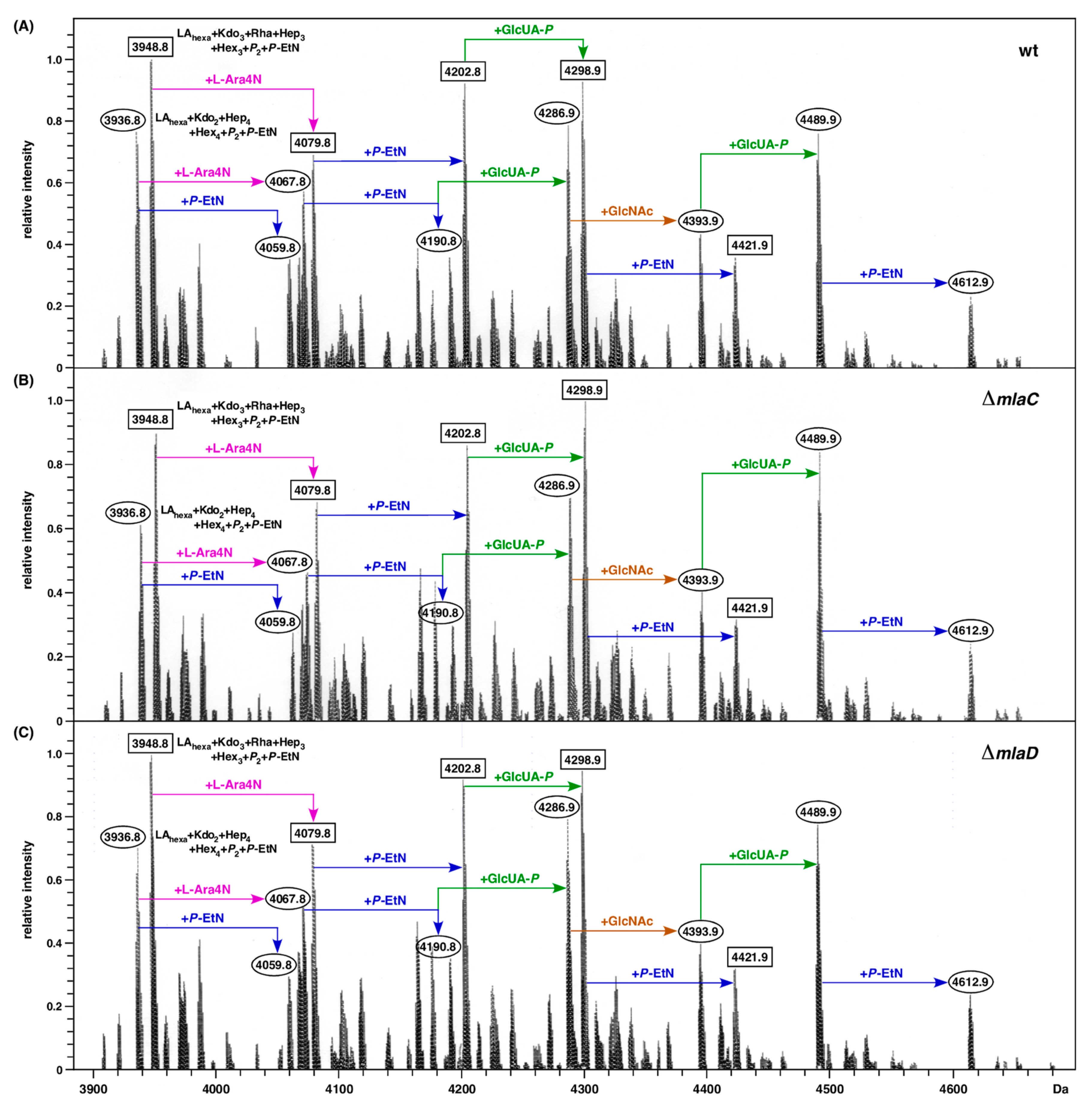
2.7. Absence of mla Genes Does Not Alter LPS Composition

In E. coli, up to now, no structural/compositional analysis of LPS has been reported when the Mla system is impaired. To investigate if the deletion derivatives of mla genes cause any major alterations in the LPS composition or in the incorporation of non-stoichiometric modifications, purified LPS obtained from the wild type and its isogenic derivatives were examined using mass spectrometric analysis. For such experiments, LPS was extracted after growth in phosphate-limiting growth medium as described earlier, since such growth conditions are inducive for lipid A and LPS core modifications due to the induction of BasS/R and PhoB/R two-component systems [13,43]. Examination of mass spectra of LPS obtained from the wild type and isogenic ΔmlaC and ΔmlaD derivatives shows similar mass peaks corresponding to the presence of complete glycoform I represented by mass peaks at 3936.7 Da and its derivatives with predicted incorporation of additional 1 or 2 P-EtN residues and also modification by L-Ara4N and GlcUA moieties as indicated. These mass peaks can be explained as LAhexa (Kdo2Hep4Hex4P2) accompanied by additional substitutions with P-EtN and L-Ara4N, as shown (Figure 7). The mass peaks at 4489.9 Da and 4612.9 Da can be attributed to the addition of GlcNAc and GlcUA to glycoform I (Figure 7). These spectra from LPS obtained from either the wild type or its ΔmlaC and ΔmlaD derivatives also reveal a prominent presence of mass peaks corresponding to glycoforms IV and V, which are represented by mass peaks at 3948.7, 4079.8, 4202 and 4298.9 Da. These mass peaks are explained by the incorporation of a third Kdo and Rha linked to the Kdo disaccharide with a concomitant truncation of the outer core with a predicted composition of LAhexa (Kdo3RhaHep3Hex3P2). The mass differences in these derivatives of glycoforms with a third Kdo and Rha are explained by the additional incorporation of P-EtN, L-Ara4N and GlcUA, as shown (Figure 7). The main findings from mass spectrometry data are that a deletion of mla genes does not alter LPS core and lipid A structural properties under these defined growth conditions.

2.8. SrrA-Mediated Suppression of lapC190 Mutant Bacteria Requires the Presence of Cardiolipin Synthase A and ClsA Is Essential for lapC190

Among various multicopy suppressors that were identified, SrrA has a distinguishing property that the corresponding gene in high dosage can suppress the Ts phenotype of strains lacking six cytoplasmic peptidyl-prolyl cis/trans isomerases, ΔlapD bacteria and is again reisolated as a suppressor of the Ts phenotype of lapC190 bacteria [28,36]. Since a modest overproduction of SrrA was found to restore LpxC amounts (Figure 5), we further investigated a possible pathway that can explain this suppression. The same strategy of isolating transposon insertion mutants was used as shown above with the marA gene. Thus, the strain SR23951 carrying the chromosomal lapC190 mutation and expressing the srrA gene from a tightly regulated PT5-lac promoter was used to serve as a host to generate a saturated library of Tn10 Kan insertion mutants under permissive growth conditions at LA 30 °C. This library was next screened on LA agar supplemented with 75 μM IPTG to induce the expression of the srrA gene at 43.5 °C. Transposon mutants that exhibit a Ts phenotype even with the induction of the srrA gene were retained. Following bacteriophage P1-mediated transductions of such Tn10 Kan mutants in the wild type, those conferring per se a Ts phenotype in the wild type were not followed. The identity of the Tn10 insertion was obtained after DNA sequencing of inverse PCR products obtained from the template chromosomal DNA of the remaining Tn10 insertion-carrying mutations, which specifically abrogated the multicopy suppression of the srrA gene in lapC190 mutant bacteria. Analysis of DNA sequencing revealed that the majority of strains (8 out of 11) had Tn10 insertion in the coding region of the clsA gene encoding cardiolipin synthase A. Two other strains had a mutation in the cpxR gene. CpxR acts as the DNA-binding response regulator and works with CpxA IM kinase, constituting one of the main two-component systems that responds to protein folding defects in the periplasm and severe defects in LPS biosynthesis [13,44,45,46,47]. One Tn10 insertion mutant that did not allow the multicopy suppression by the srrA gene has an identical Tn10 insertion in the heat shock promoter of the groSL operon at nt position -101, which was earlier found to abolish DksA-mediated suppression of Δ6ppi and ΔdnaK/J strains [36]. GroSL chaperonins constitute the major player in protein folding and are indispensable for bacterial viability [48,49]. This Tn10 insertion in the groSL operon is located 3 nt downstream of the -35 heat shock promoter element, which is 101 nt upstream of the translational initiation codon as described previously with a reduced amount of GroL with its impaired induction at a high temperature [36]. Since the majority of Tn10 insertion mutations mapped to the clsA gene, this suggests that the presence of cardiolipin is essential for SrrA-mediated restoration of the growth of lapC190 mutant bacteria at elevated temperatures.
To further understand the requirement of the clsA gene when LapC function is impaired, a ΔclsA mutation was introduced in the wild type, lapC190 with vector alone and lapC190 + psrrA+ by bacteriophage P1-mediated transductions. Such transductants were subsequently verified for their growth at 43 °C. Results from such experiments show that ΔclsA is viable in the wild type under such growth conditions but cannot be introduced into lapC190 bacteria with a vector alone (Table 5). However, transductants at a highly reduced frequency with a smaller colony size were obtained in SR23951 lapC190 + psrrA+ in the presence of IPTG at only 30 °C as compared to a wild-type background. Furthermore, ΔclsA derivatives in SR23951 were unable to propagate at temperatures above 42 °C (Table 5). Thus, these results show that ClsA presence is required for SrrA to act as a multicopy suppressor to allow the growth of lapC190 bacteria at high temperatures and presumably ClsA and LapC cooperate in the maintenance of phospholipid homeostasis in the cell envelope.

2.9. SrrA Does Not Regulate Transcription of the clsA Gene

SrrA is predicted to act as a transcriptional regulator since it contains an N-terminal signal recognition Per-Arnt-Sim (PAS) domain and a C-terminal helix-turn-helix motif [36]. However, the genes whose expression is regulated by SrrA are unknown. As described above, the overexpression of the srrA gene efficiently suppresses the Ts phenotype of lapC190 bacteria and also either of the strains collectively lacking six cytoplasmic peptidyl-prolyl cis/trans isomerases or of ΔlapD bacteria [28,36]. Since SrrA-mediated suppression requires the presence of a functional clsA gene, we undertook studies to address if SrrA acts by regulating transcription of the encoding gene. Thus, total RNA was extracted from the wild type and its isogenic derivative ΔsrrA from cultures grown at 37 °C. In parallel, total RNA was also extracted from wild-type bacteria carrying either an empty vector or expressing the srrA gene from an inducible promoter. For such experiments, cultures were grown at 30 °C. Prior to heat shock, 75 μM IPTG was added to induce transcription of the srrA gene, and cultures were shifted to 43 °C for 15 min. The equivalent amount of total RNA from these four sets of RNAs was next subjected to q-RT-PCR analysis using clsA-specific oligonucleotides for the synthesis and quantification of cDNA. The quantification of the clsA transcription pattern showed nearly similar levels of clsA transcripts between the wild-type and ΔsrrA bacteria at 37 °C (Figure 8). Similarly, when the srrA gene expression was induced and when RNA was extracted from 43 °C and the relative abundance of transcripts were estimated by q-RT-PCR, no significant differences were observed in the clsA expression with or without srrA induction (Figure 8). Although a minor increase in clsA mRNA can be observed upon shift to 43 °C as compared to the relative abundance at 37 °C, such an increase is not comparable to that of heat shock genes. Thus, results from q-RT-PCR allow us to conclude that the clsA transcription is not regulated by SrrA either at 43 °C or at 37 °C, and overexpression of the srrA gene or its absence does not alter the abundance of clsA transcripts.

2.10. LapD-Mediated Suppression of a lapC190 Mutant Bacteria Requires LapD N- and C-Terminal Domains and Identification of Some of the Critical Amino Acid Residues Required for LapD Function

As overexpression of the lapD gene restored the growth of lapC190 mutant bacteria at a high temperature, we sought to address if specific domains of LapD are required for its function. LapD has a single N-terminal transmembrane anchor and a highly conserved C-terminal domain. The crystal structure of the LapD counterpart in Haemophilus ducreyi shows a unique tetrameric α-helical coiled-coil structure in the cytosolic domain, which is predicted to mediate protein–protein interactions [50]. Thus, a series of point mutations (substitution of conserved residues by Ala), deletion of the N-terminal membrane anchor (1–20 amino acids) and deletion of the C-terminal last 32 amino acids were constructed. Among the Ala substitutions, we replaced conserved amino acids D69, Y70 and R71 with three Ala residues. Similarly, L84, L85 and P86 and N93, P94 and F95 were also substituted with three Ala residues in each case. All generated constructs with in-frame mutations were expressed under a tightly regulated ptac promoter and, when required, in-frame with an N-terminal hexa-His tag, where the expression is driven from a controlled T7 promoter. Plasmids where the expression is driven from the ptac promoter were transformed either into the ΔlapD strain or in lapC190 bacteria and evaluated for the ability to suppress their Ts phenotype. Results from such experiments show that while the control plasmid overexpressing the wild-type lapD gene restores growth of lapC190 at elevated temperature, neither the N-terminal nor the C-terminal deletions were able to suppress the Ts phenotype when the expression was induced by the addition of 75 μM IPTG (Figure 9). Similarly, three plasmid sets expressing replacement by Ala substitutions of amino acids (69–71), (84–86) and (93–95) were also unable to restore the growth of lapC190 bacteria at elevated temperatures (Figure 9). These results allow us to conclude that specific conserved residues in the cytoplasmic domain are essential for LapD function. Furthermore, the single N-terminal membrane anchor and last 32 amino acid residues of the cytoplasmic domain are required for the lapD gene to act as a multicopy suppressor.

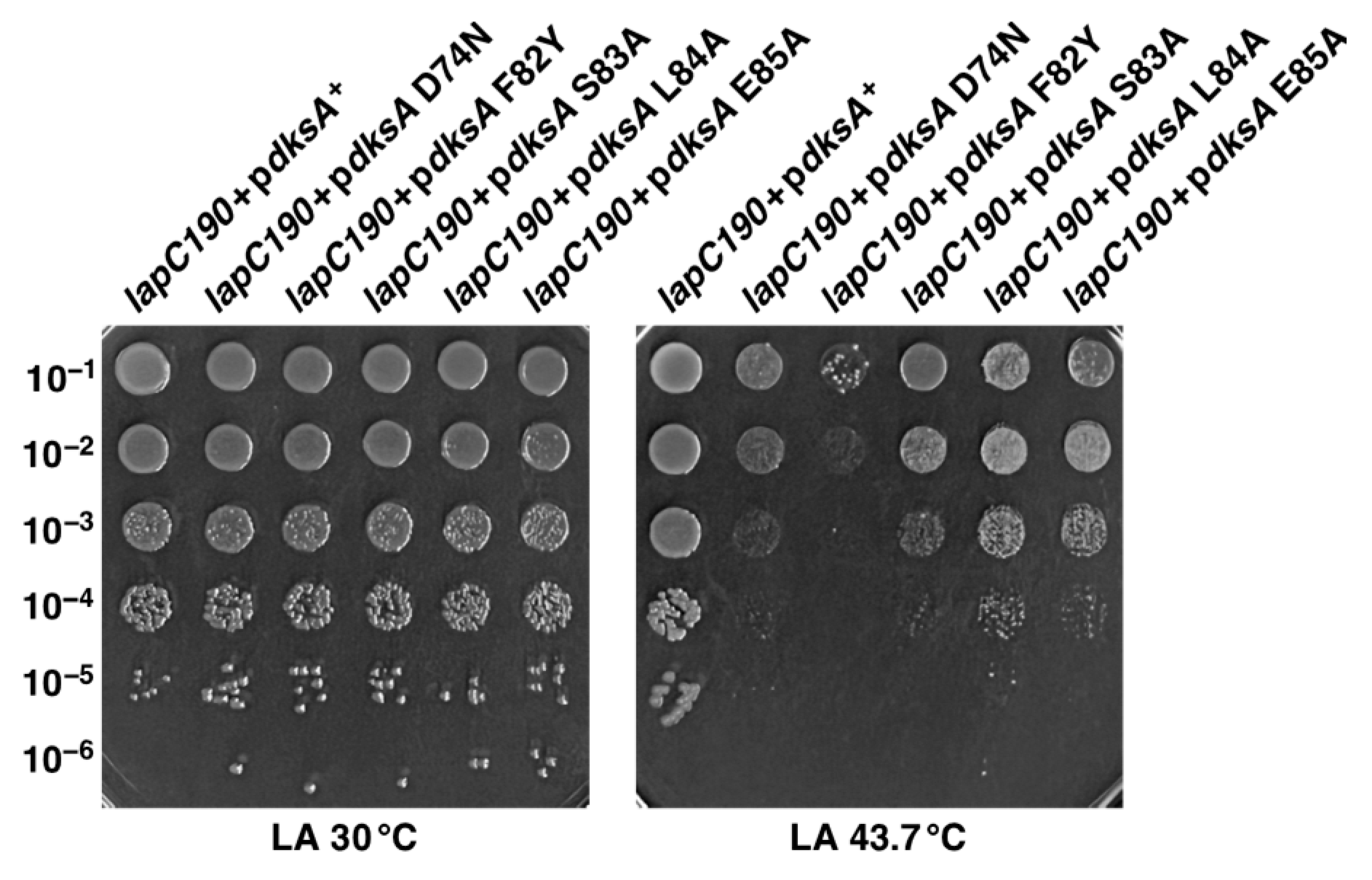
2.11. Multicopy Suppression of lapC190 Bacterial Defects Requires DksA’s PPIase and Transcriptional Activities

The results presented in the above sections have shown that overexpression of the global transcriptional regulator encoded by the dksA gene can restore the bacterial growth of lapC190 bacteria at elevated temperatures and also suppress LpxC degradation defects. We have earlier shown that DksA also suppresses the Ts phenotype of Δ6ppi bacteria and ΔlapD bacteria [28,36]. Characterization of the biochemical properties of DksA further revealed that besides acting as a global transcriptional regulator, it also exhibits the PPIase activity, which can be inhibited by the FK506 macrolide [36]. Several studies have led to the conclusion that the coiled-coil domain of DksA inserts into the secondary channel of RNAP and that residues at the tip of the coiled-coil domain of DksA are important for its activity [51,52,53]. More recent studies suggest that amino acid residues in this coiled-coil domain play an essential role for the transcriptional and PPIase activities of DksA [36]. Thus, to examine which activity of DksA is required for the suppression of growth defects of lapC190 bacteria, cloned DNA of D74N, F82Y, S83A, L84A and E85A variants of the dksA gene were transformed into lapC190 bacteria, using wild-type pdksA+ as a control. It is important to mention that D74, F84, S83 and E85 amino acid residues are important for DksA-mediated transcriptional regulation of reporter genes such as the rrnBP1 promoter [36]. However, the replacement of the F82 amino acid residue by the Tyr residue abolishes the PPIase activity of DksA, although such a variant behaves like wild type concerning the regulation of the rrnBP1 promoter [36]. Our complementation experiments reveal that cloned D74N, F82Y, S83A, L84A and E85A mutants of the dksA gene have all lost the ability to fully restore the growth of lapC190 bacteria at high temperatures upon their overexpression as compared to the complete suppression of Ts phenotype by the wild-type dksA gene (Figure 10). Viable colonies at all dilutions, albeit with smaller colony size, are observed when D74N, S83A, L84A and E85A mutants of the dksA gene were overexpressed in a lapC190 strain (Figure 10). However, replacement of F82 by Y, which is a critically important residue for DksA’s PPIase activity, completely abrogates the DksA-mediated multicopy suppression of lapC190 bacteria (Figure 10). These results suggest that the PPIase activity of DksA is most critical for DksA to function as a multicopy suppressor to suppress the Ts phenotype of lapC mutant bacteria, and the transcriptional role of DksA, although important, could only play a minor role. This is particularly striking due to the abrogation of suppression by overexpression of the DksA F82Y variant in comparison to only a partial loss of function with other DksA mutants concerning the LPS-related function.

3. Discussion

In this study, we identified new additional factors that regulate LpxC levels. Regulation of LpxC is critical for our understanding of the mechanism of balancing biosynthesis of LPS and phospholipids, since they both share the same (R)-3-hydroxymyristate-ACP as the common metabolic precursor. Recent studies have shown that LpxC stability is regulated by the FtsH-LapB complex, wherein FtsH degrades LpxC, which requires LapB function for its proteolytic activity towards LpxC [19,22]. However, this activity is counteracted by the essential LapC (YejM) protein [6,23,25]. In addition to FtsH-LapB and LapC, many additional factors participate in the regulation of LpxC amounts and, hence, the overall LPS content. Mutations in either the ftsH or lapB genes lead to an increased abundance of LPS due to the stabilization of LpxC, whereas loss-of-function mutations in the lapC gene exhibit enhanced LpxC degradation with a concomitant reduction in the overall LPS content. During earlier work, we isolated temperature-sensitive lapC190, lapC377fs and lapC F349S mutant bacteria [6]. Because lapC190 mutant bacteria exhibit a tight Ts phenotype, we exploited such a property to isolate multicopy suppressors that restore growth at elevated temperatures to identify additional factors that regulate LpxC amounts. Such an approach should also reveal what are the limiting factors that confer the Ts phenotype to lapC190 mutant bacteria. Using such a procedure, thirteen genes were identified whose mild induction overcomes the Ts phenotype. As lapC190 bacteria exhibit permeability defects and reduced LpxC and LPS levels, we examined whether overexpression of any of these genes also suppress such defects. Quite satisfactorily, a major proportion of such genes can be implicated either directly or indirectly in functions related to LPS, phospholipoid/fatty acid biosynthesis and regulation of LpxC protease FtsH amount (lapD, pldA, acpP, acpT, accB, gnsA, yfgM, marA) or envelope stress response (srrA, dksA, ymgG). These thirteen genes were thus grouped into different categories based on their known or predicted functions. Three out of them (marA, dksA and srrA) act as transcriptional factors and should contribute by their regulation of downstream target gene(s), whose gene products either directly regulate LPS or phospholipid biosynthesis. Another group of suppressors include genes encoding the LPS assembly protein LapD, the essential AcpP protein that shuttles fatty acid chains in the lipid A and fatty acid/phospholipid biosynthesis, the outer membrane phospholipase A encoded by the pldA gene. Isolation of these genes is interesting since this shows that lapC mutant bacteria have imbalanced phospholipid and LPS biosynthesis. Importantly, PldA degrades phospholipids that have been mislocalized to the outer leaflet in the OM, which signal stabilization of LpxC and an increased production of LPS [29], explaining its isolation as a multicopy suppressor. However, it should be noted that overexpression of the pldA gene does not overcome the permeability defects of lapC190 bacteria, as reflected by an inability to restore growth on bile-salt-containing MacConkey agar. Interestingly, up to now, the sole dosage-dependent suppressor of lapC mutant bacteria expressing only the periplasmic domain has been the acpT gene, whose product encodes a phosphopantetheinyl transferase [54]. In this work, we also isolated the acpT gene as a multicopy suppressor; however, this suppressing phenotype was much weaker than the others. Surprisingly, it has been shown that AcpT-mediated suppression of lapC (yejM) mutant bacteria for the Ts phenotype does not require its enzymatic activity [54].
The global transcriptomes of MarA and DksA are known [55,56,57], but only sketchy information about SrrA is available. Thus, in this study, we systematically attempted to identify genes whose presence is required for multicopy suppression by MarA and SrrA. In E. coli, MarA acts as a transcriptional activator and is known to activate the expression of acrAB-tolC-encoded efflux pump, thereby contributing to resistance against antimicrobial compounds [58,59,60]. More recently, approximately 30 transcriptional units have been identified that could be regulated by MarA [61]. Here, we used saturated transposon mutagenesis to identify genes whose inactivation abrogates the marA- and srrA-mediated restoration of the growth of lapC190 mutant bacteria at high temperatures. The vast majority of transposon mutants that prevented MarA-mediated multicopy suppression mapped to the mla operon, whose products are implicated in lipid trafficking, leading to the removal of the mislocalized phospholipids from the OM [29]. Consistent with our results, a MarA-binding motif has been identified in the promoter region of the mla operon [61]. Thus, our results provide a rationale for the identification of the marA gene as a multicopy suppressor of lapC190 mutant bacteria, and this suppression requires the functionality of mla genes. In accordance with these findings, the pldA gene was isolated as a multicopy suppressor since both PldA and Mla pathways sense OM disturbance due to the accumulation of phospholipids in the OM when LPS is limiting.
In a similar manner, we addressed the pathway of SrrA-mediated suppression. Our results show that the overexpression of the srrA gene efficiently restores the growth at high temperatures, LpxC levels as well as permeability defects. This suppression was found to be contingent on the presence of the functional copy of the clsA gene encoding cardiolipin synthase A and the envelope-responsive two-component system CpxA/R. The CpxA/R system, along with the RpoE sigma factor, responds to severe defects in LPS assembly and biosynthesis as well as protein misfolding in the periplasm [13,19,39,62]. We have previously shown that ClsA is required for the viability of strains synthesizing either penta- or tetraacylated lipid A, or when LapD assembly protein is missing [15,28]. Since LPS is presumably sensed in the inner membrane by LapC, the absence of ClsA could further accentuate this defect as it is required to assist MsbA-mediated LPS translocation. However, this requires further studies because the clsA gene is per se essential in lapC190 bacteria. Our gene expression studies did not show that SrrA regulates transcription of the clsA gene based on q-RT-PCR results. Thus, we are initiating a global transcriptosome study to identify genes that are regulated by SrrA, and initial results, which require further in-depth experimentation, suggest that SrrA acts as a transcriptional repressor of the envelope stress response and fatty acid metabolism in E. coli.
Another important finding from this study is the partial overlap between LapD and LapC functions in the regulation of LpxC and LPS levels. This is based on the following results: (i) Some of the multicopy suppressors identified in this work that relieve the Ts phenotype of lapC190 bacteria were earlier also identified as suppressors of ΔlapD. These include dksA, srrA, acpP, accB and yfgM [28]. (ii) Of significance is the isolation of the lapD gene as a multicopy suppressor of lapC mutant bacteria for the Ts phenotype. We recently proposed that LapD could also act upstream of LapB-FtsH in a manner similar to LapC, since suppressors mapping to lapB, ftsH and lpxC that stabilize LpxC, can suppress ΔlapD and lapC190 bacteria. (iii) However, some suppressors, like the marA gene and the acpT gene cannot relieve the Ts phenotype of ΔlapD bacteria upon their overexpression and are specific to lapC mutant bacteria. Consistent with these results, the overexpression of the lapC gene does not relieve the Ts phenotype of ΔlapD bacteria. These results are not surprising because, at the biochemical level, despite the LapC and LapD co-purification with LapB, several other interacting partners of LapD are unique to it and were not found to be part of the LapC complex [6,24,28].
To further understand LapD-mediated multicopy suppression of lapC mutant bacteria, we constructed a series of mutations in the lapD gene to identify the essential domains of the encoded protein. This allowed us to express such mutant versions in a controlled manner from a regulated inducible promoter in lapC mutant bacteria. This revealed that the N-terminal membrane anchor and the last 32 conserved C-terminal amino residues are critical for LapD function. Furthermore, several residues that were predicted for LapD to interact with its partners [50] were also found to be essential for its function since their replacement by Ala amino acid residues abolished LapD function (inability to suppress the Ts phenotype of either ΔlapD or lapC190 mutant bacteria). This includes a motif constituted by the N93 P94 F96 triplet, which we show is essential for the LapD function and has been predicted to interact with LapA. This conserved NFP motif is located after the long cytoplasmic helical domain.
Regarding the mechanism of suppression by overproduction of the global transcriptional regulator DksA, we show that, most critically, its PPIase activity is required for this activity. However, the replacement of conserved amino acid residues in the tip region of the coiled-coil domain of DksA which are known to be essential for its role in interaction with RNA polymerase and hence the transcriptional regulation caused a highly attenuated ability to confer suppressing ability of lapC190 bacteria. However, the most severe phenotype was the replacement of F82 by Y, which resulted in the total abolishment of the multicopy-suppressing ability of DksA. This amino acid residue is important for conferring the PPIase activity of DksA but is not essential for transcriptional function [36]. Hence, the PPIase activity of DksA is critical for its multicopy-suppressing ability, and its transcriptional activity also plays an important role. Further studies are required to determine the mechanism(s) of the restoration of growth at elevated temperatures by other multicopy suppressors that were identified in this study.
It would be interesting to determine whether the suppression by gnsA overexpression is due to an increase in the unsaturated fatty acid synthesis that could lead to the stabilization of LpxC. The gnsA gene was initially identified as a multicopy suppressor of the secG null mutant and was shown to increase the acidic phospholipid content and inhibit phosphatidylethanolamine accumulation via an unknown mechanism [63]. Furthermore, overexpression of the gnsA gene also suppresses the Ts phenotype of fabA6 mutation and causes an increase in the unsaturated fatty acid content, such as cis-vaccenic acid (18:1), at the expense of palmitic acid (16:0), particularly at low temperatures [64]. At present, the molecular basis of the suppression of either lapC190 bacteria or the fabA6 Ts mutation or the ascribed increase in the unsaturated fatty acid content is not known. To this end, we have started to measure the impact of gnsA overexpression on fatty acid and phospholipid content in various lapC mutant bacteria. It is also intriguing why the central cofactor acyl carrier protein (AcpP) is limiting in lapC190 bacteria. AcpP is the fourth most abundant protein in E. coli, its excess is usually toxic in wild-type bacteria [65], and yet its mild induction quite effectively overcomes the various growth defects of lapC mutant bacteria without increasing LpxC stability or restoring LPS amounts. AcpP is involved in not only LPS and phospholipid biosynthesis, but its interactome contains more than 35 proteins, some of which are unrelated to LPS and fatty acid synthesis [66]. Since the replacement of catalytically active site residues confers a toxic growth phenotype due to the accumulation of apo-ACP [67], we could not address if the catalytic activity of AcpP is required for the suppressing phenotype. However, AcpP overproduction does not enhance either LPS or LpxC amounts, and thus, its action could be either rebalancing fatty acid composition or via some another mechanism. Isolation of other multicopy suppressors, such as the yfgM gene, can be rationalized, since its product has been found to be a substrate of FtsH protease [68]. Overexpression of the yfgM gene could potentially titrate out FtsH, and this could lead to the stabilization of LpxC. The function of YceJ is currently unknown. However, the expression of the yceJ gene is induced by a variety of stress conditions such as heat shock, the induction of extracytoplasmic responsive CpxR regulon and when the Lol system (lipoprotein sorting) is inhibited [69]. Thus, YceJ may be involved in stress response management that could directly or indirectly modulate LpxC levels and hence more studies are required to address its function.
In summary, in this study, we identified new proteins, such as the transcriptional activator MarA, factors that are required to maintain OM asymmetry (PldA, Mla system), and the essentiality of cardiolipins which participate in the regulation of LPS assembly summarized in the Figure 2. To date, MarA has not been shown to directly regulate LpxC stability, which we have now established here. Isolation of the srrA gene as a multicopy suppressor is interesting because its overexpression stabilizes LpxC. We also identified essential amino acid residues that are required for LapD function and its partial overlap with LapC in regulating LpxC amounts.

4. Materials and Methods

4.1. Bacterial Strains, Plasmids and Media

The various bacterial strains and plasmids used in this study are described in Table 6. Luria-Bertani agar (LA), LB broth (Difco, Franklin Lakes, NJ, USA), M9 minimal media and MacConkey agar (Difco) were prepared as described previously [13,19]. Growth medium was supplemented with antibiotics kanamycin (30 or 50 μg/mL), chloramphenicol (10 or 30 μg/mL) and ampicillin (100 μg/mL). The strain carrying the lapC190 mutation in BW25113 has been previously described and when required was transduced into different genetic backgrounds using bacteriophage P1-mediated transductions at 30 °C [6]. Non-polar ΔmarA and ΔmlaCD mutations were constructed by using the λ Red recombinase mediated recombination system [70]. The kanamycin resistance cassette was amplified using pKD13 as a template [70]. PCR products from such amplification reactions were electroporated into BW25113 derivative GK1942 containing the λ Red recombinase-encoding plasmid pKD46. The aph cassette was removed using the plasmid pCP20 expressing FLP recombinase at 30 °C and verified to have lost the antibiotic marker by streaking on LA plates with an appropriate antibiotic. The construction of some of the deletion derivatives used in this study, ΔdksA, ΔsrrA, ΔclsA, ΔlpxL, ΔlpxM and ΔlapD has been previously described [13,28,36]. Isogenic deletion combinations were constructed using bacteriophage P1-mediated transductions at 30 °C on either the LA or M9 medium.

4.2. Identification of Multicopy Suppressors Whose Overexpression Suppresses the Temperature-Sensitive Phenotype of lapC190 Bacteria

As E. coli K-12 strains carrying the chromosomal lapC190 mutation exhibit the Ts phenotype (inability to grow above 42 °C), we used such a growth defect to identify genes whose overexpression can allow growth under such non-permissive conditions. To achieve this, competent cells of SR23529 and SR23583 lapC190 derivatives of BW25113 were transformed with a complete genomic library of all predicted ORFs of E. coli cloned in pCA24N (ASKA collection). Transformants were selected at 43 °C in the presence of IPTG (75 μM). In this library, the expression of an individual gene is driven from the tightly regulated IPTG-inducible PT5-lac promoter. The concentration of IPTG used in this study has been optimized to provide a mild induction of genes without any toxicity [19]. Cultures of such Ts+ colonies were grown to obtain plasmid DNAs and used to retransform parental lapC190 mutant bacteria. Transformants were plated on growth medium in the presence of IPTG at 43 °C and 43.5 °C, and plasmids which allowed restoration of growth were retained. DNA insert of all relevant plasmids that restored the growth upon retransformation on LA-rich medium at 43.5 °C was sequenced.

4.3. The Isolation of Chromosomal Transposon Insertion Mutations That Prevent the Suppression by Overexpression of Either the marA Gene or the srrA Gene

As overexpression of either the marA gene or the srrA gene restore the growth of lapC190 bacteria, chromosomal Tn10 transposon insertions were isolated to understand the mechanism of their suppression as previously described [36]. Thus, the strains SR23660 and SR23951 lapC190 derivatives, which can grow at 43.5 °C carrying either the marA gene or the srrA gene, respectively, were used as a host to isolate Tn10 insertions. In these strains, suppressing marA and srrA genes are cloned in pCA24N vector with expression regulated by PT5-lac promoter. For the mutagenesis, SR23660 (lapC190 + pmarA+) and SR23951 (lapC190 + psrrA+) isogenic strains served as recipients using λTn10 kan bacteriophage to obtain random saturated transposon insertions. More than 50,000 transposon insertion mutants were isolated in each strain background in LA medium at 30 °C (permissive growth conditions) as described previously [36]. Transductants were screened for a Ts phenotype on LA medium at 43.5 °C supplemented by 75 μM IPTG to induce the expression of marA and srrA genes. Transductants that were unable to grow at 43.5 °C were retained and a bacteriophage P1 was grown on them individually. Such bacteriophage P1 lysates were used to retransduce SR23660 and SR23951 strains. Transductants were tested for their inability to grow at 43.5 °C and those which bred true (Ts phenotype and hence lack suppression) were further used. Such transposon mutants were next transduced into the wild-type strain BW25113 to identify a transposon insertion conferring the Ts phenotype in the wild-type background. Generally, only those Tn insertions that did not allow multicopy suppression by either the marA gene or the srrA gene were further used to obtain chromosomal DNA to identify the position of Tn10 insertion. The position of Tn10 was determined by the inverse PCR with nested primers and sequenced using the Tn10 primer as described earlier [19].

4.4. Estimation of LpxC Amounts by Immunoblotting

The isogenic bacterial cultures of wild type, its derivative with the lapC190 mutation with the vector alone and its isogenic cognates carrying multicopy suppressor encoding genes were grown in LB medium at 30 °C. Exponentially grown cultures were adjusted to an OD595 of 0.05 and allowed to further grow up to an OD595 of 0.2. For the induction of expression of the suppressing gene, IPTG at the final concentration of 75 μM was added and shifted in prewarmed flasks held at 43 °C for 2 h. Cultures were harvested by centrifugation at 10,000 rpm for 15 min and pellets were solubilized in the sample buffer. The protein concentration of each sample was measured using the Pierce BCA protein assay kit (Thermo Scientific, Waltham, MA, USA). Equivalent amounts of proteins were applied to a 12% SDS-PAGE and transferred by Western blotting. Blots were probed with polyclonal antibodies against LpxC, or with anti-TrxA as a loading control as described earlier [6]. Blots were revealed by a chemiluminescence kit from Thermo Scientific as per the manufacturer’s instructions.

4.5. Site-Directed Mutagenesis of the lapD Gene and In Vivo Complementation Analysis

To identify amino acid residues and specific domains in LapD, PCR and Gibson cloning was used. Firstly, two sets of deletions of the lapD gene were constructed using oligonucleotides for PCR amplification to clone only the region encoding either the soluble domain or removal of the coding region for the last 32 C-terminal amino acid residues. PCR products were cloned in the pCA24N vector where the expression is tightly regulated from PT5-lac promoter and for the protein induction in T7 promoter-based pIVEX 4.2 vector (Roche diagnostics, Basel, Switzerland). Three specific sets of mutations (replacement of D69, Y70 and R71 with Ala substitutions in the coding region of the lapD gene were introduced by using gene synthesis and Gibson cloning. Similarly, L84, L85 and P86 were substituted by three Ala, and in the third set, N93, P94 and F95 were also replaced with Ala residues. In the three resulting plasmid sets, the expression of mutated lapD gene is regulated from PT5-lac promoter. For in vivo complementation, ΔlapD and lapC190 mutant bacteria were transformed by empty vector and plasmid expressing the wild-type gene and where the lapD gene is mutated. Transformants were plated on LA agar at 30 °C. Cultures of such transformed derivatives subsequently were grown to an exponential growth phase and growth properties measured by spot dilution assays on LA medium at 30 °C and 43.5 °C.

4.6. LPS Extraction, Mass Spectrometry and Measurement of LPS Levels

Typically, 400 mL cultures of wild type, its isogenic ΔmlaC, ΔmlaD and Δ(mlaC mlaD) derivatives were grown in phosphate-limiting medium at 37 °C. Cultures were harvested by centrifugation at 10,000 rpm for 30 min and the pellets were lyophilized. LPS was extracted by the phenol/chloroform/petroleum ether procedure [72]. For the LPS analysis, lyophilized material was dispersed in water by sonication and resuspended at a concentration of 2 mg/mL. For mass spectrometric data acquisition, LPS samples were dissolved at a concentration of ~10 ng/μL and analyzed as described previously [13,73]. Electrospray ionization-Fourier transform ion cyclotron (ESI-FT-ICR)-mass spectrometry was performed on intact LPS in the negative ion mode using an APEX QE (Bruker Daltonics, Breman, Germany) equipped with a 7-tesla actively shielded magnet and dual ESI-MALDI. Mass spectra were charge deconvoluted, and mass numbers given refer to the monoisotopic peaks.
For the estimation of LPS levels, an equivalent amount of bacterial cultures (wild type, its isogenic lapC190 derivative and its derivatives carrying various multicopy suppressor-encoding genes) were grown up to an OD595 of 0.5. Cultures were harvested by centrifugation and pellets were resuspended in 1X sample buffer. To obtain whole cell lysates, samples were boiled for 10 min, followed by digestion with Proteinase K. Equivalent portions of obtained whole cell lysates were applied to a 15.5% SDS-Tricine gel. After the electrophoresis, LPS was transferred by Western blotting. Immunoblots were probed for LPS amounts using the WN1 222-5 monoclonal antibody as described previously [6]. The WN1 222-5 monoclonal antibody [74] was used at a dilution of 1:10,000. Blots were revealed using a chemiluminescence kit from Thermo Scientific as per the manufacturer’s instructions.

4.7. RNA Purification and qRT-PCR Analysis

Exponentially grown cultures of the wild-type strain BW25113, its ΔsrrA derivative SR19957, SR19761 (BW25113+vector pCA24N) and SR14857 (BW25113+psrrA+ in pCA24N) were used to extract total RNA. For comparison of wild type and its ΔsrrA derivative, cultures were grown at 37 °C in LB and adjusted to an optical density OD600 of 0.05. Cultures were allowed to grow up to an OD595 of 0.2 and were then harvested by centrifugation. To quantify changes in the mRNA abundance when the expression of the srrA gene is induced, the bacterial culture of SR19761 and SR14857 were grown at 30 °C in LB-rich medium to an OD595 of 0.1. Prior to heat shock, IPTG at the final concentration of 75 μM was added. Aliquots were immediately shifted to a prewarmed medium held at 43 °C and incubated for another 15 min. Equivalent amounts of culture samples were collected from cultures grown at 30 °C and after the heat shock of 43 °C, they were harvested by centrifugation. Total RNA was extracted using hot phenol extraction as described previously [75]. Purified total RNA was treated RQ1 DNase (Promega, Madison, WI, USA) to remove any genomic DNA and ethanol precipitated. Pellets were resuspended in DEPC-treated water. RNA was subjected to another round of purification using the GeneElute universal total RNA isolation kit from Sigma Aldrich, St. Louis, MO, USA. RNA amounts were quantified and their integrity verified using agarose gel electrophoresis. q-RT-PCR was used to quantify changes in the clsA gene expression, using gene-specific primers. Two μg of purified mRNA was converted to cDNA using Maxima H-Minus Reverse Transcriptase (Thermo Scientific). Reactions were conducted for 40 cycles using PowerUp SYBR® Green PCR Master Mix (Thermo Scientific), as described previously [6]. Q-RT-PCR was performed using the CFX Connect Real-Time PCR Detection System (Bio-Rad, Warsaw, Poland). Data were analyzed by software Bio-Rad CFX Maestro 2.3.

4.8. Bacterial Growth Analysis

For the measurement of relative restoration of growth by overexpression of different genes identified in this work, growth ability was quantified. For such experiments, exponentially grown cultures grown at 30 °C were adjusted to an optical density OD595 of 0.1, and ten-fold dilutions were spot tested on LA agar plates or on MacConkey agar at different temperatures in the presence of 75 μM IPTG to induce the expression of the suppressing gene present on the plasmid. Five μL of each dilution were spotted and the bacterial growth was analyzed after incubation for 24 h at 30 °C and 43 °C or at 43.5 °C as indicated in specific experiments.

Author Contributions

Conceptualization, methodology, validation, writing, review and editing, S.R. and G.K.; investigation, G.K., A.M., A.W., A.A., W.S. and S.R.; supervision, S.R.; funding acquisition, S.R. All authors have read and agreed to the published version of the manuscript.

Funding

This research was funded by National Science Center (NCN) Grant 2017/25/B/NZ6/02021 to S.R.

Institutional Review Board Statement

Not applicable.

Informed Consent Statement

Not applicable.

Data Availability Statement

Data are contained within the article.

Acknowledgments

We gratefully thank B. Lindner for the mass spectrometric analysis, F. Narberhaus and S. Müller-Loennies for anti-LpxC and WN1 222-5 antibodies, respectively. We also thank previous and current members of the Laboratory of Bacterial Genetics who participated in some of the initial studies.

Conflicts of Interest

The authors declare no conflict of interest.

References

  1. Nikaido, H. Molecular basis of bacterial outer membrane permeability revisited. Microbiol. Mol. Biol. Rev. 2003, 67, 593–656. [Google Scholar] [CrossRef] [PubMed]
  2. Klein, G.; Raina, S. Regulated control of the assembly and diversity of LPS by noncoding sRNAs. BioMed. Res. Int. 2015, 2015, 153561. [Google Scholar] [CrossRef] [PubMed]
  3. Raetz, C.R.H.; Whitfield, C. Lipopolysaccharide endotoxins. Annu. Rev. Biochem. 2002, 71, 635–700. [Google Scholar] [CrossRef] [PubMed]
  4. Mohan, S.; Kelly, T.M.; Eveland, S.S.; Raetz, C.R.; Anderson, M.S. An Escherichia coli gene (fabZ) encoding (3R)-hydroxymyristoyl acyl carrier protein dehydrase. Relation to fabA and suppression of mutations in lipid A biosynthesis. J. Biol. Chem. 1994, 269, 32896–32903. [Google Scholar] [CrossRef]
  5. Ogura, T.; Inoue, K.; Tatsuta, T.; Suzaki, T.; Karata, K.; Young, K.; Su, L.H.; Fierke, C.A.; Jackman, J.E.; Raetz, C.R.H.; et al. Balanced biosynthesis of major membrane components through regulated degradation of the committed enzyme of lipid A biosynthesis by the AAA protease FtsH (HflB) in Escherichia coli. Mol. Microbiol. 1999, 31, 833–844. [Google Scholar] [CrossRef]
  6. Biernacka, D.; Gorzelak, P.; Klein, G.; Raina, S. Regulation of the first committed step in lipopolysaccharide biosynthesis catalyzed by LpxC requires the essential protein LapC (YejM) and HslVU protease. Int. J. Mol. Sci. 2020, 21, 9088. [Google Scholar] [CrossRef]
  7. Anderson, M.S.; Raetz, C.R. Biosynthesis of lipid A precursors in Escherichia coli. A cytoplasmic acyltransferase that converts UDP-N-acetylglucosamine to UDP-3-O-(R-3-hydroxymyristoyl)-N-acetylglucosamine. J. Biol. Chem. 1987, 262, 5159–5169. [Google Scholar] [CrossRef]
  8. Young, K.; Silver, L.L.; Bramhill, D.; Cameron, P.; Eveland, S.S.; Raetz, C.R.; Hyland, S.A.; Anderson, M.S. The envA permeability/cell division gene of Escherichia coli encodes the second enzyme of lipid A biosynthesis. UDP-3-O-(R-3-hydroxymyristoyl)-N-acetylglucosamine deacetylase. J. Biol. Chem. 1995, 270, 30384–30391. [Google Scholar] [CrossRef]
  9. Anderson, M.; Bull, H.; Galloway, S.; Kelly, T.; Mohan, S.; Radika, K.; Raetz, C. UDP-N-acetylglucosamine acyltransferase of Escherichia coli. The first step of endotoxin biosynthesis is thermodynamically unfavorable. J. Biol. Chem. 1993, 268, 19858–19865. [Google Scholar] [CrossRef]
  10. Bartling, C.M.; Raetz, C.R.H. Steady-state kinetics and mechanism of LpxD, the N-acyltransferase of lipid A biosynthesis. Biochemistry 2008, 47, 5290–5302. [Google Scholar] [CrossRef]
  11. Raetz, C.R.; Guan, Z.; Ingram, B.O.; Six, D.A.; Song, F.; Wang, X.; Zhao, J. Discovery of new biosynthetic pathways: The lipid A story. J. Lipid Res. 2009, 50, S103–S108. [Google Scholar] [CrossRef] [PubMed]
  12. Belunis, C.J.; Raetz, C.R. Biosynthesis of endotoxins. Purification and catalytic properties of 3-deoxy-D-manno-octulosonic acid transferase from Escherichia coli. J. Biol. Chem. 1992, 267, 9988–9997. [Google Scholar] [CrossRef] [PubMed]
  13. Klein, G.; Lindner, B.; Brabetz, W.; Brade, H.; Raina, S. Escherichia coli K-12 suppressor-free mutants lacking early glycosyltransferases and late acetyltransferases: Minimal lipopolysaccharide structure and induction of envelope stress response. J. Biol. Chem. 2009, 284, 15369–15389. [Google Scholar] [CrossRef] [PubMed]
  14. Vorachek-Warren, M.K.; Ramirez, S.; Cotter, R.J.; Raetz, C.R.H. A triple mutant of Escherichia coli lacking secondary acyl chains on lipid A. J. Biol. Chem. 2002, 277, 14194–14205. [Google Scholar] [CrossRef] [PubMed]
  15. Gorzelak, P.; Klein, G.; Raina, S. Molecular basis of essentiality of early critical steps in the lipopolysaccharide biogenesis in Escherichia coli K-12: Requirement of MsbA, cardiolipin, LpxL, LpxM and GcvB. Int. J. Mol. Sci. 2021, 22, 5099. [Google Scholar] [CrossRef] [PubMed]
  16. Douglass, M.V.; Cléon, F.; Trent, M.S. Cardiolipin aids in lipopolysaccharide transport to the gram-negative outer membrane. Proc. Natl. Acad. Sci. USA 2021, 118, e2018329118. [Google Scholar] [CrossRef]
  17. Klein, G.; Wieczorek, A.; Szuster, M.; Raina, S. Checkpoints that regulate balanced biosynthesis of lipopolysaccharide and its essentiality in Escherichia coli. Int. J. Mol. Sci. 2021, 23, 189. [Google Scholar] [CrossRef]
  18. Sorensen, P.G.; Lutkenhaus, J.; Young, K.; Eveland, S.S.; Anderson, M.S.; Raetz, C.R. Regulation of UDP-3-O-[R-3-hydroxymyristoyl]-N-acetylglucosamine deacetylase in Escherichia coli: The second enzymatic step of lipid a biosynthesis. J. Biol. Chem. 1996, 271, 25898–25905. [Google Scholar] [CrossRef]
  19. Klein, G.; Kobylak, N.; Lindner, B.; Stupak, A.; Raina, S. Assembly of lipopolysaccharide in Escherichia coli requires the essential LapB heat shock protein. J. Biol. Chem. 2014, 289, 14829–14853. [Google Scholar] [CrossRef]
  20. Mahalakshmi, S.; Sunayana, M.R.; SaiSree, L.; Reddy, M. yciM is an essential gene required for regulation of lipopolysaccharide synthesis in Escherichia coli. Mol. Microbiol. 2014, 91, 145–157. [Google Scholar] [CrossRef]
  21. Möller, A.M.; Brückner, S.; Tilg, L.J.; Kutscher, B.; Nowaczyk, M.M.; Narberhaus, F. LapB (YciM) orchestrates protein-protein interactions at the interface of lipopolysaccharide and phospholipid biosynthesis. Mol. Microbiol. 2023, 119, 29–43. [Google Scholar] [CrossRef] [PubMed]
  22. Shu, S.; Mi, W. Regulatory mechanisms of lipopolysaccharide synthesis in Escherichia coli. Nat. Commun. 2022, 13, 4576. [Google Scholar] [CrossRef] [PubMed]
  23. Guest, R.L.; Samé Guerra, D.; Wissler, M.; Grimm, J.; Silhavy, T.J. YejM modulates activity of the YciM/FtsH protease complex to prevent lethal accumulation of lipopolysaccharide. mBio 2020, 11, e00598-20. [Google Scholar] [CrossRef] [PubMed]
  24. Clairfeuille, T.; Buchholz, K.R.; Li, Q.; Verschueren, E.; Liu, P.; Sangaraju, D.; Park, S.; Noland, C.L.; Storek, K.M.; Nickerson, N.N.; et al. Structure of the essential inner membrane lipopolysaccharide-PbgA complex. Nature 2020, 584, 479–483. [Google Scholar] [CrossRef] [PubMed]
  25. Fivenson, E.M.; Bernhardt, T.G. An essential membrane protein modulates the proteolysis of LpxC to control lipopolysaccharide synthesis in Escherichia coli. mBio 2020, 11, e00939-20. [Google Scholar] [CrossRef]
  26. Nguyen, D.; Kelly, K.; Qiu, N.; Misra, R. YejM controls LpxC levels by regulating protease activity of the FtsH/YciM complex of Escherichia coli. J. Bacteriol. 2020, 202, e00303-20. [Google Scholar] [CrossRef]
  27. Cian, M.B.; Giordano, N.P.; Masilamani, R.; Minor, K.E.; Delabroux, Z.D. Salmonella enterica serovar Typhimurium uses PdgA/YejM to regulate lipopolysaccharide assembly during bacteremia. Infect. Immun. 2020, 88, e00758-19. [Google Scholar]
  28. Wieczorek, A.; Sendobra, A.; Maniyeri, A.; Sugalska, M.; Klein, G.; Raina, S. A new factor LapD is required for the regulation of LpxC amounts and lipopolysaccharide trafficking. Int. J. Mol. Sci. 2022, 23, 9706. [Google Scholar] [CrossRef]
  29. Goodall, E.C.A.; Isom, G.L.; Rooke, J.L.; Pullela, K.; Icke, C.; Yang, Z.; Boelter, G.; Jones, A.; Warner, I.; Da Costa, R.; et al. Loss of YhcB results in dysregulation of coordinated peptidoglycan, LPS and phospholipid synthesis during Escherichia coli cell growth. PLoS Genet. 2021, 17, e1009586. [Google Scholar] [CrossRef]
  30. May, K.L.; Silhavy, T.J. The Escherichia coli phospholipase PldA regulates outer membrane homeostasis via lipid signaling. mBio 2018, 9, e00379-18. [Google Scholar] [CrossRef]
  31. Emiola, A.; Andrews, S.S.; Heller, C.; George, J. Crosstalk between the lipopolysaccharide and phospholipid pathways during outer membrane biogenesis in Escherichia coli. Proc. Natl. Acad. Sci. USA 2016, 113, 3108–3113. [Google Scholar] [CrossRef]
  32. Kettles, R.A.; Tschowri, N.; Lyons, K.J.; Sharma, P.; Hengge, R.; Webber, M.A.; Grainger, D.C. The Escherichia coli MarA protein regulates the ycgZ-ymgABC operon to inhibit biofilm formation. Mol. Microbiol. 2019, 112, 1609–1625. [Google Scholar] [CrossRef] [PubMed]
  33. Kitagawa, M.; Ara, T.; Arifuzzaman, M.; Ioka-Nakamichi, T.; Inamoto, E.; Toyonaga, H.; Mori, H. Complete set of ORF clones of Escherichia coli ASKA library (a complete set of E. coli K-12 ORF archive): Unique resources for biological research. DNA Res. 2005, 12, 291–299. [Google Scholar] [CrossRef]
  34. Alekshun, M.N.; Levy, S.B. The mar regulon: Multiple resistance to antibiotics and other toxic chemicals. Trends Microbiol. 1999, 7, 410–413. [Google Scholar] [CrossRef] [PubMed]
  35. Holden, E.R.; Yasir, M.; Turner, A.K.; Wain, J.; Charles, I.G.; Webber, M.A. Genome-wide analysis of genes involved in efflux function and regulation within Escherichia coli and Salmonella enterica serovar Typhimurium. Microbiology 2023, 169, 001296. [Google Scholar] [CrossRef] [PubMed]
  36. Wojtkiewicz, P.; Biernacka, D.; Gorzelak, P.; Stupak, A.; Klein, G.; Raina, S. Multicopy suppressor analysis of strains lacking cytoplasmic peptidyl-prolyl cis/trans isomerases identifies three new PPIase activities in Escherichia coli that includes the DksA transcription factor. Int. J. Mol. Sci. 2020, 21, 5843. [Google Scholar] [CrossRef] [PubMed]
  37. Baird, L.; Lipinska, B.; Raina, S.; Georgopoulos, C. Identification of the Escherichia coli sohB gene, a multicopy suppressor of the HtrA (DegP) null phenotype. J. Bacteriol. 1991, 173, 5763–5770. [Google Scholar] [CrossRef] [PubMed]
  38. Strauch, K.L.; Johnson, K.; Beckwith, J. Characterization of degP, a gene required for proteolysis in the cell envelope and essential for growth of Escherichia coli at high temperature. J. Bacteriol. 1989, 171, 2689–2696. [Google Scholar] [CrossRef]
  39. Klein, G.; Stupak, A.; Biernacka, D.; Wojtkiewicz, P.; Lindner, B.; Raina, S. Multiple transcriptional factors regulate transcription of the rpoE gene in Escherichia coli under different growth conditions and when the lipopolysaccharide biosynthesis is defective. J. Biol. Chem. 2016, 291, 22999–23019. [Google Scholar] [CrossRef]
  40. Malinverni, J.C.; Silhavy, T.J. An ABC transport system that maintains lipid asymmetry in the gram-negative outer membrane. Proc. Natl. Acad. Sci. USA 2009, 106, 8009–8014. [Google Scholar] [CrossRef]
  41. Kolich, L.R.; Chang, Y.T.; Coudray, N.; Giacometti, S.I.; MacRae, M.R.; Isom, G.L.; Teran, E.M.; Bhabha, G.; Ekiert, D.C. Structure of MlaFB uncovers novel mechanisms of ABC transporter regulation. Elife 2020, 9, e60030. [Google Scholar] [CrossRef] [PubMed]
  42. Klein, G.; Lindner, B.; Brade, H.; Raina, S. Molecular basis of lipopolysaccharide heterogeneity in Escherichia coli: Envelope stress-responsive regulators control the incorporation of glycoforms with a third 3-deoxy-α-D-manno-oct-2-ulosonic acid and rhamnose. J. Biol. Chem. 2011, 286, 42787–42807. [Google Scholar] [CrossRef] [PubMed]
  43. Klein, G.; Müller-Loennies, S.; Lindner, B.; Kobylak, N.; Brade, H.; Raina, S. Molecular and structural basis of inner core lipopolysaccharide alterations in Escherichia coli: Incorporation of glucuronic acid and phosphoethanolamine in the heptose region. J. Biol. Chem. 2013, 288, 8111–8127. [Google Scholar] [CrossRef] [PubMed]
  44. Missiakas, D.; Raina, S. Signal transduction pathways in response to protein misfolding in the extracytoplasmic compartments of E. coli: Role of two new phosphoprotein phosphatases PrpA and PrpB. EMBO J. 1997, 16, 1670–1685. [Google Scholar] [CrossRef] [PubMed]
  45. Danese, P.N.; Silhavy, T.J. The σE and the Cpx signal transduction systems control the synthesis of periplasmic protein-folding enzymes in Escherichia coli. Genes Dev. 1997, 11, 1183–1193. [Google Scholar] [CrossRef]
  46. Price, N.L.; Raivio, T.L. Characterization of the Cpx regulon in Escherichia coli strain MC4100. J. Bacteriol. 2009, 191, 1798–1815. [Google Scholar] [CrossRef]
  47. Zhao, Z.; Xu, Y.; Jiang, B.; Qi, Q.; Tang, Y.J.; Xian, M.; Wang, J.; Zhao, G. Systematic identification of CpxRA-regulated genes and their roles in Escherichia coli stress response. mSystems 2022, 7, e0041922. [Google Scholar] [CrossRef] [PubMed]
  48. Fayet, O.; Ziegelhoffer, T.; Georgopoulos, C. The groES and groEL heat shock gene products of Escherichia coli are essential for bacterial growth at all temperatures. J. Bacteriol. 1989, 171, 1379–1385. [Google Scholar] [CrossRef]
  49. Shewmaker, F.; Kerner, M.J.; Hayer-Hartl, M.; Klein, G.; Georgopoulos, C.; Landry, S.J. A mobile loop order-disorder transition modulates the speed of chaperonin cycling. Protein Sci. 2004, 13, 2139–2148. [Google Scholar] [CrossRef] [PubMed]
  50. Mehla, J.; Liechi, G.; Morgenstein, R.M.; Caufield, J.H.; Hosseinnia, A.; Gagarinova, A.; Phanse, S.; Goodacre, N.; Brockett, M.; Sakhawalkar, N.; et al. ZapG (YhcB/DUF1043), a novel cell division protein in gamma-proteobacteria linking the Z-ring to septal peptidoglycan synthesis. J. Biol. Chem. 2021, 296, 100700. [Google Scholar] [CrossRef]
  51. Lee, J.H.; Lennon, C.W.; Ross, W.; Gourse, R.L. Role of the coiled-coil tip of Escherichia coli DksA in promoter control. J. Mol. Biol. 2012, 416, 503–517. [Google Scholar] [CrossRef] [PubMed]
  52. Parshin, A.; Shiver, A.L.; Lee, J.; Ozerova, M.; Schneidman-Duhovny, D.; Gross, C.A.; Borukhov, S. DksA regulates RNA polymerase in Escherichia coli through a network of interactions in the secondary channel that includes sequence insertion 1. Proc. Natl. Acad. Sci. USA 2015, 112, E6862–E6871. [Google Scholar] [CrossRef] [PubMed]
  53. Gourse, R.L.; Chen, A.Y.; Gopalkrishnan, S.; Sanchez-Vazquez, P.; Myers, A.; Ross, W. Transcriptional responses to ppGpp and DksA. Annu. Rev. Microbiol. 2018, 72, 163–184. [Google Scholar] [CrossRef]
  54. De Lay, N.R.; Cronan, J.E. Genetic interaction between the Escherichia coli AcpT phosphopantetheinyl transferase and the YejM inner membrane protein. Genetics 2008, 178, 1327–1337. [Google Scholar] [CrossRef] [PubMed]
  55. Barbosa, T.M.; Levy, S.B. Differential expression of over 60 chromosomal genes in Escherichia coli by constitutive expression of MarA. J. Bacteriol. 2000, 182, 3467–3474. [Google Scholar] [CrossRef] [PubMed]
  56. Pomposiello, P.J.; Bennik, M.H.; Demple, B. Genome-wide transcriptional profiling of the Escherichia coli responses to superoxide stress and sodium salicylate. J. Bacteriol. 2001, 183, 3890–3902. [Google Scholar] [CrossRef]
  57. Sanchez-Vazquez, P.; Dewey, C.N.; Kitten, N.; Ross, W.; Gourse, R. Genome-wide effects on Escherichia coli transcription from ppGpp binding to its two sites on RNA polymerase. Proc. Natl. Acad. Sci. USA 2019, 116, 8310–8319. [Google Scholar] [CrossRef]
  58. White, D.G.; Goldman, J.D.; Demple, B.; Levy, S.B. Role of the acrAB locus in organic solvent tolerance mediated by expression of marA, soxS, or robA in Escherichia coli. J. Bacteriol. 1997, 179, 6122–6126. [Google Scholar] [CrossRef]
  59. Rhee, S.; Martin, R.G.; Rosner, J.L.; Davies, D.R. A novel DNA-binding motif in MarA: The first structure for an AraC family transcriptional activator. Proc. Natl. Acad. Sci. USA 1998, 95, 10413–10418. [Google Scholar] [CrossRef]
  60. Zhang, A.; Rosner, J.L.; Martin, R.G. Transcriptional activation by MarA, SoxS and Rob of two tolC promoters using one binding site: A complex promoter configuration for tolC in Escherichia coli. Mol. Microbiol. 2008, 69, 1450–1455. [Google Scholar] [CrossRef]
  61. Sharma, P.; Haycocks, J.R.J.; Middlemiss, A.D.; Kettles, R.A.; Sellars, L.E.; Ricci, V.; Piddock, L.J.V.; Grainger, D.C. The multiple antibiotic resistance operon of enteric bacteria controls DNA repair and outer membrane integrity. Nat. Commun. 2017, 8, 1444. [Google Scholar] [CrossRef] [PubMed]
  62. Betton, J.M.; Boscus, D.; Missiakas, D.; Raina, S.; Hofnung, M. Probing the structural role of an αβ loop of maltose-binding protein by mutagenesis: Heat-shock induction by loop variants of the maltose-binding protein that form periplasmic inclusion bodies. J. Mol. Biol. 1996, 262, 140–150. [Google Scholar] [CrossRef] [PubMed]
  63. Sugai, R.; Shimizu, H.; Nishiyama, K.; Tokuda, H. Overexpression of gnsA, a multicopy suppressor of the secG null mutation, increases acidic phospholipid contents by inhibiting phosphatidylethanolamine synthesis at low temperatures. J. Bacteriol. 2004, 186, 5968–5971. [Google Scholar] [CrossRef]
  64. Sugai, R.; Shimizu, H.; Nishiyama, K.; Tokuda, H. Overexpression of yccL (gnsA) and ydfY (gnsB) increases levels of unsaturated fatty acids and suppresses both the temperature-sensitive fabA6 mutation and cold-sensitive secG null mutation of Escherichia coli. J. Bacteriol. 2001, 183, 5523–5528. [Google Scholar] [CrossRef] [PubMed]
  65. Rock, C.O.; Jackowski, S. Regulation of phospholipid synthesis in Escherichia coli. Composition of the acyl-acyl carrier protein pool in vivo. J. Biol. Chem. 1982, 257, 10759–10765. [Google Scholar] [CrossRef]
  66. Butland, G.; Peregrín-Alvarez, J.M.; Li, J.; Yang, W.; Yang, X.; Canadien, V.; Starostine, A.; Richards, D.; Beattie, B.; Krogan, N.; et al. Interaction network containing conserved and essential protein complexes in Escherichia coli. Nature 2005, 433, 531–537. [Google Scholar] [CrossRef]
  67. Keating, D.H.; Carey, M.R.; Cronan, J.E., Jr. The unmodified (apo) form of Escherichia coli acyl carrier protein is a potent inhibitor of cell growth. J. Biol. Chem. 1995, 270, 22229–22235. [Google Scholar] [CrossRef]
  68. Bittner, L.M.; Westphal, K.; Narberhaus, F. Conditional proteolysis of the membrane protein YfgM by the FtsH protease depends on a novel N-terminal degron. J. Biol. Chem. 2015, 290, 19367–19378. [Google Scholar] [CrossRef] [PubMed]
  69. Lorenz, C.; Dougherty, T.J.; Lory, S. Transcriptional responses of Escherichia coli to a small-molecule inhibitor of LolCDE, an essential component of the lipoprotein transport pathway. J. Bacteriol. 2016, 198, 3162–3175. [Google Scholar] [CrossRef]
  70. Datsenko, K.A.; Wanner, B.L. One-step inactivation of chromosomal genes in Escherichia coli K-12 using PCR products. Proc. Natl. Acad. Sci. USA 2000, 97, 6640–6645. [Google Scholar] [CrossRef]
  71. Gully, D.; Bouveret, E. A protein network for phospholipid synthesis uncovered by a variant of the tandem affinity purification method in Escherichia coli. Proteomics 2006, 6, 282–293. [Google Scholar] [CrossRef] [PubMed]
  72. Galanos, C.; Lüderitz, O.; Westphal, O. A new method for the extraction of R lipopolysaccharides. Eur. J. Biochem. 1969, 9, 245–249. [Google Scholar] [CrossRef] [PubMed]
  73. Kondakova, A.; Lindner, B. Structural characterization of complex bacterial glycolipids by Fourier-transform mass spectrometry. Eur. J. Mass Spectrom. 2005, 11, 535–546. [Google Scholar] [CrossRef] [PubMed]
  74. Müller-Loennies, S.; Brade, L.; MacKenzie, C.R.; Di Padova, F.E.; Brade, H. Identification of a cross-reacting epitope widely present in lipopolysaccharide from enterobacteria and recognized by the cross-protective monoclonal antibody WN1 222-5. J. Biol. Chem. 2003, 278, 25618–25627. [Google Scholar] [CrossRef] [PubMed]
  75. Raina, S.; Missiakas, D.; Baird, L.; Kumar, S.; Georgopoulos, C. Identification and transcriptional analysis of the Escherichia coli htrE operon which is homologous to pap and related pilin operons. J. Bacteriol. 1993, 175, 5009–5021. [Google Scholar] [CrossRef]
Figure 1. Schematic drawing depicting various players that regulate the first committed step in LPS biosynthesis catalyzed by LpxC and maintain a balance between LPS and phospholipids. Shown is the utilization of the common metabolic precursor (R)-3-hydroxymyristate by LpxA and by FabZ in LPS and phospholipid biosynthesis, respectively. LpxC amounts are regulated by its turnover by the FtsH-LapB complex and at high temperature by HslVU protease. LapC and LapD act as antagonist of LapB to regulate LPS biosynthesis as per its demand. Also shown is LapB interaction with LpxA and FabZ, thus acting as a central hub. LpxC amounts can also be fine-tuned by GcvB sRNA.
Ijms 24 15174 g001
Figure 2. Schematic depiction of various approaches that identify new regulators of LpxC. Multicopy suppressors of sensitivity to high temperature, growth on either MacConkey agar or erythromycin of lapC190 identified several genes, some of which act by increasing LpxC and LPS amounts (bottom panel). Transposon mutagenesis showed that while as MarA-mediated suppression requires the functional presence of Mla system, SrrA-dependent suppression needs the presence of cardiolipin synthase A.
Ijms 24 15174 g002
Figure 3. Overexpression of specific genes, including marA, srrA and acpP genes, restore the growth of lap190 bacteria at 43 °C. Overexpression of some of them, such as srrA (A) and yfgM (B), also restore growth on MacConkey agar. Growth of isogenic cultures of lap190 bacteria transformed with either plasmid DNA of the vector alone or when the specific multicopy suppressing gene is present on the plasmid was quantified by spot dilution on LA 30 °C and 43 °C. For the restoration of growth on MacConkey agar, plates were incubated at 37 °C (A,B). The relevant genotype and temperature of incubation are indicated.
Ijms 24 15174 g003
Figure 4. Overexpression of the lapD gene can restore the growth of lap190 bacteria at 43 °C. Growth of isogenic cultures of lap190 bacteria transformed with either plasmid DNA of the vector alone or when the lapD gene is present on the plasmid was quantified by spot dilution on LA 30 °C and 43 °C. The relevant genotype and temperature of incubation are indicated.
Ijms 24 15174 g004
Figure 5. Restoration of LpxC amounts to different extents upon the overexpression of various multicopy suppressor encoding genes in lap190 bacteria. Immunoblots of whole cell lysates obtained from isogenic strains grown at 30 °C, followed by the addition of 75 μM IPTG and a temperature shift for 2 h at 43 °C (A,B). For immunoblotting, LpxC-specific antibodies were used and the relevant genotype is indicated. An equivalent amount of total proteins was resolved by an SDS-PAGE prior to immunoblotting. As controls, lane 2 in panel (A) and panel (B) samples from lapC190 bacteria are loaded, which have reduced LpxC amounts. Additional control involves samples from ΔftsH sfhC21 which expectedly reveal higher amounts of LpxC due to its stabilization lane 10 (panel (A)) and lane 9 (panel (B)). As loading control, the same samples were probed with anti-TrxA antibodies.
Ijms 24 15174 g005
Figure 6. Restoration of LPS amounts by overexpression of marA, yceJ, srrA and dksA to different extents in lap190 bacteria. Immunoblots of whole cell lysates obtained from isogenic strains grown at 30 °C, following a temperature shift for 2 h at 43 °C. Expression of the specific gene was induced by the addition of IPTG. An equivalent portion of whole cell lysates were applied and samples were resolved on a 15.5% SDS-Tricine gel and LPS was transferred by Western blotting. Amounts of LPS were revealed using a LPS-specific WN1 222-5 monoclonal antibody. For immunoblotting, LpxC-specific antibodies were used and the relevant genotype is indicated. An equivalent amount of total proteins was resolved by an SDS-PAGE prior to immunoblotting using chemiluminescence detection kit.
Ijms 24 15174 g006
Figure 7. In the absence of either the mlaC or mlaD gene, LPS structure is similar to that of parental wild type. Charge-deconvoluted mass spectra in the negative ion mode of LPS from the wild type (A) and its ΔmlaC (B) and ΔmlaD (C) derivatives. Cultures were grown in phosphate-limiting medium at 30 °C. Mass numbers refer to monoisotopic peaks. Mass peaks with rectangular boxes correspond to the glycoform containing the third Kdo. Mass peaks marked with an oval box correspond to predicted LPS containing two Kdo residues.
Ijms 24 15174 g007
Figure 8. Transcription of the clsA gene is not regulated by SrrA. qRT-PCR analysis of mRNA isolated from wild-type bacteria, its isogenic derivative ΔsrrA, grown up to an OD595 of 0.2 in LB medium at 37 °C. For heat shock experiments isogenic cultures of wild type with empty vector and lapC190 bacteria carrying the inducible srrA gene on a plasmid were grown at 30 °C in M9 minimal medium up to an OD595 of 0.2 and supplemented with 75 μM IPTG prior to a 15-min shift to 43 °C. Data presented are from RNA isolated from three biological replicates and error bars are indicated.
Ijms 24 15174 g008
Figure 9. The N-terminal membrane anchor of LapD and its conserved amino acid residues in the C-terminal domain are essential for the restoration of the growth of lap190 bacteria at 43.7 °C. In-frame deletion of N-terminal 20 amino acid residues, ΔN20, deletion of last 32 amino acid residues Δ100-132 and a triple Ala replacement of conserved amino acid were constructed by gene synthesis and cloned in expression vector. Plasmid DNA with the wild-type lapD gene and plasmids DNAs with various substitutions or deletions in the lapD gene were used to transform lapC190 bacteria. Growth of such isogenic cultures of lap190 was quantified by serial spot dilution assay on LA 30 °C and 43.7 °C. The relevant genotype and temperature of incubation are indicated.
Ijms 24 15174 g009
Figure 10. Amino acid residues that are essential for DksA’s transcriptional activity and those required for its peptidyl-prolyl cis/trans isomerase activity are required for its multicopy suppressing ability of lapC190 bacteria. Competent cells of lapC190 bacteria were transformed by plasmid DNA carrying either the wild-type dksA gene or with specific single-amino amino acid replacement in the tip domain of the coiled-coil domain of DksA. Cultures of such isogenic cultures of lap190 bacteria were grown at 30 °C in LB medium and bacterial growth was quantified by serial spot dilution assay on LA 30 °C and 43.7 °C. The relevant genotype and temperature of incubation are indicated.
Ijms 24 15174 g010
Table 1. Identification of the multicopy suppressors of lapC190 bacteria that restore the growth at high temperatures and on MacConkey agar.
Growth Conditions
GeneLA 43 °CMacConkey Agar 37 °CΔlapDFunction
yfgM+++FtsH substrate, ancillary SecYEG translocon subunit
dksA+++RNA polymerase-binding transcription factor
artJ+++L-arginine ABC transporter periplasmic binding protein
acpP+++acyl carrier protein
accB+-+biotin carboxyl carrier protein
lapD+ND+LPS assembly protein
pldA+--outer membrane phospholipase A
marA+±-DNA-binding transcriptional dual regulator
yceJ+--putative cytochrome b561
gnsA+--putative phosphatidylethanolamine synthesis regulator
ymgG+-±PF13436 family protein toxin-antitoxin system
srrA+++transcription (stress response regulator A)
acpT++-holo-(acyl carrier protein) synthase 2
ND denotes not determined. Sign + indicates restoration of growth, - indicates lack of growth and ± refers to partial restoration of growth.
Table 2. Synthetic lethal combinations reveal essentiality of marA, srrA and lapD genes in lapC190 bacteria.
Numbers of Transductants on LA 30 °C
DonorWild TypelapC190
ΔmarA127492 1
ΔsrrA6502
ΔdksA>2000163 2
ΔpldA146034 tiny
ΔlpxL7426
ΔlpxM96012
ΔlapD73325
1 not viable. 2 viable but heterogenous.
Table 3. Synthetic lethal combinations reveal essentiality of lpxL and lpxM genes in lapC190 bacteria, which can be overcome in the presence of ectopic plasmid containing a wild-type copy of either of the genes.
Numbers of Transductants on LA 30 °C
DonorWild TypelapC190
wild type + plpxLlapC190 + plpxL
P1 ΔlpxL12341164
wild type + plpxMlapC190 + plpxM
P1 ΔlpxM9681005
wild type + plapDlapC190 + plapD
P1 ΔlapD811840
wild type + psrrAlapC190 + psrrA
P1 ΔsrrA794736
Table 4. MarA-mediated multicopy suppression of lapC190 bacteria requires the presence of Mla system.
RecipientP1 Donor
30 °C43 °C
ΔmlaCΔmlaDΔmlaCDΔmlaCΔmlaDΔmlaCD
wt BW2511312601380142914121274980
lapC190 (SR23583)62371069012149
lapC190 + pmarA (SR23660)6547405659128
Table 5. The srrA-mediated multicopy suppression of lapC190 bacteria requires the presence of cardiolipin synthase A encoded by the clsA gene.
RecipientP1 Donor ΔclsA
30 °C43 °C
wt840759
lapC190 + vector alone3-
lapC190 + psrrA+ with IPTG331 small colonies38
Table 6. Bacterial strains and plasmids used in this study.
StrainsGenotypeReference
BW25113lacIq rrnBT14 ΔlacZWJ16 hsdR514 ΔaraBADAH33 ΔrhaBADLD78[70]
GK1942BW25113+pKD46[19]
W3110λ, IN (rrnD-rrnE)1, rph-1CGSC, Yale
GK6075BW25113 lapC190<>cat[6]
SR23529GK6075 lapC190<>frtThis study
SR23583BW25113 lapC190<>aphThis study
SR23679BW25113 lapD<>aph[28]
SR23743SR23679 lapD<>frt[28]
SR9073BW25113 mlaC<>aph[42]
SR9078BW25113 mlaD<>aph[42]
SR23627BW25113 mlaCD<>aphThis study
SR23630GK6075 mlaCD<>aphThis study
SR19957BW25113 srrA<>aph[36]
SR23605BW25113 marA<>aphThis study
SR7092BW25113 clsA<>aphOur collection
SR23320BW25113 clsA<>frt[28]
SR20066BW25113 dksA<>aph[36]
SR24224BW25113 pldA<>aphThis study
GK1275W3110 lpxM<>aph[13]
GK1077W3110 lpxL<>aph[13]
SR23941GK6075 lpxL<>aph msbA L412PThis study
SR23946GK6075 lpxM<>aph msbA L412PThis study
SR19761BW25113 + pCA24NThis study
SR23951SR23529 + psrrA+This study
SR23952SR23951 clsA::Tn10This study
SR23992SR23529 + pyfgM+This study
SR23994SR23529 + pdksA+This study
SR23996SR23529 + partJ+This study
SR23998SR23529 + pacpP+This study
SR24000SR23529 + paccB+This study
SR24027SR23529 + plapD+This study
SR24097SR23529 + ppldA+This study
SR23660SR23591 + pmarA+ This study
SR24099SR23529 + pmarA+ mlaC<>aphThis study
SR24100SR23529 + pmarA+ mlaC::Tn10This study
SR24101SR23529 + pgnsA+This study
SR24103SR23529 + pyceJ+This study
SR24105SR23529 + pacpT+This study
SR24107SR23529 + pymgG+This study
GK3592BW25113 sfhC21 zad220::Tn10 ΔftsH3::Kan[19]
SR24021SR23529 + pdksA+This study
SR24011SR23529 + pdksA D74NThis study
SR24013SR23529 + pdksA F82YThis study
SR24015SR23529 + pdksA S83AThis study
SR24017SR23529 + pdksA L84AThis study
SR24019SR23529 + pdksA E85AThis study
SR24056SR23529 + plapD ΔN20This study
SR24058SR23529 + plapD (Δ100-132)This study
SR24167SR23529 + plapD D69A Y70A R71AThis study
SR24170SR23529 + plapD L84A L85A P86AThis study
SR24173SR23529 + plapD N93A P94A F95AThis study
PlasmidsGenotypeReference
pCA24NIPTG-inducible expression vector cmR[33]
pDUETExpression vectorOur collection
pKD3oriR6Kg, bla(AmpR), kan, rgnB(Ter), cat[70]
pKD13oriR6Kg, bla(AmpR), kan, rgnB(Ter)[70]
pKD46araBp-gam-bet-exo, bla(AmpR), repA101(ts)[70]
pCP20ts replicon with inducible FLP recombinase[70]
pSR23599lapD+ in pDUETThis study
pSR22189dksA+ in pBR322 lacIq tetS ampR[36]
pSR22505dksA+ D74N in pBR322 lacIq tetS ampR[36]
pSR22511dksA+ F82Y in pBR322 lacIq tetS ampR[36]
pSR22519dksA+ S83A in pBR322 lacIq tetS ampR[36]
pSR22498dksA+ L84A in pBR322 lacIq tetS ampR[36]
pSR22525dksA+ E85A in pBR322 lacIq tetS ampR[36]
JW0141dksA+ in pCA24N cmR[33]
JW0844artJ+ in pCA24N cmR[33]
JW0976gnsA+ in pCA24N cmR[33]
JW1044yceJ+ in pCA24N cmR[33]
JW1080acpP+ in pCA24N cmR[33]
JW2497yfgM+ in pCA24N cmR[33]
JW3223accB+ in pCA24N cmR[33]
JW3440acpT+ in pCA24N cmR[33]
JW3794pldA+ in pCA24N cmR[33]
JW5178ymgG+ in pCA24N cmR[33]
JW5249marA+ in pCA24N cmR[33]
JW5539lapD+ in pCA24N cmR[33]
pSR14857srrA+ in pCA24N cmRThis study
pEB797 pBAD-CBP-ACP (S36T)[71]
Disclaimer/Publisher’s Note: The statements, opinions and data contained in all publications are solely those of the individual author(s) and contributor(s) and not of MDPI and/or the editor(s). MDPI and/or the editor(s) disclaim responsibility for any injury to people or property resulting from any ideas, methods, instructions or products referred to in the content.

