Article

Pulsatilla vulgaris Inhibits Cancer Proliferation in Signaling Pathways of 12 Reporter Genes

by
Grażyna Łaska
1,
Elwira Sieniawska
2,*,
Magdalena Maciejewska-Turska
3,
Łukasz Świątek
4,
David S. Pasco
5 and
Premalatha Balachandran
5
1
Department of Agri-Food Engineering and Environmental Management, Bialystok University of Technology, 15-351 Bialystok, Poland
2
Department of Natural Products Chemistry, Medical University of Lublin, 20-093 Lublin, Poland
3
Department of Pharmacognosy with the Medicinal Plant Garden, Medical University of Lublin, 20-093 Lublin, Poland
4
Department of Virology with SARS Laboratory, Medical University of Lublin, 20-093 Lublin, Poland
5
National Center for Natural Products Research, Research Institute of Pharmaceutical Sciences, School of Pharmacy, University of Mississippi, University, MS 38677, USA
*
Author to whom correspondence should be addressed.
Int. J. Mol. Sci. 2023, 24(2), 1139; https://doi.org/10.3390/ijms24021139
Submission received: 26 November 2022 / Revised: 22 December 2022 / Accepted: 4 January 2023 / Published: 6 January 2023

Abstract

:
This study aimed to examine if methanolic extracts of Pulsatilla vulgaris Mill. can inhibit HeLa cell proliferation through the modulation of cancer-related signaling pathways. The cytotoxicity and chemical composition of P. vulgaris leaves and root extracts were also determined. Research showed that root extract of P. vulgaris inhibited 12 signaling pathways in a cervical cancer cell line and the most potent activation inhibition was observed for MYC, Notch, Wnt, E2F, Ets, Stat3, Smad, Hdghog, AP-1, and NF-κB, at a concentration of 40 µg/mL. The methanolic extracts of P. vulgaris enhanced apoptotic death and deregulated cellular proliferation, differentiation, and progression toward the neoplastic phenotype by altering key signaling molecules required for cell cycle progression. This is the first study to report the influence of P. vulgaris on cancer signaling pathways. Additionally, our detailed phytochemical analysis of the methanolic extracts of P. vulgaris gives a conclusion that compounds, which strongly suppressed the growth and proliferation of HeLa cancer cells were mainly triterpenoid saponins accompanied by phenolic acids.

1. Introduction

The Pulsatilla genus comprises 70 species, including 38 taxa in the Northern Hemisphere [1], with a long history of use in Traditional Chinese Medicine [2,3,4]. The biological and chemical diversity of the plants of the Pulsatilla genus identified them as a natural remedy for numerous ailments, including malaria, bacterial infections [4] or treatment of bronchitis, coughs, asthma, insomnia, hyperactivity, stress, anxiety, neuralgia, headaches, earache, and rheumatism [5]. Triterpenoid saponins, flavonoids, and phenolic acids were considered the predominant classes of specialized metabolites documented in aerial and underground parts of these plants [6].
In the present study, we focused on Pulsatilla vulgaris subsp. vulgaris Mill. (Pasque flower), a little-known representative of the family Ranunculaceae. Widespread in Central and Eastern Europe [7] P. vulgaris is classified as an extinct species (RE category) in Poland [8] due to agricultural intensification. Our previous paper reported the antifungal activity of this taxon. The extracts from P. vulgaris were active against the fungus Candida glabrata with the half-maximal inhibitory concentration (IC50) values of 11 μg/mL [9]. Previous phytochemical studies showed that P. vulgaris can be a potential source of bioactive compounds, such as triterpenoid saponins, mainly of the oleanane and lupane-type [10,11], which may have anticancer activity. The root methanolic extract from P. vulgaris showed the presence of Pulsatilla saponin D (hederagenin 3-O-α-l-rhamnopyranosyl (1→2)-[β-d-glucopyranosyl (1→4)]-α-l-arabinopyranoside) [12]. However other Pulsatilla species were much more explored for their antiproliferative activity. For example, the oleanane saponin from the methanolic extract of the root of P. patens subsp. multifida (G. A. Pritzel) Zämelis inhibited the growth of skin cancer [13]. Pulsatilla saponin A and Pulsatilla saponin B (oleanane and lupane-type, respectively), isolated from the root methanolic extract of P. chinensis (Bunge) Regel showed high cytotoxic activity against malignant lung cancer cells [14,15] and human liver tumor [16]. P. chinensis triterpene saponins have high antifungal [17], antiparasitic [18], antibacterial [19], antiprotozoal [20], and molluscicides [21] activities and are used in the treatment of inflammatory diseases, indigestion, premenstrual syndrome, and psychosomatic disorders [3]. Similarly, the saponin D from P. koreana Nakai inhibited the growth of cancer cells [22,23] and is used in the treatment of Alzheimer’s disease [24]. From the roots of P. koreana, 11 saponins were isolated, including five lupane-type, and one oleanane [25], which showed anticancer [26], antibacterial [27], antiparasitic [28], anti-inflammatory [29], antifungal and antibiotic activities [25]. The saponins produced by P. chinensis [14,15], P. koreana [23,25], P. pratensis (L.) Mill. [30], P. nigricans Storck [31], P. cernua (Thunb.) Bercht. et Opiz. [32,33], P. turczaninovii Kryl. et Serg. [34], P. dahurica (Fisch. ex DC.) Spreng. [35], show high biological activity not only as an anticancer [16,22,26] but also antioxidant [36], antimicrobial [37], immunomodulating [38], neuroactive [24,39,40], and cytotoxic [41,42].
In the present study, the cytotoxicity assessment of P. vulgaris extracts was performed using a panel of seven (normal and cancerous) mammalian cell lines after 48 h incubation in the neutral red assay [43,44] and the panel of two cell lines (VERO green monkey kidney, HeLa cervical adenocarcinoma) after 72 h incubation using the MTT-based assay [45,46]. Other Pulsatilla species and their active secondary metabolites have also been studied by many researchers. Flavonoids and anthocyanidins were found in the plant material of P. slaviankae (Zimmer.) Jordanov and Kožuharov, P. montana (Velen.) Zämelis and Paegle, P. halleri (Stoj. et Stef.) K. Krause, P. cernua [32,47,48], and P. nigricans [49], which were evaluated for the treatment of breast, colon, prostate cancer, and leukemia [50,51].
As can be seen from the above study of the literature, the species of P. vulgaris was not extensively studied. The first chemical composition reports of this taxon were in the 20th (1926–1927) and 40th (1949) years of the XX century [52,53,54], in which saponins, tannins [52,53], and anemonine drugs [54] were described. Research on this taxon is mainly focused on population ecology [7,55,56], life history [57], and life cycle biology [58,59] or molecular genetics [7,60]. P.vulagris is classified in the EU as a taxon “critically endangered” (CR category) in Austria [61], as “endangered” (EN category) in Germany [62] and Switzerland [63], and as species “near threatened“ by the International Union for Conservation of Nature [64]; therefore, it was rarely pharmacologically investigated. P. vulgaris is a “vulnerable” (VU category) species in the United Kingdom [65], Sweden [66], Slovakia, and Ukraine [67]; hence, the availability of this taxon in natural sites is low, also in Poland [8,68], what it is the cause that it is mainly cultivated artificially in Botanical Gardens and Greenhouses.
This is the first study to report a detailed phytochemical analysis of P. vulgaris extracts and the influence of this species on cancer signaling pathways, through which the growth and proliferation of HeLa cancer cells were strongly suppressed. For the evaluation of the activity of methanolic extracts from roots and leaves of P. vulgaris, we selected 13 luciferase reporter gene vectors. These vectors were represented by transcription factors, such as MYC, Ets, Notch, Wnt Stat3, Smad, AP-1, NF-κB, E2F, Hdghog, miR-21, k-Ras, FoxO, and pTK—control. Our study focused on the signal transduction in the presence of various inducers (IL-6, TGF-β, PMA, wnt-3a) when cells were exposed to extracts for 4–6 h. Additionally, a cytotoxicity assessment of P. vulgaris extracts was performed using a panel of seven (normal and cancerous) mammalian cell lines and a detailed phytochemical analysis of this species was completed, which allowed the identification of triterpenoid saponins and phenolic acids responsible for the antiproliferative activity.

2. Results and Discussion

The activity of P. vulgaris extracts in inhibiting HeLa cell proliferation in the signaling pathways of 13 reporter genes was described in this work. The cytotoxicity of the extracts was determined, and their chemical composition was described.

2.1. Cytotoxicity Studies

The roots and leaf extracts of P. vulgaris showed in vitro cytotoxicity to the mammalian cell lines in the neutral red assay (Table 1). The criteria for cytotoxicity evaluation of tested plant extracts were based on the guidelines set by the National Cancer Institute (NCI) and were as follows: CC50 < 20 μg/mL (high cytotoxic activity), CC50: 21–200 μg/mL (moderate cytotoxic activity), CC50: 201–500 μg/mL (weak cytotoxic activity), CC50 > 500 μg/mL (no cytotoxic activity) [69]. The P. vulgaris roots extract showed higher cytotoxicity, with the CC50 in the range of 39–42 μg/mL for normal mammalian cell lines and 31–57 μg/mL for cancer cell lines, than P. vulgaris leaves extract (CC50 52–73 μg/mL). However, both extracts exerted a general cytotoxic activity (Table 1). Considering the above criteria, the methanolic extract of P. vulgaris can be categorized as moderately cytotoxic. The most potent inhibition of viability was observed for the HeLa cells; thus, this cancer cell line was selected for further analysis of P. vulgaris cytotoxicity and its influence on signaling pathways. However, it should be noted that Pulsatilla species producing Pulsatilla saponins (A, B, D) show general cytotoxic activity against the panel of different cancer cell lines [6,9,10,12,13,22,43,44].
The neutral red (NR) assay is a cell viability test based on the ability of live cells to take up the neutral red, which accumulates within the lysosomes of these cells. To further evaluate the cytotoxicity of P. vulgaris extracts, we have selected an alternative method, the microculture tetrazolium assay (MTT), which is based on the ability of cellular dehydrogenases to reduce the water-soluble tetrazolium salt to water-insoluble, purple formazan. The MTT assay is often used to determine cytotoxicity after exposure to toxic substances [45].
For the MTT test, we employed a longer incubation period of 72 h, and the results are presented in Figure 1, and the corresponding CC50 values are shown in Table 2. In the case of the PVR (P. vulgaris roots extract), the SI (selectivity index) value was 2.62, indicating selective anticancer activity. At the same time, the PVL (P. vulgaris leaves extract) showed similar cytotoxicity towards both normal and cancer cells. Interestingly, the PVR showed significantly lower toxicity towards VERO and HeLa when tested using MTT compared to the NR assay, whereas, for PVL, the MTT assay showed higher toxicity.
Studies showed that the NR and the MTT assay are among the most sensitive cytotoxicity assays showing statistically significant differences between the treated cells and the untreated controls. However, it was found that the results obtained using different methods are not always in agreement due to the diverse nature of each assay and the determination of different cellular functions [45,46,70]. Moreover, divergent results can be obtained when comparing these methods in assessing compounds’ toxicity on different cell lines [45].
For example, Fotakis and Timbrell [45] studied the cytotoxicity of cadmium chloride towards two hepatoma cell lines (HTC and HepG2) using various assays. After 24 h incubation, the cadmium chloride showed CC50 of 20 μM and 100 μM towards HTC cells using NR and MTT, respectively, which means that the NR indicated five times higher toxicity than the MTT. However, when HepG2 cells were used, the CC50 values obtained using NR and MTT were 8 μM and 15 μM, respectively, which indicated that NR showed less than two-fold higher toxicity than the MTT [45]. Triton X-100 showed similar toxicity towards mouse fibroblasts (L-M(TK-), ATCC CCL-1.3) using NR and MTT. Whereas, when chloroquine was tested, the NR indicated 100-fold higher toxicity than MTT because enzymatic assays like MTT may be influenced by enzyme inhibitors such as chloroquine [46]. Both NR and MTT assays use measurements of absorbance to calculate the number of viable cells after exposure to various xenobiotics, and both assays have their advantages but also significant disadvantages [70,71]. The NR assay indicates toxicity of substances that directly act on the cell membranes, while substances that damage metabolic pathways or interact with nuclear processes may not be adequately assayed. Thus, the NR is more suitable for predicting local irritation rather than systemic or delayed toxicity [71]. The MTT, by assessing the activity of cellular dehydrogenases, shows the disruption of a critical biochemical function and indicates early cytotoxicity in affected cells. However, it was shown that the mitochondria with active succinate dehydrogenase may remain in the dead cells, which leads to inappropriate interpretation as biologically active, surviving cells. Moreover, tested compounds may influence (enhance or reduce) succinate dehydrogenase activity without affecting the survival of the cells or may even directly interact with the MTT. The possibility of chemical reactions with MTT is particularly important when assessing the cytotoxicity of plant extracts since they are complex mixtures of compounds with versatile properties, including reductive activity, such as antioxidants [70]. Since there is no universal approach to cytotoxicity testing, using at least two assays based on the assessment of different parameters may be considered a good practice.

2.2. The Chemical Composition of the Studied P. vulgaris Methanolic Extracts

In this study, methanolic extracts obtained from the roots and aboveground parts of P. vulgaris were subjected to sophisticated analysis involving a combination of chromatographic and spectroscopic methods (LC/ESI-QToF/MS-MS). As a result, 39 constituents were determined for the first time in P. vulgaris (Table 3 and Figure 2). Compounds presented in P. vulgaris samples were identified by comparing their UV spectra and high-resolution mass spectral data with relevant literature and publicly available databases such as HMDB (HMDB, https://hmdb.ca/, accessed on 4 May 2022) and PubChem (https://pubchem.ncbi.nlm.nih.gov, accessed on 4 May 2022). The identity of fourteen triterpenoid saponins, sixteen phenolic acids, four coumarins, one flavonoid, and three fatty acids was confirmed. The triterpenoid saponins and hydroxycinnamic acid derivatives represented the main class of specialized metabolites in P. vulgaris. However, similarly to previously reported data regarding P. patens constituents, a difference in phytochemical profile was observed depending on the analyzed plant part [6]. Saponins were distributed exclusively in the root samples. The MS data acquired in negative ionization mode are presented in Table 3 according to compounds elution order.
The saponin fraction is a distinct group of bioactive constituents distributed in various Pulsatilla species [12]. Regarding sapogenin skeleton constituents belonging to either oleanane- or lupine-type were identified, while the cleavage of the entire α-sugar chain or successive losses of single sugar fragments from C-28 or C-3, respectively, gave information about the position of substituents in oligoglycoside part [72,73]. Due to the similarity in the retention of chromatographic parameters and fragmentation mechanism of compounds 22, 29, and 33 to that described in our previous study, we identified them as 3-O-glucopyranosyl—arabinopyranosyl—23-hydroxybetulinic acid 28-O-rhamnopyranosyl(1-4)-glucopyranosyl(1-6)-glucopyranosyl ester, 3-O-arabinopyranosyl-23-hydroxybetulinic acid 28-O-rhamnopyranosyl(1→4)-glucopyranosyl(1→6)-glucopyranosyl ester and 28-O-rhamnopyranosyl (1-4)-glucopyranosyl (1-6)-glucopyranosyl 23-hydroxybetulinic acid ester, respectively. Compound 27 shared a similar fragment pathway to compound 22. Compound 27 afforded the precursor ion at m/z 1205.5922 and the major fragment ion at m/z 735.3446. The mass difference between these two ions resulted from a typical neutral loss of 470 Da, supporting the substitution of two glucose and one rhamnose residue in the α-sugar chain. Moreover, the decrease of 30 Da of these two ions compared to that of compound 22 (m/z 1235.5926 and 765.4470) suggested the presence of one xylopyranosyl (−132 Da) rather than glucosyl group (−162 Da) linked to C-3 position. Therefore, according to [74], compound 27 was assigned to be 3-O-xylopyranosyl(1-2)-α-L-arabinopyranosyl-23-hydroxybetulinic acid 28-O-rhamnopyranosyl (1-4)-glucopyranosyl(1-6)-glucopyranosyl ester. Four of the analyzed compounds were classified as hederagenin-type saponins, namely 3-O-glucopyranosyl—arabinopyranosyl—hederagenin 28-O-rhamnopyranosyl(1-4)-glucopyranosyl(1-6)-glucopyranosyl ester (24), 3-O- arabinopyranosyl—hederagenin 28-O-rhamnopyranosyl(1-4)-glucopyranosyl(1-6)-glucopyranosyl ester (31), 3-O-arabinopyranosyl –28-O-glucopyranosyl hederagenin ester (36) and 3-O-arabinopyranosyl-28-O- arabinopyranosyl hederagenin ester (37). In the case of compound 37, the generation of two intensive fragment ions at m/z 603.3976 and 471.3566 from adduct ion [M + HCOO] at m/z 781.4397 was presumed to be the result of sequential loss of one arabinose moiety (−132 Da) linked to C-3 and C-28 position. The fragment ions generated in MS/MS spectra for compound 28 might indicate Hederacoside C and Anemoside B4 structure; however, the insufficient data obtained does not allow for its unambiguous identification [72,74,75]. Similarly, in the case of compounds 30 and 34, the type of sapogenin skeleton was unknown, therefore compound 30 was proposed to be 3-O-rhamnopyranosyl-glucopyranosyl-hederagenin 28-O-rhamnopyranosyl(1-4)-glucopyranosyl(1-6)-glucopyranosyl ester or 3-O-rhamnopyranosyl-glucopyranosyl-23-hydroxybetulinic acid 28-O-rhamnopyranosyl(1-4)-glucopyranosyl(1-6)-glucopyranosyl ester, while compound 34 derivative of 23-hydroxybetulinic acid 28-O-rhamnopyranosyl(1-4)-glucopyranosyl(1-6)-glucopyranosyl ester or derivative of 28-O-rhamnopyranosyl(1-4)-glucopyranosyl(1-6)-glucopyranosyl hederagenin ester.
For compound 32, the deprotonated molecular ion at m/z 1189.6050 was in accordance with the empirical molecular formula of C58H94O25. The fragments generated after CID (the collision-induced dissociation) were similar to that reported by [74], hence compound 32 was tentatively assigned as 3β-[(O-β-D-xylopyranosyl (1→2)-α-L-arabinopyranosyl) oxy]lup-20-(29)-en-28-oic acid 28-O-α-L-rhamnopyranosyl-(1→4)-O-β-D-glucopyranosyl-(1→6)-β-D-glucopyranosyl ester. Two compounds, 21 and 23, annotated in roots only, were determined as 3-O-glucopyranosyl—arabinopyranosyl—bayogenin 28-O-rhamnopyranosyl(1-4)-glucopyranosyl(1-6)-glucopyranosyl ester and 3-O- arabinopyranosyl—bayogenin 28-O-rhamnopyranosyl(1-4)-glucopyranosyl(1-6)-glucopyranosyl ester, respectively, based on the presence of a specific fragment ion at m/z 487 in their MS/MS spectra, suggesting the bayogenin skeleton [6,12,73].
Table 3. The chemical composition of the studied aboveground (L) and root (R) P. vulgaris methanolic extracts.
Comp.
No
Tentative IdentificationRt (min)Molecular FormulaMW[M − H]Fragments (m/z)LRRef
1.Hydroxymelilotic acid8.99C9H10O4182.0544181.0544163.0395;
149.0229;
135.0431;
119.0505
++[6]
2.Hydroxybenzoic acid isomer 19.560C7H6O3138.0317137.0247119.0112;
109.0319;
93.0344
++
3.Caffeic acid hexoside10.382C15H18O9342.0951341.0900179.0305;
161.0221;
133.0258;
135.0419
++
4.Dihydroxycoumarin-O-glucoside11.307C15H16O9340.0794339.0750177.0236
161.0324
133.0333
++[76,77]
5.Caftaric acid12.582C13H12O9312.0481311.0373179.0345;
149.0084;
135.0476;
112.9942
++[6,78]
6.Dihydroxy-methoxycoumarin12.260C10H8O5208.0340207.0340163.0403;
135.0434;
109.0296
++[76,77]
7.Tartaric acid12.455C4H6O6150.0143149.0143149.0143;
121.0241;
87.0046;
72.9914
++[76,78]
8.Dihydroxycoumarin15.653C9H6O4178.0266177.0202133.0280;
105.0343
++[76,77]
9.Caffeic acid16.600C9H8O4180.0423179.0322135.0459++[6,78]
10.Dihydroxy-methoxycoumarin-O-glucoside17.706C16H18O10370.0900369.0894207.0318;
192.0059;
163.0046
++[76,77]
11.Coumaric acid hexoside17.756C15H18O8326.0982325.0982265.0663;
235.0568;
205.0486;
163.0359;
145.0282
-+
12.Vanillic acid18.387C8H8O4168.0423167.0338152.0089;
108.0209
++[6]
13.Ferulic acid derivative19.474--225.0790193.0580;
161.0244;
135.0492
++[76]
14.Ferulic acid hexoside20.733C16H20O10356.1107355.1045193.0522;
178.0223;
134.0387
++
15.Quercetin-O-deoxyhexoside-O-hexoside23.391C27H30O16610.1534609.1486301.0351;
300.0194;
271.0257;
255.0332;
151.0051;
117.8824
+-[76]
16.Hydroxybenzoic acid isomer 223.793C7H6O3138.0317137.026193.0365++[6]
17.Ferulic acid isomer24.273C10H10O4194.0579193.0522161.0259;
134.0383
++[6]
18.Ferulic acid25.276C10H10O4194.0579193.0520134.0424;
161.0307;
178.0260
++[6,79]
19.Ferulic acid di-hexoside26.386C22H29O1518.1752517.1752355.1153++[80]
20.Dicaffeoyltartaric acid isomer 1
(=Chicoric acid isomer 1)
28.361C22H18O12474.0798473.0712311.0373;
179.0310;
149.0059;
135.0430
++[6,81]
21.3-O-glucopyranosyl-arabinopyranosyl—bayogenin 28-O-rhamnopyranosyl(1-4)-glucopyranosyl(1-6)-glucopyranosyl ester28.687C59H96O281252.60441251.6044781.4519;
619.3788;
487.3556;
471.0539;
469.1561
-traces[6,73]
22.3-O-glucopyranosyl-arabinopyranosyl—23-hydroxybetulinic acid 28-O-rhamnopyranosyl(1-4)-glucopyranosyl(1-6)-glucopyranosyl ester29.491C59H96O271236.61391235.5926765.4470;
603.3781;
469.1522
-+[6]
23.3-O- arabinopyranosyl-bayogenin 28-O-rhamnopyranosyl(1-4)-glucopyranosyl(1-6)-glucopyranosyl ester29.760C53H86O231090.5561089.5527767.3829;
619.3829;
469.1502;
471.1573;
487.9452
-+[6]
24.3-O-glucopyranosyl-arabinopyranosyl—hederagenin
28-O-rhamnopyranosyl(1-4)-glucopyranosyl(1-6)-glucopyranosyl ester
30.381C59H96O271236.61391235.6016765.4402;
603.3883;
469.1542;
367.1197;
471.4602;
451.1425
-+[6]
25.Dicaffeoyltartaric acid isomer 2
(=Chicoric acid isomer 2)
30.586C22H18O12474.0798473.0712311.0399;
179.0327;
149.0062;
135.0412
++[6,81]
26.Caffeoyl feruloyltartaric acid30.630C23H20O12488.0957487.0957325.0517;
293.0265;
193.0431;
179.0310;
149.0115;
135.0437
++[81,82]
27.3-O-xylopyranosyl (1-2)-α-L-arabinopyranosyl-23-hydroxybetulinic acid 28-O- rhamnopyranosyl (1-4)-glucopyranosyl (1-6)-glucopyranosyl ester30.993C58H94O261206.60331205.5922735.3446;
603.4000;
469.1599
-+[74]
28.Hederacoside C (3-O-arabinopyranosyl-rhamnopyranosyl-hederagenin 28-O-rhamnopyranosyl(1-4)-glucopyranosyl(1-6)-glucopyranosyl ester)/
Anemoside B4 (3-O-arabinopyranosyl-rhamnopyranosyl-23-hydroxybetulinic acid 28-O-rhamnopyranosyl(1-4)-glucopyranosyl(1-6)-glucopyranosyl ester)
30.993C59H96O261220.6191219.6033749.4449;
603.4249;
469.1550;
471.0370
-+[74]
29.3-O-arabinopyranosyl-23-hydroxybetulinic acid 28-O-rhamnopyranosyl(1→4)–glucopyranosyl (1→6)-glucopyranosyl ester31.369C53H86O221074.56111119.5586
[M + HCOO]
1073.5510;
603.3963;
469.1590
-+[6]
30.3-O-rhamnopyranosyl-glucopyranosyl-hederagenin 28-O-rhamnopyranosyl(1-4)-glucopyranosyl(1-6)-glucopyranosyl ester
/3-O-rhamnopyranosyl-glucopyranosyl-23-hydroxybetulinic acid 28-O-rhamnopyranosyl(1-4)-glucopyranosyl(1-6)-glucopyranosyl ester
32.220C60H96O271248.62041247.62041187.6022;
777.4205;
469.1467
-+
31.3-O-arabinopyranosyl—hederagenin 28-O-rhamnopyranosyl(1-4)-glucopyranosyl(1-6)-glucopyranosyl ester33.829C53H86O221074.56111073.5628603.4048;
471.1944;
469.1651;
409.1363
-+[6]
32.3β-[(O-β-D-xylopyranosyl (1→2)-α-L-arabinopyranosyl) oxy]lup-20-(29)-en-28-oic acid 28-O-α-L-rhamnopyranosyl-(1→4)-O-β-D-glucopyranosyl-(1→6)-β-D-glucopyranosyl ester34.267C58H94O251190.60841189.6050719.4455;
587.3973;
469.1608
-+[74]
33.28-O-rhamnopyranosyl(1-4)-glucopyranosyl(1-6)-glucopyranosyl 23- hydroxybetulinic acid ester34.502C48H78O18942.5223987.519 [M + HCOO]941.4754;
471.3436;
469.1527
-+[6]
34.derivative of 23-hydroxybetulinic acid 28-O-rhamnopyranosyl(1-4)-glucopyranosyl(1-6)-glucopyranosyl ester/
derivative of 28-O-rhamnopyranosyl(1-4)-glucopyranosyl(1-6)-glucopyranosyl hederagenin ester
34.502-1027.54201026.5420941.5067;
471.3329;
469.1383
-+
35.Trihydroxy-octadecenoic acid35.697C18H34O5330.2292329.2292293.2082;
229.1428;
211.1264;
171.0929
++[76]
36.3-O-arabinopyranosyl–28-O-glucopyranosyl hederagenin ester39.886C41H66O13766.4503811.4472
[M + HCOO]
765.4380;
603.3835;
471.3425
-+[6]
37.3-O-Arabinopyranosyl –28-O-arabinopyranosyl hederagenin ester42.089C40H64O12736.4486781.4397
[M + HCOO]
735.4486;
603.3976;
471.3566
-+
38.Fatty acid46.663C18H30O3294.2175293.2175275.1987;
224.1362;
195.1427;
171.0968
tr+[76]
39.Fatty acid48.944C18H32O3296.2344295.2334277.2158;
195.1374;
177.1248
tr+[76]
In terms of phenolic acids, we characterized sixteen of them in both roots and leaves samples. Only four were classified as hydroxybenzoic acids (1,2,12,16), while the remaining majority belonged to the hydroxycinnamic acid derivatives. Among them, the most intense peak recorded in the MS spectrum belonged to compound 18 with deprotonated molecular ion at m/z 193.0520 and three abundant product ions at m/z 178.0260, 161.0307, and 134.0424. By comparison with the reference standard, compound 18 was confirmed to be ferulic acid, while compound 17, eluted earlier, was assigned as a ferulic acid isomer [83]. In addition, several ferulic acid derivatives were noticed between 19–27 min (compounds 13, 14, 19). Caffeic acid (9) and its hexoside (3) were identified based on specific caffeic acid fragment ions observed in MS/MS spectrum, resulting from molecule decarboxylation and dehydration [79,84]. Four cinnamyl tartaric acid esters were also noticed, such as caftaric acid (5), caffeoyl-feruloyltartaric acid (26), chicoric acid isomer 1 (20), and isomer 2 (25) [85]. Caffeoyl-feruloyltartaric acid previously reported in various parts of Cichorium intybus L. was identified in P. vulgaris for the first time [81,82]. Compound 11, tentatively identified as coumaric acid-hexoside, based on typical for coumaric acid fragment ions at m/z 163.0359 and 145.0282, was observed exclusively in roots, whereas quercetin-O-deoxyhexoside-O-hexoside (15), the only one flavonoid found, was annotated only in leaves. Four compounds 4, 6, 8, and 10 detected between 10–18 min in both roots and leaves were classified as coumarins. However, their profile differs from coumarins found in other Pulsatilla species [86,87]. Based on the calculated molecular formula and the characteristic for coumarins, loss of small units (CO, CO2, H2O) analyzed compounds were tentatively assigned to be dihydroxycoumarin-O-glucoside (4), dihydroxy-methoxycoumarin (6), dihydroxycoumarin (8) and dihydroxy-methoxycoumarin-O-glucoside (10) [77].

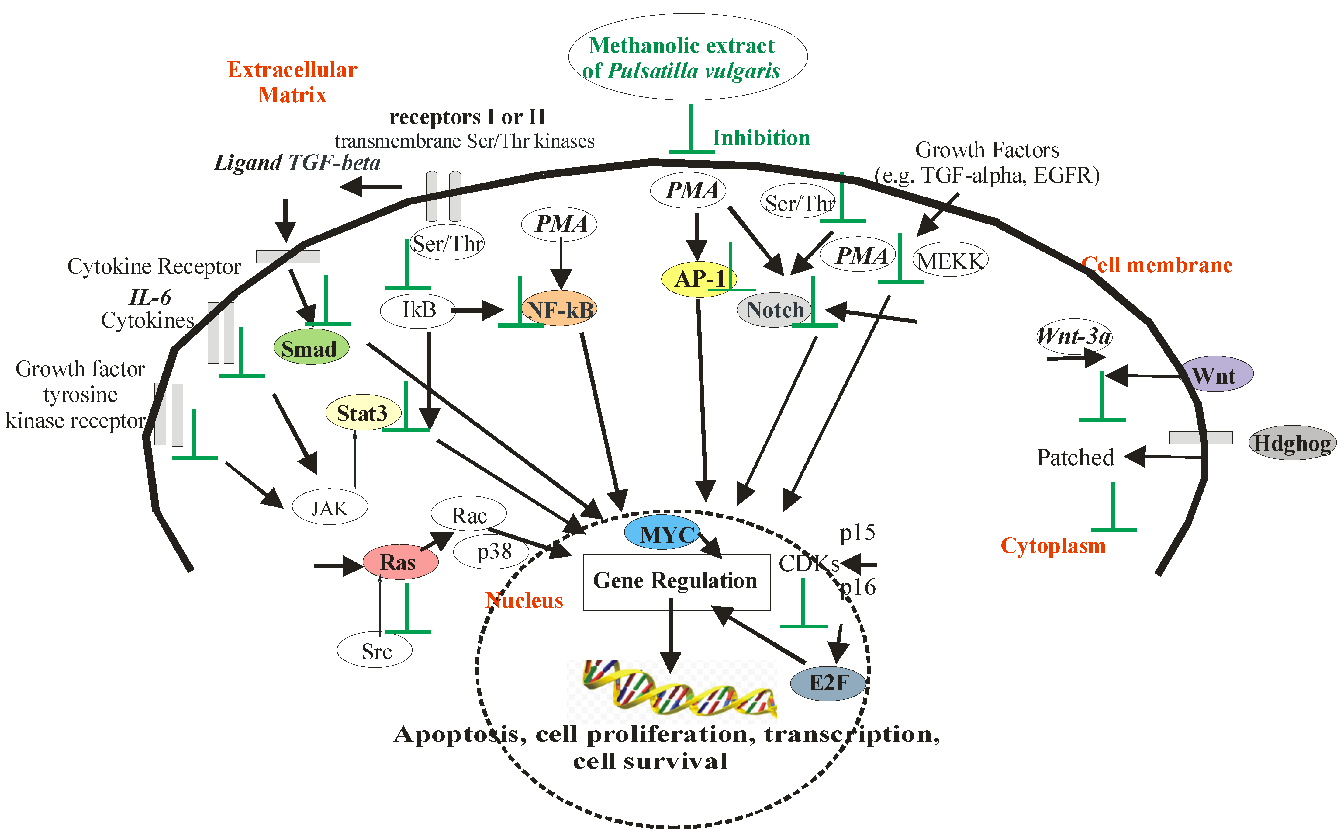
2.3. The Cancer Activity of P. vulgaris in Signaling Pathways in HeLa Cells

The antiproliferative activity of P. vulgaris extracts was assessed using a panel of 13 luciferase reporter gene vectors (Figure 3, Tables S1 and S2). The luciferase expression within each vector was driven by enhancer elements (inducers, promoters, such as IL6, TGF-β, PMA, and Wnt 3a) that bind to specific transcription factors [88]. In this study, the genus of firefly luciferase (Photinus pyralis, Pp Luc) was used. Pp Luc catalyzes the conversion of ATP, luciferin, and O2 into the products: AMP, PPi, CO2, H2O, oxyluciferin and light with a wavelength of 562 nm (Figure 4a). Firefly luciferase is in the form of a monomer, and its efficiency is much higher than that of bacterial luciferase (the marine bacteria Vibrio fischeri and Vibrio harveyi (lux) or the Renilla reniformis (sea pansy (Rr Luc) [89].
The results showed that the methanolic extract from the roots of P. vulgaris was more potent than the leaf extract of this species in inhibiting the activation of twelve pathways analyzed, except for only one, FoxO (Forkhead proteins) (Figure 3).
In Figure 3, the numerical value expresses the percentage ratio of the activity of the test samples to the activity of the tumor promoter determined for the protein encoded by the luciferase reporter gene. The lower the numerical value of the data, the activity of the test sample was greater in inhibiting Hela cell proliferation than that of the tumor promoter. The active compounds of methanolic extract from the roots of P. vulgaris were found to strongly inhibit the activation of Stat3, Smad, AP-1, NF-κB, E2F, MYC, Ets, Notch, Wnt, Hdghog, pTK—control, miR-21 and k-Ras signaling (Figure 3). Each of the apoptotic signaling mediators was inhibited stronger by compounds from root extract than from leaf extract. The methanolic extract from the roots of P. vulgaris was stronger in potency than the active antitumor compound resveratrol analog in each concentration studied for inhibition of signaling. The only exception was the apoptotic mediator FoxO, where the resveratrol analog was more effective than active compounds from both of extracts of P. vulgaris. The transcriptional activity of FoxO controls cell proliferation and apoptosis and regulates the cell cycle and apoptotic genes such as the cyclin-dependent kinase inhibitor (CKI) p27(KIP1) [91,92], Fas ligand [93], Bcl-6 [94] and Bim [95,96].
The active compounds of methanolic extract from the leaves of P. vulgaris, similarly to the resveratrol analog, inhibited the activation of Stat3 and k-Ras, but significantly less than it inhibited the activation of Wnt, NF-κB, MYC, Notch, Ets, Smad, Hdghog, Ets, pTK—control and AP-1 signaling (Figure 3).
The statistical analysis showed that the largest statistically significant differences for the methanolic extract from the roots of P. vulgaris were in inhibiting the activation of Notch, Myc, E2F, and Wnt. These transcription factors (except Wnt) were activated by phorbol 12-myristate-13-acetate, a protein kinase activator (PMA) (Figure 3). The Notch can also be activated by a specific serine/threonine (Ser/Thr) protein kinase [97], MYC by—the direct interaction with the acetyltransferases p300, and CBP [98], and E2F by—cyclins, cyclin-dependent kinases CDKs connected with the cyclin-dependent kinase inhibitors (pp. 15–16) [99].
Notch promotes proliferation signaling during neurogenesis, and its activity is inhibited by neuronal differentiation and tumor cell proliferation [97]. Notch signaling plays a vital role in regulating embryonic development and cellular processes during development and adult tissue renewal. It was found that the modulation of the Notch pathway is essential in controlling the fate of “cancer stem cells”. Mammalian cells contain four Notch receptors, the activity of which is used to study pathway dynamics in a normal and disease context. Factors that are inhibitors of Notch are being investigated as potential agents for the treatment of cancer [100].
In studies of the expression of cancer-related genes, it was found that abnormal expression of oncogenes may be responsible for carcinogenesis. Therefore, MYC (induced nuclear protein antigen) and other members of the proto-oncogene family (c-, L-, and N-myc) are central regulators of cell growth, and their deregulated expression is associated with many cancers [98]. Myc encoded by the MINA gene show elevated expression in up to 70% of all human malignancies. MYC is a pleiotropic transcription factor that controls the mediators of apoptosis and directly regulates target genes responsible for the proliferation and growth of cancer cells [101].
E2F are proteins from the family of transcription factors that are involved in regulating the cell cycle [99]. This family includes activators (E2F1, E2F2 and E2F3a) and inhibitors (E2F3b, E2F4-8). Among E2F transcriptional targets are cyclins, CDKs, replication proteins, and DNA repair. Some human tumors have concurrent tumor cell inactivation and E2F amplification and overexpression. That is why researchers propose that there are alternative tumor-promoting activities for the E2F family, which are independent of cell cycle regulation [99].
The WNT family of genes consists of structurally related genes encoding the secretion of proteins in signaling pathways involved in tumor formation. WNT gene clusters are recombination hot spots associated with carcinogenesis [102]. Among 19 WNT genes, the WNT3A gene is clustered in human chromosome 1q42, where during carcinogenesis, recombination results in chromosomal translocation, gene amplification, and deletion. Protein Wnt-3a is encoded by the WNT3A gene and might play key roles in the maintenance of cells in the undifferentiated proliferation stage through activation of the β-catenin—TCF signaling pathway [103].
In this research, it was found that methanolic extract from the root of P. vulgaris had a similar effect on the inhibition of HeLa cell proliferation in signal transduction pathways driven by the minimal thymidine kinase promoter (pTK—control) and by the transcription proteins miR-21 and K-Ras. The other signaling pathways were more significantly inhibited. MicroRNA 21 (miR-21), is a signaling pathway in the cell involved in the regulation of proteoglycans in cancer [104]. It is an RNA gene related to a class of microRNA (miRNA) genes active in the post-transcriptional regulation of gene expression, including mRNA stability and translation [105]. The k-Ras is a protein mainly involved in the regulation of cell division [106].
Stronger modulators of signaling pathways than pTK (control) against NF-κB, AP-1, Smad, Hdghog, Ets, and Stat3 were active compounds of methanolic extract from the roots of P. vulgaris (Figure 3). In our study, NF-κB (nuclear factor kappa-light-chain-enhancer of activated B cells) was activated by PMA (protein kinase activator). NF-κB can also be triggered by the enzyme IκB kinase and then translocated into the nucleus. NF-κB, a protein complex from the family of transcription factors, is present in the cytoplasm of each cell, where it plays a vital role in regulating the immune response to infection, and disturbances in its regulation are associated with tumors [107,108]. Activator protein 1 (AP-1), as a transcription factor in gene expression, controls proliferation, differentiation, and apoptosis and regulates factors of stress and growth, cytokines, and viral and bacterial infections [109]. In our study, Smad activated by TGF-β in the signaling pathway was connected with type I and type II receptors, which are transmembrane Ser/Thr kinases. Smad complexes accumulate in the nucleus and regulate target gene expression [110]. Therefore, they are essential in signaling and regulating cell functions during the whole life of the organism. Hdghog (synonyms: Hedgehog-Patched (Hh-Ptch), Hedgehog-Patched-Smoothened (Hh-Ptch-Smo)), in our modulation of cancer-related signaling pathways was activated by PMA (Figure 3). It is a transcription factor regulated by protein kinases, responsible for signal conveyance from the cell membrane into the nucleus (Figure 5). The Hdghog signal transduction pathway is important and was recently noted in the development of cancers in various organs, such as the brain, lung, mammary gland, prostate, and skin. Basal cell carcinoma, the most common form of cancerous malignancy, has the closest association with hedgehog signaling [111]. The Ets in our signaling pathways were activated by PMA, too (Figure 3). ETS (erythroblast transformation specific) belongs to the largest families of transcription factors to be associated with cancer [112]. Ets is a transcriptional activator that through changes in gene expression and their deregulation leads to carcinogenesis (cancer, tumor formation) and apoptosis (programmed cell death) [113]. It is also a key factor in angiogenesis, i.e., the neoplastic ability to form blood vessels within a neoplastic tumor that causes the tumor to further increase in mass [114]. In our study, STAT3 (signal transducers and activators of transcription) was activated by cytokine IL-6, which leads to its activation of Janus kinase (JAK) (Figure 5). Stat3 is phosphorylated by the receptor [115] and translocated to the nucleus, where it promotes prooncogenetic genes [116]. The inhibition of STAT3 in HeLa cell signaling caused by the methanolic extract of P. vulgaris is associated with the inhibition of growth and cell proliferation, and with apoptosis (Figure 5).
It was found that active compounds of methanolic extract from the root of P. vulgaris were more potent than those from the methanolic extract of leaves. We have also determined the dose–response influence of this extract on the cancer-related signaling pathways. P. vulgaris root extract at a concentration of 40 µg/mL was more potent in inhibiting the activation of six signaling pathways (Stat3, E2F, MYC, pTK, miR-21, and k-Ras), except only one—FoxO (Figure 6). At the concentration of 15 µg/mL, stronger activation of three cancer-related signaling pathways (NF-κB, Ets, Wnt) and the same influence on four signaling pathways (Smad, AP-1, Hdghog, and Notch) was observed. The P. vulgaris root methanolic extract at 10 µg/mL showed the lowest influence on all signaling pathways (Figure 6).
This is the first report describing the activity of constituents of methanolic extracts of P. vulgaris against different 13 signaling pathways of the cervical cancer-related HeLa cells. The vectors and inducers used in this study to assess the activity of the cancer-related signaling pathways were previously reported by other authors [44,86]. Similarly, resveratrol [(E)-3,5,4′-trihydroxystilbene], the parent compound of the analog used in these studies as a positive control, is used as a potential modulator of signal transduction pathways for cancer and carcinogenic response also in studies by other authors [44].
The performed analysis allowed us to tentatively identify fourteen triterpenoid saponins, sixteen phenolic acids, four coumarins, one flavonoid, and three fatty acids, which can contribute to the observed activity of P. vulgaris. Additionally, other plant secondary metabolites were studied for the inhibition of cancer-related signaling pathways [6,120]; however, their mechanism of action was related to induction of the oxidative stress in cells. In that way, they target redox reactions, and redox-sensitive cysteine [121] and additionally play a major role in reactive oxygen species (ROS) generation and the depletion of the antioxidant glutathione (GSH) [122].
The transcription factors (Stat3, Smad, AP-1, NF-κB, E2F, MYC, Ets, Notch, FoxO, Wnt, Hdghog, miR-21, k-Ras) play important roles in cancer-related signaling pathways by regulating cytological processes such as differentiation, cell death, cellular proliferation, behavior, oncogenic transformation and apoptosis [123]. Among cancer-associated genes and driver gene mutations identified, the vast belong to approximately 13+ different signal transduction pathways [6,44,86]. In our research, these vectors were used as signaling nodes for oncogenic pathways, which transduce intracellular and extracellular signals to the nucleus and control the expression of genes [124,125] responsible for physiological processes such as cell growth, proliferation, differentiation, positioning, migration, metabolism, and apoptosis [126,127]. The research found that uncontrolled cell division results from gene mutations in DNA, leading to unlimited proliferation (multiplication) of cancer cells and neoplastic processes (neoplasia, carcinogenesis) [128].
Herein, we report that P. vulgaris inhibits all of the tested signaling pathways with significantly higher potency than previously studied P. patens [6]. For example, in the case of k-Ras and Notch, the P. patens showed induction of luciferase of approx. 60%, whereas P. vulgaris completely inhibited those pathways. The action of methanolic extract P. vulgaris is based on the inhibition of mitosis in the HeLa cell cycle (Figure 5). The active compounds of P. vulgaris start the induction of apoptosis and cause deregulated cellular proliferation, which leads to apoptotic death of the HeLa cells and inhibition of the growth of cancer cells. The suggested influence of P. vulgaris on apoptosis was based on the studies of cancer-related signaling pathways in HeLa cells, being the primary goal of our research, and not on the direct measurements of apoptosis. We acknowledge that the proapoptotic activity of P. vulgaris requires additional research, such as measurements of cellular caspases (caspase-3, -7, -8 or -9) related to apoptosis, studies of gene mutations in DNA and DNA fragmentation and biological tests using fluorescence microscopy or flow cytometry. Our future scientific endeavors will focus on the isolation of bioactive molecules responsible for the observed influence on signaling pathways and detailed studies of their pro-apoptotic activity.

3. Materials and Methods

3.1. Plant Material

The leaves and roots of four years flowering individuals P. vulgaris subsp. vulgaris Mill. were collected in May 2016–2018 from cultivation conducted in the Podlasie Botanical Garden “The Herbal Corner”, Podlaskie Province, in Northeastern Poland. Plant material was identified by Prof. Grażyna Łaska from the Bialystok University of Technology. In Poland, the species P. vulgaris is currently extinct in Poland on natural sites [68]. It was only reported at four sites on the habitats of the pine forests before 1930 [129,130]. In June 2018 the plant material in the form of dry roots (59.3 g) and leaves (28.1 g) was extracted by accelerated solvent extraction (ASE) technique (SpeedExtractor E-916, Buchi) with 80% methanol. The extraction time was 30 min, the temperature was 100 °C and the pressure was 120 bar. After the evaporation of methanol using a rotor evaporator, a plant extract was obtained from roots (1.1 g) and leaves (0.95 g)

3.2. LC/ESI-QTOF-MS Procedure

The P. vulgaris samples were analyzed qualitatively by LC-ESI-QTOF-MS system composed of an Agilent 1200 Infinity chromatograph hyphenated to a 6530B Accurate-Mass Q-ToF mass spectrometer (Agilent Technologies, Santa Clara, CA, USA). After the chromatographic separation of P. vulgaris components on a Gemini® column (3 μm i.d. C18 with TMS endcapping, 110 Å, 100 × 2 mm), the detailed qualitative profiling was performed using soft, electrospray ionization (ESI) in negative mode. The LC/MS method parameters followed our previous report [131]. The MS data acquisition was performed using the MassHunter software (Agilent Technologies). The identification was carried out based on the UV and mass spectra obtained, in comparison with the fragmentation behavior of these compounds reported in scientific literature and records available in HMDB [78] and PubChem [76] databases.

3.3. Cell Cultures and Media

Cytotoxic activity in the laboratory of the National Center for Natural Products Research (NCNPR) of the University of Mississippi (MS, USA) was determined against five human cancer cell lines (HeLa, SK-OV-3, KB, SK-MEL, BT-549) and two noncancerous kidney cell lines (LLC-PK1 and VERO) (see Table 1). All cell lines were obtained from the American Type Culture Collection (ATCC, Rockville, MD, USA).
Separately, the cytotoxicity of P. vulgaris extracts was assessed against HeLa and VERO cell lines after 72 h incubation using the MTT-based assay. The VERO (ECACC, No. 84113001) and HeLa (ECACC, No. 93021013) cells were grown in T25 flasks and 96-well plates using MEM (Corning, Tewksbury, MA, USA). Cell media were supplemented with fetal bovine serum (FBS, Biowest) and antibiotics (Penicillin-Streptomycin Solution, Corning). Cells were passaged using media supplemented with 10% FBS, whereas the media used for experiments contained 2% serum only. All experiments with cell cultures were conducted at 37 °C in the 5% CO2 atmosphere (CO2 incubator, Panasonic Healthcare Co., Ltd., Tokyo, Japan). The DMSO (dimethyl sulfoxide) and 3-(4,5-dimethylthiazol-2-yl)-2,5-diphenyltetrazolium bromide (MTT) were purchased from Sigma, whereas phosphate-buffered saline (PBS) and trypsin were from Corning.

3.4. Cytotoxicity Testing

The panel of healthy and cancer cell lines (in vitro) for cytotoxicity of root and leaf extracts P. vulgaris and cell viability were determined by the neutral red method [43,44] in the NCNPR, University of Mississippi (University, MS, USA). The cytotoxicity of extracts was evaluated using the CC50 values and was tested for the viability of healthy cell lines (kidney fibroblast, kidney epithelial cells) and cancer cell lines (cervical carcinoma, ovarian carcinoma, epidermal carcinoma, skin melanoma, breast carcinoma) (see Table 1). In this assay, we used 96-well tissue culture-treated plates. The cells were seeded at a density of 25,000 cells/well and grown for 24 h. In the next step, samples of extract of P. vulgaris at different concentrations were added and cells were further incubated for 48 h. CC50 values were obtained from dose–response curves of percent growth inhibition against test concentrations. Doxorubicin was used as the control drug (a positive control), while DMSO was used as the negative (vehicle) control.
In a laboratory in Poland, the extracts obtained from P. vulgaris (PVL—leaves, PVR—roots) were dissolved in DMSO (50 mg/mL) and filtered through a syringe filter (pore diameter 0.2 µm) to obtain stock solutions used in further in vitro studies. Stock solutions were stored frozen until used in experiments.
The evaluation of cytotoxicity was carried out using an MTT microculture tetrazolium assay. Briefly, the VERO and HeLa cells were passaged into 96-well microculture plates in the concentration of 1.5 × 10⁵ cells/mL or 3 × 10⁵ cells/mL, respectively, in growth media. After overnight incubation, the growth media was removed, and serial dilutions of extract stock solution, ranging from 1000 to 0.98 µg/mL, were added, and plates were further incubated for 72 h. The cytotoxicity of DMSO used as a solvent for extract stock solutions was also evaluated. Afterward, all media was removed, the cells were washed with PBS, and 100 µL of cell media supplemented with 10% of MTT solution (5 mg/mL) was added to each well, and incubation continued for the next 4 h. Finally, 100 µL of SDS/DMF/PBS (14% SDS, 36% DMF, 50% PBS) solution per well was added, and after overnight incubation, the absorbance at 540 and 620 nm was measured using Synergy H1 Multi-Mode Microplate Reader (BioTek Instruments, Inc., Winooski, VT, USA) equipped with Gen5 software (ver. 3.09.07, BioTek Instruments, Inc., Winooski, VT, USA) and data were exported to the GraphPad Prism (ver. 8.0.1, GraphPad Software, San Diego, CA, USA) for further analysis. Based on the comparison of absorbance recorded for control and extract-treated cells, values of CC50 (concentrations of tested extract decreasing the viability of appropriate cell line by 50%) were calculated [132].

3.5. Transfection and Luciferase Assays

HeLa cell cultures bought in ATCC (Bethesda, MD, USA) were maintained in Dulbecco’s modified Eagle’s medium (DMEM) (Gibco Life Technologies, Grand Island, NY, USA) containing 10% fetal bovine serum (FBS, Atlanta Biologicals Inc., Atlanta, GA, USA). HeLa cells were plated in white opaque 384-well plates at a density of 4300 cells/well in 30 µL of growth medium supplemented with 10% FBS and 1% Pen/step. In the next step (the next day), the medium was aspirated and replaced with DMEM containing 10% FBS only. The cells were transfected with respective plasmids using an X-tremeGENE HP transfection reagent (Roche Applied Science, Indianapolis, IN, USA). The following panel of 13 inducible luciferase reporter gene vectors in this assay were used—STAT3, SMAD3/4, NF-κB, AP-1, Ets, E2F, MYC, Notch, FoxO, Wnt, Hedgehog, miR-21, k-Ras. Their expression is driven by enhancer elements (inducer, promoter). Luciferase reporter constructs were transfected into Hela cells [44]. Luciferases are enzymes encoded by genes, which catalyze the bioluminescent reaction of the transformation of the substrate—luciferin, which in the presence of ATP, Mg2+ ions, and molecular oxygen is oxidized and becomes excited, and then—returning to the initial state—causes light emission (Figure 4b). The emitted photons are counted with a luminometer. The total light emission is directly proportional to the luciferase activity in the test sample and allows the evaluation of the transcriptional activity of the reporter gene.
After 24 h of transfection, the test agents were added to the transfected cells, followed 30 min later by an inducing agent: IL-6 (50 ng/mL, R&D Systems, Inc., Minneapolis, MN, USA) for Stat3 [44], TGF-beta (5 ng/mL, R&D Systems, Inc., Minneapolis, MN, USA) for Smad [44], Wnt-3a (500 ng/mL, Peprotech Corporation, Rocky Hill, NJ, USA) for Wnt [44] and PMA (phorbol 12-myristate 13-acetate, 77 ng/mL, Sigma Chemical Company, St. Louis, MO, USA) for AP-1, NF-κB, E2F, MYC, Ets, Notch and Hdghog. No inducer was added for FoxO, miR-21, k-Ras, and pTK-control (thymidine kinase promoter). After 4 h (STAT3, SMAD3/4, NF-κB, AP-1, Ets, E2F, MYC) or 6 h (Notch, FoxO, Wnt, Hedgehog, miR-21, k-Ras) of induction, the cells were lysed by the addition of the One-Glo luciferase assay system (Promega Corporation, Madison, WI, USA). A Glomax Multi+ detection system with Instinct Software (Promega Corporation, Madison, WI, USA) was used for the light output detection. This luciferase assay determines if the test agent was able to inhibit the activation of cancer-related signaling pathways [44,88].

4. Conclusions

Our research confirmed the strong inhibition of 12 signaling pathways in HeLa cells exerted by methanolic extracts of P. vulgaris. In this study, it was found that the root extract of P. vulgaris is the most potent in inhibiting the activation of MYC, Notch, Wnt, E2F, Ets, Stat3, Smad, Hdghog, AP-1, NF-κB, k-Ras, miR-21, and pTK-control. This is the first study to report the influence of Pulsatilla vulgaris species on cancer signaling pathways. The methanolic extracts of P. vulgaris enhanced the apoptotic death of HeLa cells and deregulated cellular proliferation, differentiation, and progression toward the neoplastic phenotype by altering key signaling molecules required for cell cycle progression.
The results showed that P. vulgaris is a rich source of triterpenoid saponins and phenolic acids, wherein their triterpenoid saponins showed a different profile in comparison to other species from the Ranunculaceae family. Our research will be helpful to determine the relevance of each cancer-related signaling pathway that may be used to development of novel therapies that combine extracts of P. vulgaris with other agents including MYC, Notch, Wnt, E2F, Ets, STAT, SMAD, Hdghog, AP-1, and NF-κB blockers, to effectively treat cervical cancer, and other cancers that activate these pathways.
The study confirms the chemotherapeutic potency of P. vulgaris secondary metabolites in different types of cancer line cells and their activity at variable levels of cell signaling. Current trends in oncological pharmacology aim to use multidirectional therapies, which agrees with the pleiotropic character of extracts of P. vulgaris inhibiting cancer proliferation in signaling pathways of 12 reporter genes. However, there are still no preclinical and clinical studies that would clearly confirm the benefits of secondary metabolites of P. vulgaris in oncological therapy. The problem is the limitation of natural sites of P. vulgaris in the EU and the limited bioavailability of substances of natural origin. We need more toxicological studies of extracts of P. vulgaris and their secondary metabolites at the cellular, tissue, organ, and organism levels.

Supplementary Materials

The following supporting information can be downloaded at: https://www.mdpi.com/article/10.3390/ijms24021139/s1.

Author Contributions

Conceptualization, G.Ł.; methodology and investigation, G.Ł., E.S., M.M.-T., Ł.Ś., P.B. and D.S.P.; resources, G.Ł.; writing—original draft preparation and editing, G.Ł.; writing—review G.Ł., E.S. and Ł.Ś.; supervision, G.Ł.; project administration, G.Ł. All authors have read and agreed to the published version of the manuscript.

Funding

This research was funded by the MINISTRY OF EDUCATION AND SCIENCE OF POLAND, grant number WZ/WB-IIS/2/2020 to G.Ł. and in part by a contract from the University of Mississippi Medical Center Cancer Institute Agreement #64580110000 to P.B. and D.S.P.

Institutional Review Board Statement

Not applicable.

Informed Consent Statement

Not applicable.

Data Availability Statement

Not applicable.

Conflicts of Interest

The authors declare no conflict of interest.

Abbreviations

Transcription factors—Stat3, Smad, AP-1, NF-κB, E2F, MYC, Notch, Wnt, Hdghog, Ras; inducers—cytokines IL-6, ligand TGF-beta, PMA—protein kinase activator, phorbol 12-myristate-13-acetate, Wnt-3a protein; Ser/Thr—serine/threonine kinases, EGFR—epidermal growth factor receptor, STAT—signal transducer and activator of the transcription protein family, MEKK—mitogen-activated protein (MAP) kinase, NOTCH—neurogenic locus notch homolog protein, IkB—inhibitor of kB, JAK—Janus kinase, Src—protooncogene tyrosine-protein kinase, Rac—subfamily of the Rho family of GTPases, Ras—Ras family kinase, p38—mitogen-activated protein kinases, p15, p16—the cyclin-dependent kinase inhibitors, CDKs—cyclins, cyclin-dependent kinases, Patched—protein patched homolog.

References

  1. Wink, M.; Van Wyk, B.E. Mind-Altering and Poisonous Plants of the World. A Scientifically Accurate Guide to 1200 Toxic and Intoxicating Plants, 1st ed.; Timber Press: Portland, OR, USA, 2008; pp. 1–464. [Google Scholar]
  2. Jiangsu New Medicine College. Encyclopedia of Chinese Materia Medica, 1st ed.; Shanghai Science and Technology Press: Shanghai, China, 1977; pp. 1–294. [Google Scholar]
  3. Chinese Pharmacopoeia Commission. Pharmacopoeia of the People’s Republic of China, 1st ed.; People’s Medical Publishing House: Beijing, China, 2005; pp. 1–946.
  4. Kumar, S.; Madaan, R.; Farooq, A.; Sharma, A. The genus Pulsatilla: A review. Pharmacogn. Rev. 2008, 2, 116. [Google Scholar]
  5. Li, W.; Ding, Y.; Sun, Y.N.; Yan, X.T.; Yang, S.Y.; Choi, G.W.; Kim, E.-J.; Kang, H.K.; Kim, Y.H. Oleanane-type triterpenoid saponins from the roots of Pulsatilla koreana and their apoptosis-inducing effects on HL-60 human promyelocytic leukemia cells. Arch. Pharm. Res. 2013, 36, 768–774. [Google Scholar] [CrossRef]
  6. Łaska, G.; Maciejewska-Turska, M.; Sieniawska, E.; Świątek, Ł.; Pasco, D.S.; Balachandran, P. Extracts from Pulsatilla patens target cancer-related signaling pathways in HeLa cells. Sci. Rep. 2021, 11, 10654. [Google Scholar] [CrossRef]
  7. Hensen, I.; Oberprieler, C.; Wesche, K. Genetic structure, population size, and seed production of Pulsatilla vulgaris Mill. (Ranunculaceae) in Central Germany. Flora Morphol. Distrib. Funct. Ecol. Plants 2005, 200, 3–14. [Google Scholar] [CrossRef]
  8. Kaźmierczakowa, R.; Zarzycki, K.; Mirek, Z. Polish Red Data Book of Plants. Pteridophytes and Flowering Plants, 3rd ed.; Institute of Nature Conservation, Polish Academy of Sciences: Cracow, Poland, 2014; pp. 1–895. [Google Scholar]
  9. Łaska, G.; Sienkiewicz, A. Antifungal activity of the rhizome extracts of Pulsatilla vulgaris against Candida glabrata. Eur. J. Biol. Res. 2019, 9, 93–103. [Google Scholar]
  10. Zhang, Y.; Bao, J.; Wang, K.; Jia, X.; Zhang, C.; Huang, B.; Chen, M.; Wan, J.B.; Su, H.; Wang, Y.; et al. Pulsatilla saponin D inhibits autophagic flux and synergistically enhances the anticancer activity of chemotherapeutic agents against HeLa cells. Am. J. Chin. Med. 2015, 43, 1657–1670. [Google Scholar] [CrossRef]
  11. Łaska, G.; Sienkiewicz, A.; Stocki, M.; Sharma, V.; Zjawiony, J.K.; Jacob, M. Secondary metabolites from Pulsatilla patens and Pulsatilla vulgaris and their biological activity. Planta Med. 2015, 81, 122. [Google Scholar] [CrossRef]
  12. Łaska, G.; Sienkiewicz, A.; Stocki, M.; Zjawiony, J.K.; Sharma, V.; Bajguz, A.; Piotrowska-Niczyporuk, A.; Jacob, M.; Khan, S. Phytochemical screening of Pulsatilla species and investigation of their biological activities. Acta Soc. Bot. Pol. 2019, 88, 3613. [Google Scholar] [CrossRef] [Green Version]
  13. Ye, W.C.; Ji, N.N.; Zhao, S.X.; Che, C.T. A new cytotoxic saponin from Pulsatilla patens var. multifida. Pharm. Biol. 2001, 39, 7–10. [Google Scholar] [CrossRef] [Green Version]
  14. Ye, W.C.; Zhang, Q.W.; Hsiao, W.L.W.; Zhao, S.X.; Che, C.T. Novel cytotoxic lupane-type glycosides from Pulsatilla chinensis. Planta Med. 2002, 68, 183–186. [Google Scholar] [CrossRef]
  15. Shu, Z.; Chen, Z.; Liu, Y.L.; Zhu, W.F.; Feng, Y.L.; Xu, Q.M.; Li, X.R.; Yang, S.L. A new oleanane-type triterpenoidal saponin from Pulsatilla chinensis. Nat. Prod. Res. 2013, 27, 2196–2201. [Google Scholar] [CrossRef]
  16. Xu, Q.M.; Shu, Z.; He, W.J.; Chen, L.Y.; Yang, S.L.; Yang, G.; Liu, Y.L.; Li, X.R. Antitumor activity of Pulsatilla chinensis (Bunge) Regel saponins in human liver tumor 7402 cells in vitro and in vivo. Phytomedicine 2012, 19, 293–300. [Google Scholar] [CrossRef]
  17. Bi, Y.L.; Wang, B.; Huang, B.H.; Zhang, W.T.; Zhang, Y.H. Antifungal activity of botanical extracts against Botrytis cinerea and Alternaria solani. J. Agric. Sci. Technol. 2011, 12, 862–864. [Google Scholar]
  18. Ma, C. Treatment methods of traditional Chinese medicines against intestinal protozoan infections. In Treatment of Human Parasitosis in Traditional Chinese Medicine, 1st ed.; Mehlhorn, H., Zhongdao, W., Ye, B., Eds.; Springer: Berlin, Germany, 2014; Volume 6, pp. 11–21. [Google Scholar] [CrossRef]
  19. Li, W.C.; Shi, W.J.; Gu, Y.F.; Chen, H.L.; Chen, W.; Zhang, Y. Antibacterial effect of different extract of Pulsatilla chinensis (Bunge) Regel in vitro. J. Tradit. Chin. Vet. Med. 2011, 2, 38–40. [Google Scholar]
  20. Li, L.D.; Li, W.C.; Liu, C.W.; Shi, W.J.; Gong, P.T.; Li, J.H.; Zhang, G.C.; Yang, J.; Li, H.; Zhang, X.C. Giardia intestinalis: Effects of Pulsatilla chinensis extracts on trophozoites. Parasitol. Res. 2012, 111, 1929–1935. [Google Scholar] [CrossRef]
  21. Chen, Y.Q.; Xu, Q.M.; Liu, Y.L.; Li, X.R.; Yang, S.L.; Zhuge, H.X. Laboratory evaluation of the molluscicidal activity of Pulsatilla chinensis (Bunge) Regel saponins against the snail Oncomelania hupensis. Biomed. Environ. Sci. 2012, 25, 224–229. [Google Scholar] [CrossRef]
  22. Kim, Y.; Bang, S.C.; Lee, J.H.; Ahn, B.Z. Pulsatilla saponin D: The antitumor principle from Pulsatilla koreana. Arch. Pharm. Res. 2004, 27, 915–918. [Google Scholar] [CrossRef]
  23. Yang, H.; Cho, Y.W.; Kim, S.H.; Kim, Y.C.; Sung, S.H. Triterpenoidal saponins of Pulsatilla koreana roots. Phytochemistry 2010, 71, 1892–1899. [Google Scholar] [CrossRef]
  24. Yoo, H.H.; Lee, S.K.; Lim, S.Y.; Kim, Y.; Kang, M.J.; Kim, E.J.; Park, Y.H.; Im, G.J.; Lee, B.Y.; Kim, D.H. LC-MS/MS method for determination of hederacolchiside E, a neuroactive saponin from Pulsatilla koreana extract in rat plasma for pharmacokinetic study. J. Pharm. Biomed. Anal. 2008, 48, 1425–1429. [Google Scholar] [CrossRef]
  25. Bang, S.C.; Kim, Y.; Lee, J.H.; Ahn, B.Z. Triterpenoid saponins from the roots of Pulsatilla koreana. J. Nat. Prod. 2005, 68, 268–272. [Google Scholar] [CrossRef]
  26. Kim, Y.; Kim, S.B.; You, Y.J.; Ahn, B.Z. Deoxypodophyllotoxin; the cytotoxic and antiangiogenic komponent from Pulsatilla koreana. Planta Med. 2002, 68, 268–271. [Google Scholar] [CrossRef] [PubMed]
  27. Chung, S.W.; Chung, C.H.; Lim, S.B.; Kim, J.K.; So, E.H. The antimicrobial effect of Pulsatilla koreana extracts to oral micro-organism. J. Korean Acad. Periodontol. 2000, 30, 661. [Google Scholar] [CrossRef] [Green Version]
  28. Li, W.; Sun, Y.N.; Yan, X.Y.; Yang, S.Y.; Lee, S.J.; Byun, H.J.; Moon, C.S.; Han, B.S.; Kim, Y.H. Isolation of nematicidal triterpenoid saponins from Pulsatilla koreana root and their activities against Meloidogyne incognita. Molecules 2013, 18, 5306–5316. [Google Scholar] [CrossRef] [PubMed]
  29. Cheon, S.A.; Choi, B.K.; Jeong, C.S.; Li, D.W.; Lee, E.B. The anti-inflammatory and analgesic actions of the fractions from Pulsatilla koreana root extract. Korean J. Clin. Pharm. 2000, 31, 174–184. [Google Scholar]
  30. Schrenk, D. Feasibility study of nonclinical safety assessments on homeopathic preparations using the example of protoanemonin in Pulsatilla pratensis L. Regul. Toxicol. Pharmacol. 2013, 66, 104–108. [Google Scholar] [CrossRef] [PubMed]
  31. Samadder, A.; Das, J.; Das, S.; Khuda-Bukhsh, A.R. Dihydroxy-isosteviol-methyl-ester, an active biological component of Pulsatilla nigricans, reduces arsenic induced cellular dysfunction in testis of male mice. Environ. Toxicol. Pharmacol. 2012, 34, 743–752. [Google Scholar] [CrossRef]
  32. Fan, W.; Liu, J.; Gong, Y.; Ma, J.; Zhou, N.; Xu, Y. A new triterpenoid saponin from Pulsatilla cernua. Nat. Prod. Sci. 2013, 19, 150–154. [Google Scholar]
  33. Zhang, Q.W.; Ye, W.C.; Che, C.T.; Zhao, S.X. Triterpene saponins from Pulsatilla cernua. Acta Pharm. Sin. 2000, 35, 756–759. [Google Scholar]
  34. Xu, H.J.; Shi, X.W.; Ji, X.; Du, Y.F.; Zhu, H.; Zhang, L.T. A rapid method for simultaneous determination of triterpenoid saponins in Pulsatilla turczaninovii using microwave-assisted extraction and high performance liquid chromatography-tandem mass spectrometry. Food Chem. 2012, 135, 251–258. [Google Scholar] [CrossRef]
  35. Sun, H.; Wang, Y.; Zhang, X.Q.; Zhao, S.X.; Ye, W.C. Chemical constituents of Pulsatilla dahurica. Chem. Nat. Compd. 2009, 45, 764–765. [Google Scholar] [CrossRef]
  36. Li, H.B.; Wong, C.C.; Cheng, K.W.; Chen, F. Antioxidant properties in vitro and total phenolic contents in methanol extracts from medicinal plants. LWT—Food Sci. Technol. 2008, 41, 385–390. [Google Scholar] [CrossRef]
  37. Lee, H.S.; Beon, M.S.; Kim, M.K. Selective growth inhibitor toward human intestinal bacteria derived from Pulsatilla cernua root. J. Agric. Food Chem. 2001, 49, 4656–4661. [Google Scholar] [CrossRef] [PubMed]
  38. Dai, L.; Wang, H.; Chen, Y. The immune-enhancing effect of PcG-A-a glycoprotein isolated from dried root of Pulsatilla chinensis (Bunge) Regel. Chin. J. Biochem. Pharm. 2000, 21, 230–231. [Google Scholar]
  39. Liu, J.Y.; Guan, Y.L.; Zou, L.B.; Gong, Y.X.; Hua, H.M.; Xu, Y.N.; Zhang, H.; Yu, Z.G.; Fan, W.H. Saponins with neuroprotective effects from the roots of Pulsatilla cernua. Molecules 2012, 17, 5520–5531. [Google Scholar] [CrossRef] [PubMed] [Green Version]
  40. Seo, J.S.; Yun, J.H.; Baek, I.S.; Leem, Y.H.; Kang, H.W.; Cho, H.K.; Lyu, Y.S.; Son, H.J.; Han, P.L. Oriental medicine Jangwonhwan reduces Aβ(1–42) level and β-amyloid deposition in the brain of Tg- APPswe/PS1dE9 mouse model of Alzheimer disease. J. Ethnopharmacol. 2010, 128, 206–212. [Google Scholar] [CrossRef]
  41. Chen, Z.; Guan, Y.; Zhou, L.; Xu, Y.; Yang, M.; Liu, H. Preparation and characterization of colon-targeted particles of Pulsatilla chinensis saponins. Nat. Prod. Commun. 2015, 10, 237–238. [Google Scholar]
  42. Xu, K.; Shu, Z.; Xu, Q.M.; Liu, Y.L.; Li, X.R.; Wang, Y.L.; Yang, S.L. Cytotoxic activity of Pulsatilla chinensis saponins and their structure-activity relationship. J. Asian Nat. Prod. Res. 2013, 15, 680–686. [Google Scholar] [CrossRef]
  43. Borenfreund, E.; Babich, H.; Martin-Alguacil, N. Rapid chemosensitivity assay with human normal and tumor cells in vitro. In Vitro Cell Dev. Biol. 1990, 26, 1030. [Google Scholar] [CrossRef]
  44. Zaki, M.A.; Balachandran, P.; Khan, S.; Wang, M.; Mohammed, R.; Hetta, M.H.; Pasco, D.S.; Muhammad, I. Cytotoxicity and modulation of cancer-related signaling by (Z)- and (E)-3,4,3′,5′-tetramethoxystilbene isolated from Eugenia brigida. J. Nat. Prod. 2013, 76, 679–684. [Google Scholar] [CrossRef]
  45. Fotakis, G.; Timbrell, J.A. In vitro cytotoxicity assays: Comparison of LDH, neutral red, MTT and protein assay in hepatoma cell lines following exposure to cadmium chloride. Toxicol. Lett. 2006, 160, 171–177. [Google Scholar] [CrossRef]
  46. Weyermann, J.; Lochmann, D.; Zimmer, A. A practical note on the use of cytotoxicity assays. Int. J. Pharm. 2005, 288, 369–376. [Google Scholar] [CrossRef] [PubMed]
  47. Danova, K.; Bertoli, A.; Pistelli, L.; Dimitrow, D.; Pistelli, L. In vitro culture of Balkan endemic and rare Pulsatilla species for conservational purposes and secondary metabolites production. Bot. Serb. 2009, 33, 157–162. [Google Scholar]
  48. Xu, H.J.; Shi, X.W.; Ji, X.; Du, Y.F.; Zhu, H.; Zhang, L.T. Qualitative and quantitative determination of nine main active constituents in Pulsatilla cernua by high-performance liquid chromatography coupled to electrospray ionization tandem mass spectrometry. J. Sep. Sci. 2011, 34, 308–316. [Google Scholar] [CrossRef] [PubMed]
  49. Goyal, S.; Kumar, S. Anti-anxiety activity studies of various extracts of Pulsatilla nigricans Stoerck. Int. J. Pharm. Sci. Drug Res. 2010, 2, 291–293. [Google Scholar]
  50. Nikolova, M.; Nikolov, S.; Koeva, Y. Flavonoids in the overground part of Pulsatilla slaviankae (Rumm.) D. Yord. et Koz. (Ranunculaceae). Phytol. Balc. 1998, 4, 165–170. [Google Scholar]
  51. Nikolova, M.; Asenov, A. Surface flavonoid aglycones in newly studied plant species. Nat. Prod. Res. 2006, 20, 103–106. [Google Scholar] [CrossRef]
  52. Luft, G. The distribution of saponins and tannins in the plant. Mon. Chem. 1926, 47, 259–284. [Google Scholar] [CrossRef]
  53. Raymond-Hamet, L. Pharmacodynamic properties of Anemone pulsatilla. Bull. Sci. Pharmacol. 1927, 34, 143–151. [Google Scholar]
  54. Kroeber, L. On the pharmacology and therapeutic use of the anemonine drugs from Ranunculaceous plants. Pharmazie 1949, 4, 181–190. [Google Scholar]
  55. Walker, K.J.; Pinches, C.E. Reduced grazing and the decline of Pulsatilla vulgaris Mill. (Ranunculaceae) in England, UK. Biol. Conserv. 2011, 144, 3098–3105. [Google Scholar] [CrossRef]
  56. Piqueray, J.; Saad, L.; Bizoux, J.P.; Mahy, G. Why some species cannot colonise restored habitats? The effects of seed and microsite availability. J. Nat. Conserv. 2013, 21, 189–197. [Google Scholar] [CrossRef]
  57. Kricsfalusy, V. Variations in the life cycle of Anemone patens L. (Ranunculaceae) in wild populations of Canada. Plants 2016, 5, 29. [Google Scholar] [CrossRef] [PubMed] [Green Version]
  58. Pfeifer, E.; Holderegger, R.; Matthies, D.; Rutishauser, R. Investigation on the population biology of a flagship species of dry meadows: Pulsatilla vulgaris Mill. in north-eastern Switzerland. Bot. Helv. 2002, 112, 153–172. [Google Scholar]
  59. Liu, D.D.; Chao, W.M.; Turgeon, R. Transport of sucrose, not hexose, in the phloem. J. Exp. Bot. 2012, 63, 4315–4320. [Google Scholar] [CrossRef]
  60. DiLeo, M.F.; Graf, R.; Holderegger, R.; Rico, Y.; Wagner, H.H. Highly polymorphic microsatellite markers in Pulsatilla vulgaris (Ranunculaceae) using next-generation sequencing. Appl. Plant Sci. 2015, 3, 1500031. [Google Scholar] [CrossRef]
  61. Niklfeld, H.; Schratt-Ehrendorfer, L. Red List of Threatened Plants of Austria, 1st ed.; Grüne Reihe; Bundesministerium für Umwelt, Jugend und Familie: Vienna, Austria, 1999; pp. 1–291.
  62. Ludwig, G.; Schnittler, M. Red List of Threatened Plants in Germany, 1st ed.; Bundesamt für Naturschutz: Bonn, Germany, 1996; pp. 1–72.
  63. Moser, D.; Gygax, A.; Bäumler, B.; Wyler, N.; Palese, R. Red List of the Threatened Ferns and Flowering Plants of Switzerland, 1st ed.; Bundesamt für Umwelt, Wald und Landschaft, Zentrum des Datenverbundnetzes der Schweizer Flora: Bern, Switzerland, 2002; pp. 1–120.
  64. IUCN. The IUCN Red List of Threatened Species, Version 2021. Available online: http://www.iucnredlist.org/ (accessed on 10 August 2021).
  65. Cheffings, C.; Farrell, L. The Vascular Plant Red Data List for Great Britain, 1st ed.; Joint Nature Conservation Committee: Peterborough, UK, 2005; pp. 1–120.
  66. Gärdenfors, U. The 2010 Red List of Swedish Species, 1st ed.; ArtDatabanken: Uppsala, Sweden, 2010. [Google Scholar]
  67. Witkowski, Z.J.; Król, W.; Solarz, W. Carpathian List of Endangered Species, 1st ed.; WWF and Institute of Nature Conservation, Polish Academy of Sciences: Vienna, Austria, 2003; pp. 1–84. [Google Scholar]
  68. Piękoś-Mirkowa, H.; Zając, A.; Zając, M. Flowering Plants and Pteridophytes of Poland. A Checklist; W. Szafer Institute of Botany, Polish Academy of Sciences: Cracow, Poland, 2002; pp. 1–442. [Google Scholar]
  69. Thienthiti, K.; Tuchind, P.; Wongnoppavich, A.; Anantachoke, N.; Soonthornchareonnon, N. Cytotoxic effect of compounds isolated from Goniothalam usmarcanii Craib stem barks. Pharm. Sci. Asia 2017, 44, 86–95. [Google Scholar] [CrossRef]
  70. Figueiró, L.R.; Comerlato, L.C.; Da Silva, M.V.; Zuanazzi, J.; Von Poser, G.L.; Ziulkoski, A.L. Toxicity of Glandularia selloi (Spreng.) Tronc. leave extract by MTT and neutral red assays: Influence of the test medium procedure. Interdiscip. Toxicol. 2016, 9, 25–29. [Google Scholar] [CrossRef] [PubMed] [Green Version]
  71. Zuang, V. The Neutral Red Release Assay: A Review. Altern. Lab. Anim. 2001, 29, 575–599. [Google Scholar] [CrossRef]
  72. Jin, M.-M.; Zhang, W.-D.; Jiang, H.-H.; Du, Y.-F.; Guo, W.; Cao, L.; Xu, H.-J. UPLC-Q-TOF-MS/MS-guided dereplication of Pulsatilla chinensis to identify triterpenoid saponins. Phytochem. Anal. 2018, 29, 516–527. [Google Scholar] [CrossRef]
  73. Zhang, W.; Jiang, H.; Yang, J.; Song, G.; Wen, D.; Liu, W.; Jin, M.; Wang, Q.; Du, Y.; Sun, Q.; et al. A high-throughput metabolomics approach for the comprehensive differentiation of four Pulsatilla Adans herbs combined with a nontargeted bidirectional screen for rapid identification of triterpenoid saponins. Anal. Bioanal. Chem. 2019, 411, 2071–2088. [Google Scholar] [CrossRef]
  74. Ling, Y.; Lin, Z.; Zha, W.; Lian, T.; You, S. Rapid detection and characterisation of triterpene saponins from the root of Pulsatilla chinensis (Bunge) Regel by HPLC-ESI-QTOF-MS/MS. Phytochem. Anal. 2016, 27, 174–183. [Google Scholar] [CrossRef]
  75. Lee, K.Y.; Cho, Y.W.; Park, J.; Lee, D.Y.; Kim, S.H.; Kim, Y.C.; Sung, S.H. Quality control of Pulsatilla koreana based on the simultaneous determination of triterpenoidal saponins by HPLC-ELSD and principal component analysis. Phytochem. Anal. 2010, 21, 314–321. [Google Scholar] [CrossRef] [PubMed]
  76. Pubchem Database. Available online: https://pubchem.ncbi.nlm.nih.gov/ (accessed on 10 September 2022).
  77. Zhao, M.; Ding, W.; Wang, S.; Wang, C.; Du, Y.; Xu, H.; Wang, Q.; Jin, S. Simultaneous determination of nine coumarins in rat plasma by HPLC-MS/MS for pharmacokinetics studies following oral administration of Fraxini Cortex extract. J. Chromatogr. B Anal. Technol. Biomed. Life Sci. 2016, 1025, 25–32. [Google Scholar] [CrossRef] [PubMed]
  78. HMDB Human Metabolome Database. Available online: https://hmdb.ca/ (accessed on 10 September 2022).
  79. Hossain, M.B.; Rai, D.K.; Brunton, N.P.; Martin-Diana, A.B.; Barry-Ryan, A.C. Characterization of phenolic composition in lamiaceae spices by LC-ESI-MS/MS. J. Agric. Food Chem. 2010, 58, 10576–10581. [Google Scholar] [CrossRef] [PubMed]
  80. Usui, A.; Matsuo, Y.; Tanaka, T.; Ohshima, K.; Fukuda, S.; Mine, T.; Nakayama, H.; Ishimaru, K. Ferulic acid esters of oligo-glucose from Allium macrostemon. Nat. Prod. Commun. 2017, 12, 89–91. [Google Scholar] [CrossRef] [Green Version]
  81. Zeb, A.; Haq, A.; Murkovic, M. Effects of microwave cooking on carotenoids, phenolic compounds and antioxidant activity of Cichorium intybus L. (chicory) leaves. Eur. Food Res. Technol. 2019, 245, 365–374. [Google Scholar] [CrossRef]
  82. Mulinacci, N.; Innocenti, M.; Gallori, S.; Romani, A.; La Marca, G.; Vincieri, F.F. Optimization of the chromatographic determination of polyphenols in the aerial parts of Cichorium intybus L. Chromatographia 2001, 54, 455–461. [Google Scholar] [CrossRef]
  83. Said, R.B.; Hamed, A.I.; Mahalel, U.A.; Al-Ayed, A.S.; Kowalczyk, M.; Moldoch, J.; Oleszek, W.; Stochmal, A. Tentative characterization of polyphenolic compounds in the male flowers of Phoenix dactylifera by liquid chromatography coupled with mass spectrometry and DFT. Int. J. Mol. Sci. 2017, 18, 512. [Google Scholar] [CrossRef]
  84. Mahomoodally, M.F.; Sieniawska, E.; Sinan, K.I.; Nancy Picot-Allain, M.C.; Yerlikaya, S.; Cengiz Baloglu, M.; Altunoglu, Y.C.; Senkardes, I.; Rengasamy, K.R.; Zengin, G. Utilisation of Rhododendron luteum Sweet bioactive compounds as valuable source of enzymes inhibitors, antioxidant, and anticancer agents. Food Chem. Toxicol. 2020, 135, 111052. [Google Scholar] [CrossRef]
  85. Tardugno, R.; Pozzebon, M.; Beggio, M.; Del Turco, P.; Pojana, G. Polyphenolic profile of Cichorium intybus L. endemic varieties from the Veneto region of Italy. Food Chem. 2018, 266, 175–182. [Google Scholar] [CrossRef] [Green Version]
  86. Shumova, G.; Savelieva, E.V.; Vladymyrova, I.M.; Tishakova, T.S. Phenolic compound composition of herb of Pulsatilla pratensis (L.) Mill. Vet. Sci. Pharm. 2017, 4, 35–38. [Google Scholar]
  87. Zhao, Z.; Nian, M.; Qiao, H.; Li, B.; Zheng, X. Pulsatilla chinensis: A review of traditional uses, phytochemistry and pharmacology research progress. Arab. J. Chem. 2021, 14, 103403. [Google Scholar] [CrossRef]
  88. Hamilton, K.L.; Sheehan, S.A.; Retzbach, E.P.; Timmerman, C.A.; Gianneschi, G.B.; Tempera, P.J.; Balachandran, P.; Goldberg, G.S. Effects of Maackia amurensis seed lectin (MASL) on oral squamous cell carcinoma (OSCC) gene expression and transcriptional signaling pathways. J. Cancer Res. Clin. Oncol. 2021, 147, 445–457. [Google Scholar] [CrossRef] [PubMed]
  89. Allard, S.T.M.; Kopish, K. Luciferase reporter assays: Powerful, adaptable tools for cell biology research. Cell Notes 2008, 21, 23–26. [Google Scholar]
  90. Allard, S.T.M. Bioluminescent Reporter Genes. Adv. Biochem. 2008, 54, 350–353. [Google Scholar]
  91. Cappellini, A.; Tabellini, G.; Zweyer, M.; Bortul, R.; Tazzari, P.L.; Billi, A.M.; Fala, F.; Cocco, L.; Martelli, A.M. The phosphoinositide 3-kinase/Akt pathways regulates cell cycle progression of HL60 human leukemia cells through cytoplasmic relocalization of the cyclin-dependent kinase inhibitor p27 (Kip1) and control of cyclin D1 expression. Leukemia 2003, 17, 2157–2167. [Google Scholar] [CrossRef] [Green Version]
  92. Burgering, B.M.; Kops, G.J. Cell cycle and death control: Long live Forkheads. Trends Biochem. Sci. 2002, 27, 352–360. [Google Scholar] [CrossRef]
  93. Brunet, A.; Bonni, A.; Zigmond, M.J.; Lin, M.Z.; Juo, P.; Hu, L.S.; Anderson, M.J.; Arden, K.C.; Blenis, J.; Greenberg, M.E. Akt promotes cell survival by phosphorylating and inhibiting a Forkhead transcription factor. Cell 1999, 96, 857–868. [Google Scholar] [CrossRef] [Green Version]
  94. Tang, T.T.; Dowbenko, D.; Jackson, A.; Toney, L.; Lewin, D.A.; Dent, A.L.; Lasky, L.A. The forkhead transcription factor AFX activates apoptosis by induction of the BCL-6 transcriptional repressor. J. Biol. Chem. 2002, 277, 14255–14265. [Google Scholar] [CrossRef] [Green Version]
  95. Dijkers, P.F.; Medema, R.H.; Lammers, J.W.; Koenderman, L.; Coffer, P.J. Expression of the pro-apoptotic Bcl-2 family member Bim is regulated by the forkhead transcription factor FKHR-L1. Curr. Biol. 2000, 10, 1201–1204. [Google Scholar] [CrossRef] [Green Version]
  96. Gilley, J.; Coffer, P.J.; Ham, J. FOXO transcription factors directly activate bim gene expression and promote apoptosis in sympathetic neurons. J. Cell Biol. 2003, 162, 613–622. [Google Scholar] [CrossRef] [PubMed]
  97. Rizzo, P.; Osipo, C.; Foreman, K.; Golde, T.; Osborne, B.; Miele, L. Rational targeting of Notch signaling in cancer. Oncogene 2008, 27, 5124–5131. [Google Scholar] [CrossRef] [Green Version]
  98. Cavin, L.G.; Wang, F.; Factor, V.M.; Kaur, S.; Venkatraman, M.; Thorgeirsson, S.S.; Arsura, M. Transforming growth factoralpha inhibits the intrinsic pathway of c-Myc-induced apoptosis through activation of nuclear factor-kappaB in murine hepatocellular carcinomas. Mol. Cancer Res. 2005, 3, 403–412. [Google Scholar] [CrossRef] [PubMed] [Green Version]
  99. Chen, H.Z.; Tsai, S.Y.; Leone, G. Emerging roles of E2Fs in cancer: An exit from cell cycle control. Nat. Rev. Cancer 2009, 9, 785–797. [Google Scholar] [CrossRef] [PubMed] [Green Version]
  100. Viatour, P.; Ehmer, U.; Saddic, L.A.; Dorrell, C.; Andersen, J.B.; Lin, C.; Zmoos, A.F.; Mazur, P.K.; Schaffer, B.E.; Ostermeier, A.; et al. Notch signaling inhibits hepatocellular carcinoma following inactivation of the RB pathway. J. Exp. Med. 2011, 208, 1963–1976. [Google Scholar] [CrossRef] [PubMed] [Green Version]
  101. Chanvorachote, P.; Sriratanasak, N.; Nonpanya, N. C-myc contributes to malignancy of lung cancer. A potential anticancer drug target. Anticancer Res. 2020, 40, 609–618. [Google Scholar] [CrossRef]
  102. Katoh, M. Regulation of WNT3 and WNT3A mRNAs in human cancer cell lines NT2, MCF-7, and MKN45. Int. J. Oncol. 2002, 20, 373–377. [Google Scholar] [CrossRef]
  103. Maubant, S.; Tesson, B.; Maire, V.; Ye, M.; Rigaill, G.; Gentien, D.; Cruzalegui, F.; Tucker, G.C.; Roman-Roman, S.; Dubois, T. Transcriptome Analysis of Wnt3a-Treated Triple-Negative Breast Cancer Cells. PLoS ONE 2015, 10, e0122333. [Google Scholar] [CrossRef] [Green Version]
  104. Zheng, J.; Xue, H.; Wang, T.; Jiang, Y.; Liu, B.; Li, J.; Liu, Y.; Wang, W.; Zhang, B.; Sun, M. MiR-21 downregulates the tumor suppressor P12 CDK2AP1 and stimulates cell proliferation and invasion. J. Cell. Biochem. 2011, 112, 872–880. [Google Scholar] [CrossRef]
  105. Asangani, I.A.; Rasheed, S.A.K.; Nikolova, D.A.; Leupold, J.H.; Colburn, N.H.; Post, S.; Allgayer, H. MicroRNA-21 (miR-21) post-transcriptionally downregulates tumor suppressor Pdcd4 and stimulates invasion, intravasation and metastasis in colorectal Cancer. Oncogene 2008, 27, 2128–2136. [Google Scholar] [CrossRef] [Green Version]
  106. Medema, R.H.; Kops, G.J.; Bos, J.L.; Burgering, B.M. AFX-like Forkhead transcription factors mediate cell-cycle regulation by Ras and PKB through p27kip1. Nature 2000, 404, 782–787. [Google Scholar] [CrossRef]
  107. Nouri, M.; Massah, S.; Caradec, J.; Lubik, A.A.; Li, N.; Truong, S.; Lee, A.R.; Fazli, L.; Ramnarine, V.R.; Lovnicki, J.M.; et al. Transient Sox9 exspression facilitates resistance to androgen-targeted therapy in prostate cancer. Clin. Cancer Res. 2020, 26, 1678–1689. [Google Scholar] [CrossRef] [PubMed] [Green Version]
  108. Vlahopoulos, S.A. Aberrant control of NF-κB in cancer permits transcriptional and phenotypic plasticity, to curtail dependence on host tissue: Molecular mode. Cancer Biol. Med. 2017, 14, 254–270. [Google Scholar] [CrossRef] [PubMed] [Green Version]
  109. Tewari, D.; Nabavi, S.F.; Nabavi, S.M.; Sureda, A.; Farooqi, A.A.; Atanasov, A.G.; Vacca, R.A.; Sethi, G.; Bishayee, A. Targeting activator protein 1 signaling pathway by bioactive natural agents: Possible therapeutic strategy for cancer prevention and intervention. Pharmacol. Res. 2018, 128, 366–375. [Google Scholar] [CrossRef] [PubMed]
  110. Massagué, J. TGF-beta in cancer. Cell 2008, 134, 215–230. [Google Scholar] [CrossRef] [PubMed] [Green Version]
  111. Skoda, A.M.; Simovic, D.; Karin, V.; Kardum, V.; Vranic, S.; Serman, L. The role of the Hedgehog signaling pathway in cancer: A comprehensive review. Bosn. J. Basic Med. Sci. 2018, 18, 8–20. [Google Scholar] [CrossRef] [Green Version]
  112. Qi, T.; Qu, Q.; Li, G.; Wang, J.; Zhu, H.; Yang, Z.; Sun, Y.; Lu, Q.; Qu, J. Function and regulation of the PEA3 subfamily of ETS transcription factors in cancer. Am. J. Cancer Res. 2020, 10, 3083–3105. [Google Scholar]
  113. Oikawa, T.; Yamada, T. Molecular biology of the Ets family of transcription factors. Gene 2003, 303, 11–34. [Google Scholar] [CrossRef]
  114. Hollenhorst, P.C.; McIntosh, L.P.; Graves, B.J. Genomic and biochemical insights into the specificity of ETS transcription factors. Annu. Rev. Biochem. 2011, 80, 437–471. [Google Scholar] [CrossRef] [Green Version]
  115. Tkach, M.; Rosemblit, C.; Rivas, M.A.; Proietti, C.J.; Díaz Flaqué, M.C.; Mercogliano, M.F.; Beguelin, W.; Maronna, E.; Guzmán, P.; Gercovich, F.G.; et al. P42/p44 MAPK-mediated Stat3Ser727 phosphorylation is required for progestin-induced full activation of Stat3 and breast cancer growth. Endocr.-Relat. Cancer 2013, 20, 197–212. [Google Scholar] [CrossRef] [Green Version]
  116. Klampfer, L. Signal transducers and activators of transcription (STATs): Novel targets of chemopreventive and chemotherapeutic drugs. Curr. Cancer Drug Targets 2006, 6, 107–121. [Google Scholar] [CrossRef] [PubMed]
  117. Mirzaei, H.; Naseri, G.; Rezaee, R.; Mohammadi, M.; Banikazemi, Z.; Mirzaei, H.R.; Salehi, H.; Peyvandi, M.; Pawelek, J.M.; Sahebkar, A. Curcumin: A new candidate for melanoma therapy? Int. J. Cancer 2016, 139, 1683–1695. [Google Scholar] [CrossRef] [PubMed]
  118. Bhattacharyya, S.; Mandal, D.; Saha, B.; Sen, G.S.; Das, T.; Sa, G. Curcumin prevents tumor-induced T cell apoptosis through Stat-5a-mediated Bcl-2 induction. BioMed Cent. J. Biol. Chem. 2007, 282, 15954e64. [Google Scholar] [CrossRef] [PubMed] [Green Version]
  119. Terlikowska, K.; Witkowska, A.; Zujko, M.; Dobrzycka, B.; Terlikowski, S. Potential application of curcumin and its analogues in the treatment strategy of patients with primary epithelial ovarian Cancer. Int. J. Mol. Sci. 2014, 15, 21703–21722. [Google Scholar] [CrossRef] [PubMed] [Green Version]
  120. Shanmugam, M.K.; Rane, G.; Kanchi, M.M.; Arfuso, F.; Chinnathambi, A.; Zayed, M.E.; Alharbi, S.A.; Tan, B.K.H.; Kumar, A.P.; Sethi, G. The multifaceted role of curcumin in cancer prevention and treatment. Molecules 2015, 20, 2728–2769. [Google Scholar] [CrossRef] [Green Version]
  121. Wondrak, G.T. Redox-directed cancer therapeutics: Molecular mechanisms and opportunities. Antioxid. Redox Signal. 2009, 11, 3013–3069. [Google Scholar] [CrossRef] [Green Version]
  122. Snezhkina, V.A.; Kudryavtseva, A.V.; Kardymon, O.L.; Savvateeva, M.V.; Melnikova, N.V.; Krasnov, G.S.; Dmitriev, A.A. ROS generation and antioxidant defense systems in normal and malignant cells. Oxid. Med. Cell. Longev. 2019, 2019, 6175804. [Google Scholar] [CrossRef] [Green Version]
  123. Son, M.K.; Jung, K.H.; Lee, H.S.; Lee, H.; Kim, S.J.; Yan, H.H.; Ryu, Y.L.; Hong, S.S. SB365, Pulsatilla saponin D suppresses proliferation and induces apoptosis of pancreatic cancer cells. Oncol. Rep. 2013, 30, 801–808. [Google Scholar] [CrossRef]
  124. Landry, J.J.M.; Pyl, P.T.; Rausch, T.; Zichner, T.; Tekkedil, M.M.; Stütz, A.M.; Jauch, A.; Aiyar, R.S.; Pau, G.; Delhomme, N.; et al. The genomic and transcriptomic landscape of a HeLa cell line. G3 2013, 3, 1213–1224. [Google Scholar] [CrossRef] [Green Version]
  125. Adey, A.; Burton, J.N.; Kitzman, J.O.; Hiatt, J.B.; Lewis, A.P.; Martin, B.K.; Qiu, R.; Lee, C.; Shendure, J. The haplotype-resolved genome and epigenome of the aneuploid HeLa cancer cell line. Nature 2013, 500, 207–211. [Google Scholar] [CrossRef] [Green Version]
  126. Forment, J.V.; Abderrahmane, K.; Jackson, S.P. Chromothrypsis and cancer: Causes and consequences of chromosome shattering. Nat. Rev. Cancer 2012, 12, 663–670. [Google Scholar] [CrossRef]
  127. Korbel, J.O.; Campbell, P.J. Criteria for inference of chromothripsis in cancer genomes. Cell 2013, 152, 1226–1236. [Google Scholar] [CrossRef] [PubMed] [Green Version]
  128. Hong, S.W.; Jung, K.H.; Lee, H.S.; Son, M.K.; Yan, H.H.; Kang, N.S.; Lee, J.; Hong, S.S. SB365, Pulsatilla saponin D, targets c-Met and exerts antiangiogenic and antitumor activities. Carcinogenesis 2013, 34, 2156–2169. [Google Scholar] [CrossRef] [PubMed] [Green Version]
  129. Zając, A.; Zając, M. Distribution Atlas of Vascular Plants in Poland, 1st ed.; Laboratory of Computer Chorology, Institute of Botany, Jagiellonian University: Cracow, Poland, 2001; pp. 1–319. [Google Scholar]
  130. Den Virtuella Floran—Naturhistoriska Riksmuseet. 1996. Available online: http://linnaeus.nrm.se/flora/di (accessed on 1 August 2017).
  131. Zengin, G.; Sieniawska, E.; Senkardes, I.; Picot-Allain, M.C.N.; Sinan, I.; Fawzi, K.; Mahomoodally, M. Antioxidant abilities, key enzyme inhibitory potential and phytochemical profile of Tanacetum poteriifolium Grierson. Ind. Crops Prod. 2019, 140, 111629. [Google Scholar] [CrossRef]
  132. Łaska, G.; Sieniawska, E.; Świątek, Ł.; Zjawiony, J.; Khan, S.; Boguszewska, A.; Stocki, M.; Angielczyk, M.; Polz-Dacewicz, M. Phytochemistry and biological activities of Polemonium caeruleum L. Phytochem. Lett. 2019, 30, 314–323. [Google Scholar] [CrossRef]
Figure 1. Cytotoxicity of P. vulgaris root (PVR) and leaf extracts (PVL) tested using MTT assay.
Ijms 24 01139 g001
Figure 2. The base peak chromatograms of P. vulgaris aboveground parts (higher) and roots (lower) extracts. Numbers correspond to compounds described in Table 3.
Ijms 24 01139 g002
Figure 3. The inhibition of activations mainly of Notch, MYC, E2F, Wnt and NF-κB, Ap-1, Smad, Hdghog, Ets, Stat3 by methanolic extract of Pulsatilla vulgaris and reference/control compound—resveratrol analog. IL-6, TGF-β, PMA, wnt 3a—Inducer (promoter) of cancer processes. Resveratrol analog (RA)—the analog 3,5,4′-trihydroxy-trans-stilbene, trans-resveratrol or (E)-resveratrol—is a stilbenoid, a type of natural phenol, and a phytoalexin with anticancer activity. PVR—Pulsatilla vulgaris root extract (40 µg/mL); PVL—Pulsatilla vulgaris leaves extract (40 µg/mL); PMA—Phorbol 12—myristate 13—acetate; TGF-ß—Transforming growth factor beta; IL-6—Interleukin 6; wnt 3a—Wnt family member 3A. The obtained data were statistically analyzed using GraphPad Prism (two-way ANOVA, Dunnett’s multiple comparisons test), where * p < 0.05; ** p < 0.01.
Ijms 24 01139 g003
Figure 4. The reaction catalyzed by Firefly (Photinus pyralis) luciferase (a) and treatment of transfected cells and measurement of biological stimulation by determining the amount of active luciferase (b). Reprinted with permission from Ref. [90]. Copyright 2008, Promega Corporation.
Ijms 24 01139 g004
Figure 5. Oncogenic signaling pathways targets of Pulsatilla vulgaris extract. Mechanism of extract action in HeLa cells. The methanolic extracts of Pulsatilla vulgaris enhance apoptotic death, deregulated cellular proliferation, differentiation, and progression towards the neoplastic phenotype by altering key signaling molecules required for cell cycle progression [117,118,119]. Colors in the figure: Green captions–plant extract and inhibition mechanism; red captions–cell and its elements; Colors of transcription proteins: green–Smad, sand–Stat3 (Signal transducers and activators of transcription), yellow–AP1 (Activator protein 1), pink–Ras (Ras family kinases), violet–Wnt (WNT gene family), light grey–Notch (Neurogenic locus notch homolog protein), gray–Hdghog, blue–MYC (induced nuclear protein antigen), graphite–E2F protein, light brown–NF-κB (nuclear factor kappa-light-chain-enhancer of activated B cells); White colors of protein kinase: PMA (protein kinase activator), Ser/Thr (serine/threonine protein kinase), JAK (Janus kinase), MEKK (MAP kinase), IkB (Inhibitor of kB), Src (Protooncogene tyrosine-protein kinase), Rac (Subfamily of the Rho family of GTPases), p38 (mitogen-ctivated protein kinases), Wnt-3a protein; Gray color through cell membrane–receptors–Receptors I or II (transmembrane Ser/Thr kinases), Cytokine receptors–cytokines IL-6, Ligand TGF-beta, Growth factor tyrosine kinase receptor, EGFR–Epidermal growth factor receptor, Patched (protein patched homolog).
Ijms 24 01139 g005
Figure 6. The active compounds from the roots methanolic extract of P. vulgaris against cancer−related signaling pathways in HeLa cells with different concentrations of 40 µg/mL, 15 µg/mL, and 10 µg/mL.
Ijms 24 01139 g006
Table 1. Cytotoxicity of root (PVR) and leaf (PVL) extracts of Pulsatilla vulgaris in a panel of normal and cancerous mammalian cell lines tested after 48h incubation using the neutral red assay (normal cell lines: VERO—kidney fibroblast, LLC-PK1—kidney epithelial; cancer cell lines: HeLa—cervical carcinoma, SK-OV-3—ovarian carcinoma, KB—epidermal carcinoma, SK-MEL—skin melanoma, BT-549—breast cancer).
Cytotoxic Activity (CC50 μg/mL) 1
Mammalian Cell LinesNormal Cell LinesCancer Cell Lines
Sample NameVEROLLC-PK1HeLaSK-OV-3KBSK-MELBT-549
PVR39423135424457
PVL52705060736068
Doxorubicin 2>51.63.92.31.71.72.2
1 CC50 μg/mL is the concentration that affords 50% inhibition of cell growth; 2—positive control drug; PVR—P. vulgaris root extract; PVL—P. vulgaris leaves extract.
Table 2. The CC50 (µg/mL) values obtained for P. vulgaris root (PVR) and leaf extracts (PVL) after 72 h incubation using the MTT-based assay.
ExtractsCC50 1 µg/mL (±SD)Statistical SignificanceSI
VEROHeLa
PVR188.3 ± 7.1171.87 ± 0.79**2.62
PVL17.57 ± 1.7721.88 ± 1.73ns0.8
1 CC50—50% cytotoxic concentration; selectivity index (SI)—(CC50VERO/CC50HeLa); **—statistically highly significant (p < 0.001); ns—not significant; PVR—P. vulgaris root extract; PVL—P. vulgaris leaves extract.
Disclaimer/Publisher’s Note: The statements, opinions and data contained in all publications are solely those of the individual author(s) and contributor(s) and not of MDPI and/or the editor(s). MDPI and/or the editor(s) disclaim responsibility for any injury to people or property resulting from any ideas, methods, instructions or products referred to in the content.

