Review

Channeling the Natural Properties of Sindbis Alphavirus for Targeted Tumor Therapy

Department of Pathology, NYU Grossman School of Medicine, New York University, New York, NY 10016, USA
*
Author to whom correspondence should be addressed.
Int. J. Mol. Sci. 2023, 24(19), 14948; https://doi.org/10.3390/ijms241914948
Submission received: 22 August 2023 / Revised: 21 September 2023 / Accepted: 3 October 2023 / Published: 6 October 2023
(This article belongs to the Section Molecular Microbiology)

Abstract

:
Sindbis alphavirus vectors offer a promising platform for cancer therapy, serving as valuable models for alphavirus-based treatment. This review emphasizes key studies that support the targeted delivery of Sindbis vectors to tumor cells, highlighting their effectiveness in expressing tumor-associated antigens and immunomodulating proteins. Among the various alphavirus vectors developed for cancer therapy, Sindbis-vector-based imaging studies have been particularly extensive. Imaging modalities that enable the in vivo localization of Sindbis vectors within lymph nodes and tumors are discussed. The correlation between laminin receptor expression, tumorigenesis, and Sindbis virus infection is examined. Additionally, we present alternative entry receptors for Sindbis and related alphaviruses, such as Semliki Forest virus and Venezuelan equine encephalitis virus. The review also discusses cancer treatments that are based on the alphavirus vector expression of anti-tumor agents, including tumor-associated antigens, cytokines, checkpoint inhibitors, and costimulatory immune molecules.

1. Introduction

A Sindbis viral vector platform was developed for the treatment of multiple types of tumors [1,2,3,4,5,6]. Sindbis virus (SV) is an enveloped, single-stranded, positive-sense RNA virus [7]. Like other members of the Alphavirus genus and Togavirus family, SV is transmitted to vertebrates via arthropods and is hence known as an arthropod-borne or arbovirus [7]. Alphaviruses are classified as either Old World or New World viruses based on (1) their geographic location and relative distributions; (2) their pathogenicity in humans and animals; (3) their utilization of different host-cell proteins for replication [7,8].
SV is an Old World virus which was first isolated from mosquitos within the Egyptian Sindbis district [9]. Old World alphaviruses, such as SV, Chikungunya virus (CHIKV), Semliki Forest Virus (SFV), and Ross River virus (RRV), cause fever and arthralgia-like disease, while New World viruses like Venezuelan Equine Encephalitis Virus (VEEV), Eastern Equine Encephalitis Virus (EEEV), and Western Equine Encephalitis Virus (WEEV) cause more severe encephalitic infections.
Comprehensive reviews of alphavirus structure, expression, replication, and evolution have been published [7,10,11]. This review, however, focuses on SV and related studies that promote alphavirus vectors as effective therapies for cancer treatment.

1.1. Alphavirus Vectors

SV has long been recognized as an effective vector system [12,13]. Alphaviruses, in general, have many attributes that render them advantageous as gene expression vectors, including: (1) a broad host range for entry into mammalian cells [13]; (2) the formation of a replication complex in the cytoplasm of infected cells [14,15,16] that produces approximately 106 copies of viral RNA which, coupled with a strong subgenomic promoter, enables very high expression levels of recombinant proteins [12,17]; and (3) RNA replication without DNA intermediates, avoiding the risk of chromosomal integration or insertional mutagenesis [18]. Finally (4), alphavirus vectors are easy to manipulate and can accommodate at least 8000 bp’s of heterologous mRNA [19].

1.2. Alphavirus Vectors as an Oncolytic-Virus-Mediated Therapy

Oncolytic viruses can infect and specifically replicate in tumor cells, eliciting cell death and stimulating an anti-tumor immune response. Oncolytic viruses can be DNA-based (e.g., adenoviruses, vaccinia, and herpes simplex) or RNA-based, including single- stranded, negative-sense viruses (e.g., measles, Newcastle disease, and vesicular somatitus viruses), positive-sense viruses (e.g., polio, Coxsackie, Sindbis, Semliki forest, and Venezuelan equine encephalitis viruses), or double-stranded reoviruses. As of 2021, there are 153 DNA and 70 RNA oncolytic-virus-mediated clinical trials in progress investigating the viruses as either monotherapies or in combination with other agents [20]. Recent comprehensive reviews discussed the different modalities and progression of oncolytic-virus-mediated therapies [20,21,22,23,24,25,26,27].
With Sindbis virus as a prototype, this review promotes the utilization of single-stranded RNA alphaviruses, which have thus far been under-represented, for clinical treatments of cancer. Several characteristics make alphaviruses particularly suitable as cancer-therapeutic agents: (1) like other oncolytic viruses, alphaviruses are cytopathic, causing infected cells to die via apoptosis [5,28,29]; apoptotic bodies could induce immune responses via the cross-priming of tumor-associated antigens [30,31,32]; (2) double-stranded RNA replication intermediate elicits “danger signals”, activating antiviral pathways that stimulate innate immune responses, further enhancing adaptive immunity [33,34,35,36,37,38,39]; (3) alphaviruses are known to target lymph nodes [3,31,40] and infect monocytes, macrophages, and dendritic cells [41,42]; () (4) transmitted via insect bites, alphaviruses are blood-borne, allowing recombinant vectors to be stably delivered systemically [7]; (5) the repeated administration of vectors remains efficacious [43,44,45].

2. Development of Sindbis Virus as an Effective Vector for Cancer Treatment

2.1. Vector Safety

Sindbis virus has the safest profile among alphaviruses. Mostly asymptomatic infections occur. When rare symptomology occurs, it predominantly involves a mild fever, rash, and arthralgia that promptly resolve [46,47,48]. To further minimize infection accompanied by symptomology, SV vectors were constructed by splitting the SV genome into two plasmid segments [12,17]. A replicon plasmid encodes the non-structural replicase proteins and contains a strong subgenomic promoter to express heterologous “genes of interest”. The viral packaging signal is removed from the separate helper DNA plasmid that encodes the structural capsid and envelope proteins that would normally be expressed from the genome subpromoter. The removal of the packaging signal prevents the inclusion of the helper RNA strand into the virus particle, precluding subsequent virus formation due to the absence of the necessary structural proteins. The SV sequences, preceded by a bacteriophage promoter, are transcribed in vitro, and RNA is transfected into baby hamster kidney cells (BHKs) to produce SV particles that only contain a replicon genome and are propagation-defective vectors (see Figure 1).

2.2. Early Investigations: Engineered Targeting

Initial studies to harness SV for cancer treatment presumed the need to design vectors that target tumor cells while failing to infect normal cells. The discovery by Dubuisson et al. [49] of a mutated site within the SV envelope E2 protein that impeded viral entry, but not virus particle assembly and release, facilitated the creation of a chimeric SV E2 vector that could target cells. Two IgG binding domains of the Staphylococcus aureus protein A were inserted between amino acids 71 and 74 of the E2 protein [50]. Protein A, having a strong affinity for the Fc region of many mammalian IgG antibodies [51], allowed the modified vector to target many types of both adherent and suspension tumor cells only when coupled with monoclonal antibodies that react with cell-surface tumor antigens [50].
A more specific chimeric envelope vector was created via the insertion of human chorionic gonadotrophic glycoprotein α and β subunits within the same E2 site. This vector was able to infect human choriocarcinoma cell lines that express luteinizing hormone and chorionic gonadotropin receptors in a dose-dependent manner [52].
When the modified envelope vectors were applied to treat in vivo tumors, unexpectantly, the unmodified control SV vectors were as effective at decreasing the tumor burden without infecting normal cells [5,6,53,54,55]. These surprising results led to the examination of SV vector localization in mouse tumor models.

2.3. In Vivo Imaging Studies

Unmodified SV vectors that expressed LacZ were initially used for the in vivo localization of an injected vector [54]. C.B-17-SCID (severe combined immunodeficiency) mice were injected subcutaneously with baby hamster kidney (BHK) or human tumor cell lines representing colon, ovarian, pancreatic, and hepatocellular malignancies. The SV-LacZ vector was injected intraperitoneally (i.p.) after tumors were well established. Tumors in mice that received the SV-LacZ vector began to decrease in size by day 6 to 7 after treatment while the control mice receiving physiological saline required euthanasia by day 12 due to high tumor burdens. After 30 days of treatment, complete tumor regression was observed in four of the five mice treated with the SV-LacZ vector. The immunohistochemistry of the treated mice showed LacZ staining in the necrotic areas of the tumors but not in viable tissue. Serial tumor sections stained with antibodies to both LacZ and an endothelial marker, Factor VIII, suggested a colocalization of the SV vector with the tumor vasculature. Later imaging studies which used two different near-infrared fluorescent probes, Qtracker and AngioSense, for the in vivo molecular imaging of tumor vasculature provided bio-optical imaging evidence that SV vectors correlate with vascular leakiness in tumors [56]. The association of tumor vascularization, vascular leakiness, and tumoral virus distribution was confirmed in mouse studies, using positron emission tomography (PET) with radioactive thymidine kinase substrates to image the SV-thymidine kinase vector infection of tumors [57]. Their evolution from arboviruses provides SV vectors the advantage of stable delivery through the bloodstream to target tumors and metastases.
The advent of bioluminescent in vivo imaging systems (IVISs) enabled the noninvasive monitoring of SV vector delivery and its infection and eradication of tumor cells in live mice. Human ES-2 cells, derived from a clear cell ovarian carcinoma known for its resistance to many chemotherapeutic agents [58], were injected intraperitoneally (i.p.) into immunodeficient C.B-17-SCID mice and 5 days later, an SV vector expressing firefly luciferase (SV-Fluc) was i.p. injected. Bioluminescence measurements showed the specific tumor infection with the Sindbis/Fluc vector throughout the peritoneal cavities of the ES-2–inoculated mice. Control tumor-free mice showed low signals in fat deposits which disappeared after a second vector injection.
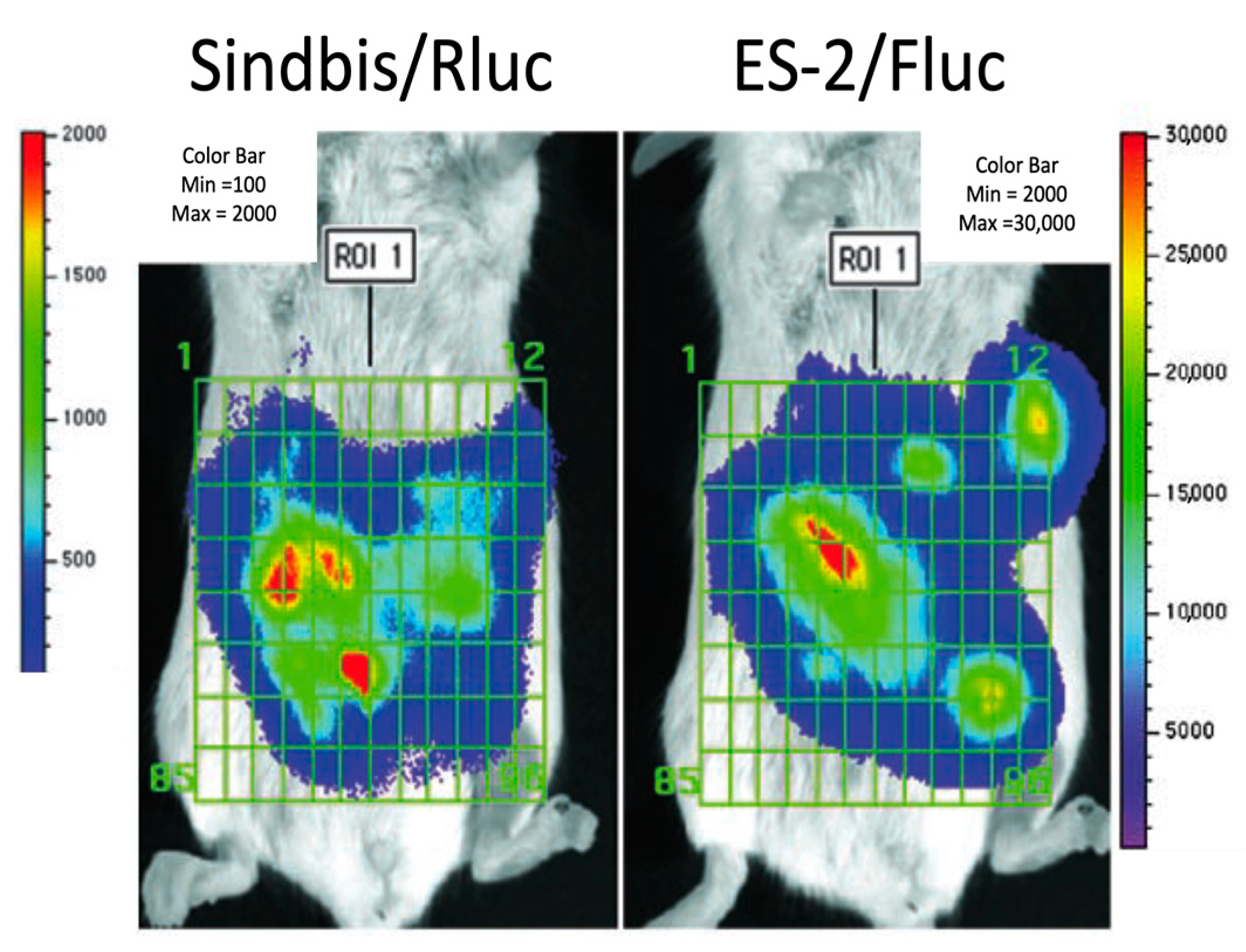
To confirm the degree and specificity of the Sindbis infection of the tumor cells, imaging studies were conducted that measured independent bioluminescent signals from ES-2 tumor cells and SV vector [5]. The cell lines were transfected with firefly luciferase expression plasmids (Fluc), while the SV vector expressed Renilla luciferase (Rluc). Firefly luciferase uses D-luciferin, whereas Renilla luciferase uses coelenterazine to generate bioluminescence. Both luciferases are highly substrate-specific and do not cross-react, however, coelenterazine has a shorter half-life [59]. For a quantitative analysis, bioluminescence signals were generated in the same animal by first injecting the short-lived Renilla substrate; after signal fading, this was followed by D-luciferin. Measurements were acquired and quantitated using Living Image software (Xenogen). A significant correlation (p < 0.0001) was established between the signals from SV-Rluc and Fluc tumor cells, indicating that a single i.p. delivery of Sindbis vectors led to the efficient infection of the metastasized ES-2 tumor cells throughout the peritoneal cavity (Figure 2). Further imaging experiments also showed that Sindbis vectors target syngeneic [5] and spontaneous [6] tumors in immune-competent mice. Therefore, SV vectors infected human and mouse cell-line-derived tumors as well as in vivo-generated mouse tumors.

2.4. Sindbis Virus Infection, Tumor Targeting, and the 67 kDa Laminin Receptor

Screening monoclonal antibodies for the ability to block infection, Wang et al. identified the 67 kDa laminin receptor protein (LAMR) as a receptor for Sindbis virus [60]. The presence and conservation of LAMR among distantly related species correlates with the broad host range of Sindbis virus. The transduction of LAMR cDNA into BHK cells was also found to increase levels of Sindbis virus infection. The study of LAMR was gaining interest at this time as earlier reports indicated its overexpression in some tumor cells [60].
LAMR has a high affinity for laminin, a major component of cell basement membranes that plays an important role in cellular adhesion, morphology, differentiation, and migration (reviewed in [61]). LAMR is a multifaceted protein that evolved from a 37 kDa ribosomal protein, although the nature of the precursor’s conversion from 37 to 67 kDa is still not well understood. It also has diverse functions and localizations within the nucleus, the ribosome, and the cell membrane (reviewed in [62]).
Many immunohistochemical studies of patient tumor biopsies went on to show increased levels of expression of LAMR on breast cancers [63,64,65], colon carcinomas [66,67], gastric cancer [68], lung cancer [69], pancreatic tumors [70], uterine adenocarcinoma [71], ovarian cancer [72,73,74,75], and thyroid cancer [76]. High levels of LAMR were shown in hepatocellular carcinoma via RT-PCR [77].
Recognizing the potential tumor targeting of SV vector via increased levels of LAMR on tumor cells motivated further studies to understand the association among SV, LAMR, and tumor cells. First, the replicon and envelope sequences of wild-type and laboratory-adapted SV strains were compared with respect to tumor targeting and suppression [53]. It was found that the SV Ar-339 wild-type strain provided the best suppression of tumor growth, whereas the E2 region of the Toto1101 Ar-339 showed better tumor targeting. A change of only one amino acid from E70 to K70 in the Ar-339 E2 protein suppressed the ability to target metastatic tumor implants in mice [53]. Further experiments utilized an SV vector derived from an AR-339 laboratory-adapted strain that contained amino acid E70 in the E2 envelope protein.
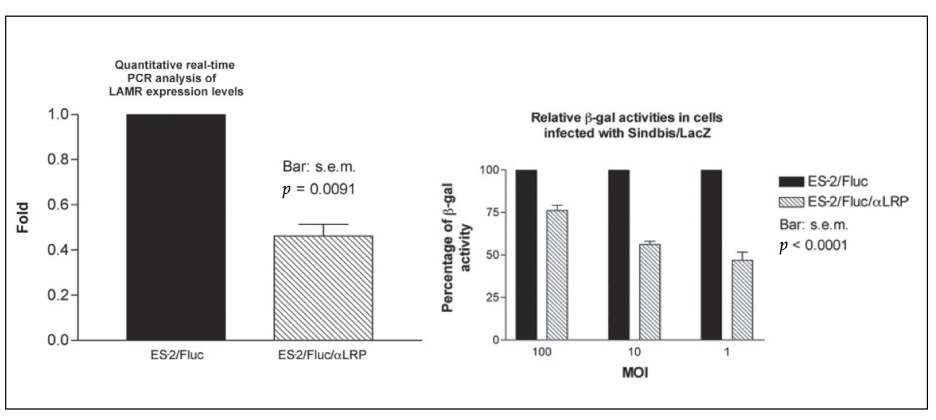
To investigate the correlation between laminin receptor expression and Sindbis vector infection, a small interfering RNA (siRNA) was incorporated into a pcDNA-Fluc vector (pcDNA-Fluc/iLAMR) and stably transfected into ES-2 ovarian carcinoma cells. A quantitative RT-PCR indicated that the ES-2/Fluc/iLAMR cells expressed 40% of 37/67 kDa LAMR RNA levels compared with E2/Fluc cells (Figure 3, left panel). As hypothesized, the ES-2/Fluc/iLAMR cells expressing fewer laminin receptors were infected less effec tively by the Sindbis vectors compared with the ES-2/Fluc cells (Figure 3 right panel) [5].
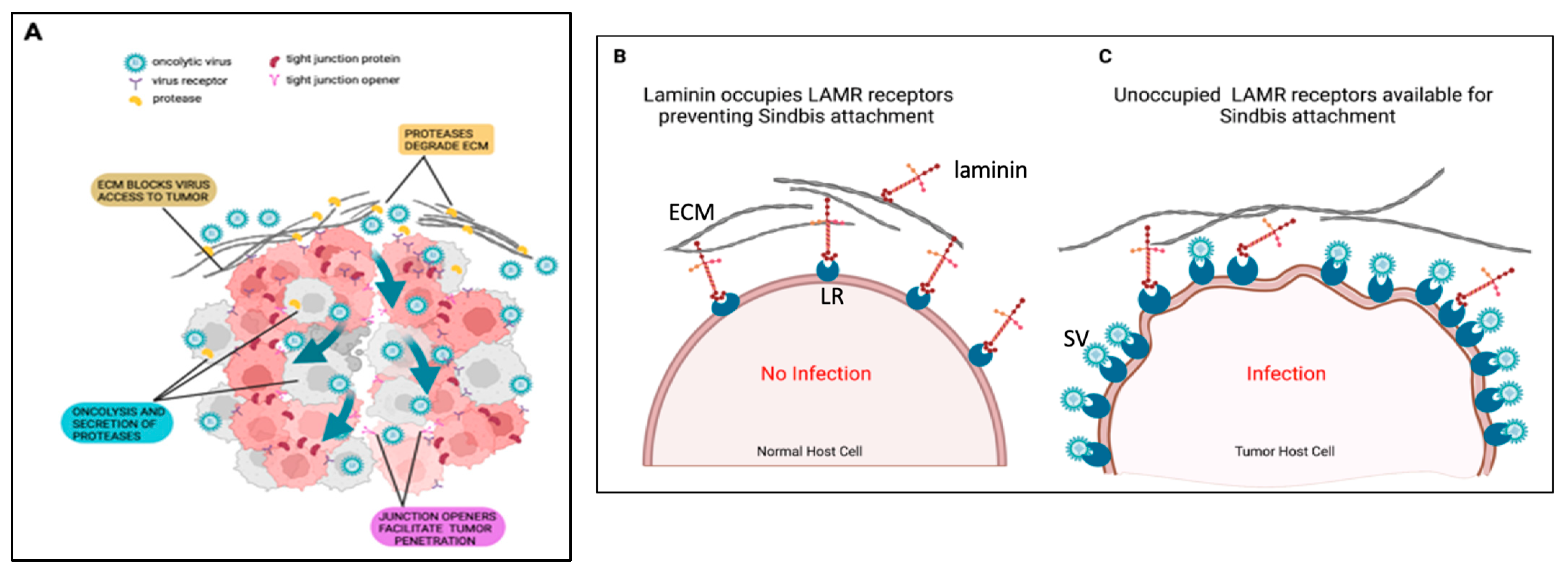
Binding studies conducted with LAMR monoclonal antibodies suggest that tumor cells have varying degrees of unoccupied cell-surface LAMRs that conceivably results from the increased biosynthesis of LAMR or the dissolution of the surrounding basement membrane and extracellular matrix by tumor cells [78,79,80,81]. Thus, it is plausible that SV uses unoccupied LAMRs as fortuitous binding sites on tumor cells (Figure 4).

2.5. LAMR as a Cancer Therapy Target

The increased expression of LAMR on tumor cells and its interaction with the extracellular membrane (ECM) suggested a role for LAMR in tumor migration and metastases [61,80]. Blocking the LAMR–laminin interaction using antibodies, siRNA, or glycan molecules demonstrated a reduction in the invasiveness of a tumorigenic HT1080 fibrosarcoma cell line [82]. LAMR was also shown to modify laminin within the ECM to alter cell signaling [83] and tumor aggressiveness [84].
The connection between SV, LAMR, and tumorigenesis is intriguing. To understand LAMR functions an siRNA pool that targets different regions of LAMR and a non-targeting control siRNA was introduced into human cell lines. With this system, >90% of the LAMR RNA and protein were knocked down [85]. HeLa, HEK 293T, and HepG2 human cell lines transfected with the siLAMR were found to undergo cell-cycle arrest in the G1 phase, and the expression of cell-cycle-related RNA and protein were altered. At 37 kDa, LAMR is a ribosomal protein, and the effect of siLAMR on protein translation was examined. A striking reduction in protein synthesis was observed, along with a disruption of the poly-ribosomal profile.
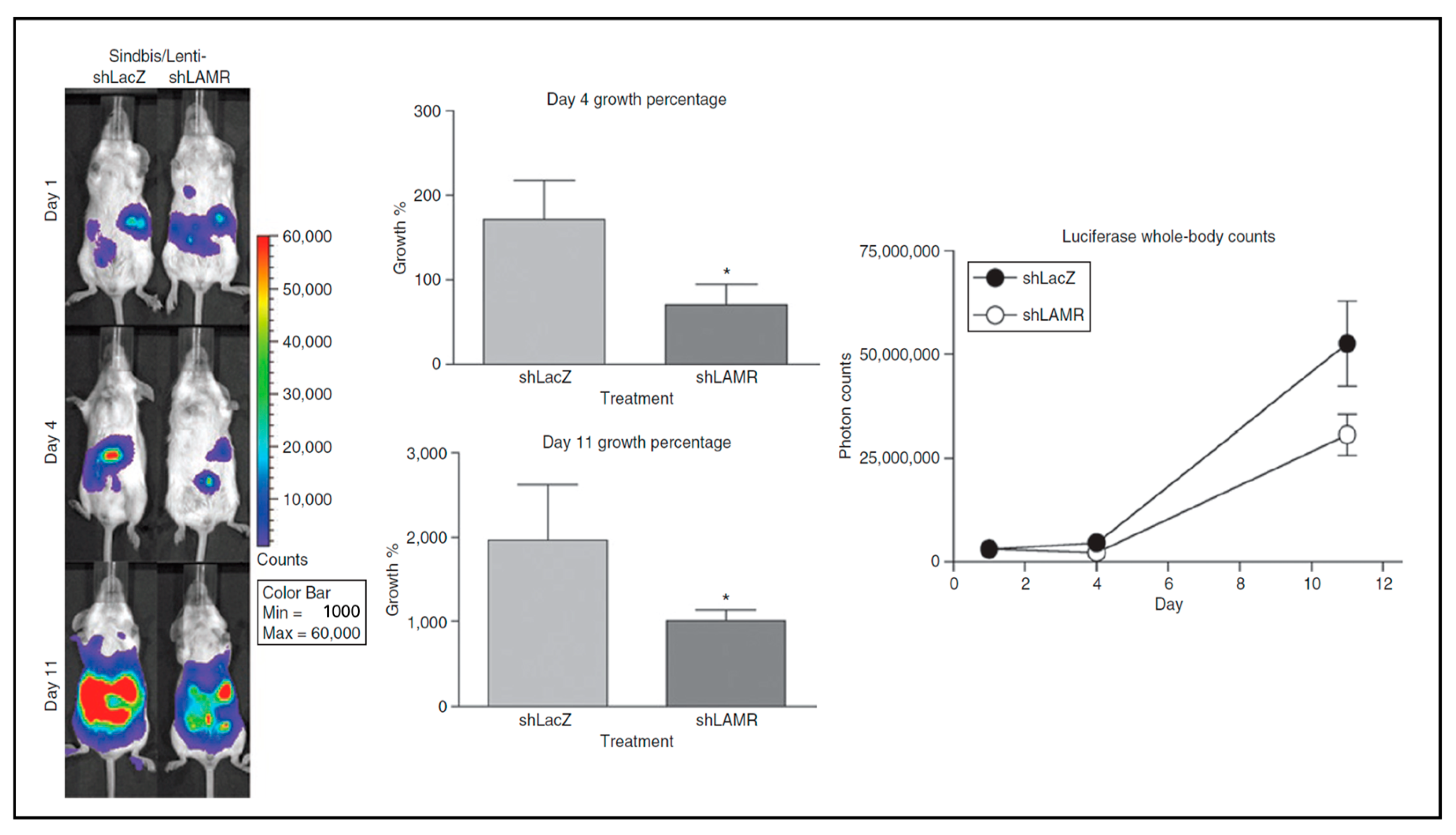
To explore the effect of LAMR reduction in tumor cells, a Sindbis/Lentivirus pseudotype vector was developed which carried short-hairpin RNA (shRNA) designed against LAMR [85]. SCID mice with tumors from ES-2/FLuc cells were treated with the Sinbis/Lenti viruses (Figure 5). The mice that received treatment with the pseudotype vector that targets LAMR showed a reduction in tumor growth between ~60 and 50% tumor growth compared with a control pseudotype vector expressing LacZ on days 4 and 11, respectively. Further studies to understand how LAMR regulates cell survival revealed that the C-terminal 75 amino acid residues of LAMR were required for cell viability [86].
The function of LAMR as a cell-surface protein that interacts with the ECM and effects cell signaling and migration [83] prompted a study into the association of LAMR 37/67 kDa with the cytoskeleton. Other studies have shown that LAMR could alter gene expression [87] and cell degranulation [88] through its interactions with the cytoskeleton.
LAMR and actin play important roles in cell migration. When cells are plated on laminin, actin filaments are reorganized and lamellipodia are formed. LAMR is also required for components of the 40S ribosomal subunit to colocalize with α-tubulin and is important for protein translation [89]. A mutational analysis indicated that the binding sites for actin and tubulin are separate from the laminin binding site [90].
Several investigations have focused on 37/67 kDa LAMR as a target for tumor treatment. The knock-down of LAMR with siRNA blocked metastasis and induced apoptosis in MDA_MB231 human breast cancer cells and also inhibited telomerase activity [91]. Hypoxia-inducible factor-1 (Hif-1), which was found to increase 37/67 kDa LAMR and the metastases of human gastric tumor cells, showed diminished invasiveness when siRNA for either Hif-1 or LAMR was used in knock-down studies [92]. LAMR siRNA was also found to induce apoptosis in breast and esophageal cancer cells [93]. The application of a specific antibody for LAMR (IgG1-iS8) to melanoma cells inhibited invasion and adhesion [94]. The same antibody showed similar results for pancreatic cancer and neuroblastoma cells [95].
A LAMR tumor-associated antigen, designated oncofetal antigen immature laminin receptor protein (OFA-iLRP), was found to be immunogenic in rodent and human tumors (reviewed in [96]). OFA-iLRP was shown to be identical to the 37 kDa LAMR [97]. While the mature, dimeric, and modified 67 kDa LAMR becomes non-immunogenic, the 37 kDa OFA-iLRP remains immunogenic and is only expressed in early embryonic stages and is re-expressed in many types of cancer. Tumor vaccine studies conducted to induce antibodies in OFA-iLRP have led to early-phase clinical trials to treat renal cell carcinoma [98], lymphocytic leukemia [99], and hematological cancers [100]; however, no results have been published yet.

2.6. LAMR as a Receptor for Other Viruses

LAMR has been identified as a receptor for the alphavirus Venezuelan equine encephalitis virus (VEE) [101], several flavivirus serotypes, dengue [102,103], classical swine fever virus [104], West Nile virus [105], and the oncolytic adeno-associated DNA virus (AAV) [106]. Of these viruses, vectors generated from VEE have been tested in preclinical studies for human papilloma virus [107] and in clinical trials for prostate [108], breast [109], and metastatic cancers expressing a carcinoembryonic antigen (CEA) [110]. Adeno-associated virus vectors have also been used in several clinical trials (reviewed in [111]). In all these studies, however, levels of LAMR expression on tumor cells were not correlated with vector efficacy.

2.7. Additional Alphavirus Receptors

Although LAMR has been identified as a receptor for several viruses, it is important to recognize that the mechanisms of LAMR-mediated binding and cell internalization have not been determined. There are also instances in which cancer cells showing marked expression of LAMR were not infected very well [57,112]. Attachment factors that concentrate viruses at the cell surface, like heparan sulfate, C-type lectins, or phosphatidyl-serine residues, can also play a role in viral entry (reviewed in [11,113]). The possibility that defects in anti-viral IFN signaling in tumor cells affect tumor tropism has also been discussed [55,114].
Additional potential receptors were found more recently. A genome-wide RNAi screen in Drosophila cells revealed the natural resistance-associated macrophage protein (NRAMP), a divalent metal transport protein, as a receptor for Sindbis virus [115]. The vertebrate homolog NRAMP2 was shown to mediate the infection of mammalian cells with Sindbis; mouse cells rendered deficient for NRAMP2 were not infected with Sindbis. The association between Sindbis tumor targeting and NRAMP2 expression, however, has not been reported.
Affinity purification followed by mass spectrometry identified the CD147 membrane protein as a receptor for CHIKV, RRV, Eastern and Western equine encephalitis, and SV in human cells [116]. CD147 has been found to be overexpressed on many cancer cells and cells within the tumor microenvironment, and it plays a role in tumor proliferation and the inhibition of apoptosis [117].
Additional receptors have been detected via CRISPER/Cas9 screens for single-guide RNAs that prevent virus infection. The low-density lipoprotein receptor class A domain containing protein 3 (LDLRAD3) was found to be a VEE receptor [118]. VLDLR and ApoER2, members of the low-density lipoprotein family, were found to be receptors for SV, SFV, and Eastern equine encephalitis virus [119]. A matrix-remodeling-associated protein 8 (MXRA8) was shown to serve as a receptor for O’nyong nyong virus (ONNV), Mayaro virus (MAYV), CHIKV, SFV, and RRV [120]. Although it is not clear whether these receptors can be utilized as targets for cancer therapy, MXRA8 has been shown to be increased in thyroid carcinoma cells [121], is abundant in most solid tumors [122], and correlates with tumor cell aggressiveness and an immunosuppressive environment [123].

3. The Delivery of Anti-Tumor Agents

Extensive summaries of preclinical and clinical trials using alphavirus vectors to deliver anti-tumor agents for cancer therapy have been published [124,125,126]. Studies with SV, SFV, and VEE virus vectors are presented here.

3.1. Cytokines

Early studies indicated that the SV vector expression of IL-12 [6] and IL-15 [1] enhanced anti-tumor activity. Sin/IL12 was used to treat intraperitoneal tumors of ES-2 human ovarian carcinoma in SCID mice [35]. The efficacy of mouse survival was linked to natural killer cell (NK) activation. Treatment showed a significant intraperitoneal influx of NK cells and increased IFNγ levels that upregulated MHC class II expression on peritoneal macrophages. The effect of SV.IL12 was also tested in immunocompetent mice bearing CT26 colon cell tumors [4]. Although SV.IL12 produced a modest increase in survival, an examination of T cells one week after treatment showed the upregulation of OX40 on effector CD4 T cells and the stimulation of macrophages. This observation suggests the use of combined treatment (see Section 4.3). Notably, plasma IL-12 levels did not significantly increase with treatment, indicating that IL-12, produced at high levels in infected cells in vitro, was produced locally by the SV.IL12 vector in vivo and was, therefore, less likely to cause toxicity.
Semliki forest alphavirus (SFV) expressing IL-12 has been used in several pre-clinical trials. The co-administration of SFV-IL-12 and SFV-E6,E7 (oncogenic proteins of human papilloma virus) was shown to augment the induction of cytotoxic T cells, and 28% of mice remained tumor-free for 45 days [127]. The delivery of IL-12 via a self-amplifying RNA, derived from SFV intratumorally electroporated into mice, to treat MC38 subcutaneous colon and hepatocarcinoma tumors showed anti-tumor effects and the induction of CD8 T cells [128]. The intratumoral injection of an SFV-IL-12 vector resulted in the regression of a B16 murine melanoma. In this study, IFNγ was induced; however, no increases in NK cells or T-cell mediated cytotoxicity were observed. The tumor inhibition was attributed to the antiangiogenic activity of IL-12 [129]. Antiangiogenic activity and tumor suppression were also observed when SFV expressing murine vascular endothelial growth factor (VEGF2) was used to treat mice with CT26 colon carcinoma and 4T1 metastatic mammary carcinoma [130].

3.2. Tumor-Associated Antigens

Tumor-associated antigens (TAAs), predominantly expressed by tumor cells compared with normal cells, can be indirectly targeted by virus vectors encoding the TAA to stimulate an immune response. This process was demonstrated using a CT26 colon carcinoma cell line transduced to express β-galactosidase (LacZ), CT26.CL25 [31]. An SV vector encoding LacZ (SV/LacZ) was i.p. injected into immunocompetent mice bearing CT26.CL25 tumors. Although SV/LacZ does not infect CT26 tumors, a strong therapeutic effect was observed. Knowing that the intraperitoneal injection of SV/luciferase leads to an infection of the mediastinal lymph nodes (MLNs) within three hours, T cells within the MLN were analyzed 24 h after treatment. A significant influx of activated CD8 T cells was observed that were shown to be required for the antigen-specific effect of SV/LacZ. Surprisingly, splenocytes from the SV/LacZ-treated tumor-cured mice acquired cytotoxicity against not only CT26.CL25 cells but also LacZ-negative CT26.WT cells. An immune response to endogenous CT26.CL25 antigens was shown to occur via the process known as epitope spreading. This study is notable for the observation that the direct targeting of tumors is not necessarily required. The stimulation of a potent immune response via the SV vector delivery of antigens to lymph nodes can produce an effective tumor treatment.
Numerous studies of therapeutic TAAs expressed by alphaviruses were previously summarized [124,126,131]. An SFV vector encoding the P1A testis antigen, expressed on mast cell tumors, was shown to be effective at treating murine mastocytomas [124,126]. The SFV-P1A vector was shown to induce the highest level of tumor protection compared with adenovirus and vaccinia virus vectors expressing the P1A antigen [132].
Preclinical therapies for mouse melanoma used SV and VEE viruses to express the tyrosinase-related proteins (TRP), TRP-1 and TRP-2 and the adhesion proteins MCAM/MUC18. Mice immunized with a DNA Sindbis replicon vector encoding mouse TRP-1 was shown to provide significant protection against B16 melanoma [36]. An SV-based DNA plasmid was also used to express the melanoma antigen, MUC18. Vaccination with this vector reduced the incidence of B16 melanoma and metastasis and induced humoral and cellular immunity [133]. Similarly, a VEE-VRP expressing TRP-2 proved to be an effective therapeutic vaccine against melanoma in mice, providing long-term protection through the activation of antibodies and T cell responses [134].
Human epidermal growth factor (HER2)/neu proto-oncogene (neu) was expressed by a VEE-VRP to treat murine breast cancers [124,126,131]. VRP-neu vaccination prevented tumor formation and induced high levels of neu-specific CD8 T cells and anti-neu antibody [135].
To treat murine prostate tumors, VEE vectors producing prostrate membrane proteins, PMSA [136] and STEAP [137], were utilized.
Endostatin, an anti-angiogenic protein, was expressed from an SFV to treat brain tumors. This study highlighted the therapeutic importance of the anti-viral response to the alphavirus vector treatment as endostatin alone did not inhibit angiogenesis or tumor growth [138].
Human papilloma virus (HPV) oncogenic proteins E6 and E7 were expressed in SFV and VEE to treat murine cervical cancer [126,139]. A phase I trial of SFV-E6,E7 reported safety and immunological responses in 12/12 participants [140].
Vectors based on VEE with TAA for a carcinoembryonic protein were used in a phase I clinical trial for several metastatic tumors. The results were encouraging, showing improved survival and immune response. The prostate cancer PSMA antigen and breast cancer antigen HER2/neu expressed by VEE vectors have also entered phase I trials [125].

The Modification of TAAs

Several groups have modified TAAs to enhance their immunogenicity. Fusing the HPV E7 oncogene to HSP70 increased the efficacy of a Sindbis RNA replicon [141] and virus vector [142]. Calreticulin, which interacts with the transport proteins involved with antigen presentation, was also fused to the HPV E7 protein and expressed from an SV vector [143]. To compensate for the inability of the plasmid or virus-particle-based Sindbis vectors to proliferate, Cheng et al. fused HPV E7 to the Herpes Simplex virus VP22 protein, which possesses a transport function, to disperse E7 among tissue cells [144]. These strategies were found to increase T-cell-mediated responses and long-term tumor-specific immunity.

4. Combination Therapy with Immunomodulatory Proteins

4.1. SV-NYESO1 and PD-1 Antibody

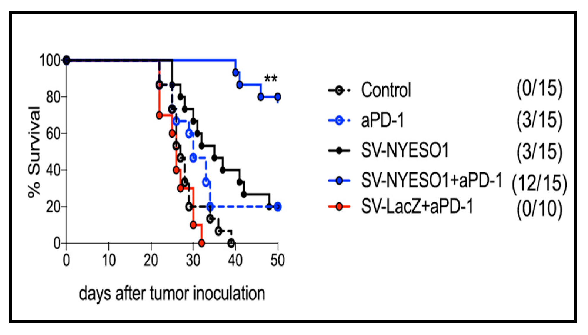
Attempts to optimize the SV vector to achieve long-term curative results focused on combining SV vector therapy with immunomodulatory agents. CT26 colon carcinoma cells were transfected with the TAA NY-ESO-1, a human cancer testis antigen which is expressed in many tumoral cells but absent from normal tissue [145]. Tumors were established in syngenetic, immunocompetent BALB/c mice. In this model, the oncolytic effects of the SV vector treatment were precluded as CT26 cells are not susceptible to SV infection. As the CT26 cells were shown to express programmed death protein ligand 1 (PDL-1), a treatment combining SV-NYESO1 with the antibody to PD-1 was examined. Figure 6 shows that while the SV vector expressing either NY-ESO-1 or PD-1 antibody prolongs survival, the combination of SV-NYESO1 and the antibody to PD-1 resulted in significant tumor regression. Mice rechallenged with tumor cells after 200 days remained tumor-free. A further analysis of T cells from splenocytes and lymph nodes showed that SV-NYESO1 is a potent immunostimulatory agent that induced a systemic pro-inflammatory reaction and lymphocyte activation within 2 days. The addition of the anti-PD-1 checkpoint to SV-NYESO1 therapy induced a stronger systemic and an intratumoral immune response indicated via increased T cell cytotoxicity and the production of IFNγ [3]. Importantly the results showed that the SV infectivity of macrophage or antigen-presenting cells within lymph nodes was sufficient to induce a systemic immune response.

4.2. SV Vector and 4-1BB Agonistic Antibody

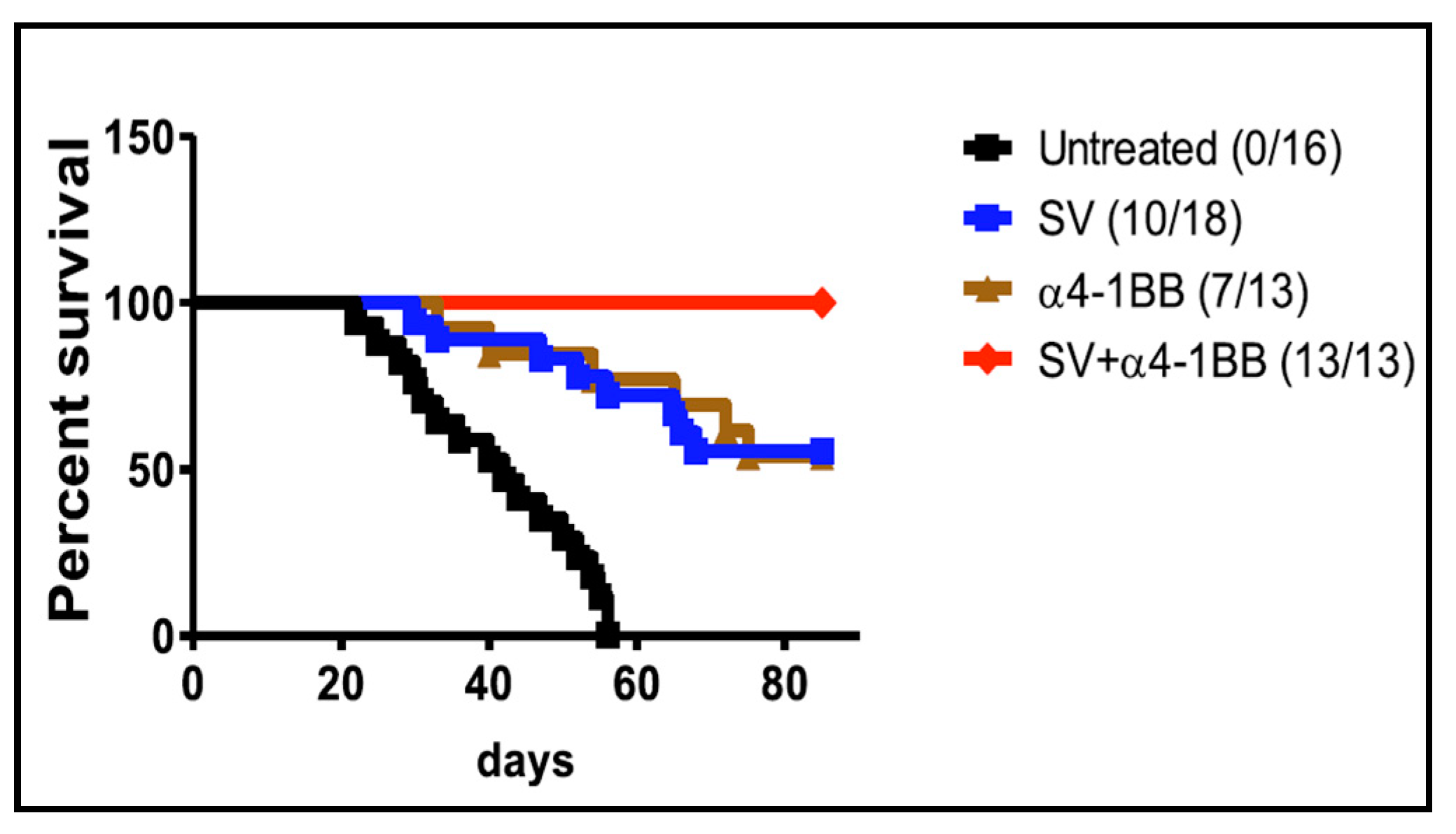
Following the observation that potent anti-lymphoma activity was elicited by an agonistic antibody to the T cell co-stimulatory molecule 4-1BB (CD137) [146], anti-4-1BB was combined with an empty SV vector to treat an A20 B cell lymphoma BALB/c tumor model [147]. All mice receiving combined treatment survived long-term (Figure 7). Mice surviving >4 months and rechallenged with A20 tumor cells all survived. Significantly, this study indicated that the expression of a TAA was not required for an SV vector plus anti-41BB to successfully treat A20 tumors.
To understand the nature of survival, at 7 days after treatment, T cells purified of splenocytes and tumor cells were analyzed via RNA-seq transcriptomics, ELISPOT, flow cytometry, and cytotoxicity and metabolic assays. The results indicated that combined treatment enhances T cell proliferation, activation, migration, and cytotoxicity. An influx of activated and metabolically energized T cells were found within the tumor mass, while immune-suppressive T reg cells were decreased [147].

4.3. SV.IL12 and Anti-OX40 Antibody

As mentioned in Section 3.1, an SV vector expressing IL-12 (SV.IL12) activates T cells and increases the expression of OX40 on CD4 cells. OX40 is a TNF-family co-stimulatory receptor that is expressed on activated T cells [148] and also represses regulatory T cell induction [149]. Activated OX40 promotes the function and survival of Th1, a proinflammatory T cell [150].
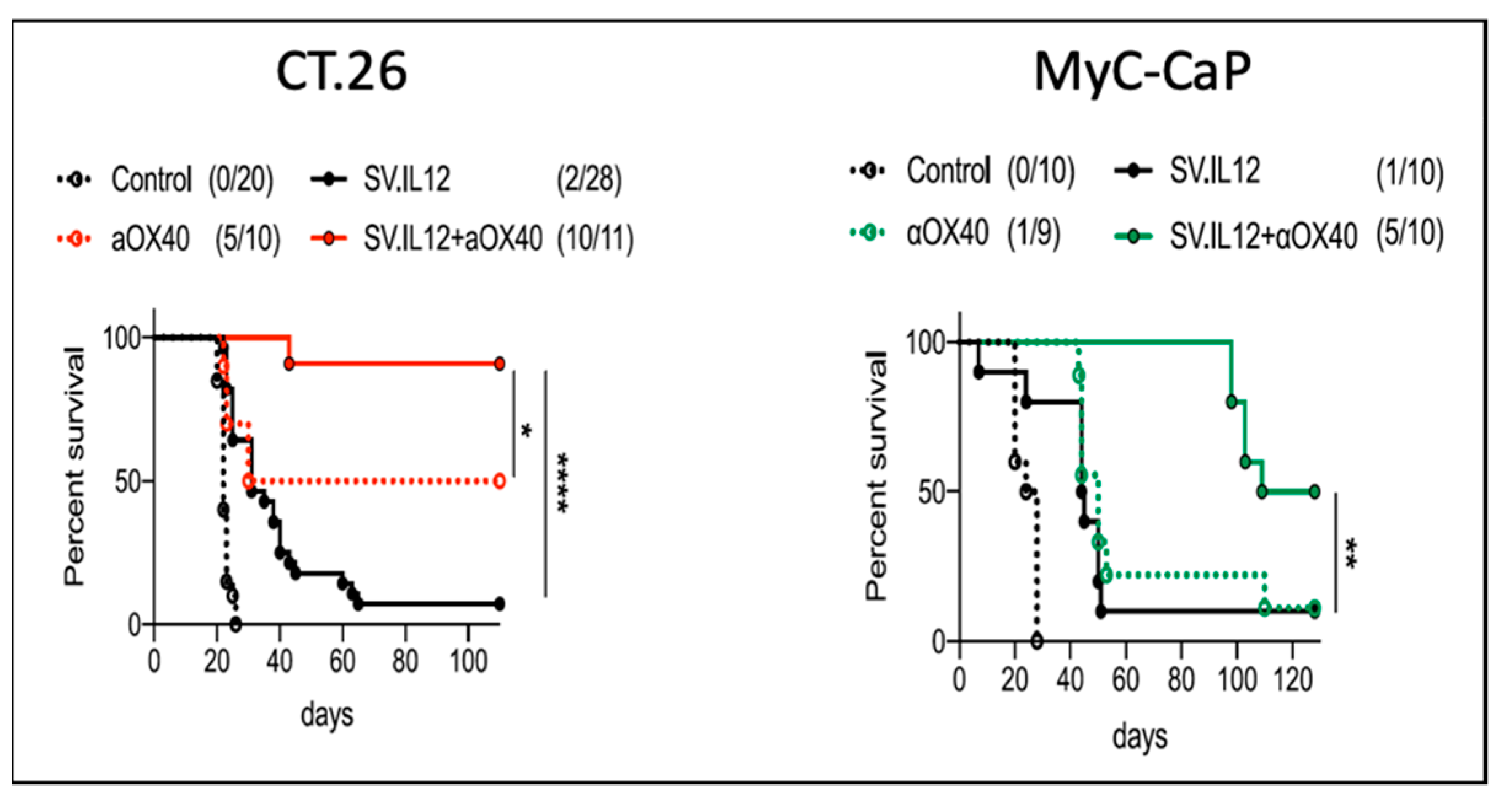
To test the effect of SV.IL12 with the agonistic anti-OX40 antibody, SV-susceptible prostate cancer, MyC-CaP, and non-susceptible CT26 colon cancer tumor cell models were injected into immunocompetent FVB/NJ and BALB/c mice, respectively. As shown in Figure 8, the combined SV.IL12 and anti-OX40 (αOX40) treatment showed a significant regression of tumors in the two distinct models. Again, the oncolytic activity of SV was not required to stimulate an effective anti-tumor response.
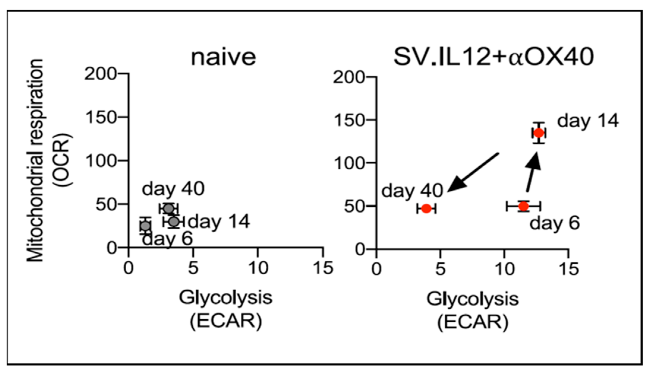
Splenocytes and tumor cells were analyzed, as in Section 4.2. Collectively, the results demonstrated that only the combined treatment with SV.IL12 and αOX40 generated the induction of differentiated effector T cells. Metabolic assays of CT.26 splenic T cells indicated that the combined treatment boosted T cells to a highly energetic state by 14 days (Figure 9), a hallmark of highly activated T cells [151].
The combined treatment increased the infiltration of T cells into the tumor, as assessed via flow cytometry and multiplex immunofluorescence. Flow cytometry showed that the CD4 T cells expressed ICOS+T-bet+ and granzyme B, which are indicative of an anti-tumor phenotype. The CD4 T cells were also found to produce the highest levels of IFNγ [4]. This observation is noteworthy as the role of CD4 anti-tumor cytotoxic lymphocytes, independent of CD8 T cells, is increasingly being recognized [152]. Overall, the data suggest that independent of the tumor model, the distinct T cell response induced by SV.IL12 plus αOX40 stems from the interaction with the peripheral immune cells that are activated to infiltrate the tumor to eliminate the tumor cells.

4.4. SV Vector Expressing αOX40

The combination of SV.IL12 and αOX40 was used to treat a mouse ovarian surface epithelial cell (MOSEC.ID8) that was adapted to be more stable in vivo (MOSEC.p11). MOSEC.p11 expresses firefly luciferase and is susceptible to SV infection. Initial experiments showed an increased migration of immune cells into tumor tissue when mice were treated with either SV.IL12, αOX40, or SVIL.12 plus αOX40. In metabolic studies, however, only a combined treatment stimulated highly energized T cells [2].
Studies in several mouse models indicate that the SV.IL12 vector in combination with the agonistic antibody for OX40 was efficacious and devoid of adverse effects. The encouraging results with the CT.26, MyC-CaP, and MOSEC.p11 tumor models motivated the generation of a single SV vector encoding both αOX40 and IL-12 to facilitate local delivery to peripheral lymphoid tissue and to tumors susceptible to SV infection [2]. This vector, SV.IgGOX40.IL12, effectively suppressed ovarian cancer tumors in vivo. Survivors rechallenged with MOSEC.p11 cells after 140 days were protected from tumor recurrence (Figure 10). In this ovarian cancer model, SV.IL12 was as effective as the combined therapy. As IL-12 stimulates OX40 expression on CD4 cells, it is likely that SV.IL12 tumor infection locally increases OX40 signaling.
Depletion studies indicated the importance of CD4 and CD8 T cells and, once again, CD4 T cells were shown to play a pivotal role in treatment effects. An RNA transcriptome analysis of tumor tissue also indicated that the treatment reprogrammed cells within the tumor to express genes related to an upregulated immune response.

4.5. Alphavirus Combined with Immunotherapy

Other alphaviruses have been used in conjunction with immunomodulatory agents. Co-immunizations with SFV vectors encoding VEGF-2 and IL-4 were found to induce high titers of VEGF antibodies and increase the survival of mice bearing CT.26 colon cancer or 4T1 metastatic breast cancer [130]. SFV-IL-12 and an anti-PD-1 antibody were able to synergistically treat B16 melanoma and MC38 colorectal tumors [153,154]. A VEE vector expressing a tyrosinase-related protein TAA , was used in combination with the antagonistic anti-CTLA-4 antibody or the agonistic GITR antibody to treat a B16F10 mouse melanoma model. The addition of the immunomodulatory antibodies increased the immunity of the VEE-TAA vector alone [155]. A phase II clinical trial testing a VEE-based vector, VRP-HER2, combined with an anti-PD-1 antibody produced promising results. Tumor biopsies, sera, and peripheral blood mononuclear cells (PBMCs) were collected before and after the immunizations. The vector treatment showed overall enhancement of the immune response with the combined treatment [125].
Table 1 and Table 2 present preclinical and clinical trials performed within the last 5 years using alphaviruses. In most cases, the alphavirus vectors combine the expression of additional agents with their oncolytic action. Previous preclinical and clinical trials with alphaviruses have been reported in recent reviews [124,125,156].

5. Summary

Sindbis virus has emerged as a viable vector for the treatment of cancer. Imaging studies have demonstrated a natural proclivity of Sindbis vectors for tumor cells, achieved through direct infection or indirectly through an interaction with peripheral lymphoid tissue, thereby augmenting and mobilizing pre-existing immune cells to target tumors. In several preclinical in vivo models, SV vectors have inhibited tumor growth and conferred long-term protection against tumor rechallenge. Treatment efficacy is dependent upon T-cells that are transcriptionally reprogrammed, leading to the expression of immune response genes and metabolic alterations that result in higher energy states. A notable influx of immune cells into the tumor microenvironment occurs.
A Sindbis virus (SV) vector platform has been developed, combining the expression of interleukin-12 (IL-12) and an agonistic antibody targeting the co-stimulatory OX40 receptor. This platform has yielded promising results in treating various preclinical mouse models. Its translation to clinical applications is feasible as SV vectors can be produced and purified under Good Manufacturing Practice (GMP) standards, ensuring high titers (1011 transducing units per milliliter (Tu/mL)) to compensate for dilution in the bloodstream.
As mentioned in Section 1.1 and Section 1.2, Sindbis virus vectors produce high levels of expressed proteins over a short period of time (24–48 h), building a robust T cell priming in the lymph nodes. Despite this immune activation, in some cases, treatment efficacy may require multiple doses. While alphaviruses are not highly immunogenic, the requirement for many doses could pose challenges and may lead to patient reluctance to undergo treatment. New vector designs to improve efficacy with fewer doses are now being studied in preclinical models. The lack of clinical trials involving Sindbis virus vectors thus far makes it difficult to evaluate their transition from animal models to human patients.
A number of other alphavirus vectors, primarily based on Venezuelan equine encephalitis (VEE) or Semliki Forest virus (SFV), have been generated for cancer treatment and cancer immunotherapies. Some vectors have advanced to clinical trials, demonstrating promising safety profiles and therapeutic potential. The continued optimization of vectors and administration protocols is expected to enhance the therapeutic efficacy of these versatile vectors.

Author Contributions

D.M., C.P., A.H. and S.O. conceptualized the review; original papers, all authors ; writing, C.P.; editing, D.M., A.H., S.O.; preparation of figures, A.H., S.O., C.P.; supervision, project administration, funding acquisition, D.M. All authors have read and agreed to the published version of the manuscript.

Funding

Funding was provided by NIH 5R44CA250627 and through a Research and Licensing. Agreement between Cynvec and NYU Langone, which licenses the Sindbis technology to Cynvec. This work was also supported, in part, by The Experimental Pathology Research Laboratory at NYU Langone which is partially supported by the Cancer Center Support Grant P30CA016087. The Vectra3 multispectral imaging system was purchased through Shared Instrumentation Grant S10 OD021747.

Institutional Review Board Statement

All experiments were performed in accordance with the Institutional Animal Care and Use Committee of New York University Health.

Informed Consent Statement

Not applicable.

Data Availability Statement

All sequencing data that support the findings of this study will be deposited in the National Center for Biotechnology Information Gene Expression Omnibus (GEO) and are accessible through the GEO Series accession number that will be provided, including all other relevant data included in the article. Further inquiries can be directed to the corresponding authors.

Acknowledgments

We wish to thank all the postdoctoral fellows, graduate students, and technicians who contributed to the studies described in this review. We appreciate the funding that was provided by NIH 5R44CA250627. We would like to thank the NYU High Throughput Biology Laboratory for the Seahorse usage, the NYU Genome Technology Center for the RNA sequencing service, and the Experimental Pathology Research Laboratory at NYU Langone, which also supported this work in part via the Experimental Pathology Research Laboratory at NYU Langone, which is partially supported by the Cancer Center Support Grant P30CA016087. The Vectra3 multispectral imaging system was purchased through Shared Instrumentation Grant S10 OD021747.

Conflicts of Interest

All authors are employed by the NYU Langone School of Medicine and have no employment relationship or consultancy agreement with Cynvec, a biotechnology company that supports some studies under a Research and Licensing agreement with NYU. S.O., A.H., C.P. and D.M. are inventors on one or several issued patents and/or patent applications held by NYU that cover Sindbis treatment of neoplasia and COVID-19. As part of the Research and Licensing agreement, authors who are inventors on patents are entitled to a portion of NYU Langone’s royalties received should Sindbis vectors be approved by the FDA for therapeutic or vaccination use. Regarding data and material availability, correspondence should be addressed to D.M.

References

  1. Hurtado, A.; Tseng, J.C.; Meruelo, D. Gene therapy that safely targets and kills tumor cells throughout the body. Rejuvenation Res. 2006, 9, 36–44. [Google Scholar] [CrossRef] [PubMed]
  2. Opp, S.; Hurtado, A.; Pampeno, C.; Lin, Z.; Meruelo, D. Potent and Targeted Sindbis Virus Platform for Immunotherapy of Ovarian Cancer. Cells 2022, 12, 77. [Google Scholar] [CrossRef] [PubMed]
  3. Scherwitzl, I.; Hurtado, A.; Pierce, C.M.; Vogt, S.; Pampeno, C.; Meruelo, D. Systemically Administered Sindbis Virus in Combination with Immune Checkpoint Blockade Induces Curative Anti-tumor Immunity. Mol. Ther. Oncolytics 2018, 9, 51–63. [Google Scholar] [CrossRef] [PubMed]
  4. Scherwitzl, I.; Opp, S.; Hurtado, A.M.; Pampeno, C.; Loomis, C.; Kannan, K.; Yu, M.; Meruelo, D. Sindbis Virus with Anti-OX40 Overcomes the Immunosuppressive Tumor Microenvironment of Low-Immunogenic Tumors. Mol. Ther. Oncolytics 2020, 17, 431–447. [Google Scholar] [CrossRef]
  5. Tseng, J.C.; Hurtado, A.; Yee, H.; Levin, B.; Boivin, C.; Benet, M.; Blank, S.V.; Pellicer, A.; Meruelo, D. Using sindbis viral vectors for specific detection and suppression of advanced ovarian cancer in animal models. Cancer Res. 2004, 64, 6684–6692. [Google Scholar] [CrossRef]
  6. Tseng, J.C.; Levin, B.; Hurtado, A.; Yee, H.; Perez de Castro, I.; Jimenez, M.; Shamamian, P.; Jin, R.; Novick, R.P.; Pellicer, A.; et al. Systemic tumor targeting and killing by Sindbis viral vectors. Nat. Biotechnol. 2004, 22, 70–77. [Google Scholar] [CrossRef]
  7. Strauss, J.H.; Strauss, E.G. The alphaviruses: Gene expression, replication, and evolution. Microbiol. Rev. 1994, 58, 491–562. [Google Scholar] [CrossRef]
  8. Kim, D.Y.; Reynaud, J.M.; Rasalouskaya, A.; Akhrymuk, I.; Mobley, J.A.; Frolov, I.; Frolova, E.I. New World and Old World Alphaviruses Have Evolved to Exploit Different Components of Stress Granules, FXR and G3BP Proteins, for Assembly of Viral Replication Complexes. PLoS Pathog. 2016, 12, e1005810. [Google Scholar] [CrossRef]
  9. Taylor, R.M.; Hurlbut, H.S. The isolation of Coxsackie-like viruses from mosquitoes. J. Egypt. Med. Assoc. 1953, 36, 489–494. [Google Scholar]
  10. Jose, J.; Snyder, J.E.; Kuhn, R.J. A structural and functional perspective of alphavirus replication and assembly. Future Microbiol. 2009, 4, 837–856. [Google Scholar] [CrossRef]
  11. Zimmerman, O.; Holmes, A.C.; Kafai, N.M.; Adams, L.J.; Diamond, M.S. Entry receptors—The gateway to alphavirus infection. J. Clin. Investig. 2023, 133, e165307. [Google Scholar] [CrossRef]
  12. Bredenbeek, P.J.; Frolov, I.; Rice, C.M.; Schlesinger, S. Sindbis virus expression vectors: Packaging of RNA replicons by using defective helper RNAs. J. Virol. 1993, 67, 6439–6446. [Google Scholar] [CrossRef]
  13. Xiong, C.; Levis, R.; Shen, P.; Schlesinger, S.; Rice, C.M.; Huang, H.V. Sindbis virus: An efficient, broad host range vector for gene expression in animal cells. Science 1989, 243, 1188–1191. [Google Scholar] [CrossRef] [PubMed]
  14. Frolova, E.I.; Gorchakov, R.; Pereboeva, L.; Atasheva, S.; Frolov, I. Functional Sindbis virus replicative complexes are formed at the plasma membrane. J. Virol. 2010, 84, 11679–11695. [Google Scholar] [CrossRef] [PubMed]
  15. Froshauer, S.; Kartenbeck, J.; Helenius, A. Alphavirus RNA replicase is located on the cytoplasmic surface of endosomes and lysosomes. J. Cell Biol. 1988, 107, 2075–2086. [Google Scholar] [CrossRef] [PubMed]
  16. Kujala, P.; Ikaheimonen, A.; Ehsani, N.; Vihinen, H.; Auvinen, P.; Kaariainen, L. Biogenesis of the Semliki Forest virus RNA replication complex. J. Virol. 2001, 75, 3873–3884. [Google Scholar] [CrossRef]
  17. Frolov, I.; Hoffman, T.A.; Prágai, B.M.; Dryga, S.A.; Huang, H.V.; Schlesinger, S.; Rice, C.M. Alphavirus-based expression vectors: Strategies and applications. Proc. Natl. Acad. Sci. USA 1996, 93, 11371–11377. [Google Scholar] [CrossRef]
  18. Strauss, J.H.; Wang, K.S.; Schmaljohn, A.L.; Kuhn, R.J.; Strauss, E.G. Host-cell receptors for Sindbis virus. Arch. Virol. Suppl. 1994, 9, 473–484. [Google Scholar] [CrossRef]
  19. Scaglione, A.; Opp, S.; Hurtado, A.; Lin, Z.; Pampeno, C.; Noval, M.G.; Thannickal, S.A.; Stapleford, K.A.; Meruelo, D. Combination of a Sindbis-SARS-CoV-2 Spike Vaccine and alphaOX40 Antibody Elicits Protective Immunity Against SARS-CoV-2 Induced Disease and Potentiates Long-Term SARS-CoV-2-Specific Humoral and T-Cell Immunity. Front. Immunol. 2021, 12, 719077. [Google Scholar] [CrossRef]
  20. Lauer, U.M.; Beil, J. Oncolytic viruses: Challenges and considerations in an evolving clinical landscape. Future Oncol. 2022, 18, 2713–2732. [Google Scholar] [CrossRef]
  21. Ehrlich, M.; Bacharach, E. Oncolytic Virotherapy: The Cancer Cell Side. Cancers 2021, 13, 931. [Google Scholar] [CrossRef] [PubMed]
  22. Groeneveldt, C.; van den Ende, J.; van Montfoort, N. Preexisting immunity: Barrier or bridge to effective oncolytic virus therapy? Cytokine Growth Factor Rev. 2023, 70, 1–12. [Google Scholar] [CrossRef] [PubMed]
  23. Lin, D.; Shen, Y.; Liang, T. Oncolytic virotherapy: Basic principles, recent advances and future directions. Signal Transduct. Target. Ther. 2023, 8, 156. [Google Scholar] [CrossRef]
  24. Lundstrom, K. Viral Vectors in Gene Therapy: Where Do We Stand in 2023? Viruses 2023, 15, 698. [Google Scholar] [CrossRef] [PubMed]
  25. Ma, R.; Li, Z.; Chiocca, E.A.; Caligiuri, M.A.; Yu, J. The emerging field of oncolytic virus-based cancer immunotherapy. Trends Cancer 2023, 9, 122–139. [Google Scholar] [CrossRef] [PubMed]
  26. Swift, E.A.; Pollard, S.M.; Parker, A.L. Engineering Cancer Selective Virotherapies: Are the Pieces of the Puzzle Falling into Place? Hum. Gene Ther. 2022, 33, 1109–1120. [Google Scholar] [CrossRef] [PubMed]
  27. Yang, L.; Gu, X.; Yu, J.; Ge, S.; Fan, X. Oncolytic Virotherapy: From Bench to Bedside. Front. Cell Dev. Biol. 2021, 9, 790150. [Google Scholar] [CrossRef]
  28. Venticinque, L.; Meruelo, D. Sindbis viral vector induced apoptosis requires translational inhibition and signaling through Mcl-1 and Bak. Mol. Cancer 2010, 9, 37. [Google Scholar] [CrossRef] [PubMed]
  29. Zhang, J.; Frolov, I.; Russell, S.J. Gene therapy for malignant glioma using Sindbis vectors expressing a fusogenic membrane glycoprotein. J. Gene Med. 2004, 6, 1082–1091. [Google Scholar] [CrossRef]
  30. Huckriede, A.; Bungener, L.; Holtrop, M.; de Vries, J.; Waarts, B.L.; Daemen, T.; Wilschut, J. Induction of cytotoxic T lymphocyte activity by immunization with recombinant Semliki Forest virus: Indications for cross-priming. Vaccine 2004, 22, 1104–1113. [Google Scholar] [CrossRef]
  31. Granot, T.; Yamanashi, Y.; Meruelo, D. Sindbis viral vectors transiently deliver tumor-associated antigens to lymph nodes and elicit diversified antitumor CD8+ T-cell immunity. Mol. Ther. 2014, 22, 112–122. [Google Scholar] [CrossRef]
  32. Chen, Z.; Moyana, T.; Saxena, A.; Warrington, R.; Jia, Z.; Xiang, J. Efficient antitumor immunity derived from maturation of dendritic cells that had phagocytosed apoptotic/necrotic tumor cells. Int. J. Cancer 2001, 93, 539–548. [Google Scholar] [CrossRef]
  33. Alexopoulou, L.; Holt, A.C.; Medzhitov, R.; Flavell, R.A. Recognition of double-stranded RNA and activation of NF-kappaB by Toll-like receptor 3. Nature 2001, 413, 732–738. [Google Scholar] [CrossRef]
  34. Fournier, P.; Zeng, J.; Schirrmacher, V. Two ways to induce innate immune responses in human PBMCs: Paracrine stimulation of IFN-alpha responses by viral protein or dsRNA. Int. J. Oncol. 2003, 23, 673–680. [Google Scholar] [CrossRef] [PubMed]
  35. Granot, T.; Venticinque, L.; Tseng, J.C.; Meruelo, D. Activation of cytotoxic and regulatory functions of NK cells by Sindbis viral vectors. PLoS ONE 2011, 6, e20598. [Google Scholar] [CrossRef] [PubMed]
  36. Leitner, W.W.; Hwang, L.N.; deVeer, M.J.; Zhou, A.; Silverman, R.H.; Williams, B.R.; Dubensky, T.W.; Ying, H.; Restifo, N.P. Alphavirus-based DNA vaccine breaks immunological tolerance by activating innate antiviral pathways. Nat. Med. 2003, 9, 33–39. [Google Scholar] [CrossRef]
  37. Martin-Fontecha, A.; Thomsen, L.L.; Brett, S.; Gerard, C.; Lipp, M.; Lanzavecchia, A.; Sallusto, F. Induced recruitment of NK cells to lymph nodes provides IFN-gamma for T(H)1 priming. Nat. Immunol. 2004, 5, 1260–1265. [Google Scholar] [CrossRef] [PubMed]
  38. Vivier, E.; Tomasello, E.; Baratin, M.; Walzer, T.; Ugolini, S. Functions of natural killer cells. Nat. Immunol. 2008, 9, 503–510. [Google Scholar] [CrossRef] [PubMed]
  39. Wang, L.; Smith, D.; Bot, S.; Dellamary, L.; Bloom, A.; Bot, A. Noncoding RNA danger motifs bridge innate and adaptive immunity and are potent adjuvants for vaccination. J. Clin. Investig. 2002, 110, 1175–1184. [Google Scholar] [CrossRef]
  40. Choi, Y.; Chang, J. Viral vectors for vaccine applications. Clin. Exp. Vaccine Res. 2013, 2, 97–105. [Google Scholar] [CrossRef]
  41. Gardner, J.P.; Frolov, I.; Perri, S.; Ji, Y.; MacKichan, M.L.; zur Megede, J.; Chen, M.; Belli, B.A.; Driver, D.A.; Sherrill, S.; et al. Infection of human dendritic cells by a sindbis virus replicon vector is determined by a single amino acid substitution in the E2 glycoprotein. J. Virol. 2000, 74, 11849–11857. [Google Scholar] [CrossRef] [PubMed]
  42. MacDonald, G.H.; Johnston, R.E. Role of dendritic cell targeting in Venezuelan equine encephalitis virus pathogenesis. J. Virol. 2000, 74, 914–922. [Google Scholar] [CrossRef]
  43. Osada, T.; Morse, M.A.; Hobeika, A.; Lyerly, H.K. Novel recombinant alphaviral and adenoviral vectors for cancer immunotherapy. Semin. Oncol. 2012, 39, 305–310. [Google Scholar] [CrossRef] [PubMed]
  44. Pushko, P.; Parker, M.; Ludwig, G.V.; Davis, N.L.; Johnston, R.E.; Smith, J.F. Replicon-helper systems from attenuated Venezuelan equine encephalitis virus: Expression of heterologous genes in vitro and immunization against heterologous pathogens in vivo. Virology 1997, 239, 389–401. [Google Scholar] [CrossRef] [PubMed]
  45. Uematsu, Y.; Vajdy, M.; Lian, Y.; Perri, S.; Greer, C.E.; Legg, H.S.; Galli, G.; Saletti, G.; Otten, G.R.; Rappuoli, R.; et al. Lack of interference with immunogenicity of a chimeric alphavirus replicon particle-based influenza vaccine by preexisting antivector immunity. Clin. Vaccine Immunol. 2012, 19, 991–998. [Google Scholar] [CrossRef] [PubMed]
  46. Brummer-Korvenkontio, M.; Vapalahti, O.; Kuusisto, P.; Saikku, P.; Manni, T.; Koskela, P.; Nygren, T.; Brummer-Korvenkontio, H.; Vaheri, A. Epidemiology of Sindbis virus infections in Finland 1981-96: Possible factors explaining a peculiar disease pattern. Epidemiol. Infect. 2002, 129, 335–345. [Google Scholar] [CrossRef]
  47. Hardwick, J.M.; Levine, B. Sindbis virus vector system for functional analysis of apoptosis regulators. Methods Enzymol. 2000, 322, 492–508. [Google Scholar] [CrossRef] [PubMed]
  48. Manni, T.; Kurkela, S.; Vaheri, A.; Vapalahti, O. Diagnostics of Pogosta disease: Antigenic properties and evaluation of Sindbis virus IgM and IgG enzyme immunoassays. Vector Borne Zoonotic Dis. 2008, 8, 303–311. [Google Scholar] [CrossRef]
  49. Dubuisson, J.; Rice, C.M. Sindbis virus attachment: Isolation and characterization of mutants with impaired binding to vertebrate cells. J. Virol. 1993, 67, 3363–3374. [Google Scholar] [CrossRef]
  50. Ohno, K.; Sawai, K.; Iijima, Y.; Levin, B.; Meruelo, D. Cell-specific targeting of Sindbis virus vectors displaying IgG-binding domains of protein A. Nat. Biotechnol. 1997, 15, 763–767. [Google Scholar] [CrossRef]
  51. Surolia, A. Interaction of Protein-a with the Domains of Fc Fragment. Trends Biochem. Sci. 1982, 7, 318–319. [Google Scholar] [CrossRef]
  52. Sawai, K.; Meruelo, D. Cell-specific transfection of choriocarcinoma cells by using Sindbis virus hCG expressing chimeric vector. Biochem. Biophys. Res. Commun. 1998, 248, 315–323. [Google Scholar] [CrossRef] [PubMed]
  53. Hurtado, A.; Tseng, J.C.; Boivin, C.; Levin, B.; Yee, H.; Pampeno, C.; Meruelo, D. Identification of amino acids of Sindbis virus E2 protein involved in targeting tumor metastases in vivo. Mol. Ther. 2005, 12, 813–823. [Google Scholar] [CrossRef] [PubMed]
  54. Tseng, J.C.; Levin, B.; Hirano, T.; Yee, H.; Pampeno, C.; Meruelo, D. In vivo antitumor activity of Sindbis viral vectors. J. Natl. Cancer Inst. 2002, 94, 1790–1802. [Google Scholar] [CrossRef] [PubMed]
  55. Tseng, J.C.; Zheng, Y.; Yee, H.; Levy, D.E.; Meruelo, D. Restricted tissue tropism and acquired resistance to Sindbis viral vector expression in the absence of innate and adaptive immunity. Gene Ther. 2007, 14, 1166–1174. [Google Scholar] [CrossRef]
  56. Tseng, J.C.; Granot, T.; DiGiacomo, V.; Levin, B.; Meruelo, D. Enhanced specific delivery and targeting of oncolytic Sindbis viral vectors by modulating vascular leakiness in tumor. Cancer Gene Ther. 2010, 17, 244–255. [Google Scholar] [CrossRef] [PubMed]
  57. Stelter, L.; Tseng, J.C.; Torosjan, A.; Levin, B.; Longo, V.A.; Pillarsetty, N.; Zanzonico, P.; Meruelo, D.; Larson, S.M. Tumor-specific targeting with modified Sindbis viral vectors: Evaluation with optical imaging and positron emission tomography in vivo. Mol. Imaging Biol. 2013, 15, 166–174. [Google Scholar] [CrossRef] [PubMed]
  58. Lau, D.H.; Lewis, A.D.; Ehsan, M.N.; Sikic, B.I. Multifactorial mechanisms associated with broad cross-resistance of ovarian carcinoma cells selected by cyanomorpholino doxorubicin. Cancer Res. 1991, 51, 5181–5187. [Google Scholar]
  59. Bhaumik, S.; Gambhir, S.S. Optical imaging of Renilla luciferase reporter gene expression in living mice. Proc. Natl. Acad. Sci. USA 2002, 99, 377–382. [Google Scholar] [CrossRef]
  60. Wang, K.S.; Kuhn, R.J.; Strauss, E.G.; Ou, S.; Strauss, J.H. High-affinity laminin receptor is a receptor for Sindbis virus in mammalian cells. J. Virol. 1992, 66, 4992–5001. [Google Scholar] [CrossRef]
  61. Liotta, L.A.; Rao, C.N.; Wewer, U.M. Biochemical interactions of tumor cells with the basement membrane. Annu. Rev. Biochem. 1986, 55, 1037–1057. [Google Scholar] [CrossRef]
  62. DiGiacomo, V.; Meruelo, D. Looking into laminin receptor: Critical discussion regarding the non-integrin 37/67-kDa laminin receptor/RPSA protein. Biol. Rev. Camb. Philos. Soc. 2016, 91, 288–310. [Google Scholar] [CrossRef] [PubMed]
  63. Martignone, S.; Menard, S.; Bufalino, R.; Cascinelli, N.; Pellegrini, R.; Tagliabue, E.; Andreola, S.; Rilke, F.; Colnaghi, M.I. Prognostic significance of the 67-kilodalton laminin receptor expression in human breast carcinomas. J. Natl. Cancer Inst. 1993, 85, 398–402. [Google Scholar] [CrossRef]
  64. Viacava, P.; Naccarato, A.G.; Collecchi, P.; Ménard, S.; Castronovo, V.; Bevilacqua, G. The spectrum of 67-kD laminin receptor expression in breast carcinoma progression. J. Pathol. 1997, 182, 36–44. [Google Scholar] [CrossRef]
  65. Menard, S.; Tagliabue, E.; Colnaghi, M.I. The 67 kDa laminin receptor as a prognostic factor in human cancer. Breast Cancer Res. Treat. 1998, 52, 137–145. [Google Scholar] [CrossRef]
  66. Sanjuan, X.; Fernandez, P.L.; Miquel, R.; Munoz, J.; Castronovo, V.; Menard, S.; Palacin, A.; Cardesa, A.; Campo, E. Overexpression of the 67-kD laminin receptor correlates with tumour progression in human colorectal carcinoma. J. Pathol. 1996, 179, 376–380. [Google Scholar] [CrossRef]
  67. Cioce, V.; Castronovo, V.; Shmookler, B.M.; Garbisa, S.; Grigioni, W.F.; Liotta, L.A.; Sobel, M.E. Increased expression of the laminin receptor in human colon cancer. J. Natl. Cancer Inst. 1991, 83, 29–36. [Google Scholar] [CrossRef]
  68. de Manzoni, G.; Guglielmi, A.; Verlato, G.; Tomezzoli, A.; Pelosi, G.; Schiavon, I.; Cordiano, C. Prognostic significance of 67-kDa laminin receptor expression in advanced gastric cancer. Oncology 1998, 55, 456–460. [Google Scholar] [CrossRef] [PubMed]
  69. Tagliabue, E.; Pastorino, U. Re: Killing of laminin receptor-positive human lung cancers by tumor-infiltrating lymphocytes bearing gamma delta + T-cell receptors. J. Natl. Cancer Inst. 1996, 88, 1241–1242. [Google Scholar] [CrossRef]
  70. Pelosi, G.; Pasini, F.; Bresaola, E.; Bogina, G.; Pederzoli, P.; Biolo, S.; Menard, S.; Zamboni, G. High-affinity monomeric 67-kD laminin receptors and prognosis in pancreatic endocrine tumours. J. Pathol. 1997, 183, 62–69. [Google Scholar] [CrossRef]
  71. van den Brule, F.A.; Buicu, C.; Berchuck, A.; Bast, R.C.; Deprez, M.; Liu, F.T.; Cooper, D.N.; Pieters, C.; Sobel, M.E.; Castronovo, V. Expression of the 67-kD laminin receptor, galectin-1, and galectin-3 in advanced human uterine adenocarcinoma. Hum. Pathol. 1996, 27, 1185–1191. [Google Scholar] [CrossRef]
  72. van den Brule, F.A.; Berchuck, A.; Bast, R.C.; Liu, F.T.; Gillet, C.; Sobel, M.E.; Castronovo, V. Differential expression of the 67-kD laminin receptor and 31-kD human laminin-binding protein in human ovarian carcinomas. Eur. J. Cancer 1994, 30A, 1096–1099. [Google Scholar] [CrossRef] [PubMed]
  73. van den Brule, F.A.; Castronovo, V.; Menard, S.; Giavazzi, R.; Marzola, M.; Belotti, D.; Taraboletti, G. Expression of the 67 kD laminin receptor in human ovarian carcinomas as defined by a monoclonal antibody, MLuC5. Eur. J. Cancer 1996, 32A, 1598–1602. [Google Scholar] [CrossRef] [PubMed]
  74. Liebman, J.M.; Burbelo, P.D.; Yamada, Y.; Fridman, R.; Kleinman, H.K. Altered expression of basement-membrane components and collagenases in ascitic xenografts of OVCAR-3 ovarian cancer cells. Int. J. Cancer 1993, 55, 102–109. [Google Scholar] [CrossRef]
  75. Song, T.; Choi, C.H.; Cho, Y.J.; Sung, C.O.; Song, S.Y.; Kim, T.J.; Bae, D.S.; Lee, J.W.; Kim, B.G. Expression of 67-kDa laminin receptor was associated with tumor progression and poor prognosis in epithelial ovarian cancer. Gynecol. Oncol. 2012, 125, 427–432. [Google Scholar] [CrossRef] [PubMed]
  76. Basolo, F.; Pollina, L.; Pacini, F.; Fontanini, G.; Ménard, S.; Castronovo, V.; Bevilacqua, G. Expression of the Mr 67,000 laminin receptor is an adverse prognostic indicator in human thyroid cancer: An immunohistochemical study. Clin. Cancer Res. 1996, 2, 1777–1780. [Google Scholar] [PubMed]
  77. Ozaki, I.; Yamamoto, K.; Mizuta, T.; Kajihara, S.; Fukushima, N.; Setoguchi, Y.; Morito, F.; Sakai, T. Differential expression of laminin receptors in human hepatocellular carcinoma. Gut 1998, 43, 837–842. [Google Scholar] [CrossRef] [PubMed]
  78. Hand, P.H.; Thor, A.; Schlom, J.; Rao, C.N.; Liotta, L. Expression of laminin receptor in normal and carcinomatous human tissues as defined by a monoclonal antibody. Cancer Res. 1985, 45, 2713–2719. [Google Scholar]
  79. Hayman, E.G.; Engvall, E.; Ruoslahti, E. Concomitant loss of cell surface fibronectin and laminin from transformed rat kidney cells. J. Cell Biol. 1981, 88, 352–357. [Google Scholar] [CrossRef]
  80. Liotta, L.A. Tumor invasion and metastases: Role of the basement membrane. Warner-Lambert Parke-Davis Award lecture. Am. J. Pathol. 1984, 117, 339–348. [Google Scholar]
  81. Ardini, E.; Sporchia, B.; Pollegioni, L.; Modugno, M.; Ghirelli, C.; Castiglioni, F.; Tagliabue, E.; Ménard, S. Identification of a novel function for 67-kDa laminin receptor: Increase in laminin degradation rate and release of motility fragments. Cancer Res. 2002, 62, 1321–1325. [Google Scholar]
  82. Zuber, C.; Knackmuss, S.; Zemora, G.; Reusch, U.; Vlasova, E.; Diehl, D.; Mick, V.; Hoffmann, K.; Nikles, D.; Fröhlich, T.; et al. Invasion of tumorigenic HT1080 cells is impeded by blocking or downregulating the 37-kDa/67-kDa laminin receptor. J. Mol. Biol. 2008, 378, 530–539. [Google Scholar] [CrossRef] [PubMed]
  83. Givant-Horwitz, V.; Davidson, B.; Reich, R. Laminin-induced signaling in tumor cells: The role of the M(r) 67,000 laminin receptor. Cancer Res. 2004, 64, 3572–3579. [Google Scholar] [CrossRef] [PubMed]
  84. Berno, V.; Porrini, D.; Castiglioni, F.; Campiglio, M.; Casalini, P.; Pupa, S.M.; Balsari, A.; Ménard, S.; Tagliabue, E. The 67 kDa laminin receptor increases tumor aggressiveness by remodeling laminin-1. Endocr. Relat. Cancer 2005, 12, 393–406. [Google Scholar] [CrossRef] [PubMed]
  85. Scheiman, J.; Tseng, J.C.; Zheng, Y.; Meruelo, D. Multiple functions of the 37/67-kd laminin receptor make it a suitable target for novel cancer gene therapy. Mol. Ther. 2010, 18, 63–74. [Google Scholar] [CrossRef]
  86. Scheiman, J.; Jamieson, K.V.; Ziello, J.; Tseng, J.C.; Meruelo, D. Extraribosomal functions associated with the C terminus of the 37/67 kDa laminin receptor are required for maintaining cell viability. Cell Death Dis. 2010, 1, e42. [Google Scholar] [CrossRef]
  87. Blum, J.L.; Zeigler, M.E.; Wicha, M.S. Regulation of mammary differentiation by the extracellular matrix. Environ. Health Perspect. 1989, 80, 71–83. [Google Scholar] [CrossRef]
  88. Fujimura, Y.; Umeda, D.; Kiyohara, Y.; Sunada, Y.; Yamada, K.; Tachibana, H. The involvement of the 67 kDa laminin receptor-mediated modulation of cytoskeleton in the degranulation inhibition induced by epigallocatechin-3-O-gallate. Biochem. Biophys. Res. Commun. 2006, 348, 524–531. [Google Scholar] [CrossRef]
  89. Venticinque, L.; Jamieson, K.V.; Meruelo, D. Interactions between laminin receptor and the cytoskeleton during translation and cell motility. PLoS ONE 2011, 6, e15895. [Google Scholar] [CrossRef]
  90. Jamieson, K.V.; Hubbard, S.R.; Meruelo, D. Structure-guided identification of a laminin binding site on the laminin receptor precursor. J. Mol. Biol. 2011, 405, 24–32. [Google Scholar] [CrossRef]
  91. Naidoo, K.; Malindisa, S.T.; Otgaar, T.C.; Bernert, M.; Da Costa Dias, B.; Ferreira, E.; Reusch, U.; Knackmuss, S.; Little, M.; Weiss, S.F.; et al. Knock-Down of the 37kDa/67kDa Laminin Receptor LRP/LR Impedes Telomerase Activity. PLoS ONE 2015, 10, e0141618. [Google Scholar] [CrossRef] [PubMed]
  92. Liu, L.; Sun, L.; Zhao, P.; Yao, L.; Jin, H.; Liang, S.; Wang, Y.; Zhang, D.; Pang, Y.; Shi, Y.; et al. Hypoxia promotes metastasis in human gastric cancer by up-regulating the 67-kDa laminin receptor. Cancer Sci. 2010, 101, 1653–1660. [Google Scholar] [CrossRef]
  93. Khumalo, T.; Ferreira, E.; Jovanovic, K.; Veale, R.B.; Weiss, S.F. Knockdown of LRP/LR Induces Apoptosis in Breast and Oesophageal Cancer Cells. PLoS ONE 2015, 10, e0139584. [Google Scholar] [CrossRef] [PubMed]
  94. Munien, C.; Rebelo, T.M.; Ferreira, E.; Weiss, S.F. IgG1-iS18 impedes the adhesive and invasive potential of early and late stage malignant melanoma cells. Exp. Cell Res. 2017, 351, 135–141. [Google Scholar] [CrossRef]
  95. Rebelo, T.M.; Chetty, C.J.; Ferreira, E.; Weiss, S.F. Anti-LRP/LR-specific antibody IgG1-iS18 impedes adhesion and invasion of pancreatic cancer and neuroblastoma cells. BMC Cancer 2016, 16, 917. [Google Scholar] [CrossRef] [PubMed]
  96. Barsoum, A.L.; Schwarzenberger, P.O. Oncofetal antigen/immature laminin receptor protein in pregnancy and cancer. Cell Mol. Biol. Lett. 2014, 19, 393–406. [Google Scholar] [CrossRef] [PubMed]
  97. Coggin, J.H., Jr.; Barsoum, A.L.; Rohrer, J.W. 37 kiloDalton oncofetal antigen protein and immature laminin receptor protein are identical, universal T-cell inducing immunogens on primary rodent and human cancers. Anticancer Res. 1999, 19, 5535–5542. [Google Scholar]
  98. Holtl, L.; Zelle-Rieser, C.; Gander, H.; Papesh, C.; Ramoner, R.; Bartsch, G.; Rogatsch, H.; Barsoum, A.L.; Coggin, J.H., Jr.; Thurnher, M. Immunotherapy of metastatic renal cell carcinoma with tumor lysate-pulsed autologous dendritic cells. Clin. Cancer Res. 2002, 8, 3369–3376. [Google Scholar]
  99. Friedrichs, B.; Siegel, S.; Kloess, M.; Barsoum, A.; Coggin, J., Jr.; Rohrer, J.; Jakob, I.; Tiemann, M.; Heidorn, K.; Schulte, C.; et al. Humoral immune responses against the immature laminin receptor protein show prognostic significance in patients with chronic lymphocytic leukemia. J. Immunol. 2008, 180, 6374–6384. [Google Scholar] [CrossRef]
  100. Siegel, S.; Wagner, A.; Kabelitz, D.; Marget, M.; Coggin, J., Jr.; Barsoum, A.; Rohrer, J.; Schmitz, N.; Zeis, M. Induction of cytotoxic T-cell responses against the oncofetal antigen-immature laminin receptor for the treatment of hematologic malignancies. Blood 2003, 102, 4416–4423. [Google Scholar] [CrossRef]
  101. Ludwig, G.V.; Kondig, J.P.; Smith, J.F. A putative receptor for Venezuelan equine encephalitis virus from mosquito cells. J. Virol. 1996, 70, 5592–5599. [Google Scholar] [CrossRef]
  102. Thepparit, C.; Smith, D.R. Serotype-specific entry of dengue virus into liver cells: Identification of the 37-kilodalton/67-kilodalton high-affinity laminin receptor as a dengue virus serotype 1 receptor. J. Virol. 2004, 78, 12647–12656. [Google Scholar] [CrossRef]
  103. Tio, P.H.; Jong, W.W.; Cardosa, M.J. Two dimensional VOPBA reveals laminin receptor (LAMR1) interaction with dengue virus serotypes 1, 2 and 3. Virol. J. 2005, 2, 25. [Google Scholar] [CrossRef]
  104. Chen, J.; He, W.R.; Shen, L.; Dong, H.; Yu, J.; Wang, X.; Yu, S.; Li, Y.; Li, S.; Luo, Y.; et al. The laminin receptor is a cellular attachment receptor for classical Swine Fever virus. J. Virol. 2015, 89, 4894–4906. [Google Scholar] [CrossRef]
  105. Bogachek, M.V.; Protopopova, E.V.; Loktev, V.B.; Zaitsev, B.N.; Favre, M.; Sekatskii, S.K.; Dietler, G. Immunochemical and single molecule force spectroscopy studies of specific interaction between the laminin binding protein and the West Nile virus surface glycoprotein E domain II. J. Mol. Recognit. 2008, 21, 55–62. [Google Scholar] [CrossRef] [PubMed]
  106. Akache, B.; Grimm, D.; Pandey, K.; Yant, S.R.; Xu, H.; Kay, M.A. The 37/67-kilodalton laminin receptor is a receptor for adeno-associated virus serotypes 8, 2, 3, and 9. J. Virol. 2006, 80, 9831–9836. [Google Scholar] [CrossRef] [PubMed]
  107. Da Silva, D.M.; Martinez, E.A.; Bogaert, L.; Kast, W.M. Investigation of the Optimal Prime Boost Spacing Regimen for a Cancer Therapeutic Vaccine Targeting Human Papillomavirus. Cancers 2022, 14, 4339. [Google Scholar] [CrossRef] [PubMed]
  108. Slovin, S.F.; Kehoe, M.; Durso, R.; Fernandez, C.; Olson, W.; Gao, J.P.; Israel, R.; Scher, H.I.; Morris, S. A phase I dose escalation trial of vaccine replicon particles (VRP) expressing prostate-specific membrane antigen (PSMA) in subjects with prostate cancer. Vaccine 2013, 31, 943–949. [Google Scholar] [CrossRef]
  109. Crosby, E.J.; Gwin, W.; Blackwell, K.; Marcom, P.K.; Chang, S.; Maecker, H.T.; Broadwater, G.; Hyslop, T.; Kim, S.; Rogatko, A.; et al. Vaccine-Induced Memory CD8+ T Cells Provide Clinical Benefit in HER2 Expressing Breast Cancer: A Mouse to Human Translational Study. Clin. Cancer Res. 2019, 25, 2725–2736. [Google Scholar] [CrossRef]
  110. Morse, M.A.; Hobeika, A.C.; Osada, T.; Berglund, P.; Hubby, B.; Negri, S.; Niedzwiecki, D.; Devi, G.R.; Burnett, B.K.; Clay, T.M.; et al. An alphavirus vector overcomes the presence of neutralizing antibodies and elevated numbers of Tregs to induce immune responses in humans with advanced cancer. J. Clin. Invest. 2010, 120, 3234–3241. [Google Scholar] [CrossRef]
  111. Bower, J.J.; Song, L.; Bastola, P.; Hirsch, M.L. Harnessing the Natural Biology of Adeno-Associated Virus to Enhance the Efficacy of Cancer Gene Therapy. Viruses 2021, 13, 1205. [Google Scholar] [CrossRef] [PubMed]
  112. Suzme, R.; Tseng, J.C.; Levin, B.; Ibrahim, S.; Meruelo, D.; Pellicer, A. Sindbis viral vectors target hematopoietic malignant cells. Cancer Gene Ther. 2012, 19, 757–766. [Google Scholar] [CrossRef] [PubMed]
  113. Backovic, M.; Rey, F.A. Virus entry: Old viruses, new receptors. Curr. Opin. Virol. 2012, 2, 4–13. [Google Scholar] [CrossRef]
  114. Huang, P.Y.; Guo, J.H.; Hwang, L.H. Oncolytic Sindbis virus targets tumors defective in the interferon response and induces significant bystander antitumor immunity in vivo. Mol. Ther. 2012, 20, 298–305. [Google Scholar] [CrossRef] [PubMed]
  115. Rose, P.P.; Hanna, S.L.; Spiridigliozzi, A.; Wannissorn, N.; Beiting, D.P.; Ross, S.R.; Hardy, R.W.; Bambina, S.A.; Heise, M.T.; Cherry, S. Natural resistance-associated macrophage protein is a cellular receptor for sindbis virus in both insect and mammalian hosts. Cell Host Microbe 2011, 10, 97–104. [Google Scholar] [CrossRef]
  116. De Caluwe, L.; Coppens, S.; Vereecken, K.; Daled, S.; Dhaenens, M.; Van Ostade, X.; Deforce, D.; Arien, K.K.; Bartholomeeusen, K. The CD147 Protein Complex Is Involved in Entry of Chikungunya Virus and Related Alphaviruses in Human Cells. Front. Microbiol. 2021, 12, 615165. [Google Scholar] [CrossRef]
  117. Zhang, J.; Wang, Z.; Zhang, X.; Dai, Z.; Zhi-Peng, W.; Yu, J.; Peng, Y.; Wu, W.; Zhang, N.; Luo, P.; et al. Large-Scale Single-Cell and Bulk Sequencing Analyses Reveal the Prognostic Value and Immune Aspects of CD147 in Pan-Cancer. Front. Immunol. 2022, 13, 810471. [Google Scholar] [CrossRef] [PubMed]
  118. Basore, K.; Ma, H.; Kafai, N.M.; Mackin, S.; Kim, A.S.; Nelson, C.A.; Diamond, M.S.; Fremont, D.H. Structure of Venezuelan equine encephalitis virus in complex with the LDLRAD3 receptor. Nature 2021, 598, 672–676. [Google Scholar] [CrossRef]
  119. Clark, L.E.; Clark, S.A.; Lin, C.; Liu, J.; Coscia, A.; Nabel, K.G.; Yang, P.; Neel, D.V.; Lee, H.; Brusic, V.; et al. VLDLR and ApoER2 are receptors for multiple alphaviruses. Nature 2022, 602, 475–480. [Google Scholar] [CrossRef]
  120. Zhang, R.; Kim, A.S.; Fox, J.M.; Nair, S.; Basore, K.; Klimstra, W.B.; Rimkunas, R.; Fong, R.H.; Lin, H.; Poddar, S.; et al. Mxra8 is a receptor for multiple arthritogenic alphaviruses. Nature 2018, 557, 570–574. [Google Scholar] [CrossRef]
  121. Wu, L.; Zhou, Y.; Guan, Y.; Xiao, R.; Cai, J.; Chen, W.; Zheng, M.; Sun, K.; Chen, C.; Huang, G.; et al. Seven Genes Associated With Lymphatic Metastasis in Thyroid Cancer That Is Linked to Tumor Immune Cell Infiltration. Front. Oncol. 2021, 11, 756246. [Google Scholar] [CrossRef]
  122. Song, D.; Jia, X.; Liu, X.; Hu, L.; Lin, K.; Xiao, T.; Qiao, Y.; Zhang, J.; Dan, J.; Wong, C.; et al. Identification of the receptor of oncolytic virus M1 as a therapeutic predictor for multiple solid tumors. Signal Transduct. Target. Ther. 2022, 7, 100. [Google Scholar] [CrossRef]
  123. Tan, L.; Fu, D.; Liu, F.; Liu, J.; Zhang, Y.; Li, X.; Gao, J.; Tao, K.; Wang, G.; Wang, L.; et al. MXRA8 is an immune-relative prognostic biomarker associated with metastasis and CD8+ T cell infiltration in colorectal cancer. Front. Oncol. 2022, 12, 1094612. [Google Scholar] [CrossRef]
  124. Lundstrom, K. Alphaviruses in Cancer Therapy. Front. Mol. Biosci. 2022, 9, 864781. [Google Scholar] [CrossRef]
  125. Morse, M.A.; Crosby, E.J.; Force, J.; Osada, T.; Hobeika, A.C.; Hartman, Z.C.; Berglund, P.; Smith, J.; Lyerly, H.K. Clinical trials of self-replicating RNA-based cancer vaccines. Cancer Gene Ther. 2023, 30, 803–811. [Google Scholar] [CrossRef] [PubMed]
  126. Quetglas, J.I.; Ruiz-Guillen, M.; Aranda, A.; Casales, E.; Bezunartea, J.; Smerdou, C. Alphavirus vectors for cancer therapy. Virus Res. 2010, 153, 179–196. [Google Scholar] [CrossRef] [PubMed]
  127. Riezebos-Brilman, A.; Regts, J.; Chen, M.; Wilschut, J.; Daemen, T. Augmentation of alphavirus vector-induced human papilloma virus-specific immune and anti-tumour responses by co-expression of interleukin-12. Vaccine 2009, 27, 701–707. [Google Scholar] [CrossRef] [PubMed]
  128. Silva-Pilipich, N.; Lasarte-Cía, A.; Lozano, T.; Martín-Otal, C.; Lasarte, J.J.; Smerdou, C. Intratumoral electroporation of a self-amplifying RNA expressing IL-12 induces antitumor effects in mouse models of cancer. Mol. Ther. Nucleic Acids 2022, 29, 387–399. [Google Scholar] [CrossRef]
  129. Asselin-Paturel, C.; Lassau, N.; Guinebretière, J.M.; Zhang, J.; Gay, F.; Bex, F.; Hallez, S.; Leclere, J.; Peronneau, P.; Mami-Chouaib, F.; et al. Transfer of the murine interleukin-12 gene in vivo by a Semliki Forest virus vector induces B16 tumor regression through inhibition of tumor blood vessel formation monitored by Doppler ultrasonography. Gene Ther. 1999, 6, 606–615. [Google Scholar] [CrossRef]
  130. Lyons, J.A.; Sheahan, B.J.; Galbraith, S.E.; Mehra, R.; Atkins, G.J.; Fleeton, M.N. Inhibition of angiogenesis by a Semliki Forest virus vector expressing VEGFR-2 reduces tumour growth and metastasis in mice. Gene Ther. 2007, 14, 503–513. [Google Scholar] [CrossRef]
  131. Zappasodi, R.; Merghoub, T. Alphavirus-based vaccines in melanoma: Rationale and potential improvements in immunotherapeutic combinations. Immunotherapy 2015, 7, 981–997. [Google Scholar] [CrossRef]
  132. Näslund, T.I.; Uyttenhove, C.; Nordström, E.K.; Colau, D.; Warnier, G.; Jondal, M.; Van den Eynde, B.J.; Liljeström, P. Comparative prime-boost vaccinations using Semliki Forest virus, adenovirus, and ALVAC vectors demonstrate differences in the generation of a protective central memory CTL response against the P815 tumor. J. Immunol. 2007, 178, 6761–6769. [Google Scholar] [CrossRef]
  133. Leslie, M.C.; Zhao, Y.J.; Lachman, L.B.; Hwu, P.; Wu, G.J.; Bar-Eli, M. Immunization against MUC18/MCAM, a novel antigen that drives melanoma invasion and metastasis. Gene Ther. 2007, 14, 316–323. [Google Scholar] [CrossRef]
  134. Avogadri, F.; Merghoub, T.; Maughan, M.F.; Hirschhorn-Cymerman, D.; Morris, J.; Ritter, E.; Olmsted, R.; Houghton, A.N.; Wolchok, J.D. Alphavirus replicon particles expressing TRP-2 provide potent therapeutic effect on melanoma through activation of humoral and cellular immunity. PLoS ONE 2010, 5, e12670. [Google Scholar] [CrossRef]
  135. Wang, X.; Wang, J.P.; Maughan, M.F.; Lachman, L.B. Alphavirus replicon particles containing the gene for HER2/neu inhibit breast cancer growth and tumorigenesis. Breast Cancer Res. 2005, 7, R145–R155. [Google Scholar] [CrossRef]
  136. Durso, R.J.; Andjelic, S.; Gardner, J.P.; Margitich, D.J.; Donovan, G.P.; Arrigale, R.R.; Wang, X.; Maughan, M.F.; Talarico, T.L.; Olmsted, R.A.; et al. A novel alphavirus vaccine encoding prostate-specific membrane antigen elicits potent cellular and humoral immune responses. Clin. Cancer Res. 2007, 13, 3999–4008. [Google Scholar] [CrossRef]
  137. Garcia-Hernandez Mde, L.; Gray, A.; Hubby, B.; Kast, W.M. In vivo effects of vaccination with six-transmembrane epithelial antigen of the prostate: A candidate antigen for treating prostate cancer. Cancer Res. 2007, 67, 1344–1351. [Google Scholar] [CrossRef]
  138. Yamanaka, R.; Zullo, S.A.; Ramsey, J.; Onodera, M.; Tanaka, R.; Blaese, M.; Xanthopoulos, K.G. Induction of therapeutic antitumor antiangiogenesis by intratumoral injection of genetically engineered endostatin-producing Semliki Forest virus. Cancer Gene Ther. 2001, 8, 796–802. [Google Scholar] [CrossRef]
  139. Singh, A.; Koutsoumpli, G.; van de Wall, S.; Daemen, T. An alphavirus-based therapeutic cancer vaccine: From design to clinical trial. Cancer Immunol. Immunother. 2019, 68, 849–859. [Google Scholar] [CrossRef]
  140. Komdeur, F.L.; Singh, A.; van de Wall, S.; Meulenberg, J.J.M.; Boerma, A.; Hoogeboom, B.N.; Paijens, S.T.; Oyarce, C.; de Bruyn, M.; Schuuring, E.; et al. First-in-Human Phase I Clinical Trial of an SFV-Based RNA Replicon Cancer Vaccine against HPV-Induced Cancers. Mol. Ther. 2021, 29, 611–625. [Google Scholar] [CrossRef]
  141. Cheng, W.F.; Hung, C.F.; Chai, C.Y.; Hsu, K.F.; He, L.; Rice, C.M.; Ling, M.; Wu, T.C. Enhancement of Sindbis virus self-replicating RNA vaccine potency by linkage of Mycobacterium tuberculosis heat shock protein 70 gene to an antigen gene. J. Immunol. 2001, 166, 6218–6226. [Google Scholar] [CrossRef] [PubMed]
  142. Lin, C.T.; Chang, T.C.; Shaw, S.W.; Cheng, P.J.; Huang, C.T.; Chao, A.; Soong, Y.K.; Lai, C.H. Maintenance of CD8 effector T cells by CD4 helper T cells eradicates growing tumors and promotes long-term tumor immunity. Vaccine 2006, 24, 6199–6207. [Google Scholar] [CrossRef]
  143. Cheng, W.F.; Lee, C.N.; Su, Y.N.; Chai, C.Y.; Chang, M.C.; Polo, J.M.; Hung, C.F.; Wu, T.C.; Hsieh, C.Y.; Chen, C.A. Sindbis virus replicon particles encoding calreticulin linked to a tumor antigen generate long-term tumor-specific immunity. Cancer Gene Ther. 2006, 13, 873–885. [Google Scholar] [CrossRef]
  144. Cheng, W.F.; Hung, C.F.; Hsu, K.F.; Chai, C.Y.; He, L.; Polo, J.M.; Slater, L.A.; Ling, M.; Wu, T.C. Cancer immunotherapy using Sindbis virus replicon particles encoding a VP22-antigen fusion. Hum. Gene Ther. 2002, 13, 553–568. [Google Scholar] [CrossRef] [PubMed]
  145. Gnjatic, S.; Nishikawa, H.; Jungbluth, A.A.; Güre, A.O.; Ritter, G.; Jäger, E.; Knuth, A.; Chen, Y.T.; Old, L.J. NY-ESO-1: Review of an Immunogenic Tumor Antigen. Adv. Cancer Res. 2006, 95, 1–30. [Google Scholar] [CrossRef] [PubMed]
  146. Houot, R.; Goldstein, M.J.; Kohrt, H.E.; Myklebust, J.H.; Alizadeh, A.A.; Lin, J.T.; Irish, J.M.; Torchia, J.A.; Kolstad, A.; Chen, L.; et al. Therapeutic effect of CD137 immunomodulation in lymphoma and its enhancement by Treg depletion. Blood 2009, 114, 3431–3438. [Google Scholar] [CrossRef]
  147. Yu, M.; Scherwitzl, I.; Opp, S.; Tsirigos, A.; Meruelo, D. Molecular and metabolic pathways mediating curative treatment of a non-Hodgkin B cell lymphoma by Sindbis viral vectors and anti-4-1BB monoclonal antibody. J. Immunother. Cancer 2019, 7, 185. [Google Scholar] [CrossRef] [PubMed]
  148. Aspeslagh, S.; Postel-Vinay, S.; Rusakiewicz, S.; Soria, J.C.; Zitvogel, L.; Marabelle, A. Rationale for anti-OX40 cancer immunotherapy. Eur. J. Cancer 2016, 52, 50–66. [Google Scholar] [CrossRef] [PubMed]
  149. Zhang, X.; Xiao, X.; Lan, P.; Li, J.; Dou, Y.; Chen, W.; Ishii, N.; Chen, S.; Xia, B.; Chen, K.; et al. OX40 Costimulation Inhibits Foxp3 Expression and Treg Induction via BATF3-Dependent and Independent Mechanisms. Cell Rep. 2018, 24, 607–618. [Google Scholar] [CrossRef] [PubMed]
  150. Zander, R.A.; Obeng-Adjei, N.; Guthmiller, J.J.; Kulu, D.I.; Li, J.; Ongoiba, A.; Traore, B.; Crompton, P.D.; Butler, N.S. PD-1 Co-inhibitory and OX40 Co-stimulatory Crosstalk Regulates Helper T Cell Differentiation and Anti-Plasmodium Humoral Immunity. Cell Host Microbe 2015, 17, 628–641. [Google Scholar] [CrossRef]
  151. Buck, M.D.; O’Sullivan, D.; Pearce, E.L. T cell metabolism drives immunity. J. Exp. Med. 2015, 212, 1345–1360. [Google Scholar] [CrossRef] [PubMed]
  152. Kruse, B.; Buzzai, A.C.; Shridhar, N.; Braun, A.D.; Gellert, S.; Knauth, K.; Pozniak, J.; Peters, J.; Dittmann, P.; Mengoni, M.; et al. CD4+ T cell-induced inflammatory cell death controls immune-evasive tumours. Nature 2023, 618, 1033–1044. [Google Scholar] [CrossRef] [PubMed]
  153. Quetglas, J.I.; Labiano, S.; Aznar, M.A.; Bolanos, E.; Azpilikueta, A.; Rodriguez, I.; Casales, E.; Sanchez-Paulete, A.R.; Segura, V.; Smerdou, C.; et al. Virotherapy with a Semliki Forest Virus-Based Vector Encoding IL12 Synergizes with PD-1/PD-L1 Blockade. Cancer Immunol. Res. 2015, 3, 449–454. [Google Scholar] [CrossRef]
  154. Ballesteros-Briones, M.C.; Martisova, E.; Casales, E.; Silva-Pilipich, N.; Buñuales, M.; Galindo, J.; Mancheño, U.; Gorraiz, M.; Lasarte, J.J.; Kochan, G.; et al. Short-Term Local Expression of a PD-L1 Blocking Antibody from a Self-Replicating RNA Vector Induces Potent Antitumor Responses. Mol. Ther. 2019, 27, 1892–1905. [Google Scholar] [CrossRef] [PubMed]
  155. Avogadri, F.; Zappasodi, R.; Yang, A.; Budhu, S.; Malandro, N.; Hirschhorn-Cymerman, D.; Tiwari, S.; Maughan, M.F.; Olmsted, R.; Wolchok, J.D.; et al. Combination of alphavirus replicon particle-based vaccination with immunomodulatory antibodies: Therapeutic activity in the B16 melanoma mouse model and immune correlates. Cancer Immunol. Res. 2014, 2, 448–458. [Google Scholar] [CrossRef]
  156. Lundstrom, K. Alphaviruses in Immunotherapy and Anticancer Therapy. Biomedicines 2022, 10, 2263 . [Google Scholar] [CrossRef]
  157. Hu, C.; Liu, Y.; Lin, Y.; Liang, J.K.; Zhong, W.W.; Li, K.; Huang, W.T.; Wang, D.J.; Yan, G.M.; Zhu, W.B.; et al. Intravenous injections of the oncolytic virus M1 as a novel therapy for muscle-invasive bladder cancer. Cell Death Dis. 2018, 9, 274. [Google Scholar] [CrossRef]
  158. Zhang, J.; Liu, Y.; Tan, J.; Zhang, Y.; Wong, C.-W.; Lin, Z.; Liu, X.; Sander, M.; Yang, X.; Liang, L.; et al. Necroptotic virotherapy of oncolytic alphavirus M1 cooperated with Doxorubicin displays promising therapeutic efficacy in TNBC. Oncogene 2021, 40, 4783–4795. [Google Scholar] [CrossRef]
  159. Design, I. Phase 1 Study of Intradermal LV305 in Patients with Locally Advanced, Relapsed or Metastatic Cancer Expressing NY-ESO-1. Available online: https://classic.clinicaltrials.gov/ct2/show/NCT02122861 (accessed on 15 September 2023).
  160. Yigit, R. Immune Modulating Effects and Safety of Vvax001, a Therapeutic Semliki Forest Virus Based Cancer Vaccine, in Patients with a History of (Pre) Malignant Cervical Lesions. Available online: https://clinicaltrials.gov/study/NCT03141463 (accessed on 15 September 2023).
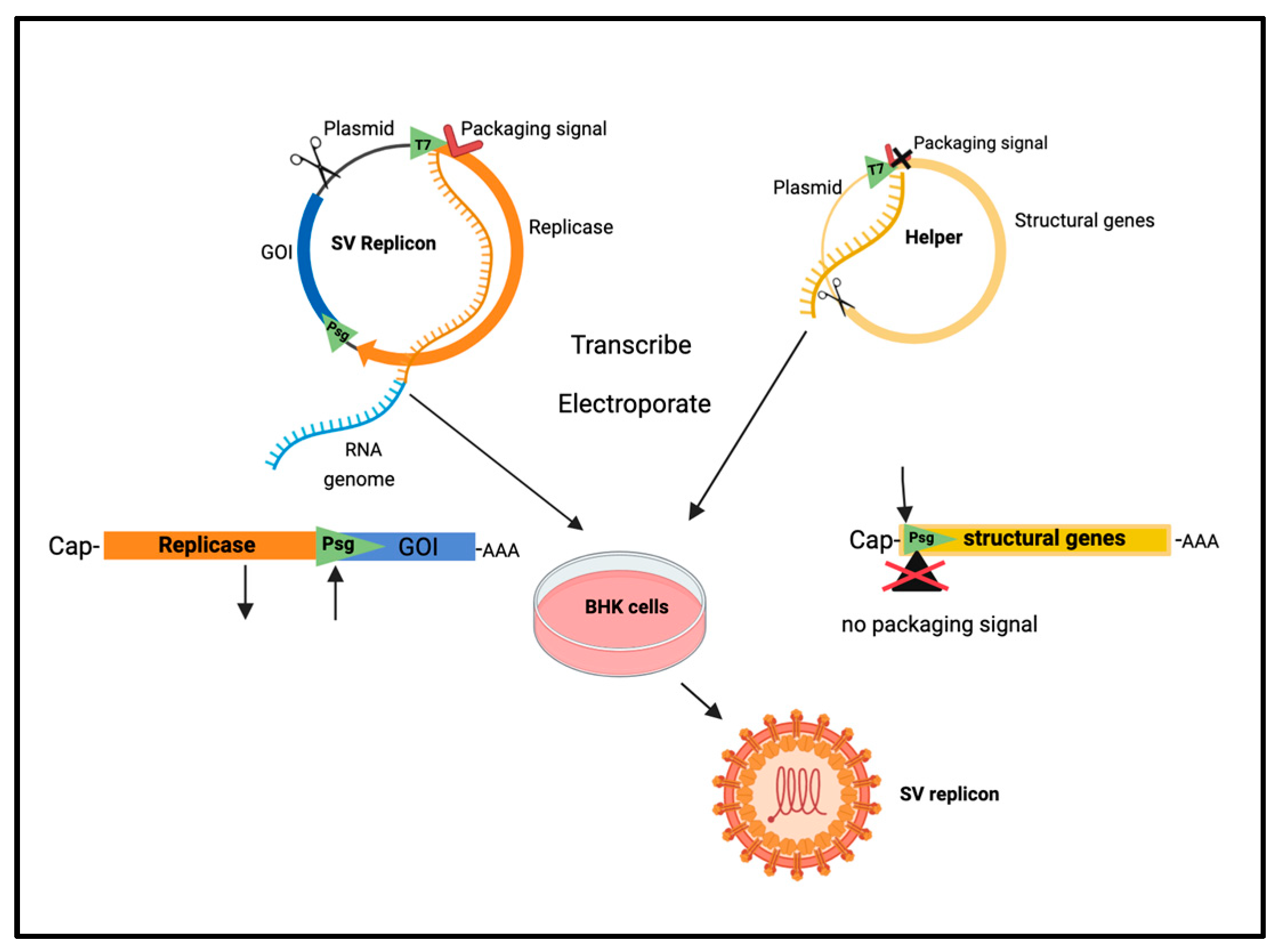
Figure 1. Preparation of SV vector. Plasmid and linear maps of replicon with gene of interest and helper with structural genes. T7, in vitro transcription promoter; Psg , Sindbis subgenomic promoter; GOI, gene of interest; AAA, polyA sequence. Created with BioRender.com.
Ijms 24 14948 g001
Figure 2. Sindbis/Rluc infection colocalized with the metastasized ES-2/Fluc tumors in the peritoneal cavity as determined by the IVIS system. Color bars show photo counts [5].
Ijms 24 14948 g002
Figure 3. Infectivity of Sindbis vectors correlates with expression of the laminin receptor. ES-2/Fluc/αLRP are cells infected with pcDNA-Fluc/iLAMR.
Ijms 24 14948 g003
Figure 4. (A) Schematic of the tumor extracellular matrix being degraded, creating space for virus entry. (B) LAMRs in normal cells are bound to laminin and generally not overexpressed. (C) As a result of extracellular matrix degradation by metalloproteinases during metastasis, laminin detaches from LAMR, creating unoccupied receptors available for SV attachment. Additionally, LAMRs are overexpressed in tumors because of increased ribosome production, which increases targets for SV attachment and entry (via receptor-mediated endocytosis). ECM, extracellular matrix; LR, laminin receptor; SV, Sindbis virus vector. Created using BioRender.com.
Ijms 24 14948 g004
Figure 5. Targeting laminin receptor (LAMR) with Sindbis/Lenti pseudotype vectors inhibits ES-2 tumor growth in vivo. ES-2 cells express Fluc. IVIS RLU used to quantify tumor growth, color bar shows photo counts [85]. * p < 0.05, Student's t-test. [85].
Ijms 24 14948 g005
Figure 6. SV-NYESO1 plus anti-PD1 increases mouse survival. aPD-1, antibody to PD-1; **, significance determined by Mantel-Cox test p <0.01. [3].
Ijms 24 14948 g006
Figure 7. Survival curve of BALB/c mice with A20 lymphoma treated with SV vector and anti-4-1BB alone and in combination. Ratio is survived number/total number [147].
Ijms 24 14948 g007
Figure 8. Survival curves of control and SV vector treated mice in two tumor models. Statistical analysis determined with Mantel-Cox test *, p < 0.05; **, p < 0.005; ****, p < 0.00005 [4].
Ijms 24 14948 g008
Figure 9. SV.IL12 plus anti-OX40 promotes metabolic programming of T cells.
Ijms 24 14948 g009
Figure 10. (A) Survival curve shows SV vector platform suppresses MOSEC ovarian tumors in vivo. (B) Survivors (140 days) rechallenged with MOSEC tumor cells are protected from tumor recurrence [2].
Ijms 24 14948 g010
Table 1. Preclinical Trials: Alphavirus Vectors.  α PD-1, programmed cell death check point antibody; VRP, vector replicative particle; i.p., intraperitoneal; i.m., intramuscular; i.t., intratumoral; i.v., intravenous; TILs, tumor infiltrating lymphocytes; Ki-67, proliferation marker; ND, not determined; M1, alphavirus that targets ZAP (-) tumor cells.
Vector/AgentDeliveryCancerImmune ResponseClinical Outcome
SV-NY-ESO-1
+ α PD-1 (VPR)
i.p.colonT cell activation
TILs
tumor growth inhibition[3]
SV + α 4-1BB
(VRP)
i.p.B cell
Lymphoma
T cell activationtumor growth inhibition,
protection against rechallenge
[147]
SV-IL12 (VPR)
SV-IL12 + α OX40
i.p.colon
prostate
T cell activation,
metabolic reprogramming
TILs
tumor elimination[4]
SV-IL12 + α OX40
(VPR)
i.p.ovarianT cell activation,
metabolic reprogramming
TILs
tumor elimination,
protection against rechallenge
[2]
VEE-HPV-E6/E7
(VRP)
i.m.cervicalCD8 T cells, IFNγ
T memory cells
protection against rechallenge[107]
SFV- α PDL-1 (VPR)i.t.colonT cells, IFNγ,TILs>40% tumor regression[154]
M1i.v.bladderLower Ki-67 signals
in tumor
tumor growth inhibition,
increased survival
[157]
M1 + doxorubicini.v.breastNDtumor growth inhibition[158]
Table 2. Clinical Trials: Alphavirus vectors. (SV PV), Sindbis pseudovirus; i.d., intradermal; OS, overall survival; CEA, carcinoembryonic antigen; E6/E7, HPV.
Vector/AgentDeliveryCancerImmune ResponseClinical Outcome
Phase I
VEE(VPR)-CEA
i.m.Stage III
colon
T cells, IFNγ
α CEA
Improved OS in IFNγ (+) patients[125]
Phase I
Vvax001
SFV-E6/E7
i.m.cervicalT cells, IFNγ
Th1 response
ND[159]
Phase I
LV305
LV-NY-ESO-1
(SV PV)
i.d.metastatic
NY-ESO-1 (+)
tumor
T cell response in 85%
patients
tumor growth inhibition
improved OS
[160]
Disclaimer/Publisher’s Note: The statements, opinions and data contained in all publications are solely those of the individual author(s) and contributor(s) and not of MDPI and/or the editor(s). MDPI and/or the editor(s) disclaim responsibility for any injury to people or property resulting from any ideas, methods, instructions or products referred to in the content.